the magazine from carl zeiss can be found on the internet ... · the magazine from carl zeiss issue...
TRANSCRIPT

Innovation –
The Magazine from Carl Zeiss can
be found on the Internet at
www.zeiss.com/innovation

The Magazine from Carl Zeiss Issue 229/ 2010
(Re)solution: New Methods Provide Exciting Insights
Better Vision: With and Without Glasses
Essay: Reunification of Carl Zeiss East and West

With brain mapping, science is under- taking a new attempt to unravel the secrets of the human thinking machine.
Aided by high-resolution images generated by an electron microscope, brain researchers aim to produce a type of three-dimensional map of the brain and, above all, record the intricate networking of the nerve cells via the synapses. They hope that this will assist them in discovering the causes of diseases like schizophrenia. Brain mapping repre-sents a new and promising application for particle beam systems.

3Innovation 22, 9 / 2010
Dear Readers,
What happens when a virus enters a cell? Understanding the processes in
living cells is a key requirement for advances in medicine and thus for
improved health and quality of life.
Those who deal with cell structures and intracellular processes on a daily
basis use light microscopes for their work. However, the laws of physics set
a boundary which Ernst Abbe – business partner to company founder Carl
Zeiss, scientific mind and later owner of the company – recognized way back
in the 1870s. This resolution limit of 200 nanometers named after Abbe was
valid for more than 130 years.
Only recently have intelligent solutions made it possible to lower this limit
of traditional light microscopy by a factor of 10 and thus open the door
to a new dimension in research. The option of examining living organisms
is something that primarily benefits cell biologists such as Leibniz Award
winner Petra Schwille.
Carl Zeiss has been able to take things a step farther. The company, the
only one in the world to manufacture both light and electron microscopes,
has combined both technologies, to enable and considerably simplify
correlative microscopy. Correlative microscopy can be used, for example,
to determine how a virus penetrates a cell.
We at Carl Zeiss are proud of our 160-year history of providing innovative
tools and solutions that have repeatedly led science to new insights. Rest
assured, this will remain our ambition in the future.
Enjoy reading!
Best wishes,
Dr. Dieter Kurz,
President & CEO of Carl Zeiss AG
Editorial

4 Innovation 22, 9 / 2010
Table of Contents
Bridging the Micro and Nano Worlds 16
“Freedom is the Greatest Gift” 22An Interview with Dr. Petra Schwille
Unveiling the Secrets of Thinking 24
Cover Story: (Re)solution
Editorial 3
Panorama 6
Always Connected 10Photo Contest
16
Fluorescence-labeled vesicles in a neuron, overlaid with scanning electron microscope image.
Essay 44
Twenty Years Later

5Innovation 22, 9/ 2010
Report: Heavens in Motion
If Spaceships Could Fly through Galaxies 26
Report: Sailing the Seven Seas
Unveiling the Secrets of the Oceans 30
Report: Better Vision
The Beautiful World of Vision 38Revolution in Laser Surgery 40The Solution with the Cylinder 42
26
See more. Live more.
38
Bochum Planetarium.
Look at Laureates 50Roger Tsien
Feature Capturing the Heat of the Moment 36

6 Innovation 22, 9 / 2010
Panorama
Distracted Patients Are Relaxed Patientscinemizer Plus video glasses from Carl Zeiss take the edge off
self-contained system that simulates a 44 inch screen at a distance of two meters. Sound is provided by two speakers integrated into the frame, or by headphones that can be easily connected. The cinemizer Plus is not only for dentists, but can also be used in other medical centers when a local anesthetic is required for minor procedures in a hospital, for example.
Sitting in the dentist chair totally oblivious to what‘s going on – wouldn‘t it be great if it were always like this? The cinemizer Plus makes it possible. Around 400 dentists in Germany already use these video glasses from Carl Zeiss to make the treatment more relaxing. Patients can watch music videos or movies depending on the length of the procedure. Experienced patients bring their favorite shows with them on an iPhone, iPod or a Nokia smart phone. Previous attempts to distract patients with movies shown on monitors on the ceiling failed due to reflections. However, with the video goggles, everything is contained in a single,

7Innovation 22, 9 / 2010
Knowledge grows when it is shared. This is the idea behind the Metrology Portal from Carl Zeiss, a unique global social network for all matters concerning measuring technology. With their free registration, us-ers receive access to a wide range of offers. For example, they can research the basics of measuring technology, enter forum discussions, get tips for specific problems and download software for their daily work. Customers with a software maintenance agreement can also download the latest software updates and obtain information about their measuring machines. This ensures that they remain up to date from both a technical and scientific standpoint. The benefit to the user was the main focus of attention during setup of the portal and is also its greatest advantage: “It is amazing how quickly you can find answers to your metrology problems,” says Dieter Finner, Production Planner at tedrive Germany in Düren. Carl Zeiss also benefits from this exchange with users and is able to expand its know-how. The Metrology Portal is a classic win-win situation for everyone involved.
Teaching Each Other to Measure The Metrology Portal from Carl Zeiss is a unique Internet forum
Further information is available athttp://www.zeiss.de/metrology-portal
Moore’s Law EUV lithography reaches maturity for the volume production of high performance computer chips
In 1965 Intel founder Gordon Moore predicted that the number of components on a computer chip would double every 18 months. This forecast became known as Moore‘s Law. He assumed that, after ten to 15 years, the semiconductor industry would reach an insur-mountable technological limit, after which the law would lose its validity. However, the development is continuing at a tremendous pace. Chip structures
are still getting smaller and smaller, allowing more and more components to be applied to the chip surface. No end to this trend is in sight.
This is partly attributable to EUV lithography, a new manufacturing method that is revolutionizing the volume production of microchips. Carl Zeiss delivers the optical system. This leading-edge technology required 15 years of intensive research. Therefore, it was no surprise that developer Peter Kürz was nominated for the Future Award of the German Presi-dent in 2007. EUV lithography uses extremely short-wave radiation of 13.5 nanometers. This radiation, which is more than one-tenth shorter than the current standard of 193 nanometers, lays the foundation for the imaging of chip structures with dimensions of 10 nanometers and less. ASML, the Dutch partner of Carl Zeiss, integrates the optical system into its EUV chip fabrication equipment. The first system is due for deliv-ery in the second half of this year.

8 Innovation 22, 9 / 2010
The Third DimensionSurfMax inspects the surfaces of 3D parts fully automatically
Machines are more reliable than people when it comes to routine jobs. Machines do not have good days or bad days, and they make few mistakes when they are programmed properly. This also applies to the visual inspection of surfaces. Visual inspections are required wherever there is no guarantee that technical or de-corative surfaces will make it through the production process without defects. Traditionally, an employee examines the part, holds it up against the light, ro-tates it and turns it over – a time-consuming form of inspection that can easily be performed by a machine.
Fully automatic surface inspection in production – Optical Inline Metrology (OIM) – is the domain of SurfMax, the first configurable tester capable of in-specting three-dimensional objects. During a visual inspection, SurfMax works much like a human: it uses the reflections on the surface to determine whether it is flawless or if scratches and grooves arepresent. This works on matte or glossy flat and curved metal, plastic and ceramic surfaces. Unlike people, the machine does not need any special skills or experience to deliver an expert opinion. Along
the way, it learns and always works with the same precision. This frees up people to take care of jobs that machines cannot.
Carl Zeiss in Oberkochen established a new factory after World War II. This resulted in Fine Measuring departments in East and West Germany, which fought bitterly over trademarks. Fine Measuring West quickly jumped ahead and in 1950 developed the UMM univer-sal measuring microscope. At the 1973 Microtecnic trade fair in Zurich, its successor, the UMM 500, caused a real sensation. This 3D measuring machine truly revolutionized metrology and became a legend in its own right. When it became obvious in 1976 that 3D measuring machines were the future of the Fine Mea-suring Department it was renamed to Industrial Metrol-ogy (IMT).
90 Years of Industrial Metrology at Carl Zeiss from the August-Dreesbach-Verlag chronicles the exciting development of industrial metrology at Carl Zeiss between 1919 and 2009 in a nearly 100-page book. (ISBN 978-3-940061-37-9)
A 24-page short version is also available.
It all began with the Abbe comparator principle. Physicist Ernst Abbe, Carl Zeiss’ partner and founder of the Carl Zeiss Foundation, formulated this principle of precise measurement in 1890. Without him, indus-trial metrology would not be where it is today. The first measuring machines from Carl Zeiss for industrial use were developed by Otto Eppenstein who for nearly 20 years headed the Fine Measuring Department in Jena founded in 1919. During this time, he applied for 78 patents. Eppenstein’s machines were so advanced that they continued to be built without changes for decades.
Fine Measuring to IMT A look back at 90 years of industrial measuring technology at Carl Zeiss is now in book form
Further information is available at www.august-dreesbachverlag.de/html/buecher/imt.html

9Innovation 22, 9 / 2010
A spotting scope is somewhere between a telescope and a pair of binoculars. Marksmen use them to check their accuracy from far away. Nature lovers utilize them to watch animals, primarily birds, from long range without disturbing them. Spotting scopes uncover details, such as the white spot on the tenth primary feather of a seagull, which would remain hidden with binoculars.The Victory DiaScope spotting scope from Carl Zeiss opens upnew possibilities for nature lovers: the Vario eyepiece not only provides 75x magnification of the image frame, but also delivers bright,
A decision is only as good as the information on which it is based. In the future, neurosurgeons will be able to more quickly record what they need to know for surgery. FLOW 800 supports them in the visual analysis of the blood flow – an aid that must not be under-estimated for deciding whether a clip has been correct-ly positioned on a blood vessel in the brain and if the blood is flowing freely in the bypass around a clogged vessel.
To date, the surgeons have been able to see on a screen during surgery how and where the blood is flowing through the vessels. This is made possible by visual-ization using INFRARED 800, with which the OPMI® Pentero® surgical microscope from Carl Zeiss is equipped. However, in order to expertly interpret the data, doctors have had to examine the recordings several times. To establish how effective the procedure has been, they have had to perform a “before and after“ comparison, for which they had to constantly scroll back and forth in a time-consuming process.
This is all easier and faster with FLOW 800. The infor-mation from the INFRARED 800 videos is presented at
Analysis at a GlanceFLOW® 800 facilitates neurosurgical procedures through excellent visualization of the blood flow
a glance. The program images complex situations on colored overview maps and shows how the blood flow changes during the procedure. The doctor can select and directly compare sequences, making it easier to make a sound medical analysis.
White Spot on the WingNew spotting scopes from Carl Zeiss show more of nature and are user friendly
razor-sharp images deep into the twilight without irritating color fringes. This is all made possible by thespecial design of the lens and the high-performance fluoride glass. The Vario eyepiece with an expanded zoom range is a real innovation. Users can quickly and seamlessly switch from a wide field of view to high magnification. Another highlight is the dual speed focus system that gives users two focusing speeds on one control element: the image definition is set with pinpoint accuracy in the fine mode, while the system automatically switches to the coarse mode for fast focusing when the wheel is turned quickly.The Victory DiaScope 85 T* FL recently received the 2010 red dot design award in the product design category. It impresses users as well as the red dot jury with its technology and functional design.

Innovation 22, 9 / 20101010

Always ConnectedDo people live differently in the age of
computers, mobile phones and the Internet?
The pictures for the Carl Zeiss “Digital Culture“
contest demonstrate that new media are
solidly entrenched in modern society.
11Innovation 22, 9 / 2010

Innovation 21, 12 / 200812
Love in the Time of the Internet
12 Innovation 22, 9 / 2010
This picture is for everyone, Janusz Chwiolka, PolandTaken with: Nokia N95 Smartphone with Carl Zeiss optics
Previous double page:Digital technology brings cultures closer together, Jens Oeltjebruns, GermanyTaken with: Sony DCS-H9 with Carl Zeiss optics

13Innovation 21, 12 / 2008 13Innovation 22, 9 / 2010
A picture is worth a thousand words: photo contest. The Carl Zeiss Photo Contest is held every year in December. The 2009 contest focused on capturing the digital shift in images – with a lens from Carl Zeiss, of course. From mobile phones to medium format, anything was possi-ble and allowed.
Digital media simplify life. They change the way people treat each other, how they exchange informa-tion, work, play, meet friends and make music. An international jury, including well-known photogra-phers Edith Held and Jürgen Müller, focused primarily on creativity and implementation, and less on image processing.
The winning photo – Digital Blind Date from Netanel Hadad in Israel – shows a man talking to a woman. They’re looking in each other‘s eyes – but cannot touch. On the table, next to candles and wineglasses: two notebooks. One monitor shows her face, the other his. This picture took first place over 1018 images from 58 countries. Hadad met his own wife on the Internet.
Boy with a beret and his PlayStation Maxime Ballesteros, GermanyTaken with: Contax T2 with Sonnar T* 2,8/38
Winning photo: Digital Blind Date Netanel Hadad, IsraelTaken with: Hasselblad 501 CMwith Carl Zeiss Planar T* 2,8/80 CB

14 Innovation 21, 12 / 200814 Innovation 22, 9/ 2010
Right: Times have changed, Bernd Geh, USA
Taken with: Canon EOS-1D Mark III with Planar T* 1,4/85 ZE
Computer repair shop in a Serbian village Miodrag Trajkovic, SerbiaTaken with: Sony HVR-Z7E with Carl Zeiss optics
“Digital” is fully entrenched in our lives Holger Forst, GermanyTaken with: Nikon D3 with Distagon T* 2,8/21 ZF
A new way to play Juan Leon, SpainTaken with: Nikon D700 with Carl Zeiss Planar T* 1,4/50 ZF

15Innovation 22, 9 / 2010
16 Innovation 22, 9 / 2010Innovation 22, 9 / 201016
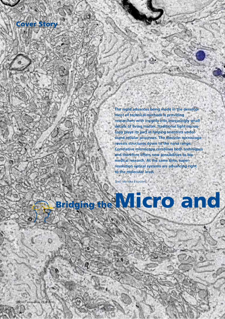
Bridging the Micro and Nano Worlds
Cover Story
The rapid advances being made in the develop-
ment of technical methods is providing
researchers with insights into increasingly small
details of living matter. Traditional light micros-
copy plays its part in helping scientists under-
stand cellular processes. The electron microscope
reveals structures down to the nano range.
Correlative microscopy combines both techniques
and therefore offers new possibilities to bio-
medical research. At the same time, super-
resolution optical systems are advancing right
to the molecular level.
Text: Monika Etspüler

17Innovation 22, 9 / 2010
Report: Lorem ipsum
17Innovation 22, 9 / 2010
Bridging the Micro and Nano Worlds
18 Innovation 22, 9 / 2010
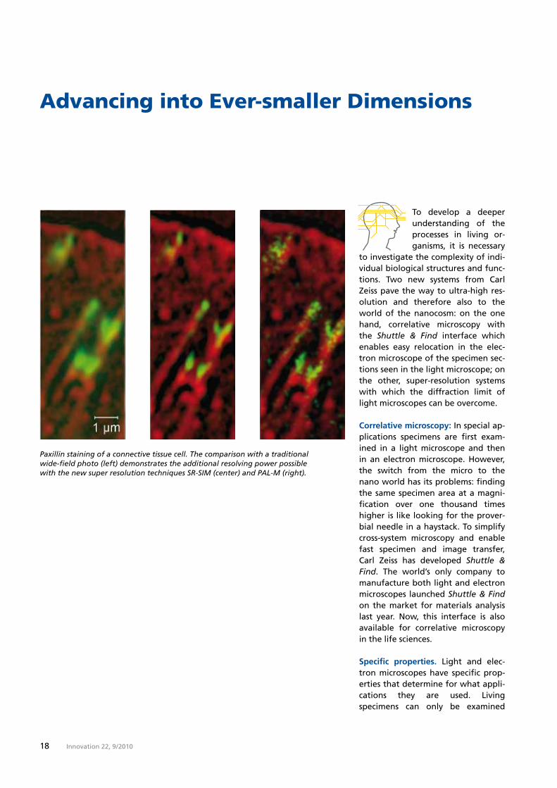
Advancing into Ever-smaller Dimensions
Paxillin staining of a connective tissue cell. The comparison with a traditional wide-field photo (left) demonstrates the additional resolving power possible with the new super resolution techniques SR-SIM (center) and PAL-M (right).
To develop a deeper understanding of the processes in living or- ganisms, it is necessary to investigate the complexity of indi-vidual biological structures and func-tions. Two new systems from Carl Zeiss pave the way to ultra-high res-olution and therefore also to the world of the nanocosm: on the one hand, correlative microscopy with the Shuttle & Find interface which enables easy relocation in the elec-tron microscope of the specimen sec-tions seen in the light microscope; on the other, super-resolution systems with which the diffraction limit of light microscopes can be overcome.
Correlative microscopy: In special ap-plications specimens are first exam-ined in a light microscope and then in an electron microscope. However, the switch from the micro to the nano world has its problems: finding the same specimen area at a magni-fication over one thousand times higher is like looking for the prover-bial needle in a haystack. To simplify cross-system microscopy and enable fast specimen and image transfer, Carl Zeiss has developed Shuttle & Find. The world’s only company to manufacture both light and electron microscopes launched Shuttle & Find on the market for materials analysis last year. Now, this interface is also available for correlative microscopy in the life sciences.
Specific properties. Light and elec-tron microscopes have specific prop-erties that determine for what appli-cations they are used. Living specimens can only be examined

Cover Story: (Re)solution
19Innovation 22, 9 / 2010
Advancing into Ever-smaller Dimensions
Previous double page:Ultra-thin section through brain of zebra finch. Fluorescence-labeled vesicles in a neuron, overlaid with scanning electron microscope image..
with light and laser scanning micro-scopes. These achieve a resolution limit up to roughly 200 nanometers (1 nanometer = 10-9 meters). Electron microscopes are used for advancing farther into the nano world. Their resolving power is more than two orders of magnitude above that of a light microscope. Due to the tech-nology and functionality of the elec-tron microscope, however, it can be used solely for static examinations on non-living material. Until now, a combination of the two techniques for the analysis of a defined region of interest was very complicated, if not impossible.
Use in materials analysis. Aalen Uni-versity in Germany was one of the first facilities to use Shuttle & Find for its research activities. Here, Carmen Hafner and Timo Bernthaler are examining lithium-ion batteries from small electronic appliances. And as with all rechargeable batter-ies, the ravages of time do not make an exception here either. However, the issue of ageing processes and performance loss has become in-creasingly more urgent now that lithium-ion batteries have practically become an emblem of electro-mo-bility and an emission-free future. The composition and granularity of a structure allow conclusions to be drawn not only about the type of production used, but also about the material properties.
The Elyra PS.1 microscope system combines SR-SIM and PAL-M technology in a single system.
Timo Bernthaler attaches Shuttle & Find to the motorized specimen stage of the Axio Imager.2 light mi-croscope. The specimen holder with the specimen is calibrated via three markings on the surface, to which it first has to move. The cross-section of a battery appears with 25x magni-fication on the monitor. “One year’s development work was necessary to produce such a good sample,” he ex-plains almost in passing. The spirally arranged separators between the an-ode and the cathode have definitely seen better days. Decomposition is already evident in some areas. Timo Bernthaler marks the “trouble spots” that are stored electronically to en-able their subsequent relocation in the electron microscope. Another calibration is required before the ex-

20 Innovation 22, 9 / 2010
amination of the region of interest in the EM. The structural damage of the material is plain to see on the monitor – but at 16,000x magnifica-tion this time.
“One year‘s development
work was necessary to
produce such a good sample“
Timo Bernthaler
Biological perspectives. Whether it is used for the life or materials scienc-es, Shuttle & Find works along the same principles in each case. The dif-ferences lie, above all, in the design of the specimen holder, which must fulfill much more stringent require-ments for biological material.
n In light microscopy cover slips are used for living specimens. How- ever, the electron beams of an electron microscope cannot pene- trate the cover slip material. Therefore, the holder had to be
constructed so that the specimen can be examined from two sides in the microscope.
n In light microscopy immersion oil is used at very high magnifica- tions. But, oil contaminates elec- tron microscopes. Therefore, it must be possible to remove it in its entirety. For this purpose, Carl Zeiss has applied for a patent for a technique in which a thin film applied between the immersion oil and the cover slip can be sim- ply “stripped off” with the oil. n After all, in the specimen holder it should also be possible to pre- pare the object being examined, i.e. fixing, dyeing and embed- ding, without affecting the direct surrounding area with the mark- ings.
Shuttle & Find opens up interesting perspectives in cell biology. One ex-ample: it enables the overlay of images taken with the light and electron microscopes. A light and la-ser scanning microscope makes it possible to observe how large viruses labeled with fluorescent dye pene-trate a host cell. Electron microscopy delivers information about the sur-face morphology of the cell in the regions of interest. The fluorescence signals indicate whether and in what area a virus docks onto a cell or to what extent it has already invaded it.
Shuttle & Find being used in the specimen area of an electron microscope.

Cover Story: (Re)solution
21Innovation 22, 9 / 2010
“SR-SIM allows the spatial
imaging of structures. This
is a huge benefit”
Martin Bastmeyer
Super-resolution systems. In the years ahead an important research goal will be to visualize the diversity of the workings of a cell down to the molecular level. This requires light microscopes with fluorescence-based technology and extremely high reso-lution, also known as “super resolu-tion.” Elyra from Carl Zeiss combines these two quality features. Elyra S.1 (SR-SIM), which stands for Structured Illumination Microscopy, has a reso-lution that is twice as high as that of traditional fluorescence microscopes. Elyra P1 (PAL-M), also known as Pho-to Activated Localization Microscopy, achieves a resolution of 20 nanome-ters and therefore functions in a range in which single molecules can be localized.
Elyra PS.1, a combination of PAL-M und SR-SIM, is installed in the labora-tory of Prof. Dr. Martin Bastmeyer, who holds the chair of Cell and Neu-robiology at the Karlsruhe Institute of Technology (KIT). One of the ap-plications for which the scientist uses SR-SIM is to examine the cytoskele-ton which provides stability and elas-ticity to a cell. It consists of a net-work of actinium filaments and microtubules. These are protein fi-bers that ensure that the cell keeps its shape. In the SR-SIM these struc-tures are discernible as fine threads. With the aid of PAL-M, Bastmeyer is
succeeding in localizing individual paxillin proteins. Paxillin, which ap-pears as a green fluorescent dot in the specimen, is one of over 100 pro-teins that together form the docking point of the actinium filaments on the cell membrane.
The application determines which of the two super-resolution microscopy techniques is used. “SR-SIM allows the spatial imaging of structures. This is a huge benefit,” explains Mar-tin Bastmeyer. “PAL-M requires a large amount of preparation time. In addition, the optical sections must be extremely thin,” he adds. For PAL-M, only fluorophores from special GFP mutants (Green Fluorescent Pro-teins) are used, while all fluorescent dyes can be utilized for SR-SIM.
Outwitting diffraction limits. Al-though the technology behind SR-SIM and PAL-M is totally different, the two systems nevertheless have one thing in common: to work at all, they have to circumvent a law of physics formulated by Ernst Abbe as far back as 1873. The general rule applies that the wave nature of light sets limits to the resolving power of a microscope. The maximum resolv-ing power of even the best micro-scope lies at a minimum distance of 200 nanometers between two points. If more detail is required, the microscope and microscopic images must be modified in such a way that resolution beyond Abbe‘s law is pos-sible.
In structured illumination (SR-SIM), therefore, a defined grating struc-ture is projected into the focal plane
of the fluorescence microscope. The generated modulation contrast be-tween bright and dark areas in the image can then be used to distin-guish between two closely adjacent points. To ensure that the super reso-lution is not only available in individ-ual areas of the image, the grating is laterally displaced across several positions and rotated. This guaran-tees a uniform increase in resolution in all three spatial directions. The individual raw images generated in this process have little to do with “real” imaging. Subsequent software computation is needed to turn this into a realistic microscope image.
Photoactivated localization microsco-py (PAL-M) uses switchable fluoro-phores to circumvent the diffraction limit. The specimen is irradiated with different wavelengths so that only a few molecules can be excited at any one time. This makes it possible to determine the exact position of an individual molecule with nanometer accuracy. This process is repeated in 20,000 to 40,000 single images, and the results are subsequently added to form an overall image.
More networking ahead. The future trend is heading toward further networking of the different micros-copy techniques. It is not yet possi-ble to “marry” super-resolution light microscope systems and electron microscopes via Shuttle & Find. If this is successful, there is nothing left to hinder the determination of the position of a single protein and its function in a highly resolved cell structure.

22 Innovation 22, 9 / 2010
Interview
“Freedom Is the Greatest Gift”Leibniz Prize winner Petra Schwille explains the attraction of cell biology
Were the conditions you found at the Bio- technological Center (BIOTEC) of the Techn-calUniversityofDresdenoneofthereasons why you won the LeibnizPrize? Yes, definitely. This is a fantastic environment to work in. It’s really in-spiring. And my colleagues are great, too. There’s a pioneering spir-it about the place. You have the feeling that you’re part of a great movement, and that’s always fun.
WhatdoestheLeibnizPrizemeantoyou? It’s a wonderful feeling to be hon-ored with such a prestigious prize. It boosts your confidence and encour-ages you to go on. The prize means you can continue your work at this level, no matter what happens in the next five years.
Youwereoriginallyaphysicist. Biological phenomena were al-ways what interested me most, which is why I went to Göttingen to study. I moved from physics to physi-cal chemistry, then on to physical biochemistry and biophysical chemis-try, until I more or less landed in cell biology.
The 1990s to mid-2000 were what could be called the “single molecule years,“ but the initial hype is over now. Scientists now want to do something sensible with these tech-nologies. What fascinates me is fun-damental questions like “How does the cell become an organism?” There’s an incredible number of
processes involved: symmetry diffrac-tion, polarization, the emergence of local concentrations ... I’m interested in finding out what specific property of these molecules allows them to form such complex structures.
Just what attracted you to cellbiology? There are two important reasons: in my doctoral thesis I dealt with the subject of Fluorescence Correlation Spectroscopy (FCS), and I think I was pretty successful, too. But my entire data consisted of curves, curves and more curves. Then at a conference I met people who actually did confo-cal microscopy. They showed images and videos that really swept me off my feet. I wanted to do it too! The other reason was the environment, the people here and the issues we discuss. Dresden was definitely a stroke of luck for me. The way peo-ple at the Max Planck Institute for Molecular Cell Biology and Genetics pose fundamental biological ques-tions – that truly enriched me.
Youaremovingawayfromcellbiol-ogytobiomedicalresearch:howwillyourresultsbenefitpeople?
I can’t really say. I want to under-stand how the cell works. I want to understand what drives the mole-cules to create something as inter-esting as a cell. And what is special about it. What does a molecule need to build something like this? How does it differ from any other “bor-ing” molecules?
In recent years we have simplified systems and discovered that only a few molecules or molecule species can be used to generate real pat-terns; biological patterns with a front and a back. When it comes down to it, it‘s all a matter of phys-ics. There is some sort of switch on the molecule that somehow works, another molecule operates the switch in a special way and then, abracadabra, a pattern is there.

23Innovation 22, 9 / 2010
The person
Dr. Petra Schwille
42-year-old Petra Schwille studied physics and philosophy at the universities of Stuttgart and Göttingen. She obtained her degree in 1993.
She began her doctorate in the workgroup of Nobel laureate Manfred Eigen and finished it with a dissertation in the field of fluorescence correlation spectroscopy at the Technical University of Braunschweig in 1996.
After holding various postdoc-toral positions, e.g. at Cornell University in Ithaca, NY, Petra Schwille became Junior Group Leader for experimental bio-physics at the Göttingen Max Planck Institute in 1999. She has been Professor of Biophysics at the Technical University of Dresden since 2002. In 2010 she received the Leibniz Prize from the German Research Foundation (DFG).
How long did it actually take fromthe initial test tube experiments toexperimentswithlivingcells? Before understanding the funda-mental cellular mechanisms, we had to succeed in our endeavors to mea-sure quantities in a living cell. It took us a long time to achieve this. Almost 15 years, in fact. In 1996, during the work for my PhD, I devel-oped the FCS method precisely for this purpose. In my time in America we then examined cells with two-photon excitation. The first major publications didn’t appear until 2002, 2004. And now we work in the living embryo, the “high end system“ as I suppose you could call it, where absolutely everything is moving. That is one step up on the complexity ladder.
As a professor, how much time doyour bureaucratic duties leave youtodoresearchwork? It’s difficult to separate. With very few exceptions, everything I do is linked to research. I invest a lot of work writing official reports, and I spend a great deal of time compiling project applications. The Leibniz Prize has given me a little breathing space.
I have to actively pick the times I need to sit down and reflect during the working day. But it’s good fun nevertheless.
In other words, having fun to youmeanssittingdownandreflecting? Maybe fun is the wrong word for it. I like to reflect upon things in peace and quiet, but I also love talking with people and letting them inspire me to think up new ideas.
How early did you decide to dophysics? At school: I was good at math and science. I didn’t want to study biolo-gy – that was more a subject for girls who just wanted to learn things by heart. My father was a chemist, so that possibility was ruled out too. Physics seemed to be a good option. In the first few years I more or less regretted my decision. But then, I suppose from the time I actually started to experiment myself, I began to enjoy my physics studies.
And the final kick came with yourPhD? Yes. Thanks to the equipment at the Max Planck Institute for Bio-physical Chemistry in Göttingen. My boss, the funds available, the possi-bilities – it was really super. This free-dom, the experience of freedom, it was wonderful. Freedom is the greatest gift.
Thank you.
The interview was conducted by Dieter Brocksch and Silke Schmid.
Cover Story: (Re)solution

24 Innovation 22, 9 / 2010
Unveiling the Secrets of Thinking
How does the brain ac- tually think? This is a question that scientists have been addressing for a long time now. What exactly is the relationship between the struc-ture and the function of the human thinking machine? Researchers are using high-resolution electron mi-croscope systems to find an answer.
The human being has about 100 bil-lion nerve cells and around 100 tril-lion synapses. These figures may be impressive, but they do not auto-matically lead to a deeper under-standing of the processes occurring in this central organ. With brain mapping, science is undertaking a new attempt to unravel the secrets of the human thinking machine.
The Connectome project. In this ap-proach to research, the focus of at-tention is no longer being placed solely on the recording of the nerve cords, but the networking or con-nectivity of individual nerve cells is also being examined and registered
with meticulous accuracy. “Connec-tome” is the term used to describe the system of all nerve cells and their networking in the brain – based on the word “genome” that represents the sum of all genes in a cell. The long-term goal is to achieve a type of brain map with a three-dimensional circuit dia-gram.
High expectations are being placed on brain mapping. The potential that it offers would appear to be in-exhaustible. It is hoped that it will be possible to discover the causes of such diseases as autism or schizo-phrenia and obtain a better under-standing of how medicines work. Ex-act knowledge of the structure and function of the human brain may also provide information on the de-velopment of personality in the course of a human life. Science is still more or less at the begin-ning of this large-scale proj-ect. Even with today’s fast-est solutions, projections show that it will take 30
Cover Story

25Innovation 22, 9 / 2010
years to record the brain of a mouse, not to
mention the mapping of the human think-
ing apparatus.
A puzzle with many fac-ets. Even the initial
preparation of a brain section is no easy matter.
The material is subsequent-ly embedded in synthetic res-in and cut with a microtome, a type of planing device, into
wafer-thin slices that are ap-plied to a wafer and imaged in a
scanning electron microscope. Such a specimen has a volume of about one cubic millimeter; this cube is cut up into about 20,000 slices. Vast pro-cessing times are required to subse-quently merge the resulting huge quantities of image data into a three-dimensional image in the com-puter again.
Speed means success. Carl Zeiss is supporting this research work with intelligent solutions. With the aid of the SIGMATM field emission scanning electron microscope (FE-SEM), biolo-gist Jeff Lichtman from Harvard Uni-versity in Cambridge, Massachusetts, is examining pieces of a mouse’s brain. The system is equipped with a special detector system and software that allow 100 times faster image generation and storage than with traditional systems. For John Men-denhall of the University of Texas in Austin, speed is an important argu-ment in favor of using a ZEISS FE-SEM. Together with a special applica-tion solution, the system has an image memory with a capacity of up to one gigapixel and therefore per-mits the recording of large-area specimens at maximum resolution.
Dr. Marco Cantoni of the Ecole Poly-technique Fédérale de Lausanne in Switzerland examines mouse brains with a CrossBeam® microscope. In-stead of a mechanical technique, an ion beam is used to remove the spec-imen slice by slice, with the result then being examined in the scan-ning electron microscope. This pro-cess is largely automated. “The re-sult was incredible. In 48 hours we generated 1600 images of specimen slices, each with a thickness of just six nanometers,” explains Dr. Canto-ni. “This gives us an insight into the three-dimensional structure of the tissue to be examined.” The ORION® helium ion microscope also offers possible applications in the field of brain mapping. The potential of this technology lies, above all, in its ex-
tremely large depth of field and the innovative contrast mechanisms of-fered by imaging with ions.
Brain research as an industry. The quest for images and data already shows that this scientific challenge cannot be mastered without broad-based research. The opinion is now increasingly being voiced that Con-nectome research should be industri-alized, much like the situation with genome research at the end of the last century. The solution could be that entire “farms” will emerge comprising dozens of electron micro-scope systems in which brain sections will be imaged day and night.
The computer industry is also faced with a major challenge. Its job will be to create gigantic storage media. Even one cubic millimeter of mouse brain delivers information totaling 1000 terabytes; this would be one million times 1000 terabytes for the human brain.
However, knowing the circuit dia-gram still does not allow any de- ductions to be made about the activ-ities of the thinking machine. The question as to the effect that a nerve cell really has on its direct surround-ings remains unresolved for the time being.
Monika Etspüler
Cover Story: (Re)solution

24 Innovation 22, 9 / 2010Innovation 22, 9 / 201026
If Spaceships Could Fly through Galaxies
Report

27Innovation 22, 9 / 2010
If Spaceships Could Fly through GalaxiesPlanetariums open the door to virtual excursions
in space. A new projection system makes it possible
to embark on journeys of a different type with
even more realism.

28 Innovation 22, 9 / 2010

Report: Heavens in Motion
29Innovation 22, 9 / 2010
A trip to Saturn and back, feeling how the earth shrivels under your feet and life palls into insignificance. Providers of pri-vate trips to space are not yet offer-ing such breathtaking experiences. Although 55 million dollars will buy you a ticket to the international space station this is where the expe-dition ends. Planetariums, on the other hand, can take you to the deepest regions of the cosmos. Audi-ences are whisked away to the farthest corners of the universe without leaving the safe confines of the facility.
Setting new standards. Visitors to the planetariums in Wolfsburg and Bochum have been able to experi-ence this magic for several months now. The Wilhelm Foerster Planetari-um in Berlin recently reopened its doors. The powerdome®VELVET pro-jection system developed and manu-factured by Carl Zeiss is being used for the first time in all three cities. It fills the entire planetarium dome with digital images and gives audiences the impression of actually being in space. What makes this all-dome video system totally different from its predecessors is its ability to generate an absolutely black back-ground, in front of which the celes-tial bodies appear to hover and the stars twinkle.
A New Element to Planetarium Domes
Flexibility is the key. “VELVET lays to rest the notion of antiquated plane-tariums,” says Dirk Schlesier, a scien-tific employee at the planetarium in Wolfsburg. “We are bringing dyna-mism to the dome,” commented Prof. Dr. Susanne Hüttemeister, Di-rector of the Bochum Planetarium. The traditional planetarium has evolved into a showroom where psy-chedelic music from Pink Floyd causes the images on the dome to swirl and the Beagle sails the seven seas with Charles Darwin on board.
In addition to numerous new sub-jects from science and culture, the workings of the heavens remain the primary focus of attention. Many events are directed at kids and young people who “sometimes un-derstand for the first time that they are part of the universe as a result of such presentations,” explains Dirk Schlesier.
Completely renovated. The stars have been shining at the planetari-um in Bochum since 1964, in Wolfs-burg since 1983. Both required ex-tensive renovations before the new projection system could be used. “One of the main reasons for the complete overhaul in Bochum is the fact that the Ruhr region is one of the Capitals of Culture this year,“ says Susanne Hüttemeister.
Fascinating night sky. The heart of both planetariums is still the opto-mechanical star projector. Stars are recreated via glass fiber, a techno- logy that has only been mastered by Carl Zeiss. It ensures that the image of the night sky is as realistic as
possible. The UNIVERSARIUM star projector is used in large planetari-ums such as those in Bochum and Berlin with a dome diameter of 20 meters; domes with a diameter of 15 meters such as that in Wolfsburg use the STARMASTER projector.
The universe is alive. Until now, tra-ditional slide and video projectors were responsible for showing mo-tion in space. 50 were in operation in Wolfsburg alone. However, these instruments are not suited for bril-liant full-dome presentations. The background remains visible, and gray, washed-out, sometimes dirty images are generated. Carl Zeiss has solved this problem with VELVET, which eliminates any residual light. Black in the image is black in the projection. The technical challenge was to eliminate residual and stray light from all projector components and thus vastly increase the contrast. The result: VELVET generates con-trast of 2.5 million to 1, compared to standard projectors that offer 30,000 to 1 at most.
Analog meets digital. powerdome VELVET and the opto-mechanical star projector are coupled via a joint controller to create a brilliant night sky and enable a spectacular, virtual journey through the depths of the cosmos. VELVET eliminates the need for traditional slide and video sys-tems. These have been replaced by six VELVET projectors in Wolfsburg, eight in Bochum and Berlin. Audi-ences really appreciate the techno-logical advances.
Monika Etspüler
Recently equipped with the powerdome® VELVET projection system, the Bochum Planetarium will show “tempus.ruhr,” the full-dome production from Rocco Helmchen during the 2010 Capital of Culture.

Innovation 22, 9 / 201030
Report
the OceansTara Oceans, a three-year expedition whose
scope exceeds anything that has gone before,
was launched last fall. Its goal is to explore
the diversity of life in the oceans and investigate
the condition of marine ecosystems. The Tara
schooner was converted into an advanced,
floating laboratory for this purpose.
Unveiling the Secrets of

31Innovation 22, 9/2010

32 Innovation 22, 9 / 2010
Scientists Explore the Marine Ecosystems
Petro-Pavlovsk
Vladivostok
Tokyo
Shanghai
Honkkong Taipei
Manila
Jakarta
Cocos Keeling
Darwin
Sydney
Nouméa
Auckland
Papeete
Marquesas Islands
Clipperton
Galapagos Islands
Guayaquil
Easter Island
Valparaiso
Puerto Montt
Puerto Williams Ushuaia
Port Stanley
Buenos Aires
Rio de Janeiro
Cape Town
Europa Toliara
Mayotte Antsiranana
St DenisPort Louis
Saint Brandon
Malé
Goa
MumbaiMascatAbu DhabiJeddah
Beirut
Limassol
Port Said
Djibouti
Sharm el Sheikh
Athens
Dubrovnik
Lavalette
St Pierre et Miquelon
New York
Bermuda Island
Caribbean
Boston
Resolute Bay
Anchorage
Seattle
Hawaii
St Helene
Naples
Tripoli
BizerteAlgiers
NiceBarcelona
TangierLisbon
Lorient
Sept. 2011Sept. 2010
Sept. 2009
Sept. 2012
March 2012
March 2011
March 2010
Erstes Jahr (Lorient – Kapstadt)
Zweites Jahr (Kapstadt - Auckland)
Drittes Jahr (Auckland - Europa)
Anlaufhafen
Tara on a world tour. 137 years were to pass before a ship with a similar assignment would weigh anchor again. On 5 September 2009 the Tara schooner set out for the Mediterra-nean from the French port of Lorient. And this is where the past meets the present: the schooner will cruise the oceans for three years, covering a distance of 81,000 nautical miles, pass the Cape of Good Hope and cross the Atlantic, Pacific and Arctic waters. The organizers of the expedition are Etienne Bourgois, President of the Fonds Tara (Tara Foundation) and Eric Karsenti of the European Molecular
Biology Laboratory (EMBL) in Heidel-berg, Germany. Tara Oceans is being supported by the United Nations En-vironment Program among others.
A gigantic research program. The goal of the expedition is to provide the foundation for an extensive anal-ysis of marine ecosystems. “We aim to explore the diversity of life in the oceans in order to gain a better un-derstanding of relationships and in-teractions in marine life that are still largely unexplained,” is how Dr. Em-manuel Reynaud, one of the scientif-ic coordinators of the expedition and
On 21 December 1872 the HMS Challenger set sail from Portsmouth harbor in the UK. The aim of the expedition was to gather information about the oceans. The voyage to even the remotest corners of the earth lasted three-and-a-half years. When the three-master re-turned to England in May 1876, it had covered a total of 68,890 nautical miles. The crew had performed al-most 300 deep-sea soundings, around 250 deep-sea temperature measure-ments and just as many investigations using trawling nets.

Report: Sailing the seven Seas
33Innovation 22, 9 / 2010
The expedition set sail from the French port of Lorient on
5 September 2009. The voyage will last for three years.
The Tara Oceans expedition will cover a distance of 81,000 nautical miles
in its quest to examine the marine ecosystems.
Scientists Explore the Marine Ecosystems
Petro-Pavlovsk
Vladivostok
Tokyo
Shanghai
Honkkong Taipei
Manila
Jakarta
Cocos Keeling
Darwin
Sydney
Nouméa
Auckland
Papeete
Marquesas Islands
Clipperton
Galapagos Islands
Guayaquil
Easter Island
Valparaiso
Puerto Montt
Puerto Williams Ushuaia
Port Stanley
Buenos Aires
Rio de Janeiro
Cape Town
Europa Toliara
Mayotte Antsiranana
St DenisPort Louis
Saint Brandon
Malé
Goa
MumbaiMascatAbu DhabiJeddah
Beirut
Limassol
Port Said
Djibouti
Sharm el Sheikh
Athens
Dubrovnik
Lavalette
St Pierre et Miquelon
New York
Bermuda Island
Caribbean
Boston
Resolute Bay
Anchorage
Seattle
Hawaii
St Helene
Naples
Tripoli
BizerteAlgiers
NiceBarcelona
TangierLisbon
Lorient
Sept. 2011Sept. 2010
Sept. 2009
Sept. 2012
March 2012
March 2011
March 2010
Erstes Jahr (Lorient – Kapstadt)
Zweites Jahr (Kapstadt - Auckland)
Drittes Jahr (Auckland - Europa)
Anlaufhafen
First year
Second year
Third year
Port of call
a cell biologist at University College Dublin (UCD) in Ireland, described the objective of the expedition.
Everything the sea has to offer is collected. This includes not only bacteria and viruses but also, and most especially, protists, i.e. creatures comprising one or only a few cells. Marine protists are artisans of the atmosphere that we breath on earth and transfer carbon from the atmo-sphere right down to deep sedi-ments. The scientists hope to obtain more exact information on the distri-bution of microorganisms in the oceans. In the northern part of the Indian Ocean they have analyzed the acid content of the water which is higher in this region than, for example, in the Pacific. The examina-tion of coral reefs was an integral part of the research program. Sam-ples were taken on site to glean information about the actual condi-tion of the 425 million-year-old un-derwater structures. Oceanographic
data such as water temperature, salt content, and nitrogen, oxygen and nitrate concentration is also deter-mined.
It is the sheer scope of the research being performed that makes the Tara Oceans expedition so unique. More than 100 scientists, including ocean-ographers, biologists, geneticists and physicists from around 50 scientific laboratories and institutes around the globe are taking part in the proj-ect. Another unique milestone in the history of seafaring is also the con-version of the schooner into an advanced, floating laboratory in which – to Emmanuel Reynaud’s great delight – all instruments func-tion flawlessly.
The world under the microscope. Carl Zeiss equipped the expedition with two stereomicroscopes of the type Stemi® DV4 and one SteREO Dis-covery.V20 and provided lenses, cam-eras and image analysis software.
34 Innovation 22, 9 / 2010
1.
2
3.
4
5.
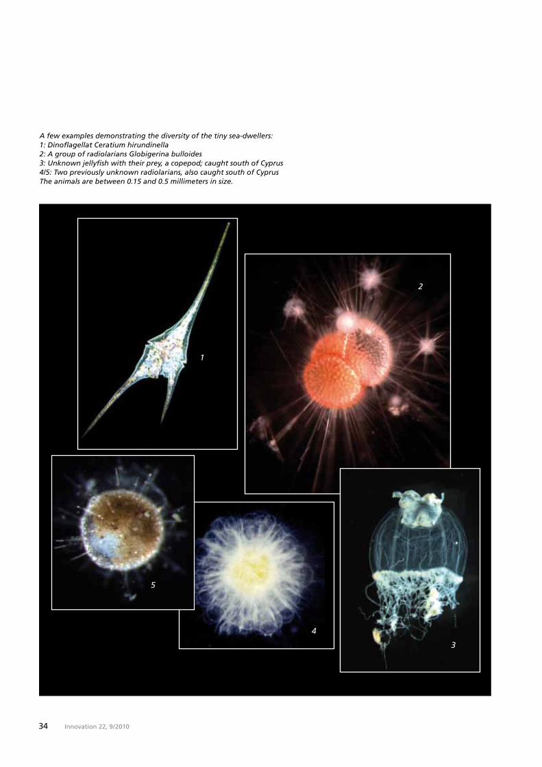
A few examples demonstrating the diversity of the tiny sea-dwellers:1: Dinoflagellat Ceratium hirundinella2: A group of radiolarians Globigerina bulloides3: Unknown jellyfish with their prey, a copepod; caught south of Cyprus4/5: Two previously unknown radiolarians, also caught south of CyprusThe animals are between 0.15 and 0.5 millimeters in size.

Report: Sailing the seven Seas
35Innovation 22, 9 / 2010
Initially, the organisms collected at various depths will be roughly sorted using the Stemi DV4 systems. The “harvest” is then taken to the dry lab located in the belly of the ship. There, the tiny organisms are exam-ined in more detail, classified and photographed. To ensure that the scientists do not lose track of the samples due to vast amounts of ma-terial concerned, barcodes are allocated to the data.
The SteREO Discovery.V20 is located below deck. It makes it possible to study the anatomy and motion processes of the organisms with extreme accuracy. These observations then allow conclusions to be drawn about the way of life and behavior of the sea-dwellers. A large number of samples are first frozen in the wet lab before being sent directly to the Eu-ropean Molecular Biology Laboratory.
Adapting their lifestyle. Very little space remains for the 15-strong crew on the 36-meter-long schooner. “To ensure that everything goes smoothly on board, everyone has to adapt their lifestyle to this situation. This can sometimes prove to be difficult,” admits Emmanuel Reynaud. “At the start there were three separate teams on board,” he continues. “These were the crew, the scientists and the journalists, and each group had its own goals and ideas.” This inevitably led to tension. And the turbulent Mediterranean seas caused by the autumn storms near Nice made things even more challenging for the crew. Thankfully, Tara is now sailing in calmer waters, and the processes on board have become a matter of
The details
Stereomicroscopes
The SteREO Discovery.V20 (see photo) has a zoom factor of 20, i.e. starting from an ini-tial overview of a sample, it is then possible to visualize or zoom in on small details. The selected magnification can be activated via motor control. The high final magnification permits the three-dimensional viewing of objects which could only be examined two-dimen-sionally under a light micro-scope until now. Stemi DV 4 combines sturdy technology with operating simplicity. When the system is combined with the compact C stand, the user can switch between reflected, transmitted and mixed light.
Further information is available at www.taraexpeditions.org
routine. “I never cease to be amazed at how this expedition actually works on such a small ship – and the situation is constantly improving,“ says a happy Emmanuel Reynaud.
Unknown creatures. When the HMS Challenger entered Portsmouth harbor in 1876, it had almost 4000 previously unknown creatures on board. This time, once again, scientists will probably uncover or-ganisms in the depths of the oceans that no one ever thought existed. The goal is to build up a database in which both well-known and newly discovered sea-dwellers are recorded in order to allow faster detection of any subsequent changes occurring in the oceans. “We are primarily interested in what we can’t see with the naked eye,” says Emmanuel Rey-naud. 98 percent of the marine bio-mass is composed of microorganisms. “Our very existence depends on these creatures. They are the start of the food chain that ends with human be-ings.”
Verified results from the Tara Oceans expedition are expected in five years’ time at the latest. In the Challenger expedition it was 30 years before the last research results were finally published in a total of 50 volumes.
Monika Etspüler

42 Innovation 22, 6/ 2010
Feature
Capturing the Heat of the Moment
It’s in the details. It doesn‘t matter if you’re watching a sporting event, checking out nature or hunting – you want to see every little thing from far away. High-quality binocu-lars are an indispensable tool. How-ever, it is hardly possible to improve modern optics or clearly make them stand out from the competition us-ing traditional mechanical and opti-cal methods. Therefore, the trend is moving towards the integration of electronic components that pro-vide added value for each specific application.
“I like technical innovations,“ admits German biathlon national team coach Frank Ullrich, referring to the new Victory PhotoScope 85 T* FL from Carl Zeiss. This spotting scope featuring a fully integrated digital camera is one of his primary sources of information. He uses it not only to follow the excitement of the compe-tition, but also to capture those spe-cial moments on camera or video, which he can analyze in peace and quiet afterwards. “In my job, being able to observe, take pictures and even videos at the same time is a great benefit,“ says Frank Ullrich. This was not possible in the past. On traditional setups, the camera is simply attached to the rear of the spotting scope.
Binoculars with added functionality. The Victory PhotoScope is the latest development in a line of products
hobby ornithologist Stephen Ingra-ham has been on the trail of our feathered friends for 20 years. “It’s often difficult to even see the ani-mals. Getting a really good look at them demands not only patience, but also good technology,“ he ex-plains. With an objective lens focal length of 600-1800 millimeters and the integrated seven megapixel cam-era, the Victory PhotoScope is the perfect tool. From five meters to far away, objects can be seen brilliantly and crisply even in poor light. The spotting scope itself provides 15-45x magnification. For Stephen Ingra-ham, the ease of use and the robust-ness of the instrument that easily weathers the rain, cold and heat un-scathed, are added bonuses.
Outstanding design. The Victory PhotoScope was honored with the red dot design award last year thanks to its attractive appearance. Victory RF binoculars received an award in 2008 for their “ease of use, clearly arranged display and suitabili-ty for practical use.”
Monika Etspüler
that also includes the Victory RF (8x and 10x45 T*) binoculars and the Victory Diarange 2.5-10x50 T* rifle-scope. They all feature additional electronic functions to provide users with even more improvements: the Victory PhotoScope a digital camera, the riflescope a laser rangefinder and the Victory RF binoculars a laser rangefinder and the BIS® ballistic in-formation system.
Fast and precise information. The Victory RF is a flexible system. It can be used for hunting or animal and bird watching. The integrated digital laser rangefinder ensures particularly fast and precise measurement be-tween 10 and 1200 meters. A self-il-luminating LED display projects the result directly into the field of view of the binoculars. In addition to the exact range, hunters also need infor-mation on the trajectory of the bul-let and the holdover. The ballistic system automatically calculates the required information in a fraction of a second. Six stored ballistic curves cover the flight paths of practically all hunting calibers.
Nature at its best. One of the driving forces behind the development of the Victory PhotoScope was to enable users to experience nature in close-up and capture its fascination. As a result, the product precisely meets the needs of a Carl Zeiss em-ployee in Chester, Virginia. Equipped with binoculars and a camera,
36 Innovation 22, 9/ 2010

43Innovation 22, 6/ 2010
Capturing the Heat of the Moment
37Innovation 22, 9/ 2010

38 Innovation 22, 9 / 2010
Report

Report: Better Vision
39Innovation 22, 9 / 2010
The Beautiful World of Vision
“ZEISS Experience” is a newmarketingstrategy. Since May 2009 Carl Zeiss Vision has beenoffering analytical, training andmarketing tools under the motto“Seemore.Livemore.”Eyecarepro-fessionalsusethesetocommunicatethe importance of good vision totheirpatientsandtheneedtoselecttherighteyeglassesfortheireyes.
The eyes are our most important sense organ; in effect, they are our “gateway to the world.” It is not until we see badly and therefore need glasses that it becomes obvious how important our eyes are for the quality of life we enjoy. Finding the ideal pair of eyeglasses is no easy matter. First and foremost, they must improve vision, but they should also make the wearer look good and, of course, they should be affordable. Being spoilt for choice does not only mean being confronted by a vast selection of colorful frames. What is even more difficult, and indeed more important, is the choice of the right lens. Very few people know what lens can solve their personal visual problem. Usually, they know nothing about the advances that have been made in technology or about the possibilities offered by modern eyeglass lenses.
Expert advice. The days when eye care professionals used to have a large number of low-price frames and only a handful of expensive frames for well-heeled clients, or
one test chart and one instrument for measuring visual acuity, are well and truly gone. Nowadays, eye care professionals who are contractual partners of Carl Zeiss have numerous systems for a wide variety of differ-ent tasks: the i.Profiler® uses wave-front technology and automatically measures sphere, cylinder and axis, the i.Polatest® measures visual acu-ity, three-dimensional vision and the interaction of the eyes, and on the i.Terminal® the eyeglasses are exactly centered, as even the smallest cen-tration error can lead to a loss of over 40 percent in visual perfor- mance.
Patientwishes. As technological ad-vances have now been made that were totally inconceivable just a few years ago, the patient is reliant on the expert advice provided by the eye care professional. He or she must explain the examination methods and the special features of the lenses to provide patients with factual cri-teria as a basis for selecting the lens that best meets their requirements. One important field is, for example, progressive lenses that initially re-quire a certain adaptation process from the wearer. Here, not only careful consultation but also ongo-ing support is required.
People purchasing eyeglasses want to know exactly what they can expect from the advice and care of-fered by an expert. A current survey of eyeglass wearers in ten countries showed that they are very prepared to invest in high quality eyewear if they are convinced of the benefits offered by the lenses beforehand.
They want to obtain detailed infor-mation and a customized solution for their personal visual problem. The lens brand has a considerable in-fluence on the decision they make, but only if the patient knows it and has learned to value it.
Comfortable vision. Lenses precisely tailored to the wearer’s needs help avoid eye fatigue. Impairments of vision are minimized, and custom-ized lenses for work, leisure or sport offer maximum visual comfort.
Ursula Walther

40 Innovation 22, 9 / 2010
Report
40 Innovation 22, 9 / 2010
Revolution in Laser Surgery
The new ReLExTM treat- mentmethodfromCarl Zeiss heralds the dawn of a new era in refrac-tivesurgery.ReLExcombinespreciserefractivefemtosecondlasertechnol-ogyandthegentleremovalofacor-neallamellatocreateanew,revolu-tionaryprinciple.Onedecisivediffer-ence from the current LASIK correc-tive procedure is that the acuitycorrection isperformed in the intactcornea.ReLExhasalreadybeenusedsuccessfullymorethan1000times.
Quality of life. Sometimes even the best eyeglasses are not good enough. When swimming, playing football or entering a heated room in the win-ter, every wearer is promptly remind-ed of their glasses. Small wonder, therefore, that more and more peo-ple are now opting for laser treat-ment.
Abriefhistory. The first clinical stud-ies on surgical procedures on the eye were conducted in the 1930s. Researchers were looking for effec-tive methods of changing the refrac-tive power of the cornea. Surgeons started by making circular incisions aimed at destabilizing the cornea so that it lost part of its surface tension and became flatter. However, the incisions frequently led to inflamma-tion and scarring. In 1963 the Span-iard Jose Ignacio Barraquer devoted
his attention to the me-chanical remov-al of the inner layers of the cornea – a method that was then regularly used to treat shortsighted patients from 1978 onwards. In 1988 Theo Seiler of the University Hospital of the Free University of Berlin used a laser for the removal of thin corneal layers for the very first time.
Optical planer. The techniques were enhanced, but the basic principle remained the same: the doctor folds back the topmost layer of the cor-nea, removes a thin layer of the underlying tissue and then folds the upper layer back over the wound. LASIK is the name given to the tech-
nique now most commonly used. A femtosecond laser (a femtosecond equals 10-15 seconds) is increasingly being utilized for this purpose. It transmits millions of high-intensity laser pulses to thecornea and there-fore severs the tissue. It generates

41Innovation 22, 9 / 2010 41Innovation 22, 9 / 2010
to the cornea. The result: an optimal corneal shape across the entire opti-cal zone.
Noticeable comfort. Compared to LASIK, ReLEx offers noticeable bene-fits in regard to patient comfort. For example, there is no longer any need for the patient to move to another instrument during the thera-py. This considerably reduces treat-ment times. The procedure is over in a matter of minutes – without noise and without any unpleasant odor. Thanks to the innovative design of the contact lens, the patient’s tempo-rary loss in vision associated with LASIK is avoided. ReLEx ensures that patients enjoy maximum visual comfort in their new lives without eyeglasses.
Ursula Walther
Revolution in Laser Surgery
the corneal flap precisely and with-out risk or pain. This is then folded open. A medical excimer laser, a type of optical planer, then flattens the cornea to the desired curvature. Each procedure therefore requires two workflows with two different instru-
ments, which means lengthy treatment
time for both doctor and patient.
A brand-new approach. ReLEx from Carl Zeiss is a gen-
uine alternative to LASIK. The Vi-suMax® femtosecond laser first cuts
a thin lens-shaped segment, a lamel-la, into the intact cornea. The laser then generates a corneal flap that provides the doctor with an access route for removal of the lamella. The integrated, high-quality ZEISS surgi-cal microscope gives the doctor opti-mum control during the manual re-moval of the lamella. The technique offers many clinical benefits, particu-larly for patients requiring high pre-scriptions. Unlike LASIK treatment, the VisuMax with ReLEx transfers the pre-calculated lamella profile exactly
Report: Better Vision

In the future the inser- tion of artificial lenses forthecorrectionofas- tigmatismwillbefasterandmorereliablethanwithcurrentmethods. For measurement of theeye as well as the calculation andpositioning of the lens during sur-gery, the ZEISS Toric Solution fromCarl Zeiss Meditec combines se-veral tools that increase workflowefficiency.
42 Innovation 22, 9 / 2010
The Solution with the Cylinder
Report
Astigmatism is one of the visual defects that can be corrected with an artificial lens. It is caused by irreg-ular curvature of the cornea. Nor-mally, the cornea of the human eye is as round as a barrel. In astig-matism, however, it looks more like an egg or an American football. Peo-ple with this malformation of the cornea have a blurred image of the world, as the focal point required for clear vision becomes a focal line.
The solution. Doctors can correct minor astigmatism during laser treatment of the eye. For severe

43Innovation 22, 9 / 2010
astigmatism, the intraocular lens is the means of choice. If a patient suffers from both astigmatism and cataract, the doctor inserts a toric intraocular lens during cataract sur-gery. However, this only corrects the visual defect if it is positioned at the right angle in the patient’s eye. This angle must be carefully calculated by doctors – a process that required considerable time and effort in the past. For example, they had to place a transparency with the right dimen-sions over the monitor on which they were following the procedure. The ZEISS Toric Solution from Carl Zeiss Meditec simplifies the complex workflow by linking several systems and products.
Goldstandardofbiometry. To some extent, the IOLMaster® can be con-sidered as the gold standard of optical biometry. In other words, developers see it as the benchmark system. The doctor uses the IOL- Master to measure the patient’s eye without touching it and receives precise, reliable and reproducible measuring results. Maximum preci-sion is important for the calculation in Z CALC. Z CALC is the official online calculator for all toric ZEISS intraocular lenses. It is used directly online by the doctor before the surgery begins. This means he or she can change both the sphere and cyl-inder of the recommended lens in real time prior to the procedure and estimate in advance how the new lens will influence the refraction of light in the eye.
Thespecial cylinder. When selecting a suitable intraocular lens, doctors have various possibilities at their
disposal. Carl Zeiss Meditec is the only company in the world to offer bitoric lenses, i.e. lenses in which the refractive power of the cylinder is distributed symmetrically over the front and back of the lens. This leads to considerably improved im-aging, particularly in the case of high cylinder powers, because the radii of curvature do not differ on the two sides of the lens. Bitoric lenses are either monofocal or bifocal, which means that they have either one or two focal points. With monofocal lenses patients have to decide whether they would prefer to have good distance and less good near vision, or vice-versa, after the surgery. With a bifocal lens, they can enjoy both.
Therightangle. As part of the intel-ligent CALLISTO eye assistance sys-tem, Z ALIGN supports the doctor during the surgical procedure. For orientation when aligning the lens, doctors use a target axis that they project on top of the video image of the eye on a touchscreen and then adjust with degree accuracy. Even if the patient’s eye moves during the alignment or the insertion of the lens, the target axis remains stable as Z ALIGN tracks the movement of the eye automatically. The result: the ZEISS Toric Solution allows the procedure to be performed more quickly than ever before by eliminat-ing the risk of slipping.
Ursula Walther
Report: Better Vision

44 Innovation 22, 9 / 2010
Essay
Twenty Years Later 1990 saw the reunification of Carl Zeiss West and East
45Innovation 22, 9/ 2010
The company Carl Zeiss was partitioned in 1945 in the wake of the Second World War and remerged after the reunification of Germany. During the cere- mony held to mark the 150 years of Carl Zeiss on 19 November 1996, German Chancellor Helmut Kohl stated that “Carl Zeiss mirrors the problems, but also the opportunities inherent in German unity.” The 20th anniversary of German reunification is reason en-ough to examine how Carl Zeiss has become a single, united company within the new Germany. This at times dramatic story impressively shows how new strength and dynamism can be generated by a unification process.
The events took place two decades ago: the fall of the Berlin Wall, monetary union, the reunification of Germany. Today, a new generation of young people who know this period from hearsay only is entering the job market. Political ceremonies on 3 October, the Day of German Unity, and memorials in East German cities have remained as testimonies to this momentous period in the country’s history.
The process of national convergence between East and West has been continuing for two decades now, but it is not yet complete. The Solidarity Pact between the East and West lasts until 2019. The process of complete-ly restructuring the totally decrepit East German eco-nomy is still underway. Few state combines in the for-mer East German state have achieved the transition to a global player; only a small number of brands have survived into the new era. And that a once partitioned company has managed to go down in history by achie-ving its own reunification process is an even rarer accomplishment.
Twenty years ago the history of Carl Zeiss took a new direction. Two decades have now passed since the two factories in Oberkochen and Jena made the first tentative steps toward reconciliation. Steps that were aimed at bringing together two firms that had emerged from the former Jena enterprise as a result of the World War and that had since gone their sepa-rate ways. Two independent companies, one domina-ting the Western optical markets from its base in Ober-
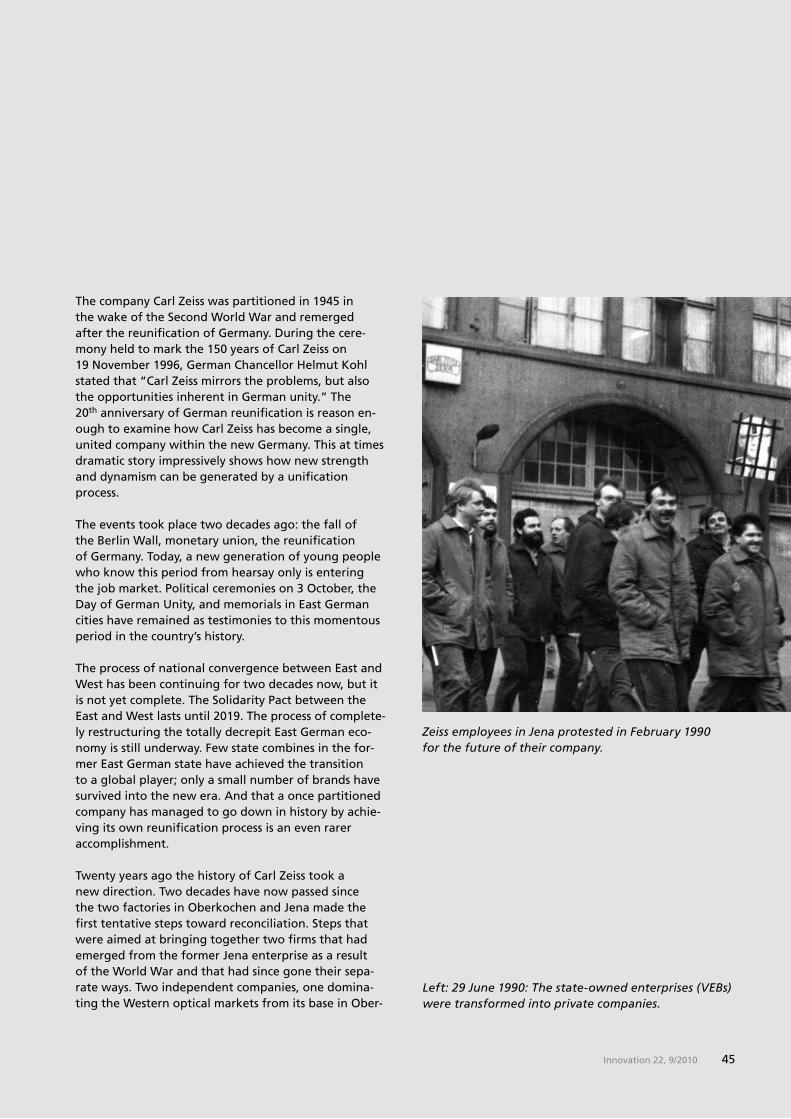
Left: 29 June 1990: The state-owned enterprises (VEBs) were transformed into private companies.
Zeiss employees in Jena protested in February 1990 for the future of their company.

46 Innovation 22, 9 / 2010
Essay
The Foundation is the key. Having the Foundation as the sole owner ensures independence from capital markets and promotes science; it guarantees profit sha-ring for the workforce and safeguards the legacy of Carl Zeiss and Ernst Abbe. Strictly speaking, it offered nothing from which the “state of workers and far-mers,“ as East Germany described itself, could benefit. The Jena Foundation enterprises were converted into state-owned enterprises (VEBs) in 1948. This meant that the Jena Foundation lost its function as the owner of the two factories. Technically, however, it continued to exist.
The Foundation, and the problems associated with it, was the linchpin on which the fate of Carl Zeiss turned after 1990. The future had to be shaped with due regard to tradition. In a highly charged environment comprising politicians, a privatization agency (“Treu-handanstalt”), trade unions and the workforce in both the East and West, a solution had to be found to the unique Carl Zeiss scenario.
The ownership structure had two consequences: it initially posed legal challenges on the road to a uni-fied Carl Zeiss Group in Germany. Above all, however, it played a decisive role in ensuring that the Carl Zeiss
kochen in West Germany and the other the markets in the Eastern Bloc from its Jena headquarters in the East. Separate brands, separate developments, but a common name and a common tradition.
Who gets what and where? Just a few days after the fall of the Berlin Wall the companies started to test the waters. Staff from Jena ventured to Oberkochen, and Zeissians from Oberkochen made their way to the city of Carl Zeiss and Ernst Abbe. Everyone was curious: after all, they all shared a unique history that began with the company founder Carl Zeiss in 1846. Then as now, Zeissians knew that their company was one of the world leaders in the field of optics, even if this world was split into two halves – the West and the East – and they acted as rivals in the competitive arena.
East Germany conducted the longest lawsuit in its history in London – and it was all about the ZEISS brand. In 1971, after 18 years of litigation, an agree-ment was reached that perfectly reflected the spirit of the Cold War: Carl Zeiss West and East divided up the hemispheres for their respective fields of business and pledged never to use the ZEISS brand in the part of the world where the other company operated. This ava-lanche of lawsuits was partly triggered by the renaming of the Oberkochen-based company, initially founded under a different name, to Carl Zeiss in 1951.
Tradition initially determines the future. Two compa-nies that had now grown apart began a dispute about their most precious heritage: the ZEISS brand and the legitimate successor to the Carl Zeiss Stiftung (Carl Zeiss Foundation) as the owner of the companies Carl Zeiss and Schott.
Ernst Abbe, a companion of Carl Zeiss and his successor as head of the company, incorporated the enterprise in a foundation that was and still is committed to the successful continuation of the company, the well-being of its employees and the advancement of science. The firm of the colleague and friend of Zeiss and Abbe, Otto Schott, followed this example. Without the company Schott Glas, the revolutions achieved in optics would never have been possible.
In 1990 President and CEO Dr. Horst Skoludek (right) visited the grave of Carl Zeiss in Jena accompanied by press spokesman Manfred Berger.
47Innovation 22, 9/ 2010
state combine did not suffer a similar fate to other enterprises in the crumbling East German economy: a resolution passed by the privatization agency, transfer to a competing group of companies, a guarantee of employment followed by investment aid and mass layoffs and, in many cases, a future as an “extended workbench” of companies in the West.
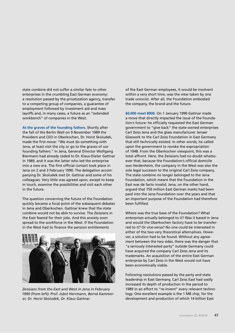
At the graves of the founding fathers. Shortly after the fall of the Berlin Wall on 9 November 1989 the President and CEO in Oberkochen, Dr. Horst Skoludek, made the first move: “We must do something with Jena, at least visit the city or go to the graves of our founding fathers.” In Jena, General Director Wolfgang Biermann had already ceded to Dr. Klaus-Dieter Gattnar in 1989, and it was the latter who led the enterprise into a new era. The first official contact took place in Jena on 2 and 3 February 1990. The delegation accom-panying Dr. Skoludek met Dr. Gattnar and some of his colleagues. Very little was agreed upon, except to keep in touch, examine the possibilities and visit each other in the future.
The question concerning the future of the Foundation quickly became a focal point of the subsequent debates in Jena and Oberkochen. Gattnar knew that the state combine would not be able to survive. The Zeissians in the East feared for their jobs. And this anxiety soon spread to the workforce in the West. If the Foundation in the West had to finance the pension entitlements
of the East German employees, it would be insolvent within a very short time, was the view taken by one trade unionist. After all, the Foundation embodied the company, the brand and the future.
60,000 meet 8000. On 1 January 1990 Gattnar made a move that directly impacted the issue of the Founda-tion’s future: he officially requested the East German government to “give back“ the state-owned enterprises Carl Zeiss Jena and the glass manufacturer Jenaer Glaswerk to the Carl Zeiss Foundation in East Germany that still technically existed. In other words, he called upon the government to revoke the expropriation of 1948. From the Oberkochen viewpoint, this was a total affront. Here, the Zeissians had no doubt whatso-ever that, because the Foundation’s official domicile was Heidenheim, the company in the West was also the sole legal successor to the original Carl Zeiss company. The state combine no longer belonged to the Jena Foundation, which meant that the Foundation in the East was de facto invalid. Jena, on the other hand, argued that 750 million East German marks had been paid into the Jena Foundation over the years and that an important purpose of the Foundation had therefore been fulfilled.
Where was the true base of the Foundation? What enterprises actually belonged to it? Was it based in Jena and would the Oberkochen factory have to be transfer-red to it? Or vice-versa? No one could be interested in either of the two very theoretical alternatives. Howe-ver, a solution had to be found. Without any agree-ment between the two sides, there was the danger that “a seriously interested party” outside Germany could have acquired the company Carl Zeiss Jena and its trademarks. An acquisition of the entire East German enterprise by Carl Zeiss in the West would not have been economically viable.
Following resolutions passed by the party and state leadership in East Germany, Carl Zeiss East had vastly increased its depth of production in the period to 1989 in an effort to “re-invent” every relevant techno-logy. One excellent example is the 1 MB chip, for the development and production of which 14 billion East
Zeissians from the East and West in Jena in February 1990 (from left): Prof. Jobst Herrmann, Bernd Kammer-er, Dr. Horst Skoludek, Dr. Klaus Gattnar.

48 Innovation 22, 9 / 2010
Essay
Speech during the ceremony to mark the 150th anniver-sary: Chancellor Helmut Kohl in 1996.
German marks had to be invested. In 1989 Carl Zeiss Jena generated sales of 64,000 marks (East) per emplo-yee, compared to 145,000 marks (West) in Oberkochen. The state combine had a total of over 60,000 emplo-yees, while Carl Zeiss West had 8,278.
The contrast could not have been greater. A hugely bloated state combine on one side, and a lean, mid-sized company on the other. On one side, the federal state of Thuringia in the East with many other worries to deal with than the question where a foundation was going to be based in the future, and on the other the Foundation Commissioner in the West – the Educa-tion Minister responsible for universities in the federal state of Baden-Wuerttemberg – who firmly believed that Heidenheim was the legal domicile of the Carl Zeiss Foundation. This was also seen as the legitimate heir of the company and the ZEISS brand.
The Foundation issue turned out to be resolvable as soon as the regional political system was formed after the first and last free elections in the East German state took place on 18 March 1990. With the Declaration of Biebelried of 29 May 1990, the parties involved agreed to strive to incorporate the industrial enterprises with-in a single Foundation.
The power of unity. Dr. Dieter Kurz, current President and CEO of Carl Zeiss AG, says in retrospect: “Our common origin and name were the very strong forces that ultimately brought the two companies together.” And these forces were certainly put to the test in the period to the mid-1990s. A few days before 3 October 1990, on the express initiative of the Zeissians in Thurin-gia, the Volkskammer – the East German Parliament – transferred 20 percent of its shares in the state combine to the Foundation in Jena. This ruled out the possibility of Carl Zeiss West regulating the future of the factories in the East by acquisition.
In March 1991 Detlev Karsten Rohwedder, head of the government privatization agency, announced the decision that had been reached about Carl Zeiss. The Carl Zeiss enterprises and the Schott factories in Thurin-gia were to be transferred to the Jena Foundation. This included the responsibility for the then still 30,000 em-ployees, layoffs and redundancy programs. Transfer to the Carl Zeiss Foundation in Heidenheim would have placed this burden on the companies in the West.The
29 May 1990: In Biebelried representatives from Schott and Carl Zeiss document the intention to unite the companies in the East and West in one Carl Zeiss Foundation.

49Innovation 22, 9/ 2010
Other companies relocated as a result of the War now have branches in the east of Germany again or have acquired parts of former state combines. However, no other company has focused on its “place of birth” with greater determination than Carl Zeiss. The Zeissians in Jena and Oberkochen can rightly say that they have united their sites in the East and West into a single, joint company. “The two companies clearly have the same genes,“ says Dr. Kurz. “That was also a key factor why a new, joint phase of success began after a difficult transitional period.” The changes that the company has experienced have made Carl Zeiss strong. The expe-rience gained during reunification also helped to over-come the crisis in the mid-1990s. The brand is now undi-vided and globally present. Important business groups within the company conduct their worldwide business operations from Jena. Carl Zeiss Jena GmbH, head-quartered in Jena, has been the shared production company for East and West since 2009 – with an opera-ting site in Oberkochen.
The authors from the East and West: Gudrun Vogel has worked at Carl Zeiss in Jena since 1979, where she now heads the Press Office. Thomas R. Zecher headed the Press Department at Carl Zeiss in Oberkochen until 1992 and has been working as a freelance PR consultant in Frankfurt since 1998. Joachim Kuss is a Communications Consultant at Ketchum Pleon.
“mammoth meeting” held in the headquarters of the privatization agency on 11 June 1991 found a financing solution for restructuring the Jena enterprises that had succeeded the state combine Carl Zeiss Jena. On 25 June 1991 an agreement in principle was reached: the Foun-dation was now to be domiciled in “Heidenheim an der Brenz and Jena.” This period also saw the splitting of the companies succeeding the state combine. Many car-ve-outs from the former East German combine were launched. On 22 October the following was documented in the commercial register: with a workforce now tota-ling 3,000 people, Carl Zeiss Jena GmbH was once again an independent company fully owned by the Carl Zeiss Foundation.
Financial losses, downsizing both the East and West and mutual distrust overshadowed the company‘s history until 1995. Much has been written and speculated about whether Oberkochen simply wanted its sister company in Jena to disappear from the market. Carl Zeiss was cited as a prime example of how badly the reunification process was proceeding. “At Zeiss the reunification pro-cess has gone totally wrong. The enemy is in the East,“ wrote the news magazine Der Spiegel in October 1994. 20 years later what the two companies had in common has proven to be stronger than what divided them. Dr. Kurz now says: “On the basis of their shared roots the two companies committed themselves totally to innova-tion and to attaining the highest possible quality.”
The return to common roots was emphasized in 1991 by the newly designed, uniform logo: the “square” of the previous Oberkochen company logo was combined with the curved “lens” of the logo used by Carl Zeiss Jena.
Logo in the west Logo in the east Current logo
+ =

50 Innovation 22, 9/2010
Look at Laureates
Observing Molecules in Real Time
Together with Osamu Shimomura and Martin Chalfie, Roger Tsien received the Nobel Prize for Chemis-try in 2008 for the discovery of the green fluorescent protein (GFP).
Tsien studied chemistry and physics at Harvard. He has been a professor of pharmacology, chemistry and biochemistry at the University of California in San Diego since 1989.
For more than 20 years now, Roger Tsien has played a pivotal role in the development of important fluorescence methods that have set trends in molecular cell biology.
An initial highlight in his career as a researcher was the use and further development of fluorescent dyes that can visualize cellular calcium in single living cells. He received world-wide recognition for his research work on the GFP and its use. GFP revolutionized research in cell and neurobiology in particular: since its inception, scientists have been able to look into the living cell and molecules in real time. Virtually all biomedical disciplines, from brain to cancer research, benefit from the discovery of the GFP.
In 2004 the German Society for Cell Biology (DGZ) declared Roger Tsien as the winner of the Carl Zeiss Lecture Award.
substrates for certain proteases, protein-splitting enzymes that are secreted by the tumor cells. One long-term objective is the develop-ment of fluorescent molecules that are activated without gene transfer. This allows them to be used as clinical biomarkers for the diagnosis and follow-up of diseases.
Since then, the focus of Tsien’s research has changed. “I‘ve always wanted to do something clinically relevant in my career, if possible, and cancer is the ultimate challenge.”The imaging and treatment of can-cer tumors are now the center of his attention. His initial discoveries led to a U-shaped peptide that carries either an imaging molecule or a drug. The peptide can dock on to the cancer cell. Peptides are

51Innovation 22, 9/2010
EN_9
0_01
0_02
2 P
rin
ted
in G
erm
any
CM
-MSW
_IX
201
0 U
Ao
o
Twelve Images to Create a Picture
Photographic art from Bryan Adams – photographer and rock star
In the heart of Manhattan, a very short man and a very tall woman are carrying a very large package across the street in front of the Guggen-heim Museum. A movie scene? Art theft? Not even remotely close – it is one of the images in the 2011 Carl Zeiss calendar. A world-famous artist staged 12 moments for the calendar, which will accompany customers and friends of Carl Zeiss throughout the coming year: Bryan Adams.
Bryan Adams is known to many of his fans as a rock star. He is also a highly professional photographer. Magazines around the globe print his pictures. He also started his own award-winning photo magazine – ZOO.
Two other celebrities will feature in the photos: movie and TV star Michael J. Fox and super model Tatjana Patitz. Fox has been a global name since his blockbuster movie
Back to the Future. He has also re-ceived numerous awards for his roles in many major movie and television productions. Tatjana Patitz is an in-ternational super model. Born in Hamburg, Germany, she now lives in California and still works in front of the camera around the world. For the Carl Zeiss calendar, they de-pict a short man and a tall woman – sometimes with grand, sometimes almost casual gestures, but always with a certain tongue-in-cheek irony.
The Carl Zeiss calendar from Bryan Adams follows in the footsteps of Wim Wenders´ 2009 special-edition calendar titled Tomorrow Morning.
Innovation – The Magazine from Carl ZeissIssue 22, September 2010
Issued by:Carl Zeiss AG, Oberkochen, GermanyCorporate CommunicationsJörg Nitschke
Editors:Silke Schmid (Editor-in-Chief), Dr. Dieter Brocksch, Gudrun VogelCarl Zeiss AGCorporate CommunicationsCarl-Zeiss-Str. 2273446 Oberkochen, [email protected] +49 (0) 7364 20-8208Fax +49 (0) 7364 20-3122
Contributors to this issue:Monika Etspüler, Ursula Walther, MSW;Thomas R. Zecher, Joachim Kuss
English Translation: Clive Poole and Paul Soos
Conception and design:Overall coordination Nicola Schindler MSW, Manfred Schindler Werbeagentur OHG, Aalenwww.msw.de
Pictures courtesy of: Cover Photo: Fotolia/MSW; Page 7 top: Courtesy ASML; Page 8 bottom: Carl Zeiss OIM GmbH, J. Packeiser; Page 16-17: M. Kirschmann, D. Oberti, R. Hahnloser, Institut für Neuroinformatik, Uni. Zürich und ETH Zürich, Schweiz; Page 18: Prof. Dr. Martin Bastmeyer, Uni. Karlsruhe (TH) Zoologisches Institut I; Page 26-27, 28: Rocco HeimchenPage 30-31, 33, 35: F. Latreille/Fonds Tara; Page 34: 1/3/5: Dr. Emmanuel G. Reynaud, UCD-Tara Oceans, 2: Dr. Fabrice Not, Roscoff-Tara Oceans, 4: Johan Decelle, Roscoff-Tara Oceans; Page 36-37: Stephen Ingraham; Page 40-41,42: Fotolia; Page 44: Frank Dö-bert; Page 50: Wikipedia;
Printing: C. Maurer Druck und Verlag, Geislingen an der Steige
Innovation – The Magazine from Carl Zeiss appears in German and English
ISSN 1431-8040
The contents of the articles do not alwaysreflect the opinion of the publisher. Reprintonly with the exclusive written permissionof Carl Zeiss.
To order an issue or change your mailing address, please contact Renate König +49 (0) 7364/20-2878 or [email protected]