the management of congenital muscular dystrophy (cmd)€¦ · the management of congenital muscular...
TRANSCRIPT
The Management of Congenital Muscular Dystrophy (CMD)A guide for families

The Management of Congenital Muscular Dystrophy (CMD)
A guide for families
PREFACEThis family guide summarizes an international consensus on congenital muscular dystrophy (CMD) diagnosis and medical care. This effort was supported by Cure CMD (curecmd.org), TREAT-NMD (treat-nmd.eu), AFM-Association Française contre les Myopathies (afm-france.org), and Telethon Italy (telethon.it). The main document is published in the Journal of Child Neurology (Ching H Wang, et al. Consensus Statement on Standard of Care for Congeni-tal Muscular Dystrophies, J Child Neurology 2010;25(12):1559 –1581. Published online 15 Nov 2010). The main document can be down-loaded free from:http://jcn.sagepub.com/content/25/12/1559.
This family-oriented CMD treatment guideline is based on medical management recommenda-
tions by a group of 82 international experts from 7 medical subspecialties: pathology, neurol-ogy, pulmonary/ICU care, gastrointestinal/nutri-tion/speech/oral care, orthopedics/rehabilitation, cardiology, and palliative care. To build consen-sus, the team used the following strategies: • a comprehensive literature review • an online expert survey of how CMD care is currently provided in their practice • an online survey of families’ opinions on key care issues and care gaps in CMD • a 2-day CMD Standard of Care work shop, held in Brussels in November 2009.

DISCLAIMER The information and advice published or made available in this booklet is not intended to replace the services of a physician, nor does it constitute a physician-patient relationship. This advice should be taken in conjunction with medical advice from your medical clinician whom you should consult in all matters relating to your health, in particular with respect to symptoms that may require diagnosis or medical attention. Any action on your part in response to the information provided in this booklet is at your own discretion.
1
Table of Contents 1 Introduction… 1
2 Comprehensive Management… 5 Care at Diagnosis, Ongoing and Hospital Stays
3 Neurologic Management… 9 Care of Seizures and Cognitive Impairment
4 Respiratory Management… 10 Care of Breathing
5 Gastrointestinal Management… 15 Nutrition, Feeding, and Oral Care
6 Cardiac Management… 19 Taking Care of the Heart
7 Orthopedics and Rehabilitation Management… 23 Care of Contractures and Scoliosis
8 Palliative Care… 27 Individual and Family Emotional Well-being
Appendix A - Definition of Subtypes… 29Appendix B - Definition of Experts Providing Specialty Care… 31Appendix C - Glossary of Terminology (terms underlined in text)… 33 Appendix D - Diagnostic Tools… 39

2
You or your child may have just received a diagnosis of congenital muscular dystrophy (CMD). You may be feeling overwhelmed with the amount of information presented to you. It is important that families and individuals affected with CMD understand the medical issues surrounding this diagnosis so that they can anticipate and participate in their or their child’s health care and management.
The purpose of this guide is to assist you in understanding the many different symptoms that may be present and the types of care that may be required over time. Understanding this information will help you to better anticipate the needs associated with a diagnosis of CMD and to become a more effective advocate.
The CMDs are a group of mostly inherited rare diseases with symptoms starting within the first 2 years of life. Early symptoms may include weakness (hypotonia), contractures, and breathing and feeding problems. The CMDs are part of the spectrum of muscular dystrophy. This means that the same gene that can lead to a CMD can also lead to a limb-girdle muscular dystrophy or later-onset muscular dystrophy. People with CMD with the same subtype may have different experiences; they may be stronger or weaker than others with the same subtype or may have had symptoms earlier or later than someone else. Within this group of CMD diagnoses, a percentage of people have a subtype in which the genetic mutation responsible has not yet been identified. Many researchers around the world are working to identify all the genetic mutations that cause the CMDs, with new discoveries made yearly.(Continued on page 4)
1 INTRODUCTION What is Congenital Muscular Dystrophy?
How to Use this DocumentThis document first gives an overview of the essential areas of care. It is further broken down into the specific body systems that can be affected by the CMDs, such as heart or lung, and other problems that can be seen in people with the same diagnosis. Some of the CMDs have specific problems that are not nec-essarily seen with other types of CMD. These differences are described in this document.
The areas of specialty care involved with the treatment of CMD, and described in this guide, are neurology and neuromuscular, pulmonary (respiratory), GI/nutrition/oral care, cardiology, orthopedics and rehabilita-tion, and mental health/palliative care. Although these areas of care appear to be separate and distinct, the best way to manage your child’s health care needs is with a multidisciplinary team that includes subspecialists, allied health professionals (physical therapy, respiratory therapy), and the family engaging in discussion and management decisions.
Although multidisciplinary care is the ideal, you may find that your child’s care is difficult to coordinate without access to CMD experts and subspecialists. Identifying and obtaining a referral to a national center of pediatric neuromuscular excellence can be the first step in obtaining coordinated care.
You may wish to read this guide all at once to begin to understand the issues related to the diagnosis of CMD. Others may choose to reference it only when specific issues present themselves for their child.The decision to learn more about CMD is different for each family affected; this guide will provide valuable assistance however you may choose to utilize it. We acknowledge that the reader of this document might be the affected individual. To make this document easier to read, however, it will refer to the affected individual as “your child.”
Definitions for terms used in this document that are underlined can be found in the glossary (Appendix C).
3
(continued on page 4)
Box 1
4
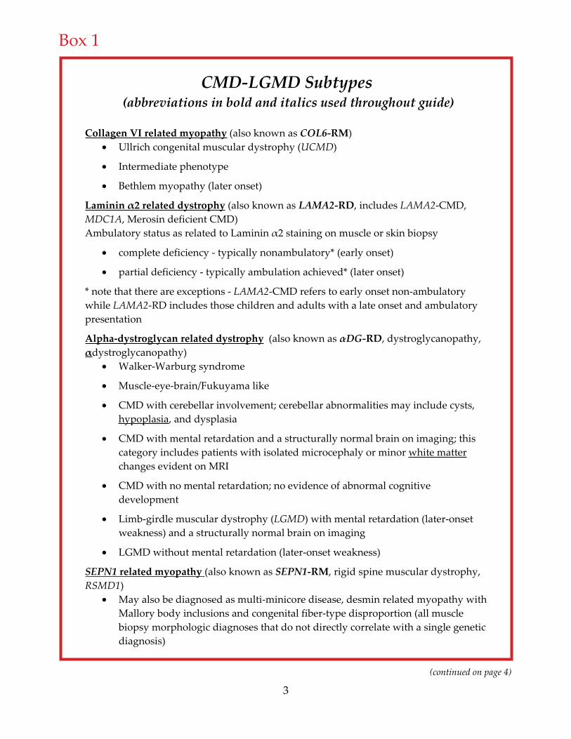
CMD-LGMD Subtypes (abbreviations in bold and italics used throughout guide)
Collagen VI related myopathy (also known as COL6-RM)Ullrich congenital muscular dystrophy (UCMD)
Intermediate phenotype
Bethlem myopathy (later onset)
Laminin 2 related dystrophy (also known as LAMA2-RD, includes LAMA2-CMD,MDC1A, Merosin deficient CMD) Ambulatory status as related to Laminin 2 staining on muscle or skin biopsy
complete deficiency - typically nonambulatory* (early onset)
partial deficiency - typically ambulation achieved* (later onset)
* note that there are exceptions - LAMA2-CMD refers to early onset non-ambulatory while LAMA2-RD includes those children and adults with a late onset and ambulatory presentation
Alpha-dystroglycan related dystrophy (also known as DG-RD, dystroglycanopathy, dystroglycanopathy)
Walker-Warburg syndrome
Muscle-eye-brain/Fukuyama like
CMD with cerebellar involvement; cerebellar abnormalities may include cysts, hypoplasia, and dysplasia
CMD with mental retardation and a structurally normal brain on imaging; this category includes patients with isolated microcephaly or minor white matterchanges evident on MRI
CMD with no mental retardation; no evidence of abnormal cognitive development
Limb-girdle muscular dystrophy (LGMD) with mental retardation (later-onset weakness) and a structurally normal brain on imaging
LGMD without mental retardation (later-onset weakness)
SEPN1 related myopathy (also known as SEPN1-RM, rigid spine muscular dystrophy, RSMD1)
May also be diagnosed as multi-minicore disease, desmin related myopathy with Mallory body inclusions and congenital fiber-type disproportion (all muscle biopsy morphologic diagnoses that do not directly correlate with a single genetic diagnosis)
Children who acquire the ability to walk or those who first have symptoms in late childhood or adulthood may be referred to as having Limb Girdle Muscular Dystrophy (LGMD). All CMD subtypes are on a spectrum with CMD (early onset, weaker) on one end and LGMD (later onset, milder) on the other. Box 1 lists the known subtypes of CMD. See Appendix A for a full description.
4 5
RYR1 related myopathy (also known as RYR1-RM, includes RYR1-CMD)
Overlaps with RYR1 related myopathies (RYR1-RM), central core and centronuclear myopathy
Considered CMD if muscle biopsy is dystrophic without typical central cores
LMNA related dystrophy (also known as LMNA-RD, includes L-CMD, LMNA-CMD)Dropped head syndrome, foot-drop, non-ambulatory
Ambulatory presentation may be called early-onset Emery-Dreifuss muscular dystrophy.
This demonstrates L-CMD is a part of the LMNA related Dystrophies which includes Dropped head syndrome L-CMD, Ambulatory L-CMD and Emery-Dreifuss.
CMD Undiagnosed
People with CMD may carry a clinical diagnosis of CMD without genetic confirmation. While clinical presentation and/or muscle biopsy are consistent with CMD, genetic testing may not provide a diagnosis as not all CMD genes have been discovered. Genetic testing under the guidance of a CMD expert is encouraged.
How to Use this Document
Box 1 continued
(Continued from page 2)
Some of the known genetic mutations cause muscles to break down faster than they can repair or grow, leading to muscular weakness. A child with CMD may also have different types of neurologic or physical problems related to the CMD. Some children walk by themselves or with supports; other children learn how to walk but then become weaker and stop walking; and some others may never walk at all.
It is important to remember that not all people with CMD have all the symptoms or need all the treatments you will read about in this guide. Although there may be similarities, each person’s course with CMD will be unique with differing needs at different points in time. This means care must be individualized and it may be difficult to meet someone whose CMD is exactly the same.

5
2 ComPrEhEnSivE mAnAgEmEnT Care at Diagnosis, Ongoing and Hospital Stays
Providing well-coordinated multidisciplinary care and creating strong provider–patient relationships and individualized care plans are essential throughout the changing course of the disease.
This section is divided into three important topics, reflecting care at diagnosis, outpatient visits, and acute hospitalization (going to the hospital when sick or injured).
9
Five Key Topics to Discuss at the Initial Meeting
Five key areas should be addressed:
Diagnosis. The clinician should explain what is known about the cause of the disorder and how it may affect other functions, such as including motor function, breathing and heart function, and cognitive function (mental abilities).
Prognosis. There is a wide range of severity and life expectancy in CMD. However, for most forms of CMD, the prognosis has improved due to recent advances in medical technology.
Recurrence risk and impact on future family planning. Even if this is not the most important issue to address at the time of diagnosis, the clinician should discuss the risk of having another child with the same disorder. If the exact genetic diagnosis is known, this recurrence risk can typically be calculated. Even if it is not known, recurrence risks may be roughly estimated.
Treatment plan. A multidisciplinary approach is required, most often including a pediatric neurologist, pulmonologist, cardiologist, ophthalmologist, physical therapist, orthopedist, and others (see Appendix B). Ideally, a palliative carespecialist should also be included from early on to improve quality of life. Ingeneral, the treatment plan will be similar whether a specific genetic diagnosis has been obtained or not. Approximately 50% of children with CMD do not have a specific genetic diagnosis.
Family support and community resources. You should receive information about advocacy and family support groups (on-line and in-person) and relevant educational resources. Families often find connecting with other families who have children with similar diseases to be extremely helpful. If this information is not offered, you should request it, or can find it at curecmd.org.
Care at Diagnosis Once your child receives a diagnosis of CMD, appropriate care, outlined below, should be put into place with ongoing support and education. Ideally this care is guided by a neurologist or neuromuscular specialist well-informed regarding CMDs who works with the family as a team. The specialist clinician needs to help your family plan for possible health care
Box 2
6
problems before they happen and keep your child healthy and doing the most that your child can do for as long as possible. To do this, both medical and psychosocial aspects need to be considered. Both multisystemic and multidisciplinary monitoring are required as part of an effective treatment plan.
An initial meeting with your clinician should take place as soon as a clinical diagnosis of CMD is available, even if the specific genetic type of CMD is not yet known. At this meeting, the clinician should explain the diagnosis of CMD in a way you and your family can understand, even if you do not have a medical background. You are encouraged to write down your questions and take notes in this meeting because it is often difficult to remember what was actually covered during this initial discussion. If it feels helpful, supportive family or friends should be welcomed to participate in this meeting. From this point forward, regular appointments will most often be required and will need to be scheduled.
Box 2 offers an overview of topics to discuss at that initial discussion.
Outpatient Clinic Visits
Your child should be seen regularly—probably once every 4 to 6 months—in a pediatric neurology/neuromuscular clinic experienced in CMD, preferably with a multidisciplinary team that includes specialists in many different areas (see Appendix B). Infants with CMD who are younger than age 12 months, or older children with severe or worsening medical problems (such as seizures that do not get better with medication, severe hypotonia, respiratory issues, or nutrition issues), should be seen at least every 3 to 4 months.
At these visits, it is recommended that your child have the following things checked: blood pressure, heart rate, respiratory rate, weight and BMI (body mass index), height, and—for in-fants and toddlers—head circumference. If your child is unable to stand or has scoliosis, height can be approximated by measuring arm length (ulnar length). Other tests may also be relevant for your child, such as measurement of
joint angles (goniometry), muscle strength testing (myometry), electrocardiogram (ECG), pulmonary function tests (for example, forced vital capacity, or (FVC), and blood oxygen measurement (pulse oximetry).
Other important things that may be evaluated in these visits may include:• Development: Children at risk for developmental delay or learning difficulties should receive early interventions, including physical therapy, occupational therapy, and speech therapy. Developmetal delay may mean a motor delay (physical movements like sitting, walking, or holding a bottle) or associated cognitive delay (language/speaking or learning problems).
• Lungs: Prevention of severe respiratory infections (using vaccines or early antibiotic treatment, for example) is important. Weak cough, shortness of breath, sleep disturbances, morning headaches, failure to gain weight, and repeated infections in particular are warning signs that should be discussed with a pediatric pulmonary expert (see Respiratory Care section).
• heart: At least one heart evaluation that includes an ECG and a cardiac ultrasound (echocardiogram) should be performed if your child has a type of CMD known to affect the heart (such as LMNA-RD, aDG-RD, LAMA2-RD) or if the CMD subtype is unknown. Monitoring with a Holter and/or event monitor is necessary for LMNA-RD. A cardiac workup is also necessary for any CMD diagnosis with symptoms suggestive of an abnormal heart rhythm (arrhythmia) or enlarged heart (cardiomyopathy). More frequent evaluation may be recommended depending on CMD subtype (see Cardiac Management section).
• Eyes: If your child has an undefined CMD or CMD subtype with known eye involvement (such as αDG), it is important to involve an ophthalmologist early to help with diagnosis and watch for other eye problems, such as cataracts, near-sightedness, retinal detachment, and glaucoma.
7
• Nutrition and growth: Children with CMD should not be expected to follow typical growth curves. However, if your child is not gaining weight, is losing weight or gaining excess weight, or has swallowing difficulties, stomach reflux, intestinal dysmotility, constipation, or an oral deformity, he or she should be referred to a dietitian, gastroenterologist, and swallowing expert see Gastrointestinal Management section). Monitoring calcium and vitamin D intake to promote maximal bone density is important.
• Skeletal system: If your child develops contractures or scoliosis, an early referral to a pediatric orthopedist or spine surgeon should be made (see Orthopedics and Rehabilitation Management section).
• Body movement: Your child’s physical therapy program should be focused on maintenance of function and mobility. This includes prevention or treatment of joint contractures and spine deformities, as well as
performance of activities to improve respiratory function. It is also important that your child is provided with the right type of seating and wheelchair support, as well as adaptive equipment (tools to make everyday activities easier) for functional activities.
• Emotion and behavior: If you have concerns regarding your child’s mood, behavior, or other psychiatric issues, support should be offered and referrals to psychology/ psychiatric professionals should be made (see Palliative Care section).
• Psychosocial: You and your family members may benefit from services to assist with some of the more practical aspects of living with CMD (like insurance coverage, services availability, or school access). Social work support from your child’s place of medical care should be made available to help you and your family with many of the emotional challenges you may experience.

Hospital Care Your child may require unplanned hospitaliza-tions (see Table 1). Your child’s pediatric neuro-muscular specialist or neurologist may play a
major role in coordinating medical care during any acute or critical illness, though this role may also be played by your pediatric pulmonologist.
Symptoms of CmD that may result in acutehospitalization and their associated CMD subtypes
13
Symptom Requiring Hospitalization
Subtypes that May Have Issues in Infancy (Early)
Subtypes with Issues in Childhood to Adolescence
Breathing problems requiring breathing support
DG-RDLAMA2-RD
COL6-RMSEPN1-RM
Heart failure or arrhythmias requiring medications
DG-RD(Fukutin, FKRP, POMT1)*LAMA2-RDLMNA-RD
Feeding problems requiring gastrostomy (G-tube)
LAMA2-CMD**RYR1-RM
DG-RD
COL6-RM
Seizures requiring medication
DG-RD(includingFukuyama, WWS, MEB)
LAMA2-RD
Malignant hyperthermia SEPN1-RMRYR1-RM
SEPN1-RMRYR1-RM
Abbreviations: DG-RD, alpha-dystroglycanopathy; FKRP, fukutin-related protein CMD; LAMA2-RD, merosin-deficient CMD; MEB, muscle-eye-brain disease; POMT1, proteinO-mannosyltransferase 1; SEPN1-RM, rigid spine muscular dystrophy; WWS, Walker-Warburg syndrome; LMNA-RD, lamin A/C CMD. *Fukutin, FKRP, and POMT1 are genes that can lead to a DG-RD. The first two are more highly associated with heart failure, although the third may also be associated with it. If one has a DG-RD caused by one of these three genes, increased cardiac surveillance is warranted. **LAMA2-CMD is used to refer to form of LAMA2-RD (Merosin Deficiency)that presents at birth and does not achieve ambulation, while LAMA2-RD incorporates the milder ambulant form and early onset non-ambulant form.
Table 1
Common reasons for acute care hospitalization include:
• respiratory infections or respiratory failure
• seizures
• failure to thrive (poor weight gain or excessive weight loss).
If your child needs to have a planned hospitalization due to surgery or a procedure that will involve the use ofanesthesia, your child’s doctor should first talk with you about the potential risks involved and then co-ordinate the planning and management for the care of your child during the procedure and through recovery.
8

9
3 nEurologiC mAnAgEmEnT Care of Seizures and Cognitive Impairment
Various neurologic symptoms are related to some of the known subtypes of CMD. The most common are abnormalities in brain structure or function and seizures.
Brain MalformationTwo groups of CMD are most often associated with brain abnormalities: LAMA2-RD and the αDG-RD. To evaluate for a structural brain abnormality (malformation), a magnetic resonance imaging (MRI) scan of the brain is performed.
Children with αDG-RD who have normal brain structure on MRI may or may not have a prob-lem with learning and cognitive function. In ad-
dition, a wide spectrum of brain MRI findings may be found in children with αDG-RD; they can range from normal to profound (very severe) structural abnormalities.
The most common brain malformation in LAMA2-RD is a white matter
abnormality that is not associated with cognitive impairment. The white matter change is usu-ally stable over time and does not require repeat brain imaging.
Functional brain abnormalities associated with CMD can cause multiple problems, including cognitive impairment; behavioral, language, and learning problems; emotional problems; motor delays; seizures; and vision problems.
If your child is believed to have a functional brain problem such as cognitive impairment, he or she should undergo psychometric testing and be referred to early intervention and augmented/specialized school or communica-tion programs. Communication strategies for the nonverbal or minimally verbal child need to be implemented early and include sign language, picture or symbol cards (PECS, Picture Exchange
Seizures Seizures are frequently associated with CMDs, particularly in those children with known brain involvement. Seizure types can include absence, atypical absence, or convulsive seizures. Seizures can start at any age from newborn to adolescence; in people who are at risk for developing seizures, the seizures may be provoked by fever and illness. Seizures may also start without anything known triggering them. If you have concerns about any activity or behavior your child exhibits that you feel may be a seizure, please discuss this with your child’s health care provider.
To determine if your child is having seizures, your child’s neurologist may recommend a detailed workup. This evaluation should include a thorough history of the events that raise concerns that seizure activity is occurring or a history of known seizures, a comprehensive neurologic evaluation, and at least one routine electroencephalogram (EEG). Depending on the results of the EEG, further or longer EEGs may be recommended. An MRI of the brain or a repeat MRI of the brain may be recommended. The definition of epilepsy is two or more unprovoked seizures (that is, seizures not caused by fever or illness). If your child is diagnosed with epilepsy, the neurologist will likely recommend an anticonvulsant medication to reduce the frequency and severity of seizures.
Seizures in children with LAMA2-RD are often successfully treated with a specific anticonvulsant, valproic acid, but other treatments have also been used successfully. Occasionally seizures can be difficult to control. In children with αDG-RD, for example, management of seizures can be more difficult because of possible underlying structural abnormalities. There are many different anticonvulsants, so if your child does not respond to the first medicine, your neurologist may recommend different or multiple medications to try to control the seizures.
Communication System), voice output devices (DynaVox, TapToTalk), and ongoing speech therapy to practice vocalization.
10
4 rESPirATory mAnAgEmEnT Care of Breathing
A primary purpose of the lungs and breathing (respirations) is to bring oxygen (O2) into the blood that circulates in the body and release carbon dioxide (CO2) out of the body. This process of trading O2 and CO2 is also called gas exchange; it occurs in all humans and is a critical element in your child’s health.
The need for respiratory support for a child with CMD can vary considerably between and within each CMD subtype. Children with all types of CMD have an increased risk of developing pulmonary (lung) problems due to weak breathing muscles. The age when breathing problems may emerge, as well as the severity of the respiratory problems that appear, varies from individual to individual. Typically, breathing problems begin to be noticed between ages 8 and 15 years. Younger children with CMD and breathing problems may not show typical symptoms; it is important that parents and caregivers be aware of the early signs of breathing problems. It is recommended that once your child has been diagnosed with CMD, he or she be evaluated by a pulmonologist to get a baseline assessment. The pulmonologist will teach you about the early signs of respiratory problems in young children. Your child’s coordinating provider and pulmonologist will work with you toward effective respiratory care.
Signs and SymptomsA two-step proactive approach to the care for your child’s respiratory problems is important in helping to maintain his or her best possible functioning over time. The recognition by parents and caregivers of early signs and symptoms, in addition to regularly scheduled pulmonary evaluations, pulmonary testing, and treatment, is of utmost importance.
Signs of early symptoms and problems with your child’s breathing muscles may be subtle and can change over time. If you have concerns about your child’s respiratory function, please contact
your pulmonologist. If the situation appears urgent, have your child evaluated in the emergency room. Be on the lookout for the following signs and symptoms:• weak cry• ineffective coughing• repeated respiratory infections, irregular breathing patterns, or general irritability • choking during feedings or on their own secretions • weight loss or poor weight gain (often referred to as failure to thrive). Some additional symptoms may be related to problems with breathing function at night.
Breathing problems may start at night because this is when all humans breathe more shallowly. These signs can include:• interrupted sleep or an increased need to turn at night• awakening in the morning feeling tired or in disturbed mood, even when he or she has slept for enough hours• a faster breathing rate or a feeling of breathlessness• morning headaches, nausea• poor concentration during the day • fear of going to sleep and nightmares.
Curvature of the spine (scoliosis) and chest wall deformities can also develop, in part due to weak chest muscles and a weakened diaphragm, which may further limit your child’s breathing capacity. See Orthopedics and Rehabilitation Management section.

11
Your child’s diaphragm muscles may be weak without producing any other obvious symptoms. This is unique to several CMD subtypes; respiratory problems may begin while your child is still walking (see Table 2) even though in most other forms of muscular dystrophy, respiratory prob-lems do not start until after a child can no longer walk. This fact makes it even more important that your child be evaluated by a pulmonologist before symptoms are seen.
Onset of typical breathing problems in known CMD subtypesTable 2
21
Abbreviations: CMD, congenital muscular dystrophy; FKRP, fukutin-related protein; LAMA2, laminin A2; LGMD, limb-girdle muscular dystrophy; MEB, muscle-eye-brain disease; SEPN1, selenoprotein; WWS, Walker-Warburg syndrome.
CMD Subtype Onset of Breathing Problems COL6-RM Early-onset night-time breathing
problems with diaphragm weaknessBreathing support needed on average by age 11 years
SEPN1-RM Early onset of night-time breathing problems; may occur before losing the ability to walk Breathing support needed on average by age 10 years
LAMA2-RD Association seen between declines in motor function and respiratory functionBreathing support needed on average by age 11 years
DG-RD with cognitive impairment (WWS, MEB, Fukuyama)
Severe progression of muscle weakness and respiratory failure Breathing management may start at birth or in first decade if severe muscle weakness
DG-RD without cognitive impairment and LGMDforms
Association seen between declines in motor function and respiratory functionBreathing management starts when person loses the ability to walk

12
Types of Pulmonary Function Testing • Spirometry is used to document breathing function; this test should be done on at least an annual basis by at least age 6 years. Spirometry testing can occur during your child’s regular pulmonology visit or at a separate appointment. Most often this testing is administered by a respiratory therapist before your child’s exam with the clinician. This noninvasive testing may include the measurement of your child’s forced vital capacity (FVC) and peak cough flow; these are measured by having the child breathe into a tube or mask. These tests may also be referred to as pulmonary function tests, or PFTs. • Nocturnal (night- time) oximetry (“pulse ox”) pain lessly measures blood oxygen saturation levels using a sensor attached typically to a finger or toe. Sometimes the sen sor looks like a big Band-Aid or is kept on the finger or toe with just a piece of tape. • Polysomnography, or a sleep study, is an outpatient overnight testing procedure conducted in a sleep lab; it is done as recommended by your child’s pulmonologist, who may recommend it on an annual basis. This test is helpful in the monitoring of night-time respiration, and can discover if sleep apnea is present and how severe it is. Sleep studies can also be used to monitor the results of bilevel positive airway pressure (BiPAP) use and to guide adjustments to such use.
• Blood gases are measured through a blood draw. This procedure is used to measure the O2 and CO2 levels in the blood if a child is having new or severe breathing problems.
• End-tidal CO2: This is measured using a device that measures the CO2when a person breathes out. It can help a pulmonologist understand how well a person who is on
breathing support (BiPAP or ventilator) is breathing and whether adjustments in settings need to be made. This device may also be used by a pulmonologist to check CO2 levels for people with CMD who are just starting to have breathing issues but are not on breathing support.
• A speech and swallow evaluation may be considered whenever there are symptoms indicating risk for aspiration, such as cough, choking, difficulty swallowing, poor feeding, or failure to thrive.
Preventive Respiratory CarePneumococcal (pneumonia) and annual influenza (flu) vaccines are recommended for all children and adults with CMD. It is also recommended that Synagis, the vaccine to prevent respiratory syncytial virus (RSV), be given to children under age 2 years.your child will benefit from the following methods that improve his or her ability to move secretions, cough efficiently, and help keep their airway and lungs open:
• Cough assistance using a mechanical insufflator-exsufflator apparatus (“cough machine,” “coughalator,” or Cough Assist) may help to remove mucus from the lower airways.
• Breath stacking techniques, as taught by your child’s pulmonologist, may help reduce the risk of chronic collapse of areas of the lungs (atelectasis).
• Chest physical therapy using a daily intrapulmonary percussive ventilation (IPV) regimen may also assist in the clearance of secretions. • A bronchial drainage chest compression vest (percussive vest) provides rapid chest compression to mobilize secretions.
Your child may have breathing problems due to other factors not related to CMD. Although asthma is not a symptom of CMD, if it is diagnosed in your child it should be treated with bronchodilators and inhaled steroids as needed. Treatment of asthma in children with CMD is no different from treatment for children without CMD.

13
when breathing out. A variety of interface options are available, based on your child’s age, skin condition, face shape, and ability to tolerate this intervention.
Once your child begins to use noninvasive venti-lation, he or she will need to undergo overnight monitoring (sleep study), at least annually, to adjust ventilator settings and to check and adjust the fit of the mask or other interface.
Special care should be directed to the young child receiving long-term ventilation to help address the potential complications of abnormal facial development (midface hypoplasia). The use of individually fitted masks or alternating between nasal pillows, nasal masks, and full face masks may be helpful in preventing this complication. Sip ventilation with a mouthpiece may also be recommended for people who require breathing support during the day.
Sometimes long-term mechanical ventilation may need to be delivered via a surgically placed tube in the neck, called a tracheostomy tube. Indications for this include chronic aspiration with repeated pneumonias or ineffective clearing of airway secretions despite the use of assistive interventions. Some people also prefer a tracheostomy tube if they require noninvasive ventilation for the majority of the day and night.
InterventionsSevere scoliosis can make it harder for the lungs to expand all the way and prevent a person from taking a “deep breath.” Your child may require spinal bracing to slow the progression of scoliosis and maintain improved posture during daily activities. When bracing is used it is important to consider its effect not only on the scoliosis, but also on the child’s breathing. Each brace needs to be evaluated to make sure it does not have a potentially negative effect on breathing function. Your child’s orthopedist and pulmonologist should work together to ensure that the brace is supportive enough for the spine and doesn’t worsen breathing function.
To assist with difficulties with effective breath-ing, your child’s clinician may recommend the use of supportive breathing equipment (noninvasive or ventilator equipment), which has been shown to improve gas exchange, decrease chest infections, and decrease the frequency and duration of hospital stays.
Noninvasive ventilation is most commonly recommended when there is evidence of hypoventilation (weak breathing ability) or for any of the resulting signs and symptoms of respiration problems. Noninvasive ventilation techniques are provided through a mask or other easily removable device.
Bi-PAP (bilevel positive airway pressure) is a commonly used noninvasive ventilator usually begun for night-time support. It consists of asmall machine that pumps air through tubing connected to an interface or mask that goes over your child’s nose or mouth. Pressurized air that supports your child’s breath helps remove CO2
Management of Acute Respiratory Illness Respiratory tract infections (common cold and pneumonia) are the most frequent cause of hospital admissions and life-threatening situations in individuals with CMD. When an acute respiratory infection is suspected, it is important to have your child evaluated, making sure that you tell the clinician the type of CMD your child has and what you know about the disease course. Signs of acute respiratory distress can be subtle but may include: • paleness • increased sleepiness • decreased appetite • unusual movement of chest and belly

14
• fast heart rate or breathing rate • weak cough • increasing fatigue.
Any of these signs deserve a careful evaluation but if, in addition, the oxygen saturation is less than 94% or is lower than your child’s baseline, your child should be seen by his or her medical provider or evaluated in the emergency department immediately.
To evaluate the severity of your child’s illness, the clinician will perform a physical exam and listen to your child’s chest. Other diagnostic methods may include: • assessment of cough effectiveness • pulse oximetry and possible CO2 measurement to evaluate breathing problems • chest X-ray to identify pneumonia and collapsed areas of lungs (comparison with previous films may be needed for the most accurate evaluation) • sputum culture if your child is able to produce mucus by coughing; this culture may provide information about the type of bacteria causing the pneumonia.
Treatment of your child’s acute respiratory infection aims at maintaining stable breathing function.
Antibiotics should be used in most respiratory infections to treat potential underlying bacterialpneumonia in CMD with ongoing monitoring of respiratory status if pneumonia is diagnosed. If your child’s oxygen saturation is low, supplemental O2 should be provided (sometimes through a nasal cannula or mask). However, it is important to note that if there is evidence of CO2 retention it is more appropriate to provide ventilator support rather than O2 alone.
If there are signs of respiratory failure and your child is not yet using noninvasive ventilation support, this should be initiated. If your child is already using breathing support, re-evaluation of his or her ventilator settings or an increase in the number of hours the child uses it may be necessary to stabilize your child’s breathing function. In more serious illnesses, intubation
may be required if noninvasive ventilation is not helpful, your child is unable to clear secretions, or your child is losing the ability to protect his or her airway, increasing the risk of aspiration.
Treatments to help mobilize your child’s secretions should be intensified, including use of a cough machine, IPV, chest insufflations, and manually assisting hie or her cough. Bronchodi-lators and chest percussion may also be used as recommended by your pulmonologist. Ventilation only assists with the process of gas exchange; thus, these methods of airway clearance are critical to recovery and should continue to be used even if the patient is on assisted ventilation.
imPorTAnT FACTS To rEmEmBEr 1. Keep a general written description of the subtype of CMD your child has, if known, and a copy of the latest breathing test (pulmonary function test, forced vital capacity) to show the medical clincian in an emergency situation. 2. Your child´s breathing function needs to be checked before any surgery. 3. Lower respiratory tract infections should be treated aggressively with an aim to maintain a steady level of adequate oxygenation and CO2 levels. Most often, antibiotics should be used to treat the infection. If your child has chest muscle weakness, additional help with coughing is essential. 4. Symptoms of inadequate breathing function include paleness, sleepiness, decreased appetite or weight loss, abnormal breathing pattern, weak cough, repeated chest infections/pneumonias, increased fatigue, decreased concentration ability, and morning headache. Symptoms may initially be subtle.

Assessment
Growth assessments for your child need to occur at every regular visit by measuring weight and height. Ulnar length may be used for height measurement if your child is older than age 5 years and is unable to stand.
Children with CmD often have a growth curve below what is expected for age. This is acceptable if your child is in good health, without signs of fatigue, recurrent infections, or cardiac and respiratory problems. It is important to get an accurate weight when your child is evaluated and to keep frequent track of your child’s weight curve to ensure continued weight gain along his or her own course.
If your child’s growth or health is not adequate, a feeding assessment may be recommended. This should include an oral-facial examination, observation and evaluation of his or her feeding and swallowing skills, and assessment of his or her seating and positioning.
Videofluoroscopy or fiber-endoscopic evaluation can be helpful in diagnosing difficulties your child may be having with swallowing that can increase the risk of aspiration.
15
4 gASTroinTESTinAl mAnAgEmEnT
Nutrition, Feeding, and Oral Care
Feeding and nutrition problems are frequently encountered with children who have CMD. Other frequently encountered problems may include gastroesophageal reflux (GER), aspiration, constipation, speech difficulties, poor bone health, and difficulties with oral and dental hygiene. Management of these issues is a significant priority for optimizing your child’s care and is best addressed by a multidisciplinary team, including specialists experienced in feeding and swallowing evaluation, a dietitian or nutritionist, and a gastroenterologist.
Feeding and Nutritional Symptoms A common problem in people with CMD is difficulty with gaining weight, or failure to thrive. For other people with CMD, weight gain, often related to the loss of walking, may become an issue.
Other symptoms of feeding concerns with your child may include: • frequent pulmonary infections
• chest/upper abdominal pain, vomiting
• difficulties with chewing, choking, or coughing
• poor oral coordination and excessive drooling
• constipation or diarrhea
• difficulties with eating independently past an appropriate age
• length of mealtimes; meals lasting longer than 30 minutes are considered to be pro longed; this may be a sign of feeding difficulty
• increased family stress or decreased enjoyment of mealtimes for the child and caregivers.
Other associated factors that need to be considered in the feeding and swallowing assessment include neck weakness, jaw and neck
contractures, weak or high arched palate, poor tongue lateralization, dental crowding, scoliosis, weak or ineffective cough, respiratory fatigue, insufficient night-time breathing function, poor appetite, gastroesophageal reflux (GER), and dysmotility.

16
If difficulties with weight gain continue or there is a concern that your child’s nutritional status is affecting his or her ability to fight recurrent respiratory infections, a referral to a gastroenter-ologist needs to be made to consider the option of tube feeding.
• For short-term use, such as before and after surgery or during acute illness, a nasogastric (NG) tube (feeding tube through the nose) may be used.
• For long-term use, a surgical gastrostomy tube (G-tube) or jejunostomy tube (J-tube) insertion may be needed. If due to severe reflux, a Nissen fundoplication is recommended for your child, this may be done at the same time as the tube placement.
• How often and how much nutrition your child needs to receive through the tube feedings will be determined by the GI team to ensure that your child meets his or her fluid and nutrient requirements.
Management Safety and adequate nutritional intake are very important in the treatment and management of your child’s feeding-related problems. Obtaining instructions and information about healthy eating habits from a specialist in feeding and nutrition starting at diagnosis is a proactive way to help prevent under nutrition or overweight problems, as well as to maintain optimal bone health.
If your child has difficulty with feeding, strategies to improve this may include: • making changes in the way your child is positioned or sitting during mealtimes • modifying utensils and other aids that support self-feeding
• learning and using safe swallowing techniques
• changing the texture of foods (for example, making liquids thicker or cutting food into very small bites)
• increasing meal frequency and selecting foods with more calories if underweight (having several smaller meals and regular snacks up to every 2 hours throughout the day) • using sensory interventions and oral therapy to improve the movement of jaw, tongue, head, and neck
• getting referrals to a dietitian to assess food and calorie intake and to discuss supplemental calorie drinks if underweight or calorie reduction if overweight.
As long as it is safe for your child to swallow, having a feeding tube does not mean your child will not eat by mouth anymore. Instead, tube feedings may become an option for necessary nutritional support for your child so that eating can be pleasurable for everyone and so that the stress surrounding nutrition can be reduced.
Gastrointestinal MotilityChildren with CMD often have reflux or constipation.
Symptoms of gastroesophageal reflux (gEr) may include chest/upper abdominal pain, vomit-ing, aspiration, and recurrent respiratory infec-tions. Medical management of GER includes the use of various medications and antacid treatment as well as dietary and positional changes.
Constipation is due to many factors and may be improved by changing food textures and fiber content, increasing fluid intake, position changes
17
and movement, and the use of laxatives as prescribed by your child’s clinician. Children with CMD often have difficulty effectively moving his or her bowels and may require sitting on the toilet with assistance for a longer time period.
SpeechChildren with CMD may have speech difficulties due to facial weakness, jaw contractures, weak breath, weak or high arched palate, problems with lip closure, and brain involvement.
Oral motor therapy and exercises may help to
maintain the range of movement in your child’s jaw and mouth. Speech therapy services mayalso help with communication strategies and options. Some children benefit from communica-tion devices if they have difficulty pronouncing words or speaking loud enough for others to hear them or are deaf and hard of hearing.
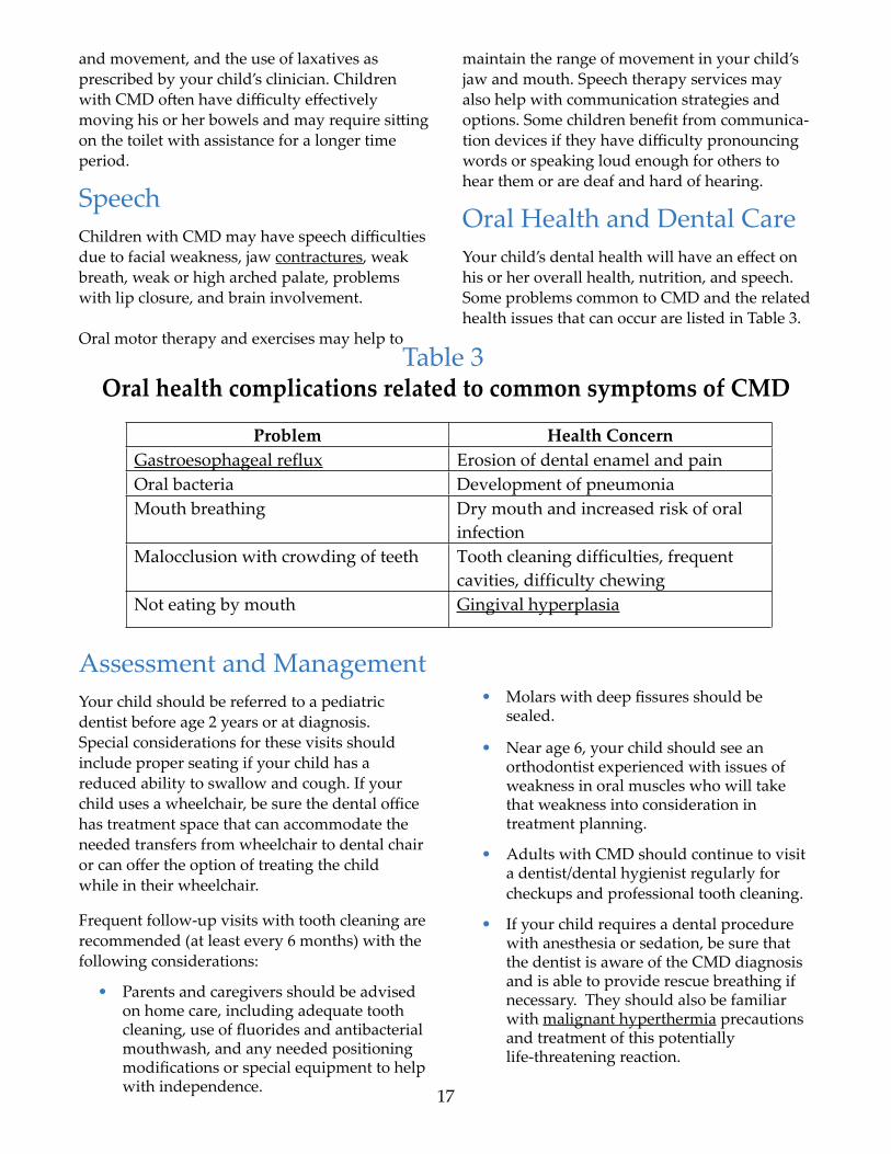
Oral Health and Dental CareYour child’s dental health will have an effect on his or her overall health, nutrition, and speech. Some problems common to CMD and the related health issues that can occur are listed in Table 3.
Oral health complications related to common symptoms of CMDTable 3
33
Problem Health ConcernGastroesophageal reflux Erosion of dental enamel and pain Oral bacteria Development of pneumonia Mouth breathing Dry mouth and increased risk of oral
infection Malocclusion with crowding of teeth Tooth cleaning difficulties, frequent
cavities, difficulty chewing Not eating by mouth Gingival hyperplasia
Assessment and ManagementYour child should be referred to a pediatric dentist before age 2 years or at diagnosis. Special considerations for these visits should include proper seating if your child has a reduced ability to swallow and cough. If your child uses a wheelchair, be sure the dental office has treatment space that can accommodate the needed transfers from wheelchair to dental chair or can offer the option of treating the child while in their wheelchair.
Frequent follow-up visits with tooth cleaning are recommended (at least every 6 months) with the following considerations:
• Parents and caregivers should be advised on home care, including adequate tooth cleaning, use of fluorides and antibacterial mouthwash, and any needed positioning modifications or special equipment to help with independence.
• Molars with deep fissures should be sealed. • Near age 6, your child should see an orthodontist experienced with issues of weakness in oral muscles who will take that weakness into consideration in treatment planning.
• Adults with CMD should continue to visit a dentist/dental hygienist regularly for checkups and professional tooth cleaning.
• If your child requires a dental procedure with anesthesia or sedation, be sure that the dentist is aware of the CMD diagnosis and is able to provide rescue breathing if necessary. They should also be familiar with malignant hyperthermia precautions and treatment of this potentially life-threatening reaction.
18

6 CArDiAC mAnAgEmEnT
Taking Care of the Heart
The goal of cardiac management is the early diagnosis and treatment of heart problems that may, at any age, be associated with CMD. In some CMD forms heart problems are likely to develop and so regular cardiac screening is necessary; others do not have heart involvement and will not require regular cardiac screening. Sometimes heart involvement can be due to weakness that develops in the heart muscle as part of CMD. It can also be caused by breathing problems that have not been diagnosed or treated appropriately, leading to strain on the heart (see Respiratory Care section). In these cases or if there is a concern that symptoms may
be due to heart arrhythmia or heart enlargement, cardiac screening and a visit to a cardiologist may be needed. If the CMD subtype is unknown, cardiac screening may be needed.
The two most commonly diagnosed heart problems are arrhythmias (an abnormal heart rhythm) and cardiomyopathy (abnormally functioning heart muscle and enlarged heart). Either condition may occur as the main heart problem in certain CMD subtypes, but not all individuals with that particular subtype may have cardiac problems (see Table 4).
Cardiac issues in various CMD subtypesTable 4
37
CMD Subtype Issue DG-RD Increased risk of developing cardiomyopathy
LAMA2-RD Mild heart enlargement that does not affect heart and arrhythmias that require treatment have been reported
LMNA-RD Increased and serious risk for both arrhythmias and cardiomyopathy. Early cardiology evaluation and regular follow-up is most important.
COL6-RM The heart muscle does not seem to be affected, but cardiomyopathy can be caused by untreated lung problems. An echocardiogram when breathing support begins is recommended.
SEPN1-RM The heart muscle does not seem to be affected, but cardiomyopathy can be caused by untreated lung problems. An echocardiogram when breathing support begins is recommended.
RYR1-RM The heart muscle does not seem to be affected, but cardiomyopathy can be caused by untreated lung problems. An echocardiogram when breathing support begins is recommended.
Abbreviations: DG-RD, alpha-dystroglycanopathy; CMD, congenital muscular dystrophy; COL6-RM, collagen VI related myopathy; LAMA2-RD, laminin 2 relateddystrophy, including MDC1A; RYR1-RM, ryanodine receptor 1related myopathy; SEPN1-RM, selenoprotein N1 related myopathy; LMNA-RD, lamin A/C CMD.
19
20
Cardiac Symptoms Typical symptoms of cardiac problems are listed here. However, it is important to note that young children may not be able to describe these symptoms.
• Fatigue • Shortness of breath • Paleness of the skin and mucous membranes • Periods of fast heartbeat (tachycardia) • Palpitations • Loss of consciousness • Light-headedness • Dizziness
AssessmentThe first cardiac evaluation should take place around the time when your child is diagnosed with CMD. This evaluation typically includes an electrocardiogram (ECG) and echocardiogram (heart ultrasound). Your child’s cardiologist may also request a 72-hour ECG (Holter ECG) or event monitor (2-week monitor) to check for abnormal heart rhythms. The frequency of follow-up evaluations will be determined by your cardiologist and depends on your child’s subtype, if known, and cardiac symptoms or concerns.
As noted in Table 4, children with L-CMD have the highest risk of cardiac problems and require frequent evaluation starting at diagnosis and every 6 months therafter. Children with αDG-RD (related to Fukutin and FKRP) require frequent cardiac evaluations at diagnosis and annually. Children with αDG-RD (related to other genes or unknown gene involvement) and LAMA2 subtypes have an increased risk of cardiac problems and require evaluations at diagnosis, age 5 years, age 10 years, and annually thereafter. If a heart abnormality is detected by ECG, echocardiogram, or Holter/event monitor, more frequent follow-up may be required.
ManagementIf your child has any signs of cardiomyopathy, medications such as ACE inhibitors or beta-blockers should be started. The management of severe cardiomyopathy or heart failure in children with CMD is no different than in the general pediatric population.
The heart has four chambers: two upper and two lower. The heart “beats” (contracts, pumping blood out of the heart to circulate through the body) when the right upper chamber sends a signal to the rest of the heart. Problems with the way that this signal is sent, or conducted, through the heart are called arrhythmias. People who have arrhythmias may say that they feel like the heart is beating abnormally.
There are two types of arrhythmias: • Supraventricular arrhythmias are caused by the upper heart chambers and conduction system and are usually treated with beta-blockers. • ventricular arrhythmias occur in the lower heart chambers and are life-threatening. When these types of arrhythmias occur, the heart does not beat as well and blood doesn’t circulate through the body. This type of arrhythmia may be seen in people with LMNA-RD and may require placement of an implantable defibrillator (known as an AICD, for automatic implantable cardioverter defibrillator) because ventricular arrhythmias don’t get better on their own. A defibrillator treats the arrhythmia by making sure the heart beats the right way and thereby prevents sudden cardiac death. The implantation of an AICD should be discussed if your child has progressive and severe heart enlargement and is at risk for ventricular arrhythmias, has had a loss of consciousness, or after resuscitation from cardiac arrest.
21
imPorTAnT FACTS To rEmEmBEr
Be aware of these symptoms of potential cardiac symptoms:
• fatigue
• shortness of breath
• paleness
• periods of irregular or fast heartbeat (palpitations or tachycardia)
• loss of consciousness
• light-headedness
• dizziness. Regular cardiac screening will help in the early diagnosis and treatment of heart problems for those CMD subtypes with possible heart involvement.
22

23
7 ORTHOPEDICS AND REHABILITATION MANAGEMENT Care of Contractures and ScoliosisPeople with all forms of CMD are commonly faced with orthopedic problems of the limbs, joints, and spine. Access to orthopedic care and different types of rehabilitation management is important throughout your child’s life to pre-serve and optimize function; promote comfort, safety, and independent mobility; relieve pain; and maximize quality of life.
Orthopedic problems may include joint and neck contractures, hypotonia, scoliosis, foot deformity, and hip dislocation or subluxation. • Conditions that may be present at birth include arthrogryposis, hypotonia, torticollis, hip dislocation, scoliosis, and clubfoot. • Common orthopedic problems that happen when a child is older include development of contractures and scoliosis, which may affect your child’s respiratory health (see Respiratory Care section). Orthopedic treatment and rehabilitation interventions must be seen as both short-term and long-term issues; they should be viewed as an investment for the future.
AssessmentYour child’s multidisciplinary team should include an orthopedist and a physicalmedication and rehabilitation team. The rehabilitation management team includes physical and occupational therapists, orthotists, and wheelchair, seating, and equipment specialists.
At least annually, your child should have an evaluation of his or her spine curvature, joint mobility, sitting comfort, and activities of daily living. Commonly used assessment tools include physical examination, spinal X-ray, goniometry, and myometry.
For younger children with severe hypotonia, respiratory insufficiency, or an unstable or rapid progression in the curvature of the spine, or
when there is a poor response to treatment measures, more frequent evaluations by his or her team will be necessary.
Parents and caregivers are important participants in monitoring and assisting with their child’s orthopedic interventions. You are encouraged to seek expert consultation regarding any orthopedic concerns.
Orthopedic ComplicationsAlthough orthopedic complications can occur in all the subtypes of CMD, their severity, type, and location differs between the various subtypes of CMD (see Table 5). Contractures are discussed in more detail in Box 3.
Contractures in CMD• A contracture is a joint that no longer moves all the way. Most joints in the body (like the elbow or knee) are like doors that sit on hinges and can open and close completely. When a contracture happens, the hinges don’t work properly and the door remains stuck in a half-open, half-closed position. • Having a contracture can make life more difficult because one loses the ability to move arms or legs, which remain “stuck” in one position. • Most contractures start gradually and get worse over time. The only intervention currently available for contractures, with limited success, is stretching and low-impact exercise that encourages full range of supported motion (for example, swimming). • Neck or jaw contractures may have a significant impact on functional ability (movement, feeding) and require special consideration regarding anesthesia prior to surgery.
Box 3

24
Table 5Age of onset of orthopedic complications related to specific CmD
45
Typical Orthopedic Complication
CMD Subtype When?
Joint laxity (wrist, ankles, fingers, toes)
COL6-RM, DG-RD,SEPN1-RM
At birth; may turn into contractures
Joint contractures Ullrich CMD*,complete LAMA2-RD
May present at birth; contractures start before losing walking ability if walking
DG-RD, partial LAMA2-RDLMNA-RD, COL6-RM
Contractures start after losing ability to walk
Hip dislocation COL6-RM At birth Neck contractures UCMD, LAMA2-RD,
LMNA-RDDevelop from age 0–10 years of life
Spinal rigidity SEPN1-RM, LMNA-RD, COL6-RM,LAMA2-RD
Progressive lower spinal rigidity
Scoliosis UCMD At birth (kyphoscoliosis)LMNA-RD, SEPN1-RM, LAMA2-RD,RYR1-RM
Early-onset: early childhood
DG-RD Late-onset (lumbar lordosis): teenage years with loss of ambulation
*Note in this table, Ullrich CMD (UCMD) is separated from COL6 to show that UCMD,or the early-onset more progressive form of COL6, may be affected earlier. COL6 in thistable means intermediate and Bethlem forms of Collagen VI myopathy. Similarly complete and partial LAMA2-RD are separated out to denote, complete (early onset, MDCIA) andpartial (late onset, ambulatory MDC1A).Abbreviations: DG-RD, alpha-dystroglycanopathy; CMD, congenital muscular dystrophy; COL6-RM, collagen VI related myopathy; LAMA2-RD, laminin 2 relateddystrophy, including MDC1A; RYR1-RM, ryanodine receptor 1related myopathy; SEPN1-RM, selenoprotein N1 related myopathy; LMNA-RD, lamin A/C CMD.
ManagementA proactive preventive approach is an essential part of managing the orthopedic complications of CMD.
Communication between the orthopedist, rehabilitation team, and your family is important so that interventions make the most sense for your child.
Your child should be referred to physical or occupational therapy before development of contractures, loss of motor function, altered gait, abnormal positioning, pain, scoliosis, problems with transfers, joint deformity, or loss of activities of daily living occur.

25
Therapy, including daily stretching of the joints of the limbs, the hip, neck, spine, and jaw can be helpful in the management of contractures. The use of orthotics and some splinting techniques may also be recommended for day or nighttime use. Examples include ankle-foot orthoses (AFO), including dynamic AFO (DAFO), molded AFO (MAFO), and the knee-ankle-foot orthosis (KAFO), as well as dynamic and passive hand, knee, and elbow splints.
Spinal bracing may be recommended in efforts to prevent the progression of scoliosis. The effects on respiratory function must be considered with any bracing or orthopedic intervention (see Respiratory Care section).
Supportive equipment may become a part of supporting your child’s daily activities. Assistance for standing, ambulation, and other forms of mobility include canes, walking frames, swivel walkers, orthotics, standing frames, scooters, and wheelchairs. Other types of equipment may be needed for help with transferring, eating and drinking, communication, turning in bed, toileting, and bathing. It is essential to collaborate with a rehabilitation management team experienced in treating individuals with neuromuscular disorders.
If your child has pain, rehabilitation specialists may help manage or improve the pain. Positioning for sitting, standing, and sleeping, as well as finding the correct fit and use of orthoses and braces, may help with pain. Swimming or physical therapy in the water may also be helpful.
Surgical ManagementSurgery may be recommended for your child to improve or maintain function, reduce pain, or improve sitting position or the fit of orthoses to allow standing. Surgery in CMD is not without risk; good preoperative counseling is manda-tory, and the benefits and risks of any surgery should always be discussed with your doctor. The end goal of orthopedic surgery is functional benefit.
Hip Instability • If your child is walking, hip surgery may be considered at an early stage to improve standing or walking ability. However, the need to limit movement for a period of time after surgery can lead to further joint contractures and more difficulty walking. • If your child is nonambulatory, then surgery is only recommended if the hip dislocation causes chronic pain, which is uncommon.
Knee Contractures • Surgery to correct this condition is rarely done, but may be recommended if severe contractures (>90 degrees) prevent your child from sitting comfortably.
Ankle Contractures • Surgery for Achilles tendon (heel cord) lengthening is common and can be considered to improve walking or to maintain good posture or the ability to wear shoes or orthoses. However, again, postsurgical risks may outweigh the benefits.
Scoliosis • The goal of spinal fusion is to preserve the best possible posture for comfort and function. The type and extent of fusion performed will depend on the ambulatory status of your child and the degree of spinal curvature. The surgery should be performed by spinal surgeons experienced in neuromuscular disorders. • Surgery of the spine in very young children should be performed only when conservative management with bracing cannot be applied or has failed.
• nonfusion techniques, such as “growing rods,” can be used to allow continued growth of the spine of the child; however, this technique requires multiple surgical interventions to expand the growing rod.
• Surgery for spinal deformities in the older child has been shown to improve quality of life. However, this is a major surgery and significant risks are involved that should be thoroughly discussed with your child’s doctors and medical team.
26
Box 4
49
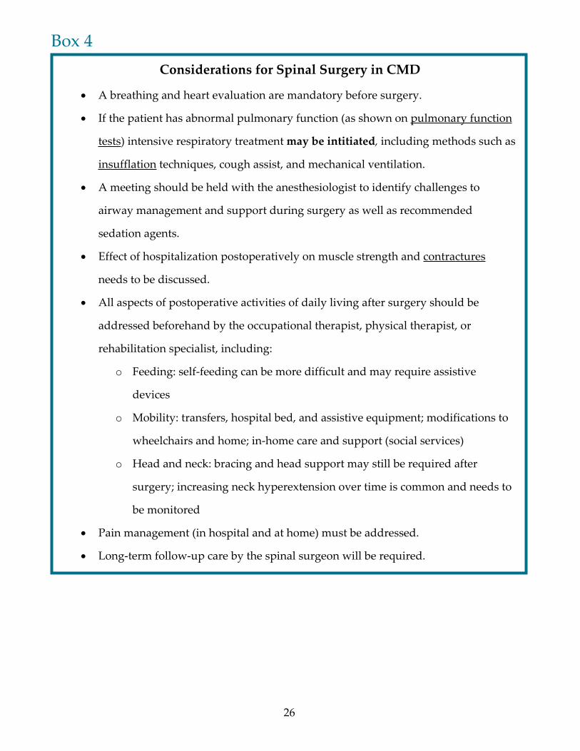
Considerations for Spinal Surgery in CMD
A breathing and heart evaluation are mandatory before surgery.
If the patient has abnormal pulmonary function (as shown on pulmonary function
tests) intensive respiratory treatment may be intitiated, including methods such as
insufflation techniques, cough assist, and mechanical ventilation.
A meeting should be held with the anesthesiologist to identify challenges to
airway management and support during surgery as well as recommended
sedation agents.
Effect of hospitalization postoperatively on muscle strength and contractures
needs to be discussed.
All aspects of postoperative activities of daily living after surgery should be
addressed beforehand by the occupational therapist, physical therapist, or
rehabilitation specialist, including:
o Feeding: self-feeding can be more difficult and may require assistive
devices
o Mobility: transfers, hospital bed, and assistive equipment; modifications to
wheelchairs and home; in-home care and support (social services)
o Head and neck: bracing and head support may still be required after
surgery; increasing neck hyperextension over time is common and needs to
be monitored
Pain management (in hospital and at home) must be addressed.
Long-term follow-up care by the spinal surgeon will be required.

27
8 Palliative Care Individual and Family Emotional Well-being
Palliative care aims to provide comfort by integrating the emotional, spiritual, develop-mental, and physical dimensions of life into the care of individuals with life-threatening diseases. Incorporating palliative care from the time of diagnosis can benefit you, your child, and the medical team as you anticipate and make decisions regarding interventions that affect your child’s quality of life.
Although palliative care may seem to offer a broad range of services, the goals of palliative treatment are concrete: relief from suffering, treatment of pain and other distressing symptoms, psychological and spiritual care, a support system to help your child live as actively as possible, and a support system to sustain the entire family. Many people associate palliative care with “giving up” or when end of life is near. However, palliative care is much more than that: it is a holistic approach to treating symptoms caused by serious diseases.
Pain/FatiguePain may be a significant and under-recognized problem that can be due to various conditions in different systems of the body. For example, the pain arising from progressive muscle weakness, scoliosis, and contractures may require adjustments with seating and splinting. Emotional and psychological aspects, including anxiety, depression, and fear, may also contribute to pain and fatigue. The interrelationship between these areas may be considerable and needs to be explored.
Effective management for your child’s pain be-gins with a comprehensive assessment of acute and chronic symptoms. Determining the pres-ence, frequency, and duration of painful episodes will help to identify contributing factors and those that help provide relief.
Fatigue is commonly reported by children with CMD. Activity level, respiratory status, sleep habits, and various medications may cause or worsen fatigue.
Mental Health
Because CMD can be difficult to diagnose, with many uncertainties about the course of the disease, you, your child, and other family members are naturally at an increased risk for emotional distress; among these are feelings of depression, anxiety, fear, and grief.
It is important to monitor the emotional well-being of your child. Signs of concern may be either direct, such as sadness, or indirect, like anger or restlessness. If you have any concerns about your child’s mental well-being, speak with your child’s medical team about obtaining a supportive psychological consultation and discussing helpful resources for coping. It is also important to monitor your emotional well-being as a parent or caregiver. Everyone has different ways of coping with stress and emotions; it is very common for parents to have difficulty dealing with their feelings when it comes to pediatric chronic illnesses such as CMD. When parents and family members are stressed, children will also be stressed. Oftentimes family counseling is helpful.
Such consultations should help bring about open discussion, relationship building, and the acknowledgement of fears, tension, and sadness.
Other resources for support may include: • Internet lists and groups (for example, Facebook groups such as Merosin Positive Mums for Merosin Negative Kids and Yahoo support groups for LMNA-RD, SEPN1-RM, Ullrich CMD, Bethlem myopathy, Walker-Warburg syndrome, lissencephaly and αDG-RD)
• Cure CMD website (curecmd.org), information and message boards • in-person support groups at hospitals or other agencies • Support from your faith tradition if relevant. These supports can help to enable you and your

family to plan meaningfully and effectively anticipate and participate in care decisions for the future when things may feel confusing and unclear.
28
The journey of life with CMD is not a straight path,
but rather spirals from issue to issue and back and forward again.
Along the way, ongoing attention, care, and patience in the areas of
medical, emotional, practical, and spiritual needs are most
important to support and enrich the lives of the affected individual
and those most closely involved with his or her care.
While reaching in for strength, be sure to also reach out.
In addition to the CMD medical community, there is also a
growing community of families that may be able to share information
and insight as you continue along the journey of life with CMD,
in all of its complexities.
End-of-Life Care Family members and health care providers understandably often find it difficult to discuss the possibility of death, but CMD can be a potentially life-limiting disease and so discussion of end-of-life care is appropriate.
It is important that your child’s health care providers help to guide you through potential end-of-life concerns. Ideally this would happen before the occurrence of a major life-threatening event, allowing you as a family time to clearly explore options and gather information before decisions need to be made.
The last decade has offered tremendous progress to those with CMD. The drafting of care guidelines, development of an international registry and growing momentum in research to identify possible treatments have contributed to hope for the future. This building of infrastructure and raising awareness to support improved health care and science will lead to new discoveries and continue to prolong and improve the quality of life for your children.
The need and timing for such discussion varies depending on the diagnosis and course of the disease, and is often more urgent when the diagnosis is more severe or is unknown. The goal is always for your child’s health care team and your family to work together through these painful issues.

29
Alpha-dystroglycan related dystrophies (αDG-RD, dystroglycanopathies): The dystroglycanopathies are a group of diseases that represent a spectrum of neurologic and physical impairment. Those that present in infancy are classified as congenital muscular dystrophy and often have brain involvement, including seizures and developmental delay, although these children may be cognitively normal. Those that present in childhood or adulthood are classified as limb-girdle muscular dystrophy with predominantly muscle involvement, although they may have mild cognitive involvement. Speech may be affected.
Infants who present with more severe involvement are labeled as having Walker-Warburg syndrome (WWS), muscle-eye-brain disease (MEB), or Fukuyama muscular dystrophy, many with abnormal brain MRI findings, including structural abnormalities and lissencephaly (abnormal neuronal migration during brain development as an embryo). Seizures, feeding issues, and eye problems (extreme near-sightedness, retinal problems, cataracts) are common in these three forms of αDG-RD.
Bethlem myopathy: This collagen VI myopathy forms a continuum with Ullrich CMD. This means that they are not two distinct diseases but rather represent a spectrum. The collagen VI myopathies (Ullrich CMD and Bethlem) share progressive contracture development, skin findings, and mutations in one of the three collagen VI genes. Adults with Bethlem myopathy can have tight tendons at the back of his or her ankles, as well as tightness of various other joints (elbows, knees, joints in the back) and especially of some of the muscles in the hands. Other symptoms, such as poor stamina/poor exercise tolerance and difficulties walking up stairs or doing tasks that require lifting the arms above the head, are related to the subtle muscle weakness that tends
APPEnDiX ADefinition of Subtypes
to be found in Bethlem myopathy. As with all the CMDs, because it is a rare disorder, people with Bethlem myopathy may often have had other diagnoses suggested in the past.
LMNA related dystrophy: This recently recognized CMD subtype (L-CMD) is caused by a mutation in the lamin A/C gene (LMNA), not to be confused with the laminin A2 gene (LAMA2), which is affected in merosin-deficient or LAMA2-related CMD. Some children with LMNA-RD present with extremely weak necks, leading to difficulty in keeping his or her head up. This is referred to as dropped head syn-drome. Children with LMNA-RD may have foot drop, meaning the foot is not able to lift itself while strength is preserved in the legs. In LMNA-RD a loss of strength and function may be observed in the first 2 years of life, which sets this CMD apart from other CMDs, in which typically the patients slowly gain function during this time period. The loss of function observed may be “ability to get into crawling position.” Children with LMNA-RD require early and frequent monitoring of his or her breathing and heart status.
Limb-girdle muscular dystrophy (LGMD): Limb-girdle muscular dystrophy typically refers to a group of muscular dystrophies that start in late childhood, adolescence, or adulthood. There are several distinct genetically defined forms of LGMD. The CMDs sit on a spectrum with LGMD. Some children with a mutation in LAMA2, collagen VI , LMNA or one of the αDG genes may have a milder form, present later in life, and achieve and maintain ambulation. In other words, CMD and LGMD are bookends on the same shelf and are not diagnoses in and of themselves. Obtaining genetic confirmation is critical for both CMD and LGMD.
laminin α2 related dystrophy (mDC1A, merosin Deficient CmD): This is also known as LAMA2-related CMD.
30
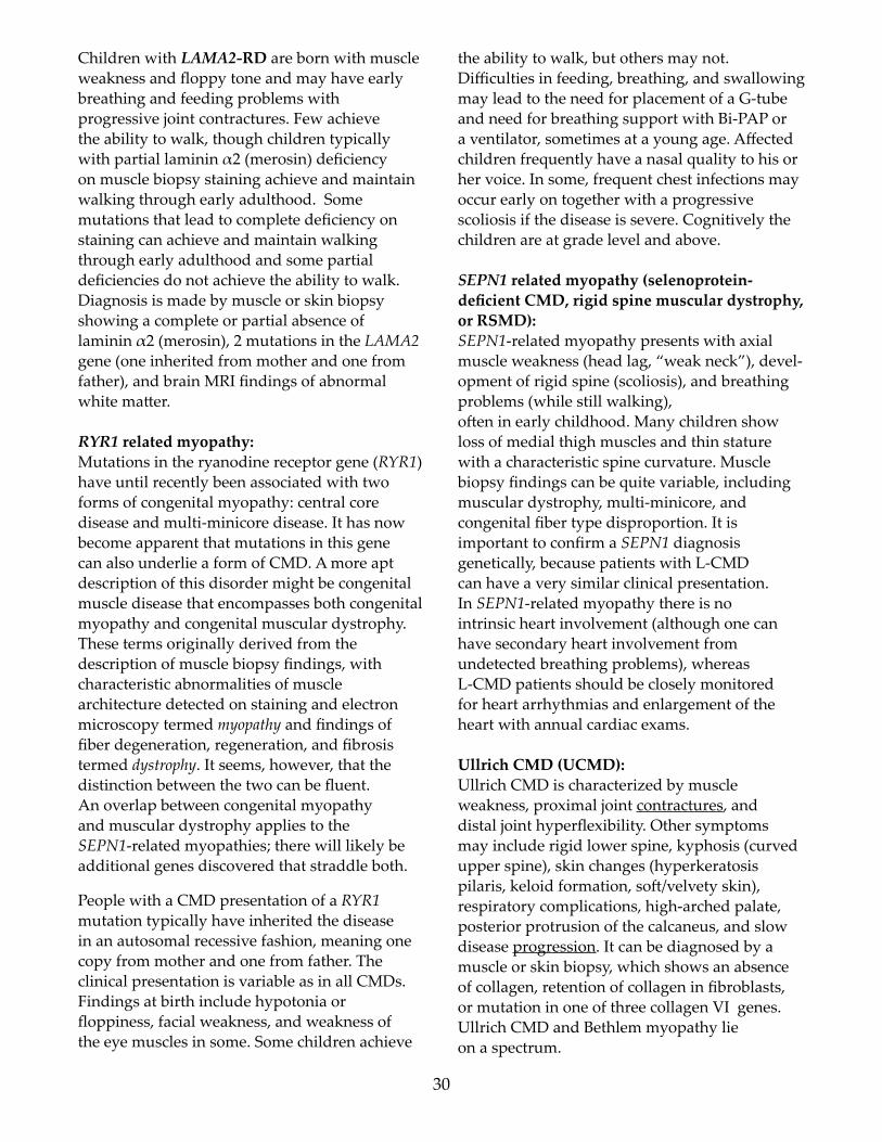
Children with LAMA2-RD are born with muscle weakness and floppy tone and may have early breathing and feeding problems with progressive joint contractures. Few achieve the ability to walk, though children typically with partial laminin α2 (merosin) deficiency on muscle biopsy staining achieve and maintain walking through early adulthood. Some mutations that lead to complete deficiency on staining can achieve and maintain walking through early adulthood and some partial deficiencies do not achieve the ability to walk. Diagnosis is made by muscle or skin biopsy showing a complete or partial absence of laminin α2 (merosin), 2 mutations in the LAMA2 gene (one inherited from mother and one from father), and brain MRI findings of abnormal white matter.
RYR1 related myopathy: Mutations in the ryanodine receptor gene (RYR1) have until recently been associated with two forms of congenital myopathy: central core disease and multi-minicore disease. It has now become apparent that mutations in this gene can also underlie a form of CMD. A more apt description of this disorder might be congenital muscle disease that encompasses both congenital myopathy and congenital muscular dystrophy. These terms originally derived from the description of muscle biopsy findings, with characteristic abnormalities of muscle architecture detected on staining and electron microscopy termed myopathy and findings of fiber degeneration, regeneration, and fibrosis termed dystrophy. It seems, however, that the distinction between the two can be fluent. An overlap between congenital myopathy and muscular dystrophy applies to the SEPN1-related myopathies; there will likely be additional genes discovered that straddle both.
People with a CMD presentation of a RYR1 mutation typically have inherited the disease in an autosomal recessive fashion, meaning one copy from mother and one from father. The clinical presentation is variable as in all CMDs. Findings at birth include hypotonia or floppiness, facial weakness, and weakness of the eye muscles in some. Some children achieve
the ability to walk, but others may not. Difficulties in feeding, breathing, and swallowing may lead to the need for placement of a G-tube and need for breathing support with Bi-PAP or a ventilator, sometimes at a young age. Affected children frequently have a nasal quality to his or her voice. In some, frequent chest infections may occur early on together with a progressive scoliosis if the disease is severe. Cognitively the children are at grade level and above.
SEPN1 related myopathy (selenoprotein-deficient CmD, rigid spine muscular dystrophy, or rSmD): SEPN1-related myopathy presents with axial muscle weakness (head lag, “weak neck”), devel-opment of rigid spine (scoliosis), and breathing problems (while still walking), often in early childhood. Many children show loss of medial thigh muscles and thin stature with a characteristic spine curvature. Muscle biopsy findings can be quite variable, including muscular dystrophy, multi-minicore, and congenital fiber type disproportion. It is important to confirm a SEPN1 diagnosis genetically, because patients with L-CMD can have a very similar clinical presentation. In SEPN1-related myopathy there is no intrinsic heart involvement (although one can have secondary heart involvement from undetected breathing problems), whereas L-CMD patients should be closely monitored for heart arrhythmias and enlargement of the heart with annual cardiac exams.
Ullrich CMD (UCMD): Ullrich CMD is characterized by muscle weakness, proximal joint contractures, and distal joint hyperflexibility. Other symptoms may include rigid lower spine, kyphosis (curved upper spine), skin changes (hyperkeratosis pilaris, keloid formation, soft/velvety skin), respiratory complications, high-arched palate, posterior protrusion of the calcaneus, and slow disease progression. It can be diagnosed by a muscle or skin biopsy, which shows an absence of collagen, retention of collagen in fibroblasts, or mutation in one of three collagen VI genes. Ullrich CMD and Bethlem myopathy lie on a spectrum.

31
Undiagnosed CMD: In the past two decades, 18 new genes that lead to a clinical diagnosis of CMD have been identified, with new discoveries increasing as whole-exome sequencing technology moves forward. This technology is enabling us to better understand the complex genetic causes of CMD. As a result, we can better understand known subtypes; in patients with a clinical diagnosis of Walker-Warburg syndrome (WWS), for example, a genetic mutation will be identified in the six known genes only 40% of the time. (This means that 60% of individuals with WWS
harbor unknown genes.) And, most importantly, a person with CMD who does not have a genetic diagnosis can use this guideline to plan medical treatment with the medical team and register in a CMD registry (cmdir.org). Registering enables you or your child to participate in ongoing gene discovery studies by providing you with information on these studies. Although knowing the genetic mutation allows a team to anticipate certain key issues, many of the CMDs face similar medical issues and these treatment guidelines will provide assistance to those without a final genetic diagnosis.
APPEnDiX BDefinition of Experts Providing Specialty Care
Advance Practice Nurse: An umbrella term used for a licensed registered nurse who has one of four roles: a clinical specialist, certified registered nurse anesthetist, certified nurse-midwife, or certified nurse practitioner. Advance practice nurses have their Master’s degrees and are board certified in a population group (for example, pediatrics). Advance practice nurses can work independently and collaboratively with physicians and other members of the multidisciplinary team and can be experts in a subspecialty, such as neuromuscular disorders or cardiomyopathy.
Cardiologist: A physician who has specialized education and training in problems with the heart. Cardiologists treat different conditions, such as arrhythmias (abnormal heartbeat), high blood pressure, and heart disease. Some cardiologists have additional training and expertise in problems with the heart muscle (cardiomyopathy specialists).
Certified nurse Practitioner (nP): A Master’s or doctorally prepared nurse who examines, diagnoses, makes treatment recommendations, prescribes medications, and directs follow-up
care within his or her area of expertise. The nurse practitioner also advocates for and teaches the patient and family about the patient’s condition.
Endocrinologist: A physician who has specialized education and training in problems with the body organs that make and release hormones (chemicals made by our body that have different functions). Endocrinologists treat different diseases, such as diabetes, short stature, and delayed puberty.
Gastroenterologist: A physician who has specialized education and training in problems with the digestive tract and problems with breaking down food. Gastroenterologists treat different diseases, such as severe constipation, failure to thrive, and gastroesophageal reflux.
Genetic Counselor: A health care provider who has a Master’s degree with education and training in medical genetics and counseling. A genetic counselor can help explain which genetic mutation is causing your child’s symptoms and may be able to help you figure out if you could have another child with the same condition.
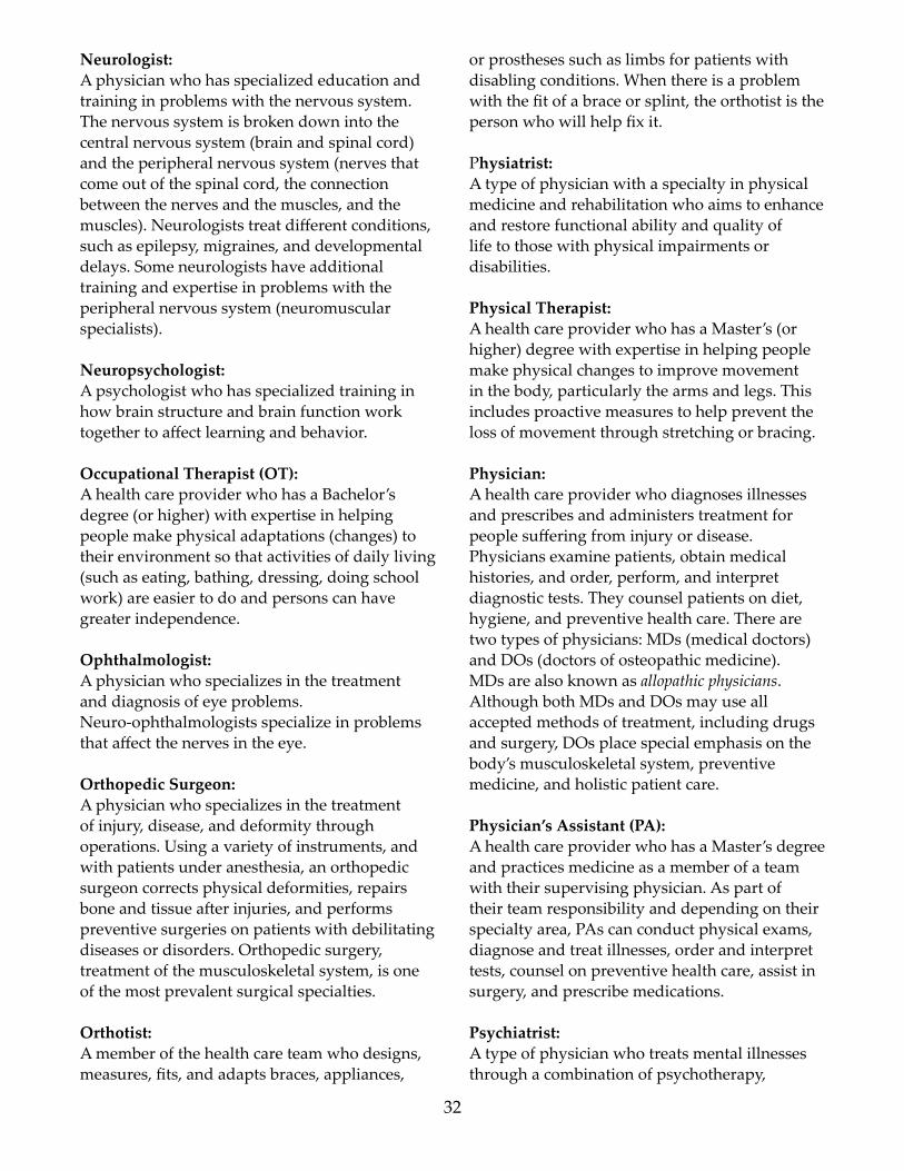
32
Neurologist: A physician who has specialized education and training in problems with the nervous system. The nervous system is broken down into the central nervous system (brain and spinal cord) and the peripheral nervous system (nerves that come out of the spinal cord, the connection between the nerves and the muscles, and the muscles). Neurologists treat different conditions, such as epilepsy, migraines, and developmental delays. Some neurologists have additional training and expertise in problems with the peripheral nervous system (neuromuscular specialists).
Neuropsychologist: A psychologist who has specialized training in how brain structure and brain function work together to affect learning and behavior.
Occupational Therapist (OT): A health care provider who has a Bachelor’s degree (or higher) with expertise in helping people make physical adaptations (changes) to their environment so that activities of daily living (such as eating, bathing, dressing, doing school work) are easier to do and persons can have greater independence.
Ophthalmologist: A physician who specializes in the treatment and diagnosis of eye problems. Neuro-ophthalmologists specialize in problems that affect the nerves in the eye.
orthopedic Surgeon: A physician who specializes in the treatment of injury, disease, and deformity through operations. Using a variety of instruments, and with patients under anesthesia, an orthopedic surgeon corrects physical deformities, repairs bone and tissue after injuries, and performs preventive surgeries on patients with debilitating diseases or disorders. Orthopedic surgery, treatment of the musculoskeletal system, is one of the most prevalent surgical specialties.
Orthotist: A member of the health care team who designs, measures, fits, and adapts braces, appliances,
or prostheses such as limbs for patients with disabling conditions. When there is a problem with the fit of a brace or splint, the orthotist is the person who will help fix it.
Physiatrist: A type of physician with a specialty in physical medicine and rehabilitation who aims to enhance and restore functional ability and quality of life to those with physical impairments or disabilities.
Physical Therapist: A health care provider who has a Master’s (or higher) degree with expertise in helping people make physical changes to improve movement in the body, particularly the arms and legs. This includes proactive measures to help prevent the loss of movement through stretching or bracing.
Physician: A health care provider who diagnoses illnesses and prescribes and administers treatment for people suffering from injury or disease. Physicians examine patients, obtain medical histories, and order, perform, and interpret diagnostic tests. They counsel patients on diet, hygiene, and preventive health care. There are two types of physicians: MDs (medical doctors) and DOs (doctors of osteopathic medicine). MDs are also known as allopathic physicians. Although both MDs and DOs may use all accepted methods of treatment, including drugs and surgery, DOs place special emphasis on the body’s musculoskeletal system, preventive medicine, and holistic patient care.
Physician’s Assistant (PA): A health care provider who has a Master’s degree and practices medicine as a member of a team with their supervising physician. As part of their team responsibility and depending on their specialty area, PAs can conduct physical exams, diagnose and treat illnesses, order and interpret tests, counsel on preventive health care, assist in surgery, and prescribe medications.
Psychiatrist: A type of physician who treats mental illnesses through a combination of psychotherapy,
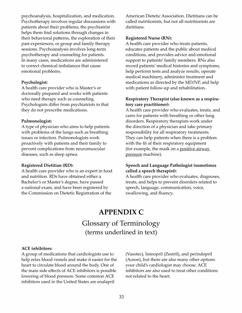
33
psychoanalysis, hospitalization, and medication. Psychotherapy involves regular discussions with patients about their problems; the psychiatrist helps them find solutions through changes in their behavioral patterns, the exploration of their past experiences, or group and family therapy sessions. Psychoanalysis involves long-term psychotherapy and counseling for patients. In many cases, medications are administered to correct chemical imbalances that cause emotional problems.
Psychologist: A health care provider who is Master’s or doctorally prepared and works with patients who need therapy such as counseling. Psychologists differ from psychiatrists in that they do not prescribe medications.
Pulmonologist: A type of physician who aims to help patients with problems of the lungs such as breathing issues or infection. Pulmonologists work proactively with patients and their family to prevent complications from neuromuscular diseases, such as sleep apnea.
Registered Dietitian (RD): A health care provider who is an expert in food and nutrition. RDs have obtained either a Bachelor’s or Master’s degree, have passed a national exam, and have been registered by the Commission on Dietetic Registration of the
American Dietetic Association. Dietitians can be called nutritionists, but not all nutritionists are dietitians.
Registered Nurse (RN): A health care provider who treats patients, educates patients and the public about medical conditions, and provides advice and emotional support to patients’ family members. RNs also record patients’ medical histories and symptoms, help perform tests and analyze results, operate medical machinery, administer treatment and medications as directed by the MD/NP, and help with patient follow-up and rehabilitation.
Respiratory Therapist (also known as a respira-tory care practitioner): A health care provider who evaluates, treats, and cares for patients with breathing or other lung disorders. Respiratory therapists work under the direction of a physician and take primary responsibility for all respiratory treatments. They can help patients when there is a problem with the fit of their respiratory equipment (for example, the mask on a positive airway pressure machine).
Speech and language Pathologist (sometimes called a speech therapist): A health care provider who evaluates, diagnoses, treats, and helps to prevent disorders related to speech, language, communication, voice, swallowing, and fluency.
APPEnDiX CGlossary of Terminology (terms underlined in text)
ACE inhibitors: A group of medications that cardiologists use to help relax blood vessels and make it easier for the heart to circulate blood around the body. One of the main side effects of ACE inhibitors is possible lowering of blood pressure. Some common ACE inhibitors used in the United States are enalapril
(Vasotec), lisinopril (Zestril), and perindopril (Aceon), but there are also many other options your child’s cardiologist may choose. ACE inhibitors are also used to treat other conditions not related to the heart.

34
Advocate: To work with your child’s health care providers as a team member to do the best thing for your child. This sometimes may mean speaking up when you are uncomfortable with a situation or plan of care, seeking out a second opinion, or identifying an advocate within your child’s health care team.
Anticoagulants: A group of medications that thin the blood to prevent it from forming clots (which cause blockages in the blood vessels). The heart does not have to work as hard to pump thinner blood, but when people are on this type of medication they can bleed easily. Some common anticoagulant medications used in the United States are warfarin (Coumadin), heparin, and aspirin, but there are also other options your child’s cardiologist may choose.
Anticonvulsant therapy: Medications that reduce the frequency and severity of seizures. Sometimes seizures can stop completely when a person is on an anticonvulsant. Some common anticonvulsants used in the United States are valproic acid (Depakote), levetiracetam (Keppra), zonisamide (Zonegran), and topiramate (Topamax), but there are also many other options your child’s neurologist may choose. Anticonvulsants are often used for other conditions not related to seizures or epilepsy.
Arrhythmia: A change in the rhythm of the heart’s beating.
Arthrogryposis: A condition that results in an infant being born with multiple contractures. This can be an early symptom of CMD but may be confused with other causes for contractures seen at birth.
Aspiration: When something (food, liquid, mucus, etc.) goes into the lungs instead of into the stomach or out the mouth or nose. When a substance is aspirated into the lungs, it can lead to a lung infection (like pneumonia).
Atelectasis: collapse of part (or all) of a lung. This can be caused by a blockage of the airways or by pressure on the airways from outside the lungs.
Beta-blockers: A group of medications that cardiologists use to decrease the heart rate by slowing down the speed at which the heart beats. These medications also help open the blood vessels and lower the blood pressure. Some common beta-blockers used in the United States are atenolol, nadolol (Corgard), and propranolol (Inderal), but there are also many other options your child’s cardiologist may choose. Beta-blockers are also used for other conditions not related to the heart.
Bi-PAP: Abbreviation for bilevel positive airway pres-sure, one of the most commonly used forms of noninvasive ventilation. Bi-PAP has two levels of airway pressure: a high pressure when the person breathes in and a low pressure when the person breathes out. A Bi-PAP machine can be programmed to cycle when the person breathes, or it can be set to a timed cycle.
Blood gases: A test to measure the concentrations of oxygen (O2) and carbon dioxide (CO2) in the blood, along with blood pH and bicarbonate level.
Breath stacking techniques: A type of respiratory therapy. The patient uses a special bag equipped with a one-way valve and mouthpiece to take a series of breaths without exhaling, expanding the lungs beyond what he or she can accomplish with a single breath. This stretches the lungs and opens clogged airways.
Cardiomyopathy: A disease of heart muscle that causes enlargement of the heart and rigidity of the walls of the heart. It can be a complication of other heart diseases.
Composite motor skills: A way of measuring several different types of

35
motor skills, such as handwriting and eating, and putting these measurements together to see how various aspects of a person’s motor skills are doing overall.
Continuous positive airway pressure (CPAP): One of the most commonly used forms of noninvasive ventilation, CPAP increases the pressure of the air in the lungs for the whole time someone is using the machine. This is helpful for people who have weak airways that get too small at times (as with obstructive sleep apnea).
Contracture: A tightness in the muscles or tendons around a joint, which prevents the joint from moving the full amount. For example, a contracture in the knee can prevent the knee from straightening out or bending completely.
Diagnosis: The specific name of a medical disorder.
Dysmotility: When digested food does not move through the stomach or intestines at the right speed. Digested food moves through our body when the muscles in our intestines move like a wave to push the food along. Sometimes the wave moves too slowly and can cause constipation. Other times it moves too quickly and can cause diarrhea.
Echocardiogram (echo): An ultrasound of the heart. This test looks at the structure of the heart and can help show how the heart is functioning.
Electrocardiogram (ECg or EKg): A device that looks at the pattern and speed of the heartbeat. This test is performed by placing electrodes (monitors) on the chest, arms, and legs. Routine ECGs usually last for less than 1 hour.
Electroencephalogram (EEg): A test of brain activity that looks for the cause of seizures by placing electrodes (monitors) on the head. The brain communicates to our body by sending messages (signals) from one nerve to
another, producing a regular pattern we can expect to see when the brain is functioning normally. When an irregular pattern is seen on the EEG, a person can be at risk for having seizures, but the EEG doesn’t tell us why that person may be at risk. To use an analogy, nerves are like telephone wires connecting houses. The EEG monitors the activity that is happening on the telephone wires but does not listen to the people talking to each other.
Failure to thrive: A term used to describe infants or young children who are not growing or gaining weight as expected. It is usually related to not taking in enough food to meet the child’s calorie needs or to an inability to absorb nutrients from food.
Fiber-endoscopic evaluation (or endoscopy): A procedure that uses a thin, long tube and light to look inside a person. For example, endoscopy can be used to look at the intestines (colonoscopy) or lungs (bronchoscopy).
Forced vital capacity (FvC): The maximum amount of air someone can blow out after taking the biggest breath possible. The FVC can help measure if there is a problem with lung function, such as respiratory muscle weakness, or if an infection is present.
Gas exchange: The body process in which oxygen (O2) is moved from air to body tissues to use by the cells and carbon dioxide (CO2) is moved from tissues to the air. It occurs in the lungs and the bloodstream.
gastroesophageal reflux (gEr): When stomach acid overflows out of the stomach and up into the esophagus (the tube that connects the throat to the stomach).
Gastrostomy tube (G-tube):A type of feeding tube that is surgically inserted through the skin and directly into the stomach. Some specific types of G-tubes are PEG tubes, Mic-Key buttons, and Bard buttons.

Genetic mutation: A change in a person’s genes that alters some-thing about his or her body or how it functions. Genes are the blueprints or directions for how everything in your body is made. We inherit genes from our biological parents. Our genes can be said to make up the letters that link together to produce sentences in an instruction manual. Using the same analogy, a mutation, when there is a bad change in the genes, is like a spelling mistake or when a sentence or section of the instruction manual is missing. Everyone has some changes in their genes, just as every book has some spelling mistakes. Most of these mutations do not cause major problems, but some genetic mutations can cause problems or diseases. For example, pretend that you bought a dresser and need to put it together at home. There may be some spelling mistakes in the instruction manual, but you can ignore them because you can figure out what to do. However, if words are missing from a sentence or a section is missing from the instruction manual, you may not know that you have to use screws to hold all the parts of the dresser together. Or you may unknowingly leave the drawers in the box and turn the dresser into shelves.
Gingival hyperplasia: An overgrowth of the tissue that makes up the gums around the teeth in the mouth. This is often a side effect in patients who cannot close their mouths (because of hypotonia or muscle weakness) or in patients treated with phenytoin, a drug used to control seizures.
Goniometry: the measurement of a joint angle, or how much a joint can bend and extend.
holter monitor: A device that is placed on a patient that allows an electrocardiogram to be produced over a longer period of time, usually 2 or 3 days. This device records the electrical activity of the heart and is used together with a patient diary to identify times of day or symptoms that may reflect a change in the recorded electrical activity. Once the Holter monitor is placed on a patient, the patient usually can go home and does not need to stay in the office or hospital.
36
hypoplasia: Underdevelopment of a body part. For example, midface hypoplasia is a flattening of the area around the nose that may be related to the use of a facemask.
hypotonia: Tone is a term that describes the amount of tension or resistance to movement in a muscle. Hypotonia refers to low tone (also sometimes called floppy), and the body part moves easier than it should. High tone is called hypertonia or spasticity and is when a joint is stiff. Tone is a dif-ferent measure than strength (a hypotonic child can have residual strength in muscles), but it is often hard to tell the difference between tone and strength in infants.
insufflator-exsufflator device: A machine used to help encourage good lung function by simulating a cough; the lungs are filled full of air (like taking a deep breath) and then the air is briefly sucked out of the lungs (like a forceful cough). Usually, these devices are set on a cycle of a certain number of coughs each time it is used. These machines are also called coughalators or are known by the brand name CoughAssist. Some children say that these machines take some time to get used to using, but that once that happens they feel much better after using it.
intrapulmonary percussive ventilation (iPv): A type of chest physical therapy in which a device (machine) provides very fast vibration to the chest to help mobilize secretions (get mucus out of the lungs). There are many different types of IPV devices; some are hand-held; another is a vest that is worn by the patient.
Jejunostomy tube (J-tube): A type of feeding tube that is surgically inserted through the skin and directly into the lowest part of the stomach, a region called the jejunum.
Magnetic resonance imaging (MRI): A detailed picture of the structure of a body part. An MRI provides much more details (that is, has a higher resolution) than a CT scan or X-ray; in addition, an MRI does not use any type of

37
radiation. An MRI is useful when looking at soft tissues like brain and muscle, but is not the ideal tool for looking at bones. To use an analogy, an MRI of the brain is like looking at the “Satellite” view on Google Maps. It can tell us where the houses, streets, and parks are and what they all look like or if a tornado came through and caused lots of structural damage. But with an MRI (the picture of the city) we cannot see the individual brain cells or nerves (people in the houses), nor determine the function of the brain (tell if the people are going to work on time or if there is a lot of traffic one day). An MRI can only show us structure
Malignant hyperthermia: An allergic reaction to some types of anesthesia (medicines given to people to make them sleep through a procedure). This can be a life-threatening reaction that causes the body to become too hot. Like all allergies, only some people have this problem, but certain genetic mutations may increase the risk of malignant hyperthermia.
Multidisciplinary care: When health care providers with different areas of expertise work together as a team—for example, when a neurologist, pulmonologist, physical therapist, and dietitian all work together to help improve the health of one patient.
Multisystemic: When multiple different body systems are affected by a disease or condition or when they are monitored or examined together by a health care provider.
Myometry: The formal name for measuring muscle strength using a special device that gauges the amount of force exerted by a given muscle or muscle group.
Nasogastric tube (NG tube): A type of temporary feeding tube that is inserted through the nose and ends in the stomach.
Nissen fundoplication: A “knot” that is surgically tied in the upper part of the stomach to help prevent severe gastroesophageal reflux (GER).
Noninvasive ventilation: A way to help people who cannot breath on their own or are not breathing well. This type of ventilatory (breathing) support is given by noninvasive methods, like through a mask rather than through an invasive method like a tracheostomy tube, and can be used at specific times, like only at night or only during illnesses. Noninvasive ventilation is often preferred over invasive ventilation. Positive airway pressure (PAP) is an example of a noninvasive ventilation technique.
Orthosis: An artificial or mechanical aid, such as a brace, to support or assist movement of a body part. Examples of orthoses include AFO, which stands for ankle-foot orthosis. An AFO is a one-piece hard plastic splint that is molded to the posterior lower leg and under the foot, is typically fastened with Velcro, and can be worn over a sock and in a shoe. The AFO provides support to children with low tone and may assist them with achieving and maintaining ambulation.
Oximetry: Measurement of the oxygen content of blood.
Palliative care: A type of multidisciplinary care for people with serious medical diseases. Palliative care is different from end-of-life care or hospice care. The goal of palliative care is to improve the patient’s and family’s quality of life by reducing the symptoms of the disease.
Peak cough flow: Ameasurement of how hard someone can cough; this helps measure lung function and the person’s ability to clear secretions (that is, get mucus out of the lungs).
Polysomnography (sleep study): A recording of the many changes in a person’s body that happen during sleep. During the study, a sleeping patient’s lungs, heart, and brain function, along with eye movement and muscle movement, are monitored using different tests. It is useful in understanding the cause of daytime fatigue.
38
Positive airway pressure (PAP): A type of noninvasive ventilation that was originally developed for people with sleep apnea but is also used for people with neuromuscular diseases. There are two types of PAP: continuous positive airway pressure (CPAP) and bilevel positive airway pressure (Bi-PAP). Once the airway is open with this type of machine, a person can breathe normally. Proactive: to do something before there is a problem or before the problem gets worse. For example, wearing a seat belt is a proactive action to prevent head injuries in a car accident.
Prognosis: How the disease is expected to change over time and what those changes mean for your child’s health and life.
Progression: The process that a disease takes over time.
Psychometric testing: The name for a group of tests that evaluate learning, cognition, behavior, mood, and personality traits. This type of testing can also be called a psychoeducational evaluation. The specific tests performed are not the same for every child. They can change due to a child’s age or specific concerns that need to be evaluated.
Pulmonary function tests (PFTs): A group of tests that measure how well the lungs work to take in and release air and how well they move oxygen into the bloodstream.
Scoliosis: An abnormal sideways curve in the spine (back bones) that makes the spine look like a “C” or “S” shape. This type of curve is different than a curve in the lower back (lumbar region) which makes the stomach stick out (lumbar lordosis) or a curve in the upper back (thoracic region), which some people call “hunchback” (thoracic kyphosis). When both kyphosis and scoliosis are present, this is called kyphoscoliosis.
Seizure: An excessive surge of electrical activity in the
brain. This surge can stay in only one part of the brain (a partial/focal seizure) or surge through the whole brain all at once (generalized seizure). Because the brain controls everything we do, a seizure can look different in different people, depending on where the seizure is coming from in the brain. Some people can have seizures where their whole body shakes, or just an arm or leg shake. Other people can have a seizure that just looks like they are staring, or staring can be combined with abnormal movements of their mouth, eyes, or hands. The definition of epilepsy is two or more unprovoked seizures. If you have concerns about potential seizure activity in your child, please speak with your health care provider. For more information on seizures and epilepsy, please see efa.org or epilepsy.com.
Sleep apnea: Abnormal pauses in breathing during sleep. It is normal for the respiratory (breathing) rate to slow down when someone is sleeping; however, sometimes it slows down too much. If someone has long pauses between breaths, carbon dioxide can build up in the bloodstream. When this happens, it is possible that not enough oxygen gets to the brain (hypoventilation). Sleep apnea is stressful for the body. When someone has untreated chronic (long-term) hypoventilation, it can lead to heart failure or other multisystemic problems.
Spirometry: The most common of the pulmonary function tests, spirometry measures the amount of air entering and leaving the lungs.
Subluxation: When a bone comes partially out of a joint but does not completely dislocate. In CMD, the hips frequently subluxate.
Torticollis: A type of neck contracture in which the neck is twisted, making the head tilt to one side and the ear move closer to the shoulder. When a child has torticollis, they cannot turn their head all the way from one side to the other.
39
Ulnar length: The length of the lower part of the arm, from the wrist to the elbow, which can be used to calculate height when someone cannot stand up straight.
valproic acid (vPA): One of the specific types of anticonvulsants. This medication is known by the brand names Depakote (pills/capsules) and Depakene (liquid).
videofluoroscopy: A type of X-ray that takes a video while someone swallows foods or liquids to test for aspiration. This test is also called a modified barium swallow study.
White matter: When we look directly at the brain, we can see that it has two different colors: white and gray. The white matter (white stuff) is the outside layer of the brain and the gray matter (gray stuff) is on the inside of the brain. The gray matter is made up of the nerve cell bodies (where the signals start) and the white matter is made up of the nerve fibers (axons, the part that connects one nerve to something else). Axons have a coating called myelin that makes signals travel faster. The myelin is what gives this part of the brain a white appearance.
APPEnDiX DDiagnostic Tools
A diagnosis of CMD starts with a clinical diagnosis. This means that a physician, allied health professional, or physical therapist must realize that a person (infant, child, teen, adult) has symptoms or signs of a CMD: early-onset muscle weakness with or without contractures, breathing difficulties, or scoliosis. Although the result of the CK (creatine kinase) blood test may be high in CMD, it can also be normal.
If the clinician feels that the individual has a clinical diagnosis and the symptoms match a known CMD subtype pattern, the clinician may start directly with genetic testing (a blood test), if the gene for that CMD subtype is known. As an example, if a clinician sees a child with a rigid lower spine, flexible fingers, flushed cheeks, keloid scar, bumpy skin (hyperkeratosis pilaris), and elbow contractures, he or she may recognize the pattern as collagen VI myopathy and go directly to genetic testing for COL6A1, COL6A2, and COL6A3 mutations.
If the clinician feels the individual has CMD but does not recognize a pattern, the next best step is muscle biopsy or skin biopsy. A skin biopsy can help diagnose LAMA2 and COL6. (αDG testing is available only as part of research study at the University of Iowa).
Other types of CMDs require a muscle biopsy for diagnosis. Muscle is looked at under the microscope for structural problems (called histopathology) and then special stains are used to look for missing proteins (immunohistochemistry) whose absence might cause a CMD. Currently most pathology labs have access to staining for dystrophin (Duchenne), merosin, and sarcogly-cans. COL6 staining and dystroglycan staining are performed at only a few labs in the United States and are not routinely done as part of muscle biopsy testing. If the muscle biopsy shows a complete absence or a reduction in an important protein and matches the individual’s symptoms, genetic testing is then performed to identify the problem mutation(s), if possible.
Additional tools that may help guide a diagnosis include muscle ultrasound and muscle MRI to look at which muscles are involved. Certain CMD subtypes, such as SEPN1-related myopathy, have involvement of the muscles on the inner thigh, and this is not seen in other forms of muscular dystrophy. A brain MRI can contribute to the diagnosis in dystroglycanopathies and LAMA2-related CMD secondary to characteristic structural and white matter abnormalities, respectively. Genetic testing is the ultimate confirmation

40
of CMD. It is important for people with CMD to obtain genetic confirmation, building our knowledge of mutations that are disease-causing, the relationship between any given mutation and disease severity, and new gene
discovery. At this time, not all the genes that cause CMDs have been identified; however, research is advancing at a much faster rate than in the past. The hope is to identify all genes involved in CMDs in the future.
78
Steps toward a Diagnosis of CMD Subtype
Clinical Diagnosisof CMD?
Muscle BiopsyDiagnosis of CMD: staining for proteins and looking for signs of muscular dystrophy
Skin Biopsy Diagnosis of CMD: only if clinical diagnosis makes LAMA2 or COL6 the leading contenders for diagnosis (aDG skin biopsy testing at U of Iowa on research basis)
NoYes
Protein deficiency identified by muscle or skin biopsy: go to genetic testing
Genetic Diagnosis of CMD: this is where we would like to see all patients with CMD end up—with genetic testing confirming the gene involved
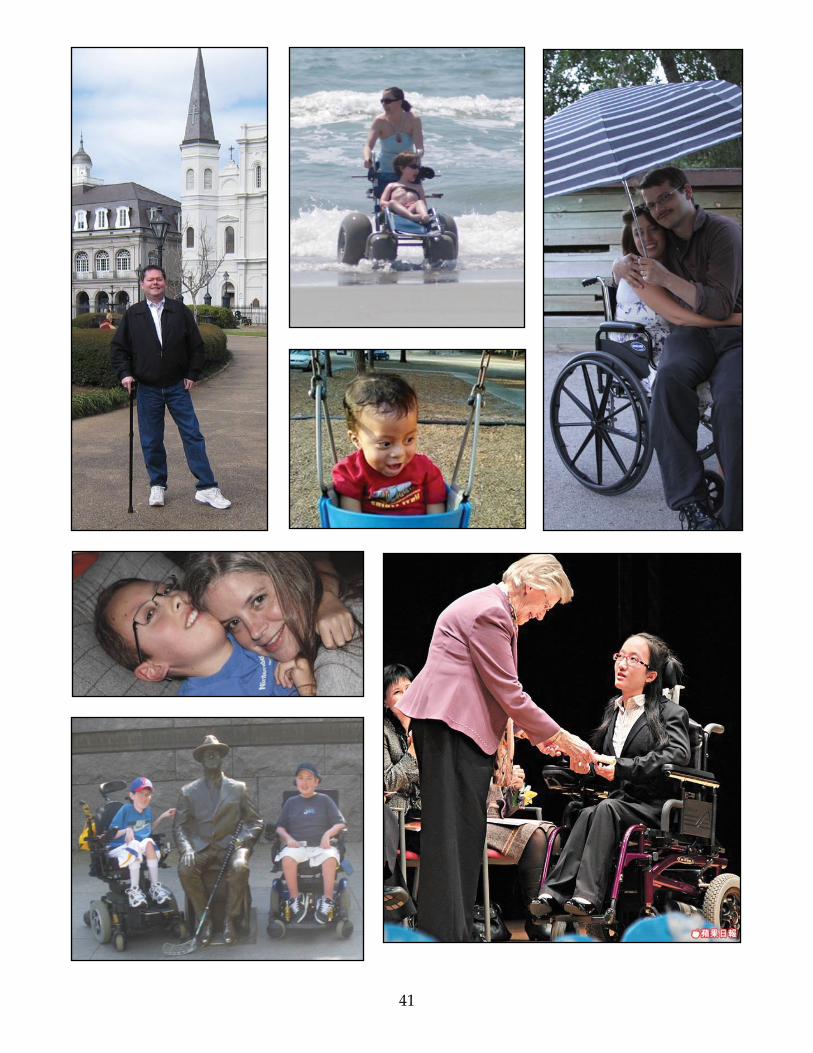
41
42
Acknowledgments:
This guide was made possible by the vision of Anne Rutkowski, MD, Chair of Cure CMD, and was adapted, compiled, and edited by Susan Sklaroff-Van Hook and Diane Smith-Hoban.
Additional expert contributions were made by Meganne Leach, MSN, APRN, PNP-BC, Katy Meilleur, PhD, CRNP National Institutes of Health; Thomas Sejersen, MD Karolinska Institutet; Kate Bushby, MD Newcastle University; Ching H. Wang Stanford University; and Carsten Bonnemann, MD, Neuromuscular and Neurogenetic Disorders of Childhood Section, National Institutes of Health. Medical editing and publishing support generously donated by Mary T. Durkin, Diane True and Erin McGuirk.
Photographs used in this guide were provided by families affected by CMD. We would like to thank all the children and families who allowed their photos to be used..If you have any comments, questions or additional feedback for future revisions of this guide, please contact Cure CMD ([email protected]) or email [email protected]