the map kinase fus3 associates with and phosphorylates the
TRANSCRIPT

The MAP kinase Fus3 associates with and phosphorylates the upstream signaling component Ste5 Janice E. Kranz/"^ Brett Satterberg/ and Elaine A. Elion^"*
^Department of Biological Chemistry and Molecular Pharmacology, Harvard Medical School, Boston, Massachusetts 02115 USA; ^Department of Cellular and Developmental Biology, Harvard University, Cambridge Massachusetts, 02138 USA
Activation of the Saccbaromyces cerevisiae MAP kinase Fus3 is thought to occur via a linear pathway involving the sequential action of three proteins: Ste5, a protein of unknown function, Stell, a MAPKK kinase homology and Ste7, a MAPK kinase homolog which phosphorylates and activates Fus3. In this report, we present evidence for a novel mechanism of Fus3 activation that involves a direct association with Ste5, a protein not predicted to interact with Fus3. First, overexpression of Ste5 suppresses fus3 point mutations in an allele-specific manner and increases Fus3 kinase activity in vitro. Second, Ste5 associates with Fus3 in vivo as demonstrated by the two-hybrid system and by two methods of copurification. Third, Ste5 and Fus3 associate prior to pheromone stimulation even when Fus3 is inactive, and in strains lacking Ste7 and Stell. Fourth, Ste5 is phosphorylated by Fus3 in purified complexes and copurifies with an additional protein kinase(s). These observations suggest the possibility that SteS promotes signal transduction by tethering Fus3 to its activating protein kinase(s).
[Key Words: Saccharomyces cerevisiae-, MAP kinase; signal transduction; Fus3; SteS]
Received November 16, 1993; accepted in revised form December 21, 1993.
Haploid Saccharomyces cerevisiae cells exposed to mating pheromone undergo cell-cycle arrest at Gi/Start, transcriptional activation of mating-specific genes, and cell elongation prior to mating (for review, see Sprague and Thomer 1994). Genetic analysis has identified numerous signal transduction components which regulate these responses (Mackay and Marmey 1974; Hartwell 1980; for review, see Kurjan 1992; Sprague and Thomer 1994). Based on genetic data, these signal transduction components have been ordered into a linear pathway upstream of the transcription factor, Stell, that is activated and phosphorylated in response to pheromone (Dolan et al. 1989; Dolan and Fields 1990). In this linear pathway model, signal transduction initiates by pheromone binding to a cell surface receptor, activating a heterotrimeric G-protein, then continues successively through the putative protein kinase Ste20, the SteS protein, the protein kinases Stell and Ste7, and culminating in the activation of Fus3 and Kssl, related protein kinases that activate Stel2 (Blinder et al. 1989; Elion et al. 1990, 1991b; Whiteway et al. 1990; Cairns et al. 1992; Gartner et al.
P̂resent address: Cubist Phannaceuticals, 24 Emily Street, Cambridge, MA 02139 USA. ^Conesponding author.
1992; Leberer et al. 1992; Stevenson et al. 1992; Errede et al. 1993; Ramer and Davis 1993). Although this linear pathway is favored for its simplicity, interdependent or branched models have not been excluded (Elion et al. 1990; Stevenson et al. 1992; Elion et al. 1993).
The protein kinases Fus3, Kssl, SteZ, and Stell comprise a highly conserved regulatory module found in mammals as well as yeast (Errede and Levin 1993; Ko-sako et al. 1993; Lange-Carter et al. 1993; Neiman et al. 1993). Fus3 and Kssl are homologues of MAP kinases (mitogen-activated protein kinases, MAPKs) or ERKs (extracellular signal-responsive kinases) that are activated by phosphorylation of tyrosine and threonine residues (Ray and Sturgill 1987; Boulton et al. 1991; for review, see Cobb et al. 1991; Pelech and Sanghera 1992a, 1992b; Posada and Cooper 1992). Fus3 is also phosphorylated on these residues in response to a-factor, and requires this modification for function (Gartner et al. 1992; Elion et al. 1993). Ste7 is related to the dual-specificity MAP kinase kinases (MAPKKs or MEKs, for MAP kinase/ERK kinase) (Crews and Erikson 1992; Seger et al. 1992; Ko-sako et al. 1993) that activate the MAP kinases (L'Alle-main et al. 1992; Rossomando et al. 1992). Consistent with this, Ste7 immune complexes phosphorylate Fus3 in vitro (Errede et al. 1993; Zhou et al. 1993). Stell is
GENES & DEVELOPMENT 8:313-327 © 1994 by Cold Spring Harbor Uboratory Press ISSN 0890-9369/94 $5.00 313
Cold Spring Harbor Laboratory Press on January 30, 2022 - Published by genesdev.cshlp.orgDownloaded from

Kianz et al.
related to the MAPK kinase regulator, MAPKKK or MEKK (Lange-Carter et al. 1993), although it has yet to be shown that Stell directly activates Ste7. Ste20 is a putative protein kinase which to date lacks a mammalian counterpart and physiological target (Leberer et al. 1992; Ramer and Davis 1993).
Ste5 is the only signal transduction component that lacks obvious homology to a protein of known function (Leberer et al. 1993; Mukai et al. 1993; Perlman et al. 1993), making its role difficult to assess. The STE5 gene encodes a 917-residue open reading frame with limited homology to two types of proteins, fueling speculation about its function (Leberer et al. 1993; Mukai et al. 1993; Perlman et al. 1993). A 150-residue amino-terminal domain of Ste5 is related to Farl, suggesting that Ste5 and Farl share a fimction in Gj arrest (Leberer et al. 1993; Mukai et al. 1993; Perlman et al. 1993). This region overlaps a potential cysteine/histidine metal-binding motif, which together with a carboxy-terminal acidic domain, suggest that Ste5 is a transcription factor (Leberer et al. 1993; Mukai et al. 1993; Perlman et al. 1993; Sprague and Thomer 1994). Neither possibility is consistent with the current placement of Ste5 in the signalling cascade. Thus, while genetic experiments demonstrate that Ste5 is essential for signal transduction (MacKay and Manney 1974; Hartwell 1980), the function of Ste5 remains a mystery.
Ste5 is thought to act at a single step between the Ste20 and Stell protein kinases on the basis of genetic analysis with hyperactive forms of Ste5 and Stell (Cairns et al. 1992; Leberer et al. 1992; Stevenson et al. 1992; Hasson et al. 1994); however, several observations suggest a more complex picture. First, hyperactive forms of Stel 1 do not fully suppress ste5 mutations (Stevenson et al. 1992) and hyperactive forms of SteS only partially suppress G-protein mutants (Hasson et al. 1993). Second, overexpression of SteS suppresses ste4 (i.e., G-proteiu; MacKay 1983), fus3 (Elion et al. 1991a), and ste20 (Leberer et al. 1993) mutations that block at different points in the signal transduction pathway. This ubiquitous pattern of suppression suggests that SteS function cannot be defined in simple linear terms. Therefore, more complicated models for the role of SteS in signal transduction, such as an interdependence with other signal transduction components (e.g., the G-protein, Stell, or Fus3), cannot be excluded (Elion et al. 1991a; Stevenson et al. 1992; Hasson, Blinder, Thomer, Jenness, 1994).
We present here a connection between SteS and Fus3, proteins not previously thought to interact directly on the basis of classical genetic experiments. Overexpression of SteS suppresses multiple defects of fus3 point mutants in an allele-specific fashion, and also increases Fus3 kinase activity in vitro, suggesting a direct interaction between the two proteins. Consistent with these results, SteS associates with Fus3 in vivo, independently of both pheromone-stimulation and Fus3 catalytic activity. Furthermore, SteS is phosphorylated by Fus3 and additional associated kinases in vitro. Taken together, these data suggest SteS associates in a complex with Fus3 and other protein kinase(s).
Results
Excess SteS suppresses Gj airest and transciiption defects of fus3 mutants
We isolated the STES gene in two screens for suppressors of fus3 mutations. In the first screen, STES was identified as a suppressor of the mating defect of the fusS-S mutant (Elion et al. 1991a). Subsequent analysis showed that STES overexpression increases mating and FUSl transcription l.S- to 2-fold, and partially suppresses the Gi arrest defect of a fusS null mutant (Elion et al. 1991a). In contrast, excess STES does not bypass a fusS kssl double mutant, suggesting STES either functions before Fus3 and Kssl, or requires either kinase for function (Elion et al. 1991a). STES was also isolated as a suppressor of the a-factor resistance of the fus3-2 mutant in a second screen that identified other efficient suppressors including PARI and STE12 (Elion et al. 1993; B. Satterberg and E.A. Elion, details to be published elsewhere). Subsequent analysis showed that overexpression of other upstream components (e.g., STE2, STE4, STE7, STEll, STE20] did not suppress the fus3-2 mutant as efficiently as STES (data not shown). Thus, STES is a conspicuously strong suppressor of fus3 mutations.
SteS activates Fus3 kinase activity in vitro
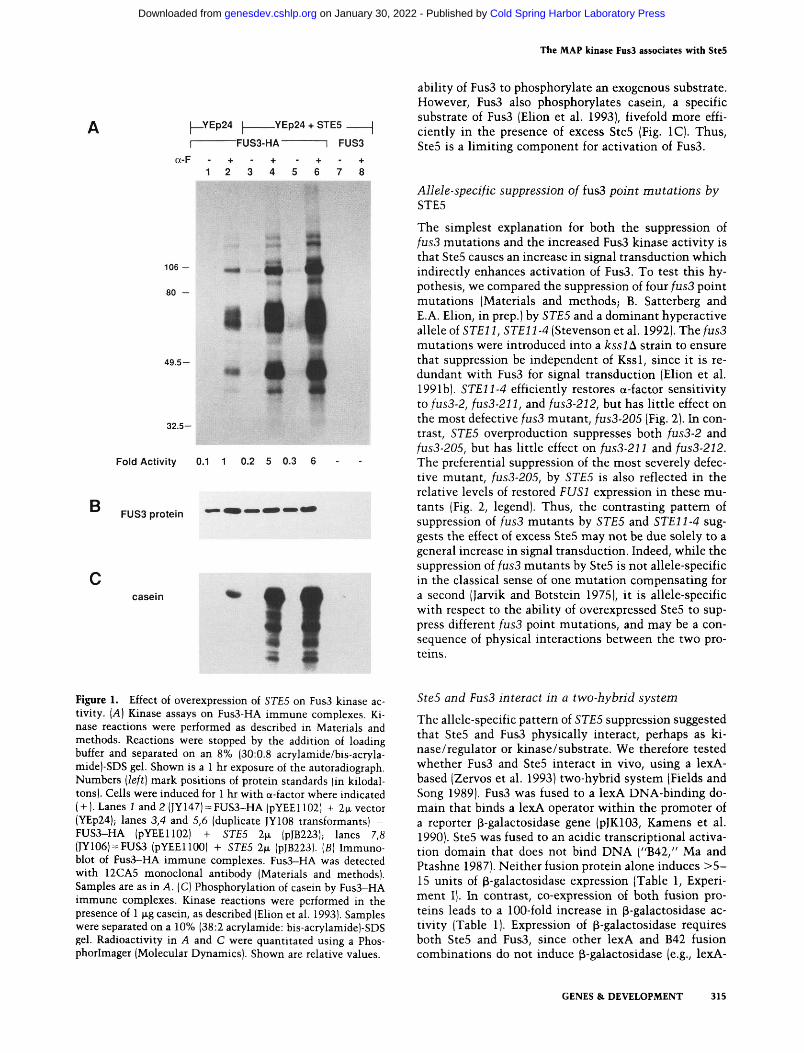
We determined the effect of SteS overexpression on Fus3 kinase activity using an immune complex kinase assay that employs a hemagglutinin (FIA) epitope-tagged Fus3, Fus3-HA (Elion et al. 1993). A typical assay reveals a profile of five or more associated proteins that are phosphorylated when Fus3 is activated by pheromone (Fig. I A, lane 2). Fus3 kinase activity increases approximately fivefold when SteS is overexpressed, as shown by a five-to sixfold increase in intensity of all phosphoproteins in the profile and the presence of at least three new associated substrates (apparent molecular mass of —US kD, ~9S kD, and ~S5 kD; Fig. lA, lanes 4,6). SteS overexpression also increases Fus3 kinase activity two- to threefold in the absence of pheromone induction (Fig. I A, lanes 3,5). In contrast, a steS deletion mutation blocks Fus3 kinase activity, as expected (data not shown).
SteS overproduction could cause an apparent increase in Fus3 kinase activity by several mechanisms. First, the kinase activity could be due to SteS; however, no kinase activity is seen in immune, complexes prepared from strains with Fus3 lacking the HA epitope (Fig. lA, lanes 7,8), nor in strains containing Fus3R42-HA, an inactive form of Fus3-HA (Elion et al. 1993; data not shown). Second, the effect could be an indirect consequence of an increase in the level of Fus3 due to enhanced activity of Stel2, which normally induces FUS3 mRNA ~3-fold (Elion et al. 1991b). However, equivalent amounts of Fus3 are immunoprecipitated in the absence and presence of excess SteS (Figure IB, cf. lanes 1 and 2 with 3-6). Third, SteS overproduction could simply increase the levels of the substrates that coprecipitate with Fus3. If this were the case, then SteS levels should not affect the
314 GENES & DEVELOPMENT
Cold Spring Harbor Laboratory Press on January 30, 2022 - Published by genesdev.cshlp.orgDownloaded from
The MAP kinase Fus3 associates with Ste5
|—YEp24 I YEp24 + STE5
I FUS3-HA
a-F 1 FUS3
1 2 3 4 5 6 7 8
106
80 -
49.5-
32.5-
B
Fold Activity 0.1 1 0.2 5 0.3 6
FUS3 protein
casein
ability of Fus3 to phosphorylate an exogenous substrate. However, Fus3 also phosphorylates casein, a specific substrate of Fus3 (Elion et al. 1993), fivefold more efficiently in the presence of excess Ste5 (Fig. IC). Thus, Ste5 is a limiting component for activation of Fus3.
AUele-specific suppression of fus3 point mutations by STE5
The simplest explanation for both the suppression of fus3 mutations and the increased Fus3 kinase activity is that Ste5 causes an increase in signal transduction which indirectly enhances activation of Fus3. To test this hypothesis, we compared the suppression of four fus3 point mutations (Materials and methods; B. Satterberg and E.A. Elion, in prep.) by STE5 and a dominant hyperactive allele of ST£n, ST£11-4 (Stevenson etal. 1992). The/us3 mutations were introduced into a ksslA strain to ensure that suppression be independent of Kssl, since it is redundant with Fus3 for signal transduction (Elion et al. 1991b). STEll-4 efficiently restores a-factor sensitivity to fus3-2, fus3-211, and fus3-212, but has Httle effect on the most defective fus3 mutant, fus3-205 (Fig. 2). In contrast, STE5 overproduction suppresses both fus3-2 and fus3-205, but has httle effect on fus3-211 and fus3-212. The preferential suppression of the most severely defective mutant, fus3-205, by STE5 is also reflected in the relative levels of restored FUSl expression in these mutants (Fig. 2, legend). Thus, the contrasting pattern of suppression of fus3 mutants by STE5 and STEll-4 suggests the effect of excess SteS may not be due solely to a general increase in signal transduction. Indeed, while the suppression of fus3 mutants by SteS is not allele-specific in the classical sense of one mutation compensating for a second (Jarvik and Botstein 1975), it is allele-specific with respect to the ability of overexpressed SteS to suppress different fus3 point mutations, and may be a consequence of physical interactions between the two proteins.
Figure 1. Effect of overexpression of STES on Fus3 kinase activity. [A] Kinase assays on Fus3-HA immune complexes. Kinase reactions were performed as described in Materials and methods. Reactions were stopped by the addition of loading buffer and separated on an 8% (30:0.8 acrylamide/bis-acryla-mide)-SDS gel. Shown is a 1 hr exposure of the autoradiograph. Numbers (left] mark positions of protein standards (in kilodal-tons). Cells were induced for 1 hr with a-factor where indicated ( + ). Lanes i and 2 (JY147) = FUS3-HA (pYEE1102| + 2|JL vector (YEp24); lanes 3,4 and 5,6 (duplicate JY108 transformants) = FUS3-HA (pYEE1102) + STES 2 .̂ (pJB223); lanes 7,8 (JY106) = FUS3 (pYEEllOO) + STES 2ti (pJB223). [B] Immuno-blot of Fus3-HA immune complexes. Fus3-HA was detected with 12CA5 monoclonal antibody (Materials and methods). Samples are as in A. (C) Phosphorylation of casein by Fus3-HA immune complexes. Kinase reactions were performed in the presence of 1 |xg casein, as described (Elion et al. 1993). Samples were separated on a 10% (38:2 acrylamide: bis-acrylamide)-SDS gel. Radioactivity in A and C were quantitated using a Phos-phorlmager (Molecular Dynamics). Shown are relative values.
SteS and Fus3 interact in a two-hybrid system
The allele-specific pattern of STES suppression suggested that SteS and Fus3 physically interact, perhaps as ki-nase/regulator or kinase/substrate. We therefore tested whether Fus3 and SteS interact in vivo, using a lexA-based (Zervos et al. 1993) two-hybrid system (Fields and Song 1989). Fus3 was fused to a lexA DNA-binding domain that binds a lexA operator within the promoter of a reporter p-galactosidase gene (pJK103, Kamens et al. 1990). SteS was fused to an acidic transcriptional activation domain that does not bind DNA ("B42," Ma and Ptashne 1987). Neither fusion protein alone induces > 5 -IS units of p-galactosidase expression (Table 1, Experiment I). In contrast, co-expression of both fusion proteins leads to a 100-fold increase in p-galactosidase activity (Table 1). Expression of p-galactosidase requires both SteS and Fus3, since other lexA and B42 fusion combinations do not induce p-galactosidase (e.g., lexA-
GENES & DEVELOPMENT 315
Cold Spring Harbor Laboratory Press on January 30, 2022 - Published by genesdev.cshlp.orgDownloaded from

Ktanz et al.
YEp24 YEp24 + STE11-4
STE5
fus3-2
fus3-205
fus3-211
1US3-212
Figure 2. Effect of overexpressed STE5 and hyperactive STEl 1 -4 on a-factor sensitivity of fus3 mutants. Lawns of MATa sstlA kssl\ strain EY966 containing mutations fus3-2 (BY369), fus3-205 (BY360), or fus3-211 (BY366) and bearing either YEp24 {URA3 2̂ JL), PJB223 (STE5 URA3 2fi), pSL1655 [STEll-4, integrated) were tested for a-f actor sensitivity by a halo assay using 5 l̂l of 0.1 mM a-factor in dimethylsulfoxide. Strains were grown in SC medium lacking uracil to select for all plasmids, including the integrated STEll-4. Plates were photographed after 24 hr at 30°C. We measured FUSl expression in a subset of these strains after a 1 hr exposure to a-factor with a Fusl-p-galactosidase reporter gene (pJB207). In the absence of excess Ste5, the fus3-2, fus3-205, and fus3-211 strains produce equivalent low levels of p-galactosidase activity (1.5±0.1 units). Excess Ste5 has little effect on FUSl expression in the fus3-211 strain (1.7±0.1 units), a threefold effect in the fus3-2 strain (4.8±0.1 units) and a fourfold effect in the fus3-20S strain (6.0±0.2 units). Numbers are averages of duplicate transfor-mants assayed twice.
leucine (data not shov^n). In addition^ Fus3 and Ste5 interact equivalently in an isogenic ste5 deletion strain (Table 1, Experiment II), indicating that endogenous Ste5 is not required for their association. The fact that Ste5 and Fus3 still interact in the absence of these signal transduction proteins strongly suggests their association is direct.
Ste5 and Fus3 copurify
We investigated whether Ste5 and Fus3 copurify under conditions in which both proteins are functional, as predicted by the two-hybrid results. To detect Ste5, we constructed a fusion between glutathione-S-transferase and Ste5 (GST-Ste5) that is expressed from the GALl promoter and complements a ste5A strain for mating (see Materials and methods). Fus3-HA immune complexes were prepared from strains containing either GST or GST-Ste5, then immunoblotted with anti-GST antiserum. As shown in Figure 3A, GST-Ste5, but not GST alone, coprecipitates with Fus3-HA, from both STE5 and ste5A strains (cf. lanes 3, 4, 6 and 7 to lane 5). The presence of GST-Ste5 is not due to nonspecific binding to the antibody or protein-A-Sepharose as it is not recovered from strains lacking the epitope tag on Fus3 (lane I). Thus, the ability to copurify GST-Ste5 with Fus3-HA is dependent on both Ste5 and Fus3. Furthermore, Ste5 and Fus3 associate regardless of whether Fus3 is active (a-factor-induced, lanes 4,7) or inactive (uninduced, lanes 3,6).
To confirm the specificity of the association between Ste5 and Fus3, we did the complementary experiment of purifying GST and GST-Ste5 with glutathione-agarose, and assaying for the presence of Fus3-HA. Identical results were seen: Fus3-HA copurifies with GST-Ste5 but
bicoid, B42-CDC28 in Table 1). Thus, by the two-hybrid system, Ste5 and Fus3 proteins interact.
Ste5 and Fus3 interact in the absence of Ste7, Stell, and Stel2
It is possible that the two-hybrid interaction we detect is not due to a simple association between Fus3 and Ste5. For example, overexpression of Ste5 could activate the endogenous signal transduction pathway and cause lexA-Fus3 to associate with another protein, such as the transcription factor Stel2, a Fus3 substrate (Elion et al. 1993). Alternatively, B42-Ste5 could associate with lexA-Fus3 via intermediary proteins such as Ste l l or Ste7, which function between Ste5 and Fus3. We therefore repeated the two-hybrid experiment in strains deleted for STE7, STEll, or STEl2, using a lexA-dependent LEU2 reporter gene, and assayed growth on plates lacking leucine. Fus3 and Ste5 still associate in the absence of Ste7, S te l l , or Stel2 as shown by equivalent growth of STE, stelL, stellii, and stel2A strains on media lacking
Table 1. ^-Galactosidase activity induced by interaction of Fus3 and Ste5 fusion proteins in a two-hybrid system
I
II
Host strain
STE5
STE5 steSA
lexA fusion plasmid^
0 bicoid FUS3 FUS3 FUS3
0
9 10 13 15 15
p-Galactosidase activity (units)^
B42 fusion plasmid^
CDC28
8 14 13
N.D. N.D.
STE5
7 14
1200 310 360
(N.D.) Not determined. ""Specific plasmids are listed in Table 4. 0 refers to the vector alone. LexA vector is LexA202PL, bicoid fusion is pHM0, FUS3 is LexA-FUS3; B42 fusion vector is pJG4-5, CDC28 fusion is pYBSlSO; STE5 fusion is pYBS146. ''Values are Miller units of p-galactosidase activity (Craven 1965), averaged from two experiments in which two independent plasmid-bearing transformants were assayed. All assays were done in yeast strain EGY048 (ST£5) or a steSA. derivative carrying a LacZ reporter plasmid.
316 GENES & DEVELOPMENT
Cold Spring Harbor Laboratory Press on January 30, 2022 - Published by genesdev.cshlp.orgDownloaded from

The MAP kinase Fus3 associates with Ste5
GST +/- STE5
Dex or Gal
a-F
106-
G G G
GST-ST£5 •*-
106' 80-
-106
g-GST
HA IMMUNOPRECIPITATES GSTPREClPfTATES Figute 3. GST-Ste5 and Fus3-HA co-pre-steSA STE5 . c g^_ cipitate in vivo. Immunoblot analysis of
n̂^ I ~^ ^ I ~^ ^ I — 1 I 1 GST-Ste5 in Fus3-HA immune com-D G G G G G D G G R R R R plcxcs. (Le/t) Fus3-HA was immunoprccip-
itated with 12CA5 monoclonal antibody from ste5A (lanes 1—4) or STE5'^ (lanes 5-7] strains carrying FUS3-HA on pYEEllOl (except lane 1, which has FUSS on pYEEllOO) and either GST (lane 5) or GST-STE5 (lanes 1-4 and 6,7). Cells were pre-grown in either galactose (G) or dextrose (D), prior to being induced for 1 hr with a-factor, where indicated (+). Duplicate samples were run on separate gels for immunoblot analysis with either antiserum against GST [top] or 12CA5 [bottom]. Note that the secondary antibody (goat anti-rabbit IgG-HRP) cross-reacts with the 12CA5 Ig in the immune complexes. Numbers [left] mark positions of protein standards (in kilodaltons). The expected position of GST protein is indicated. Strains are BY845 (lane 1], BY846 (lanes 2-4], BY 784 (lane 5), and BY786 (lanes 6,7] [right]. Immunoblot
analysis of Fus3-HA associated with GST-Ste5. GST or GST-Ste5 was purified with glutathione-agarose (see Materials and methods), and duplicate samples were run on separate gels and immunoblotted with either 12CA5 [top] or affinity-purified rabbit antiserum against GST [bottom]. Numbers mark positions of protein standards (in kilodaltons). Results are shown from two separate experiments. Cells were grown in either galactose (G) or dextrose (D), and were induced for 1 hr with a-factor before extract preparation, where indicated ( + ). Lanes 1-3 (BY846) = ste5A + GST-STE5 (pYBSI86), lane 4 (BY844) = ste5A + GST (pEMBI^GST), lane 5 (BY784) = ST£5^ + GST (pEMBL-GST), and lanes 6,7 (BY786) = ST£5* + GST-STE5 (pYBS186).
49.5-
32.5-
g-HA
- • I Q G H
•^GST
80-
49.5-
32.5-
-49.5
32.5
FUS3-HA -» -
not with GST (Fig. 3A, cf. lanes 2 and 3 with 4; lanes 6 and 7 with 5). Again, the association between Ste5 and Fus3 could be detected in extracts of both a-factor-in-duced (lanes 3,7) and -uninduced (lanes 2,6) cells. Thus, Ste5 and Fus3 associate in vivo independently of phero-mone induction.
SteS associates with a catalytically inactive form of Fus3
The association between SteS and Fus3 prior to phero-mone stimulation (Fig. 3A) and in ste strains suggests that activation of Fus3 is not a prerequisite for association with SteS. We tested this possibility by repeating both types of copurification with a catalytically impaired form of Fus3-HA, Fus3R42-HA. As shown in Figure 4, GST-SteS associates equivalently with both functional (Fus3-HA, lanes 3,4, left and right) and nonfunctional (Fus3R42-HA, lanes 6,7, left and right) forms of Fus3. Thus, Fus3 need not be catalytically active in order to associate with SteS.
Fus3 phosphorylates SteS in purified complexes
We next determined whether SteS is phosphorylated by Fus3 as evidence of a direct association. Fus3 was first tested for its ability to phosphorylate SteS using the B42-SteS protein (Table 1) as an exogenous substrate. Equivalent amounts of B42-Ste5 and Fus3-HA were immuno-precipitated (Fig. S, right) and tested in the kinase assay
(Elion et al. 1993). B42-SteS is only slightly phosphorylated when assayed alone (Fig. S, left, lane S) or in the presence of inactive Fus3R42-HA (lane 6). In contrast, the addition of active Fus3 causes a significant increase in the amount of B42-SteS phosphorylation (Fig. S, left, lanes 2, 4). Equivalent results were seen when B42-SteS was prepared from either /us3A or FUSS strains (data not shown). Moreover, this phosphorylation is specific for SteS, since B42 alone is not phosphorylated (lane 1 and data not shown).
The fact that Fus3 could phosphorylate SteS in vitro suggested that phosphorylation could be used to detect a direct association between the two proteins when they associate in vivo. We therefore performed kinase assays on Fus3-F1A immunoprecipitated from strains containing GST-SteS (Fig. 6A, lanes 1-9). As seen previously for wild-type SteS (Fig. 1), overexpression of GST-SteS causes a large increase in Fus3 kinase activity both in the absence and presence of a-factor (Fig. 6A, cf. lanes 8 and 9 with 6 and 7), even under repressive conditions when GST-SteS is produced at very low levels (Fig. 6A, lanes 13,18). Significantly, overexpression of GST-SteS produces a prominent new phosphoprotein of —120 kD, the size observed for GST-SteS (Fig. 6A, lanes 8,9), and consistent with the expression of GST-SteS (Fig. 6A, cf. lanes 8 and 9 with lanes 6 and 7). Furthermore, the —120-kD phosphoprotein is present in extracts from both untreated (lane 8) and pheromone-treated (lane 9) cells, consistent with the association of SteS and Fus3.
To prove that the ~120-kD substrate is GST-SteS, we
GENES & DEVELOPMENT 317
Cold Spring Harbor Laboratory Press on January 30, 2022 - Published by genesdev.cshlp.orgDownloaded from

Kranz et al.
HA IMMUNOPRECIPITATES GST PRECIPITATES
Figure 4. Association of GST-Ste5 with catalytically-inactive Fus3. Fus3R42-HA and GST-STE5 were purified exactly as described in Fig. 3 from STES'*^ strains carrying FusR42-HA (on pYBS351) and either GST (lane IJ or GST-STE5 (lanes 2-7). Duplicate 12CA5 immunoprecipitates (left) and glutathione-agarose precipitates (right) were run on separate gels for immunoblot analysis with either anti-GST antiserum [top) or anti-HA monoclonal antibody (bottom). Strains are STE5'' fusS'' (EY1095) containing Fus3-HA and GST (lane i =BY784), Fus3-HA and GST-Ste5 (lanes 2-4=BY786), or fus3R42-HA and GST-Ste5 (lanes 5-7 = Pfl65). Samples were s eparated on 10% (30:0.8 acrylamide/bis-acrylaraide)-SDS gels.
GST *h STE5 Dex or Gal
a-F
g-GST
FUS3-HA fus3R42-HA
D G G D G G
FUS3-HA fus3R42-HA
G + 1 2
• + + I • + + 1
0 G G D G G
mmmm' -" GST-STE5 -"
igG
g-HA
GST
— FUS3-HA -»-
determined whether it could be purified with glutathione-agarose. Fus3—^HA kinase reactions were solubilized and incubated with glutathione-agarose (see Materials and methods). Of the nine or more phosphoproteins.
only the 120-kD protein bound to glutathione-agarose (Fig. 6B, lane 2), demonstrating that it is GST-Ste5. Furthermore, treatment of the purified protein with thrombin, which recognizes a site engineered between GST
B Substrate A d d e d : B42 B42-STE5
Gal or Dex G 1 G D G G G |
a-HA
FUS3-HA IP FUS3-HA 0 R42
aP 1+ . + +1
aF n.a. + 1 2 3 r ^ 5
106-
80-
Mm • AAA AAA
106-
80-
49.5-
B42-STE5
igG,
FUS3-HA
32.5-
49.5-
32.5-
Figure 5. Phosphorylation of B42-Ste5 by Fus3-HA immune complexes. Kinase assays (left) were performed on B42-Ste5 and B42 immunoprecipitates alone (lane 5) or in the presence of Fus3-HA immune complexes. Strains containing the galactose-inducible fusion proteins B42 (BY872) or B42-Ste5 (BY873) were grown in galactose (G) or dextrose (D), as indicated. Fusion proteins were immunoprecipitated with 12CA5 monoclonal antibody from 200 IN^ of yeast extract in RIPA buffer, then washed 5 times
before being added to Fus3-HA immune complexes. Fus3-HA immune complexes were prepared from Fus3-HA-containing strain EY960 (lanes 1-4) or fus3R42-HA (catalytically inactive)-containing strain EYl 144 (lane 6). Fus3-HA strains were treated for 1 hr with a-factor where indicated (+) . Samples were separated on an 8% (30:0.8 acrylamide/bis-acrylamide)-SDS gel. (Right) Immunoblot of Fus3-HA and B42-Ste5-HA immunoprecipitates. Duplicate immunoprecipitates from those used in the kinase assay were performed using 12CA5 monoclonal antibody.
318 GENES & DEVELOPMENT
Cold Spring Harbor Laboratory Press on January 30, 2022 - Published by genesdev.cshlp.orgDownloaded from

The MAP kinase Fus3 associates with SteS
Gal or Dex aF
FUS3-HA
106
80
49.5
STES'
cA^^
oG^^ oG^^ ^G^'^ oG -̂̂
AsteS
5^0
n0 -̂.A^^
D D G G - + - +
1 2 3 4
G D D G G + - + - +
5 6 7 8 9
G G D D G G + + • + • +
10 11 12 13 1415
G 0 D G G + - + - +
16 1718 19 20
B 32-P Coomassie blue D a-GST
10'at30°C thrombin
106
80
49.5
32.5
* GST-STE5
* STES
10'at SOX - + + thrombin - - +
1 2 3
106 -
80 -
49.5 - y
10'at30°C thrombin
+ + +
2 3
* GST-STE5
* STES
I * GST)
106
80
49.5
32.5
Wi * GST-STE5
* STES )
* GST
32.5 * GST
Figure 6. Effect of GST-Ste5 on Fus3-HA kinase assay. [A] Kinase assays of Fus3-HA immune complexes. Total protein (200 .̂g) of each strain was immunoprecipitated with 12CA5 monoclonal antibody, and Fus3-HA kinase assays were performed as described in Materials and methods. All strains contained Fus3-HA (pYEE1102) except those in lanes 5, 11, and 16, which contained Fus3 lacking the HA epitope (pYEEllOO) indicated by (-). Lanes 1-9 are from a STE5^ strain (EY1095) containing GST (lanes J-4) or GST-Ste5 (lanes 5-9). Lanes 10-20 are from a steSA strain (BY819) containing GST (lane 10], GST-Ste5 (lanes 11-15], or STES CRN (lanes 16-20]. Cells were grown in either galactose (G), to induce expression of the GST fusion proteins, or dextrose (D), to repress their expression, and were induced for 1 hr with a-factor, where indicated (+ ). (B] Purification and thrombin digestion of GST-Ste5 from kinase assays of Fus3-HA immune complexes. A kinase reaction identical to lane 9 of A was solubilized in RIPA buffer and incubated with glutathione-agarose. The GST precipitates were washed, analyzed directly (lane 2), incubated with thrombin (lane 4], or incubated without thrombin (lane 3). (C) Thrombin digestion of purified GST-Ste5. GST-Ste5 was purified with glutathione-agarose from 200 |xg of whole cell extract from the STE5* strain (BY786) containing Fus3-HA + GST-STE5. Thrombin was added (lane 3] or not added (lanes 1,2], and the precipitates were incubated at 30°C for 10 min (lanes 2,3]. Samples were separated on a 10% SDS-polyacrylamide gel and visualized with Coomassie blue. (D) Immunoblot with a-GST of duplicate samples from C. Note that a GST doublet is typically seen but the smaller species was inadvertently run off this gel.
GENES & DEVELOPMENT 319
Cold Spring Harbor Laboratory Press on January 30, 2022 - Published by genesdev.cshlp.orgDownloaded from

Kianz et al.
and Ste5, shows that the vast majority of the phosphorylation is on Ste5; not GST. Cleavage of the ~120-kD phosphoprotein with thrombin produces a single phos-phoprotein of -100 kD equivalent in size to the Ste5 portion of GST-Ste5 (Fig. 6B, lane 4). In contrast, cleavage of purified GST-Ste5 with thrombin produces three major polypeptides, one of -100 kD, and two of -30 kD that are detected by GST antiserum (Fig. 6C, D). Thus, Fus3 phosphorylates Ste5 when the two proteins associate, proving they directly contact one another.
SteS associates with an additional protein kinase(s)
One explanation for the interaction between SteS and Fus3 is that SteS mediates the association between Fus3 and its upstream activating kinases. To test this possibility, we examined whether SteS associates with additional protein kinases. We first determined whether GST-SteS is still phosphorylated in vitro when copuri-fied with catalytically inactive Fus3R42-HA. Normally, no Fus3 kinase activity is detected in kinase assays of Fus3R42-HA (Fig. 7A, lane 4; Elion et al. 1993). Nevertheless, GST-SteS is still residually phosphorylated in immune complexes with Fus3R42 (Fig. 7A, lanes 5, 6). This phosphorylation could reflect residual Fus3 kinase activity that is now detectable in the presence of excess GST-SteS. Indeed, an equivalent ERK2 MAP kinase mutant retains S% activity in vitro when assayed with myelin basic protein (Robbins et al. 1993). However, identical Fus3R42-HA/GST-SteS immune complexes do not phosphorylate a large excess of 2VLBP (data not shown), a proven Fus3 substrate (Elion et al. 1993). Thus, GST-SteS may be phosphorylated by another protein kinase(s) present in the complex whose activity is independent of Fus3 and specific for GST-SteS. This possibility is strongly supported by the fact that GST-SteS purified from both PUS3 and /us3A strains is associated with a protein kinase(s) which phosphorylates GST-SteS in the kinase assay (Fig. 7B; see Materials and methods). Significantly, only full-length GST-SteS is phosphorylated, although immunoblot analysis shows the presence of shorter degradation products (Figs. 3 and 4). Thus, an additional protein kinase(s) that specifically recognizes SteS can copurify with both Fus3 and SteS, suggesting the existence of a multimeric complex.
FUS3-HA fus3R42-HA
Dex or Gal D G G D G G a-F + - • + - +
1 2 3 4 5 6
106 -
80 -
49.5 -
32.5 _
1^
•
IP m
m
WT A 1 2
106 -
80 -
• GST-STES
Figure 7. GST-SteS is associated with a Fus3-independent protein kinase(s|. [A] Kinase assays of active Fus3-HA (BY786, lanes 1-3] and inactive Fus3R42-HA ([Y165, lanes 4-6} from strains harboring GST-SteS (pYBS186). Kinase assays were performed as described in Materials and methods. GST-SteS was expressed by growth in galactose (G), repressed by growth in dextrose (D). Cells were induced for 1 hr with a-factor where indicated (-I-). [B] Phosphorylation of GST-SteS by an associated protein kinase(s). Strains were grown in galactose to induce expression of GST-SteS and then induced for 1 hr with a-factor. GST-SteS was purified from FUS3 (lane 1, EY1462) and /us3A (lane 2, EY1450) extracts with glutathione-agarose and incubated in a standard kinase assay for 8 min (Elion et al. 1993). Samples were separated on a 10% SDS-polyacrylamide gel.
Fus3 is inactive, and in the absence of Ste7 and Stell. Fifth, SteS is phosphorylated by Fus3 and additional protein kinase(s) in purified complexes. These data strongly suggest that SteS and Fus3 directly interact. The simpler explanation for the effects of SteS on Fus3, that of over-expressing a limiting component upstream of Fus3 in the signal transduction pathway, is highly imlikely. This purely indirect mechanism for the effects of SteS on Fus3 does not explain the allele-specific suppression, the physical interaction, nor the fact that SteS can be phosphorylated by Fus3.
Discussion
A novel linkage between SteS and Fus3
We demonstrate five features of a previously unknown relationship between SteS and Fus3, proteins believed to be positioned three steps apart in the signal transduction pathway and not predicted to interact. First, overexpres-sion of SteS suppresses fus3 point mutations in an allele-specific manner. Second, SteS is a limiting component for activation of Fus3 kinase. Third, SteS associates with Fus3 in vivo as demonstrated by the two-hybrid system and by two methods of copurification. Fourth, SteS and Fus3 associate prior to pheromone stimulation, when
Evidence of a direct role for SteS in FusS activation: a model for SteS function
Our results can best be explained by a model in which SteS serves a structural role to tether Fus3 to its activating protein kinases, Ste7 and Stell (Fig. 8A). In this model, SteS positively regulates Fus3 kinase activity by virtue of its association with Fus3; SteS overproduction could cause a higher proportion of Fus3 molecules to be associated in an activating complex. The allele-specific suppression of a loss-of-function fus3 mutation by excess SteS, coupled with the ability of SteS to enhance Fus3 kinase activity, and to associate with Fus3 and an additional protein kinase(s), are all consistent with this
320 GENES & DEVELOPMENT
Cold Spring Harbor Laboratory Press on January 30, 2022 - Published by genesdev.cshlp.orgDownloaded from

The MAP kinase Fus3 associates with Ste5
ARREST TX MATING
C STE20
STE5
I STE11
; STE7
I FUSS
i STE5
ARREST TX MATING
Figure 8. Three models to explain the interaction between Ste5 and Fus3. The three models stem from the proposed linear pathway of signal transduction in the pher-omone response pathway, established by genetic experiments using hyperactive forms of STEll (Cairns et al. 1992; Gartner et al. 1992; Leberer et al. 1992; Stevenson et al. 1992; Zhou et al. 1993). See text for explanation. {A] Ste5 directly regulates Fus3 by serving as a nucleation site for Fus3 and activating kinases, Ste7, and Stell, facilitating rapid signal amplification. The phosphorylation reactions follow the established linear pathway (e.g., STEll-*STE7-^FUS3). Ste20 is positioned directly upstream of Ste5 as hypothesized by Leberer et al. (1992). This model neither precludes nor requires a catalytic role for Ste5. [B] Feedback regulation between Fus3 and Ste5, which could be negative (\) Jor positive (^|. (C) An additional role for SteSp downstream of Fus3.
model. Moreover, the association of Ste5 with Fus3 in the absence of pheromone induction and catalytically active Fus3 is consistent with a positive regulatory role for Ste5 that would be required prior to pheromone stimulation. Indeed, this interaction contrasts sharply with the interaction between Fus3 and Stel2, a proven Fus3 substrate that appears to associate preferentially with active Fus3 (Elion et al. 1993). Furthermore, a prediction of this model is that Ste5 should also interact with Stel 1 and Ste7, and Fus3 should interact with Stel 1. Both the two-hybrid analysis and copurification studies suggests that this is the case (B. Satterberg, D. Lyons, and E.A. Elion, in prep.).
Thus, the primary role for Ste5 as a nucleation site for the assembly of protein kinases involved in signal transduction is consistent with the previous epistasis analyses (MacKay 1983; Blinder et al. 1989; Elion et al. 1991a, b; Cairns et al. 1992; Leberer et al. 1992, 1993; Stevenson et al. 1992; Hasson et al. 1994; Ramer and Davis 1993), if we assume that the enzymatic reactions, but not necessarily the physical associations, are in a linear series. The fact that Ste5 is so limiting for Fus3 activation may suggest Ste5 is in low abundance, consistent with a critical role as a potent activator. Furthermore, the existence of a complex between Ste5 and Fus3 also permits feedback regulation between the two proteins, which would explain why Fus3 phosphorylates Ste5.
Alternative inteipietations of the Ste5/Fus3 interaction
Alternative explanations for the interaction between SteS and Fus3 are based on the premise that Ste5 interacts with Fus3 only as a substrate. The first alternative is that SteS and Fus3 associate solely to feedback regulate SteS, resulting in either down-regulation or activation of
signal transduction (Fig. 8B). The feedback model does not readily explain the allele-specific suppression of fus3-205, nor the fact that the association between Fus3 and SteS is not enhanced by pheromone-induction as might be expected. However, numerous MAP kinases have been shown to phosphorylate upstream signal transduction components in vitro, including the EGF receptor (for review, see Pelech and Sanghera 1992a) and Raf-1 kinase (Lee et al. 1992). Moreover, Fus3 has been shown to phosphorylate SteZ in vitro (Zhou et al. 1993), and genetic analysis suggests Fus3 negatively regulates its own activation (Gartner et al. 1992). Thus, it is plausible that Fus3 may feedback regulate SteS, but perhaps in the context of a multimeric complex.
A second alternative is that SteS acts in a second place directly downstream of Fus3 to promote Gj arrest, transcription, and mating, requiring phosphorylation by Fus3 for function (Fig. 8C). Both the interaction and phosphorylation data are consistent with SteS interacting with Fus3 as a substrate. This interpretation could explain the inability of hyperactive alleles of STEl 1 to fully suppress a steSA mutant (Stevenson et al. 1992), the ability of high copy STES to suppress a farl mutant (Leberer et al. 1993), and resemblance of SteS to transcription factors (Mukai et al. 1993; Perlman et al. 1993). However, many observations argue against this interpretation. First, overproduced SteS is an allele-specific suppressor of a mutant, fus3-205, that appears to be defective in activation (B. Satterberg and E.A. Elion, in prep.). Second, SteS associates with Fus3 prior to pheromone stimulation regardless of Fus3 activity, as might be expected for a regulatory interaction. Third, a farl mutation can be suppressed by increasing the activity of other upstream components besides SteS (E.A. Elion, unpubl.). Fourth, SteS does not appear to play a direct role in transcription activation, because a steSA stel2A double mutant ap-
GENES & DEVELOPMENT 321
Cold Spring Harbor Laboratory Press on January 30, 2022 - Published by genesdev.cshlp.orgDownloaded from

Kianz et al.
pears no more defective in transcription than a stel2^. single mutant (E.A. Elion, unpubl.).
Precedents for protein complexes mediating signal transduction
The notion of a signal transduction pathway operating through a multimeric protein complex, with at least one protein playing a primarily structural role, is not unprecedented. One example comes from bacterial chemotaxis. Though originally ordered by double mutant studies in a linear pathway [receptors -^ CheW (a protein of unknown function) -^ CheA (a histidine kinase) -^ CheY (a phospho-activated response regulator)] (for review, see Stock et al. 1992), more rigorous biochemical experiments now show that CheW's function is solely as a "coupling protein" between the CheA kinase and the receptors, stimulating CheA activity more than 10-fold (Borkovich and Simon 1990; McNally and Matsumara 1991; Ninfa et al. 1991; Gegner et al. 1992). A second example comes from mammalian signal transduction. Recent reports reveal that the proteins Raf-1 kinase and its regulator Ras.GTP interact to form a complex (Vojtek et al. 1993; Wame et al. 1993; Zhang et al. 1993), and that MAP kinase kinase can be found in this complex (Moodie et al. 1993; Van Aelst et al. 1993).
Further investigation will determine how SteS's interaction with Fus3 and other protein kinases contributes to signal transduction. The association of multiple protein kinases into a ternary complex with SteS might enable the proper set of protein kinases to interact, assuring specificity. Such specificity might be important in light of the existence of at least three MAP kinase cascades in S. cerevisiae (Errede and Levin 1993). The conservation of MAP kinase signal transduction modules across widely divergent organisms raises the question of whether SteS is also conserved in other MAP kinase signal transduction cascades. It is tempting to speculate that the SteS-dependent associations we detect in yeast provide a paradigm for an analogous form of regulation in higher eukaryotes.
Materials and methods
Media, strains, and yeast strain construction
Yeast media, containing either 2% dextrose, 2% raffinose or 2% galactose, were prepared as described (Sherman et al. 1986). All strains (Table 2) except the EGY048 derivatives are isogenic sstlA derivatives of W303 (EY699, Elion et al. 1991b). SSTl encodes a protease that degrades a-factor (Ciejek and Thomer 1979). The fus3 point mutants are described elsewhere (B. Sat-terberg and E.A. Elion, in prep.). Yeast transformations were performed by the lithium acetate procedure (Ito et al. 1983). Gene replacement (Rothstein 1983) was used to construct ste derivatives using pJB220, pJB221, pNC149, pNC202, and pNC163.
Plasmids and plasmid construction
Plasmids are listed in Table 3. Standard methods were used for bacterial transformations, plasmid DNA preparation, and plas
mid constructions (Sambrook et al. 1989) using restriction and DNA-modifying enzymes from New England Biolabs. pYBS351 was made by cloning the 2-kb £coRI fus3R42-HA fragment from pYEE127 (Ebon et al. 1993) into pYEE1102. pYBS186 was made by first subcloning the 3-kb Kpnl STES fragment of plB215 (gift of J. Brill, MIT) into the Kpnl site of pUCllS, to make pYBS97; then subcloning the 3-kb BamHl STES fragment (containing all but the first 23 amino acids of the STES gene) from pYBS97 mto pEMBL-GST to make pYBS186. Note that this GST-Ste5 construct complements the mating defect of a steSA. strain when it is expressed under either repressive (i.e., glucose) or inducing (i.e. galactose) conditions. pYBS138 is YCplac33 (Gi-etz and Sugino 1988) with STES on a 4-kb BamHl-Xbal fragment. pYBS146 contains the 3-kb BamHI STES fragment from pYBS97 in-frame after a tripartite fusion protein that contains, in order, an ATG start codon, the SV40 nuclear localization signal, the transcriptional acidic-activation domain B42, and HA, in p[G4-5 (Zervos et al. 1993). pYBSlSO (Elion et al. 1993) contains a 1-kb BamHl-Bglll fragment containing the CDC28 open reading frame (derived from pGALCDC28, gift of I. Ce-lenza), into the BamHl site of pYBS139 (a derivative of pYBS135 in which the £coRI site was filled-in to create the proper reading frame).
a-Factoi sensitivity, and ^-galactosidase assays
a-factor sensitivity was measured by a halo assay as described (Elion et al. 1993), using 50 |xl of an overnight culture of yeast cells and 5 \JA of 0.1 mM synthetic a-factor in dimethylsulfoxide (synthesized by C. Dahl, Harvard Medical School). All halos were done in duplicate. p-Galactosidase activity was measured as described (Craven et al. 1965), using yeast extracts prepared by glass bead breakage, as described below.
Preparation of yeast extracts
Yeast strains containing plasmids were grown at 30°C in selective (SC) media with 2% dextrose to an Agoo of 0.4—0.8 and then induced for 1 hr at 30°C with 50 nM a-factor. Genes under control of the GALl promoter were induced by first growing strains overnight in selective media with 2% raffinose to an A^QO between 0.5 and 1.2, followed by growth for 5-6 hr in 2-3 volumes of selective media containing 2% galactose, and then, where indicated, induced with a-factor. Cells were collected at 4°C, washed once with cold sterile water, then either frozen in dry ice/ethanol or used immediately for protein extract preparation. Whole cell extracts were prepared by lysis with glass beads, as described in (Elion et al. 1993), including 250 mM NaCl in the lysis buffer. Protein concentrations were determined with the Bio-Rad protein assay.
Preparation of immune complexes for kinase assays and for coimmunoprecipitation experiments
All immune complexes and kinase assays were performed exactly as described (Elion et al. 1993), except for the B42 fusion proteins, which were immunoprecipitated in RJPA buffer (Sam-brook et al. 1989). BSA (1%) was included in coimmunoprecip-itation experiments. Casein and MBP were added as substrates in the kinase assays at a final concentration of 0.5 mg/ml.
Western blotting
Samples were electrophoresed by SDS-PAGE on 7.5%, 8% or 10% poly aery lamide gels (with acrylamide/bis-acrylamide ratios of either 38:2 or 30:0.8), as noted in each figure legend.
322 GENES & DEVELOPMENT
Cold Spring Harbor Laboratory Press on January 30, 2022 - Published by genesdev.cshlp.orgDownloaded from

Table 2. Yeast strains
The MAP kinase Fus3 associates with Ste5
Strain Genotype Source
All strains are W303 background, except EGY048
EY957 MATa ade2-l his3-ll,15 leu2-3,112 sstlA trpl-1 UTa3-l canl-100 G a r Elion et al. (1993) EY940 EY957 fus3-6::LEU2 Elion et al. (1993) EY959 EY940 + pYEElM Elion et al. (1993) EY960 EY940 + pYEE121 Elion et al. (1993) EY1144 £Y940 + pY££128 Elion et al. (1993)
EY966 EY940kssl::HIS3 BY360 EY966 fus3::LEU2 replaced with fus3-205 BY366 EY966 fus3::LEU2 replaced with fus3-211 BY367 EY966 /us3;;L£U2 replaced with fus3-212 BY369 £Y966 fus3::LEU2 replaced with fus3-2 EY1466 BY360 + pJB207 + YEP24 EY1467 BY366 + pJB207 + pJB223 EY1469 BY369 + p[B207 + YEp24 EY1470 BY366 + pJB207 + YEp24 EY1471 BY360 + pJB207 + plB223 EY1472 BY369 + pJB207 + plB223
Elion e t a l . (1993) B. Satterberg and E. Elion (in prep. B. Satterberg and E. Elion (in prep. B. Satterberg and E. Elion (in prep. B. Satterberg and E. Elion (in prep. this study this study this study this study this study this study
EGY048 MATi LEU2::pLexAop6-LEU2 uTa3 his3 trpl Gal BY872 EGY048 + pfK103 + LexA202PL + pJG4-5 BY874 EGY048 + pJK103 + LexA202PL + pYBSlBO BY873 EGY048 + pJK103 + LexA202Pl + pYBS146 BY875 EGY048 + pfK103 + pHM0 + pfG4-5
Zervos et al. this study this study this study this study
(1993)
BY876 EGY048 + pJKlOS + pHM0 + pYBSlSO BY818 EGY048 + pJK103 + pHM0 + pYBS146 BY716 EGY048 + pfK103 + LexA-FUS3 + pJG4-5 BY719 EGY048 + pJK103 + LexA-FUS3 + pYBSlSO BY718 EGY048 + p]K103 + LexA-FUS3 + pYBS146
EY1095 £Y957 fus3-8::ADE2 BY783 EY1095 + pYEEllOO + pEMBL-GST BY784 EY1095 + pYEE1102 + pEMBL-GST BY785 EY1095 + pYEEllOO + pYBS186 BY786 EY1095 + pYEE1102 + pYBS186 JY165 EY1095 + pYBS351 + pYBS186 EY1450 EY1095 + YCp403 + pYBS186 EY1452 EY1095 + pYEE1102 + pYBS186
this study this study this study this study this study
this study this study this study this study this study this study this study this study
BY819 EY1026 with ste5::TRPl replaced for ste5::URA3 BY843 BY819 + pYEEllOO + pEMBL-GST BY844 BY819 + pYEE1102 + pEMBL-GST BY845 BY819 + pYEEllOO + pYBSl86 BY846 BY819 + pYEE1102 + pYBSl86 BY847 BY819 + pYEEllOO + pYBS138 BY848 BY819 + pYEE1102 + pYBSl38 JY161 BY819 + pYBS351 + pYBS186
this study this study this study this study this study this study this study this study
IY106 EY940 + pfB223 + pYEEllOO JY108 EY940 + p]B223 + pYEE1102 JY177 EY940 + YEp24 + pYEEllOO JY147 EY940 + YEp24 + pYEE1102 IY173 EY940 + YEp24 + pYBS351
EY1026 EY940ste5::URA3 JYl 79 EY1026 + pYEEllOO JY181 EY1026 + pYEE1102 JY183 EY1026 + pYBS351
this study this study this study this study this study
this study this study this study this study
GENES & DEVELOPMENT 323
Cold Spring Harbor Laboratory Press on January 30, 2022 - Published by genesdev.cshlp.orgDownloaded from

Kianz et al.
Table 3. Plasmids
Plasmid Markers Source
pYEE114 pYEElZl pYEE128
YCp403 pYEEllOO pYEE1102 pYBS351
YEp24 pYBSlOl pYBS102 prB223 pYBS138 pSL1655
PJB220 PJB221 pNC149 pNC202 pNC163
URA3 CEN FUS3 URA3 CEN FUS3-HA URA3 CEN fus3R42-HA
HIS3 CEN HIS3 CEN FUS3 HISS CEN FUS3-HA HIS3 CEN fus3R42-HA
URA3 2y. URA3 2M, STE12 URA3 2M. PARI URA3 2[i STE5 URA3 CEN STE5 URA3 STEll-4
steS 101::TRP1 steS 102::URA3 ste7-3::URA3 stell-4::URA3 stel2-l::URA3
PIB207
LexA202PL LexAFUS3 p H M 0 PJG4-5 pYBS146 pYBSlSO PJK103
pEMBL-GST pYBS186
FUSl-lacZ LEU2 2̂ Jl
HIS3 ly. pADHl-LexA HIS3 2M. pADHl-LexA-FUS3 HISS 2M. pADHl-LexA-Bicoid TRPl 2M. pGALl-B42-HA TRPl 2M. pGALl-B42-HA-STES TRPl 2fjL pGALl-B42~HA~CDC28 URAS 2M. pLexA-lacZ
URAS 2M pGAL-GST URAS 2M pGAL-GST-STE5
El ione ta l . (1993) E l ione ta l . (1993) E l ione ta l . (1993)
Stearns e t a l . (1990) E l ione ta l . (1993) E l ione ta l . (1993) this study
B. Satterberg and E. Elion (in prep.) E l ione ta l . (1993) J. Brill (MIT) this study Stevenson et al. (1992)
J. Brill I. Brill Company e t a l . (1988) B. Errede (University of NC, Chapel Hill) Company e t a l . (1988)
J. Brill
Zervoset al. (1993) Zervoset al. (1993) Zervoset al. (1993) Zervose ta l . (1993) this study El ione ta l . (1993) Kamense ta l . (1990)
C. Chan (University of Texas, Austin) this study
Western blotting was performed essentially as described (Bur-nette 1981), with the modifications noted in (Ehon et al. 1993). Nitrocellulose filters were blocked in 20 mM Tris at pH 7.5, 150 mM NaCl, 0.05% Tween-20 (TBST) containing 5% nonfat-dry milk, then incubated with primary antibody diluted in TBST-5% milk overnight at 4''C or 2 hr at room temperature. Anti-HA mouse monoclonal antibody 12CA5 tissue culture supernatant was provided by Harvard University antibody facility; anti-GST affinity-purified rabbit antiserum was a gift of R. Van Etten, Harvard Medical School. Immunoblots were washed five times with TBST at room temperature, and then incubated for 1-2 hr at room temperature with secondary antibody (HRP-conjugated goat-anti-mouse IgG or HRP-conjugated goat-anti-rabbit IgG, purchased from Bio-Rad) in TBST-5% milk. After five washes in TBST and two washes in TBS, the blots were developed with the Amersham ECL kit, according to manufacturer's instructions using Fuji RX X-ray film.
Precipitation, purification, and thrombin-cleavage, of GST proteins
GST-Ste5 was purified with glutathione-agarose (Sigma) following the same approach used for immunoprecipitations (EHon et al. 1993), using 200 jig of whole cell extract in the presence of modified H buffer, 150 mM NaCl, and 1% BSA.
Non-specific aggregates were first pelleted by a 15 min mi-crofuge spin at 4°C. The supernatant was then incubated with 25 M1 of glutathione agarose (at 75 [i-g/iil] at 4°C for 30-60 min with constant rotation. Glutathione precipitates were pelleted by a 15-sec spin at 1500 rpm in a Sorvall RT6000B centrifuge, washed two times with modified H buffer with 150 mM NaCl, and then three times with modified H buffer. Samples to be analyzed by gel electrophoresis were resuspended in 30 M^ 2x SDS-PAGE sample buffer (Sambrook et al. 1989) and boiled for 5 min. Samples to be cleaved with thrombin were washed once in PBS with 1% TX-lOO, once in 50 mM Tris at pH 7.5 with 150 mM NaCl, and once in TCB (thrombin cleavage buffer: 2.5 TUM CaClj, 50 mM Tris at pH 7.5, 150 mM NaCl), as described (Ausu-bel et al. 1992), except that all buffers were cold. The samples were then resuspended in a final volume of —20 \i.l TCB, to which was added either 5 M1 of thrombin (0.1 unit/M.1 in TCB; Sigma T-6759; stored at -20°C) and 2 M-1 of heparin (10 units/M-l in TCB; Sigma H-3393; stored at -20°C), or 7 M-1 of TCB. Reactions were carried out for 10 min at 30°C. These cleavage conditions were determined empirically, based on previously described conditions (Chang 1985). Reactions were stopped by addition of 30 M.1 of 2x SDS-PAGE sample buffer iollowed by boiling for 5 min.
Phosphorylated GST-Ste5 was purified from Fus3-HA immune complexes as follows: First, 1 ml of RIPA buffer (either at
324 GENES & DEVELOPMENT
Cold Spring Harbor Laboratory Press on January 30, 2022 - Published by genesdev.cshlp.orgDownloaded from

The MAP kinase Fus3 associates with Ste5
room temperature or 42°C) was added to Fus3-HA immunopre-cipitates after an 8 min kinase reaction, and the sample was mixed by inverting ~10 times over a 5-10 min period at 23°C. The protein-A-Sepharose and associated proteins were then pelleted, and the supernatant was transferred to a tube containing 25 fjil glutathione-agarose. The glutathione precipitation was then done at 4°C in RIPA buffer, and the subsequent thrombin cleavage was performed exactly as described above.
Preparation of GST-SteS for kinase assays
GST-Ste5 was purified from 200 jig of whole cell extract using glutathione agarose exactly as described above. Glutathione precipitates were washed once with modified H buffer containing 150 mM NaCl, twice with modified H buffer and twice with kinase buffer. Kinase reactions were then performed exactly as described (Elion et al. 1993).
Acknowledgments
We especially thank J.A. Brill and G.R. Fink for STE5 plasmids. We also thank B. Stevenson and G.F. Sprague, Jr. for the STEl 1 -4 plasmid, B. Errede for the ste disruption plasmids, C. Chan for the pEMBI^GST plasmid, and R. Van Etten for the antiserum to GST. We thank Fred Winston and Joan Ruderman for helpful comments on the manuscript. D. Lyons provided superb technical assistance. This work was supported by the National Institutes of Health grant (ROI-GM46962-01) and a new investigator grant from the General Cinema Corporation Charitable Foundation.
The publication costs of this article were defrayed in part by payment of page charges. This article must therefore be hereby marked "advertisement" in accordance with 18 USC section 1734 solely to indicate this fact.
References
Ausubel, F.M., R. Brent, R.E. Kingston, D.D. Moore, J.G. Seidman, J.A. Smith, and K. Struhl. 1992. Current protocols in molecular biology. John Wiley &. Sons, New York.
Blinder, D., S. Bouvier, and D. Jenness. 1989. Constitutive mutants in the yeast pheromone response: Ordered function of the gene products. Cell 56: 479-486.
Borkovich, K.A. and M.I. Simon. 1990. The dynamics of protein phosphorylation in bacterial chemotaxis. Cell 63: 1339-1348.
Boulton, T.G., S.H. Nye, D.J. Robbins, N.Y. Ip, E. Radziejewska, S.D. Morgenbesser, R.A. DePinho, N. Panayotatos, M.H. Cobb, and G.D. Yancopoulos. 1991. ERKs: A family of pro-tein-serine/threonine kinases that are activated and tyrosine phosphorylated in response to insulin and NGF. Cell 65: 663-675.
Bumette, W.N. 1981. Western blotting: Electrophoretic transfer of proteins from sodium dodecyl sulfate polyacrylamide gels to unmodified nitrocellulose and radiographic detection with antibody and radio-iodinated Protein A. Anal. Bio-chem. 112: 195-203.
Cairns, B.R., S.W. Ramer, and R.D. Komberg. 1992. Order of action of components in the yeast pheromone response pathway revealed with a dominant allele of the STEl 1 kinase and
the multiple phosphorylation of the STE7 kinase. Genes &. Dev. 6: 1305-1318.
Chang, J.-Y. 1985. Thrombin specificity: Requirement for apo-lar amino acids adjacent to the thrombin cleavage site of polypeptide substrate. Eur. J. Biochem. 151:217-224.
Ciejek, E. and J. Thomer. 1979. Recovery of S. cerevisiae a cells from Gl arrest by a factor requires endopeptidase action. Cell 18: 623-635.
Cobb, M.H., T.G. Boulton, and D.J. Robbins. 1991. Extracellular signal-regulated kinases: ERKs in progress. Cell Reg. 2: 965-978.
Courchesne, W.E., R. Kunisawa, and J. Thomer. 1989. A putative protein kinase overcomes pheromone-induced arrest of cell cycling in S. cerevisiae. Cell 58: 1107-1119.
Craven, G.R., E.J. Steers, J.L. Bethune, and C.B. Anfinsen. 1965. Purification, composition, and molecular weight of the p-ga-lactosidase of Escherichia coli K12. /. Biol. Chem. 240: 2468-2477.
Crews, CM. and R.L. Erikson. 1992. Purification of a murine protein-tyrosine/threonine kinase that phosphorylates and activates the Erk-1 gene product: Relationship to the fission yeast hyrl gene product. Proc. Natl. Acad. Sci. 89: 8205-8209.
Dolan, J.W. and S. Fields. 1990. Overproduction of the yeast STE 12 protein leads to constitutive transcriptional induction. Genes & Dev. 4: 492-502.
Dolan, J.W., C. Kirkman, and S. Fields. 1989. The yeast STE12 protein binds to the DNA sequence mediating pheromone induction. Proc. Natl. Acad. Sci. 86: 5703-5707.
Elion, E.A., P.L. Grisafi, and G.R. Fink 1990. FUS3 encodes a cdc2/CDC28-related kinase required for the transition from mitosis into conjugation. Cell 60: 649-664.
Elion, E.A., J.A. Brill, and G.R. Fink. 1991a. Functional redundancy in the yeast cell cycle: FUS3 and KSSl have both overlapping and unique functions. Cold Spring Harbor Symp. Quant. Biol 56: 41-^9.
. 1991b. FUS3 represses CLNl and CLN2 and in concert with KSSl promotes signal transduction. Proc. Natl. Acad. Sci. 88: 9392-9396.
Elion, E.A., B. Satterberg, and J.E. Kranz. 1993. FUS3 phosphorylates multiple components of the mating signal transduction cascade: Evidence for STE 12 and FARl. Mol. Biol. Cell 4: 495-510.
Errede, B. and D.E. Levin. 1993. A conserved kinase cascade for MAP kinase activation in yeast. Curr. Op. Cell Biol. 5: 254-260.
Errede, B., A. Gartner, Z. Zhou, K. Nasmyth, and G. Ammerer. 1993. MAP kinase-related FUS3 from 5. cerevisiae is activated by STE7 in vitro. Nature 362: 261-264.
Fields, S. and O. Song. 1989. A novel genetic system to detect protein-protein interactions. Nature 340: 245-246.
Gartner, A., K. Nasmyth, and G. Ammerer. 1992. Signal transduction in Saccharomyces cerevisiae requires tyrosine and threonine phosphorylation of FUS3 and KSSl. Genes & Dev. 6: 1280-1292.
Gegner, J.A., D.R. Graham, A.F. Roth, and F.W. Dahlquist. 1992. Assembly of an MCP receptor, CheW, and kinase CheA complex in the bacterial chemotaxis signal transduction pathway. Cell 70; 975-982.
Gietz, R.D. and A. Sugino. 1988. New yeast-Escherichia coli shuttle vectors constructed with in vitro mutagenized yeast genes lacking six-base pair restriction sites. Gene 74: 527-534.
Hartwell, L.H. 1980. Mutants of Saccharomyces cerevisiae unresponsive to cell division control by polypeptide mating pheromone. /. Cell Biol. 85: 811-822.
GENES & DEVELOPMENT 325
Cold Spring Harbor Laboratory Press on January 30, 2022 - Published by genesdev.cshlp.orgDownloaded from

Kianz et al.
Hasson, M.S., D. Blinder, J. Thomer, and D.D. Jenness. 1994. Mutational activation of the STE5 gene product bypasses the requirement for G protein p and y subunits in the yeast pheromone response pathway. Mol. Cell Biol. 14: (in press).
Ito, H., Y. Fukada, K. Murata, and A. Kimura. 1983. Transformation of intact yeast cells with alkali cations. /. Bactehol. 153: 163-168.
Jarvik, J. and D. Botstein. 1975. Conditional lethal mutations that suppress genetic defects in morphogenesis by altering structural proteins. Proc. Natl. Acad. Sci. 72: 2738-2742.
Kamens, J., P. Richardson, G. Mosialos, R. Brent, and T. Gil-more. 1990. Oncogenic transformation by vRel requires an amino-terminal activation domain. Mol. Cell. Biol. 10: 2840-2847.
Kosako, H., E. Nishida, and Y. Gotoh. 1993. cDNA cloning of MAP kinase kinase reveals kinase cascade pathways in yeasts to vertebrates. EMBO J. 12: 787-794.
Kurjan, J. 1992. Pheromone response in yeast. Annu. Rev. Bio-chem. 61: 1097-1129.
Lange-Carter, C.A., C M . Pleiman, A.M. Gardner, K.J. Blumer, and G.L. Johnson. 1993. A divergence in the MAP kinase regulatory network defined by MEK kinase and Raf. Science 260:315-319.
Leberer, E., D. Dignard, D. Harcus, D.Y. Thomas, and M. White-way. 1992. The protein kinase homologue Ste20p is required to link the yeast pheromone response G-protein ^-y subunits to downstream signaling components. EMBO J. 11: 4815-4824.
Leberer, E., D. Dignard, D. Harcus, L. Hougan, M. Whiteway, and D.Y. Thomas. 1993. Cloning of the Saccharomyces cer-evisiae STE5 as a suppressor of a Ste20 protein kinase mutant: Structural and functional similarity of Ste5 to Pari. Mol. Gen. Genet. 241: 241-254.
Lee, R.-M., M.H. Cobb, and P.J. Blackshear. 1992. Evidence that extracellular signal-regulated kinases are the insulin-activated Raf-1 kinase kinases. /. Biol. Chem. 267: 1088-1092.
Ma, J. and M. Ptashne. 1987. A new class of transcriptional activators. Cell 51: 113-119.
MacKay, V.L. 1983. Cloning of yeast STE genes in 2-micron vectors. Methods Enzymol. 101: 325-343.
MacKay, V.L. and T.R. Manney. 1974. Mutations affecting sexual conjugation and related processes in Saccharomyces cei-evisiae. II. Genetic analysis of nonmating mutants. Genetics 76:273-288.
McNally, D.F. and P. Matsumara. 1991. Bacterial chemotaxis signaHng complexes: Formation of a CheA/CheW complex enhances autophosphorylation and affinity for CheY. Proc. Natl. Acad. Sci. 88: 6269-6273.
Moodie, S.A., B.M. Willumsen, M.J. Weber, and A. Wolfman. 1993. Complexes of Ras-GTP with Raf-1 and mitogen-acti-vated protein kinase kinase. Science 260: 1658-1661.
Mukai, Y., S. Harashima, and Y. Oshima. 1993. Function of the Ste signal transduction pathway for mating pheromones sustains MATal transcription in Saccharomyces cerevisiae. Mol. Cell. Biol. 13: 2050-2060.
Neiman, A.M., B.J. Stevenson, H.-P. Xu, G.F. Sprague, I. Her-skowitz, M. Wigler, and S. Marcus. 1993. Functional homology of protein kinases required for sexual differentiation in Schizosaccharomyces pombe and Saccharomyces cerevisiae suggests a conserved signal transduction module in eukary-otic organisms. Mol. Biol. Cell 4: 107-120.
Ninfa, E.G., A. Stock, S. Mowbray, and J. Stock. 1991. Recon-stitution of the bacterial chemotaxis signal transduction system from purified components. /. Biol. Chem. 266: 9764-9770.
Pelech, S.L. and J.S. Sanghera. 1992a. MAP kinases: Charting
the regulatory pathways. Science 257: 1355-1356. 1992b. Mitogen-activated protein kinases: Versatile
transducers for cell signalling. Trends Biochem. Sci. 17: 233-238.
Perlman, R., D. Yablonski, G. Simchen, and A. Levitzki. 1993. Cloning of the STE5 gene of Saccharomyces cerevisiae as a suppressor of the mating defect of cdc25 temperature-sensitive mutants. Proc. Natl. Acad. Sci. 90: 5474-5478.
Peter, M., A. Gartner, J. Horecka, G. Ammerer, and I. Hersko-witz. 1993. FARl links the signal transduction pathway to the cell cycle machinery in yeast. Cell 73: 747-760.
Posada, J. and J.A. Cooper. 1992. Molecular signal interplay between serine, threonine, and tyrosine phosphorylation. Mol. Biol. Cell 3: 583-592.
Ramer, S.A. and R.W. Davis. 1993. A dominant truncation allele identifies a gene, STE20, that encodes a putative protein kinase necessary for mating in Saccharomyces cerevisiae. Proc. Natl. Acad. Sci. 90: 452-456.
Ray, L.B. and T.W. Sturgill. 1987. Rapid stimulation by insulin of a serine/threonine kinase in 3T3-L1 adipocytes that phos-phorylates microtubule-associated protein 2 in vitro. Proc. Natl. Acad. Sci. 84: 1502-1506.
Robbins, D.J., E. Zhen, H. Owaki, C.A. Vanderbilt, D. Ebert, T.D. Geppert, and M.Pi. Cobb. 1993. Regulation and properties of extracellular signal-regulated protein kinases 1 and 2 in vitro, f. Biol. Chem. 268: 5097-5106.
Rothstein, J. 1983. One-step gene disruption in yeast. Methods Enzymol. 101:202-211.
Sambrook, ]., E.F. Fritsch, and T. Maniatis. 1989. Molecular cloning: A laboratory manual. Cold Spring Harbor Laboratory Press, Cold Spring Harbor, New York.
Seger, R., D. Seger, F.J. Lozeman, N.G. Ahn, L.M. Graves, J.S. Campbell, L. Ericsson, M. Harrylock, A.M. Jensen, and E.G. Krebs. 1992. Human T-cell mitogen-activated protein kinase kinases are related to yeast signal transduction kinases. /. Biol. Chem. 267:25628-25631.
Sherman, F., G.R. Fink, and J.B. Hicks. 1986. Methods in yeast genetics. Cold Spring Harbor Laboratory, Cold Spring Harbor, New York.
Sprague, G.F. Jr. and J.W. Thomer. 1994. Pheromone response and signal transduction during the mating process of Saccharomyces cerevisiae. In The molecular and cellular biology of the yeast saccharomyces. Cold Spring Harbor Laboratory Press, Cold Spring Harbor, New York. (In press.)
Steams, T., H. Ma, and D. Botstein. 1990. Manipulating the yeast genome using plasmid vectors. Methods Enzymol. 185: 280-297.
Stevenson, B.J., N. Rhodes, B. Errede, and G.F. Sprague Jr. 1992. Constitutive mutants of the protein kinase STE 11 activate the yeast pheromone response pathway in the absence of the G protein. Genes & Dev. 6: 1293-1304.
Stock, J.B., M.G. Surette, W.R. McCleary, and A.M. Stock. 1992. Signal transduction in bacterial chemotaxis. /. Biol. Chem. 267: 19753-19756.
Van Aelst, L., M. Barr, S. Marcus, A. Polverino, and M. Wigler. 1993. Complex formation between RAS and RAF and other protein kinases. Proc. Natl. Acad. Sci. 90: 6213-6217.
Vojtek, A.B., S.M. Hollenberg, and J.A. Cooper. 1993. Mammalian Ras interferes directly with the serine/threonine kinase Raf. Cell 74: 205-214.
Wame, P.H., P.R. Viciana, and J. Downward. 1993. Direct interaction of Ras and the amino-terminal region of Raf-1 in vitro. Nature 364: 352-355.
Whiteway, M., L. Hougan, and D.Y. Thomas. 1990. Overexpres-sion of the STE4 gene leads to mating response in haploid Saccharomyces cerevisiae. Mol. Cell. Biol. 10: 217-222.
326 GENES & DEVELOPMENT
Cold Spring Harbor Laboratory Press on January 30, 2022 - Published by genesdev.cshlp.orgDownloaded from

The MAP kinase Fus3 associates with Ste5
Zervos, A.S., J. Gyuris, and R. Brent. 1993. Mxil, a protein that specifically interacts with Max to bind Myc-Max recognition sites. Cell 72: 223-232.
Zhang, X., J. Settleman, J.M. Kyriakis, E. Takeuchi-Suzuki, S.J. Eledge, M.S. Marshall, J.T. Bruder, U.R. Rapp, and J. Avruch. 1993. Normal and oncogenic p21ras proteins bind to the amino-terminal regulatory domain of c-Raf-1. Nature 364:308-313.
Zhou, Z., A. Gartner, R. Cade, G. Ammerer, and B. Errede. 1993. Pheromone-induced signal transduction in Saccharomyces ceievisiae requires the sequential function of three protein kinases. Mol. Cell. Biol. 13: 2069-2080.
GENES & DEVELOPMENT 327
Cold Spring Harbor Laboratory Press on January 30, 2022 - Published by genesdev.cshlp.orgDownloaded from

10.1101/gad.8.3.313Access the most recent version at doi: 8:1994, Genes Dev.
J E Kranz, B Satterberg and E A Elion upstream signaling component Ste5.The MAP kinase Fus3 associates with and phosphorylates the
References
http://genesdev.cshlp.org/content/8/3/313.full.html#ref-list-1
This article cites 61 articles, 33 of which can be accessed free at:
License
ServiceEmail Alerting
click here.right corner of the article or
Receive free email alerts when new articles cite this article - sign up in the box at the top
Copyright © Cold Spring Harbor Laboratory Press
Cold Spring Harbor Laboratory Press on January 30, 2022 - Published by genesdev.cshlp.orgDownloaded from