the mechanism of anemia - ccjm
TRANSCRIPT

THE MECHANISM OF ANEMIA* RUSSELL L . HADEN, M . D .
Anemia is a reduction below normal of the capacity of the blood to transport the oxygen necessary for all animal life. The body tissues must be supplied with many times as much oxygen as can be carried in physical solution in the plasma. The hemoglobin normally present (15 to 16 gm. per 100 c.c. of blood) increases one hundred times the power of the blood to transport oxygen by carrying it in chemical combination. This amount of hemoglobin in solution in the circulating blood would greatly increase the osmotic pressure of the plasma beyond that of the surrounding tissues and so dehydrate the tissues. Hemoglobin in a red cell is outside the plasma, does not affect the osmotic pressure, and yet functions efficiently as an oxygen carrier since absorption and release of oxygen is as efficient as if the hemoglobin were in solution in the plasma. The red cell is thus simply a container1 for the necessary hemoglobin and functions as a cup on an endless-chain conveyor. It is normally filled with hemoglobin and is constantly making round trips from the lungs to the tissues. This conception of the function of the red cell applied to the different laboratory types of anemia is illustrated in Fig. 1. In addition to thinking of the red cell as a cup on an endless-
« TISSUES
A a C 0 E F FIG. 1.—Schematic illustration of red cells functioning as units ( cups) on an endless-
chain conveyor. A, Normal red cells. B, Red cells of normal size partly filled with hemoglobin (normocytic , hypochromic anemia) . C, Small red cells completely filled with hemoglobin (microcytic , hypochromic anemia) . D, Small cells, partly filled with hemoglobin (microcytic , hypochromic anemia) . E, Large cells, completely filled with hemoglobin (macrocytic , hyperchromic anemia) . F, Large cells containing a normal amount of hemoglobin (macrocytic , normochromic anemia) .
•Reprinted f r o m : The Journal of Laboratory and Clinical Medicine, 22:439-456, (February) 1937.
270
uses require permission. on January 18, 2022. For personal use only. All otherwww.ccjm.orgDownloaded from

THE MECHANISM OF ANEMIA*
chain conveyor, we should also visualize the total mass of circulating red cells as a vessel containing hemoglobin. The size of this vessel varies enormously in blood dyscrasias affecting the red cell as illustrated in Fig. 2.
Weiqht inK<5. 55 97.2 55 82 69 82 46 81 80 84 61 74, cT cT $ . $ cT cT $ $ ci cf $ CT Normal Normal Polycythemia Vera Idiopathic Hypochromic Aptotic Anemia Pernicious Anemia
Anemia
FIG. 2.—Variation in total mass of circulating red cells in various conditions. The circle indicates the relative volume of the unit of mass (the red cell) in each instance.
The problem of anemia is primarily concerned with hemoglobin and its carrier, the red cell. The span of life of a red cell averages thirty days. About a trillion red cells are formed and destroyed each day since the number normally in the circulation shows little variation. The life history of the erythrocyte is shown in Table I. To form red cells, nonspecific substances necessary for building all cells are needed. These are protein, fat, carbohydrate, water, vitamins, and mineral salts. Two specific substances are also required, the iron necessary for hemoglobin formation and the substance supplied by liver and liver substitutes nec-essary for the maturation of red cells. To evaluate an anemia, we must understand red cell formation in the marrow, know the level of circulat-ing elements, and visualize the rate of red cell and hemoglobin destruction.
271
uses require permission. on January 18, 2022. For personal use only. All otherwww.ccjm.orgDownloaded from

RUSSELL L. HADEN
T A B L E I
L I F E HISTORY OF THE ERYTHROCYTE
SUBSTANCES necessary for all cell life and growth
ENDOTHELIAL CELL MEGALOBLAST
Factors necessary for change unknown.
MEGALOBLAST NORMOBLAST
Erythrocyte maturing factor (EMF) in liver and liver substitutes necessary for change.
Some multiplication at megaloblast stage.
NORMOBLAST RECTICULOCYTE MATURE ERYTHROCYTE
MATUR
Multiplication most active at normoblast stage. Iron necessary for normal division and growth of
normoblast and formation of hemoglobin.
E ERYTHROCYTE BLOOD STREAM
Lives two to six weeks in circulation.
I Dies by fragmentation and is engulfed by reticulo-
endothelial cells largely in the spleen. I
End products
J / N Iron Bilirubin
Certain indicators of red cell activity are necessary to evaluate the formation, circulation, and destruction of red cells. The red cell count and the hemoglobin content record only the balance between red cell formation and red cell destruction. Young red cells have the property of staining with certain dyes before they are fully matured. The number of reticulocytes or young cells which take this stain is an index of the rate of production of red cells ready to function in the blood stream or at least the rate of delivery of such cells from the marrow. The marrow may be hyperplastic or hyperactive with a low reticulocyte count in the circulation if the delivery of cells from the marrow is im-paired. If the reticulocyte count in the circulation is high, the marrow is necessarily hyperplastic; if below normal the marrow may be aplastic, hypoplastic, or hyperplastic.
When a red cell is destroyed, hemoglobin is set free, iron is split off from the hemoglobin molecule, and bilirubin is formed as the end product of the pigment metabolism. Bilirubin so formed is adsorbed
272
uses require permission. on January 18, 2022. For personal use only. All otherwww.ccjm.orgDownloaded from

THE MECHANISM OF ANEMIA*
by protein and is not easily excreted by the kidney. The capacity of the liver cells to excrété bilirubin so formed is also quickly exceeded, so an excessive destruction of red cells and hemoglobin is soon reflected in an increased bile pigment content of the plasma. In the absence of biliary obstruction and liver disease, the amount of bilirubin present in the plasma is an indicator of the rate of red cell and hemoglobin destruction. A correlation of the bilirubin content of the plasma and the reticulocyte level is shown in Table II.
T A B L E I I
RELATION OF BLOOD F O R M A T I O N AND DESTRUCTION TO BILIRUBIN AND RETICULOCYTE LEVEL
BILIRUBIN CONTENT
(ICTERUS INDEX)
REÇTICULOCYTE COUNT BILIRUBIN CONTENT
(ICTERUS INDEX) INCREASED
(OVER 1 . 5 % ) NORMAL
(0.5-1.5%) DECREASED
(UNDER 0 . 5 % )
Increased (over 6 units)
Increased blood de-struction with active bone marrow
Increased destruction without good mar row response
increased destruction with inactive bone marrow or impaired delivery of red cells
Normal (4-6 units)
Active bone marrow without excessive de-struction
Decreased marrow with-out excessive destruc-tion of red cells
Decreased formation or impaired delivery of red cells without exces-sive destruction.
Decreased (under 4 units)
Decreased destruction of hemoglobin due to iron deficiency; active cell formation in marrow
Decreased destruction of hemoglobin. De-creased formation of hemoglobin ; normal cell formation in mar row
Decreased destruction of hemoglobin. Decreased formation of hemoglo-bin. Decreased cell formation in marrow or impaired delivery of cells
If all elements necessary for red cell formation are deficient, the marrow cannot make the normal number of cells at the normal rate. The marrow functions at a low rate of speed but such cells as are delivered into the circulation are usually normal. The two specific elements, iron and erythrocyte maturing factor (EMF), are necessary if the marrow is to make a normal cell with a normal complement of hemoglobin. As the red cells develop in the bone marrow, they multiply actively at the megaloblast stage but are not ready for delivery from the marrow until completed by a substance formed by the interaction of a secretion of the stomach (the intrinsic factor of Castle) on food elements (the extrinsic factor of Castle) and stored in the liver. This substance has been designated by many names, as "liver principle," the "antianemic principle of Castle," the "pernicious anemia principle," and "anti-megalocyte principle." Its fundamental action is to mature the red cell, or prepare it for emergence from the marrow, so we have designated it
273
uses require permission. on January 18, 2022. For personal use only. All otherwww.ccjm.orgDownloaded from
RUSSELL L. HADEN
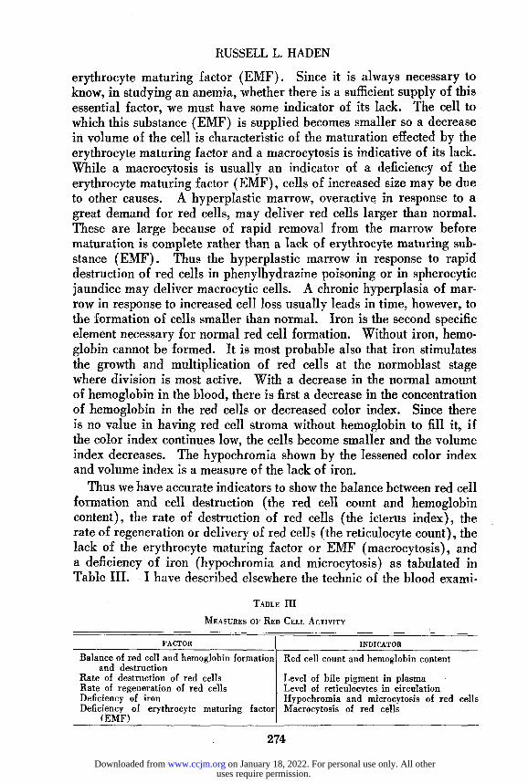
erythrocyte maturing factor (EMF). Since it is always necessary to know, in studying an anemia, whether there is a sufficient supply of this essential factor, we must have some indicator of its lack. The cell to which this substance (EMF) is supplied becomes smaller so a decrease in volume of the cell is characteristic of the maturation effected by the erythrocyte maturing factor and a macrocytosis is indicative of its lack. While a macrocytosis is usually an indicator of a deficiency of the erythrocyte maturing factor (EMF), cells of increased size may be due to other causes. A hyperplastic marrow, overactive in response to a great demand for red cells, may deliver red cells larger than normal. These are large because of rapid removal from the marrow before maturation is complete rather than a lack of erythrocyte maturing sub-stance (EMF). Thus the hyperplastic marrow in response to rapid destruction of red cells in phenylhydrazine poisoning or in spherocytic jaundice may deliver macrocytic cells. A chronic hyperplasia of mar-row in response to increased cell loss usually leads in time, however, to the formation of cells smaller than normal. Iron is the second specific element necessary for normal red cell formation. Without iron, hemo-globin cannot be formed. It is most probable also that iron stimulates the growth and multiplication of red cells at the normoblast stage where division is most active. With a decrease in the normal amount of hemoglobin in the blood, there is first a decrease in the concentration of hemoglobin in the red cells or decreased color index. Since there is no value in having red cell stroma without hemoglobin to fill it, if the color index continues low, the cells become smaller and the volume index decreases. The hypochromia shown by the lessened color index and volume index is a measure of the lack of iron.
Thus we have accurate indicators to show the balance between red cell formation and cell destruction (the red cell count and hemoglobin content), the rate of destruction of red cells (the icterus index), the rate of regeneration or delivery of red cells (the reticulocyte count), the lack of the erythrocyte maturing factor or EMF (macrocytosis), and a deficiency of iron (hypochromia and microcytosis) as tabulated in Table III. I have described elsewhere the technic of the blood exami-
T A B L E I I I
MEASURES OF RED CELL ACTIVITY
FACTOR INDICATOR
Balance of red cell and hemoglobin formation and destruction
Rate of destruction of red cells Rate of regeneration of red cells Deficiency of iron Deficiency of erythrocyte maturing factor
(EMF)
Red cell count and hemoglobin content
Level of bile pigment in plasma Level of reticulocytes in circulation Hypochromia and microcytosis of red cells Macrocytosis of red cells
274
uses require permission. on January 18, 2022. For personal use only. All otherwww.ccjm.orgDownloaded from

THE MECHANISM OF ANEMIA*
nation to supply such data.2 A careful laboratory study is first necessary in every anemia to furnish the data outlined above. From the laboratory examination the anemia is classified on the basis of the number, size, and hemoglobin content of the mean red cell.3 These studies are illustrated in Fig. 3. The relation of the blood findings to red cell formation and destruction is shown in Table IV.
T A B L E I V
RELATION OF BLOOD FINDINGS TO R E D CELL F O R M A T I O N AND DESTRUCTION
Active bone marrow
Inactive bone marrow
Increased red cell and he-moglobin destruction
Decreased hemoglobin destruc-tion
Deficiency in erythrocyte ma-turing factor (pernicious anemia)
Deficiency in iron (iron defi-ciency anemia; chronic hem-orrhagic anemia)
Hemolytic anemia
Anemia due to decrease in amount or activity of mar-row (aplastic or hypoplastic anemia)
Increased number of reticulocytes, basophilia, nucleation. Slight increase in mean erythrocyte volume if re-ticulocytosis is marked. Often an increase in leuco-cytes and platelets unless destruction is more active than normal. The number of cells is increased
Decrease or absence of reticulocytes, basophilia and nuclea-tion. If blood destruction is normal or increased, the cell count decreases
Increase in bilirubin content of plasma; decrease in num-ber of cells unless compensated for by increased marrow activity
Decrease in bilirubin content of plasma
Anemia with increase in mean erythrocyte volume (in-creased volume index)
Anemia with hypochromia of red cells (decreased color index) ; microcytosis (decreased volume index) ; if hypochromia continues
J Anemia with increased icterus index; reticulocytosis if i marrow responds to increased need
Anemia with cells of normal size and hemoglobin content; decrease in reticulocytes
Since an anemia represents a loss of balance between red blood cell formation and destruction, an anemia can result only from increased blood loss without a compensating increase in blood formation, by decreased formation with a normal or accelerated blood loss, or by a combination of increased blood loss and decreased formation. A clinical classification of anemia on the basis of method of production with the more important clinical causes is given in Table V. In every anemia it is necessary to make both a laboratory and clinical classifi-cation.
275
uses require permission. on January 18, 2022. For personal use only. All otherwww.ccjm.orgDownloaded from

RUSSELL L. HADEN
Reticulocytes $ (in percent]
•
Icterus Index
[in units')
• Volume Index
©
Color Index
150 140 130 120 110 idol 9C 8C 70
• 60 Red Cells in -j®
Millions 30 10 IjO
Packed Cells and Hemoglobin in per cent of Normal
110 100 90 80 70 60 50 40 30 20 10
Normal Less Than
Determined by Direct; Counting of Red Cells Stained with Brilliant. Cresyl Blue
Jf high, more young red cells than normal are getting into the circulation .
, If very low, less young red celte than normal are getting into circulation.
Normal [4 -6 ]
. Determined by Comparison of Color of •v Serum with a solut ion of K 2 C r 2 0 T
NA high index shows increased hemoglobin destruction. [If no biliary obstruction]
A—-A low index shows decreased hemoglobin destruction.
Normal Volume and Color Index for Men awl Women
Calculated from Red Cell Count and Hematocrit and Hemoglobin Determinations.
A hiij\ volume index indicates cells larger than normal.
A low index indicates » cells smaller than
normal.
® A high color index indicates cells containing
more hemoglobin than normal.
A low color index ® indicates cells containing
less hemoglobin than normal.
Normal for
Women Determined by Direct Counting
Nomiai^ for~ Determined by Hematocrit Reading in c.c. Women and Hemoglobin in grams per 100cc blood.
I l l 7Indicates incomplete filling of cells with hemoglobin.
Saturation Index shows the filling or saturation of the red cells in relation to normal. It must be 1.00 or less .
FIG. 3.—Study of blood and interpretation of findings in anemia.
T A B L E V
CLINICAL CLASSIFICATION OF A N E M I A
I. Increased blood loss 1. Mechanical loss from hemorrhage 2. Accelerated red cell destruction by:
a. Hemolytic agents (as phenylhydrazine or bacterial toxin) b. Rapid red cell removal from an abnormality of cell shape (as congenital
hemolytic icterus), overactivity of reticuloendothelial system, or defect in cell structure
II. Decreased blood formation 1. Quantitative decrease in red marrow from aplasia as in benzol poisoning, or
crowding out of erythrogenic tissue as in leucemia or myeloma 2. Quantitative depression of marrow activity as by malignancy, hypometabolism,
chronic toxemia such as nephritis or cachexia 3. Qualitative decrease in marrow activity from deficiency of specific substances
necessary for normal marrow activity a. Deficiency in supply, absorption, or use of erythrocyte maturing factor (EMF)
as in pernicious anemia or sprue b. Deficiency in supply, absorption, or use of iron as in chronic hemorrhage,
dietary lack, and idiopathic hypochromic anemia
276
uses require permission. on January 18, 2022. For personal use only. All otherwww.ccjm.orgDownloaded from

THE MECHANISM OF ANEMIA*
With a careful study of the blood and determination of each of the indicators of red cell activity, a clinical study of the patient and a clinical classification of the anemia, the different types of anemia can be visualized by means of diagrams. In each diagram, the blood is depicted in relation to the three phases of the red cell, viz., (1) forma-tion, (2) circulation, and (3) destruction. The fundamental fault in the production of the anemia is apparent in such a diagram, so the point of attack in treatment is evident.
In Fig. 4, the normal cell is shown in relation to formation, circula-tion, and destruction. The bone marrow is thought of as a gristmill with three hoppers supplying materials for making red cells. One hopper supplies the nonspecific elements and the other two the specific elements. Normally, the hoppers are full. The level in the mill indi-cates the relative fullness of the bone marrow. To maintain the normal balance between formation and destruction, nearly one trillion cells and 25 gm. hemoglobin must be formed daily. In the circle showing the normal circulation are 100 red cells with one reticulocyte. The cells are of normal size and hemoglobin content. The normal findings are shown below the circle. Old red cells are taken out by the reticulo-endothelial cells, largely those of the spleen. If the blood count remains constant as it normally does, the same number must be taken out as are delivered to the blood stream by the marrow. As the hemoglobin is destroyed, iron is split off. Some of the iron is excreted but the larger part (85 per cent) is returned to the marrow to be used again. The end product of hemoglobin destruction is bilirubin which is excreted by the liver. The normal amount of bile pigment and iron formed is indicated by the level of these substances in the containers in which they are received. We think of the mill as functioning at a constant rate of speed so as to supply the same number of cells with the same hemoglobin con-tent as are destroyed each day. The normal mean elapsed time between the beginning of formation of the cell and the ultimate disposal of it is thirty days.
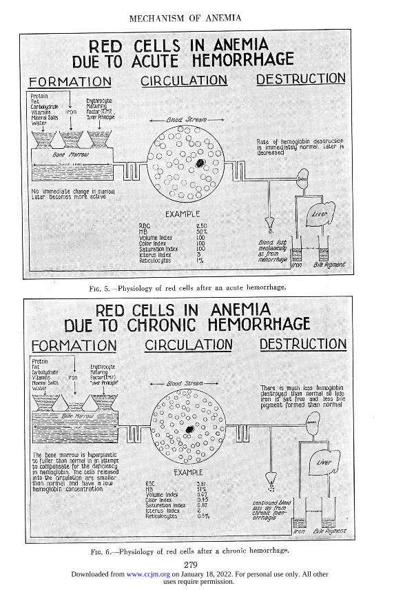
Every anemia can be illustrated by such a diagram. In Fig. 5 is shown the red cell mechanism immediately after a large hemorrhage from the uterus. The cells remaining in the circulation are normal and the process of formation and destruction is unaltered. This state per-sists for only a short time, however, after the hemorrhage, when there is increased activity of the marrow to compensate for the blood lost. The icterus index falls (2 units) and the amount of bilirubin and iron set
277
uses require permission. on January 18, 2022. For personal use only. All otherwww.ccjm.orgDownloaded from

RUSSELL L. HADEN
free decreases, and the picture is now one of an iron deficiency anemia, shown in Fig. 6, where there is a defect in the supply of iron to the
NORMAL RED BLOOD CELL FORMATION
Protein Fat Carbohydrate Vitamins Mineral Salts Water *
Erythrocyte Maturing Factor CE.HÍ-'Liver Principie)
4
NORMAL ADULT
Red cells formed largely in ribs, , skull and vertebrae Total normal volume of marrow is 1400 c c Normal color is greyish red For red cell formation, non-specific substances necessary for all cells are required as well as too specif-ic substances, iron and the erythro-cyte maturing factor CE.M.F) One trillion cells and 25 gms. HB. formed each day Rate of formation measured oq re-ticulocyte percentage in circulation Normal life of red cell about todays
CIRCULATION Twenty-five trillion red cells in blood stream. Each cell during its life, mate 50,000 to 100,000 complete circuits from lungs to tissues. Functions as conveyor for hemoglobin which carries oxygen from lungs to tissues Blood count represents balance be-tween red cell and hemoglobin for-mation and destruction
-ß/ood
E X A M P L E
R.B.C 5.0 Million ne. 100% 05.4-gms) Volume Index loo-OocuDic microns) Color Index loo-ßiamkromkrograr^ Saturation Index I.OO-C317Ä) Icterus Index 4-6 units Reticulocytes a s - t o i
DESTRUCTION Old red cells taken out by reticulo-endothelial cells, largely those of spleen I trillion red cells destroyed each day 25 gms. MB destroyed each day 100 mg. of iron released 85 mg. used again 15 mg. lost and replaced by food 500 c.c. bile formed and excreted by the liver containing 300 to 400 mg. of bile pigment Rate of destruction gauged by iron released and bile pigment formed Only clinical laboratory measure is icterus index or quantitative Van den Bergh
Eighty-five percent of iron released returns to marrow to be used again ?
Iron Bile Pigment
Fic. 4.—Norma] red cell physiology.
marrow with the result that the cells are small (volume index 0 .067) and have a decreased hemoglobin content (color index 0 . 4 ) . The therapeutic indication is to stop the blood loss and supply an adequate amount of iron.
278
uses require permission. on January 18, 2022. For personal use only. All otherwww.ccjm.orgDownloaded from
MECHANISM OF ANEMIA
RED CELLS IN ANEMIA DUE TO ACUTE HEMORRHAGE
F O R M A T I O N CIRCULATION DESTRUCTION
Rate of hemoglobin destruction is immediately normal. Later is decreased
Bi/eP/gmen(
Fic. 5.—Physiology of red cells after an acute hemorrhage.
RED CELLS IN ANEMIA DUE TO CHRONIC HEMORRHAGE
FORMATION CIRCULATION DESTRUCTION Protein Fat Carbohydrate Vitamins Mineral Salts Water
iron
Erutti rocyte naturine) Factor (EUR "Liver Principle'
The bone marrow is hyperplastic so fuller than normal in an attempt to compensate for the deficiency in hemoglobin. The cells released into tiie circulation are smaller than normal and have a low hemoglobin concentration
- Blood Stream There is much less hemoglobin destroyed than normal so less iron is set free and less bile pigment formed than normal
RBC tie Volume Index Color Index Serturation Index Icterus Index Reticulocytes
Iron Bi/e P/g/K/}t
FIC. 6.—Physiology of red cells after a chronic hemorrhage.
279
uses require permission. on January 18, 2022. For personal use only. All otherwww.ccjm.orgDownloaded from

RUSSELL L. HADEN
In Fig. 7 is illustrated the red cell mechanism in an anemia due to excessive hemolysis resulting from the improper use of phenylhydrazine. This patient was given this drug on the basis of a wrong diagnosis of polycythemia vera. The polycythemia was a symptomatic one since the blood volume was normal. When first seen, the patient had a well-marked anemia (hemoglobin 58 per cent) with a high icterus index (25 units) and a high reticulocyte count (10.3 per cent). The bone marrow here is overactive and increased in volume as indicated by the reticulocytosis to compensate for the excessive red cell destruction. The supply of building materials is normal. The cells damaged by the
Red celis damaged in cirsulaticrt are rapidly removed Du the spleen so jnore iron is set free and bile pigment forrr«a ;,-»=.-normal.Spleen «ienjed
Bone harrow
Liver
RED CELLS IN ANEMIA DUE TO HEMOLYTIC AGENT
AS PHENYLHYDRAZINE FORMATION CIRCULATION DESTRUCTION Protein fat Carbohydrate vitamins dineral Sans water
i
Erythrocyte + Maturino
Iron Factor (EHF) I liver Principle
The marrow is overactive to compensate for increased red cell destruction and quickly be-comes fuller than normal
-Blood Stremr:
RBC H B volume Index Color Index Saturation Index Icterus index Reticulocytes
E X A M P L E
10.3-7, P/gmenz
FIG. 7.—Physiology of red cells with excessive destruction by a toxic substance.
phenylhydrazine are rapidly removed from the circulation so the spleen is overactive and larger than normal. The output of iron and bile pigment is necessarily greater than normal. In this patient the primary difficulty is the damage to the red cells in the circulation, so the thera-peutic indication is to stop the cell damage. There is no need for iron, liver preparations, or marrow stimulation unless the process has pro-ceeded to the point of exhaustion. In this instance, after the action of the
280
uses require permission. on January 18, 2022. For personal use only. All otherwww.ccjm.orgDownloaded from

THE MECHANISM OF ANEMIA*
drug was past, the spleen, icterus index, the reticulocyte count, the red cells, and hemoglobin all returned to normal.
In Fig. 8 is shown the red cell mechanism in congenital hemolytic icterus and spherocytic jaundice. This patient had a well-marked anemia with a high icterus index (15 units) indicating excessive blood destruction and a high recticulocyte count (10.8 per cent) showing good marrow response. The supply of building materials is ample so the hoppers are full and the active marrow is fuller than normal. The fundamental difficulty in this disease is an anatomic defect in the shape of the red cells which are spherocytic rather than normal biconcave
RED CELLS IN ANEMIA DUE TO SPHEROCYTIC SHAPE OF CELLS
CHRONIC HEMOLYTIC ICTERUS FORMATION CIRCULATION DESTRUCTION
Red cells rapidly removed from circulation by spleen and destroy-ed so mope iron set free and bile pigment formed than normal Spleen large
FIG. 8.—Physiology of red cells with excessive filtration by spleen (spherocytic anemia).
disks. As a result of this abnormal shape the cells are more fragile than normal4 and are rapidly removed from the circulation by the spleen which is enlarged as a result of the increased activity. More iron and bilirubin than normal are poured out. Here the average length of l ife of the red cell is a few days instead of the usual thirty days. There is a rapid stream of cells from the site of origin, the bone marrow, to the place of destruction, the spleen. We cannot correct the anatomic defect
281
uses require permission. on January 18, 2022. For personal use only. All otherwww.ccjm.orgDownloaded from

RUSSELL L. HADEN
so the patient is treated by removing the filter. The abnormally shaped cells function normally if allowed to remain in the circulation. The anemia, reticulocytosis and jaundice all disappear after splenectomy, showing that the increased activity of the spleen is the cause of the anemia although the fundamental defect is in the bone marrow.
Sickle-cell anemia, shown in Fig. 9, has much in common with spherocytic anemia (congenital hemolytic icterus). Here also, there is a fundamental defect in the marrow with the delivery of cells of abnormal shape and probably with a greater tendency to
BJte Pigment
RED CELLS IN ANEMIA DUE TO SICKLE SHAPE OF CELLS
FORMATION CIRCULATION DESTRUCTION Erythrocyte Maturing ,
Iron factor (JMF) liver Principle"
I < Blood. Stream Red cells rapidly removed from circulation by Spleen and de-stroyed so more iron set free and bile pigment formed than
Marrow very full and active in attempt to compensate for in-creased red cell destruction.. Many young cells (reticulocytes) in circulatioTi EXAMPLE
RBC 4.47 tIB 587= Volume Index 0.77 Color Index 0.65 Saturation Index 0.84 Icterus index i5 Reticulocytes 7 "J.
FIG. 9.—Physiology of red cells with excessive filtration by spleen (sickle-cell anemia).
fragment. The supply of building material is adequate, the marrow is overactive as shown by the reticulocytosis, and red cell destruction is excessive as shown by the increased icterus index. The spleen is enlarged at least early in the disease due to overactivity in removing excessive numbers of abnormal cells from the circulation. Splenectomy helps the anemia5'6 but here the result differs from that seen in spherocytic anemia in that the patient continues to have some anemia after removal of the spleen. The increased cell destruction and forma-tion also continue so the excessive activity of the spleen cannot be the
282
uses require permission. on January 18, 2022. For personal use only. All otherwww.ccjm.orgDownloaded from

THE MECHANISM OF ANEMIA*
sole cause of the anemia. It is most probable that the red cells fragment more easily than normal and this fragmentation continues after splenectomy. There is no treatment for this phase of the disorder. Splenectomy removes only one factor in the anemia.
Fig. 10 illustrates the anemia due to marrow aplasia caused by the prolonged use of arsphenamine. The amount of functioning marrow tissue is decreased. In this instance the blood examination shows a marked anemia with cells of normal size (volume index, 0.97) and
RED CELLS IN ANEMIA DUE TO APLASIA OF MARROW BY MYELOTOXIC AGENT
AS ARSPHENAMINE FORMATION CIRCULATION DESTRUCTION Protein Fat Carbohydrate vitamins Mineral Salts Water
1 Erythrocyte Maturing
Iron Factored F) liver Principi
The marroui is partly destroyed by the toxic agent so is smaller and less full than normal. The activity of the remaining marrow cells is impaired also
Volume Index Color Index Saturation index Icterus Index Reticulocytes
Usually less red cells end herno-qlobin are destroyed so less iron B set free and less bile pigment is formed than normal
Iron Bile ftymait
FIG. 10.—Physiology of red cells when formation of cells is decreased by aplasia of marrow.
hemoglobin content (color index, 1.02). The reticulocyte count is very low (0.2 per cent). The marrow is at a low level as indicated by the low red cell, white cell, reticulocyte, and platelet counts. The mill is greatly decreased in size although the supply of raw material is ample. There is less destruction of cells so the iron and bile pigment output are much below normal. It is apparent that this anemia can be treated only by measures designed to improve the size and function of the marrow. In this instance the marrow was permanently damaged and the patient finally died of the anemia.
283
uses require permission. on January 18, 2022. For personal use only. All otherwww.ccjm.orgDownloaded from

RUSSELL L. HADEN
Another type of anemia due to marrow deficiency is illustrated in Fig. 11. This is a myeloid leucemia with a marked anemia (hemoglobin 42 per cent). Here the marrow is full, but the increase in size is due to the hyperplasia of myeloid tissue at the expense of erythrogenic tissue, so there is a great decrease in red-cell-forming tissue and a consequent anemia. The spleen is also enlarged from infiltration of myeloid tissue and not from overactivity due to excessive cell destruction. There is less red cell destruction and so less iron is set free and less bile pigment formed. Here again, the indication for treatment of the anemia is to decrease the mass of myeloid tissue in the marrow by radiation or
RED CELLS IN ANEMIA DUE TO CROWDING OUT OF ERYTHROGENIC TISSUE IN MARROW
AS IN LEUKEMIA FORMATION CIRCULATION DESTRUCTION Protein fat Carbohydrate Vitamins Mineral Salts Water
i
1 Iron
Erythrocyte Maturing FactorffMF) "Liver Principle'
The marroui is full but this is due to hyperplasia of the myeloid tissue which crouds out the eruthrogenio tissue. There is probably also some mechanical blockage interfering with the release of red cells into the circulation
-B/ood Stream-
RBC ÜB Volume Index Color index Saturation index Icterus index Reticulocytes Leucocyte Count
Less red cells and hemoglobin are destroyed so less iron is set free and less bile pigment is formed
FIG. 11.—Physiology of red cells where the formation of red cells is diminished by crowding out of erythrogenic tissue.
medication to make room for the erythrogenic tissue. The red cell count often reveals more in leucemia than the number of white cells as it gauges the state of hyperplasia of the marrow which is more important than the white cell count.
Instead of a quantitative decrease in erythrogenic tissue, the total amount may be unchanged but the function be quantitatively depressed.
284
uses require permission. on January 18, 2022. For personal use only. All otherwww.ccjm.orgDownloaded from

MECHANISM OF ANEMIA
Protein Fat Carbohydrate vitamins Mineral Salts Water
Iron Factor(tMF) I 'liver Principle
Bone Mârrou/
RED CELLS IN ANEMIA DUE TO QUANTITATIVE DEPRESSION OF MARROW ACTIVITY AS BY TOXEMIA FROM CHRONIC NEPHRITIS
FORMATION CIRCULATION DESTRUCTION
tess cells and hen)ogloJ)in are destroyed 50 less iron is set free and less bile pigment formed than normal
Here the marrouj is normal in a-mount but does not function at the normal rate so fewer cells than normal are released into the circulation
ß/ood Sir earn
EXAMPLE RUB MB Volume Index Color Index Saturation Index Icterus Index Reticulocytes
FIG. 12.—Physiology of red cells when the function of marrow is slowed up.
RED CELLS IN ANEMIA DUE TO DEFICIENCY OF ERYTHROCYTE MATURING FACTOR-EMF-
O R PERNICIOUS A N E M I A
FORMATION CIRCULATION DESTRUCTION
«—ô/ood Stream—>• The red cells cannot be complet-ed in the marrow due to loss of erythrocyte maturing factor (EMF) The red cells die in excessive numbers in the marrow 30 more iron is set free and more bile pigment formed than normal
Erythrocyte Maturino Factor fEW) •Liver Principle"
The marrow is hyperplastic in an attempt to compensate for diffi-culty in maturing cells. Relatively few cells are released into circula-tion since they cannot be complet-ed due to lacK of erythrocyte mat-uring factor (EnFJ. Such cell; as are released are abnormal in shape and size
EXAMPLE RBC 0.87 (IB zii° Volume Index 1.29 Color Index 1.83 Saturation Index 0.95 Icterus index 10 Reticulocytes 0.51,
FIG. 13.—Physiology of red cells when building materials are deficient (pernicious anemia). 285
uses require permission. on January 18, 2022. For personal use only. All otherwww.ccjm.orgDownloaded from

RUSSELL L. HADEN
This type of mechanism is shown in Fig. 12. It is responsible for many cases of anemia such as malignancy, infections, toxemia, and hypo-metabolism. We can best visualize this mechanism by thinking of it as normal except for the speed with which the apparatus works. It is greatly slowed up, although the supply of building material is normal. Such cells as are turned out are normal and less cells are disposed of. The total number circulating is decreased. The time interval between the beginning of cell formation and the end of cell destruction is in-creased to varying degrees just as it is decreased in spherocytic anemia or sickle-cell anemia.
Protein Fat Carbohydrate vitamins Mineral Salts Water •ß/ood âtream
^oO^To^
~ fJcrrrwl tevöri-~--.~
¿/wr
RED CELLS IN ANEMIA DUE TO DEFICIENT INTAKE OF IRON
FORMATION
iron
Erythrocyte Maturino factor[Eflf liver Principle)
4
Bone marrow more active and fuller than normal. Usually more red cells and always less hemo-globin formed than normal due to iron deficiency
CIRCULATION
RBC I I B Volume Index Color index Saturation index icterus index Reticulocytes
4.0 50% 0.75 0.63 0.83 2 27.
D E S T R U C T I O N
Less hemoglobin destroyed so less iron s e t free ana less bile pigment jormed
EXAMPLE
FIG. 14.—Physiology of red cells when building materials are deficient (iron deficiency anemia).
The anemias due to a defect in supply of building material are most important especially since the lack can readily be supplied. It is in this group that the greatest advances in treatment have been made in recent years. The mechanism of development is now well understood. In Fig. 13 is illustrated the red cell mechanism in an anemia due to a defect in supply to the marrow of the erythrocyte maturing factor (EMF) fur-nished by the liver and liver substitutes. The anemia I have shown is a typical pernicious anemia. The mechanism is the same in sprue and
286
uses require permission. on January 18, 2022. For personal use only. All otherwww.ccjm.orgDownloaded from

MECHANISM OF ANEMIA
RED CELLS IN ANEMIA DUE TO QUALITATIVE DEPRESSION OF MARROW FUNCTION
AS IN MYXEDEMA
FORMATION CIRCULATION DESTRUCTION
Cells which cannot be matured in the marrow may die in situ in excessive numbers, so the iron set free and bile pigment form-ed may be greater than normal
In certain cases the bone marrou) cannot use the erythrocyte mat-uring factor (EMFJ although pres-ent in normal amount. Here the marrou) becomes hyperplastic to compensate for the deficiency in cell maturation. Many of the ceils released are larger than normal or abnormal in shape
E X A M P L E
R 8 C 3.14 MB 6i7* Volume Index 1.06 Color index 0.37 Saturation index 031 Kterus Index 4 Reticulocyte 11,
8//e%me/rt
Erythrocyte Maturing Factor (EMF)
FIG. 15.—Physiology of red cells when the marrow is unable to utilize the erythrocyte maturing factor ( E M F ) normally.
RED CELLS IN ANEMIA DUE TO QUALITATIVE DEPRESSION OF MARROW ACTIVITY
AS IN LEAD POISONING FORMATION CIRCULATION DESTRUCTION
In certain cases the utilization of iron in the marrou), although present in normal amounts, is interfered uiith. Here there is hyperplasia of the marrou) to compensate for the insufficiency of hemoglobin. The cells released become small and have a low hemoglobin content
E X A M P L E
RSC 3.30 "B 46% Volume Index 0.83 Color index 0.70 Saturation Index o.6< Icterus Index 5 Reticulocyte 37.
Less hemoglobin is destroyed so less iron is set free and less bile pigment is formed
Protein Fat Carbonydrate Vitamins iron Mineral Salts
Erythrocyte Maturing Factor ft MF) 'Liver Rinaple' -Blood Stream *
FIG. 16.—Physiology of red cells when the marrow is unable to utilize iron normally. 287
uses require permission. on January 18, 2022. For personal use only. All otherwww.ccjm.orgDownloaded from

RUSSELL L. HADEN
similar disorders in which the macrocytic anemia occurs. This whole group should be designated the erythrocyte-maturing-factor (EMF) deficiency anemias. Pernicious anemia is the most important of the group. The patient cited during the active phase of the disease had the macrocytosis of red cells (volume index, 1.29) characteristic of such a deficiency. The marrow in active pernicious anemia is shown by marrow puncture and necropsy studies to be hyperplastic, but very few cells are delivered to the blood stream so the reticulocyte percentage is low (0.5 per cent). The bile pigment of the plasma is high (icterus index, 10 units), indicating an excessive destruction of red cells which in pernicious anemia takes place in the marrow and not in the circulating blood or reticuloendothelial system. The output of iron is high. Here the therapy is evident. It consists in supplying adequately the deficient erythrocyte maturing factor by giving liver, gastric tissue, or liver con-centrates.
The mechanism in an anemia due to a deficient intake or impaired assimilation of iron is shown in Fig. 14. The mechanism is similar to that already shown in a chronic hemorrhagic anemia (Fig. 5) except for the mechanical loss of iron in the hemorrhage. In such an instance there is also an iron deficiency anemia due to the loss of iron more rapidly than it is normally supplied by food. If sufficient iron is given, the hemorrhage may continue, but the anemia is relieved so long as the marrow is able to stand the added strain. The bone marrow has to cope with the same deficiency if there is a defect in assimilation so the iron taken in does not reach the marrow. This is the condition in idiopathic hypochromic anemia. In the example cited, insufficient iron has been taken in. The marrow in this instance is hyperplastic in an attempt to compensate, but such red cells as do get out are small (volume index, 0.75) and deficient in hemoglobin (color index, 0.67). The low volume and color index indicate the iron deficiency. Here there is much less destruction of hemoglobin so very little iron is set free and the bile pigment content of the plasma is less than normal (icterus index, 2 units). Again the therapy of the anemia is clearly indicated from the diagram. It consists in filling the iron hopper by providing an adequate supply of iron.
If an organ does not receive an adequate supply of a necessary factor, a deficiency necessarily develops. Under certain conditions an organ may receive a necessary factor and for some reason not use it so a deficiency state results just as if the factor were not supplied. . The condi-tions influencing the use of nutritional factors in general have been discussed elsewhere.7 We may find the clinical and laboratory picture of a deficiency anemia even though an adequate amount of iron and erythrocyte maturing factor be supplied to the marrow.
288
uses require permission. on January 18, 2022. For personal use only. All otherwww.ccjm.orgDownloaded from

THE MECHANISM OF ANEMIA*
It is well known that myxedema may show the typical blood picture of pernicious anemia. Such a state is illustrated in Fig. 15. This patient had an anemia with a mild macrocytosis in myxedema. The defect is in the normal completion of the red cells just as it is in idiopathic pernicious anemia. In such a case, the addition of thyroid extract alone should relieve the anemia since the marrow can then use the erythrocyte maturing factor (EMF) already supplied in adequate amounts.
It is quite common to encounter an iron deficiency anemia which will not respond to adequate dosage of iron. There seem to be many more extraneous factors influencing the use of iron by the marrow than of the erythrocyte maturing factor. The example cited in Fig. 16 is the case of a patient with lead poisoning. The giving of iron does not influence the anemia, although the laboratory findings of low volume and color index are characteristic of an iron deficiency anemia. The lead seems to prevent the normal utilization of the iron by the marrow, so treatment must first consist of removing the influencing factor before iron is given. The findings here are exactly like those shown in chronic hemorrhagic anemia (Fig. 6) , and an anemia due to a deficient intake of iron (Fig. 14). The laboratory findings indicate the fundamental defect so far as the marrow is concerned but do not show whether the marrow defect is due to excessive loss, deficient intake, or impaired utilization of iron.
S U M M A R Y
In every case of anemia, the rate of red cell formation and delivery from the marrow, the rate of destruction, and the balance between these two factors must be determined.
Measures are available for gauging accurately the state of the mar-row and all important factors in red cell activity.
The anemia must be studied and classified from both the clinical and laboratory standpoints.
Red cell formation, circulation, and destruction in all the common anemias are illustrated by diagrams.
REFERENCES
1. Barcroft, J.: The Raison d'être of the Red Corpuscle, Harvey Lectures, Series 17, pp. 146-163, 1921-1922.
2. Haden, R. L.: Technic of Blood Examination, J. Lab. & Clin. Med. 17: 843, 1932. 3. Haden, R. L.: Clinical Significance of Volume and Hemoglobin Content of Red Blood
Cell, Arch. Int. Med. 49: 1032, 1932. 4. Haden, R. L.: Mechanism of the Increased Fragility of Erythrocytes in Congenital
Hemolytic Jaundice, Am. J. M. Sc. 188: 441, 1934. 5. Landon, J. F., and Patterson, H. A.: Evaluation of Splenectomy in Treatment of
Sickle-Cell Anemia, J. Pediat. 7: 472, 1935. 6. Haden, R. L., and Evans, F. D.: Sickle-Cell Anemia in the White Race. Report of
Two Cases Benefitted by Splenectomy. (Publication pending.) 7. Haden, R. L.: Multiple Specific Nutritional Deficiency Disease in Adult, J. A. M. A.
106 : 261, 1936.
289
uses require permission. on January 18, 2022. For personal use only. All otherwww.ccjm.orgDownloaded from