the membrane attack complex, perforin and cholesterol ... · commentary the membrane attack...
TRANSCRIPT

COMMENTARY
The membrane attack complex, perforin and cholesterol-dependent cytolysin superfamily of pore-forming proteinsNatalya Lukoyanova1, Bart W. Hoogenboom2,3 and Helen R. Saibil1,*
ABSTRACTThe membrane attack complex and perforin proteins (MACPFs) andbacterial cholesterol-dependent cytolysins (CDCs) are two branchesof a large and diverse superfamily of pore-forming proteins thatfunction in immunity and pathogenesis. During pore formation,soluble monomers assemble into large transmembrane poresthrough conformational transitions that involve extrusion andrefolding of two α-helical regions into transmembrane β-hairpins.These transitions entail a dramatic refolding of the protein structure,and the resulting assemblies create large holes in cellularmembranes, but they do not use any external source of energy.Structures of the membrane-bound assemblies are required tomechanistically understand and modulate these processes. In thisCommentary, we discuss recent advances in the understanding ofassembly mechanisms and molecular details of the conformationalchanges that occur during MACPF and CDC pore formation.
KEY WORDS: CDC, MACPF, Pore-forming proteins
IntroductionPore-forming proteins (PFPs) are initially soluble proteins that canbind to membranes, oligomerise and convert to membrane-insertedpores of 1–50 nm in diameter that are formed of six to 50 or moresubunits (Peraro and van der Goot, 2016). PFPs are produced by avariety of organisms and are often involved in attack or defencemechanisms. Depending on the secondary structure elements thatform the transmembrane pores, PFPs are broadly classified asα-PFPs, which utilise amphipathic α-helices for pore formation(e.g. Cry toxins produced by Bacillus thuringiensis (Ounjai et al.,2007; Pardo-López et al., 2013), diphtheria toxin (Leka et al., 2014;Ghatak et al., 2015) and colicin produced by Escherichia coli(Cascales et al., 2007; Housden and Kleanthous, 2012); or asβ-PFPs with pores built of amphipathic β-hairpins organised into atransmembrane β-barrel (for example, anthrax protective antigen(Collier, 2009; Jiang et al., 2015), aerolysin toxin produced byAeromonas hydrophila (Degiacomi et al., 2013) and α-hemolysin ofStaphylococcus aureus (Menestrina et al., 2003; Sugawara et al.,2015). Many PFPs are bacterial toxins and are able to damagehost membranes to gain access to cells or cell contents, or to killcells. For example, perfringolysin O, a β-PFP produced byClostridium perfringens, is cytotoxic at high concentrationsagainst host leucocytes and macrophages, thereby eliminatinginflammatory cells of the host immune system at the site of
clostridial gangrene lesions (Popoff, 2014). Other PFPs cantranslocate across membranes. For example, colicins are α-PFPsproduced by some Escherichia coli strains to reduce competitionfrom other strains. Colicins translocate across the outer membraneof target cells and then deliver a lethal hit by forming small pores inthe inner membrane, or by exerting their endonuclease activity onDNA or RNA (Cascales et al., 2007). Other PFPs, such as anthraxtoxin produced by Bacillus anthracis (Collier, 2009) or perforin,a protein of the immune system (Voskoboinik et al., 2015),enable passage of effector proteins. Anthrax toxin delivers edemafactor and lethal factor, which lead to killing of macrophagesand facilitate tissue invasion by bacteria. The transfer of granzymeproteases through perforin pores initiates apoptosis of target cellsand is a key step in the immune surveillance of cytotoxiclymphocytes against virus-infected or transformed cells(Voskoboinik et al., 2015).
In this Commentary, we focus on the structure and mechanism ofone of the largest families of β-PFPs, the membrane attack complex,perforin and cholesterol-dependent cytolysin (MACPF–CDC)superfamily. This superfamily of PFPs includes the key immunemediators perforin and the terminal components of the complementsystem, as well as a family of bacterial toxins that is important invirulence. We will review here recent structural and biophysicalstudies that have afforded insights into assembly intermediates andmembrane-inserted pore forms.
The MACPF–CDC superfamilyThe MACPF family of proteins was initially defined by thestructural and functional similarities between the C7, C8 and C9proteins of the membrane attack complex (MAC) in the complementsystem and perforin, the above-mentioned pore-forming protein thatis secreted by activated T-lymphocytes (Tschopp et al., 1986). Thecomplement system was discovered over a century ago by theimmunologist Jules Bordet, who described it as the ability of bloodplasma factors to lyse bacteria. It is now understood that thecomplement system involves a cascade of activation reactions thatultimately triggers pore formation by the C9 complementcomponent of the MAC (Bubeck, 2014; Merle et al., 2015). Inthe 1980s, a related pore-forming protein, perforin, wascharacterised in cytotoxic T-lymphocytes and natural killer cellsin the blood (Podack and Dennert, 1983; McCormack et al., 2013).As noted above, perforin plays a key role in immune surveillancebecause perforin pores enable delivery of lethal granzyme proteasesinto virus-infected or cancerous cells (Voskoboinik et al., 2015).The MACPF family is extremely diverse − its members function inimmunity and pathogenesis across all kingdoms of life (Kondoset al., 2010). For example, MACPF proteins facilitate the invasionand/or proliferation of intracellular pathogens, such as Toxoplasmagondii, Plasmodium falciparum and Chlamydia, all of which musttraverse cellular membrane barriers during their life cycles(Blackman and Carruthers, 2013; Taylor and Nelson, 2014, Wade
1Department of Crystallography/Biological Sciences, Institute of Structural andMolecular Biology, Birkbeck College, London WC1E 7HX, UK. 2London Centre forNanotechnology, University College London, London WC1H 0AH, UK.3Department of Physics and Astronomy, University College London,London WC1E 6BT, UK.
*Author for correspondence ([email protected])
H.R.S., 0000-0002-2266-8891
2125
© 2016. Published by The Company of Biologists Ltd | Journal of Cell Science (2016) 129, 2125-2133 doi:10.1242/jcs.182741
Journal
ofCe
llScience
and Tweten, 2015). In addition, several MACPF proteins have rolesin embryonic development (Estévez-Calvar et al., 2011; Johnsonet al., 2015) and in neural migration (Kawano et al., 2004; Giousohet al., 2015).The CDC family includes some important virulence factors,
most of which are produced by Gram-positive bacteria, such asClostridium, Streptococcus and Listeria (Tweten, 2005; Heucket al., 2010). CDC proteins form large transmembrane pores thatcomprise 35–50 monomers and range from 25 to 30 nm indiameter. These pores disrupt plasma membranes and thus causecell death by necrosis. CDCs are secreted, with the exception ofpneumolysin, a major virulence factor of Streptococcuspneumoniae. Pneumolysin lacks an N-terminal secretion signal(Walker et al., 1987) and is released after bacterial autolysis. It isalso released in high concentrations from bacterial cells afterantibiotic therapy, which results in host cell necrosis and can, insome circumstances, lead to permanent tissue and organ damage(Hirst et al., 2004).Some pathogens (Listeria monocytogenes and Listeria
ivanovii) release their CDC (listeriolysin) inside host cells.Following their internalisation, Listeria initially reside in anendosome. Here, listeriolysin pore formation is essential for thebacteria to escape from this phagocytic vesicle into the cytoplasm,where they can divide and then spread from cell to cell; thispathway avoids exposure of the bacteria to the extracellularenvironment and thus detection by immune surveillance systems(Seveau, 2014).Proteins of the MACPF and CDC families have no detectable
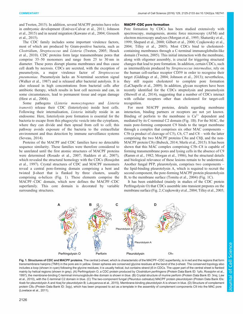
sequence similarity. These families were therefore considered tobe unrelated until the first atomic structures of MACPF proteinswere determined (Rosado et al., 2007; Hadders et al., 2007),which revealed the structural homology with the CDCs (Rossjohnet al., 1997). Crystal structures of CDC and MACPF monomersreveal a central pore-forming domain comprising a bent andtwisted β-sheet that is flanked by three clusters, usuallycomprising α-helices (Fig. 1). These elements comprise theMACPF–CDC domain, which now defines the MACPF–CDCsuperfamily. This core domain is decorated by variablesurrounding structures.
MACPF–CDC pore formationPore formation by CDCs has been studied extensively withspectroscopy, mutagenesis, atomic force microscopy (AFM) andelectron microscopy analyses (Morgan et al., 1995; Shatursky et al.,1999; Shepard et al., 2000; Gilbert et al., 2000; Czajkowsky et al.,2004; Tilley et al., 2005). Most CDCs bind to cholesterol-containing membranes through a C-terminal immunoglobulin-likedomain (Tweten, 2005). This initial interaction with the membrane,along with oligomer assembly, is crucial for triggering structuralchanges that lead to pore formation. In addition, certain CDCs, suchas intermedilysin produced by Streptococcus intermedius, rely onthe human cell-surface receptor CD59 in order to recognise theirtarget (Giddings et al., 2004; Johnson et al., 2013); nevertheless,they still require cholesterol to complete pore formation(LaChapelle et al., 2009). In addition, glycan receptors have beenrecently identified for the CDCs streptolysin and pneumolysin(Shewell et al., 2014), suggesting that a number of CDCs interactwith cellular receptors other than cholesterol for target-cellrecognition.
For most MACPF proteins, details regarding membraneinteraction, binding partners or receptors are not yet known.Binding of perforin to the membrane is Ca2+ dependent andmediated by its C-terminal C2 domain (Fig. 1B). For the MAC, themain pore-forming component C9 binds to the target membranethrough a complex that comprises six other MAC components –C5b (a product of cleavage of C5), C6, C7 and C8 – with the lattercomprising the two MACPF proteins C8α and C8β, and the non-MACPF protein C8γ (Bubeck, 2014; Merle et al., 2015). It has beenshown that this MAC complex comprising C5b–C8 is capable offorming transmembrane pores and lysing cells in the absence of C9(Ramm et al., 1982; Morgan et al., 1986), but the structural detailsand biological relevance of these lesions remain to be understood.Another fungal PFP, pleurotolysin, comprises two components –the lipid-binding pleurotolysin A, which is required to recruit thesecond component, the pore-forming MACPF protein pleurotolysinB, to the membrane surface (Tomita et al., 2004) (Fig. 1C).
It has been established (mainly in studies of the CDC proteinPerfringolysin O) that CDCs assemble into transient prepores on themembrane surface (Fig. 2;Czajkowskyet al., 2004; Tilley et al., 2005;
Perfringolysin O Perforin Pleurotolysin C8α
Pleurotolysin A
Pleurotolysin B
B DA C
Fig. 1. Structures of CDC andMACPF proteins. The central β-sheet, which is characteristic of the MACPF–CDC superfamily, is in red and the regions that formtransmembrane hairpins (TMH) in the pore are in yellow. Green spheres are conserved glycine residues at the bend of the β-sheet. The conserved topology alsoincludes a loop (shown in cyan) following the glycine residues; it is usually helical, but contains strand β5 in CDCs. The upper part of the central sheet is flankedmainly by helical regions (shown in grey). (A) Perfringolysin O, a CDC protein produced by Clostridium perfringens (Protein Data Bank ID: 1pfo, Rossjohn et al.,1997); the membrane-binding C-terminal immunoglobulin-like domain is shown in blue. (B) Crystal structure of murine perforin (Protein Data Bank ID: 3nsj, Lawet al., 2010), with the C-terminal C2 domain in blue. (C) The two-component fungal (Pleurotus ostreatus) MACPF protein pleurotolysin (Protein Data Bank IDs:4oeb for pleurotolysin A and 4oej for pleurotolysin B; Lukoyanova et al., 2015). Membrane-binding pleurotolysin A is shown in blue. (D) Structure of complementprotein C8α (Protein Data Bank ID: 3ojy), which has been proposed to act as a template in the assembly of complement components C9 into the MAC pore(Lovelace et al., 2011).
2126
COMMENTARY Journal of Cell Science (2016) 129, 2125-2133 doi:10.1242/jcs.182741
Journal
ofCe
llScience
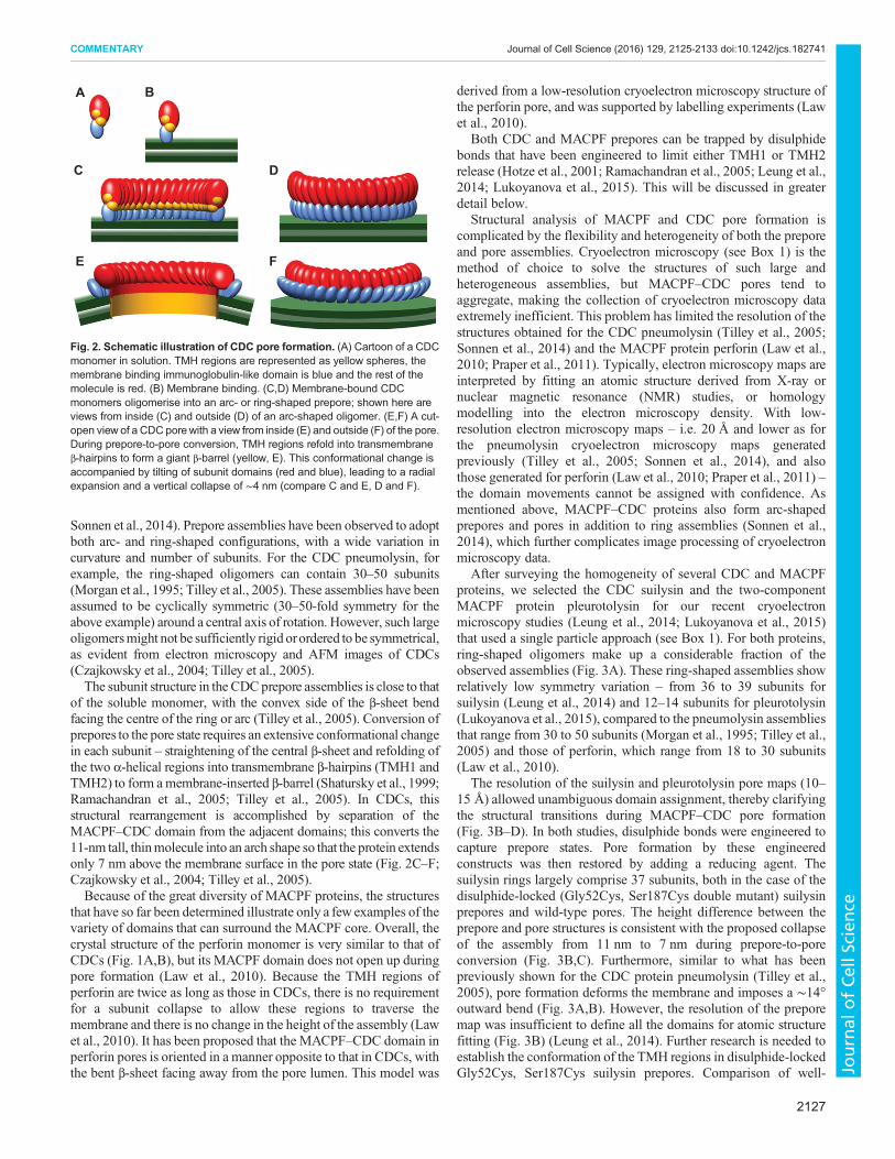
Sonnen et al., 2014). Prepore assemblies have been observed to adoptboth arc- and ring-shaped configurations, with a wide variation incurvature and number of subunits. For the CDC pneumolysin, forexample, the ring-shaped oligomers can contain 30–50 subunits(Morgan et al., 1995; Tilley et al., 2005). These assemblies have beenassumed to be cyclically symmetric (30–50-fold symmetry for theabove example) around a central axis of rotation. However, such largeoligomersmight not be sufficiently rigid orordered to be symmetrical,as evident from electron microscopy and AFM images of CDCs(Czajkowsky et al., 2004; Tilley et al., 2005).The subunit structure in the CDCprepore assemblies is close to that
of the soluble monomer, with the convex side of the β-sheet bendfacing the centre of the ring or arc (Tilley et al., 2005). Conversion ofprepores to the pore state requires an extensive conformational changein each subunit – straightening of the central β-sheet and refolding ofthe two α-helical regions into transmembrane β-hairpins (TMH1 andTMH2) to form amembrane-inserted β-barrel (Shatursky et al., 1999;Ramachandran et al., 2005; Tilley et al., 2005). In CDCs, thisstructural rearrangement is accomplished by separation of theMACPF–CDC domain from the adjacent domains; this converts the11-nm tall, thinmolecule into an arch shape so that the protein extendsonly 7 nm above the membrane surface in the pore state (Fig. 2C–F;Czajkowsky et al., 2004; Tilley et al., 2005).Because of the great diversity of MACPF proteins, the structures
that have so far been determined illustrate only a few examples of thevariety of domains that can surround the MACPF core. Overall, thecrystal structure of the perforin monomer is very similar to that ofCDCs (Fig. 1A,B), but its MACPF domain does not open up duringpore formation (Law et al., 2010). Because the TMH regions ofperforin are twice as long as those in CDCs, there is no requirementfor a subunit collapse to allow these regions to traverse themembrane and there is no change in the height of the assembly (Lawet al., 2010). It has been proposed that the MACPF–CDC domain inperforin pores is oriented in a manner opposite to that in CDCs, withthe bent β-sheet facing away from the pore lumen. This model was
derived from a low-resolution cryoelectron microscopy structure ofthe perforin pore, and was supported by labelling experiments (Lawet al., 2010).
Both CDC and MACPF prepores can be trapped by disulphidebonds that have been engineered to limit either TMH1 or TMH2release (Hotze et al., 2001; Ramachandran et al., 2005; Leung et al.,2014; Lukoyanova et al., 2015). This will be discussed in greaterdetail below.
Structural analysis of MACPF and CDC pore formation iscomplicated by the flexibility and heterogeneity of both the preporeand pore assemblies. Cryoelectron microscopy (see Box 1) is themethod of choice to solve the structures of such large andheterogeneous assemblies, but MACPF–CDC pores tend toaggregate, making the collection of cryoelectron microscopy dataextremely inefficient. This problem has limited the resolution of thestructures obtained for the CDC pneumolysin (Tilley et al., 2005;Sonnen et al., 2014) and the MACPF protein perforin (Law et al.,2010; Praper et al., 2011). Typically, electron microscopy maps areinterpreted by fitting an atomic structure derived from X-ray ornuclear magnetic resonance (NMR) studies, or homologymodelling into the electron microscopy density. With low-resolution electron microscopy maps – i.e. 20 Å and lower as forthe pneumolysin cryoelectron microscopy maps generatedpreviously (Tilley et al., 2005; Sonnen et al., 2014), and alsothose generated for perforin (Law et al., 2010; Praper et al., 2011) –the domain movements cannot be assigned with confidence. Asmentioned above, MACPF–CDC proteins also form arc-shapedprepores and pores in addition to ring assemblies (Sonnen et al.,2014), which further complicates image processing of cryoelectronmicroscopy data.
After surveying the homogeneity of several CDC and MACPFproteins, we selected the CDC suilysin and the two-componentMACPF protein pleurotolysin for our recent cryoelectronmicroscopy studies (Leung et al., 2014; Lukoyanova et al., 2015)that used a single particle approach (see Box 1). For both proteins,ring-shaped oligomers make up a considerable fraction of theobserved assemblies (Fig. 3A). These ring-shaped assemblies showrelatively low symmetry variation – from 36 to 39 subunits forsuilysin (Leung et al., 2014) and 12–14 subunits for pleurotolysin(Lukoyanova et al., 2015), compared to the pneumolysin assembliesthat range from 30 to 50 subunits (Morgan et al., 1995; Tilley et al.,2005) and those of perforin, which range from 18 to 30 subunits(Law et al., 2010).
The resolution of the suilysin and pleurotolysin pore maps (10–15 Å) allowed unambiguous domain assignment, thereby clarifyingthe structural transitions during MACPF–CDC pore formation(Fig. 3B–D). In both studies, disulphide bonds were engineered tocapture prepore states. Pore formation by these engineeredconstructs was then restored by adding a reducing agent. Thesuilysin rings largely comprise 37 subunits, both in the case of thedisulphide-locked (Gly52Cys, Ser187Cys double mutant) suilysinprepores and wild-type pores. The height difference between theprepore and pore structures is consistent with the proposed collapseof the assembly from 11 nm to 7 nm during prepore-to-poreconversion (Fig. 3B,C). Furthermore, similar to what has beenpreviously shown for the CDC protein pneumolysin (Tilley et al.,2005), pore formation deforms the membrane and imposes a ∼14°outward bend (Fig. 3A,B). However, the resolution of the preporemap was insufficient to define all the domains for atomic structurefitting (Fig. 3B) (Leung et al., 2014). Further research is needed toestablish the conformation of the TMH regions in disulphide-lockedGly52Cys, Ser187Cys suilysin prepores. Comparison of well-
B
DC
A
FE
Fig. 2. Schematic illustration of CDC pore formation. (A) Cartoon of a CDCmonomer in solution. TMH regions are represented as yellow spheres, themembrane binding immunoglobulin-like domain is blue and the rest of themolecule is red. (B) Membrane binding. (C,D) Membrane-bound CDCmonomers oligomerise into an arc- or ring-shaped prepore; shown here areviews from inside (C) and outside (D) of an arc-shaped oligomer. (E,F) A cut-open view of a CDC porewith a view from inside (E) and outside (F) of the pore.During prepore-to-pore conversion, TMH regions refold into transmembraneβ-hairpins to form a giant β-barrel (yellow, E). This conformational change isaccompanied by tilting of subunit domains (red and blue), leading to a radialexpansion and a vertical collapse of ∼4 nm (compare C and E, D and F).
2127
COMMENTARY Journal of Cell Science (2016) 129, 2125-2133 doi:10.1242/jcs.182741
Journal
ofCe
llScience
resolved regions of suilysin pores and prepores with the samesymmetry revealed that the diameter of the CDC ring expands by8% upon pore formation (Fig. 3B,C). This expansion results from achange in subunit packing. In broad agreement with a previouslyproposed model of CDC domain rearrangement during poreformation (Reboul et al., 2014), the fitting of the atomic structurereveals a sideways tilt of the subunits in the pore state, which placesthe MACPF–CDC domain of one subunit above the membrane-binding immunoglobulin domain of the neighbouring subunit(Leung et al., 2014).
The cryoelectron microscopy map of the pleurotolysin pore with13-fold symmetry shows that the pore extends to a height of∼10 nmabove the membrane (Lukoyanova et al., 2015). Each subunit isformed by a pore-forming MACPF pleurotolysin B molecule on topof two pleurotolysin A molecules (Fig. 3D). The pore channel isformed by a 52-stranded β-barrel with an inner diameter of 80 Å anda height of over 100 Å. Each subunit opens up and arches toward thepore lumen, similar to what has been observed for the CDCspneumolysin and suilysin. The central β-sheet straightens and opensby ∼70° relative to the pleurotolysin B monomer crystal structure.Similar to the perforin pore, there is no height change during poreformation because the TMH1 and TMH2 regions are long enough toform a membrane-spanning β-barrel without the need for subunitcollapse.
In both the suilysin and pleurotolysin pore structures, the densitycorresponding to the β-barrel can be clearly identified. In an earlierbioinformatics study of large β-barrel architectures, it is shown thatβ-barrels are limited to discrete architectures, each with acharacteristic strand tilt relative to the barrel axis (Reboul et al.,2012). Consistent with this analysis, the diameters of suilysin andpleurotolysin pores are compatible with a ∼20° tilt of the β-strandsrelative to the pore axis. For CDC pores, this observation is alsosupported by a disulphide-bond scanning study of perfringolysin O(Sato et al., 2013). In addition to β-sheet opening, fitting of thesuilysin pore required considerable domain movements in themonomer crystal structure to accommodate the height and diameterchange during pore assembly (Leung et al., 2014). By contrast, apartfrom β-sheet straightening and refolding of TMH regions, thepleurotolysin B crystal structure fitted into the pore density withonly minor rearrangements (Lukoyanova et al., 2015).
Additional MACPF structures have been found for componentsof the MAC pore. As mentioned above, C8 is a 150-kDathree-component MACPF protein, which initiates membranepenetration and coordinates MAC pore formation. A low-resolution structure of C8 has been determined using single-particle electron microscopy (Bubeck et al., 2011). The structure ofthe intermediate MAC complex sC5b9 was also determined withelectron microscopy (Hadders et al., 2012); this contains fiveMACPF proteins and acts as a template for the formation of thecomplement C9 pore.
More recently, the structures of a detergent-solubilisedMAC pore(Serna et al., 2016) and a soluble C9 oligomer (Dudkina et al., 2016)have been solved with cryoelectron microscopy analyses to aresolution of 7–8 Å (Fig. 3) – the highest resolution so far achievedfor MACPF assemblies. This improvement in resolution resultedfrom two advantages – firstly, soluble isolated assemblies are muchmore favourable for single-particle analysis than liposome-boundstructures; and secondly, the use of direct electron detectors hasprovided greatly improved speed and sensitivity, and enablescorrection for sample motions during data acquisition. The hetero-oligomeric MAC pore comprises single copies of C5b, C6, C7, C8,C8β, C8γ and 18 copies of C9 (Fig. 3E; Serna et al., 2016). The C6,
Box 1. Cryoelectron microscopy and AFM
Electronsource
3D reconstruction
Averages of the side and end views
Specimen
Image
AFM probe
Aqueous solution
Supported lipid bilayer
Line traces of surface topography
Electron microscopy
AFM
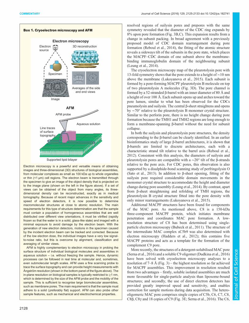
Electron microscopy is a powerful and versatile means of obtainingimages and three-dimensional (3D) structures of biological assemblies,from molecular complexes as small as 100 kDa up to whole organellesor thin (<1 µm) cell regions. The electron beam is transmitted throughthe specimen to give an image of the object density that is projected onto the image plane (shown on the left in the figure above). If a set ofviews can be obtained of the object from many angles, its three-dimensional density can be reconstructed, exactly as in medicaltomography. Because of recent major advances in the sensitivity andspeed of electron detectors, it is now possible to determinemacromolecular structures at close to atomic resolution. The mainrequirements for this type of structure determination are that the samplemust contain a population of homogeneous assemblies that are welldistributed over different view orientations, it must be vitrified (rapidlyfrozen so that the water is in a solid, glass-like state) and imaged with aminimal exposure to avoid damage by the electron beam. With thegeneration of new electron detectors, motions in the specimen causedby the incident electron beam can be tracked and corrected. Becauseof the low electron dose, the individual images have a very low signal-to-noise ratio, but this is overcome by alignment, classification andaveraging of similar views.
AFM is highly complementary to electron microscopy in probing thesurface structure of individual biological molecules and assemblies inaqueous solution – i.e. without freezing the sample. Hence, dynamicprocesses can be followed in real time at molecular and, sometimes,even submolecular length scales. AFM uses a fine scanning probe totrace the surface topography and can provide height measurements withÅngström resolution (shown in the bottom panel of the figure above). Thein-plane resolution on biological samples is typically restricted to ≥1 nm,which is determined by the size of the AFM probe and the mobility of thesample. This is sufficient to recognise large biomolecular assemblies,such as membrane pores. The main requirement is that the sample mustadhere to a solid (preferably flat) support. AFM can also probe othersample features, such as mechanical and electrochemical properties.
2128
COMMENTARY Journal of Cell Science (2016) 129, 2125-2133 doi:10.1242/jcs.182741
Journal
ofCe
llScience
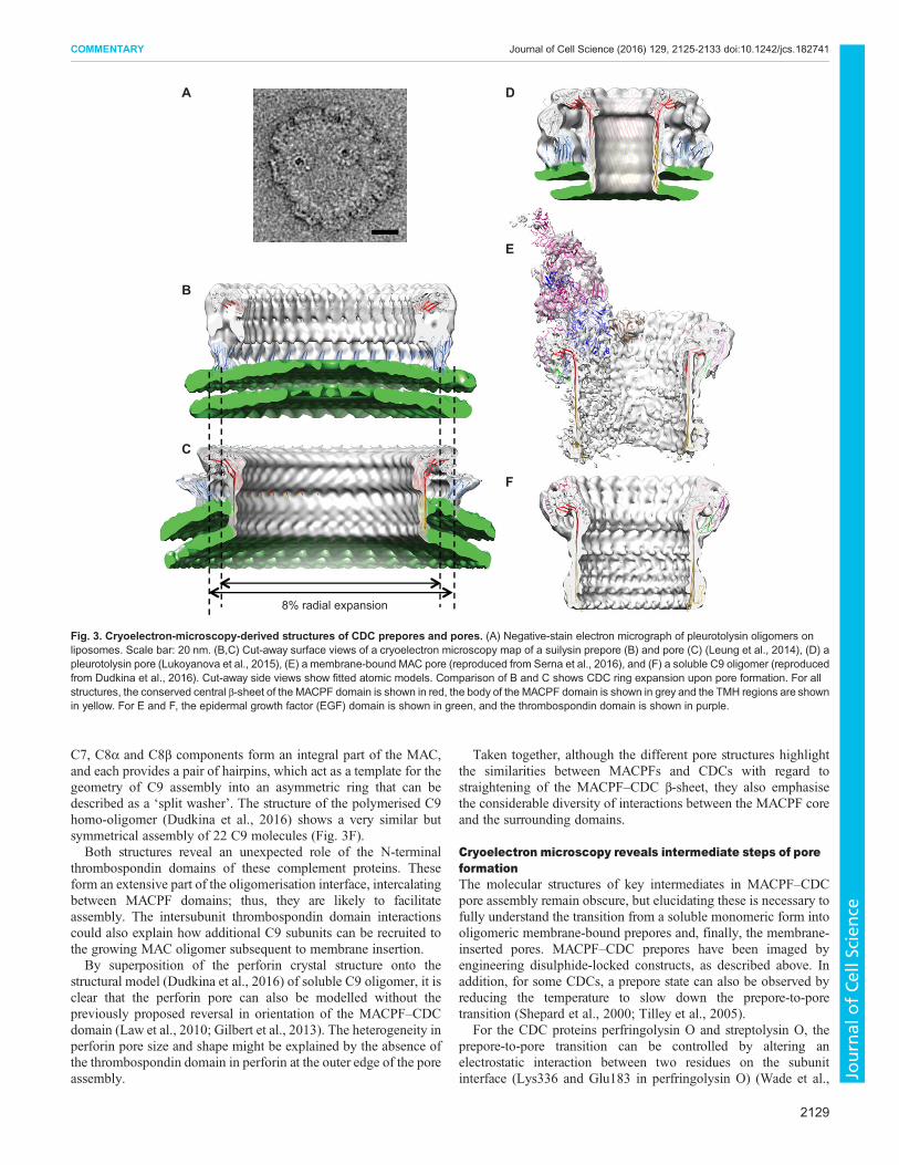
C7, C8α and C8β components form an integral part of the MAC,and each provides a pair of hairpins, which act as a template for thegeometry of C9 assembly into an asymmetric ring that can bedescribed as a ‘split washer’. The structure of the polymerised C9homo-oligomer (Dudkina et al., 2016) shows a very similar butsymmetrical assembly of 22 C9 molecules (Fig. 3F).Both structures reveal an unexpected role of the N-terminal
thrombospondin domains of these complement proteins. Theseform an extensive part of the oligomerisation interface, intercalatingbetween MACPF domains; thus, they are likely to facilitateassembly. The intersubunit thrombospondin domain interactionscould also explain how additional C9 subunits can be recruited tothe growing MAC oligomer subsequent to membrane insertion.By superposition of the perforin crystal structure onto the
structural model (Dudkina et al., 2016) of soluble C9 oligomer, it isclear that the perforin pore can also be modelled without thepreviously proposed reversal in orientation of the MACPF–CDCdomain (Law et al., 2010; Gilbert et al., 2013). The heterogeneity inperforin pore size and shape might be explained by the absence ofthe thrombospondin domain in perforin at the outer edge of the poreassembly.
Taken together, although the different pore structures highlightthe similarities between MACPFs and CDCs with regard tostraightening of the MACPF–CDC β-sheet, they also emphasisethe considerable diversity of interactions between the MACPF coreand the surrounding domains.
Cryoelectron microscopy reveals intermediate steps of poreformationThe molecular structures of key intermediates in MACPF–CDCpore assembly remain obscure, but elucidating these is necessary tofully understand the transition from a soluble monomeric form intooligomeric membrane-bound prepores and, finally, the membrane-inserted pores. MACPF–CDC prepores have been imaged byengineering disulphide-locked constructs, as described above. Inaddition, for some CDCs, a prepore state can also be observed byreducing the temperature to slow down the prepore-to-poretransition (Shepard et al., 2000; Tilley et al., 2005).
For the CDC proteins perfringolysin O and streptolysin O, theprepore-to-pore transition can be controlled by altering anelectrostatic interaction between two residues on the subunitinterface (Lys336 and Glu183 in perfringolysin O) (Wade et al.,
D
E
A
F
C
B
8% radial expansion
Fig. 3. Cryoelectron-microscopy-derived structures of CDC prepores and pores. (A) Negative-stain electron micrograph of pleurotolysin oligomers onliposomes. Scale bar: 20 nm. (B,C) Cut-away surface views of a cryoelectron microscopy map of a suilysin prepore (B) and pore (C) (Leung et al., 2014), (D) apleurotolysin pore (Lukoyanova et al., 2015), (E) a membrane-bound MAC pore (reproduced from Serna et al., 2016), and (F) a soluble C9 oligomer (reproducedfrom Dudkina et al., 2016). Cut-away side views show fitted atomic models. Comparison of B and C shows CDC ring expansion upon pore formation. For allstructures, the conserved central β-sheet of the MACPF domain is shown in red, the body of the MACPF domain is shown in grey and the TMH regions are shownin yellow. For E and F, the epidermal growth factor (EGF) domain is shown in green, and the thrombospondin domain is shown in purple.
2129
COMMENTARY Journal of Cell Science (2016) 129, 2125-2133 doi:10.1242/jcs.182741
Journal
ofCe
llScience

2015). This electrostatic interaction is prevented by mutation ofthese residues. However, pore formation can be restored by raisingthe temperature or by additional mutations. Further research isneeded to determine the importance of this electrostatic interactionin oligomerisation of other CDC and MACPF proteins.For the extensively studied CDC perfringolysin O, numerous
mutations have been described that lead to arrested prepore states.Each might provide insight into a particular stage of pore formation,but the order of these events and whether they are generally relevantto the pore formation mechanism is not understood. Forperfringolysin O, early prepores assemble through a weak andreversible association of membrane-bound monomers, as deducedfrom the presence of SDS-sensitive oligomers at this stage (Shepardet al., 2000). Conversion of this early prepore to an SDS-resistantprepore oligomer involves rotation of strand β5 of the MACPF–CDC β-sheet (Fig. 1A), disengaging it from strand β4. This allowspairing of β4 with the β1 strand of the adjacent monomer, andinitiation of β-barrel formation (Hotze et al., 2012; Dunstone andTweten, 2012). The importance of strand β5 mobility is highlightedby the location of the only two residues (glycine residues) that areconserved across the MACPF–CDC superfamily (Rosado et al.,2008; Dunstone and Tweten, 2012) at the bend between strands β4and β5. Mutation of these glycine residues in perfringolysin Oprevents oligomerisation (Ramachandran et al., 2004). In MACPF–CDC proteins, these conserved glycine residues are positioned at thebend of the β-sheet (Fig. 1) and are likely to play a role in sheetstraightening during pore formation. In MACPF proteins, such asperforin and C8α, this β-sheet contains only four strands. The fifthβ-strand of the MACPF protein pleurotolysin has a differentfunction and forms a part of TMH2 during pore formation(Lukoyanova et al., 2015).To trap one of the intermediate prepore states described, a
disulphide bond was introduced between strands β4 and β5 ofpleurotolysin (TMH2 strand lock, Fig. 4; Lukoyanova et al., 2015).The movement of either the TMH1 or TMH2 region was limited bycrosslinks to the helices in two other trapped pleurotolysin prepores(TMH1 helix lock and TMH2 helix lock, Fig. 4). These threetrapped prepores display different degrees of β-sheet opening andindicate the molecular trajectory during pore formation. The prepore
assemblies also show that the MACPF fold can oligomerise withoutsubstantial relief of the twist in the core sheet or TMH1 release.Molecular modelling has been used to analyse the trajectory ofMACPF β-sheet straightening, which requires mobility of TMH2(Lukoyanova et al., 2015). This analysis has identified theconserved helical loop motif (Figs 1 and 4) at the top of theTMH2 region following the conserved glycine residues;displacement of this motif is proposed to trigger theconformational change that leads to pore formation. This motif orequivalent is present in all the MACPF–CDC pore proteins so farcharacterised, and its displacement relative to the equivalentmonomer structure is best resolved in the C9 and MAC poresdescribed above (Dudkina et al., 2016). Straightening of theMACPF sheet results in the formation of a nascent β-barrel that actsas a template for the top-down assembly of TMH1 and TMH2 into amembrane-inserted β-barrel.
As evident from the above, electron microscopy analyses canonly provide snapshots of the dynamic processes that are capturedby rapid freezing. In contrast, other techniques, such as AFM canfollow the dynamics of pore formation at lower spatial resolution, asdiscussed below.
AFM tracks MACPF–CDC assembly and membraneperforationAFM allows the visualisation of MACPF–CDC pore formation inreal time (see Box 1). It has been previously applied toperfringolysin O (Czajkowsky et al., 2004) to reveal the heightchange during the prepore-to-pore transition of a CDC. Morerecently, several AFM studies have focused on other CDCs (Leunget al., 2014; Podobnik et al., 2015; Mulvihill et al., 2015); these allshow heterogeneous distributions of arc- and ring-shapedassemblies.
By visualising membrane removal from the lumen of suilysin andlisteriolysin O assemblies, AFM studies have established that botharc- and ring-shaped assemblies can perforate the adjacentmembrane (Leung et al., 2014; Podobnik et al., 2015; Mulvihillet al., 2015). This implies that pores with an incomplete β-barrelcreate an unsealed edge of the lipid bilayer (Fig. 5A). Furthermore,this finding unambiguously demonstrates that arc-shaped CDC
B DA C E
Pleurotolysin Bmonomer
TMH2 helix lock TMH1 helix lock TMH2 strand lock Pore
70°37° 49° 49°
Fig. 4. Schematic diagram of β-sheet opening during pleurotolysin pore formation. (A) Pleurotolysin B monomer structure: TMH regions are yellow, thecentral β-sheet is red and the helical loop region is cyan. (B–D) Sections of the cryoelectron microscopy maps and fitted domains of the three preporeintermediates that were captured by introducing disulphide bridges (pink spheres) between different parts of the TMH regions and the surrounding domains.Relative to its position in the monomer structure, the β-sheet opens to an angle of ∼37° in TMH2 helix lock (B); and opens further, to ∼49°, in TMH1 helix lock(C) and in TMH2 strand lock (D). (E) Pore model with a 70°-opened β-sheet and membrane-inserted TMH regions. The monomer crystal structure issuperimposed on the pore to show the full extent of sheet opening. The colour scheme matches that described in Fig 1, and yellow dashed lines representdisordered TMH regions. This figure has been reproduced from Lukoyanova et al., 2015.
2130
COMMENTARY Journal of Cell Science (2016) 129, 2125-2133 doi:10.1242/jcs.182741
Journal
ofCe
llScience

assemblies are functional in pore formation, confirming apreviously contentious proposition (see, for example Gilbert,2005; Gilbert et al., 2014) that had been posed based onelectrophysiology experiments (Marchioretto et al., 2013) andmore recently on cryoelectron microscopy analysis of pneumolysinarcs (Sonnen et al., 2014).When wild-type suilysin is injected into the solution above
the membrane, membrane-inserted pores form within seconds(Leung et al., 2014). Once the suilysin assemblies have transformedinto the pore state, they do not show any further change or growth.Furthermore, upon subsequent injections of suilysin monomersin solution, new pore assemblies appear, but no growth wasobserved for any previously formed pore assemblies (Leung et al.,2014). However, when arc-shaped pore assemblies of listeriolysin Oare incubated at 37°C for up to several hours, they continue tomove and merge in some cases, resulting in larger lesions of themembrane (Podobnik et al., 2015; Mulvihill et al., 2015). Hence,the prepore-to-pore transition inhibits further assembly, but in somecases, separate pore assemblies can join together to form large holes inthe membrane.To study CDC assembly and the prepore-to-pore transition in
more detail, AFM and negative-stain electron microscopy werecombined to study a disulphide mutant of suilysin (Hotze et al.,2001; Leung et al., 2014). Using this construct, pore insertion couldbe triggered by addition of reducing agent and followed in real time.This mutant, which behaves as the wild type once reduced, thereforeallows a more quantitative analysis of prepore assembly and poreformation (Leung et al., 2014; see also Fig. 5A, inset). The preporeassembly was found to occur through the addition of monomers orof small oligomers to the growing oligomers; growth is arrested
when monomers on the membrane surface are depleted. In asubsequent step, the prepore assemblies insert into the membrane assummarised in Fig. 5B (Gilbert, 2005).
Experiments on perfringolysin O show that the CDC prepore-to-pore transition is rapid compared to a preceding phase thatincludes CDC diffusion, subunit binding to the membrane andnucleation of prepore assemblies on the membrane (Tweten,2005; Hotze et al., 2012; Wade et al., 2015). SubsequentAFM experiments have provided further mechanistic details ofthe membrane insertion process. In particular, mixing thedisulphide-locked mutant with wild-type suilysin inhibited theprepore-to-pore transition in a concentration-dependent manner,and subsequent exposure to DTT, allowing the mutant to insert intothe membrane, restored normal pore formation for all assemblies(Leung et al., 2014). This initial inhibition and subsequentrestoration of function demonstrate that wild type and mutant co-assemble, as expected. From the inhibitory effect of the mutant onpore formation at equal concentrations of wild-type and mutantsuilysin, it follows that the prepore-to-pore transition requires aconcerted conformational change and cooperative insertion of allsubunits into the suilysin assembly. This is consistent with earlierfindings for perfringolysin O (Hotze et al., 2002; Hotze et al., 2012).
AFM has also provided a direct time-resolved visualisation of theprepore-to-pore transition of suilysin (Leung et al., 2014). Becausesuilysin prepores are mobile, they can only be resolved with AFM atlow temperature. At room temperature, they appear as streaks in theAFM images (Fig. 5C). Within a minute after DTT addition todisulphide-locked suilysin prepores, AFM images of the diffuserings and arcs become progressively clearer and sharper. At thesame time, the rings adopt a reduced height that is typical of a CDC
50 nm
8.0 nm6.0
4.0
2.0
0
–2.0–3.0
A B
0 5 10 15 20 25 30
Rin
gR
ingss
Arc length (monomers)35
0
500
1000
Cou
nt100 nm
+DTTds-SLY(-DTT)
–8 min 11 mm mm1 m111 m1 m1 m1 mm1111111 inin 4 m4 m4 miiiinnninninnnn 12 12 12121212122 minmininminC
Fig. 5. Formation of suilysin assemblies and membrane pores imaged by AFM. (A) AFM topography of suilysin arcs and rings on a supported lipid bilayer;these are represented by a colour scale that emphasises height of the suilysin assemblies (purple) and the membrane removal (black) with respect to themembrane surface (green). The inset shows the distribution of oligomeric states as determined by these measurements, with the dashed grey line indicating a fitwith a simple oligomerisation model. (B) Schematic model of CDC pore formation, supported by AFM data, in which prepores grow on the membrane surface.They then insert in concerted steps, after which there is no further growth, creating the final arc- or ring-shaped pores. Colour scheme as in A, figure adapted fromGilbert, 2005, with permission from Elsevier. (C) Example of a time-lapse AFM sequence of membrane perforation by suilysin. Initially, mobile disulphide-lockedsuilysin are seen as streaky features on lipid domains in the membrane (left). On addition of DTT (+DTT, at 1 min; second from left), the mobile preporesconvert into more clearly defined arc and ring shapes on the membrane; this is followed by the ejection of material (white features) from the membrane (right).The inset in panel A and panel C are adapted from Leung et al., 2014.
2131
COMMENTARY Journal of Cell Science (2016) 129, 2125-2133 doi:10.1242/jcs.182741
Journal
ofCe
llScience
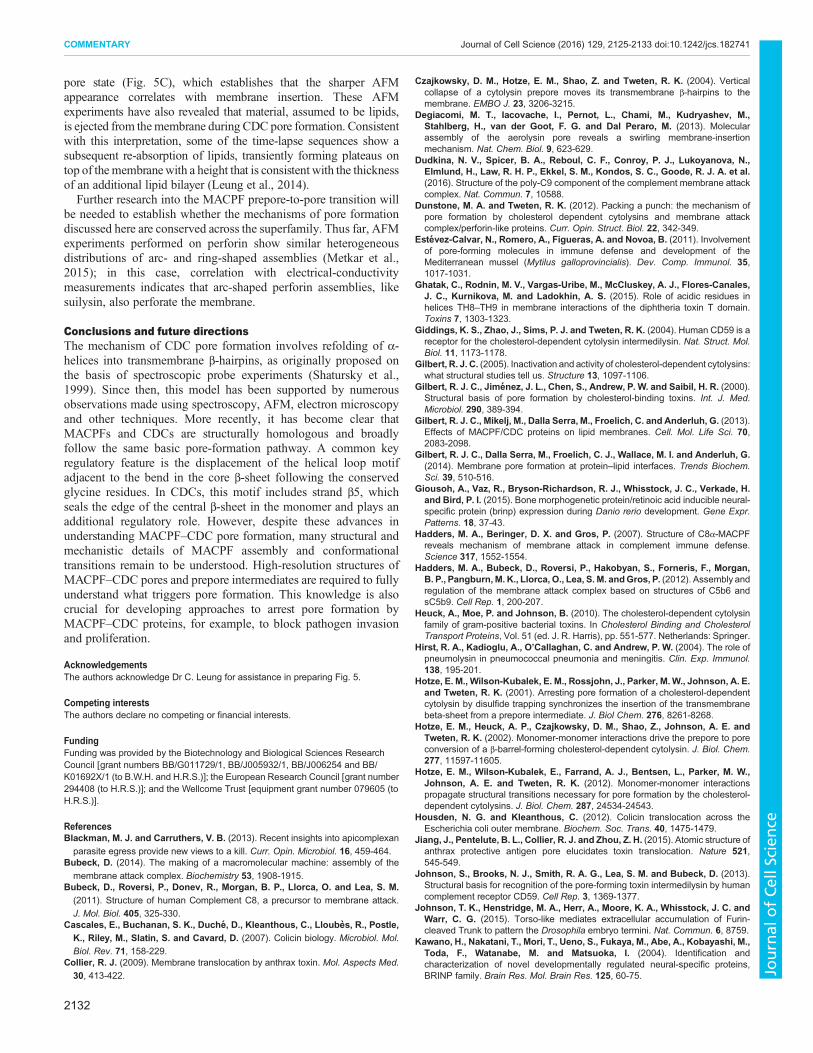
pore state (Fig. 5C), which establishes that the sharper AFMappearance correlates with membrane insertion. These AFMexperiments have also revealed that material, assumed to be lipids,is ejected from themembrane during CDC pore formation. Consistentwith this interpretation, some of the time-lapse sequences show asubsequent re-absorption of lipids, transiently forming plateaus ontop of themembranewith a height that is consistent with the thicknessof an additional lipid bilayer (Leung et al., 2014).Further research into the MACPF prepore-to-pore transition will
be needed to establish whether the mechanisms of pore formationdiscussed here are conserved across the superfamily. Thus far, AFMexperiments performed on perforin show similar heterogeneousdistributions of arc- and ring-shaped assemblies (Metkar et al.,2015); in this case, correlation with electrical-conductivitymeasurements indicates that arc-shaped perforin assemblies, likesuilysin, also perforate the membrane.
Conclusions and future directionsThe mechanism of CDC pore formation involves refolding of α-helices into transmembrane β-hairpins, as originally proposed onthe basis of spectroscopic probe experiments (Shatursky et al.,1999). Since then, this model has been supported by numerousobservations made using spectroscopy, AFM, electron microscopyand other techniques. More recently, it has become clear thatMACPFs and CDCs are structurally homologous and broadlyfollow the same basic pore-formation pathway. A common keyregulatory feature is the displacement of the helical loop motifadjacent to the bend in the core β-sheet following the conservedglycine residues. In CDCs, this motif includes strand β5, whichseals the edge of the central β-sheet in the monomer and plays anadditional regulatory role. However, despite these advances inunderstanding MACPF–CDC pore formation, many structural andmechanistic details of MACPF assembly and conformationaltransitions remain to be understood. High-resolution structures ofMACPF–CDC pores and prepore intermediates are required to fullyunderstand what triggers pore formation. This knowledge is alsocrucial for developing approaches to arrest pore formation byMACPF–CDC proteins, for example, to block pathogen invasionand proliferation.
AcknowledgementsThe authors acknowledge Dr C. Leung for assistance in preparing Fig. 5.
Competing interestsThe authors declare no competing or financial interests.
FundingFunding was provided by the Biotechnology and Biological Sciences ResearchCouncil [grant numbers BB/G011729/1, BB/J005932/1, BB/J006254 and BB/K01692X/1 (to B.W.H. and H.R.S.)]; the European Research Council [grant number294408 (to H.R.S.)]; and the Wellcome Trust [equipment grant number 079605 (toH.R.S.)].
ReferencesBlackman, M. J. and Carruthers, V. B. (2013). Recent insights into apicomplexan
parasite egress provide new views to a kill. Curr. Opin. Microbiol. 16, 459-464.Bubeck, D. (2014). The making of a macromolecular machine: assembly of the
membrane attack complex. Biochemistry 53, 1908-1915.Bubeck, D., Roversi, P., Donev, R., Morgan, B. P., Llorca, O. and Lea, S. M.(2011). Structure of human Complement C8, a precursor to membrane attack.
J. Mol. Biol. 405, 325-330.Cascales, E., Buchanan, S. K., Duche, D., Kleanthous, C., Lloubes, R., Postle,K., Riley, M., Slatin, S. and Cavard, D. (2007). Colicin biology. Microbiol. Mol.Biol. Rev. 71, 158-229.
Collier, R. J. (2009). Membrane translocation by anthrax toxin. Mol. Aspects Med.30, 413-422.
Czajkowsky, D. M., Hotze, E. M., Shao, Z. and Tweten, R. K. (2004). Verticalcollapse of a cytolysin prepore moves its transmembrane β-hairpins to themembrane. EMBO J. 23, 3206-3215.
Degiacomi, M. T., Iacovache, I., Pernot, L., Chami, M., Kudryashev, M.,Stahlberg, H., van der Goot, F. G. and Dal Peraro, M. (2013). Molecularassembly of the aerolysin pore reveals a swirling membrane-insertionmechanism. Nat. Chem. Biol. 9, 623-629.
Dudkina, N. V., Spicer, B. A., Reboul, C. F., Conroy, P. J., Lukoyanova, N.,Elmlund, H., Law, R. H. P., Ekkel, S. M., Kondos, S. C., Goode, R. J. A. et al.(2016). Structure of the poly-C9 component of the complement membrane attackcomplex. Nat. Commun. 7, 10588.
Dunstone, M. A. and Tweten, R. K. (2012). Packing a punch: the mechanism ofpore formation by cholesterol dependent cytolysins and membrane attackcomplex/perforin-like proteins. Curr. Opin. Struct. Biol. 22, 342-349.
Estevez-Calvar, N., Romero, A., Figueras, A. and Novoa, B. (2011). Involvementof pore-forming molecules in immune defense and development of theMediterranean mussel (Mytilus galloprovincialis). Dev. Comp. Immunol. 35,1017-1031.
Ghatak, C., Rodnin, M. V., Vargas-Uribe, M., McCluskey, A. J., Flores-Canales,J. C., Kurnikova, M. and Ladokhin, A. S. (2015). Role of acidic residues inhelices TH8–TH9 in membrane interactions of the diphtheria toxin T domain.Toxins 7, 1303-1323.
Giddings, K. S., Zhao, J., Sims, P. J. and Tweten, R. K. (2004). Human CD59 is areceptor for the cholesterol-dependent cytolysin intermedilysin. Nat. Struct. Mol.Biol. 11, 1173-1178.
Gilbert, R. J. C. (2005). Inactivation and activity of cholesterol-dependent cytolysins:what structural studies tell us. Structure 13, 1097-1106.
Gilbert, R. J. C., Jimenez, J. L., Chen, S., Andrew, P. W. and Saibil, H. R. (2000).Structural basis of pore formation by cholesterol-binding toxins. Int. J. Med.Microbiol. 290, 389-394.
Gilbert, R. J. C., Mikelj, M., Dalla Serra, M., Froelich, C. and Anderluh, G. (2013).Effects of MACPF/CDC proteins on lipid membranes. Cell. Mol. Life Sci. 70,2083-2098.
Gilbert, R. J. C., Dalla Serra, M., Froelich, C. J., Wallace, M. I. and Anderluh, G.(2014). Membrane pore formation at protein–lipid interfaces. Trends Biochem.Sci. 39, 510-516.
Giousoh, A., Vaz, R., Bryson-Richardson, R. J., Whisstock, J. C., Verkade, H.and Bird, P. I. (2015). Bone morphogenetic protein/retinoic acid inducible neural-specific protein (brinp) expression during Danio rerio development. Gene Expr.Patterns. 18, 37-43.
Hadders, M. A., Beringer, D. X. and Gros, P. (2007). Structure of C8α-MACPFreveals mechanism of membrane attack in complement immune defense.Science 317, 1552-1554.
Hadders, M. A., Bubeck, D., Roversi, P., Hakobyan, S., Forneris, F., Morgan,B. P., Pangburn, M. K., Llorca, O., Lea, S. M. andGros, P. (2012). Assembly andregulation of the membrane attack complex based on structures of C5b6 andsC5b9. Cell Rep. 1, 200-207.
Heuck, A., Moe, P. and Johnson, B. (2010). The cholesterol-dependent cytolysinfamily of gram-positive bacterial toxins. In Cholesterol Binding and CholesterolTransport Proteins, Vol. 51 (ed. J. R. Harris), pp. 551-577. Netherlands: Springer.
Hirst, R. A., Kadioglu, A., O’Callaghan, C. and Andrew, P. W. (2004). The role ofpneumolysin in pneumococcal pneumonia and meningitis. Clin. Exp. Immunol.138, 195-201.
Hotze, E. M., Wilson-Kubalek, E. M., Rossjohn, J., Parker, M.W., Johnson, A. E.and Tweten, R. K. (2001). Arresting pore formation of a cholesterol-dependentcytolysin by disulfide trapping synchronizes the insertion of the transmembranebeta-sheet from a prepore intermediate. J. Biol Chem. 276, 8261-8268.
Hotze, E. M., Heuck, A. P., Czajkowsky, D. M., Shao, Z., Johnson, A. E. andTweten, R. K. (2002). Monomer-monomer interactions drive the prepore to poreconversion of a β-barrel-forming cholesterol-dependent cytolysin. J. Biol. Chem.277, 11597-11605.
Hotze, E. M., Wilson-Kubalek, E., Farrand, A. J., Bentsen, L., Parker, M. W.,Johnson, A. E. and Tweten, R. K. (2012). Monomer-monomer interactionspropagate structural transitions necessary for pore formation by the cholesterol-dependent cytolysins. J. Biol. Chem. 287, 24534-24543.
Housden, N. G. and Kleanthous, C. (2012). Colicin translocation across theEscherichia coli outer membrane. Biochem. Soc. Trans. 40, 1475-1479.
Jiang, J., Pentelute, B. L., Collier, R. J. and Zhou, Z. H. (2015). Atomic structure ofanthrax protective antigen pore elucidates toxin translocation. Nature 521,545-549.
Johnson, S., Brooks, N. J., Smith, R. A. G., Lea, S. M. and Bubeck, D. (2013).Structural basis for recognition of the pore-forming toxin intermedilysin by humancomplement receptor CD59. Cell Rep. 3, 1369-1377.
Johnson, T. K., Henstridge, M. A., Herr, A., Moore, K. A., Whisstock, J. C. andWarr, C. G. (2015). Torso-like mediates extracellular accumulation of Furin-cleaved Trunk to pattern the Drosophila embryo termini. Nat. Commun. 6, 8759.
Kawano, H., Nakatani, T., Mori, T., Ueno, S., Fukaya, M., Abe, A., Kobayashi, M.,Toda, F., Watanabe, M. and Matsuoka, I. (2004). Identification andcharacterization of novel developmentally regulated neural-specific proteins,BRINP family. Brain Res. Mol. Brain Res. 125, 60-75.
2132
COMMENTARY Journal of Cell Science (2016) 129, 2125-2133 doi:10.1242/jcs.182741
Journal
ofCe
llScience

Kondos, S. C., Hatfaludi, T., Voskoboinik, I., Trapani, J. A., Law, R. H. P.,Whisstock, J. C. and Dunstone, M. A. (2010). The structure and function ofmammalian membrane-attack complex/perforin-like proteins. Tissue Antigens 76,341-351.
LaChapelle, S., Tweten, R. K. and Hotze, E. M. (2009). Intermedilysin-receptorinteractions during assembly of the pore complex: assembly intermediatesincrease host cell susceptibility to complement-mediated lysis. J. Biol. Chem. 284,12719-12726.
Law, R. H. P., Lukoyanova, N., Voskoboinik, I., Caradoc-Davies, T. T., Baran, K.,Dunstone, M. A., D’Angelo, M. E., Orlova, E. V., Coulibaly, F., Verschoor, S.et al. (2010). The structural basis for membrane binding and pore formation bylymphocyte perforin. Nature 468, 447-451.
Leka, O., Vallese, F., Pirazzini, M., Berto, P., Montecucco, C. and Zanotti, G.(2014). Diphtheria toxin conformational switching at acidic pH. FEBS J. 281,2115-2122.
Leung, C., Dudkina, N. V., Lukoyanova, N., Hodel, A. W., Farabella, I.,Pandurangan, A. P., Jahan, N., Pires Damaso, M., Osmanovic, D., Reboul,C. F. et al. (2014). Stepwise visualization of membrane pore formation by suilysin,a bacterial cholesterol-dependent cytolysin. eLife 3, e04247.
Lovelace, L. L., Cooper, C. L., Sodetz, J. M. and Lebioda, L. (2011). Structure ofhuman C8 protein provides mechanistic insight into membrane pore formation bycomplement. J. Biol. Chem. 286, 17585-17592.
Lukoyanova, N., Kondos, S. C., Farabella, I., Law, R. H. P., Reboul, C. F.,Caradoc-Davies, T. T., Spicer, B. A., Kleifeld, O., Traore, D. A. K., Ekkel, S. M.et al. (2015). Conformational changes during pore formation by the perforin-related protein pleurotolysin. PLoS Biol. 13, e1002049.
Marchioretto, M., Podobnik, M., Dalla Serra, M. and Anderluh, G. (2013). Whatplanar lipid membranes tell us about the pore-forming activity of cholesterol-dependent cytolysins. Biophys. Chem. 182, 64-70.
McCormack, R., de Armas, L., Shiratsuchi, M. and Podack, E. R. (2013). Killingmachines: three pore-forming proteins of the immune system. Immunol. Res. 57,268-278.
Menestrina, G., Dalla Serra, M., Comai, M., Coraiola, M., Viero, G., Werner, S.,Colin, D. A., Monteil, H. and Prevost, G. (2003). Ion channels and bacterialinfection: the case of beta-barrel pore-forming protein toxins of Staphylococcusaureus. FEBS Lett. 552, 54-60.
Merle, N. S., Church, S. E., Fremeaux-Bacchi, V. and Roumenina, L. T. (2015).Complement system Part I – Molecular mechanisms of activation and regulation.Front. Immunol. 6, 262.
Metkar, S. S., Marchioretto, M., Antonini, V., Lunelli, L., Wang, B., Gilbert,R. J. C., Anderluh, G., Roth, R., Pooga, M., Pardo, J. et al. (2014). Perforinoligomers form arcs in cellular membranes: a locus for intracellular delivery ofgranzymes. Cell Death Differ. 22, 74-85.
Morgan, B. P., Imagawa, D. K., Dankert, J. R. and Ramm, L. E. (1986).Complement lysis of U937, a nucleated mammalian cell line in the absence of C9:effect of C9 on C5b-8 mediated cell lysis. J. Immunol. 136, 3402-3406.
Morgan, P. J., Hyman, S. C., Rowe, A. J., Mitchell, T. J., Andrew, P.W. and Saibil,H. R. (1995). Subunit organisation and symmetry of pore-forming, oligomericpneumolysin. FEBS Lett. 371, 77-80.
Mulvihill, E., van Pee, K., Mari, S. A., Muller, D. J. and Yildiz, O. (2015). Directlyobserving the lipid-dependent self-assembly and pore-forming mechanism of thecytolytic toxin listeriolysin O. Nano Lett. 15, 6965-6973.
Ounjai, P., Unger, V. M., Sigworth, F. J. and Angsuthanasombat, C. (2007). Twoconformational states of the membrane-associated Bacillus thuringiensis Cry4Badelta-endotoxin complex revealed by electron crystallography: implications fortoxin-pore formation. Biochem. Biophys. Res. Commun. 361, 890-895.
Pardo-Lopez, L., Soberon, M. and Bravo, A. (2013). Bacillus thuringiensisinsecticidal three-domain Cry toxins: mode of action, insect resistance andconsequences for crop protection. FEMS Microbiol. Rev. 37, 3-22.
Peraro, M. D. and van der Goot, F. G. (2016). Pore-forming toxins: ancient, butnever really out of fashion. Nat. Rev. Microbiol. 14, 77-92.
Podack, E. R. and Dennert, G. (1983). Assembly of two types of tubules withputative cytolytic function by cloned natural killer cells. Nature 302, 442-445.
Podobnik, M., Marchioretto, M., Zanetti, M., Bavdek, A., Kisovec, M., Cajnko,M. M., Lunelli, L., Serra, M. D. and Anderluh, G. (2015). Plasticity of lysteriolysinO pores and its regulation by pH and unique histidine. Sci. Rep. 5, 9623.
Popoff, M. R. (2014). Clostridial pore-forming toxins: powerful virulence factors.Anaerobe 30, 220-238.
Praper, T., Sonnen, A., Viero, G., Kladnik, A., Froelich, C. J., Anderluh, G., DallaSerra, M. and Gilbert, R. J. C. (2011). Human perforin employs different avenuesto damage membranes. J. Biol. Chem. 286, 2946-2955.
Ramachandran, R., Tweten, R. K. and Johnson, A. E. (2004). Membrane-dependent conformational changes initiate cholesterol-dependent cytolysin
oligomerization and intersubunit beta-strand alignment. Nat. Struct. Mol. Biol.11, 697-705.
Ramachandran, R., Tweten, R. K. and Johnson, A. E. (2005). The domains of acholesterol-dependent cytolysin undergo a major FRET-detected rearrangementduring pore formation. Proc. Natl. Acad. Sci. USA 102, 7139-7144.
Ramm, L. E., Whitlow, M. B. and Mayer, M. M. (1982). Size of the transmembranechannels produced by complement proteins C5b-8. J. Immunol. 129, 1143-1146.
Reboul,C.F.,Mahmood,K.,Whisstock, J.C.andDunstone,M.A. (2012).Predictinggiant transmembrane β-barrel architecture. Bioinformatics 28, 1299-1302.
Reboul, C. F., Whisstock, J. C. and Dunstone, M. A. (2014). A newmodel for poreformation by cholesterol-dependent cytolysins.PLoSComput. Biol. 10, e1003791.
Rosado, C. J., Buckle, A. M., Law, R. H. P., Butcher, R. E., Kan, W.-T., Bird, C. H.,Ung, K., Browne, K. A., Baran, K., Bashtannyk-Puhalovich, T. A. et al. (2007).A common fold mediates vertebrate defense and bacterial attack. Science 317,1548-1551.
Rosado, C. J., Kondos, S., Bull, T. E., Kuiper, M. J., Law, R. H. P., Buckle, A. M.,Voskoboinik, I., Bird, P. I., Trapani, J. A., Whisstock, J. C. et al. (2008). TheMACPF/CDC family of pore-forming toxins. Cell Microbiol. 10, 1765-1774.
Rossjohn, J., Feil, S. C., McKinstry, W. J., Tweten, R. K. and Parker, M. W.(1997). Structure of a cholesterol-binding, thiol-activated cytolysin and a model ofits membrane form. Cell 89, 685-692.
Sato, T. K., Tweten, R. K. and Johnson, A. E. (2013). Disulfide-bond scanningreveals assembly state and β-strand tilt angle of the PFO β-barrel.Nat. Chem. Biol.9, 383-389.
Serna, M., Giles, J. L., Morgan, B. P. and Bubeck, D. (2016). Structural basis ofcomplement membrane attack complex formation. Nat. Commun. 7, 10587.
Seveau, S. (2014). Multifaceted activity of listeriolysin O, the cholesterol-dependentcytolysin of Listeria monocytogenes. Subcell. Biochem. 80, 161-195.
Shatursky, O., Heuck, A. P., Shepard, L. A., Rossjohn, J., Parker, M. W.,Johnson, A. E. and Tweten, R. K. (1999). The mechanism of membraneinsertion for a cholesterol-dependent cytolysin: a novel paradigm for pore-formingtoxins. Cell 99, 293-299.
Shepard, L. A., Shatursky, O., Johnson, A. E. and Tweten, R. K. (2000). Themechanism of pore assembly for a cholesterol-dependent cytolysin: formation of alarge prepore complex precedes the insertion of the transmembrane β-hairpins.Biochemistry 39, 10284-10293.
Shewell, L. K., Harvey, R. M., Higgins, M. A., Day, C. J., Hartley-Tassell, L. E.,Chen, A. Y., Gillen, C. M., James, D. B. A., Alonzo, F., III, Torres, V. J. et al.(2014). The cholesterol-dependent cytolysins pneumolysin and streptolysin Orequire binding to red blood cell glycans for hemolytic activity. Proc. Natl. Acad.Sci. USA 111:E5312-E5320.
Sonnen, A. F.-P., Plitzko, J. M. and Gilbert, R. J. C. (2014). Incompletepneumolysin oligomers form membrane pores. Open Biol. 4, 140044.
Sugawara, T., Yamashita, D., Kato, K., Peng, Z., Ueda, J., Kaneko, J., Kamio, Y.,Tanaka, Y. and Yao, M. (2015). Structural basis for pore-forming mechanism ofstaphylococcal α-hemolysin. Toxicon 108, 226-231.
Taylor, L. and Nelson, D. (2014). Chlamydial MACPF protein CT153. In MACPF/CDC Proteins - Agents of Defence, Attack and Invasion, Vol. 80 (ed. G. Anderluhand R. Gilbert), pp. 255-269. Netherlands: Springer.
Tilley, S. J., Orlova, E. V., Gilbert, R. J. C., Andrew, P.W. and Saibil, H. R. (2005).Structural basis of pore formation by the bacterial toxin pneumolysin. Cell 121,247-256.
Tomita, T., Noguchi, K., Mimuro, H., Ukaji, F., Ito, K., Sugawara-Tomita, N. andHashimoto, Y. (2004). Pleurotolysin, a novel sphingomyelin-specific two-component cytolysin from the edible mushroom Pleurotus ostreatus, assemblesinto a transmembrane pore complex. J. Biol. Chem. 279, 26975-26982.
Tschopp, J., Masson, D. and Stanley, K. K. (1986). Structural/functional similaritybetween proteins involved in complement- and cytotoxic T-lymphocyte-mediatedcytolysis. Nature 322, 831-834.
Tweten, R. K. (2005). Cholesterol-dependent cytolysins, a family of versatile pore-forming toxins. Infect. Immun. 73, 6199-6209.
Voskoboinik, I., Whisstock, J. C. and Trapani, J. A. (2015). Perforin andgranzymes: function, dysfunction and human pathology. Nat. Rev. Immunol. 15,388-400.
Wade, K. R. and Tweten, R. K. (2015). The apicomplexan CDC/MACPF-like pore-forming proteins. Curr. Opin. Microbiol. 26, 48-52.
Wade, K. R., Hotze, E. M., Kuiper, M. J., Morton, C. J., Parker, M.W. and Tweten,R. K. (2015). An intermolecular electrostatic interaction controls the prepore-to-pore transition in a cholesterol-dependent cytolysin. Proc. Natl. Acad. Sci. USA112, 2204-2209.
Walker, J. A., Allen, R. L., Falmagne, P., Johnson, M. K. and Boulnois, G. J.(1987). Molecular cloning, characterization, and complete nucleotide sequence ofthe gene for pneumolysin, the sulfhydryl-activated toxin of Streptococcuspneumoniae. Infect. Immun. 55, 1184-1189.
2133
COMMENTARY Journal of Cell Science (2016) 129, 2125-2133 doi:10.1242/jcs.182741
Journal
ofCe
llScience