the microanatomy of the central nervous system and brain
TRANSCRIPT

RESEARCH ARTICLE
ZOOLOGIA 37 e53734 | httpsdoiorg103897zoologia37e53734 | December 4 2020
The microanatomy of the central nervous system and brain of the Indo-Pacific seahorse Hippocampus barbouri during development
Sinlapachai Senarat1 Jes Kettratad23 Gen Kaneko4 Thatpon Kamnurdnin5 Chanyut Sudtongkong1
1Rajamangala University of Technology Srivijaya Faculty of Science and Fisheries Technology Department of Marine Science and Environment Trang 92150 Thailand
2Chulalongkorn University Faculty of Science Department of Marine Science Bangkok 10330 Thailand3Chulalongkorn University Aquatic Resources Research Institute Marine Ecology and Marine Resources Utilization Research Unit Bangkok 10330 Thailand
4University of Houston-Victoria School of Arts and Sciences 3007 N Ben Wilson St Victoria Texas 77901 USA5Phuket coastal Fisheries Research and Development Center Parklog sub-district Thalang district Phuket 83110 Thailand
Corresponding author Sinlapachai Senarat (sinlapachaisrmutsvacth)
httpzoobankorgA2250C62-692F-4389-BABA-ECB20335379B
ABSTRACT The central nervous system (CNS) of Teleostei is a complex system of self-governance and its morphology is
reflected in the physiological and reproductive behaviors The Indo-Pacific seahorse Hippocampus barbouri Jordan amp Richard-
son 1908 is a new candidate species for aquaculture in Thailand In this study we investigated the brain morphology of
H barbouri across various developmental windows Light microscopic observations of adult brains revealed a large optic
tectum in the mesencephalon whereas the cerebral hemispheres and the cerebellum are of medium size The detailed
brain structures were generally similar to those of other teleosts however only five distinct layers were present in the optic
tectum including the stratum marginale stratum opticum stratum album central stratum griseum central and stratum
periventriculae versus six layers observed in other fish One day after birth (1 DAB) the brain was a packed structure
without any clear sub-structures The number of capillaries in the optic tectum began to increase at 6 DAB and at 14 DAB
several features including small blood vessels in the optic tectum and Purkinje cells became noticeable By 35 DAB the
optic tectum became highly vascularized and included five layers Additionally large Purkinje cells were developed in the
cerebellum Based on the brain development pattern we speculate that the predatory ability of this fish starts to develop
from 6 to 14 days after birth
KEY WORDS Histology seahorse spinal cord Thailand
INTRODUCTION
The central nervous system (CNS) integrates the infor-mation from sensory organs and mediates the response to environmental stimuli whereas the spinal cord controls loco-motion independently of the brain (Genten et al 2009) This is a general pattern in animals including teleosts (Northcutt and Braford 1980 Nieuwenhuys and Meek 1990 Yamamoto 2008 Senarat et al 2016) and many studies have demonstrated that the brain morphology has adaptive significance and influences behavior and habits (Kotrschal et al 1998 Gonzalez-Voyer and Kolm 2010 Park and Bell 2010) For example the large cerebral
hemisphere is associated with a higher degree of learning sen-sory integration and spatial navigation (Gonzalez-Voyer and Kolm 2010 Park and Bell 2010) while a large and optic tectum is associated with good vision and orientation response (Huber et al 1997 Pollen et al 2007) Brain morphology investigations therefore elucidate the physiology and behavior of fish
It is well-known that the reproduction of teleosts is con-trolled by the hypothalamic-pituitary-gonadal axis (HPG axis) (Nagahama 2000) In this axis the hypothalamus a major area of diencephalon releases hypothalamic hormones especially gonadotropin releasing hormone (GnRH) to control the synthesis and secretion of the pituitary gonadotropic hormones (gonad-
1 11
ZOOLOGIA 37 e53734ISSN 1984-4689 (online)
zoologiapensoftnet
otropins GTHs) GTH I (FSH-like) and GTH II (LH-like) These gonadotropic hormones are essential for gonadal development and maturation as well as stimulation of gametogenesis in several species of fish (King and Millar 1992 Sherwood et al 1993 Feist and Schreck 1996 Senarat et al 2019a) There is an increasing interest in locating these reproductive hormones in the brain using immunocytochemistry and immunofluorescence (Naito et al 1991 Nyuji et al 2011 Senarat et al 2019b) which will be useful in the assessment and control of gonadal differentiation of fish (Murata et al 2012) However to interpret the results de-tailed anatomical information on the brain is required Therefore the accumulation of neuroanatomical knowledge will be also significant for the development of evidence-based aquaculture
The Indo-Pacific seahorse Hippocampus barbouri Jordan amp Richardson 1908 (Syngnathidae) is an economically important fish This fish has been reared at the Phuket Biological Center Thailand The next step to broaden the stock of this fish is to in-crease its sustainable production with appropriate management Scientific reports on the reproductive biology of this seahorse species is still limited (Nur et al 2016 Kamnurdnin 2017) and more importantly no neuroanatomical studies have been reported This study aims to provide the baseline information on the structure and development of the CNS of H barbouri in captivity To this end H barbouri were subjected to the histolog-ical observation from 1 to 35 days after birth (DAB)
MATERIAL AND METHODS
Hippocampus barbouri reared in a standard culture system of the PMBC Thailand were used for the observation We col-lected samples of juvenile (1 6 12 14 and 24 DAB) and adult (35 DAB) stages (n = 3 for each DAB) from October to Decem-ber 2017 Kamnurdnin (2017) studied the effects of food on the growth and gonadal development of this fish and we used the brains of his specimens in this study Information on the samples are shown in Table 1 All specimens were acclimatized for about 14 days in shaded concrete tanks filled with sea water at 26ndash28 degC salinity level of 31ndash33 ppt and photoperiod of 1212 hours light-dark The fish were fed wild krill twice a day The experimental protocol was approved by the Animal Care and Use Committee of Faculty of Science in accordance with the guide for the care and use of laboratory animal prepared by Chulalongkorn University (Protocol Review 1623004)
The fish used in the experiment were euthanized by the rapid cooling method (original protocol by Wilson et al 2009) and then fixed overnight in a solution containing Davidsonrsquos fixative (Dietrich and Krieger 2009) at room temperature After dissection the anatomical features of the whole brain were ex-amined from various views (dorsal longitudinal and ventral) and cross sections of the mid-body (at 35 days) were observed un-der the SZX12 stereomicroscopy (Olympus Japan) Photographs were taken with an Olympus DP 11 digital camera The major anatomical structures were subjected to a morphometric analysis (corpus cerebelli length corpus cerebelli width telencephalon width tectum opticum length lobus inferior hypothalami length lobus inferior hypothalami width cerebellum length cerebellum width and vagal lobe length) following the standard guideline from Abrahatildeo and Shibatta (2015) All morphometric parameters were measured using an automated cellular image analysis system Digimizer software version 370 Schematic diagrams were drawn using the Adobe Illustrator CS5
To examine the CNS structure all brain regions including the spinal cord of all samples (1 6 12 14 24 and 35 DAB) were processed using a standard histological technique (Presnell and Schreibman 2013 Suvarna et al 2013) The paraffin blocks were crossly and longitudinally sectioned at a thickness of 4 microm and stained with Harrisrsquos hematoxylin and eosin (HampE) All histolog-ical sections were examined for the CNS structure whereas brain development was assessed by comparing images from 1 to 35 DAB taken by the TE750-Ua camera (Leica Heidelberg Germany)
RESULTS
Gross anatomy and morphometric analysis of the brain
The CNS of H barbouri was composed of the brain and spinal cord (cerebrospinal system Figs 1ndash3) In the longitudinal view cerebral hemisphere optic tectum cerebellum hypo-thalamus and modular oblongata were clearly observed and morphometric data are shown in Table 2 The olfactory lobes were seen anteriorly from the cerebral hemisphere and optic tectum (Figs 4 5) The optic tectum was apparently the largest area (Figs 6ndash8 Table 2) followed by the cerebellum and cerebral hemisphere located anteriorly and posteriorly from the optic
Table 1 Size and number of captive Hippocampus barbouri samples used in this study
Seahorse stages Days after birth (DAB) Numbers Total length (mm)
Juveniles 1 3 156 plusmn 078
6 3 205 plusmn 104
12 3 352 plusmn 222
14 3 432 plusmn 256
24 3 483 plusmn 243
Adults 35 3 582 plusmn 365
Table 2 Morphometric analysis of brain on Hippocampus barbouri at 35 DAB
Brian regions (n = 3) Mean (microm) plusmn SD
Corpus cerebelli length 27740 plusmn 087
Corpus cerebelli width 25654 plusmn 096
Telencephalon width 78173 plusmn 102
Tectum opticum length 122040 plusmn 112
Cerebellum length 71434 plusmn 120
Cerebellum width 50321 plusmn 097
Lobus inferior hypothalami length 61068 plusmn 085
Lobus inferior hypothalami width 53034 plusmn 095
Vagal lobe length 50020 plusmn 116
S Senarat et al
ZOOLOGIA 37 e53734 | httpsdoiorg103897zoologia37e53734 | December 4 20202 11
Figures 1ndash11 The central nervous system (CNS) of Hippocampus barbouri at 35 DAB (1 2) Morphology and schematic diagram of the CNS in a longitudinal view The brain contained cerebral hemisphere (Ch) optic tectum (Otc) cerebellum (Cb) hypothalamus (Hy) and modular oblongata (Mo) The spinal cord (Sc) was also observed (3) Morphology of the brain in lateral view (4 5) Morphology and schematic diagram of the brain at high magnification The olfactory lobe (Ol) Ch Otc Cb and Mo were observed (6) Brain morphology in dorsal view (7 8) Morphology and schematic diagram of the brain in dorsal view at high magnification(9ndash11) Morphology and schematic diagram of longitudinal sections showing the olfactory tract (Ot) Ch Otc Cb Hy and Mo Scale bars 1 3 6 9 = 3 cm 4 7 = 05 cm
2
5
8
11
1
3
4
9
76
4
10
10
The microanatomy of the central nervous system and brain of Hippocampus barbouri
ZOOLOGIA 37 e53734 | httpsdoiorg103897zoologia37e53734 | December 4 2020 3 11
tectum respectively (Figs 1ndash7 Table 2) The narrow medulla oblongata connected the brain and spinal cord (Figs 7 8) In the lateral view the olfactory tract was observed anterior to the cerebral hemisphere (Figs 10 11)
Histological structure of the brain
According to the cellular composition tissue architecture and localization the brain was subdivided into five regions telencephalon mesencephalon diencephalon myelencephalon and metencephalon (Figs 12 13) The olfactory bulbs were found in the nasal pit of the anterior region as a pair of elliptical solid sacs (Figs 12 14 15) The olfactory bulb was characterized by a surface consisting of ciliated sensory cells (or receptor cells) (Fig 15) An oval nucleus with dark blue color was observed in the ciliated sensory cells (MT staining Fig 15)
Telencephalon The telencephalon consisted of paired olfactory lobes and cerebral hemispheres (Fig 12) The olfactory
lobes were connected to the olfactory bulbs in the snout via the olfactory tract a bundle of afferent nerves (Fig 16) The cerebral hemispheres contained only neuroglia (or supporting cell) (Figs 17 18) which were distinguished from neurons by their small nuclei surrounded by a thin acidophilic cytoplasm
Mesencephalon This region contained the optic tectum and is considered to be the main optic center involved in visual audito-ry and lateral line processing The optic tectum was separated from the epithalamus of the diencephalon by the third ventricle (Fig 19) Histologically this region was covered by a well-vascularized meninx primitiva The mesencephalic aqueduct (sylvian aqueduct) connected the third and fourth ventricles Five principal layers were recognized in the optic tectum (Fig 20) from the outer to inner layers tratum marginale stratum opticum stratum album centrale stratum griseum centrale and stratum periventriculare These layers had different cellular compositions in terms of neu-ronal density and afferent fiber connections
Figures 12ndash20 Schematic diagram and light micrograph of the brain of Hippocampus barbouri at 35 DAB (12 13) Overall brain structure in the dorsal view Histological observation of the brain in the longitudinal section identified five regions including telencephalon (Te) mesencephalon (Me) diencephalon (Di) metencephalon (Met) and myelencephalon (Mye) Mye was connected to the spinal cord (14) Location of the olfactory bulb (Ob) in nostril (15) High magnification image of the olfactory bulb showing the olfactory cavity (Oc) surrounding with olfactory epithelium (Oe) olfactory cavity (Oc) and ciliated sensory cells with prominent cilia () (16) Olfactory lobe (Ol) olfactory tract (Ot) and cerebral hemisphere (Ch) (17 18) Cerebral hemisphere (Ch) containing neuroglia (19) Third ventricle (Tv) was found between the optic tectum (Otc) and epithalamus (Ep) (20) Histological classification of the optic tectum including 1= stratum marginale 2 = stratum opticum 3 = stratum album central 4 = stratum griseum central and 5 = stratum periventriculae Ng = neuroglia Scale bars 13 = 500 microm 14 = 200 microm 15 16 17 19 = 50 microm 20 = 20 microm
13 16 17 18
14 19 2015
12
S Senarat et al
ZOOLOGIA 37 e53734 | httpsdoiorg103897zoologia37e53734 | December 4 20204 11
Figures 21ndash29 The diencephalon of Hippocampus barbouri at 35 DAB (21) The diencephalon was subdivided into epithalamus (Ep) thalamus (Ta) and hypothalamus (Hy) (22) The pineal gland (Pn) contained blood vessels (Bv) pinealocytes (Pc) and neuroglia (Ng) (23) Habenula ganglion (Hb) was surrounded by a thin layer of connective tissue (CNT) It contained neurons (Nu) and neuroglia (Ng) (24) The Ta contained different cells including neurons (Nu) and neuroglia (Ng) Neuronal fibers (Nf) were also present (25 26) Several important regions of the hypothalamus including nucleus periventricularis (Np) and nucleus tuberalis lateralis (Nlt) (27) Two regions in the pituitary gland (Pg) included the neurohypophysis (Np) and the adrenohypophysis (Ap) (28ndash29) The succus vasculosus (Sv) was surrounded by the epithelium (Ep) Bv = blood vessel Cc = coronet cell Nu = neuron Su = supporting glial cell Scale bars 22 27 28 29 = 20 microm 23 24 25 26 = 50 microm
21
23
24 25
22
26
28 29
27
29
The microanatomy of the central nervous system and brain of Hippocampus barbouri
ZOOLOGIA 37 e53734 | httpsdoiorg103897zoologia37e53734 | December 4 2020 5 11
Diencephalon The diencephalon was located below the mesencephalon (Figs 13 21) One of the main functions of the diencephalon was to receive the primary olfactory information from the telencephalon together with gustatory and optic in-formation from other sections of the brain The diencephalon is histologically divided into epithalamus thalamus and hypo-thalamus (Fig 21)
The epithalamus mainly contained the pineal gland (Fig 22) and habenular ganglion (Fig 23) The pineal gland contained at least two types of pineal parenchymal cells pineal cells (or pinealocytes) and neuroglia (or supporting cells) (Fig 22) A large basal nucleus was observed in the pineal cells The neuroglia was generally found at the edge region of the pineal gland with an irregularly-shaped nucleus The habenular ganglion was located close to the mesencephalon being surrounded by a thin layer of connective tissue (Fig 23) Several neurons were observed within the ganglion
The thalamus was located between the epithalamus and the hypothalamus In the thalamus we observed many nuclei of neurons and neuroglia (Fig 24) Neuronal fibers (Nf) were also present
The hypothalamus was the dominant region of the diencephalon where the ventral diencephalon formed an infundibular structure (medial lobe) (Fig 21) Several cell pop-ulations including a large number of neurosecretory cells were observed in the hypothalamus with some important areas such as nucleus periventricularis and nucleus tuberalis lateralis (Figs 25 26) Additionally the pituitary gland (Fig 27) and saccus vasculosus (Figs 28 29) were extended from the hypothalamus The pituitary gland contained two regions neurohypophysis and adrenohypophysis The saccus vasculosus was a small and capsule-like structure located in the caudal region of the hypo-thalamus Histological evaluation showed that it is surrounded by a meninx primitiva a highly vascularized endomeninx (Fig 28) Extensive folds were observed in the epithelium (Fig 28) Coronet cells with a basally located nuclei and supporting glial cells were attached to the epithelium (Fig 29)
Metencephalon The metencephalon contained the cere-bellum (Fig 30) and the posterior part of the cerebellum corpus was located below the optic tectum (Figs 32 33) There were three layers including the outer molecular layer a Purkinje cell layer and the inner granular layer of the cerebellum (Fig 31) The outer molecular layer was basically comprised of dendritic fibers whereas the Purkinje cell layer contained neurons which form synapses with the dendrites (Fig 31) The development of the inner granular layer was obvious with lining the principal neurons (Fig 31)
Myelencephalon The rostral region of the brain was the myelencephalon comprising the paired vagal lobe and the medulla oblongata (Figs 32 33) The vagal lobe was found in the dorsal medulla oblongata but was barely developed (Fig 34) A large part of the medulla oblongata was comprised of vast neuronal bodies and a fossa rhomboidea (the anterior part
of the fourth ventricle) in the caudal medulla oblongata (Fig 34) Its ventricular wall was lined with the neuronal cell called ldquoependymal cellrdquo (Fig 35)
Histology of the spinal cord
The spinal cord extended from the myelencephalon to the vertebral column (Fig 13) Neurons and neuroglia were considered to be the major component of the spinal cord (Figs 21ndash29) small and densely packed neurons were observed in the middle area of the spinal cord (Figs 36 37) The central canal of the spinal cord was open to the fourth ventricle filled with the cerebrospinal fluid (CSF) (Fig 35)
We also observed that the ganglion is a part of the ner-vous system outside the brain and spinal cord Each ganglion had similar structure containing a cluster of neural cell bodies satellite cell (supporting cell) and neuronal fiber (Fig 38)
Development of the brain
Brain development patterns are shown in Figs 39ndash51 At 1 DAB the brain was a packed structure without clear substruc-tures but it was still possible to be divided into telencephalon mesencephalon diencephalon myelencephalon and meten-cephalon (Fig 39) Both cerebral hemisphere and cerebellum were visible (Figs 40 41) however the Purkinje cells in the cerebellum were barely developed (Fig 42) Blood vessels were hardly observed in the saccus vasculosus (Fig 43) The number of capillaries began to increase at 6 DAB in the optic tectum (Fig 44) At 14 DAB several neuroglia were observed in the telencephalon (Fig 45) Blood vessels started to form a network in the saccus vasculosus (Fig 46) and the optic tectum (Fig 47) Purkinje cells in the cerebellum were obviously developed by 14 DAB (Fig 48) The pituitary gland was prominently composed of glandular tissues as positively stained in the PAS method (Fig 49) At 35 DAB highly developed blood vessels and the five layers were clearly observed in the optic tectum (Fig 50) The Purkinje cells became considerably apparent with many dendrites extending into the molecular layer (Fig 51)
DISCUSSION
Studies in the field of evolutionary neuroscience have shown that the physiological and reproductive behaviors of a species are reflected in the structure of the CNS (Kotrschal et al 1998 Nieuwenhuys et al 1998 Gonzalez-Voyer et al 2016 Tsub-oi et al 2017) We show that the brain of H barbouri contains a large optic tectum whereas the size of the cerebellum is not abnormal Such structural characteristics have been associated with a high-level sensory integration and spatial navigation (Gonzalez-Voyer and Kolm 2010 Park and Bell 2010) visual-tac-tile information and orienting response (Huber et al 1997 Pollen et al 2007) It is also associated with feeding on fast-moving prey (Kotrschal et al 1998) because the optic tectum is involved in multisensory integrations of visual signals with sensory informa-
S Senarat et al
ZOOLOGIA 37 e53734 | httpsdoiorg103897zoologia37e53734 | December 4 20206 11
Figures 30ndash38 The mylencephalon and metencephalon of Hippocampus barbouri at 35 DAB (30) The cerebellum was found behind the optic tectum (Ote) (31) High magnification image of the cerebellum layers including the outer molecular layer (MI) Purkinje cell layer (Pl) and the inner granula layer (Gl) The prominent Purkinje cells (Pc) were observed (32 33) Structure and schematic diagram of the sagittal section that show the optic tectum (Ote) next to the medulla oblongata (Mo) of the myelencelphalon (34) Vagal lobe (Vl) in the myelencephalon contained neuron (Nu) and neuroglia (Ng) (35) High magnification image showing that medulla oblongata is pene-trated with the fourth ventricle (Fv) This region prominently contained neurons (Nu) and neuroglia (Ng) (36 37) Cross section of the spinal cord (Cd) was observed which high magnification of the accumulated neuron (Nu) was seen The central canal (Cc) was lined by ependymal cell (Epc) (38) The ganglion (Gg) was connected with the dorsal or posterior root of the nerve fiber (Nf) originating from the spinal cord It contained in both neuron (Nu) and satellite cell (Sac) Scale bars 30 = 100 microm 31 35 36 37 38 = 20 microm 34 = 50 microm
30 31
34
32
35
37
36
38
33
The microanatomy of the central nervous system and brain of Hippocampus barbouri
ZOOLOGIA 37 e53734 | httpsdoiorg103897zoologia37e53734 | December 4 2020 7 11
Figures 39ndash51 Light micrograph of Hippocampus barbouri brain development (39) Packed structure of the brain at 1 DAB (40) Cerebral hemisphere of the telencephalon at 1 DAB (41) The cerebellum (Cb) contained the outer molecular layer (MI) Purkinje cell layer (Pl) and the inner granula layer (Gl) However the Purkinje cells (Pc) were rarely observed in the Pl (42) High magnification image of Pl where Pc were rarely developed (43) The absence of the blood vessel in the saccus vasculosus (44) Obvious development of the capillaries of the optic tectum at 6 DAB (45) Increased neuroglia amount of the cerebral hemisphere (46) Vascularized blood vessels in the saccus vasculosus (47) Small blood vessels in the optic tectum (48) Small Purkinje cells in the cerebellum (49) Obvious development of glandular tissue (Gg) in the adrenohypophysis (Ad) (50) Optic tectum with highly developed blood vessels and the five distinct layers (1= stratum marginale 2 = stratum opticum 3 = stratum album central 4 = stratum griseum central and 5 = stratum periventriculae) (51) Cerebellum containing Pc(MI) Molecular layer (GI) granular layer Scale bars 39 = 500 microm 40 41 43 44 45 46 47 48 49 51 = 50 microm 50 = 20 microm
39 40 41
44 45 46
47 48 49
5150
43
42
S Senarat et al
ZOOLOGIA 37 e53734 | httpsdoiorg103897zoologia37e53734 | December 4 20208 11
tion from other modalities (Davis and Northcutt 1983 Bodznick 1991 Meek and Nieuwenhuys 1998) Although no information is available on the feeding ecology of this species during the first month of development our finding probably reflects the structurally complex habitat where H barbouri catches prey This may be a unique characteristic for syngnathid fish since they commonly have the longest snout and consume highly mobile prey such as mysids shrimps and fish (Kotrschal et al 1998 Kendrick and Hyndes 2005 Garamszegi et al 2005 de Lussanet and Muller 2007 Van Wassenbergh et al 2011 MacLean et al 2014 Lefebvre et al 2016 Tsuboi et al 2017)
According to the histological observation the brain of H barbouri is subdivided into five regions telencephalon mesen-cephalon diencephalon myelencephalon and metencephalon as generally observed in other teleosts (Northcutt and Braford 1980 Nieuwenhuys and Meek 1990 Genten et al 2009 Yama-moto 2008 Senarat et al 2016) The optic tectum of H barbouri consisted of five layers including the stratum marginale stratum opticum stratum album central stratum griseum central and stratum periventriculae This result is not in line with previous observations of six layers in the optic tectum of Rastreilliger brachysoma (Bleeker 1851) (Senarat et al 2016) and other cypr-inids as Cyprinalla lulrensis (Baird amp Girard 1853) Notropis bairdi Hubbs amp Ortenburger 1929 and Notropis amabilis (Girard 1856) (Huber and Rylander 1991 Immunohistochemical observations for example targeting the estrogen receptor and neurotrophin Trk receptor expression will be needed to further test this feature
An increase in the amount of blood vessel in the saccus vasculosus of H barbouri happened at 14 DAB As reported by Nakane et al (2013) the saccus vasculosus is a complex organ and functions as a seasonal sensor by recognizing the photoperi-ods Dammerman (1910) claimed that capillaries are responsible for nutritive substances that significantly affect the function of the saccus epithelium On the other hand Purkinje cell differ-entiation has been described for Danio rerio (Hamilton 1822) (Hamling et al 2015) but no studies have addressed this issue for Syngnathidae We showed that Purkinje cells differentiate as early as 6 DAB in H barbouri and continue to multiply until 35 DAB This may be necessary to coordinate movements of various body and swimming motions in the larval fish (Kimmel et al 1995 Hamling et al 2015) To investigate possible connectivity the cerebellum function and Purkinje cells further studies using a variety of different labeling techniques will be needed
In conclusion our new description of the CNS and brain development in H barbouri provides a foundation for neurobi-ology and the potential structural basis of the ecology of this seahorse In particular the largest optic tectum implies a great capacity for learning and the propensity to feed on fast-moving prey Another important finding in this study is that the increase in blood vessels in the optic tectum and the saccus vasculosus as well as the development of the Purkinje cell layer Since these structures develop at 14 DAB we speculate that appropriate be-havior responses will be observed around this time in H Barbouri
but this hypothesis should be confirmed by future chronology studies on the feeding behavior of this fish
ACKNOWLEDGEMENTS
We thank Department of Marine Science and Environ-ment Faculty of Science and Fisheries Technology Rajamangala University of Technology Srivijaya Trang for the technical support in the laboratories
LITERATURE CITED
Abrahatildeo VP Shibatta OA (2015) Gross morphology of the brain of Pseudopimelodus bufonius (Valenciennes 1840) (Siluri-formes Pseudopimelodidae) Neotropical Ichthyology 13(2) 255ndash264 httpsdoiorg1015901982-0224-20130219
Bodznick D (1991) Elasmobranch vision multimodal integra-tion in the brain Journal of Experimental Zoology 256(S5) 108ndash116 httpsdoiorg101002jez1402560515
Dammerman KW (1910) Der saccus vasculosus der fische ein tiefeorgan Zeitschrift fuumlr wissenschaftliche Zoologie 96 654ndash726
Davis RE Northcutt RG (1983) Fish Neurobiology Ann Arbor The University of Michigan Press vol 2 375 pp
de Lussanet MHE Muller M (2007) The smaller your mouth the longer your snout predicting the snout length of Syn-gnathus acus Centriscus scutatus and other pipette feeders Journal of The Royal Society Interface 4(14) 561ndash573 httpsdoiorg101098rsif20060201
Dietrich DR Krieger HO (2009) Histological Analysis of Endo-crine Disruptive Effects in Small Laboratory Fish John Wi-ley amp Sons New Jersey 388 pp
Feist G Schreck S (1996) Brain-ituitary-gonadal axis during early development and sexual differentiation in the rain-bow trout Oncorhynchus mykiss General and Comparative Endocrinology 102(3) 394ndash409 httpsdoiorg101006gcen19960083
Garamszegi LZ Eens M Hurtrez-Bousses S Moslashller AP (2005) Testosterone testes size and mating success in birds a com-parative study Hormones and Behavior 47(4) 389ndash409 httpsdoiorg101016jyhbeh200411008
Genten F Terwinghe E Danguy A (2009) Atlas of Fish Histol-ogy Science Publishers Enfield New Hampshire 215 pp
Gonzalez-Voyer A Gonzalez-Suarez M Vila C Revilla E (2016) Larger brain size indirectly increases vulnerability to extinc-tion in mammals Evolution 70 1364ndash1375 httpsdoiorg101111evo12943
Gonzalez-Voyer A Kolm N (2010) Sex ecology and the brain evolutionary correlates of brain structure volumes in Tanganyikan cichlids PLoS One 5 e14355 httpsdoiorg101371journalpone0014355
Hamling KR Tobias ZJC Weissman TA (2015) Mapping the development of cerebellar Purkinje cells in zebrafish De-
The microanatomy of the central nervous system and brain of Hippocampus barbouri
ZOOLOGIA 37 e53734 | httpsdoiorg103897zoologia37e53734 | December 4 2020 9 11
velopmental neurobiology 75(11) 1174ndash1178 httpsdoiorg101002dneu22275
Huber R Rylander MK (1991) Quantitative histological studies of the optic tectum in six species of Notropis and Cyprinel-la (Cyprinidae Teleostei) Journal fuumlr Hirnforschung 32(3) 309ndash316
Huber R Van Staaden MJ Kaufman LS Liem KF (1997) Mi-crohabitat use trophic patterns and the evolution of brain structure in African cichlids Brain Behavior and Evolution 50(3) 167ndash182 httpsdoiorg101159000113330
Kamnurdnin T (2017) Effects of food on growth and gonadal development of seahorse Hippocampus sp MSc Thesis Chu-lalongkorn University Bangkok httpcuircarchulaacthhandle12345678961502
Kendrick A Hyndes G (2005) Variations in the dietary com-positions of morphologically diverse syngnathid fishes Environmental Biology of Fish 72 415ndash427 httpsdoiorg101007s10641-004-2597-y
Kimmel CB Ballard WW Kimmel SR Ullmann B Schilling TF (1995) Stages of embryonic development of the zebrafish Developmental Dynamics 203(3) 253ndash310 httpsdoiorg101002aja1002030302
King JA Millar RP (1992) Evolution of gonadotropin-releasing hormones Trends in Endocrinology amp Metabolism 3(9) 339ndash346 httpsdoiorg1010161043-2760(92)90113-F
Kotrschal K Van Staaden MJ Huber R (1998) Fish brains Evo-lution and environmental relationships Reviews in Fish Bi-ology and Fisheries 8 373ndash408
Lefebvre L Ducatez S Audet JN (2016) Feeding innovations in a nested phylogeny of Neotropical passerines Philosophical Transactions of the Royal Society B 371 20150188 httpsdoiorg101098rstb20150188
MacLean EL Hare B Nunn CL Addessi E Amici F Anderson RC et al (2014) The evolution of self-control Proceeding of the National Academy of Sciences of the United States of America 111(20) E2140ndashE2148 httpsdoiorg101073pnas1323533111
Meek J Nieuwenhuys R (1998) Holosteans and teleosts In Nieuwenhuys R ten Donkelaar HJ Nicholson C (Eds) The Central Nervous System of Vertebrates Springer-Verlag press Berlin 759ndash938
Murata R Kobayashi Y Karimata H Kishimoto K Kimura M Shimizu A Kakamura (2012) The role of pituitary gonado-tropins in gonadal sex differentiation in the protogynous Malabar grouper Epinephelus malabaricus General and Comparative Endocrinology 178(3) 587ndash592 httpsdoiorg101016jygcen201207012
Nagahama Y (2000) Gonadal steroid hormones major regu-lators of gonadal sex differentiation and gametogenesis in fish Proceedings of the 6th International Symposium on the Reproductive Physiology of Fish 211ndash222
Naito N Hyodo S Okumoto N Urano A Nakai Y (1991) Dif-ferential production and regulation of gonadotropins (GTH
I and GTH II) in the pituitary gland of rainbow trout Onco-rhynchus mykiss during ovarian development Cell and Tissue Research 266 457ndash467 httpsdoiorg101007BF00318586
Nakane Y Ikegami K Ligo M Ono H Takeda K (2013) The sac-cus vasculosus of fish is a sensor of seasonal changes in day length Nature Communication 4 2108
Nieuwenhuys R Meek J (1990) The telencephalon of acti-nopterygian fishes In Jones EG Peters A (Eds) Comparative structure and evolution of cerebral cortex Plenum Press New York 31ndash73
Nieuwenhuys RH ten Donkelaar HJ Nicholson C (1998) The meaning of it all In Nieuwenhuys R ten Donkelaar HJ Nicholson C (Eds) The central nervous system of verte-brates Springer-Verlag Berlin 2135ndash2195
Northcutt RG Braford MR Jr (1980) New observations on the organization and evolution of the telencephalon of acti-nopterygian fishes In Ebbesson SOE (Ed) Comparative neu-rology of the telencephalon Plenum Press New York 41ndash98
Nur FAH Christianus A Muta Harah Z Ching FF Shapawi R Saad CR Senoo S (2016) Reproductive performance of seahorse Hippocampus barbouri (Jordan and Richardson 1908) in control condition Journal of Survey in Fisheries Sciences 2(2) 17ndash33 httpsdoiorg1018331SFS2016222
Nyuji M Shiraishi T Selvaraj S Van In V Kitano H Yama-guchi A Okamoto K Onoue S Shimizu A Matsuyama M (2011) Immunoreactive changes in pituitary FSH and LH cells during seasonal reproductive and spawning cycles of female chub mackerel Scomber japonicus Fisheries Science 77(5) 731ndash739 httpsdoiorg101007s12562-011-0380-5
Park P Bell M (2010) Variation of telencephalon morpholo-gy of the threespine stickleback (Gasterosteus aculeatus) in relation to inferred ecology Journal of Evolutionary Bi-ology 23(6) 1261ndash1277 httpsdoiorg101111j1420-9101201001987x
Pollen AA Dobberfuhl AP Scace J Igulu MM Renn SC Shum-way CA Hofmann HA (2007) Environmental complexity and social organization sculpt the brain in Lake Tangan-yikan cichlid fish Brain Behavior and Evolution 70 21ndash39 httpsdoiorg101159000101067
Presnell JK Schreibman MP (2013) Humasonrsquos Animal Tissue Techniques Johns Hopkins University Press Baltimore 600 pp
Senarat S Kettretad J Jiraungkoorskul W (2016) Neuroanato-my and histology of the central nervous system in short mackerel Rastrelliger brachysoma (Bleeker 1851) Walailak Journal of Science amp Technology 13(7) 531ndash541
Senarat S Kettratad J Kangwanrangsan N Jiraungkoorskul W Amano M Shimizu A Plumley FG Tipdomrongpong S (2019a) The sbGnRH-GTH system in the female short mack-erel Rastrelliger brachysoma (Bleeker 1851) during breeding season implications for low gamete production in captive broodstocks Fish Physiology and Biochemistry 45(1) 1ndash18 httpsdoiorg101007s10695-018-0509-x
S Senarat et al
ZOOLOGIA 37 e53734 | httpsdoiorg103897zoologia37e53734 | December 4 202010 11
Senarat S Kettratad J Kangwanrangsan N Jiraungkoorskul W Plumley FG Amano M Shimizu A Boonyoung P Kaneko G (2019b) Immunoreactivity of estrogen receptor alpha in the brain and ovary of the short mackerel Rastrelliger brachyso-ma (Bleeker 1851) Asia Pacific Journal of Molecular Biology and Biotechnology 27(3) 50ndash63 httpsdoiorg1035118apjmbb2019027306
Sherwood NM Lovejoy DA Coe IR (1993) Origin of mamma-lian gonadotropin-releasing hormones Endocrine Reviews 14(2) 241ndash254 httpsdoiorg101210edrv-14-2-241
Suvarna KS Layton JD Bancroft J (2013) Bancroft Bancroftrsquos Theory and Practice of Histological Techniques Canada El-sevier 654 pp
Tsuboi M Lim ACO Ooi BL Yip MY Chong VC Ahnesjo I Kolm N (2017) Brain size evolution in pipefishes and seahorses the role of feeding ecology life history and sex-ual selection Journal of Evolutionary Biology 30(1) 1ndash11 httpsdoiorg101111jeb12995
Van Wassenbergh S Roos G Aerts P Herrel A Adriaens D (2011) Why the long face A comparative study of feeding kinematics of two pipefishes with different snout lengths Journal of Fish Biology 78(6) 1786ndash1798 httpsdoiorg101111j1095-8649201102991x
Wilson JM Bunte RM Carty AJ (2009) Evaluation of rapid cooling and tricaine methanesulfonate (MS222) as methods of euthanasia in zebrafish (Danio rerio) American Associa-tion for Laboratory Animal Science 48(6) 785ndash789
Yamamoto N (2008) Organization of the actinopterygian tel-encephalon In Watanabe S Okaichi H (Eds) Comparative study of hippocampal functions Nakanishiya Publishing Kyoto 8ndash21
Submitted April 28 2020 Accepted September 29 2020 Available online December 4 2020Editorial responsibility Carolina Arruda Freire
Author Contributions SS and TK designed the experiments SS and TK conducted the experiments SS JK GK and CS analyzed the data SS JK and GK wrote the paperCompeting Interests The authors have declared that no competing interests existcopy 2020 Sociedade Brasileira de Zoologia Published by Pensoft Publishers at httpszoologiapensoftnet
The microanatomy of the central nervous system and brain of Hippocampus barbouri
ZOOLOGIA 37 e53734 | httpsdoiorg103897zoologia37e53734 | December 4 2020 11 11
- The microanatomy of the central nervous system and brain of the Indo-Pacific seahorse Hippocampus barbouri during development
- ABSTRACT
- INTRODUCTION
- MATERIAL AND METHODS
- RESULTS
-
- Gross anatomy and morphometric analysis of the brain
- Histological structure of the brain
- Histology of the spinal cord
- Development of the brain
-
- DISCUSSION
- ACKNOWLEDGEMENTS
- LITERATURE CITED
-
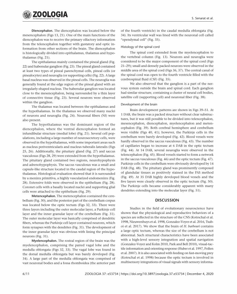