the minimum effective dose of lidocaine needed to …local anesthetics are used daily by anesthesia...
TRANSCRIPT

i
Thesis Approval Form
THE MINIMUM EFFECTIVE DOSE OF LIDOCAINE NEEDED TO BLOCKEVOKED POTENTIALS IN THE SCIATIC NERVE OF THE RAT
Bradley Wayne Stelflug
APPROVED:
Donald D. Rigamonti, Ph.D. (Co-Chair) Date
Howard Bryant, Ph.D. (Co-Chair) Date
Kenneth P. Miller, Ph.D. (Member) Date
APPROVED:
Report Documentation Page Form ApprovedOMB No. 0704-0188
Public reporting burden for the collection of information is estimated to average 1 hour per response, including the time for reviewing instructions, searching existing data sources, gathering andmaintaining the data needed, and completing and reviewing the collection of information. Send comments regarding this burden estimate or any other aspect of this collection of information,including suggestions for reducing this burden, to Washington Headquarters Services, Directorate for Information Operations and Reports, 1215 Jefferson Davis Highway, Suite 1204, ArlingtonVA 22202-4302. Respondents should be aware that notwithstanding any other provision of law, no person shall be subject to a penalty for failing to comply with a collection of information if itdoes not display a currently valid OMB control number.
1. REPORT DATE OCT 1998
2. REPORT TYPE N/A
3. DATES COVERED -
4. TITLE AND SUBTITLE THE MINIMUM EFFECTIVE DOSE OF LIDOCAINE NEEDED TOBLOCK EVOKED POTENTIALS IN THE SCIATIC NERVE OF THE RAT
5a. CONTRACT NUMBER
5b. GRANT NUMBER
5c. PROGRAM ELEMENT NUMBER
6. AUTHOR(S) Bradley Wayne Stelflug, BSN
5d. PROJECT NUMBER
5e. TASK NUMBER
5f. WORK UNIT NUMBER
7. PERFORMING ORGANIZATION NAME(S) AND ADDRESS(ES) Uniformed Services University of the Health Sciences
8. PERFORMING ORGANIZATIONREPORT NUMBER
9. SPONSORING/MONITORING AGENCY NAME(S) AND ADDRESS(ES) 10. SPONSOR/MONITOR’S ACRONYM(S)
11. SPONSOR/MONITOR’S REPORT NUMBER(S)
12. DISTRIBUTION/AVAILABILITY STATEMENT Approved for public release, distribution unlimited
13. SUPPLEMENTARY NOTES
14. ABSTRACT Local anesthetics are used daily by anesthesia providers to perform spinal, epidural, and peripheral nerveblocks. Large volumes of local anesthetic are used for peripheral nerve blocks especially if more than oneblock is performed on a patient. These local anesthetics can have side effects such as nerve compression,nerve damage, or even toxic plasma levels that can lead to seizures. Using less volumes and concentrationsof these anesthetics would reduce the chances of these complications. To discover the minimum amount oflocal anesthetic needed intraneurally, an in vivo model of the rat sciatic nerve was used. In the anesthetizedrat, the sciatic nerve was surgically exposed and then injected in the subperineural space with either 10 or20 ml of 2% lidocaine or a control solution. The proximal end of the nerve (at the greater sciatic notch) waselectrically stimulated and the tibial division of the nerve (near the ankle) was used for recording thecompound action potential (CAP). The averaged CAPs were recorded periodically for up to an hour. Thedata displayed a trend of 20ml of 2% lidocaine blocking the CAPs and that 10ml of 2% lidocaine did notconsistently block the CAPs. The data suggests that 20ml is the minimum dose of 2% lidocaine neededintraneurally to block evoked potentials in muscle and rapidly conducting sensory signals, including fast pain.
15. SUBJECT TERMS lidocaine; rats; minimum dose; in vivo; sciatic nerve
16. SECURITY CLASSIFICATION OF: 17. LIMITATION OF ABSTRACT
SAR
18. NUMBEROF PAGES
96
19a. NAME OFRESPONSIBLE PERSON
a. REPORT unclassified
b. ABSTRACT unclassified
c. THIS PAGE unclassified
Standard Form 298 (Rev. 8-98) Prescribed by ANSI Std Z39-18

ii
F.G. Abdellah,Ed.D.,Sc.D.,RN,FAAN DateDean
iv
DISCLAIMER STATEMENT
Department of Defense
“This work was supported by the Uniformed Services University of the Health Sciences
Laboratory Animal Review Board Protocol No. TO6133-01. The opinions or assertions
contained herein are the private opinions of the author and are not to be construed as
official or reflecting the views of the Department of Defense or the Uniformed Services
University of the Health Sciences.”

v
COPYRIGHT STATEMENT
The author hereby certifies that the use of any copyrighted material in the thesis entitled:
“THE MINIMUM EFFECTIVE DOSE OF LIDOCAINE NEEDED TO BLOCK
EVOKED POTENTIALS IN THE SCIATIC NERVE OF THE RAT”
beyond brief excerpts is within the permission of the copyright owner, and will save and
hold harmless the Uniformed Services University of the Health Sciences from any damage
which may arise from such copyright violations.

vi
ABSTRACT
Local anesthetics are used daily by anesthesia providers to perform spinal, epidural, and
peripheral nerve blocks. Large volumes of local anesthetic are used for peripheral nerve
blocks especially if more than one block is performed on a patient. These local
anesthetics can have side effects such as nerve compression, nerve damage, or even toxic
plasma levels that can lead to seizures. Using less volumes and concentrations of these
anesthetics would reduce the chances of these complications. To discover the minimum
amount of local anesthetic needed intraneurally, an in vivo model of the rat sciatic nerve
was used. In the anesthetized rat, the sciatic nerve was surgically exposed and then
injected in the subperineural space with either 10 or 20 µl of 2% lidocaine or a control
solution. The proximal end of the nerve (at the greater sciatic notch) was electrically
stimulated and the tibial division of the nerve (near the ankle) was used for recording the
compound action potential (CAP). The averaged CAPs were recorded periodically for up
to an hour. The data displayed a trend of 20µl of 2% lidocaine blocking the CAPs and
that 10µl of 2% lidocaine did not consistently block the CAPs. The data suggests that
20µl is the minimum dose of 2% lidocaine needed intraneurally to block evoked potentials
in muscle and rapidly conducting sensory signals, including fast pain.
KEYWORD: lidocaine, rats, minimum dose, in vivo, sciatic nerve

vii
THE MINIMUM EFFECTIVE DOSE OF LIDOCAINE NEEDED TO BLOCK
EVOKED POTENTIALS IN THE SCIATIC NERVE OF THE RAT
by
Bradley Wayne Stelflug, BSN
THESIS
Presented to the Graduate School of Nursing Faculty of
the Uniformed Services University of the Health Sciences
in Partial Fulfillment
of the Requirements
for the Degree of
MASTER OF SCIENCE DEGREE
UNIFORMED SERVICES UNIVERSITY OF THE HEALTH SCIENCES

viii
October 1998

ix
DEDICATION
I dedicate this thesis to Deanna, my wife, Hannah and Jacob, my children. Their
love and support have given me the ability to obtain any dream, and without them my
dreams would be meaningless.
x
ACKNOWLEDGEMENT
I would like express my extreme gratitude to Dr. Donald Rigamonti and Dr.
Howard Bryant, for their long hours and enthusiasm that made these experiments
possible, rewarding, and fun. I would like to thank Dr. Miller for helping me analyze the
data and for providing valuable advice.
I would like to express my appreciation to Dr. DiCarlo and Jim Schooley, for
surgical and veterinary assistance. Also, I would like to thank Dr. John Sarvey and Dr.
Paul Lea for their support of this thesis.
A special thanks to LTJG Kenneth Spence for his help and support of this thesis.

xi
TABLE OF CONTENTS
CHAPTER ONE: INTRODUCTION ……………………………………………… 1
Statement of the Problem ………………………………………………………………1
Background ……………………………………………………………………………. 2
Peripheral Nervous System …………………………………………………….2
Nerve Conduction ……………………………………………………………...3
Local Anesthetics ………………………………………………………………5
Classification of Nerve Fibers ………………………………………………… 6
Summary ……………………………………………………………………………… 7
CHAPTER TWO: REVIEW OF THE LITERATURE …………………………... 9
Introduction …………………………………………………………………………. 9
Blocking Evoked Potentials …………………………………………………………… 9
Local Anesthetics ………………………………………………………………………12
Nerve Damage with Local Anesthetics ………………………………………………...14
CHAPTER THREE: FRAMEWORK OF THE STUDY ………………………… 17
Local Anesthetics ………………………………………………………………………17
Local Anesthetics and Nerve Conduction ……………………………………………...18
CHAPTER FOUR: METHODOLOGY …………………………………………… 20
CHAPTER FIVE: DATA ANALYSIS …………………………………………….. 24
CHAPTER SIX: DISCUSSION …………………………………………………….34
Recommendations………………………………………………………………………39

xii
LIST OF REFERENCES……………………………………………………………. 40
APPENDICES………………………………………………………………………… 43
Appendix A Protocol Submitted to Laboratory Animal Review Board, USUHS form
6006……………………………………………………………………………………. 44
Appendix B Biohazards, Controlled Substances And Dangerous Materials, USUHS form
6007 (BCD) …………………………………………………………………………… 61
Appendix C Laboratory Animal Review Board Response Letter……………………..70
Appendix D Response Memorandum…………………………………………………72
Appendix E Laboratory Animal Review Board Approval Letter…………………….. 73
Appendix F Protocol Modification…………………………………………………… 74
Appendix G Protocol Modification Temporary Approval Letter……………………..75
Appendix H Protocol Modification Approval Letter……………………………….…76

xiii
LIST OF TABLES
Table 1 Lidocaine (2%, 20µl) and Control Solution (20µl) effects on Peak Amplitude
(mV) versus Time (msec) in the Sciatic Nerve………………………………………… 26
Table 2 Lidocaine (2%, 10µl) and Control Solution (10µl) effects on Peak Amplitude
(mV) versus Time (msec) in the Sciatic Nerve………………………………………… 27
Table 3 Lidocaine (2%, 20µl) and Control Solution (20µl) effects on Onset Latency
(msec) versus Time (msec) in the Sciatic Nerve……………………………………… 29
Table 4 Lidocaine (2%, 10µl) and Control Solution (10µl) effects on Onset Latency
(msec) versus Time (msec) in the Sciatic Nerve……………………………………… 30
Table 5 Lidocaine (2%, 20µl) and Control Solution (20µl) effects on Area Under the
Curve for Compound Action Potential (V/msec) versus Time (msec) in the Sciatic
Nerve……………………………………………………………………………………31
Table 6 Lidocaine (2%, 20µl) and Control Solution (20µl) effects on Area Under the
Curve for Compound Action Potential (V/msec) versus Time (msec) in the Sciatic
Nerve……………………………………………………………………………………32

xiv
LIST OF FIGURES
Figure 1 Sample Baseline Neurogram at 10 Times Threshold (volts) versus Time
(seconds) in the Sciatic Nerve…………………………………………………………. 25
Figure 2 Lidocaine (2%, 20µl) and Control Solution (20µl) effects on Peak Amplitude
(mV) versus Time (msec) in the Sciatic Nerve………………………………………… 26
Figure 3 Lidocaine (2%, 10µl) and Control Solution (10µl) effects on Peak Amplitude
(mV) versus Time (msec) in the Sciatic Nerve………………………………………… 28
Figure 4 Lidocaine (2%, 20µl) and Control Solution (20µl) effects on Area Under the
Curve for Compound Action Potential (V/msec) versus Time (msec) in the Sciatic
Nerve……………………………………………………………………………………31
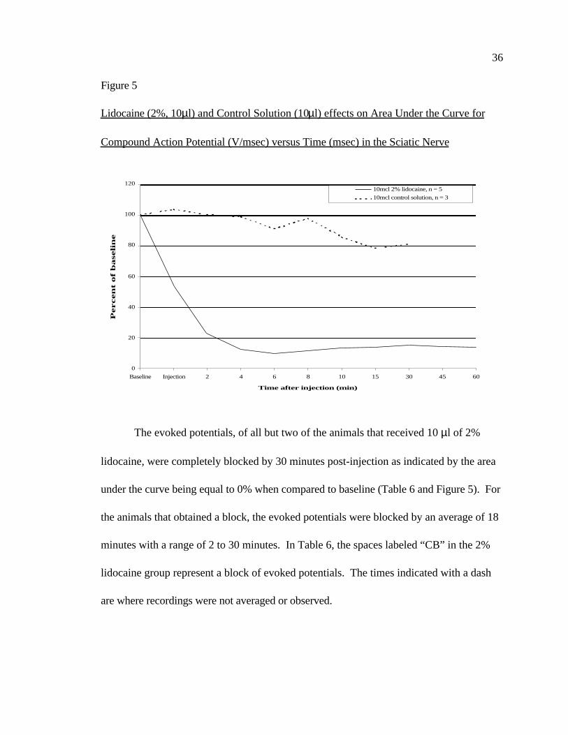
Figure 5 Lidocaine (2%, 10µl) and Control Solution (10µl) effects on Area Under the
Curve for Compound Action Potential (V/msec) versus Time (msec) in the Sciatic
Nerve……………………………………………………………………………………33
Figure 6 Lidocaine (2%, 10 & 20µl) and Control Solution (10 & 20µl) effects on Peak
Amplitude (mV) versus Time (msec) in the Sciatic Nerve …………………………… 35
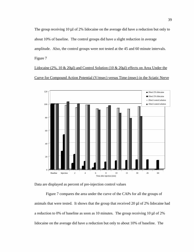
Figure 7 Lidocaine (2%, 10 & 20µl) and Control Solution (10 & 20µl) effects on Area
Under the Curve for Compound Action Potential (V/msec) versus Time (msec) in the
Sciatic Nerve ………………………………………………………………………….. 36
Figure 8 Lidocaine (2%, 10 & 20µl) and Control Solution (10 & 20µl) effects on
Conduction Velocity (m/sec) versus Time (msec) in the Sciatic Nerve ……………… 37

1
CHAPTER ONE: INTRODUCTION
Nurses have been delivering general anesthesia since Sister Mary Bernard became
the first identified nurse anesthetist in 1877 (Bankert, 1993). Regional anesthesia,
anesthetizing nerves of the peripheral nervous system, currently constitutes a large
portion of the nurse anesthetists’ practice. The first regional block was done in 1885 by
Halstead in an experiment in which he blocked the brachial plexus with cocaine (Stoelting
& Miller, 1994). Today, regional blockade of most sensory peripheral nerves is done for
procedures such as operations on shoulders, hands, legs, ankles, and feet. Anesthesia
providers are taught to use recommended volumes and concentrations, with or without
vasodilators, of local anesthetic to perform these blocks. For example, in Stoelting and
Miller’s book, Basics of Anesthesia, 25 ml of local anesthetic is recommended to block a
sciatic nerve, 25 to 40 ml to block the brachial plexus or the axillary nerve, and 10 to 20
ml for the femoral nerve. Even though many local anesthetics are available from which the
anesthesia provider may choose, there are still potential problems (Stoelting, 1991).
Statement of the Problem
Recommended volumes of anesthetic to perform a regional blockade of a
peripheral nerve can cause adverse sequelae such as nerve damage from local anesthetic
toxicity, tissue edema, or systemic toxicity which could lead to seizures (Stoelting, 1991:
Kalichman, Powell, & Myers, 1989). A large volume of local anesthetic is used for
regional anesthetic procedures to ensure that the nerve becomes anesthetized. If more
than one block is performed, such as sciatic and femoral nerve blocks, the total dose of

2
local anesthetic that may be absorbed systemically could reach toxic levels. Also, local
anesthetics have direct toxic effects on nerve tissue and may cause injury and edema.
An in vivo model has been reported in abstract form, by Paris, Pahno, Rigamonti,
Jimmerson, & Seng in 1990, to study the minimum effective dose of a local anesthetic to
block nerve conduction in the sciatic nerve of an animal. This thesis will describe what
volume of commercially prepared local anesthetic, in an intraneural injection, is needed to
block the sciatic nerve of the rat using the model developed by Paris et al. Neural signals,
recorded and analyzed by a computer, will be used to determine the minimum dose
needed to block muscle response and the fast conducting sensory system, including the
fibers commonly thought to conduct “fast pain” signals.
Background
Peripheral Nervous System
The nervous system can be divided into two parts, the central nervous system
(CNS) and the peripheral nervous system (PNS). The CNS consists of the brain and the
spinal cord, and although very important, it is not the target of 2% lidocaine which is
studied in this thesis. Thus, this section will be limited to a brief description of the PNS.
The peripheral nervous system consists of 31 pairs of spinal nerves, 12 pairs of
cranial nerves, and the autonomic nervous system (ANS). Due to the scope of this thesis,
only spinal nerves, which include the sciatic nerve, will be described. Gilman and
Newman (1996) describe the PNS in their book, Manter and Gatz’s Essentials of Clinical
Neuroanatomy and Neurophysiology. They wrote about two classes of spinal nerves,

3
efferent and afferent nerves. Afferent nerves are sensory nerves that have their cell bodies
located in a ganglia. For example, the cell body for an afferent spinal neuron is located in
the dorsal root ganglion. Sensory nerves send nerve impulses towards the spinal cord in
response to stimuli such as pain, temperature, vibration, etc. These nerves may synapse
in the dorsal horn of the spinal cord’s gray matter. The gray matter of the spinal cord is
subdivided into 10 regions termed laminae. The dorsal horn is comprised of laminae I to
VI. Lamina I, also called the marginal zone, and lamina II, also called the substantia
gelatinosa, receive impulses from C-fiber and A-δ fiber nerves that are stimulated by pain,
temperature, touch, and pressure. Laminae III and IV, also termed the nucleus proprius,
and laminae V and VI receive input from A-α and A-β fiber nerves that are stimulated by
light touch, vibration, and muscle spindles as well as Golgi tendon organs which signal
proprioception.
The nerve impulses are then either processed in the spinal cord or in the brain
(Gilman & Newman, 1996). Efferent nerves send nerve impulses towards effector organs,
muscles, glands, etc. Efferent nerves have their cell bodies located in the ventral or
intermediate horns of the spinal cord’s gray matter. Lamina VII contains the cell bodies
of the B-fiber, autonomic nerves, and lamina IX contains the cell bodies of the A-α and
A-γ fibers, motor nerves. Afferent and efferent spinal nerve fibers are bound together in
an epineural sheath and innervate distal structures of the body. They are collectively
called a nerve. For example, the sciatic nerve is composed of efferent and afferent nerves
of all fiber classifications except B fibers. The sciatic nerve carries afferent impulses from

4
the skin, muscles, joints, and fascia of the leg and efferent impulses to the muscles, glands,
and smooth muscle of the leg.
Nerve Conduction
Strichartz (1988) described nerve conduction in the book, Neural Blockade in
Clinical Anesthesia and Management of Pain. He wrote that sensory and motor messages
are transported along a nerve in the form of an electrical impulse. The sciatic nerve
consists of thousands of individual axons. An electrical potential is maintained in each
axon by maintaining a concentration gradient across its membrane. In the resting state,
there is a slightly higher concentration of potassium (K+) ions intracellularly and a higher
concentration of sodium (Na+) ions extracellularly. This, along with the semi-permeable
cell membrane, creates a resting membrane potential of approximately –70 to -80 mV. As
Na+ ions enter the cell, the resting membrane potential becomes more positive
(depolarizes). As the membranes potential depolarizes, it reaches a state called threshold
at which time voltage gated Na+ channels open up and a rapid influx of Na+ ions occurs.
This is the start of an action potential. An action potential will peak at approximately
110 mV even with different, short duration, stimuli strengths as long as the membrane
threshold is reached. Voltage gated K+ channels also open when threshold is reached but
at a slower rate, so when the Na+ channels close, the membrane potential becomes more
negative (repolarizes) until it returns approximately to its resting state. During the action
potential, the Na+ ions that entered the axon then cause the distal membrane, such as the

5
next node of Ranvier in a myelinated fiber, to depolarize and when it reaches threshold,
another action potential is generated. This is the manner in which an impulse travels
down a nerve. The conduction of this impulse is aided in myelinated nerve fiber by
Schwann cells. In myelinated fibers each Schwann cell is wrapped many times around the
nerve axon. The gap between these protective cells is called the node of Ranvier. The
voltage gated Na+ channels that produce the action potential are located predominantly in
these nodes. This allows the action potential to travel along the nerve from node to node
with the benefits of faster conduction and less Na+ crossing the membrane than an
unmyelinated axon (Gilman & Newman, 1996).
During an action potential, the voltage gated Na+ channels are in the open position
at the start of the action potential and then go into an inactivated or closed state
(Strichartz, 1988). These Na+ channels must then reset to their resting state before they
can be opened again. This time while the Na+ channels are resetting is called the
refractory period. The refractory period has two phases. The absolute refractory period,
in which the membrane cannot be further stimulated to produce another action potential,
lasts from the time the membrane reaches threshold to the time the membrane is partially
repolarized. This is followed by the relative refractory period during which the
membrane can respond to a strong stimulus and produce another action potential. An
electrically stimulated action potential that begins at the node of Ranvier can travel in
either direction up or down the axon.
Local Anesthetics

6
Local anesthetics were first used in 1884 when Carl Koller used cocaine topically
for his ophthalmic procedures (Duncum, 1947). William Halstead performed the first
regional anesthesia in 1844 by blocking the brachial plexus with cocaine. In 1885 Corning
placed cocaine extradurally to anesthetize the spinal cord. August Bier, in 1899,
performed the first planned spinal anesthetic. Alfred Einhorn developed procaine in 1904
to be used as a local anesthetic with less side effects than cocaine. The first of the amide
local anesthetics, lidocaine, was made by Nils Lofgren in 1943.
Local anesthetics are weak bases with pKa’s higher than normal physiologic pH
which means that more than half of the drug will exist in the non-active ionized form in
the blood (Stoelting, 1991). There are two different groups of local anesthetics, the esters
(procaine, chloroprocaine, tetracaine) and the amides (lidocaine, mepivacaine, bupivacaine,
etidocaine, prilocaine, ropivacaine). They are grouped by the specific type of
hydrocarbon chain they have between their lipophilic and their hydrophilic ends. The
ester local anesthetics are metabolized mainly by plasma cholinesterase. The amide local
anesthetics are mainly metabolized in the liver by microsomal enzymes. Allergic
reactions are more common with the ester local anesthetics because they are metabolized
to structures that are similar to para-aminobenzoic acid. Also, preservatives in local
anesthetics that resemble para-aminobenzoic acid may also cause allergic reactions.
Systemic toxicity is more likely to occur with use of amide local anesthetics because they
are metabolized slower.

7
Local anesthetics work by blocking passage of sodium ions through voltage gated
sodium channels located in the cellular membrane of a nerve (Stoelting, 1991). The
anesthetics attach to a specific receptor on the voltage gated sodium channels that
respond to nerve impulses. Blockage of this flow of sodium ions will stop the membrane
from reaching threshold and an action potential from propagating.
Classification of Nerve Fibers
Differentiating fibers of an amphibian nerve can be accomplished by recording the
nerve’s compound action potential (Gasser & Erlanger, 1929). Electrodes are placed on a
nerve to record nerve impulses as they travel the length of the nerve fibers. Because nerve
fibers have different conduction velocities, relative to their diameter and myelination
properties, these impulses will reach the electrodes at different times. The larger
diameter, myelinated fibers have the greatest conduction velocities (Gilman & Newman,
1996). The conduction velocities, from largest to smallest diameter in mammals, are 120
m/sec for Aα fibers, 70 m/sec for Aβ fibers, 40 m/sec for Aγ fibers, 15 m/sec for Aδ
fibers, 14 m/sec for B fibers, and 2 m/sec for C fibers. An oscilloscope records these
impulses and the tracing is displayed on a grid with the y-axis being amplitude and the x-
axis being time. The stronger impulses will create greater voltage amplitude curves. The
faster impulses will be recorded first. In a normally functioning nerve that contains these
different fiber groups, the first peak will have the greatest amplitude and represent the
Aα fibers. These fibers terminate in striated muscle and are responsible for muscle
twitch. The next peak will be smaller and represent the Aβ fibers. This continues in

8
order of fastest conduction velocity to the slowest. By interpreting the oscilloscope
tracing, data can be obtained on the blocking effects of local anesthetics on nerve fibers.
As nerve fibers were blocked, the tracings would show lessening amplitudes for that
group of fibers.
Summary
This chapter briefly discussed what potential problems there are with
administering a regional anesthetic block to a peripheral nerve. It described the main
components of this thesis, the PNS, which is a targeted anatomy in regional anesthesia,
nerve conduction, and the basics on how impulses are transmitted by nerve fibers. Also,
the other major component of this thesis, local anesthetics, was described. The
mechanism of action of local anesthetics and how they block conduction of nerve
impulses, which is the framework for this thesis, is described in chapter three.

9
CHAPTER TWO: REVIEW OF THE LITERATURE
Introduction
The literature describes how evoked potentials are blocked, how local anesthetics
work, and what damage local anesthetics can create. This review forms the physiologic
framework for this study. Evoked potentials are blocked at different rates in each kind of
nerve fiber. This is termed differential blockade. This can usually be seen clinically when
a nerve is anesthetized, the patient will lose neural function in the following order:
autonomics, temperature, pain, touch, pressure, motor, vibratory, and lastly
proprioception. Also, when a patient receives an epidural block, autonomic response is
blocked two to four dermatomes higher and sensory is blocked two dermatomes higher
than motor. It is still not fully understood how this happens, but there are different
possible explanations. Local anesthetics have been well described in the literature and
their toxic effects on nerve fibers have also been reported. Although Cauda Equina
Syndrome and Transient Radicular Irritation are important injuries from lidocaine
anesthesia, this thesis will focus on the sciatic nerve as a model which may be important
to the other syndromes.
Blocking Evoked Potentials
Nobel Prize recipients Gasser and Erlanger (1929) described the relation between
amphibian nerve fiber size and blockage of its nerve impulses by cocaine and
compression. They found that there was a differential blockade of nerve fibers but there
were no experimental data to show what quality of the nerve fiber made this happen.

10
They had previously found that velocity of nerve conduction related directly to the fibers
diameter. This was used to determine differential blockade by oscilloscope tracings.
Gasser and Erlanger found that differential blockade made by cocaine in a frog nerve was
inconsistent or not even present. They found that in every mammalian nerve, that they
tested, that the smaller fibers were blocked before the larger fibers. In one experiment,
they found that the Aδ wave, as seen on the oscilloscope, was always completely
blocked before the Aαβγ group. They did another experiment to focus on the Aαβγ
group and found, “γ was more affected than β, and β more than α; but during the blocking
of the slower waves the faster waves were undergoing alterations in form” (p.587-8).
They concluded with the sum of all their experiments that the amphibian nerve size is a
determining factor in how susceptible it is to cocaine but that during differential blockade,
fibers do not drop out strictly based upon their diameter.
Jaffe and Rowe (1996) used an in vitro model to try and explain differential nerve
block. Their model used lumbar dorsal roots and cervical area vagus nerves from adult
Sprague-Dawley rats. The section of nerve was removed and placed into an artificial
cerebrospinal fluid solution where the distal end was stimulated with 0.3 Hz, 0.2 ms
duration pulses. The proximal end was divided into small sections of one to three fibers
in which recordings were taken. After recording baseline data, the artificial cerebrospinal
fluid was replaced with a solution containing 150µM of lidocaine (Astra). If there were
axons that were not blocked at this concentration, they increased the lidocaine to 260µM
and then 540µM. Jaffe and Rowe’s found that of the dorsal root axons, 88% of

11
unmyelinated and 100% of myelinated were blocked with 520µM of lidocaine. Their
results comparing the vagus nerve axons and dorsal root axons with 260µM lidocaine
showed that fewer unmyelinated and myelinated vagal axons were blocked but that only
the myelinated difference was statistically significant. They concluded that, “compared
to unmyelinated axons, myelinated dorsal root axons are significantly more sensitive to
steady-state sodium channel blocking effects of lidocaine” (p. 1463). They hypothesized
that one reason differential block from an epidural block occurs is because with short
sections of spinal roots being anesthetized, the large diameter myelinated axons will have
fewer nodes exposed to the local anesthetic.
Raymond, Steffensen, Gugino, and Strichartz (1989) performed an in vitro study
to show how exposure length helps determine nerve impulse blockade. They exposed
different lengths (5 to 30mm) of the sciatic nerve of frogs to various concentrations of
lidocaine (Astra). They electrically stimulated the nerve and recorded the compound
action potentials from the distal end. The stimulating and recording ends were not
exposed to the local anesthetic. The researchers also took recordings from single fibers
that were teased from the distal end. In these experiments, the portion of nerve trunk
exposed still had its sheath intact. The researchers found that the slower, small diameter,
myelinated fibers were not more susceptible to being blocked by lidocaine with the
lengths of exposure that they used. Their single fiber testing lead them to conclude, “at
short exposure lengths, more than twice as much anesthetic was required than at the
longest exposure lengths (25-35mm)” (p. 567). Finally, Raymond, et al. found, “that at

12
any given concentration of lidocaine there is direct relation between the incidence of block
in a fiber population and the length of nerve exposed to LA [local anesthetics]” (p. 568).
Huang, Thalhammer, Raymond, and Strichartz (1997) tested different afferent rat
sciatic nerve fibers in vivo to determine their sensitivity to lidocaine. They exposed the
sciatic nerve and the tibial division, placed a 22mm section in a bathing tube in which
lidocaine could be added or washed out, and then they separated out nerve fibers by A-β,
A-δ, and C at the proximal end to record. The distal end of the nerve was stimulated and
the bathing tube was infused with different concentrations of lidocaine. Also, the
researchers used 14C-lidocaine to determine concentration of the lidocaine that diffused
across the neural sheath into the nerve fiber. They found that a steady state of lidocaine
uptake was obtained at approximately 20 minutes and that the mean lidocaine uptake was
1.0 ± 0.2 nmol/mg of wet nerve. Their results of differential blockade showed, "when
characterized by their median blocking concentrations (IC50s), nociceptive Aδ-fibers were
blocked at the lowest concentration, 0.32 mM, compared with 0.41 mM for LTM [light
touch mechanoreceptor] Aβ-fibers and 0.80 mM for of nociceptive C-fibers" (p. 808).
They concluded that diameter does not determine a nerve's susceptibility to local
anesthetics.
Local Anesthetics
Butterworth and Strichartz (1990) reviewed the mechanisms of local anesthetics
on voltage gated Na+ channels. Local anesthetics have both a tonic and a phasic mode of
blocking the Na+ channel. In the tonic phase, the local anesthetic binds to the Na+ channel
13
while the channel is closed resulting in less Na+ flowing into the cell. During the phasic
block, the local anesthetic binds to either the open, activated, or inactivated Na+ channel.
There are two theories for this, one of which is that the site that local anesthetics binds to
is altered when the Na+ channel changes states and this causes the local anesthetic to be
bound tighter to open and inactivated states. The other hypothesis is that the receptor is
guarded and can be accessed when the Na+ channel is such that the guarding is less. The
researchers did write that one property of a local anesthetic is whether it produces more
tonic block or more phasic block.
Butterworth and Strichartz (1990) described the structure of a Na+ channel as
being a polypeptide, which could be divided into four sections, each containing six to
eight amino acids. The four sections form four quadrants of a cylinder with the Na+
channel being in the middle. The amino acid labeled “c” for each quadrant is located in the
center and these four amino acids form the Na+ channel. The Na+ channel opens when the
“g” amino acids move intracellularly and the “d” amino acids move extracellularly allowing
the “c” amino acids to move away from each other and thus opening the Na+ channel.
The researchers wrote, “we propose that LAs [local anesthetics] bind at the dipole-
containing helices such as to inhibit their arrangement in response to membrane
depolarization” (p. 730).
Recently, Popitz-Bergez, Leeson, Strichartz, and Thalhammer (1995) conducted
an in vivo experiment to determine the amount of intraneural Lidocaine that corresponds
to different phases of blockade. All the animals were given a sciatic nerve block, with 0.1

14
ml of 1.0% 14C lidocaine HCl, by a single injection between the greater trochanter and the
ischial tuberosity. The animals were euthanized during either full blockade, when deep
pain returned, or when complete functions returned in order to measure the amount of
intraneural lidocaine. This was determined by performing an examination that had been
previously described in the literature by Thalhammer. Their results showed that for total
blockade, averaging 5 to 35 minutes, there was only 1.6 ± 0.12% of the total dose of
lidocaine injected found intraneurally. When the response to deep pain returned, average
32 to 40 minutes, there was only 0.33 ± 0.035% of the total dose of lidocaine detected.
Finally, when all functions returned, they detected only 0.065 ± 0.035% of the total dose
of lidocaine injected. In their discussion, they wrote, “It is interesting that the minimal
ratio of drug dose to body weight producing a full block of function seems to be the same
for rats and humans” (p. 590). For this statement, the researchers compared their results
with those of Cousins and Bridenbaugh who wrote that a sciatic nerve block in humans
can be accomplished with 10 cc of 2% Lidocaine. They calculated the body weight to
drug dose ratio was exactly the same when comparing a 70 kg human and a 350 gram rat,
which is the weight of the animals that these researchers studied.
Popitz-Bergez, Leeson, Strichartz, and Thalhammer (1997) repeated this study to
compare intraneural lidocaine uptake between pregnant and nonpregnant male and female
rats. In comparing pregnant and nonpregnant rats, they wrote, “The block of deep pain
sensation was prolonged by 45% in pregnant rats and that the amount of local anesthetic
present in the nerve at the time of deep pain recovery was also significantly less in

15
pregnant rats than in nonpregnant rats” (p. 369). This study yielded similar results for
the male rats as their previous study. When deep pain sensation returned, the intraneural
lidocaine content was 0.34 ± 0.03% of the total dose injected.
Nerve Damage with Local Anesthetics
Kalichman, Moorhouse, Powell, and Myers (1993) examined the toxicity of local
anesthetics to nerves. They anesthetized the exposed sciatic nerve of Sprague-Dawley
rats with 1 ml of either etidocaine, lidocaine, 2-chloroprocaine, or procaine. Then, 48
hours after applying the local anesthetic, they removed the section of nerve that was
exposed to the anesthetic, sectioned it one micron thick and stained the sections with p-
phenylenediamine and examined them under light microscopy. These sections were given
nerve injury scores of 0 (no injury) to 4 (more than half of the fibers injured in one
fascicle or if there was nerve damage in three or more fascicles). The nerve sections were
also assessed for edema. Kalichman et. al. also experimented with 57 other Sprague-
Dawley rats to find the concentration of the four local anesthetics that would produce
50% motor nerve conduction block. This was done to be able to compare nerve injury to
anesthetic potency. Anesthetic (250µl) was applied to each side of the sciatic nerve for a
total of 500µl. Then they recorded EMG responses from the interosseous muscles on the
ipsilateral foot with an oscilloscope. They used this measure to determine when the
amplitude was decreased to 50% of baseline.
Kalichman et al. (1993) found that all four of the anesthetics produced edema. In
comparing nerve injury and potency of anesthetic, they wrote, “these data support the

16
hypothesis that local anesthetic neural toxicity parallels potency for producing local
anesthesia” (p. 239). They also found that nerve injury was more severe in small fascicles
and edema was more likely to be formed in large fascicles. Lidocaine, in their study, was
shown to have 88% to 100% conduction block with 1% solution and a nerve injury score
of 2.2 +/- 0.6 at 1.3% solution. A nerve score of 2 was obtained if up to one forth of the
fibers in a fascicle were injured.
Kalichman, et al. (1989) performed a similar study earlier in which they also
looked at four different local anesthetics and their effects on nerve tissue. They wrote
that, “at high concentrations, and sometimes even at clinical concentrations, local
anesthetics are capable of producing dose-dependent injury to peripheral nerve” (p. 406).
In this study, the researchers injected 1 ml of local anesthetic perineurally on the sciatic
nerve of the rat. The local anesthetics tested were lidocaine (1.5%, 2.1%, 3%), 2-
chloroprocaine (2%, 2.8%, 4%), etidocaine (1%, 1.4%, 2%), and procaine (5%, 7%, 10%).
The sciatic nerve was removed 48 hours after the injection and dissected into 1µ thick
sections to be studied with light microscopy. A myelinated nerve fiber was considered
injured if it was swollen, had a disintegrated axon, the nerve fiber was dystrophic, or if it
became thinly myelinated or demyelinated. They found that nerve injury was
significantly different from the control solution only with the highest concentrations. The
researchers also looked at edema in the nerve tissue. They hypothesized that at clinical
concentrations, the edema most likely would not cause any lasting neurological effects.
Kalichman et al. reported that it would be possible to produce lasting deficits if higher

17
concentrations were used, epinephrine was used, or if a dose was repeated. About nerve
injury, the researchers concluded that, “four very different local anesthetic agents produce
patterns of nerve injury with relative potencies quite similar to those for producing local
anesthesia” (p. 410).
18
CHAPTER THREE: FRAMEWORK OF THE STUDY
This study will have a physiologic framework based on the current research and
interpretation of how nerves transmit impulses and how local anesthetics exert their
effects. The framework has been described in the introductory chapter during the
background section.
Local Anesthetics
In his description of the mechanism of action for local anesthetics, Stoelting
(1991) wrote that local anesthetics bind selectively to Na+ channels when they are in an
inactivated-closed state which prevents the Na+ channels from changing to a resting or
open-active state. How quickly the local anesthetic works is determined by the
anesthetic’s pK. The pK of an anesthetic equals the pH at which it exists 50% in the
nonionized form. The more nonionized form an anesthetic is in, the quicker it will be able
to work. The nonionized form is able to cross the cell membrane and then, in the lower
pH of the intracellular fluid, become ionized and attach to the receptor which is located on
the intracellular side of the sodium channel. The duration of the block can be increased by
adding epinephrine, giving a larger dose, or by giving a more lipid soluble anesthetic.
The least concentration of a local anesthetic that is needed to block nerve impulses
is termed minimum concentration (Cm). An increase in pH at the site of injection will
decrease the Cm of the local anesthetic. Also, the high frequency motor fibers require less
local anesthetic than lower frequency sensory fibers. Even though motor fibers have
higher frequencies and therefore should be blocked easier and with less anesthetic,

19
clinically it is the sensory fibers that are seen being blocked first and for the longest
duration. This happens because the sensory fibers have less length that must be exposed
to the anesthetic for the nerve impulses to be blocked. The C fibers are unmyelinated and
the A-δ fibers are lightly myelinated.
Another property of LAs is that they cause frequency-dependent blockade. The
ionized, intracellular LA only enters the Na+ channel in the activated open state, so the
more the nerve transmits evoked potentials (frequency) the more the Na+ channels will be
in the activated open state which allows the LA to bind to it. LAs also cause differential
conduction blockade. This is displayed by B-fibers blocking first and with lesser
amounts of LA, the C-fibers and Aδ-fibers blocking next with higher concentrations of
LA, and the rest of the A-fibers blocking last and requiring the highest concentration of
LA.
LOCAL ANESTHETICS AND NERVE CONDUCTION
Ichiji Tasaki (1982), in his book Physiology and electrochemistry of nerve fibers,
notes the effects of anesthetics on nerve impulses. He wrote, “local anesthetics prolong
the least interval while they shorten the absolute refractory period” (p. 79). Also, the
higher concentration of anesthetic will decrease the time needed to block conduction in the
nerve. Tasaki stated that cocaine, “conduction block takes place at a concentration high
enough to reduce the action potential amplitude of the node to about _ of the normal
value”. Another concept is that the more length of a nerve exposed to an anesthetic, the
quicker nerve conduction is blocked. This occurs until a length of approximately 6mm is

20
exposed to a local anesthetic. In large diameter myelinated fibers, 6mm corresponds to an
average of 2.5 nodes. Blockade of nerve conduction occurs more rapidly in small diameter
myelinated fibers because they have more nodes in 6 mm than large diameter fibers.

21
CHAPTER FOUR: METHODOLOGY
This is a quantitative, descriptive study to determine the minimum amount of
lidocaine (2%) needed to block recorded, averaged, evoked potentials. The model
reported by Paris et al. was followed. Animal procedures for these experiments were
approved by the Laboratory Animal Review Board at the Uniformed Services University
of the Health Sciences. A total of 16 adult Sprague-Dawley rats were operated on,
preliminary data and experimental procedures were collected in six animals and 10 animals
yielded useful data. The animals were numbered sequentially 1 to 16. Five animals were
placed in each experimental group, control group data were obtained from three animals
before they were placed in the experimental group.
The animals were initially anesthetized using Nembutal Sodium Solution (Abbott
Laboratories, North Chicago, IL) 75 mg/kg ip. Supplemental doses were administered as
needed, determined by the presence of a corneal reflex, and then given every hour after
administration of the muscle relaxant. The trachea was surgically exposed and a tracheal
tube was inserted. The internal jugular vein was cannulated, in order to administer d-
Tubocurarine, and kept patent with Heparin Sodium Injection, USP (Pharmacia and
Upjohn Company, Kalamazoo, MI). The left sciatic nerve was then surgically exposed at
the greater sciatic notch and the ankle with the aid of a surgical microscope (Zeiss).
A pair of “stimulating” electrodes was placed on the nerve at the greater sciatic
notch and another pair of “recording” electrodes was placed on the tibial nerve near the
ankle (Figure 2). The electrodes were embedded in a low melting point wax in order to
22
avoid possible signal interference from surrounding tissue and also to ensure that the
electrical impulse would go only to the nerve. The nerve was crushed and tied off
proximal to the stimulating electrodes and distal to the recording electrodes. The d-
Tubocurarine Chloride (Sigma Chemical Company, St. Louis, MO) 0.04mg/kg was
administered through the cannulated internal jugular vein. The animal was then artificially
ventilated via a Harvard Apparatus Rodent Respirator. The respirations were 50 breaths
per minute, with a tidal volume of 1 cc per 100g of rat mass. Core temperature was
physiologically maintained by placing the animal on a heating pad. Rectal temperature
monitored with Physitemp BAT-12 (Physitemp Instruments Inc., Clifton, NJ).
The recording electrodes were connected to the differential inputs of a DAM 50
(World Precision Instruments, Inc., Sarasota, FL) biological amplifier. Gains were
between 100 and 1000. Bandwidth was 0.01 to 3 kHz. The amplifier output was
displayed on an oscilloscope for continuous monitoring and recorded on a MacIntosh
computer. A MacLab/8 system connected to the computer running Scope v3.5 software
(Analog Digital Instruments) was used to initiate the electrical stimulus and to record the
nerve impulses. The nerve was stimulated with a 0.2 ms duration pulse every 5 seconds
delivered by a Grass S44 Stimulator through a Grass SIU5 Stimulus Isolation Unit (Grass
Medical Instruments, Quincy, Mass). Thirty-two responses were averaged. Baseline
neurograms were obtained at ten times threshold. Stimulus threshold was determined to
be the minimum amount of voltage needed to create a detectable positive deflection in the
recorded neurogram. After baseline values were obtained, an injection of clinical grade

23
lidocaine 2% (Abbott Laboratories, North Chicago, IL) or control solution was made
inside the nerve sheath via a glass micropipette, pulled to a OD<30µ tip, and then post
injection recordings were obtained. Recordings were made for one hour after injection.
The trials were performed at the volumes of 10µl and 20µl. The control group
(consisting of 3 animals) was injected with a modified Krebs-Henselit solution (NaHCO3
27.2mM, NaCl 118mM, KCl 4.8mM, KH2PO4 1.0mM, MgSO4 1.2mM, CaCl2 2.5mM,
and glucose 11.1mM osmolarity of 300 +/- 5 mosM and a pH of 7.4 +/- 0.05) to
determine the effect volume may play in blocking evoked potentials. After compound
action potentials return to baseline, one animal was then given an additional of rat Ringer
solution with Methylene Blue to measure the diffusion distance of the 10µl volume in the
nerve’s sheath. All solutions (control or 2% lidocaine) were injected by the same
investigator into the nerve under microscopic conditions, distal to the stimulating
electrodes, using a glass micropipette placed under the perineurium.
The remaining animals (5 per volume group) were injected with lidocaine 2%
which is sold commercially to be used clinically on humans. Data collected include:
weight of rat, volume of lidocaine used, initial recordings of the compound action
potentials, the amplitude, latency, area under the curve until blockage of evoked
potentials was complete, and spread distance of the solution. The data collected was
described in a quantitative manner. The animals were euthanized, after the experiment
end point and before the anesthesia wore off, by giving 200mg/kg ip of Nembutal. The
animals’ chests were also opened to create a pneumothorax. After the animals were

24
euthanized, the distance between the stimulating and recording electrodes was measured
by dissecting the sciatic nerve. This was used to calculate the conduction velocity of the
nerve fibers.

25
CHAPTER FIVE: ANALYSIS OF THE DATA
The equipment used to record and analyze the data included a MacLab/8 with
Scope v3.5 software, which was wired to feed the data into a MacIntosh computer. The
data collected included: animal weight, lidocaine volume, initial recordings of the
compound action potentials, the amplitude, onset latency, area under the curve until
blockage of evoked potentials is complete, and spread distance of the solution. To obtain
the CAP, the nerve was stimulated and the neural response recorded thirty-two times.
The average of these thirty-two responses became the CAP used for data analysis. The
amplitude was measured as the greatest positive amplitude (mV) after the stimulus. This
corresponded to the A-α wave, which is responsible for motor activity. Onset latency
was measured as the time from stimulus to the time of first positive deflection in the
tracing. Area under the curve was measured from the first positive deflection until the
tracing returned to zero volts. The computer calculated the area under this curve. Area
under the curve is another test to measure if impulses are transmitted along the nerve. A
dye was used to measure how far the solutions travel when injected under the neural
sheath. Also, conduction velocity was measured by dividing onset latency by the length
of the nerve. Conduction velocity slows as a nerve becomes blocked (Gasser & Erlanger,
1929).
The weight of the Sprague-Dawley rats ranged from 252 grams to 520 grams.
There were seven males and three females with the average weight of the animals being
363 grams. Evoked potentials were recorded from these animals before and after being
26
injected intraneurally with 10 µl of control solution, 20 µl of control solution, 10 µl of 2%
lidocaine, or 20 µl of 2% lidocaine. The stimulating voltage for these recordings were 10
times threshold. Threshold was determined to be the minimum amount of voltage needed
to create a positive deflection in the recorded neurogram.
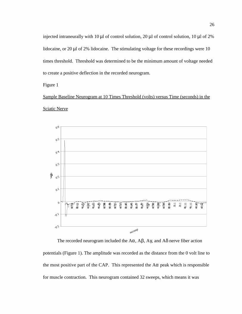
Figure 1
Sample Baseline Neurogram at 10 Times Threshold (volts) versus Time (seconds) in the
Sciatic Nerve
The recorded neurogram included the Aα, Aβ, Aγ, and Aδ nerve fiber action
potentials (Figure 1). The amplitude was recorded as the distance from the 0 volt line to
the most positive part of the CAP. This represented the Aα peak which is responsible
for muscle contraction. This neurogram contained 32 sweeps, which means it was
-0.2
-0.1
0
0.1
0.2
0.3
0.4
0.5
0.6
0
0.01
0.01
0.02
0.02
0.03
0.03
0.04
0.04
0.05
0.05
0.06
0.06
0.07
0.07
0.08
0.08
0.09
0.09
0.09 0.
1
0.1
0.11
0.11
0.12
0.12
seconds
volts

27
stimulated 32 times and the responses were averaged. A biological amplifier was used to
increase the recorded response by 100 times.
Table 1
Lidocaine (2%, 20µl) and Control Solution (20µl) effects on Peak Amplitude (mV) versus
Time (msec) in the Sciatic Nerve
Rat #2lido.
Rat #3lido.
Rat #5lido.
Rat #7lido.
Rat #13lido.
Rat #15control
Rat #16control
Rat #16control
Base 100 100 100 100 100 100 100 100Inject 18.359 76.076 26.657 0.498 57.500 99.726 100.12 96.432 min 0.103 41.097 0.411 CB 15.969 100.99 97.020 95.4414 min 0.203 23.201 CB CB 1.871 101.26 100.37 93.9186 min CB 11.665 CB CB CB 101.25 96.101 91.7438 min CB 5.526 CB CB CB 100.69 94.939 92.65710 min CB 0.033 CB CB CB 98.443 96.752 93.71015 min CB CB CB CB CB 95.102 95.006 89.17130 min 1.317 CB CB CB CB 99.493 96.752 90.51645 min 2.115 CB CB CB CB - - -60 min - CB CB 0.788 CB - - -Data are reported as percent of pre-injection control valuesCB = Complete Block- = no data
Figure 2
Lidocaine (2%, 20µl) and Control Solution (20µl) effects on Peak Amplitude (mV) versus
0
20
40
60
80
100
120
BaselineInjection
2 4 6 8 10 15 30 45 60
Time after injection
Perc
ent o
f Bas
elin
e
20mcl 2% lidocaine, n=520mcl control solution, n=3

28
Time (msec) in the Sciatic Nerve
Table 1 and Figure 2 show what percent of the baseline of the peak amplitude
attained after injection with 20µl of 2% lidocaine or 20µl of control solution. All animals
that received 20µl of 2% lidocaine showed a decrease in amplitude until the evoked
potentials were completely blocked. The animals that received 20µl control solution
showed a slight decrease in amplitude but not less than 89% of baseline. Of note is that
this protocol was not amended to add the use of d-Tubocurare until after rat #7. There
was no observable twitch for rat #2 at 30 and 45 minutes and there was no observable
twitch for rat #7 at 60 minutes. In Table 1, the spaces labeled “CB” in the 2% lidocaine
group represent a block of evoked potentials. The spaces with a dash indicate where
recordings were not averaged or obtained.
Table 2
Lidocaine (2%, 10µl) and Control Solution (10µl) effects on Peak Amplitude (mV) versus
Time (msec) in the Sciatic Nerve
Rat #8lido.
Rat #9lido.
Rat #12lido.
Rat #15lido.
Rat #16lido.
Rat #13control
Rat #13control
Rat #15control
Base 100 100 100 100 100 100 100 100Inject 98.802 41.247 69.087 58.167 6.2944 90.109 102.43 100.592 min 18.744 15.638 39.473 42.666 CB 89.166 95.371 99.4354 min 0.447 3.084 25.374 28.073 CB 88.760 92.185 98.2936 min 0.7158 CB 16.416 25.192 CB 87.281 84.125 98.0268 min 0.3071 CB 11.958 36.392 CB - - 97.33410 min 0.2178 CB 10.555 51.676 CB 88.233 70.793 96.36015 min 0.0528 CB 10.880 68.959 CB 83.269 69.505 89.23830 min CB CB 13.158 73.075 CB - - 88.92245 min 1.1837 CB 15.239 70.173 CB - - -60 min 0.9235 CB 18.298 67.820 CB - - -

29
Data are reported as percent of pre-injection control valuesCB = Complete Block- = no data

30
Figure 3
Lidocaine (2%, 10µl) and Control Solution (10µl) effects on Peak Amplitude (mV) versus
Time (msec) in the Sciatic Nerve
Table 2 and Figure 3 show the percent of baseline that the peak amplitude attained
was after injection with 10µl of 2% lidocaine or 10µl of control solution. All animals in
this group showed a decrease in amplitude after injection with 2% lidocaine, additionally,
3 of the 5 animals had a complete block of the evoked potentials. The animals that
received 10µl of control solution showed a slight decrease in amplitude but not less than
69% of baseline. All animals in this group received d-Tubocurare. In Table 2, the spaces
labeled “CB” in the 2% lidocaine group represent a complete block of evoked potentials.
The times indicated with a dash, in the control group data are intervals where recordings
0
20
40
60
80
100
120
BaselineInjection
2 4 6 8 10 15 30 45 60
Time after injection (min)
Perc
ent o
f bas
elin
e
10mcl 2% lidocaine, n = 5
10mcl control solution, n = 3

31
were not averaged or obtained.

32
Table 3
Lidocaine (2%, 20µl) and Control Solution (20µl) effects on Onset Latency (msec) versus
Time (msec) in the Sciatic Nerve
Rat #2lido.
Rat #3lido.
Rat #5lido.
Rat #7lido.
Rat #13lido.
Rat #15control
Rat #16control
Rat #16control
Baseline 1 0.75 0.9 0.9 0.6 1 1 1Injection 1 0.75 1 1.4 0.7 1 1 12 min. 2 1 2.1 CB 0.8 1 1 14 min. 2 1 CB CB 0.9 1 1 16 min. CB 1.25 CB CB CB 1 1 18 min. CB 1.5 CB CB CB 1 1 110 min. CB 3.5 CB CB CB 1 1 115 min. CB CB CB CB CB 1 1 130 min. 1 CB CB CB CB 1 1 145 min. 1 CB CB CB CB - - -60 min. - CB CB 1.6 CB - - -Data are reported in millisecondsCB = Complete Block- = no data
Latency of the A-wave CAP was also measured. The onset was measured, in
milliseconds, from the end of the stimulus to the point in the neurogram that the tracing of
the A-wave CAP started to become positive above the isoelectric line. Table 3 shows the
onset time in milliseconds for baseline and after injection with 20µl of 2% lidocaine or
20µl of control solution. The group receiving 20µl of control solution did not show an
increase in onset latency. The group receiving 20µl of 2% lidocaine showed a trend
towards increase in onset latency. In Table 3, the spaces labeled “CB” in the 2%
lidocaine group represent a block of evoked potentials. The times indicated with a dash
are intervals where recordings were not averaged or observed

33
Table 4
Lidocaine (2%, 10µl) and Control Solution (10µl) effects on Onset Latency (msec) versus
Time (msec) in the Sciatic Nerve
Rat #8lido.
Rat #9lido.
Rat #12lido.
Rat #15lido.
Rat #16lido.
Rat #13control
Rat #13control
Rat #15control
Baseline 1 1.9 0.8 1 1 0.6 0.7 1Injection 1 1.9 0.8 1 1.1 0.6 0.6 12 min. 1.1 1.9 0.8 1 CB 0.6 0.6 14 min. 1.4 1.8 0.8 1 CB 0.6 0.6 16 min. 1.3 CB 0.9 1.1 CB 0.6 0.6 18 min. 1.4 CB 0.8 1.1 CB - - 110 min. 1.3 CB 0.8 1.1 CB 0.6 0.6 115 min. 1.4 CB 0.9 1.1 CB 0.7 0.6 130 min. CB CB 0.9 1 CB - - 145 min. 1.2 CB 0.8 0.9 CB - - -60 min. 1.1 CB 0.8 0.9 CB - - -Data are reported in millisecondsCB = Complete Block- = no data
Table 4 shows the onset time in milliseconds for baseline and after injection with
10µl of 2% lidocaine or 10µl of control solution. This group did not show a consistent
increase in onset latency. Again, two of the animals receiving 10µl of 2% lidocaine and all
who received 10µl of control solution did not have a block of evoked potentials. In Table
4, the spaces labeled “CB” in the 2% lidocaine group represent a block of evoked
potentials. The times indicated with a dash are where recordings were not averaged or
observed.
34
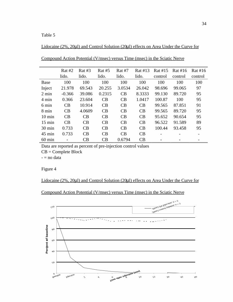
Table 5
Lidocaine (2%, 20µl) and Control Solution (20µl) effects on Area Under the Curve for
Compound Action Potential (V/msec) versus Time (msec) in the Sciatic Nerve
Rat #2lido.
Rat #3lido.
Rat #5lido.
Rat #7lido.
Rat #13lido.
Rat #15control
Rat #16control
Rat #16control
Base 100 100 100 100 100 100 100 100Inject 21.978 69.543 20.255 3.0534 26.042 98.696 99.065 972 min -0.366 39.086 0.2315 CB 8.3333 99.130 89.720 954 min 0.366 23.604 CB CB 1.0417 100.87 100 956 min CB 10.914 CB CB CB 99.565 87.851 918 min CB 4.0609 CB CB CB 99.565 89.720 9510 min CB CB CB CB CB 95.652 90.654 9515 min CB CB CB CB CB 96.522 91.589 8930 min 0.733 CB CB CB CB 100.44 93.458 9545 min 0.733 CB CB CB CB - - -60 min - CB CB 0.6794 CB - - -Data are reported as percent of pre-injection control valuesCB = Complete Block- = no data
Figure 4
Lidocaine (2%, 20µl) and Control Solution (20µl) effects on Area Under the Curve for
Compound Action Potential (V/msec) versus Time (msec) in the Sciatic Nerve
0
20
40
60
80
100
120
BaselineInjection
2 4 6 8 10 15 30 45 60
Time after injection (min)
Pe
rce
nt
of
ba
se
lin
e
20mcl 2% lidocaine, n = 5
20mcl control solution, n = 3

35
The area under the curve was also recorded. The evoked potentials, of all the
animals that received 20 µl of 2% lidocaine, were blocked by 10 minutes post-injection as
indicated by the area under the curve being equal to 0% when compared to baseline (Table
5 and Figure 4). The evoked potentials were blocked an average of 4.5 minutes with a
range of 2 to 10 minutes. In Table 5, the spaces labeled “CB” in the 2% lidocaine group
represent a block of evoked potentials. The times indicated with a dash are where
recordings were not averaged or observed.
Table 6
Lidocaine (2%, 10µl) and Control Solution (10µl) effects on Area Under the Curve for
Compound Action Potential (V/msec) versus Time (msec) in the Sciatic Nerve
Rat #8lido.
Rat #9lido.
Rat #12lido.
Rat #15lido.
Rat #16lido.
Rat #13control
Rat #13control
Rat #15control
Base 100 100 100 100 100 100 100 100Inject 103.59 37.017 63.860 50.217 10.588 90.946 123.71 96.2732 min 17.796 10.055 39.220 47.619 CB 97.787 105.41 97.5164 min -3.719 4.1437 27.105 35.065 CB 98.189 100.26 98.1376 min -2.112 CB 17.043 33.766 CB 85.714 88.144 99.0688 min -1.062 CB 12.526 46.320 CB - - 97.82610 min -1.049 CB 11.088 56.277 CB 90.946 72.680 93.47815 min -0.385 CB 9.4456 60.606 CB 78.068 72.680 83.85130 min CB CB 12.526 62.771 CB - - 81.05645 min 2.656 CB 14.579 55.844 CB - - -60 min -0.066 CB 17.659 52.381 CB - - -Data are reported as percent of pre-injection control valuesCB = Complete Block- = no data
36
Figure 5
Lidocaine (2%, 10µl) and Control Solution (10µl) effects on Area Under the Curve for
Compound Action Potential (V/msec) versus Time (msec) in the Sciatic Nerve
The evoked potentials, of all but two of the animals that received 10 µl of 2%
lidocaine, were completely blocked by 30 minutes post-injection as indicated by the area
under the curve being equal to 0% when compared to baseline (Table 6 and Figure 5). For
the animals that obtained a block, the evoked potentials were blocked by an average of 18
minutes with a range of 2 to 30 minutes. In Table 6, the spaces labeled “CB” in the 2%
lidocaine group represent a block of evoked potentials. The times indicated with a dash
are where recordings were not averaged or observed.
0
20
40
60
80
100
120
Baseline Injection 2 4 6 8 10 15 30 45 60
Time after injection (min)
Percen
t of
baseli
ne
10mcl 2% lidocaine, n = 510mcl control solution, n = 3
37
CHAPTER SIX: DISCUSSION
ANOVA and other repeated measures analysis of the data were attempted, but
because of sample size, there were insufficient data points to accomplish the analysis.
Performing a t-test analysis of the data was not an option due to the increased chance for
a type 1 error. For these reasons, the results can only be discussed as trends. The results
from these experiments are similar to the results obtained by Paris et al (1990). This
study showed a trend for 20µl of 2% lidocaine injected under the neural sheath of the rat
sciatic nerve to block the nerve’s evoked potentials. This was a strong trend in that all
five animals that received the 20µl of 2% lidocaine did have a complete block. The group
that received 10 µl of 2 % lidocaine did not show a consistent block of the evoked
potentials.
Measurements were done on the A wave CAP which includes Aα, Aβ, Aγ, and
Aδ nerve fibers. Due to the short distance between the stimulating and recording
electrodes, these A nerve fiber action potentials did not have enough time to separate out
and thus measurements were done on the entire compound action potential (CAP). The
peak of the compound action potential represents the Aα nerve fibers which are
responsible for motor function for the sciatic nerve.

38
Figure 6
Lidocaine (2%, 10 and 20µl) and Control Solution (10 and 20µl) effects on Peak Amplitude
(mV) versus Time (msec) in the Sciatic Nerve
Data are displayed as percent of pre-injection control values
Again, measurements were taken of the peak positive deflection in the compound
action potentials and not for individual nerve fiber groups. Sensory (A beta, A gamma,
and A delta) activity signals were embedded in the after peak action potential. A
reduction to 0% of baseline in the peak amplitude would represent a block of impulse
transmission to muscle fibers. The peak amplitudes of the CAPs for all the groups of
animals that were tested in shown in Figure 6. It shows that the group that received 20 µl
of 2% lidocaine had a reduction to 0% of baseline as soon as 10 minutes post injection.
0
10
20
30
40
50
60
70
80
90
100
Baseline Injection 2 4 6 8 10 15 30 45 60
Time after injection (min)
20mcl 2% lidocaine
10mcl 2% lidocaine
20mcl control solutio
10mcl control solutio
39
The group receiving 10 µl of 2% lidocaine on the average did have a reduction but only to
about 10% of baseline. The control groups did have a slight reduction in average
amplitude. Also, the control groups were not tested at the 45 and 60 minute intervals.
Figure 7
Lidocaine (2%, 10 & 20µl) and Control Solution (10 & 20µl) effects on Area Under the
Curve for Compound Action Potential (V/msec) versus Time (msec) in the Sciatic Nerve
Data are displayed as percent of pre-injection control values
Figure 7 compares the area under the curve of the CAPs for all the groups of
animals that were tested. It shows that the group that received 20 µl of 2% lidocaine had
a reduction to 0% of baseline as soon as 10 minutes. The group receiving 10 µl of 2%
lidocaine on the average did have a reduction but only to about 10% of baseline. The
0
20
40
60
80
100
120
Baseline Injection 2 4 6 8 10 15 30 45 60Time after injection (min)
20mcl 2% lidocaine
10mcl 2% lidocaine
20mcl control solution
10mcl control solution

40
control groups did have a slight reduction in average area under the curve. Also, the
control groups were not tested at the 45 and 60 minute intervals.
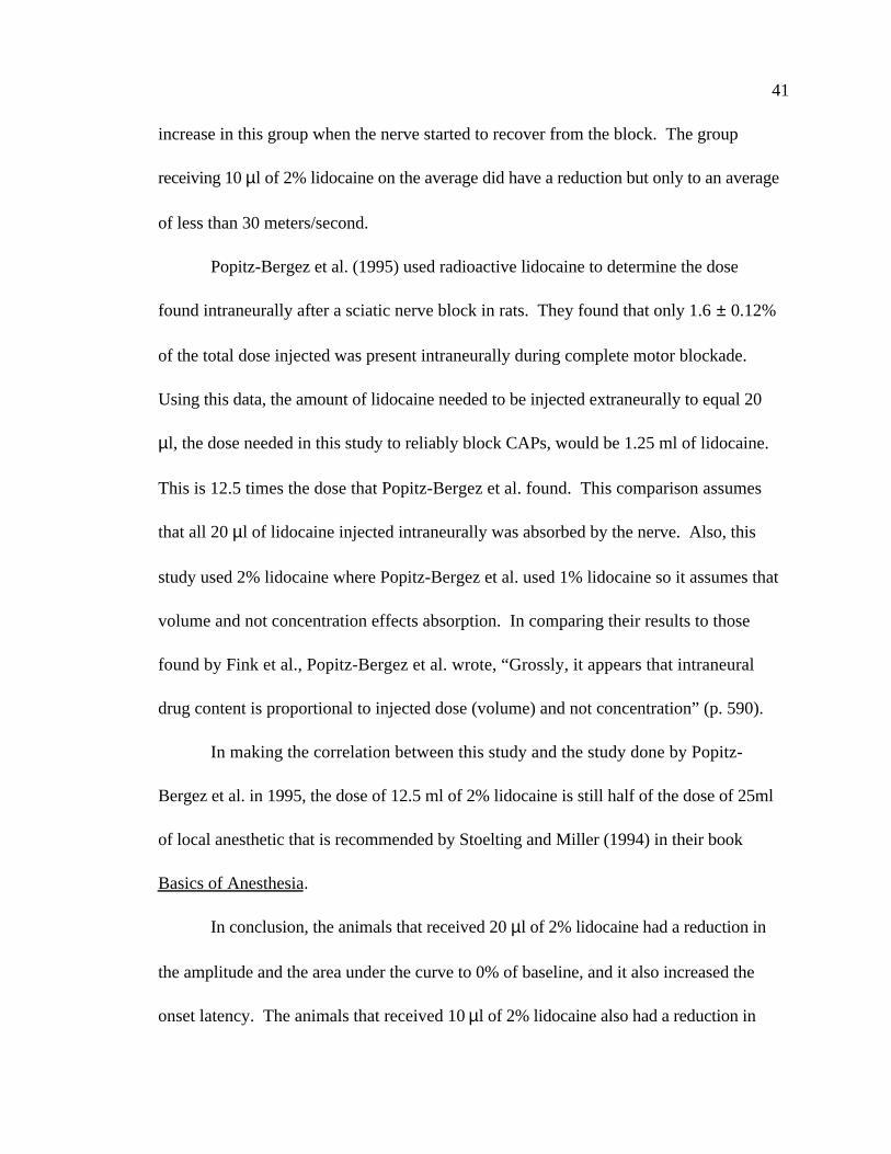
Figure 8
Lidocaine (2%, 10 & 20µl) and Control Solution (10 & 20µl) effects on Conduction
Velocity (m/sec) versus Time (msec) in the Sciatic Nerve
Figure 8 compares the conduction velocity of the CAPs for both experimental
animal groups that were tested. Gasser and Erlanger (1929) wrote, “conduction becomes
slower before it is extinguished” (p.583). It shows that the group that received 20 µl of
2% lidocaine had an average reduction in conduction velocity to 0 meters/second at 15
min when all the animals had a complete block. The conduction velocities started to
0
10
20
30
40
50
60
70
80
met
ers
per s
econ
d
Baseline Injection 2 4 6 8 10 15 30 45 60
time after injection (min)
20 mcl 2% Lidocaine10mcl 2% Lidocaine
41
increase in this group when the nerve started to recover from the block. The group
receiving 10 µl of 2% lidocaine on the average did have a reduction but only to an average
of less than 30 meters/second.
Popitz-Bergez et al. (1995) used radioactive lidocaine to determine the dose
found intraneurally after a sciatic nerve block in rats. They found that only 1.6 ± 0.12%
of the total dose injected was present intraneurally during complete motor blockade.
Using this data, the amount of lidocaine needed to be injected extraneurally to equal 20
µl, the dose needed in this study to reliably block CAPs, would be 1.25 ml of lidocaine.
This is 12.5 times the dose that Popitz-Bergez et al. found. This comparison assumes
that all 20 µl of lidocaine injected intraneurally was absorbed by the nerve. Also, this
study used 2% lidocaine where Popitz-Bergez et al. used 1% lidocaine so it assumes that
volume and not concentration effects absorption. In comparing their results to those
found by Fink et al., Popitz-Bergez et al. wrote, “Grossly, it appears that intraneural
drug content is proportional to injected dose (volume) and not concentration” (p. 590).
In making the correlation between this study and the study done by Popitz-
Bergez et al. in 1995, the dose of 12.5 ml of 2% lidocaine is still half of the dose of 25ml
of local anesthetic that is recommended by Stoelting and Miller (1994) in their book
Basics of Anesthesia.
In conclusion, the animals that received 20 µl of 2% lidocaine had a reduction in
the amplitude and the area under the curve to 0% of baseline, and it also increased the
onset latency. The animals that received 10 µl of 2% lidocaine also had a reduction in
42
the amplitude and the area under the curve but not consistently to 0% of baseline, and
they also did not have a consistent effect on onset latency.
Recommendations
My recommendations for future studies would be to repeat this experiment with
more animals so that the data could be analyzed with a repeated measures test. For
these statistical tests to be most effective, I would place each animal in each group, for
example one animal would have received injections of 10 µl of control solution, 20 µl of
control solution, 10 µl of lidocaine, and 20 µl of lidocaine. I would also recommend
using this methodology to test other local anesthetics, different concentrations of local
anesthetics, and record data until the CAP return to baseline value.
Another limitation to this study was the amount of time needed. Each
experiment took one day to complete. This study had many problems with time
constraints in order to complete the number of experiments originally intended. To save
valuable time and resources, multiple experiments were done on the later animals but not
on the first animals. As stated before it would have been preferable to do the same
number of experiments on all animals.

43
LIST OF REFERENCES
Bankert, M. (1993). The Mother of Anesthesia. Watchful care: A history of
america's nurse anesthetists (pp. 17-38). New York: Continuum Publishing Company.
Bryant, H., Harder, D., Pamnani, M., & Haddy, F. (1985). In vivo membrane
potentials of smooth muscle cells in the caudal artery of the rat. American Journal of
Physiology, 249, C78-C83.
Butterworth, J. & Strichartz, G. (1990). Molecular mechanisms of local
anesthesia: A review. Anesthesiology, 72, 711-734.
Duncum, B. (1947). Summary of events in anaesthesia. In E. Underwood (Ed.),
The development of inhalation anaesthesia, with special reference to the years 1846-1900
(pp. 614-620). London: Oxford University Press.
Gasser, H. (1943). Pain-producing impulses in peripheral nerves. In H. Wolff, H.
Gasser, J. Hinsey (Eds.), Pain (pp. 44-62). Baltimore: Williams & Wilkins Company.
Gasser, H. & Erlanger, J. (1929). The role of fiber size in the establishment of a
nerve block by pressure or cocaine. American Journal of Physiology, 88, 581-591.
Gilman, S. & Newman, S. (1996). Manter and Gatz's essentials of clinical
neuroanatomy and neurophysiology (9th ed.). Philadelphia: F.A. Davis Company.
Huang, J., Thalhammer, J., Raymond, S., & Strichartz, G. (1997). Susceptibility to
lidocaine of impulses in different somatosensory afferent fibers of rat sciatic nerve. The
Journal of Pharmacology and Experimental Therapeutics, 292(2), 802-811.

44
Jaffe, R. & Rowe, M. (1996). Differential nerve block: Direct measurements on
individual myelinated and unmyelinated dorsal root axons. Anesthesiology, 84(6), 1455-
1464.
Kalichman, M., Moorhouse, D., Powell, H., & Myers, R. (1993). Relative neural
toxicity of local anesthetics. Journal of Neuropathology and Experimental Neurology,
52(3), 234-240.
Kalichman, M., Powell, H., & Myers, R. (1989). Quantitative histologic analysis
of local anesthetic-induced injury to rat sciatic nerve. Journal of Pharmacology and
Experimental Therapeutics, 250(1), 406-413.
Paris, V., Pahno, N., Rigamonti, D., Jimmerson, V., & Seng, G. (1990). A unique
in vivo model for evaluating local anesthetic agents. Society for Neuroscience Abstracts,
16, 183.
Popitz-Bergez, F., Leeson, S., Strichartz, G., & Thalhammer, J. (1997).
Intraneural lidocaine uptake compared with analgesic differences between pregnant and
nonpregnant rats. Regional Anesthesia, 22(4), 363-371.
Popitz-Bergez, F., Leeson, S., Strichartz, G., & Thalhammer, J. (1995). Relation
between functional deficit and intraneural local anesthetic during peripheral nerve block.
Anesthesiology, 83(3), 583-592.
Raymond, S., Steffensen, S., Gugino, L., & Strichartz, G. (1989). The role of
length of nerve exposed to local anesthetics in impulse blocking action. Anesthesia and
Analgesia, 68(5), 563-570.

45
Stoelting, R. (1991). Local anesthetics. In M. Smith (Ed.), Pharmacology and
physiology in anesthetic practice (2nd ed., pp. 148-171). Philadelphia: J.B. Lippincott
Company.
Stoelting, R. & Miller, R. (1994). Peripheral nerve blocks. In B. Dickinson (Ed.),
Basics of anesthesia (3rd ed., pp. 179-190). New York: Churchill Livingstone.
Strichartz, G. (1988). Neural physiology and local anesthetic action. In M.
Cousins, & P. Bridenbaugh (Eds.), Neural blockade in clinical anesthesia and management
of pain (2nd ed., pp. 25-43). Philadelphia: J.B. Lippincott Company.
Tasaki, I. (1982). Conduction of impulses in myelinated nerve fibers. In A.
Noordergraaf (Ed.), Physiology and electrochemistry of nerve fibers (pp. 62-92). New
York: Academic Press.

46
APPENDICES
Appendix A Protocol Submitted to Laboratory Animal Review Board, USUHS form
6006……………………………………………………………………………………. 44
Appendix B Biohazards, Controlled Substances And Dangerous Materials, USUHS form
6007 (BCD) …………………………………………………………………………… 61
Appendix C Laboratory Animal Review Board Response Letter……………………..70
Appendix D Response Memorandum…………………………………………………72
Appendix E Laboratory Animal Review Board Approval Letter…………………….. 73
Appendix F Protocol Modification…………………………………………………… 74
Appendix G Protocol Modification Temporary Approval Letter……………………..
75
Appendix H Protocol Modification Approval Letter………………………………….76

47
APPENDIX A
Protocol Submitted to Laboratory Animal Review Board, USUHS form 6006
USUHS FORM 6006 (LARB) ANIMAL STUDY PROPOSAL FORM
PROTOCOL COVER SHEET
CHECK THE FOLLOWING:
[ X ] New New protocol number: (REA will assign)
[ ] Previously Submitted Old protocol number:
[ ] No modifications
[ ] Minor modifications (indicate all revisions with a bold/italic type font)
PROTOCOL TITLE: The Minimum Effective Dose of Lidocaine Needed to Block
Evoked Potentials in the Sciatic Nerve of the (adult Sprague-Dawley) Rat.
PRINCIPAL INVESTIGATOR:
_________________________1LT Bradley Stelflug 15 April 1997
Principal Investigator Signature Date
DEPARTMENT: Graduate School of Nursing
TELEPHONE: 295-6565
SCIENTIFIC REVIEW:
/ D.D. Rigamonti PhD, Howard Bryant PhD, Ken Miller
PhD /(thesis committee); CAPT Jane McCarthy PhD(Chairman of
__________ / department) 15 April 1997
Research Unit Chief/Department Head Signature Date
COORDINATION:

48
A. Attending/Consulting Veterinarian:
___________________________________________________________ _
Attending/Consulting Veterinarian Signature Date
(Only required for USDA, Category D or E proposals.)
B. Statistician:
_________________________________Dr. Ken Miller PhD 15 April 1997
Statistician Signature Date
(The PI may certify that the statistical methods are valid.)

49
USUHS FORM 6006 (LARB)
ANIMAL STUDY PROPOSAL FORM
PROTOCOL TITLE: The Minimum Effective Dose of Lidocaine Needed to Block
Evoked Potentials in the Sciatic Nerve of the (adult Sprague-Dawley) Rat.
DATE: 9 April 1997
PRINCIPAL INVESTIGATOR: 1LT Bradley Stelflug
DEPARTMENT: Graduate School of Nursing
CO-INVESTIGATOR(S): Dr. D. Rigamonti, Dr. H. Bryant, Dr. K. Miller
TECHNICIAN(S): N/A
I. NON-TECHNICAL SYNOPSIS: This study will describe the minimum volume of
local anesthetic needed to block the conduction of nerve impulses along the sciatic nerve
of a Sprague-Dawley rat. The diameter of the animal’s sciatic nerve is approximately the
size of a human intercostal nerve. Currently, large volumes of local anesthetic are used to
block nerve impulses along human nerves which has the potential of systemic toxicity,
nerve compression, and tissue necrosis. This study will show what are the minimum
volumes (concentration of clinical grade commonly used local anesthetic) needed to block
nerve impulses.
II. BACKGROUND:
A. Background: Large volumes of local anesthetic are currently recommended for
regional anesthesia of human peripheral nerves. Using these large volumes has the risks of
systemic toxicity which can lead to seizures. Also nerve compression or tissue damage
from using large volumes can cause nerve damage. Finding the minimum effective volume

50
could help in reducing the volumes currently being recommended.
B. Literature Search:
1. Literature Source(s) Searched: MEDLINE( R ) 1966-1997;
AGRICOLA 70, Feb 1997; PsycINFO ( R ) 1967-1997; EMBASE 1974-1997; BIOSIS
PREVIEWS ( R ) 1969-1997; CAB Abstracts 1972-1996.
2. Date and Number of Search: 11 March 1997
3. Key Words of Search: Sciatic Nerve, Local (Anesthe? Anasthe?, or
Anaesthe?), Evoked Potential, Nerve or Neuron, (Vitro or Isolate or Culture), Frog,
Minimum Dose, Nembutal, Pentobarbital.
4. Results of Search: The literature showed that this would not be a
duplication of effort. The only use of this in vivo model was in 1990 and written in
abstract form only published in Abstracts: Society for Neuroscience, Vol.16, 1990, p.183.
The model was shown to be a valid model to test local anesthetics and that much smaller
volumes of local anesthetics are needed for nerve blockade than are suggested clinically.
The literature search shows that the sciatic nerve of a rat can be used to test local
anesthetics, that will be used in humans, and that higher phylogenetic species are not
necessary for this experiment. The experiments, using frog nerves, used in vitro models
and were bathed in solutions which would preclude using an in vitro model for finding the
minimum effect volume. Pateromichelakis and Prokopiou (1988) found discrepancies
between results of mepivacaine in using in vivo versus in vitro models. Tsirlis et. Al.
(1994) found that at a higher pH, lidocaine block had a quicker onset and longer duration
of action. This is why an in vivo model using a mammal which would be at physiologic

51
pH is needed.
III. OBJECTIVE\HYPOTHESIS: To find the minimum effective dose of local
anesthetic needed to block nerve transmission in the sciatic nerve of the Sprague-Dawley
rat.
IV. MILITARY RELEVANCE: In both the operational theater and in peace time,
anesthesia providers will be providing regional blocks to military personnel. Currently,
large doses of local anesthetic are being used to perform these blocks. These large doses
can lead to nerve damage from compression, tissue death from compression, or even
systemic toxicity which can lead to seizures. If smaller doses of local anesthetics were
used, then the possibility of these complications would be reduced.
V. MATERIALS AND METHODS:
A. Experimental Design and General Procedures:
1. Experiment # This is a quantitative study to determine the minimum
amount of Lidocaine (2%) needed to block recorded evoked potentials in the sciatic nerve
of the rat. A total of 18 adult Sprague-Dawley rats are needed. Animals are ordered in
quantities of one to two animals per week, which is how many trials are done per week.
There will be three animals used to learn the surgical technique of these procedures and
then used in the control group. Three additional animals will be in the control group and
three animals will be in each of the groups receiving lidocaine.
Number of Animals

52
6 animals Group 1 Control group (all volumes)
3 animals Group 2 Group receiving 10µl of lidocaine
3 animals Group 3 Group receiving 20µl of lidocaine
3 animals Group 4 Group receiving 30µl of lidocaine
3 animals Group 5 Group receiving 40µl of lidocaine
18
animals
Total
The techniques will be taught by Dr. Rigamonti, a researcher who has already performed
these procedures. The sciatic nerve is tested in vivo in animals initially anesthetized using
Nembutal (50 mg/kg ip) as recommended in USUHS “The use of animals in research: A
guide for investigators. Supplemental doses will be administered as needed (as tested by
corneal reflex). The animals’ trachea is surgically exposed and a tracheal tube with T-
piece is inserted surgically. A ventilator is available if the animal is in need of artificial
respiration. The carotid artery is cannulated in order to draw arterial blood gases to
prevent respiratory alkalosis or respiratory acidosis. The left sciatic nerve is surgically
exposed and a pair of “stimulating” electrodes are placed on the nerve at the greater sciatic
notch and another pair of “recording” electrodes are placed on the nerve around the tibial
division near the ankle. The electrodes are embedded in wax in order to avoid possible
signal interference from surrounding tissue and also to ensure that the electrical impulse
goes only to the nerve. The nerve is tied off proximal to the stimulating electrodes and

53
distal to the recording electrodes. The nerve is stimulated with a 0.2 ms duration
supramaximal pulse every 5 seconds. Ten to fifty responses are averaged. After baseline
values are obtained an injection, of lidocaine or control solution, is made inside the nerve
sheath and then post injection recordings are obtained. Recordings are made for two
hours after injection. The compound action potentials are amplified, visualized, and
averaged using MacLab program. Core temperature is physiologically maintained at 37.50
+/- 0.30 C. An abstract reports that 20µl of lidocaine (2%) is the volume needed to
completely block evoked potentials in the sciatic nerve of the rat. The trials are
performed at the volumes of 10µl, 20µl, 30µl, and 40µl. The control group (consisting of
6 animals) is injected with a preparation of a rat Ringer solution prepared of NaHCO3
27.2mM, NaCl 118mM, KCl 4.8mM, KH2PO4 1.0mM, MgSO4 1.2mM, CaCl2 2.5mM,
and glucose 11.1mM to determine how much influence volume plays in blocking evoked
potentials. The solution has an osmolarity of 300 +/- 5 mosM and a pH of 7.4 +/- 0.05.
The control group is then given a second injection of the rat Ringer solution in which
Methylene Blue will be added to measure the spread of the volume in the nerve. No
recordings will be made after the injection with Methylene Blue, but the diffusion
distance will be measured. All solutions (ringer or lidocaine) are injected into the nerve
under microscopic conditions, distal to the stimulating electrodes, using a glass
micropipette with a tip OD<30µ placed under the perineurium. The remaining 12
animals (3 per volume group) are injected with Lidocaine 2% which is sold commercially
to be used clinically on humans. Data to be collected include: weight of rat, volume of
Lidocaine used, initial recordings of the compound action potentials, the amplitude,

54
latency, area under the curve until blockage of evoked potentials is complete, spread
distance of the solution, and length of nerve will be measured to determine conduction
velocity. The data collected, which includes the above plus when each fiber group is
blocked, is described in a quantitative manner. At the end of the procedure, the animals
will be euthanized by giving an overdose of Nembutal (200mg/kg ip) and then their chests
will be opened.
B. Laboratory Animals Required and Justification:
1. Non-animal Alternatives Considered: This is an in vivo model
which will look at the affects of lidocaine on the sciatic nerve in its homeostatic
environment. This will include the whole animal affect. There is no computer model
developed for this model.
2. Animal Model(s) and Species Justification: Sprague-Dawley rats
were chosen because they were used in the Paris model. To be able to use these results
with humans, a warm blooded species was needed. The Sprague-Dawley rat is the
smallest animal that still has a nerve that can be compared to a human nerve (The animals
sciatic nerve and the human intercostal nerve).
3. Total Number of Animals Required:
Genus & Species Rattus norvegius
Stock/Strain Sprague-Dawley
Sex Either Age/Weight/Size adult
Source/Vendor _______________________________
Holding Location(s) Dept.LAM (cental animal facility)

55
Number of Animals requested/total number of housing days required:
Animals Requested Housing Days
Year 1 18 126 (18 x 7)
Year 2 N/A N/A
Year 3 N/A N/A
Year 4 N/A N/A
Year 5 N/A N/A
Special Considerations:
Other: Animals will be ordered such that will only have the number of animals needed
for that week.
(Note: This table should be blocked and copied, and repeated for each species
requested)
4. Refinement, Reduction, Replacement:
a. Refinement: The procedure will have an early endpoint in that
it will end one hour after the evoked potentials are blocked and recovery of nerve function
will not be explored.
b. Reduction: Baseline data, of evoked potentials without
volumes of solutions added, will be gathered from the animals that will be used for
learning the procedure. Since the volume of solution used in the controls will disperse and
its effects will be short lived, four volumes will be used with each control animal.
c. Replacement: There are no computer models developed for
this in vivo model. Cell cultures will not allow use to test the “whole animal” affect of

56
this nerve block.
C. Technical Methods:
1. Prolonged Restraint: There will be no prolonged restraint used.
2. Surgery:
a. Procedure: The animals will be anesthetized using Nembutal 50
mg/kg ip. Supplemental doses will be administered as needed (as tested by corneal reflex).
The animals’ trachea surgically exposed and a tracheal tube with T-piece is surgically
inserted. A ventilator is available if the animal is in need of artificial respiration. The
carotid artery is cannulated in order to draw arterial blood gases to prevent respiratory
alkalosis or respiratory acidosis. The left sciatic nerve will then be surgically exposed at
the greater sciatic notch and around the tibial division near the ankle. The nerve will be
tied off proximal to the stimulating electrodes and distal to the recording electrodes. A
pair of “stimulating” electrodes will be placed on the nerve at the greater sciatic notch and
another pair of “recording” electrodes will be placed on the nerve around the tibial
division near the ankle. After the animal is euthanized, the sciatic nerve will be dissected
to determine the length between electrodes (to be used to determine conduction velocity).
b. Pre- and Postoperative Provisions: The animals will be
anesthetized using Nembutal 50 mg/kg ip. The animals will be euthanized at the study
endpoint.
c. Location: Building C, room 2081 or 2079
d. Multiple Survival Surgery Procedures:

57
(l) Procedures: N/A
(2) Scientific Justification: N/A
3. Animal Manipulations:
a. Injections: See section V.C.7.b.
b. Biosamples: Arterial Blood Gases
c. Animal Identification: Date will be placed on the cage.
d. Behavioral Studies: N/A
e. Other procedures: Oscilloscope with MacLab.
f. Location where procedure will take place: Bldg C
Rm 2079/81
4. Adjuvants: N/A
5. Study Endpoint: After complete blockage of evoked potentials is
accomplished, then stimulations and recordings will be every 5 minutes for one more
hour. This will be the study endpoint and the animals will be euthanized because the
sciatic nerve will be paralyzed from it being tied off.
6. Euthanasia: The animals will be sacrificed by giving an overdose of
Nembutal 200 mg/kg followed by opening the chest to create a pneumothorax. The
primary investigator or Dr. Rigamonti will give the overdose of Nembutal.
7. Pain:
a. USDA APHIS Form 7023 Pain Category:
(1) No Pain _________ (#) ________ % (Column C)
(2) Alleviated Pain ___18____ (#) __100___ %
58
(Column D)
(3) Unalleviated Pain or Distress ________ (#)
________ % (Column E)
b. Pain Alleviation:
(1) Anesthesia/Analgesia/Tranquilization: The animals will be
anesthetized using Nembutal 50 mg/kg ip.
(2) Paralytics: N/A
c. Alternatives to Painful Procedures:
(1) Source(s) Searched: MEDLINE( R ) 1966-1997;
AGRICOLA 70, Feb 1997; PsycINFO ( R ) 1967-1997; EMBASE 1974-1997; BIOSIS
PREVIEWS ( R ) 1969-1997; CAB Abstracts 1972-1996.
(2) Date of Search: 11 March 1997
(3) Key Words of Search: Sciatic Nerve, Local (Anesthe?
Anasthe?, or Anaesthe?), Evoked Potential, Nerve or Neuron, (Vitro or Isolate or
Culture), Frog, Minimum Dose, Nembutal, Pentobarbital.
(4) Results of Search: The literature showed that this
would not be a duplication of effort. The only use of this in vivo model was in 1990 and
written in abstract form published in Abstracts: Society for Neuroscience, Vol.16, 1990,
p.183. The model was shown to be a valid model to test local anesthetics and that much
smaller volumes of local anesthetics are needed for nerve blockade than are used clinically.
The literature search showed that the sciatic nerve of a rat can be used to test local
anesthetics that will be used in humans and that higher phylogenetic species are not

59
necessary for this experiment. The experiments, using frog nerves, used in vitro models
and were bathed in solutions which would preclude using an in vitro model for finding the
minimum effect volume. Pateromichelakis and Prokopiou (1988) found discrepancies
between results of mepivacaine in using in vivo versus in vitro models. Tsirlis et. Al.
(1994) found that at a higher pH, lidocaine block had a quicker onset and longer duration
of action. This is why an in vivo model using a mammal at physiologic pH is needed.
d. Painful Procedure Justification: The animals will be
anesthetized using Nembutal 50 mg/kg ip to avoid causing any pain to the animals from
the procedure. The animals will be euthanized in a rapid and painless way by giving
Nembutal 200 mg/kg ip at the study endpoint before the initial anesthesia wears off.
D. Veterinary Care:
1. Husbandry Considerations: The animals will be cared for at UHUHS
Central Animal Facility until they are used for the study. The animals will be ordered so
that the number arriving each week will be used that week (usually 1 animals per week,
may be up to 2). The total number of housing days was figured by each animal staying a
maximum of 7 days.
a. Study Room: N/A
b. Special Husbandry Provisions: None needed.
2. Attending Veterinary Care: Animals that are in need of Veterinary
care will be seen by a Veterinarian from the department of Laboratory Animal Medicine.
The investigator will follow any recommendations made by the Veterinarian for the
animal.

60
3. Enrichment Strategy:
a. Dogs: N/A
b. Nonhuman Primates: N/A
E. Data Analysis: Data will be analyzed with the MacLab statistical software
package. This is a standard statistical package for analyzing evoked potentials. It has the
capabilities to record and average the responses, measure the amplitude and latency and
area under the curve until blockage of evoked potentials is complete.
F. Investigator and Technician Qualifications/Training: Brad Stelflug, Don
Rigamonti, and Howard Bryant will be performing all the procedures in the entire
experiment. Dr. Rigamonti will be present for all the procedures. He helped develop the
model that will be used and he has done these procedures before. Dr. Rigamonti and Brad
Stelflug attended the USUHS Rodent Handling Course on 30 April 1997. Dr. Bryant has
worked with rodents before on protocols at USUHS. Dr. Rigamonti, Dr. Bryant, and
Brad Stelflug have attended an approved USUHS Investigator Training Course.
VI. BIOHAZARD/SAFETY:
VII. ASSURANCES: As the Primary Investigator on this protocol I provide the
following assurances
A. Animal Use: The animals authorized for use in this protocol will be used
only in the activities and in the manner described herein, unless a deviation is specifically
approved by the LARB.
B. Duplication of Effort: I have made a reasonable, good faith effort to ensure
that this protocol is not an unnecessary duplication of previous experiments.

61
C. Statistical Assurance: I assure that I have consulted with an individual who
is qualified to evaluate the statistical design or strategy of this proposal, and that the
"minimum number of animals needed for scientific validity are used."
D. Biohazard/Safety: I have taken into consideration, and I have made the
proper coordinations regarding all applicable rules and regulations regarding radiation
protection, biosafety, recombinant issues, etc., in the preparation of this protocol.
E. Training: I verify that the personnel performing the animal
procedures/manipulations described in this protocol are technically competent and have
been properly trained to ensure that no unnecessary pain or distress will be caused as a
result of the procedures/manipulations.
F. Training: I verify that I have attended an approved Uniformed Services
University of the Health Sciences (USUHS) Investigator Training Course.
G. Training: The following personnel will attend the next approved USUHS
Investigator Training Course:
1LT Brad Stelflug (Done 04 March 1997)
Dr. D. Rigamonti (Done 04 March 1997)
Dr. H. Bryant (completed)
H. Responsibility: I acknowledge the inherent moral and administrative
obligations associated with the performance of this animal use protocol, and I assure that
all individuals associated with this project will demonstrate a concern for the health,
comfort, welfare, and well-being of the research animals. Additionally, I pledge to

62
conduct this study in the spirit of the fourth "R" which the DoD has embraced, namely,
"Responsibility" for implementing animal use alternatives where feasible, and conducting
humane and lawful research.
____________________________________________________
Principal Investigator Signature Date
I. Painful Procedures: (Include above if conducting research that will cause
more than slight or momentary pain or distress [Column D or E by USDA classification]
the following statement must follow.) I am conducting biomedical experiments which
may potentially cause more than momentary or slight pain or distress to animals
that WILL BE relieved or WILL NOT (circle one) be relieved with the use of
anesthetics, analgesics and/or tranquilizers. I have considered alternatives to such
procedures; however, using the methods and sources described in the protocol, I have
determined that alternative procedures are not available to accomplish the objectives of
the proposed experiment.
______________________________________________________
Principal Investigator Signature Date
VIII. ENCLOSURES:
A. Literature Searches: MEDLINE( R ) 1966-1997; AGRICOLA 70, Feb
1997; PsycINFO ( R ) 1967-1997; EMBASE 1974-1997; BIOSIS PREVIEWS ( R )
1969-1997; CAB Abstracts 1972-1996.
B. Pathology Addendum:
C. Pain Scoring Guidelines:

63
D. Adjuvant Policy:

64
IX. PROTOCOL ABSTRACT:
A. Protocol Number: (if new, leave blank) ____________________
B. Title: The Minimum Effective Dose of Lidocaine Needed to Block Evoked
Potentials in the Sciatic Nerve of the (adult Sprague-Dawley) Rat.
C. Principal Investigator: 1LT Bradley Stelflug
D. Performing Organization: Uniformed Services of the Health Sciences
E. Funding: from the Graduate School of Nursing
F. Objective and Approach: This study will describe the minimum volume of
local anesthetic needed to block the conduction of nerve impulses along the sciatic nerve
of a Sprague-Dawley rat. The diameter of the animal’s sciatic nerve is approximately the
size of a human intercostal nerve. Currently, large volumes of local anesthetic are used to
block nerve impulses along human nerves which has the potential of systemic toxicity,
nerve compression, and tissue necrosis. This study will show what are the minimum
volumes needed.
G. Indexing Terms (Descriptors): sciatic nerve, local anesthetic, lidocaine,
animals, Sprague-Dawley rats.

65
APPENDIX B
Biohazards, Controlled Substances And Dangerous Materials,
USUHS Form 6007 (BCD)
USUHS FORM 6007 (BCD)BIOHAZARDS, CONTROLLED SUBSTANCESAND DANGEROUS MATERIALS
REA Date StampProtocol No.: TO6133-01
Principal Investigator: Bradley Stelflug
Department: GSN Phone: (301) 295-6565
Project Title: The Minimum Effective Dose of Lidocaine Needed to BlockEvoked Potentials in the Sciatic Nerve of the Rat.
PLEASE NOTE: Failure to provide ALL required information could result in projectdisapproval, or, at least add significantly to the length of the approval process. If yourequire assistance in preparing this form, contact (as needed) the EnvironmentalHealth and Occupational Safety (EHS) Department (295-3321), the Pharmacy Officer(295-3305), or the Pharmacy Technician (295-3668).
BCD categories which apply to this project:
Biohazards (Section A) Recombinant DNA {rDNA} (Section B)X Controlled Substances (Section C) X Chemicals (Section D) Others (Section E)
SECTION (A) BIOHAZARDS
1. Information to assist in the completion of this section can be found inUSUHS Instruction 6401 “Biohazards and Dangerous Materials Guide,”and the CDC/NIH Guidebook “Biosafety in Microbiological and BiochemicalLaboratories.”
a. Type of Biohazard Agent to be used:
Bacteria Virus Parasite Mold/Fungi Human Blood/Blood Products/Unfixed
Tissue

66
Others
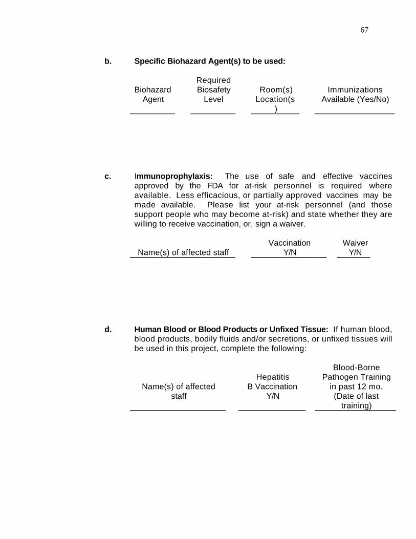
67
b. Specific Biohazard Agent(s) to be used:
BiohazardAgent
RequiredBiosafety
LevelRoom(s)
Location(s)
ImmunizationsAvailable (Yes/No)
c. Immunoprophylaxis: The use of safe and effective vaccinesapproved by the FDA for at-risk personnel is required whereavailable. Less efficacious, or partially approved vaccines may bemade available. Please list your at-risk personnel (and thosesupport people who may become at-risk) and state whether they arewilling to receive vaccination, or, sign a waiver.
Name(s) of affected staffVaccination
Y/NWaiver
Y/N
d. Human Blood or Blood Products or Unfixed Tissue: If human blood,blood products, bodily fluids and/or secretions, or unfixed tissues willbe used in this project, complete the following:
Name(s) of affectedstaff
HepatitisB Vaccination
Y/N
Blood-BornePathogen Training
in past 12 mo.(Date of last
training)

68
e. Handling of Biohazards or Human Blood/Blood Products/UnfixedTissue:Describe in sufficient detail to permit evaluation of how the agent(s)will be handled. Account for source, storage, experimentalprocedures, and disposal methodology. Emphasize precautions tobe taken to protect laboratory personnel and the public.
SECTION (B) RECOMBINANT DNA (rDNA)
2. Guidelines for the use of recombinant DNA can be found in the USUHSGuidelines for Research Involving Recombinant DNA Molecules. A copyis available in EHS and additional copies can be made available torequesting departments.
a. Have you read the USUHS rDNA Guidelines? Yes No
b. Please provide the following information:
Source(s) of DNA/rDNA:
Nature of inserted DNAsequences
Host(s) and Vector(s) to be used:
Will expression of a foreign gene be attempted? Yes NoIf yes, what protein(s) will be produced?
According to the “Guidelines”, what is the proposed Class of study?
What level of containment will be used (Section II,“Guidelines”)?
If used, what biological barriers areincorporated?

69
SECTION (C) CONTROLLED SUBSTANCES
3. Have you reviewed USUHS Instruction 6404 “Management of ControlledSubstances, Alcohol and Alcohol Liquors, Prescription Medications,Hypodermic Needles, Syringes, List I and II Chemicals, and AnabolicSteroids”?
a. Controlled Substances List: List all controlled substances to beused. List by generic form (no brand names), provide concentrationsof substances obtained as premixed medications (i.e. 100 mg/ml),and indicate the laboratory, room or other location where thesubstance(s) will be used. List only controlled substances andanabolic steroids. Do not re-list these materials in Section D.
Generic Name Amount/Concentration LocationSodium Pentobarbital 50 mg/ml C2081/79
b. Safeguards: Describe in sufficient detail to permit evaluation, howthe controlled substance(s) will be obtained (source), secured, used,accounted for, and what method(s) will be employed for disposal ofunused quantities and for waste materials generated for their use.Identify safeguards and precautions to eliminate possible loss orabuse. REMINDER: Return all “unused” quantities of controlledsubstances and anabolic steroids to the Pharmacy.Sodium Pentobarbital (50 mg/ml) purchased from the USUHSpharmacy will be given to the physiology department’s drugcustodian (Howard Bryant Ph.D.) to be stored in the safe in roomC2081. The amount of Sodium Pentobarbital (50 mg/ml) required fora day’s experiments will be obtained from the drug custodian. Anyunused drug will be returned to the drug custodian at the end of thatday.
c. List I and List II Precursor/Essential Chemicals: The followingchemicals, their slats, isomers, and slats of optical isomers inthreshold amounts are List I and List II Precursor/Essential
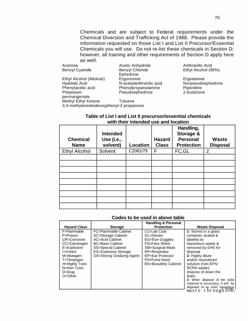
70
Chemicals and are subject to Federal requirements under theChemical Diversion and Trafficking Act of 1988. Please provide theinformation requested on those List I and List II Precursor/EssentialChemicals you will use. Do not re-list these chemicals in Section D;however, all training and other requirements of Section D apply hereas well.
Acetone Acetic Anhydride Anthranilic AcidBenzyl Cyanide Benzyl Chloride
EphedrineEthyl Alcohol (95%)
Ethyl Alcohol (Abolute) Ergonovine ErgotamineHydriotic Acid N-acetylanthranilic acid NorpseudoephedrinePhenylacetic acid Phenylpropanolamine PiperidinePotassiumpermangenate
Pseudoephedrine 2-butanone
Methyl Ethyl Ketone Toluene3,4-methylenediodeoxyphenyl-2-propanone
Table of List I and List II precursor/essential chemicalswith their intended use and location
ChemicalName
IntendedUse (i.e.,solvent) Location
HazardClass
Handling,Storage &Personal
ProtectionWaste
DisposalEthyl Alcohol Solvent C2081/79 F FC,GL 2
Codes to be used in above table
Hazard Class StorageHandling & Personal
Protection Waste DisposalF=Flammable FC=Flammable Cabinet LC=Lab Coat 1- Stored in a glassP=Poison SC=Storage Cabinet GL=Gloves container sealed &CR=Corrosive AC=Acid Cabinet EG=Eye Goggles labeled asCC=Carcinogen BC=Base Cabinet FS=Face Shied hazardous waste &E=Explosive SS=Special Cabinet SM=Surgical Mask removed by EHS forI=Irritant ES=Explosive Storage RP=Respirator disposalM=Mutagen OS=Strong Oxidizing Agent EP=Ear Protector 2- Highly diluteT=Teratogen FH=Fume Hood and/or neutralizedH=Highly Toxic BS=Biosafety Cabinet solution (non-EPA/N=Non-Toxic RCRA waste);D=Drug dispose of down theO=Other drain.
3- When disposal of the solidmaterial is necessary, it will bedisposed of as solid hazardouswaste throughEHS.

71
waste through EHS.

72
SECTION (D) CHEMICALS
4. Guidelines for the proper use of chemicals are published as the OSHALaboratory Standard. A copy of these guidelines are available in EHS. EHSstaff are available for interpretation or amplification of the guidelines.
a. Have you read:OSHA Laboratory Standard? Yes X NoUSUHS Safety Manual? X Yes NoUSUHS Waste Disposal Guide? X Yes NoUSUHS Chemical Hygiene Plan? X Yes No
Is your laboratory in compliance with the requirements of theOSHA Laboratory Standard and USUHS Chemical Hygiene Plan?
X Yes No
Have you considered the following during design of this protocol?1. The environmental impact of use of the chemicals involved
in your study?X Yes No
2. The use of less hazardous, or less toxic, chemicals?X Yes No
3. Ordering only the amount of chemicals you can utilize inyour study in order to minimize chemical waste due tooutdated or unused chemicals?
X Yes No
4. Have you established a local common storage site forchemicals?
Yes X No

73
b. Training: List the current status of those personnel (including the PI)who will be working with chemicals (in any form or amount) on thisprotocol with regard to the annual laboratory safety training requiredby OSHA regulations. If training was provided by other than EHS,provide appropriate documentation.
Name(s) of affected staff Date of last attendanceBrad Stelflug 5/5/97Howard Bryant Ph.D. 2/18/97, 4/10/97Don Rigamonti
c. Chemical List: A complete list of all chemicals used in thisresearch project must be provided. Your project cannot beapproved if chemicals mentioned in the Materials and Methods, orother sections of your protocol are not addressed on USUHS Form6007. Standard chemical reagents for each department or laboratorycan be documented in a list provided to EHS. If you have such a liston file then you may attach a copy of it to this form provided you placea check mark ( ) next to the chemicals to be used in this project.New or additional chemicals should be listed in the table providedbelow.
CHEMICAL LIST - Chemicals Not Included on a Departmental/Laboratory List
Chemical Name HazardClass
Handling,Storage &Personal
ProtectionWaste
DisposalLidocaine D SS, GL 2

74
CODES to be used in above list
Hazard Class StorageHandling & Personal
Protection Waste DisposalF=Flammable FC=Flammable Cabinet LC=Lab Coat 1- Stored in a glassP=Poison SC=Storage Cabinet GL=Gloves container sealed &CR=Corrosive AC=Acid Cabinet EG=Eye Goggles labeled asCC=Carcinogen BC=Base Cabinet FS=Face Shied hazardous waste &E=Explosive SS=Special Cabinet SM=Surgical Mask removed by EHS forI=Irritant ES=Explosive Storage RP=Respirator disposalM=Mutagen OS=Strong Oxidizing Agent EP=Ear Protector 2- Highly diluteT=Teratogen FH=Fume Hood and/or neutralizedH=Highly Toxic BS=Biosafety Cabinet solution (non-EPA/N=Non-Toxic RCRA waste);D=Drug dispose of down theO=Other drain.
3- When disposal of thesolid material is necessary,it will be disposed of assolid hazardous wastethrough EHS.
SECTION (E) OTHERS
5. List other hazards which may be involved in this project (i.e., UV light,compressed gasses, high intensity lights, lasers, etc.)N/A
6. List special safety programs or procedures involved in this project (i.e.,pre-exposure serology, immunization program, physical examination,pulmonary function test, hearing tests, etc.).N/A
PHARMACY:
7. Have you consulted with the Pharmacy on your chemical and controlledsubstance requirements?
X Yes No
8. Have you established, with the Pharmacy, a listing of chemicals requiredby this protocol detaining concentration, grade, size of container,manufacturer, etc.?
X Yes No

75
SECTION (G) STATEMENT OF RESPONSIBILITY
STATEMENT OF RESPONSIBILITY:
I have read, and understand, the USUHS Safety Manual, the USUHS WasteDisposal Guide, and the most recent guidelines for those sections of this formwhich apply to my proposal. I have been properly trainied and I will see thatrequired training is provided for my staff, and that all who work under this protocolabide by the provisions of the USUHS Biohazard, Controlled Substances, andDangerous Materials Program. I understand that EHS is available for consultationin the training of staff and for other occupational health support when required. Iwill report to EHS any instances where the safety of personnel or the environmentare threatened.
05 May 1997Principal Investigator (signature) Date
76
APPENDIX C
Laboratory Animal Review Board Response Letter
28 May 1997
MEMORANDUM FOR DR. BRADLEY W. STELFLUG, GRADUATE SCHOOL OFNURSING
SUBJECT: Laboratory Animal Care and Use Review of Protocol NumberTO6133- 01, "The Minimum Effective Dose of Lidocaine Needed to Block
Evoked Potentials in the Sciatic Nerve of the Rat"
Animal Welfare Act, Public Health Service Policy, and USUHS instruction 3204require the review of all research and teaching protocols involving the use of animals. TheUSUHS Laboratory Animal Review Board (LARB) has reviewed your Protocol No.TO6133-01, entitled, "The Minimum Effective Dose of Lidocaine Needed to Block
Evoked Potentials in the Sciatic Nerve of the Rat." Issues raised in the review ofUSUHS Form 6006, "USUHS Animal Study Proposal", which require further clarificationor explanation including the following:
1. Section II.B.1. All DoD funded research projects are required to conduct aDefense Technical Information search. Please provide evidence that the searches havebeen completed.
2. Section V.A. Reference is made to drawing arterial blood gases to preventrespiratory alkalosis or respiratory acidosis, but there is no reference made to thefrequency or volume of blood withdrawn. Please clarify.
3. Section V.A. What is the volume of rat Ringer solution as control given to theanimals? Reference is made that the control group is used to "determine how muchinfluence volume plays", but the volume is not identified.
4. Section V.A. How was the sample size determined? Will three animals pergroup provide adequate number for statistically significant results?
5. Section V.A. If 20 µl of lidocaine (2%) was reported to block evokedpotentials completely, and the aim of the study is to determine the smallest dose required,why is there only one test point (10µl) that is a lower dose? If 20 µl does provide acomplete block, then the 30µl and 40 µl volumes should not beused. Instead, doses of 5

77
µl and 2.5 µl should be used in addition to 10 µl. If volumes smaller than 10 µl presenttechnical difficulties, dilution of the 2% lidocaine should be considered. Please comment.
Respectfully request that you respond to the above by memorandum. You areresponsible for sending this memorandum to Research Administration. You are alsoresponsible for sending copies of this memorandum to me (LTC Nathaniel Powell,LAM), Dr. Richard Andre, Preventive Medicine/Biometrics, Dr. Franziska Grieder,Microbiology, Dr. Sara Contente, Pathology, Dr. Gary Francis, Pediatrics, Dr. DavidDobbins, Physiology, Dr. Sharon Juliano, Anatomy And Cell Biology, Dr. CherylDicarlo, LAM, Mr. Tim Allen (in care of LAM), Dr. Carol Bossone, Medicine, Ms.Karen Dern, University Affairs, Ms. Olga D’Onofrio (in care of LAM) (LARBmembers). Your response memorandum will subsequently become part of Protocol No.TO6133-01. Please respond within 60 days of the date on this memorandum. A lack ofresponse within this time frame will necessitate a resubmission of USUHS Form 6006.
Nathaniel Powell, Jr., D.V.M.LTC, VC, USAExecutive-Secretary, Laboratory Animal Review Board
Cc:FileLARB distribution

78
APPENDIX D
Response Memorandum
21 JULY 1997
MEMORANDIUM FOR LTC NATHANIEL POWELL, D.V.M.SUBJECT: Protocol No. T06133-01 response memorandum
1. The “Multi-Databases” from the Defense Technical Information Center wassearched on 2 July 1997. The query text used were “local anesthetics AND in vivomodel” and “local anesthetics AND minimum dose”. Two studies were in the general areaof my proposed study; “Development of Ultra Long Duration Local Anesthetic Agentsin a Rat Model” and “Analysis of the Ability of DMSO (Dimethyl Sulfoxide) andLidocaine to Penetrate Dentin”. These studies are not similar to the proposed study inthat they do not test the minimum dose of Lidocaine needed in an in vivo model.
2. After the rat is anesthetized, one of the carotid arteries in the neck will becannulated for blood gas measurements. Five or less, 0.15 ml samples will be drawnduring the course of the experiment and run on a Instrumentation Labs Model 1306 bloodgas machine that determines pH, PCO2, PO2, bicarbonate, and hemoglobin.
3. Control Group Receiving Rat Ringer SolutionAnimal #1 10µl and then 30µl*
Animal #2 20µl and then 40µl*Animal #3 10µl and then 30µl*Animal #4 20µl and then 40µl*Animal #5 10µl and then 30µl*Animal #6 20µl and then 40µl*
* The second volume will be injected after the evoked potentials have returned tobaseline. This will be done to reduce the number of animals used in this study.
4. Sample size was based on data from previous studies. Additionally, the use ofrepeated measures (specifically for the control group) will provide sufficient data pointsfor achieving statistically significant results. And finally, descriptive studies do notrequire a power analysis because they are not testing hypotheses but rather describing thedata as they exist.
5. The first experimental group will receive the 20µl volume to reconfirm the dosethat was published. If 20µl blocks the evoked potentials, then the next experimentalgroup will receive 5µl less (15µl) and so on until the minimum effective dose is reached.

79
If 20µl does not block the evoked potentials, then the next experimental group will receive5µl more (25µl ) and so on until the minimum effective dose is reached.
1LT Brad Stelflug

80
APPENDIX E
Laboratory Animal Review Board Approval Letter
27 October 1997
MEMORANDUM FOR DR. BRADLEY W. STELFLUG, GRADUATE SCHOOL OFNURSINGSUBJECT: LARB Approval of Protocol
The following application was reviewed and approved by the Uniformed ServicesUniversity of the Health Sciences (USUHS) Laboratory Animal Review Board (LARB)on 3 October 1997:
Title of Application: "The Minimum Effective Dose of Lidocaine Needed to BlockEvoked Potentials in the Sciatic Nerve of the Rat"
Protocol Number: TO6133-01
Name of Principle Investigator: Dr. Bradley Stelflug
The USUHS has a Animal Welfare Assurance on file with the Office forProtection from Research Risks, National Institutes of Health. The Assurance Number isA3448-01. The LARB approved the above referenced application as submitted.
Nathaniel Powell, Jr., D.V.M.LTC, VC, USAExecutive-Secretary, Laboratory Animal Review Board
cc:Research Administration

81
APPENDIX F
Protocol Modification
27 February 1998
MEMORANDUM FOR EXECUTIVE SECRETARY, LABORATORY ANIMALREVIEW BOARD (LARB)
SUBJECT: Request for Minor Modifications to Protocol #TO6133-01, entitled, “TheMinimum Effective Dose of Lidocaine Needed to Block Evoked Potentials in the SciaticNerve of the Rat”
The objective of these studies is to determine the minimal dose of local anesthetic(lidocaine) needed to block the sciatic nerve in a rat. To date, we have successfullyrecorded the electrical activity of the nerve before and after anesthetic injection. We havealso been able to visually note the loss of motor activity in the innervated leg musclesafter anesthetic injection. However, we are concerned that motor activity, both electrical(EMG) and mechanical (movement artifact) may be contaminating the recorded electricalneural response. Since both of these processes are synchronized with the neural signal,electronic or computer averaging techniques will not eliminate these possible artifacts.
We therefore request modification of our protocol to include the administration of thenondepolarizing neuromuscular blocking agent (d-tubocurare, 0.04-0.06 mg/kg, IV) to theanesthetized animal. This drug should have little or no effect on neural excitability whileeliminating the electrical and mechanical response of the muscle. Prior to administrationof the blocking agent, the animal will be mechanically ventilated (Volume = 1 cc per 100gof rat weight. 60 - 80 breaths per minute). The drug will be delivered through anindwelling internal jugular catheter containing heparin/saline (heparin 25u/cc) solution.The total time from administrating the muscle relaxant to termination of experiment isapproximately two hours. During this time, periodic IP injections (5mg per 30 min.Nembutal) of anesthetic will be administered at intervals, that from our previousexperience without muscle relaxant, will maintain a surgical plane of anesthesia. Also wewill monitor the animals heart rate by ECG monitoring and supplement more anestheticfor signs of light anesthetic plane (tachycardia).
Bradley W. Stelflug, BSN1LT, AN, USA
cc:

82
Dr. Don Rigamonti, Ph.D.Dr. Howard Bryant, Ph.D.
APPENDIX G
Protocol Modification Temporary Approval Letter
March 2, 1998
MEMORANDUM FOR RECORD
SUBJECT: Minor Modification for Dr. Bradley Stelflug, Graduate School of Nursing, Protocol Number TO6133-01, entitled, "The Minimum Effective Dose of Lidocaine Needed to Block Evoked Potentials in the Sciatic Nerve of the Rat"
On March 2, 1998, Dr. Brad Stelflug, Graduate School of Nursing, requested aminor modification for Protocol number TO6133-01, entitled, "The Minimum EffectiveDose of Lidocaine Needed to Block Evoked Potentials in the Sciatic Nerve of the Rat."
Dr. Stelflug's request to include the administration of nondepolarizingneuromuscular blocking agent, D-Tubocurare, to anesthetized rats in the above protocol,is approved. This approval is considered temporary and will be validated at the next fullmeeting of the USUHS LARB.
Nathaniel Powell, Jr., D.V.M.LTC, VC, USAExecutive-Secretary, Laboratory Animal Review Board
cc:FileDr. StelflugResearch Administration

83
APPENDIX H
Protocol Modification Approval Letter
24 March 1998
MEMORANDUM FOR DR. BRADLEY W. STELFLUG, GRADUATE SCHOOL OFNURSING
SUBJECT: LARB Approval of Minor Modification of Protocol
The following minor modification was reviewed and approved by the UniformedServices University of the Health Sciences (USUHS) Laboratory Animal Review Board(LARB) on March 18, 1998:
Title of Application: "The Minimum Effective Dose of Lidocaine Needed to BlockEvoked Potentials in the Sciatic Nerve of the Rat"
Protocol Number: TO6133
Name of Principle Investigator: Dr. Brad Stelflug
The USUHS has a Animal Welfare Assurance on file with the Office forProtection from Research Risks, National Institutes of Health. The Assurance Number isA3448-01. The LARB approved the above referenced application as submitted.
Nathaniel Powell, Jr., D.V.M.LTC, VC, USAExecutive-Secretary, Laboratory Animal Review Board
cc:FileResearch Administration