the mir-424(322)/503 cluster orchestrates …genesdev.cshlp.org/content/28/7/765.full.pdfthe...
TRANSCRIPT

The miR-424(322)/503 cluster orchestratesremodeling of the epitheliumin the involuting mammary gland
David Llobet-Navas,1,2,3,14 Ruth Rodrıguez-Barrueco,1,2,3,14 Veronica Castro,1,2 Alejandro P. Ugalde,4
Pavel Sumazin,5,6,7 Damian Jacob-Sendler,1,2 Berna Demircan,8 Mireia Castillo-Martın,3
Preeti Putcha,1,2 Netonia Marshall,1,2 Patricia Villagrasa,1,2 Joseph Chan,5,6,7 Felix Sanchez-Garcia,5,6,7,9
Dana Pe’er,5,6,7,9 Raul Rabadan,5,6,7 Antonio Iavarone,1,2 Carlos Cordon-Cardo,3 Andrea Califano,1,2,7,10
Carlos Lopez-Otın,4 Elena Ezhkova,11,12,13 and Jose M. Silva1,2,3,15
1Institute for Cancer Genetics, Department of Pathology, Columbia University, New York, New York 10032, USA; 2IrvingCancer Research Center, Columbia University, New York, New York 10032, USA; 3Department of Pathology, Icahn School ofMedicine at Mount Sinai, New York, New York 10029, USA; 4Departamento de Bioquımica y Biologıa Molecular, InstitutoUniversitario de Oncologıa, Universidad de Oviedo, Oviedo 33006, Spain; 5Columbia Initiative in Systems Biology, ColumbiaUniversity, New York, New York 10032, USA; 6Center for Computational Biology and Bioinformatics, Irving Cancer ResearchCenter, Columbia University, New York, New York 10032, USA; 7Department of Biomedical Informatics, Columbia University,New York, New York 10032, USA; 8Faculty of Medicine, Istanbul Medeniyet University, Istanbul 34700, Turkey; 9Department ofBiological Sciences, Columbia University, New York, New York 10027, USA; 10Department of Biochemistry and MolecularBiophysics, Columbia University, New York, New York 10032, USA; 11Black Family Stem Cell Institute, 12Department ofDevelopment and Regenerative Medicine, Icahn School of Medicine at Mount Sinai, Mount Sinai School of Medicine, New York,New York 10029, USA; 13Stem Cell Institute, Mount Sinai School of Medicine, New York, New York 10029, USA
The mammary gland is a very dynamic organ that undergoes continuous remodeling. The critical regulators of thisprocess are not fully understood. Here we identify the microRNA cluster miR-424(322)/503 as an importantregulator of epithelial involution after pregnancy. Through the generation of a knockout mouse model, we foundthat regression of the secretory acini of the mammary gland was compromised in the absence of miR-424(322)/503.Mechanistically, we show that miR-424(322)/503 orchestrates cell life and death decisions by targeting BCL-2 andIGF1R (insulin growth factor-1 receptor). Furthermore, we demonstrate that the expression of this microRNAcluster is regulated by TGF-b, a well-characterized regulator of mammary involution. Overall, our data suggesta model in which activation of the TGF-b pathway after weaning induces the transcription of miR-424(322)/503,which in turn down-regulates the expression of key genes. Here, we unveil a previously unknown, multilayeredregulation of epithelial tissue remodeling coordinated by the microRNA cluster miR-424(322)/503.
[Keywords: BCL2; IGF1R; TGFb; mammary gland development; miR-424; miR-503]
Supplemental material is available for this article.
Received January 2, 2014; revised version accepted February 19, 2014.
The mammary gland is a very dynamic organ thatpasses through continuous tissue remodeling duringthe female lifetime (Howard and Gusterson 2000; Ipand Asch 2000; Macias and Hinck 2012). The appearancesof distinct placodes containing mammary epithelialprecursors initiate the limited ingrowth of a rudimentaryductal tree during embryogenesis that undergoes exten-sive expansion at puberty in the female breast. In adultfemales, side branches and lobulo–alveolar structures
are in a continuous cycle in response to the cyclingovarian hormones. During pregnancy, secretory alveolidevelop in response to specialized hormones, forminga very dense lactiferous epithelial tree that invadesalmost the entire mammary fat pad. During weaning,regression of this architecture occurs, leading toalveoli and secretory duct collapse (a process knownas involution).
� 2014 Llobet-Navas et al. This article is distributed exclusively byCold Spring Harbor Laboratory Press for the first six months afterthe full-issue publication date (see http://genesdev.cshlp.org/site/misc/terms.xhtml). After six months, it is available under a Creative CommonsLicense (Attribution-NonCommercial 4.0 International), as described athttp://creativecommons.org/licenses/by-nc/4.0/.
14These authors contributed equally to this work.15Corresponding authorE-mail [email protected] published online ahead of print. Article and publication date areonline at http://www.genesdev.org/cgi/doi/10.1101/gad.237404.114.
GENES & DEVELOPMENT 28:765–782 Published by Cold Spring Harbor Laboratory Press; ISSN 0890-9369/14; www.genesdev.org 765
Cold Spring Harbor Laboratory Press on May 15, 2018 - Published by genesdev.cshlp.orgDownloaded from

During the last two decades, key details about the mo-lecular mechanism and regulation of the massive remod-eling that occurs during mammary gland involutionhave emerged (Watson 2006; Stein et al. 2007; Watsonand Kreuzaler 2011). It is well established that involutionoccurs in two distinct phases according to its reversibil-ity. The first 48 h after weaning are characterized byapoptotic cell death without changes in acini architec-ture. Molecularly, this step involves both the intrinsicand the extrinsic apoptotic pathways and is modulated bythe expression levels of members of the pro- and anti-apoptotic BCL-2 family. A few signaling pathways havebeen identified as upstream modulators of apoptosis,some of the most relevant being TGF-b, IGF (insulingrowth factor), LIF, NF-kB, and STAT-3. Cues for thesepathways converge to regulate Akt/PKB, which in turnbalances cell life and death decisions. The second phase,2 d after initiation of involution, is characterized by thecollapse of the alveolar structure and degradation of thesupportive extracellular matrix, which is mediated bymetalloproteases.
Despite our current knowledge of mammary glanddevelopment, our understanding of the contribution ofmiRNAs to this process is in its infancy. MicroRNAs(miRNAs) are small noncoding RNAs, ;22 nucleotides(nt) in length, with critical roles in multiple aspects of cellbiology and organism physiology (Bartel 2004, 2009).miRNAs post-transcriptionally regulate gene expressionmainly by targeting mRNAs for cleavage, translationalrepression, and mRNA destabilization. A very limitednumber of studies have identified and validated miRNAswith a role in any aspect of normal mammary gland bi-ology (Avril-Sassen et al. 2009; Tanaka et al. 2009; Ucaret al. 2010; Yang et al. 2010; Cui et al. 2011; Le Guillouet al. 2012), and even fewer have studied their function invivo (Ucar et al. 2010; Le Guillou et al. 2012).
Here we identified the miR-424(322)/503 cluster asan important regulator of involution. To investigate theprocesses influenced by this cluster in vivo, we generateda knockout mouse model in which miR-424(322)/503 wasdeleted. Although knockout mice were fully viable, de-tailed analyses of mammary gland development revealeda delayed pattern of involution after pregnancy. The studyof miR-424(322)/503 targets identified a group of geneswith a key role in apoptosis (BCL-2) and insulin signaling(IGF1R [IGF-1 receptor]). Importantly, we also demon-strated that the changes in the expression of these targetsmediated by the miR-424(322)/503 cluster impact celldeath as well as the activity of key signal transductionpathways (AKT and ERK1/2) in vitro and in vivo. Further-more, our studies linked the expression of the cluster toTGF-b, which plays a well-known role in mammary glandinvolution (Faure et al. 2000; Nguyen and Pollard 2000;Stein et al. 2007). Overall, our studies suggest a model inwhich the expression of a primary transcript containingthe miR-424(322)/503 cluster is up-regulated during in-volution via TGF-b exposure. Once processed, thesemature miRNAs are part of the mechanisms that induceinvolution by down-regulating the expression of keycomponents of signal transduction and cell death.
Results
The miRNA cluster 424(322)/503 is up-regulatedduring mammary gland involution in mammaryepithelial cells
In order to investigate the landscape of miRNAsexpressed in the mammary gland epithelium, we per-formed miRNA high-throughput sequencing (HTP-seq)studies on six different stages of development (puberty,estrus, diestrus, pregnancy [15 d], lactation [10 d], andinvolution [3 d]) (Fig. 1A). Estrus and diestrus representthe two most different stages that occur during periodicovarian cycles. Estrus is characterized by the presence ofprimary and secondary epithelial ducts with very littlealveolar budding. On the other hand, the diestrus stage ischaracterized by a higher abundance of alveolar buds.Involution of the mammary gland after pregnancy is thephase where the most dramatic changes occur. We thusdecided to investigate the miRNAs that were specifical-ly expressed in the mammary epithelial cells duringthis period. Of 1281 described mature miRNA sequences(miRBase release-19), ;200 were expressed in at least onedevelopmental stage (complete miRNA profile will bereported elsewhere) (data not shown). When we searchedfor miRNAs preferentially up-regulated during involu-tion, we found a group of 12 miRNAs with a Z-score $1.5(Fig. 1A). Additionally, we studied by quantitative RT–PCR (qRT–PCR) the expression of these miRNAs acrossa series of 15 different organs. Interestingly, miR-424(322)was expressed at the highest levels almost exclusively ininvoluting mammary epithelium, while the rest of themiRNAs in the list were expressed at higher levels inseveral other organs (Fig. 1B). Furthermore, miR-424(322)was also the most up-regulated miRNA transitioningfrom lactation to involution (72 h) (Fig. 1C). Nine of the12 miRNAs that we found preferentially up-regulatedduring involution have been previously studied duringmammary development (Avril-Sassen et al. 2009). Whilewe used HTP-seq of purified mammary epithelial cells toevaluate miRNA expression, the Avril-Sassen et al. (2009)study used a bead-based flow cytometric microarrayplatform and whole mammary gland RNA extractionwithout purification. This difference complicates thedirect comparison of the data; however, we noticed thatsix of the nine overlapping miRNAs between bothstudies showed similar expression patterns (Supplemen-tal Fig. S1A).
miR-424(322) belongs to the miR-16 family (Liu et al.2008; Finnerty et al. 2010). Some members of this miRNAfamily have been previously shown to target knownmodulators of cell cycle and apoptosis, which are funda-mental pathways during involution. We observed thatother members of the miR-16 family were expressed inthe mammary epithelium; however, their steady-statelevels did not increase significantly during involution(72 h) (Supplemental Fig. S1B). Remarkably, evolutionarystudies on the miR-16 family have revealed that while allvertebrates studied to date express miR-15a, miR-15b,miR-16, miR-103, and miR-107, only mammals are knownto express miR-195, miR-424(322), miR-497, miR-503, and
Llobet-Navas et al.
766 GENES & DEVELOPMENT
Cold Spring Harbor Laboratory Press on May 15, 2018 - Published by genesdev.cshlp.orgDownloaded from

Figure 1. Up-regulation of the miRNA cluster 424(322)/503 in the epithelium during the involution of the mammary gland. (A)Schematic representation of the strategy performed to identify stage-specific miRNAs in the epithelium of the mammary gland andlists of miRNAs differentially up-regulated during involution. Samples were collected at puberty (1 mo), estrus, diestrus, pregnancy (15 d),lactation (10 d), and involution (3 d). (B) Tissue-specific expression analysis of involution-related miRNAs and their relative expressionin the involuting mammary epithelium; n = 5. (C) Fold up-regulation of involution-related miRNAs when compared with lactatingmammary glands; n = 5. (D) Estimation of the number of molecules of miR-322 per cell during mammary gland development; n = 3.(E) Distribution of the normalized number of reads of miR-424(322) and miR-503 per sample after miRNA HTP-seq analysis in a cohortof 800 primary breast tumors (The Cancer Genome Atlas [TCGA] breast cancer). See also Supplemental Figure S1.
miR-424/503 regulates mammary involution
GENES & DEVELOPMENT 767
Cold Spring Harbor Laboratory Press on May 15, 2018 - Published by genesdev.cshlp.orgDownloaded from

miR-646 (Finnerty et al. 2010), suggesting a specializedfunction.
Prompted by these data, we decided to investigate inmore detail the role and regulation of miR-424(322) in theremodeling of involuting mammary epithelium. A moredetailed analysis of the expression of the mature miR-424(322) during early (24 h after weaning) and late (3 dafter weaning) involution confirmed that it peaks duringlate involution (Supplemental Fig. S1C). miR-424(322) islocated in chromosome X and in close proximity (;380base pairs [bp]) to miR-503 in both humans and mice.Both miRNAs have been proposed to be expressed asa cluster (Forrest et al. 2010), and, in fact, we were able toretrotranscribe a transcript containing both miRNAs(Supplemental Fig. S1D). Interestingly, miR-503 wasfiltered out in our HTP-seq analysis because of its lowexpression levels, suggesting that miR-424(322) is pref-erentially processed from the precursor miRNA (pre-miRNA). By titrating chemically synthesized maturemiRNAs, we estimated that the number of molecules ofmature miR-322 is about a few hundred per cell butincreases to a few thousand (fivefold to 10-fold) duringinvolution (Fig. 1D; Supplemental Fig. S1E). Although themature form of miR-503 also increased proportionallyduring involution, the steady-state levels of miR-503were about a log of magnitude lower than levels of miR-322 (Supplemental Fig. S1F). By comparing the number ofreads corresponding to both miRNAs in the HTP-seq datagenerated by The Cancer Genome Atlas (TCGA) breastcancer study from >800 samples (Fig. 1E; https://tcga-data.nci.nih.gov/tcga), we also confirmed that higher levels ofmiR-424(322) are maintained in humans. Finally, HTP-seq data available in the miRBase database (http://www.mirbase.org) as well as published studies in normal ovariantissue (Ahn et al. 2010) confirmed a comparable relativemiR-424(322)/miR-503 ratio of expression. Taken together,these data suggest that higher levels of mature miR-424(322) compared with miR-503 are well preservedbetween humans and mice. The reason for the unbiasedlevels between these two miRNAs is unclear. In thisregard, it has been previously reported that miR-503 isusually unstable and that this instability is mediated bythe seed region and the 39 end (Rissland et al. 2011).
miR-424(322)/503 knockout mice present defectsin mammary gland involution and displayalveoli hyperplasia after pregnancy
To study the role of the miR-424(322)/503 in the mam-mary epithelium, we generated a knockout mouse modelusing zinc finger nuclease (ZFN; Sigma) technology(Fig. 2A; Urnov et al. 2010). Two ZFNs cutting upstreamof and downstream from the cluster were engineered andinjected into 400 fertilized oocytes. These oocytes wereinjected into recipient C57BL/6 females, and 54 viablepups were born. Males and females were born at a Men-delian ratio. The genotype of these animals revealed that50% of the males carried a mutated miR-322/503 allele.In females, 30% were heterozygous, and 8.3% had bothalleles mutated. Due to the nature of the mechanisms
involved in the repair of the double-strand breaks gener-ated by the ZNFs, there were varying sizes of the deletionin mutant alleles. These deletions ranged from 22 ntto larger fragments of almost 1 kb containing the twomiRNAs (Supplemental Fig. S2A,B). For our studies, weused mice containing deletions of both mature miRNAs(alleles 7A and 38) (Fig. 2A). As expected, both knockoutalleles showed the same phenotype (see below). Conse-quently, in this study and for simplification, we do notmake any distinction between them and use the generalterm miR-424(322)/503 knockout. We also obtainedalleles where only one of the two miRNAs was deleted.Interestingly, when we analyzed the expression levels ofthe remaining miRNA, we observed that these levelswere severely reduced to barely detectable levels (Fig. 2B).This result suggests that the sequence/structure of themiR-424(322)/503 primary transcript is essential for propergeneration of mature miRNAs.
Phenotypically, knockout pups were identical to thewild type in appearance, size, and weight. We did notobserve any evident differences between knockout andwild-type animals during the first 2 mo of life. At a laterage, knockout mice presented with a higher body weightdue to increased white fat content (characterization ofthis phenotype will be reported elsewhere).
Next, we concentrated our studies on the mammarygland epithelium during the different phases of develop-ment. A systematic study of the female epithelial mammarytree was done by comparing whole-mount preparationsstained with carmine red and quantifying the area occu-pied by epithelial cells and adipocytes (Chapman et al.1999; Tiffen et al. 2008). Acini morphology and organ-ization were studied using hematoxyilin/eosin (H&E)staining, and basal (cytokeratin 5) and luminal (cytoker-atin 18) markers were studied using immunohistologicaltechniques. These studies revealed no major differencesin the organization (size, length, number, and basal/luminal marker distribution) of the ducts and lobulesduring puberty and during estrus/diestrus of nulliparousanimals at an early age (up to 3 mo) (Supplemental Fig.S2C–E). Older virgin knockout females presented a fewslightly enlarged terminal ductal lobular units (TDLUs)containing two to three times more acini than age-matched wild-type counterparts (Fig. 2C). Interestingly,parous females displayed mammary glands with multifo-cal, larger TDLUs compatible with alveoli hyperplasia(Fig. 2D).
Expression of the miR-424(322)/503 cluster increasesduring involution after pregnancy. Thus, it is plausiblethat the observed phenotype in parous animals has itsorigin at this stage. Our studies during pregnancy andinvolution revealed that during pregnancy, secretory aciniin knockout animals displayed normal morphological andfunctional (milk-producing) characteristics (SupplementalFig. S2F,G). In addition, we also observed comparablemorphological features at early involution (24 h afterweaning) between knockout and wild-type animals (Sup-plemental Fig. S2H,I). At late involution (3 d after wean-ing), wild-type females displayed massive destructionof the lobular epithelium, which led to the absence of
Llobet-Navas et al.
768 GENES & DEVELOPMENT
Cold Spring Harbor Laboratory Press on May 15, 2018 - Published by genesdev.cshlp.orgDownloaded from

Figure 2. miR-424(322)�/�/503�/� knockout (KO) mice show delayed post-lactation mammary gland involution and alveolarhyperplasia. (A) Modeling of miR-424(322)�/�/503�/� double-knockout mice by using the ZFN technology generates multiple deletedalleles. (B) qRT–PCR expression values for miR-424(322) and miR-503 in wild-type animals and miR-424(322)�/�/503�/� knockoutmice with different deleted alleles. (C) Representative carmine red staining performed on old nulliparous female mammary glandsreveals discrete enlarged terminal ductal lobular units (TDLUs). (D) In parous females, TDLUs progress into enlarged multifocalstructures compatible with alveolar hyperplasia. (E) Representative stainings of involuting mammary glands at 72 h. H&E, cytokeratin 5,cytokeratin 18, and caspase-3 stainings reveal drastic lobular epithelium death in wild-type (WT) mice, while the mammaryepithelium of miR-424(322)/503 knockout animals still presents well-organized and structured acini. (F) The graph showsquantification of caspase-3-positive cells per acini for immunostainings (between 80 and 150 acini were counted per condition). Seealso Supplemental Figure S2.
miR-424/503 regulates mammary involution
GENES & DEVELOPMENT 769
Cold Spring Harbor Laboratory Press on May 15, 2018 - Published by genesdev.cshlp.orgDownloaded from

recognizable acini structures; in contrast, knockout fe-males presented lobules with morphologically well-struc-tured acini (Fig. 2E; Supplemental Fig. S2H,I). Remark-ably, apoptosis was reduced 3 d after weaning in knockoutanimals, as indicated by fewer cells displaying activatedcaspase-3 immunostaining (Fig. 2E,F; SupplementalFig. S2J).
It is important to mention that both knockout alleles(7A and 38) were crossed with wild-type animals for sixgenerations (a 98.5% clean background) to confirm theassociation between the genotype miR-424(322)/503 knock-out and the phenotype. As expected, the phenotype segre-gated with the knockout alleles.
The miRNA cluster 424(322)/503 targets the keyregulators of mammary gland involution, BCL-2and IGF1R
The biological functions of miRNAs are mediated bytheir ability to attenuate the expression of targeted mRNAs.Thus, we investigated the targets of the miR-424(322)/503cluster. Computational prediction of targets based onseed sequence conservation (Lewis et al. 2005) as wellas nonconserved sites (Grimson et al. 2007) is a widelyused strategy to identify miRNA targets. We used theTargetScan algorithm (http://www.targetscan.org), whichhas been shown to have one of the highest specificities inpreselecting putative target genes (Shirdel et al. 2011).miR-424(322) and miR-503 have almost identical seedsequences and belong to the same miRNA family (Fig. 3A).Thus, it is not surprising that the predicted target overlapbetween both miRNAs is very high [>90% of the pre-dicted targets conserved in mammals for miR-503 areincluded in the list for miR-424(322)] (SupplementalFig. S3A). Computational predictions of miRNA targetsresult in hundreds, if not thousands, of candidates. Inorder to reduce this number of genes, we exploited thefact that up-regulation of the targeting miRNA reducedthe expression level of targets with potent binding sidesequences (Bartel 2009; Guo et al. 2010). We transducedMCF-10A, an immortalized but nontransformed humanmammary epithelial cell (HMEC) line, with lentivirusesexpressing the miR cluster or a control vector and com-pared their expression profiles after 48 h. As expected, thisstudy showed a strong statistically significant reduction inthe expression of predicted target genes (P < 10�34) (Fig. 3B).Pathway analysis (Ingenuity) of the predicted targets thatshowed reduction in mRNA levels identified a group ofgenes involved in cell cycle, apoptosis, TGF-b, and canon-ical ERK/AKT signal transduction pathways (Supplemen-tal Fig. S3B,C). Since these pathways are key regulators ofmammary gland involution (Neuenschwander et al. 1996;Nguyen and Pollard 2000; Schwertfeger et al. 2001; Steinet al. 2007; Macias and Hinck 2012), we decided to fur-ther study a series of these putative targets by following asystematic approach.
First, we assessed the ability of the predicted targetsites of these genes to regulate the expression of a reportergene. We cloned a portion of the untranslated regions(UTRs) containing the predicted miR cluster-binding sites
for 20 genes related to the above-mentioned pathwaysdownstream from a luciferase reporter gene (Luc-UTR).We then cotransduced 293T cells with the correspondingLuc-UTR reporter, a renilla luciferase expression vectorlacking any UTR used for normalization purposes, anda plasmid expressing the miR-424(322)/503 cluster. TheUTRs of 14 out of 20 candidates showed a statisticallysignificant reduction of the Luc-UTR reporter in the pres-ence of the construct expressing the miR-424(322)/503cluster (Fig. 3C).
Next, we performed additional validation using photo-activatable ribonucleoside-enhanced cross-linking im-munoprecipitation (PAR-CLIP) of the RNA-induced si-lencing complex (RISC) (Hafner et al. 2010). PAR-CLIPexperiments were performed in MCF-10A control cellsand an MCF-10A variant transduced with lentivirus vectorexpressing the miR-424(322)/503. After CLIP, qRT–PCRwas used to quantify the levels of captured mRNA forthe 14 selected targets. Our data revealed a statisticallysignificant enrichment for six of 14 targets, four ofthem showing more than twofold enrichment (CDC25A,BCL-2, IGF1R, and CCNE1) (Fig. 3D). Interestingly, therewas a positive correlation between the enrichment in thePAR-CLIP assay and the Luc-UTR reporter assay. Becausethe expression of BCL-2 (Metcalfe et al. 1999) and IGF1R(Modha et al. 2004) has been found to be down-regulatedduring involution and because apoptosis and insulinsignaling are well-known regulators of this process, wedecided to further investigate these two genes. Additionally,we also studied CCNE1, as it has previously been shownto be targeted by this miR cluster. Thus, we next mutatedthe predicted conserved miR-424(322)/503-binding sitesin the Luc-UTR constructs for these three genes. Modi-fication of the sequence complementary to the miRNAseed region in the mRNA UTR reverted completely thereduction of luciferase signal after up-regulation of themiR-424(322)/503 cluster (Fig. 3E).
Next, we analyzed the impact of the miR-424(322)/503on the levels of the endogenous proteins. In order to usethe most biologically relevant model, we engineered aMCF-10A variant to up-regulate the expression of themiRNA-424(322)/503 using a doxycycline (Dox)-induciblesystem [MCF-10A-424(322)/503miR-Dox]. Importantly,this system allowed us to up-regulate miR-424(322) andmiR-503 to levels comparable with those observed ininvoluting mouse mammary epithelial cells (MMECs)(Supplemental Fig. S3D). Up-regulation of the miR clusterin MCF-10A-424(322)/503miR-Dox clearly attenuatedthe expression levels of BCL-2 and IGF1R, but the effecton CCNE1 expression was modest (Fig. 3F). Finally, westudied the expression levels of these three targets inprimary MMECs during development as well as in telo-merase (Tert)-immortalized mammary epithelial cell linesfrom wild-type and knockout animals. Western blot stud-ies revealed an evident down-regulation of Igf1r and Bcl-2during involution that occurs at the highest peak of themature miR-424(322) and miR-503 expression, while nomajor effect was seen in Ccne1 (Fig. 3G; Supplemental Fig.S3E). Remarkably, both primary involuting MMECs andTert-immortalized miR-424(322)/503 knockout mammary
Llobet-Navas et al.
770 GENES & DEVELOPMENT
Cold Spring Harbor Laboratory Press on May 15, 2018 - Published by genesdev.cshlp.orgDownloaded from
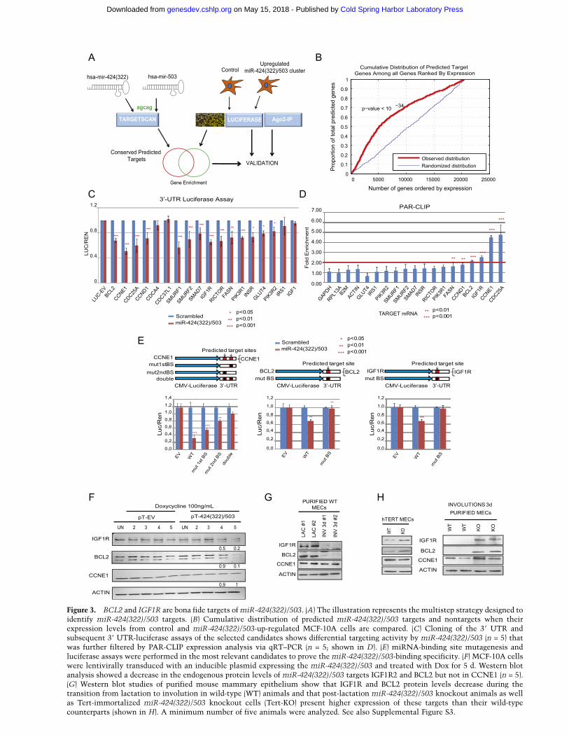
Figure 3. BCL2 and IGF1R are bona fide targets of miR-424(322)/503. (A) The illustration represents the multistep strategy designed toidentify miR-424(322)/503 targets. (B) Cumulative distribution of predicted miR-424(322)/503 targets and nontargets when theirexpression levels from control and miR-424(322)/503-up-regulated MCF-10A cells are compared. (C) Cloning of the 39 UTR andsubsequent 39 UTR-luciferase assays of the selected candidates shows differential targeting activity by miR-424(322)/503 (n = 5) thatwas further filtered by PAR-CLIP expression analysis via qRT–PCR (n = 5; shown in D). (E) miRNA-binding site mutagenesis andluciferase assays were performed in the most relevant candidates to prove the miR-424(322)/503-binding specificity. (F) MCF-10A cellswere lentivirally transduced with an inducible plasmid expressing the miR-424(322)/503 and treated with Dox for 5 d. Western blotanalysis showed a decrease in the endogenous protein levels of miR-424(322)/503 targets IGF1R2 and BCL2 but not in CCNE1 (n = 5).(G) Western blot studies of purified mouse mammary epithelium show that IGF1R and BCL2 protein levels decrease during thetransition from lactation to involution in wild-type (WT) animals and that post-lactation miR-424(322)/503 knockout animals as wellas Tert-immortalized miR-424(322)/503 knockout cells (Tert-KO) present higher expression of these targets than their wild-typecounterparts (shown in H). A minimum number of five animals were analyzed. See also Supplemental Figure S3.
Cold Spring Harbor Laboratory Press on May 15, 2018 - Published by genesdev.cshlp.orgDownloaded from

epithelial cells presented consistently higher protein levelsof these proteins than wild-type counterparts. Again nosignificant changes were observed in Ccne1 (Fig. 3H).
Expression of the miR-424(322)/503 cluster modulatesIGF-1 signal transduction and apoptosis
While the data described above clearly showed the abilityof miR-424(322)/503 to modulate the expression of BCL-2and IGF1R, it was crucial to determine whether thisregulation was biologically relevant.
IGF1R is the bona fide receptor for IGF-1 (Pollak 2012).IGF-1 binding activates the receptor kinase, leading toautophosphorylation and phosphorylation of downstreamsubstrates and resulting in the activation of two mainsignaling pathways, the PI3K–AKT/PKB and the Ras–MAPK pathways. To test the role of miR-424(322)/503in these pathways, IGF-1 was added to starved MCF-10A-424(322)/503miR-Dox cells after the miR cluster wasinduced by the addition of Dox to the medium. Down-regulation of IGF1R protein levels mediated by induc-tion of the miR cluster led to a weaker response to IGF-1stimulation, as demonstrated by reduction in the levels ofphospho-IGF1R (Tyr1135 and Tyr1131), phospho-AKT(Ser473), and phospho-ERK1/2 (Thr202/Tyr204) (Fig. 4A).Importantly, wild-type phosphorylation levels in all threeproteins were restored when a nontargetable version ofIGF1R lacking the 39 UTR was exogenously expressed(Fig. 4B; Supplemental Fig. S4A). To complement ourstudies, we performed a similar analysis with Tert-im-mortalized MMECs and primary MMECs. These revealedthat miR-424(322)/503 knockout cells, which presenthigher levels of Igf1r, were more sensitive to IGF-1 stimu-lation than their wild-type counterparts (Fig. 4C,D). Alto-gether, these data demonstrate that modulation of IGF1Rby miR-424(322)/503 affects the response to IGF-1 and itsdownstream canonical signaling.
BCL-2 is the founding member of the Bcl-2 family ofapoptosis regulator proteins (Colitti 2012), and its keyrole in the intrinsic apoptosis pathway is well established.BCL-2’s prosurvival abilities are largely due to its abilityto control mitochondrial membrane permeability by in-hibiting proapoptotic proteins. Thus, we tested whetherthe ability to undergo apoptosis was affected by the miR-424(322)/503 cluster. Apoptosis in MCF-10A-424(322)/503miR-Dox cells was induced by exposure to differentchemotherapeutic agents (cisplatin and paclitaxel). Up-regulation of the miR-424(322)/503 cluster by addition ofDox significantly increased apoptosis induced by pacli-taxel and cisplatin as shown by Annexin-V FACS analy-sis, caspase-3, and PARP cleavage, while no differenceswere observed in control cells (Fig. 4E; Supplemental Fig.S4B–D). Importantly, transduction of a BCL-2 mRNAthat lacks the 39 UTR eliminated this effect (Fig. 4F;Supplemental Fig. S4E–H). As previously done for ERK/AKT signaling studies, we also studied the response toapoptotic stress in our Tert-immortalized MMECs. Wefound that miR-424(322)/503 knockout cells, whichpresent higher levels of Bcl-2, were significantly moreresistant to apoptosis than wild-type counterparts (Fig.
4G; Supplemental Fig. S4I,J). Overall, these data demon-strate that modulation of BCL-2 by the miR-424(322)/503cluster affects the stress-induced apoptotic response.
Characterization of the 424(322)/503 promoterpri-miR and its modulation by TGF-b
To mechanistically understand the regulation of the miR-424(322)/503 in the mammary epithelium, we studiedthe genomic organization of the locus. As a first approach,we took advantage of the publicly available informationcontained in the University of California at Santa Cruzgenome (UCSC) browser (http://genome.ucsc.edu). Theclose proximity between miR-424(322) and miR-503suggests that the primary transcript containing bothmiRNAs exists as a cluster. Interestingly, a noncodingRNA (MGC16121) is described in the reference sequencegene bank whose 59 precisely matches the predictedsequence of the precursor of miR-424(322) [pre-miR-424(322)] after pre-miR has been excised by the nucle-oprocessor complex from the primary transcript (Fig. 5A).
A review of the track displaying maps of chromatinstates generated by the Broad Institute/MassachusettsGeneral Hospital ENCODE group using chromatin im-munoprecipitation (ChIP) and HTP-seq (ChIP-seq) revealeda region located ;2 kb upstream of the miR-424/503 clus-ter and strongly enriched in histone marks related toan active promoter (H3K4me2, H3K4me3, H3K9ac, andH3K27ac) in HMECs (Fig. 5A). Specifically, there wasenrichment in a histone mark associated with transcrip-tion elongation (H3K36me3) just after the terminationof the active promoter marks and spanning ;5–6 kbin a region containing the miRNA cluster (Fig. 5A).Remarkably, a comparable histone mark pattern was alsofound in the majority of the cell lines analyzed, with theexception of B cells (Supplemental Fig. S5A). In embry-onic stem cells, this putative promoter seems to be poisedfor active transcription, as it presents both active andnegative histone marks (Supplemental Fig. S5B; Bernsteinet al. 2006). Finally, FOX2 CLIP-seq (CLIP coupled withHTP-seq) used to study RNAs associated with the splic-ing regulator FOX2 showed that the H3K36me3-enrichedregion was, in fact, transcribed in the 59–39 orientation,consistent with a primary transcript starting at the pu-tative promoter upstream of the miR cluster (Fig. 5A).
In order to obtain further insights regarding the tran-scriptional regulation of the locus, we searched for con-served sequence motifs in the putative promoter region(the ;2-kb region enriched in the above-mentioned his-tone marks). Multiple alignments of 28 vertebrate speciesand measurements of evolutionary conservation using ahidden Markov model-based method (phastCons) (Pollardet al. 2010) identified several motifs conserved in verte-brates and mammals (Fig. 5B). Interestingly, one of thesemotifs represented two SMAD-binding boxes (Fig. 5B;Johnson et al. 1999). SMADs are bona fide components ofthe TGF-b and BMP pathways (Massague et al. 2005).Significantly, TGF-b ligands have well-known roles in ep-ithelial mammary gland morphogenesis (Nguyen andPollard 2000; Stein et al. 2007; Macias and Hinck 2012).
Llobet-Navas et al.
772 GENES & DEVELOPMENT
Cold Spring Harbor Laboratory Press on May 15, 2018 - Published by genesdev.cshlp.orgDownloaded from

Upon TGF-b ligand binding to its receptor, receptor-regulated SMAD-2 and SMAD-3 (R-SMADs) are activatedby phosphorylation, heterodimerize with SMAD-4, andare then shuttled to the nucleus, where they act astranscription factors. R-SMADs are weak transcriptionalactivators and commonly cooperate with additional tran-scriptional modulators, such as E2Fs (Massague et al. 2005).Remarkably, a highly conserved region containing two
E2F-binding motifs is located in close proximity (;100bases) downstream from the SMAD-3-binding site.
TGF-b ligands have a key role during mammary epi-thelial development, mainly as potent inhibitors of pro-liferation (Howard and Gusterson 2000; Macias and Hinck2012). During involution, expression of TGF-b ligands inthe mammary gland, especially TGF-b3, is induced as soonas 3 h after forced weaning, and levels of TGF-b3 continue
Figure 4. Induced miR-424(322)/503 desensitizes cells to IGF-1 and primes cells to undergo apoptosis. (A) Dox-induced miR-424(322)/
503 targets endogenous IGF1R, causing desensitization to IGF-1 stimuli, as shown by Western blot. Cells were treated with Dox for 5 d,starved with 0.5% horse serum overnight, and subsequently treated with 50 ng/mL IGF-1 for the specified time points. (B) EngineeredpT-424(322)/503 MCF-10A cells stably expressing a nontargetable form of IGF1R (pLX-IGF1R) are able to overcome IGF-1 desensitizationcaused by Dox-induced miR-424(322)/503. pLX-EV MCF-10A cells stably expressing an empty vector control. (C) Tert-immortalizedknockout (Tert-KO) epithelial cells show consistent higher sensitivity to IGF-1 stimulation, as shown by Western blot. (D) Westernblots showing increased activation of p-AKT and p-ERK in purified miR-424(322)/503 knockout involuting mammary epithelial cellscompared with their wild-type (WT) counterparts. (E) pT-424(322)/503 cells treated with Dox display higher sensitivity to apoptosisinduced by 100 ng/mL paclitaxel (PTX) and 10 mg/mL cisplatin, as shown by quantification of cell death by Annexin-V staining andFACs analysis (top panel) and analysis of active caspase-3 and cleaved PARP by Western blot (bottom panel). MCF-10A cells were treatedwith Dox and subsequently exposed to PTX for 24 h. (F) Expression of a BCL2 cDNA devoid of its 39 UTR (pLOC-BCL2) blocks apoptoticcell death induced by PTX. (G) Tert-immortalized knockout cells exert higher resistance to PTX, as shown by Annexin-V, caspase-3, andPARP studies. All the experiments were performed in triplicate. See also Supplemental Figures S4 and S5.
miR-424/503 regulates mammary involution
GENES & DEVELOPMENT 773
Cold Spring Harbor Laboratory Press on May 15, 2018 - Published by genesdev.cshlp.orgDownloaded from

Figure 5. Genomic organization of miR-424(322)/503 locus unveils transcriptional regulation mediated by TGF-b. (A) UCSC-extracted genomic locations of miR-424(322) and miR-503 and putative regulatory elements located upstream in HMECs, includinghistone marks of transcriptionally active chromatin (H3K4me2, H3K4me3, H3K9ac, and H3K27ac), transcriptional silencing orrepression (H3K27me3), and elongation (H3K36me3) as well as marks of elongation and splicing events, represented by genome-wideFOX2 CLIP-seq. The presence of additional regulatory regions such as CpG islands is also depicted. (B) Representation of theconservation status of miR-424(322), miR-503, and the upstream regulatory regions using a vertebrate and placental mammal Markovmodel. Using this method, 28 vertebrate species were aligned to assess conservation status. Magnification of a small conserved regionlocated upstream (highlighted in red) identifies two conserved SMAD-binding motifs. (C) Analysis by miRNA expression arrays ofMCF-10A treated with 10ng/mL TGF-b for 24, 48, and 72 h. (D) Results were further validated by qRT–PCR (top panel) and in situhybridization (bottom panel). For in situ hybridization, MCF-10A cells transduced with a construct expressing the cluster miR-424(322)/503 were used as a positive control. (E) TGF-b-dependent regulation of miR-424(322) and miR-503 was verified in otherHMEC lines (MCF-12A and 184B5), HuMECs, and primary Tert-immortalized MMECs. Cells were treated with 10 ng/mL TGF-b1 for48 h. (F) Expression analysis by qRT–PCR of miR-15/16 family members in MCF-10A cells upon treatment with 10 ng/mL TGF-b forseveral days. (G) TGF-b-dependent miR-424(322) and miR-503 expression was demonstrated by transfecting MCF10A cells with siRNAagainst SMAD4. Scrambled siRNAs and siRNAs against GFP were used as negative controls. The expression of bona fide TGF-b targets(top panel) and miR-424(322) and miR-503 (bottom panel) was performed by qRT–PCR. See also Supplemental Figure S5.
Cold Spring Harbor Laboratory Press on May 15, 2018 - Published by genesdev.cshlp.orgDownloaded from
to increase during the following hours to remain high forseveral days (Faure et al. 2000; Nguyen and Pollard 2000).Furthermore, expression of TGF-b type II receptor (Faureet al. 2000) and the nuclear internalization of Smad-4(Nguyen and Pollard 2000) also occur in the mammaryepithelium at about the same time. Although the role ofTGF-b3 is not fully understood, in vivo evidence fromtransgenic mice expressing Tgf-b3 under the b-lactoglobinpromoter or expressing a dominant-negative form of theTgf-b receptor-II in the mammary gland (MMTV-DNIIR)as well as from animals where Tgf-b3 knockout mam-mary epithelial cells were orthotopically transplanteddemonstrates the importance of Tgf-b signaling duringthe early and late involution phases (Nguyen and Pollard2000; Gorska et al. 2003). Based on the above information,we hypothesized that exposure to TGF-b ligands duringinvolution up-regulates the expression of miR-424(322)/503, which in turn modulates key players of cell cycle,apoptosis, and IGF-1 signaling.
The mammary cell line MCF-10A is a widely usedmodel to study TGF-b response in mammary epithelialcells (Iavarone and Massague 1997; Karakas et al. 2006).Despite being null for the INK4B gene, these cells re-tain the ability to arrest after being exposed to TGF-b.(Iavarone and Massague 1997). To study the regulation ofthe miR-424(322)/503 cluster, we exposed MCF-10A cellsto TGF-b1 and, using miRNA microarrays, tracked theexpression of 799 human miRNAs (Sanger miRbase re-lease 10.1) through time. Both miR-424(322) and miR-503emerged as the top TGF-b1-up-regulated miRNAs (Fig.5C). qRT–PCR of the mature miRNAs and in situhybridization experiments confirmed the microarray data(Fig. 5D) and showed that the observed up-regulation wastime- and dose-dependent (Supplemental Fig. S5C). In-duction of miR-424(322)/503 was not restricted to MCF-10A cells, and, although it was less pronounced, we alsoobserved induction in two additional nontransformedhuman cell lines (MCF-12A and 184B5), in primary HMECs(HuMECs), and in MMECs immortalized with TERT(Fig. 5E).
As mentioned above, TGF-b3 is a TGF-b ligand witha key role during involution (Nguyen and Pollard 2000).Thus, we also tested its ability to induce the expression ofthe miRNA cluster. Our data showed that comparablelevels of miR-424(322) and miR-503 up-regulation wereinduced with both TGF-b1 and TGF-b3 (Fig. 5F). Inter-estingly, none of the other miR-16 family members wereinduced after TGF-b1 and TGF-b3 exposure.
To evaluate the role of the canonical TGF-b pathway inthe regulation of miR-424(322)/503, we knocked downSMAD4 before exposing MCF-10A cells to TGF-b1(Fig. 5G). In knockdown cells, the induction of bona fideTGF-b targets such as SMAD7, P21, CTCF, ATF3, andPAI-1 as well as miR-424(322) and miR-503 was signifi-cantly reduced. Next we investigated which of theR-SMADs was involved in the regulation of miR-424(322)/503. Both SMAD2 and SMAD3 are activated during in-volution and when MCF-10A cells are exposed to TGF-b(Supplemental Fig. S5D). Thus, we individually silencedSMAD2 and SMAD3 (Supplemental Fig. S5E) and evalu-
ated the up-regulation of the mature miR-423(322) andmiR-503 as well as the pri-miR cluster. Our studies clearlydemonstrate that SMAD3 is the R-SMAD responsiblefor the up-regulation of the pri-miR-424(322)/503 and theproduction of the miR mature forms upon TGF-b exposure(Supplemental Fig. S5F,G).
To further confirm the role of TGF-b in regulating theexpression of miR-424(322)/503, we performed ChIPassays to investigate the presence of SMAD4 in the endog-enous putative promoter. To pursue this aim, we createda MCF-10A cell line where HA-tagged SMAD4 proteinwas constitutively expressed in MCF-10A cells. After en-suring that the exogenously introduced SMAD4 behaved asthe endogenous protein did (both were shuttled into thenucleus in the presence of TGF-b) (Supplemental Fig. S6A),we used this cell line to perform ChIP with anti-HAantibodies. These studies revealed that SMAD4 was en-riched in the putative miR-424(322)/503 promoter afterTGF-b exposure at levels comparable with the bona fideTGF-b target PAI-1 (Fig. 6A). Importantly, the strongestenrichment happened in the region near the SMAD4-binding site.
To evaluate the role of the putative SMAD4-bindingsite in the up-regulation of the miRNA cluster uponTGF-b exposure, we first engineered a reporter constructcontaining an ;5.4-kb region upstream of the miRNAcluster followed by the GFP sequence (Fig. 6B). Thisreporter was integrated into the genome of MCF-10Acells by lentiviral delivery to create a pri-miR reporter cellline (MCF-10A-pri-miR-reporter). Reverse transcriptionusing oligonucleotides complementary to GFP allowed usto study the transcript resulting exclusively from thereporter without interference of endogenous pri-miR (seethe Materials and Methods). Upon addition of TGF-bto the MCF-10A-pri-miR-reporter cells, a clear increase inthe transcription of the reporter was observed (Fig. 6C).Importantly, when we mutagenized the SMAD4-bindingsite, this increase was abrogated.
Next, we further characterized the pri-miR transcriptcontrolled by this promoter. First, we identified the tran-scription start site (TSS) by performing 59 rapid amplifi-cation of cDNA end (RACE). By using a specific primerlocated upstream of the miR cluster just before the regionenriched in promoter-specific histone marks (;300 bpdownstream from the SMAD4-binding site) for the re-verse transcription step, we identified a TSS locateddownstream from the E2F-binding sites (Fig. 6D; Supple-mental Fig. S6B). Interestingly, the addition of TGF-bshifted the TSS 181 nt upstream, which suggests the pres-ence of an alternative promoter. As expected, we wereable to amplify a region expanding from the miRNAcluster to the TSS by using overlapping PCRs (Supple-mental Fig. S6C). qRT–PCR amplification and in situhybridization confirmed the up-regulation of the pri-miRNA by TGF-b exposure (Fig. 6E; Supplemental Fig.S6C). As expected, the pri-miRNA was detected by in situhybridization at the mammary epithelium of involutingmammary glands (3 d) (Supplemental Fig. S6D). Notably,kinetic studies of the pri-miRNA, pre-miRNA, and ma-ture miRNA levels after addition of TGF-b revealed that
miR-424/503 regulates mammary involution
GENES & DEVELOPMENT 775
Cold Spring Harbor Laboratory Press on May 15, 2018 - Published by genesdev.cshlp.orgDownloaded from

Figure 6. Characterization of the miR-424(322)/503 promoter, pri-miR-424(322)/503, and transcriptional regulation by TGF-b in vivo. (A)MCF-10A cells stably expressing HA-tagged SMAD4 or a control empty vector were treated with 10 ng/mL TGF-b for 3 h, and ChIP wasperformed using an anti-HA antibody. The quantification of genomic DNA enrichment was performed using specific primers surrounding thepromoter. The putative SMAD-binding site is indicated by a red square. Quantification of DNA enrichment in the PAI-1 gene promoter wasused as a positive control, while b-Actin and Gapdh were used as negative controls. (B) A schematic illustration obtained from the UCSCgenome database showing the localization of miR-424(322) and miR-503 and the upstream putative 5.4-kb promoter region containing eitherthe wild-type or mutated SMAD-binding site that was cloned into a reporter vector, as explained in the text. (C) MCF-10A cells weretransduced with either the wild-type or mutated promoters and treated with or without 10 ng/mL TGF-b, and the promoter activity wasanalyzed by qRT–PCR. (D) Representation of the proposed miR-424(322)/503 cluster promoter region containing the SMAD-binding site andthe alternative TSSs identified by 59 RACE in the presence and absence of TGF-b. (E) Expression kinetics of pri-miR-424(322)/503 aftertreatment of MCF-10A cells with 10 ng/mL TGF-b for the specified time points by qRT–PCR using specific primers surrounding the pre-miR-
424(322) and pre-miR-503 (left) or by in situ hybridization using a probe against pri-miR-424(322)/503 (right). (F) qRT–PCR analysis showingthat increased mature miR-424(322) and miR-503 levels after treatment of MCF-10A cells with 10 ng/mL TGF-b coincide with a decrease inpre-miR-424(322) and pre-miR-503, respectively. (G) After inhibition of TGF-b signaling in vivo by the administration of the specific inhibitorLY2109761, mammary glands were examined 72 h after weaning for the presence of secretory acini by H&E and for epithelial density bywhole-mount carmine red staining. Additionally, mammary epithelial cells were purified, and milk protein production was evaluated by qRT–PCR using specific primers for a-lactalbumin and b-casein genes. (H) Inhibition of the activation of the TGF-b pathway, shown by Westernblot and immunohistochemistry for activated phospho-SMAD2/3, abrogates the transcription of pri-miR-424(322)/503 during involution, asshown by qRT–PCR. See also Supplemental Figure S6, E and F, for a similar study using the TGF-b inhibitor LY2157299.
Cold Spring Harbor Laboratory Press on May 15, 2018 - Published by genesdev.cshlp.orgDownloaded from

the increase in pri-miRNA and pre-miRNA happens firstand that generation of mature miRNAs leads to a reduc-tion of the pri-miRNA and pre-miRNA (Fig. 6E,F). Thiscould be due to the cleavage and degradation of the pri-miRNA after the pre-miRNA is excised, the existence ofa negative regulatory feedback loop, or a combination ofboth mechanisms.
Finally, to test that the regulation of miR-424(322)/503by TGF-b occurs in vivo during involution, we individu-ally treated lactating females with two different specificinhibitors of TGF-b receptor I and II with demonstratedactivity in vivo (LY2109761 and LY2157299) (Melisi et al.2008; Bouquet et al. 2011; Calon et al. 2012; Bhola et al.2013) and analyzed the induction of the miR cluster.When lactating females were pretreated with these inhib-itors starting 24 h before weaning, these animals showedan evident inhibition of involution 3 d after weaning, asindicated by carmine red and H&E staining of the mam-mary gland and qRT–PCR levels of milk proteins (Fig. 6G;Supplemental Fig. S6E). Importantly, due to the inhibi-tion of TGF-b signaling, they also presented a strongreduction in the levels of pri-miR-424(322)/503 (Fig. 6H;Supplemental Fig. S6F).
Discussion
Despite the established role of a variety of extrinsic andintrinsic signals regulating mammary gland development(Howard and Gusterson 2000; Macias and Hinck 2012),a restricted number of studies have identified and vali-dated miRNAs with a role in any aspect of normalmammary gland biology (Avril-Sassen et al. 2009; Tanakaet al. 2009; Ucar et al. 2010; Yang et al. 2010; Cui et al.2011; Le Guillou et al. 2012), and even fewer have studiedtheir function using transgenic animal models (Ucar et al.2010; Le Guillou et al. 2012). Here we unveiled themiRNA cluster miR-424(322)/503 as an important regu-lator of the massive reorganization that occurs in themammary epithelium after pregnancy. Furthermore,through the generation of a knockout mouse model, wecharacterized mechanistically its role and regulation.
Among the 12 identified miRNAs, miR-424(322) showedthe largest fold increase between lactation and involu-tion. Although this miRNA has been studied in thecontext of monocyte differentiation (Rosa et al. 2007;Forrest et al. 2010) and endothelial cell regulation (Ghoshet al. 2010; Nakashima et al. 2010; Kim et al. 2013), apotential role in mammary epithelial architecture remod-eling has not yet been described. The characterization ofthe mammary epithelium of miR-424(322)/503 knockoutfemales during development revealed a defect duringinvolution. To understand the role of miR-424(322)/503in this biological context, we considered two pertinentquestions: (1) Which are the relevant targets modulatedby the cluster in the mammary epithelium? (2) How is thepri-miR regulated?
Through our comprehensive studies in vitro and invivo, we identified BCL-2 and IGF1R as robust targetgenes of the miR-424(322)/503 cluster in the mammaryepithelium. Interestingly, our data did not validate previous
data reporting CCNE1 as a target of this miRNA cluster(Forrest et al. 2010; Nakashima et al. 2010). Since therelevance of the different miRNA targets is context-dependent (Liu and Kohane 2009; Inui et al. 2010), it isimportant to mention that we used HMECs for ourstudies and achieved mature miRNA expression levelscomparable with the observed during involution in vivo.Notably, when we augmented by lentiviral transductionthe intracellular levels of the mature miRNAs severalfold (>10 times) over the levels found in vivo, we observeda reliable reduction of CCNE1 protein (SupplementalFig. S3F). Finally, no major changes were observed inCCNE1 protein levels from lactation to involution. Thus,our data illustrate the importance of the biologicalcontext in identifying relevant miRNA targets for differ-ent processes, and, consequently, we do not considerCCNE1 a relevant target of the miR-424(322)/503 in thecontext of mammary epithelial involution and TGF-bexposure.
During the last two decades, key details about the mech-anism and regulation of mammary gland involution havebeen identified (Stein et al. 2007). The use of geneticallymodified mice with gain or loss of function has pro-vided evidence regarding the implication of Bcl-2 andIgf1r in this process. The Igf1/Igf1r axis plays critical rolesin multiple steps of mammary development (Hynes andWatson 2010). During involution, IGF signaling has beenfound to be inhibited by down-regulation of Igf1r (Modhaet al. 2004) and up-regulation of insulin-like growthfactor-binding protein 5 (Igfbp-5) (Allan et al. 2004).Importantly, the effect of IGF signaling in life or deathfate decisions has been related to its effect on the PI3K–AKT/PKB and the Ras–MAPK pathways (Allan et al.2004; Stein et al. 2007). In accordance with this, in-volution has been found to be inhibited in transgenicanimals with constitutively active Akt (Schwertfegeret al. 2001; Ackler et al. 2002). BCL-2 family membersare also involved in regulating cellular fate after thepregnancy cycle (Colitti 2012). Levels of proapoptoticBak and Bad increase during lactation, reaching theirpeak during involution. In contrast, anti-apoptotic Bcl-W and Bcl-2 are strongly down-regulated at the onset ofapoptosis (Metcalfe et al. 1999). In the proposed model,mammary epithelial cells are poised for apoptosis withhigher levels of the death effectors but are prevented fromtriggering apoptosis by anti-apoptotic Bcl-2 family pro-teins until involution, when prosurvival gene expressionlevels decline (Metcalfe et al. 1999; Stein et al. 2007;Colitti 2012). In agreement with this view, enforcedexpression of Bak induces rapid mammary apoptosis(Metcalfe et al. 1999), while high levels of Bcl-2 havebeen shown to potently regulate mammary epithelial cellsurvival and inhibit alveolar cell apoptosis during in-volution (Jager et al. 1997; Schorr et al. 1999).
By understanding the signals that initiate the transcrip-tional up-regulation of the miR cluster, we uncovered itsregulation by TGF-b. As TGF-b ligands have well-knownroles in epithelial mammary gland morphogenesis (Nguyenand Pollard 2000; Stein et al. 2007), this informationprovided us with a potential link between the induction
miR-424/503 regulates mammary involution
GENES & DEVELOPMENT 777
Cold Spring Harbor Laboratory Press on May 15, 2018 - Published by genesdev.cshlp.orgDownloaded from
of involution and the up-regulation of the miR cluster. Itis interesting to discuss that the R-SMAD that controlsthe up-regulation of the miR-424(322)/503, SMAD3, hasbeen found to control not only transcription but alsomiRNA processing in a SMAD4-independent function(Davis et al. 2008, 2010). Thus, a question is whether thisalso applies for the production of mature miR-424(322)and miR-503. SMAD3 has been found to interact withDROSHA and affect miRNA maturation by binding toa conserved 5-base motif (SBE; 59-CAGAC-39) present inthe pri-miRs. However the pri-miR-424(322)/503 clustertranscript does not contain this binding element. Addi-tionally, individual silencing of both SMAD3 and SMAD4completely abrogated the up-regulation of both pri-miR-424(322)/503 and individual mature forms. Thus, at thispoint, transcriptional up-regulation seems to be a criticalstep, and additional studies will be required to clarify apotential miRNA processing function of pri-miR-424(322)/503 by SMAD3.
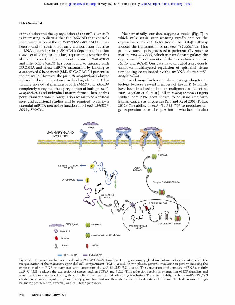
Mechanistically, our data suggest a model (Fig. 7) inwhich milk stasis after weaning rapidly induces theexpression of TGF-b3. Activation of the TGF-b pathwayinduces the transcription of pri-miR-424(322)/503. Thisprimary transcript is processed to preferentially generatemature miR-424(322), which in turn down-regulates theexpression of components of the involution response,IGF1R and BCL-2. Our data have unveiled a previouslyunknown multilayered regulation of epithelial tissueremodeling coordinated by the miRNA cluster miR-424(322)/503.
Our work may also have implications regarding tumorbiology because several members of the miR-16 familyhave been involved in human malignancies (Liu et al.2008; Aqeilan et al. 2010). All miR-424(322)/503 targetsstudied here have been shown to be associated withhuman cancers as oncogenes (Yip and Reed 2008; Pollak2012). The ability of miR-424(322)/503 to modulate tar-get expression raises the question of whether it is also
Figure 7. Proposed mechanistic model of miR-424(322)/503 function. During mammary gland involution, critical events dictate thereorganization of the mammary epithelial cell compartment. TGF-b, a well-known player, governs involution in part by inducing theexpression of a miRNA primary transcript containing the miR-424(322)/503 cluster. The generation of the mature miRNAs, mainlymiR-424(322), reduces the expression of targets such as IGF1R and BCL2. This reduction results in attenuation of IGF signaling andsensitization to apoptosis, leading the epithelial cells toward cell death during involution. The above highlights the miR-424(322)/503
cluster as a critical regulator of mammary gland homeostasis through its ability to dictate cell life and death decisions throughbalancing proliferation, survival, and cell death pathways.
Llobet-Navas et al.
778 GENES & DEVELOPMENT
Cold Spring Harbor Laboratory Press on May 15, 2018 - Published by genesdev.cshlp.orgDownloaded from

involved in breast tumorigenesis. Our data in this regardreveal that knockout animals accumulate alveoli hyper-plasia, but we did not observe tumor development inanimals followed up to 1 yr of age. Longer periods of follow-up, multiple pregnancies, or both may be necessary togenerate full cell transformation in these animals. In-terestingly, miR-424(322)/503 is located in a chromoso-mal region that has been recently identified to presentsignificant deletions in ;8% of hormone receptor-posi-tive breast cancers (Dvinge et al. 2013). Furthermore, thepromoter region that we identified is placed on a CpGisland that could be targeted by hypermethylation asa potential mechanism to down-regulate the expressionof the miRNAs during transformation. As miR-424(322)/503 is located in the silenced part of the X chromosome,a single inactivation event is enough to fully abrogate itsexpression. Interestingly, it has been recently reportedthat suppressed miR-424(322) expression contributes tothe progression of cervical cancer (Xu et al. 2013).Additional research will be necessary to fully define thepotential role of miR-424(322)/503 in breast tumorigen-esis. In this regard, our work describing the biological roleof miR-424(322)/503 during involution and its regulationestablishes the basis for future studies.
Materials and methods
Cell culture
Cell lines were obtained from the American Type CultureCollection. Normal breast epithelial MCF-10A and MCF-12cells were cultured at 37°C/5%CO2 in DMEM/Ham’s F-12supplemented with 10% penicillin/streptomycin, 20 ng/mLEGF, 10 mg/mL insulin, 100 ng/mL cholera toxin, 500 ng/mLhydrocortisone, and 5% horse serum. HuMECs (Life Technolo-gies, no. A10565) were grown according to instructions andreagents provided by the manufacturer. 184B5 cells were grownin MEGM medium (Lonza/Clonetics, no. CC-3150) supple-mented with 1 ng/mL cholera toxin. Phoenix cells were grownin DMEM–10% fetal bovine serum and 10% penicillin/strepto-mycin at 37°C/5%CO2.
TGFb1 and TGFb3 were purchased from Sigma (nos. T7039and T5425, respectively). Human recombinant IGF1 was pur-chased from PROSPEC (no. CYT-216). Cisplatin (no. P4394) andpaclitaxel (no. T7402) were both from Sigma.
miRNA extraction and HTP-seq
To perform mature miRNA and pre-miRNA expression analysisby qRT–PCR, RNA was purified using the miRVANA miRNAisolation kit (Ambion, no. AM1561) according to the instruc-tions provided. Reverse transcription was performed on 50 ng ofRNA using the miRNA reverse transcription kit (Roche, no.4366596). miRNA deep sequencing was conducted at the ColumbiaUniversity Genome Center’s core facility (http://genomecenter.columbia.edu) using the Illumina platform. RNA sequencing(RNA-seq) was performed at HiSeq single reads with a depth ofcoverage of 20 million reads per sample.
Luciferase reporter assays
39 UTRs of specific target genes were cloned downstream fromthe luciferase reporter in the pMIR-REPORT vector (Life Tech-
nologies, no. AM5795M) by PCR from human genomic DNAusing restriction enzymes. Relative luciferase units (RLUs) weremeasured using the Dual-Glo luciferase assay system (Promega,no. E2949). For a detailed description, see the SupplementalMaterial.
Mouse generation and LY2109761 treatment
To generate single miR-424(322) and miR-503 or double-knock-out mice, we used ZNF (Sigma; for a detailed description, see theSupplemental Material). To inhibit TGF-b signaling during in-volution, lactating wild-type and miR-424(322)�/�/miR-503�/�
double-knockout females were pretreated by oral gavage twicedaily with 50 mg/kg LY2109761 (Selleckchem) for 24 h beforeweaning. Thereafter, mice were treated again by oral gavagetwice daily with 50 mg/kg LY2109761 for 1 and 3 d. Mice weresacrificed, and mammary glands were dissected as mentionedpreviously. All experiments were performed under the Institu-tional Animal Care and Use Committee (IACUC) guidelines.
PAR-CLIP analysis
A PAR-CLIP assay to measure AGO2 enrichment in miR-424(322)/503 mRNA targets was performed as described pre-viously (Hafner et al. 2010). Briefly, cells were pretreated with50 mM 4-thiouridine (Sigma, no. T4509) overnight and cross-linkedat 150 mJ/cm2 at 365 nm UV on ice. Immunoprecipitation wasperformed using an anti-AGO2 antibody (Abnova, no. H00027161-M01) overnight at 4°C. RNA was extracted using the miRVANAmiRNA isolation kit, and retrotranscribed RNA was subjected toanalysis by qRT–PCR using specific primers.
Quantification of the number of miR-424(322) and miR-503molecules
The number of molecules was estimated using chemicallysynthesized RNA oligonucleotides that mimic in sequencemiR-424(322) and miR-503. RNA oligonucleotides were pre-pared in serial dilutions corresponding to 1 pg of miRNA permicroliter, 0.1 pg of miRNA per microliter, 0.01 pg of miRNAper microliter, and 1 fg of miRNA per microliter. One microliterof each dilution was retrotranscribed in parallel with 50 ng of oursamples. The number of molecules per cell was estimated byconsidering a ratio of 10 pg of RNA per cell and integrating theexperimental Ct values into the standard values obtained withthe commercial oligonucleotides.
Flow cytometry and cell viability
All flow cytometry experiments were performed on a BDFacsCalibur cell analyzer using the CellQuest Pro software on MacOSX. To analyze apoptosis induced by cisplatin and paclitaxel,we performed the Annexin-V staining following the FITCAnnexin-V apoptosis detection kit guidelines (BD Pharmingen,no. 556570). For a detailed description, see the SupplementalMaterial.
Whole-mount preparations and immunohistochemistry
To prepare whole-mount carmine red staining, mammary glandswere fixed in 4% paraformaldehyde for 1 h, stained with carminered (Sigma, no. C1022) overnight at room temperature, andultimately dehydrated and fixed in xilol. To perform immuno-histochemical analysis, formalin-fixed paraffin-embedded sam-ples were deparaffinized and rehydrated. Peroxidase inactivationand antigen retrieval were achieved by incubating samples in 1%
miR-424/503 regulates mammary involution
GENES & DEVELOPMENT 779
Cold Spring Harbor Laboratory Press on May 15, 2018 - Published by genesdev.cshlp.orgDownloaded from

H2O2 and citric buffer. The antibodies used were Ki67 (1:200;Abcam, no. ab15580), anti-cytokeratin 18 antibody (1:300; Abcam,no. ab668), anti-cytokeratin 5 (1:1000; Covance, no. PRB-160P),cleaved caspase-3 (1:500; Cell Signaling, no. 9664), and p-SMAD2/3(1:100; Cell Signaling, no. 9510).
Gene expression and miRNA expression arrays
For gene expression and miRNA expression arrays, MCF-10Acells were treated with TGFb, and RNA was extracted usingRNeasy and miRVANA extraction kits, respectively, and labeledusing the LowInput QuickAmp labeling kit (Agilent, no. 5190-2331). RNAs were hybridized on a human GE 4x44K v2 micro-array kit (Agilent, no. G4845A) and a human miRNA V2 oligomicroarray (Agilent, no. G4470B), respectively. Gene expressionarray data have been uploaded and are available in the GeneExpression Omnibus database.
ChIP
MCF-10A untreated and TGFb cells were cross-linked with 11%formaldehyde solution for 10 min at room temperature, lysed,and sonicated using a Bioruptor Standard sonication device(Diagenode, no. UCD-200 TM; see the Supplemental Materi-al). HA-SMAD4 immunoprecipitation was performed usingthe DynaMag-2 magnetic particle concentrator (Invitrogen, no.123.21D), protein G Dynabeads (Invitrogen, no. 100.03D), andan anti-HA antibody (Roche, no. 11867423001) overnight at 4°Con a rotator. Thereafter, samples were collected and washed, andphenol/chloroform/isoamyl alcohol extraction using Phaselocktubes (Prime, no. 2302840) was performed to isolate DNA. DNAenrichment was analyzed by qRT–PCR using specific primers.Data are presented as the D-normalized values between SMAD4-HA [D (TGFb-treated) � (untreated)] � EV [D (TGFb-treated) �(untreated)]. For a detailed description, see the SupplementalMaterial.
In situ hybridization
Briefly, probes against pri-hsa-miR-424(322)/503 were createdfrom human genomic DNA by PCR using specific oligonucleo-tides and were obtained by T7-driven in vitro RNA synthesis.Probes against mature miR-424(322) and miR-503 were purchasedfrom Exiqon (catalog no. 38195-08), and the assay was visualizedusing a tyramide signal amplification method (PerkinElmer,NEL741001KT). For a detailed description, see the SupplementalMaterial.
References
Ackler S, Ahmad S, Tobias C, Johnson MD, Glazer RI. 2002.Delayed mammary gland involution in MMTV–AKT1 trans-genic mice. Oncogene 21: 198–206.
Ahn HW, Morin RD, Zhao H, Harris RA, Coarfa C, Chen ZJ,Milosavljevic A, Marra MA, Rajkovic A. 2010. MicroRNAtranscriptome in the newborn mouse ovaries determined bymassive parallel sequencing. Mol Hum Reprod 16: 463–471.
Allan GJ, Beattie J, Flint DJ. 2004. The role of IGFBP-5 inmammary gland development and involution. Domest Anim
Endocrinol 27: 257–266.Aqeilan RI, Calin GA, Croce CM. 2010. miR-15a and miR-16-1
in cancer: discovery, function and future perspectives. Cell
Death Differ 17: 215–220.Avril-Sassen S, Goldstein LD, Stingl J, Blenkiron C, Le Quesne J,
Spiteri I, Karagavriilidou K, Watson CJ, Tavare S, Miska EA,et al. 2009. Characterisation of microRNA expression in
post-natal mouse mammary gland development. BMC Ge-
nomics 10: 548.Bartel DP. 2004. MicroRNAs: genomics, biogenesis, mecha-
nism, and function. Cell 116: 281–297.Bartel DP. 2009. MicroRNAs: target recognition and regulatory
functions. Cell 136: 215–233.Bernstein BE, Mikkelsen TS, Xie X, Kamal M, Huebert DJ,
Cuff J, Fry B, Meissner A, Wernig M, Plath K, et al. 2006. Abivalent chromatin structure marks key developmentalgenes in embryonic stem cells. Cell 125: 315–326.
Bhola NE, Balko JM, Dugger TC, Kuba MG, Sanchez V, SandersM, Stanford J, Cook RS, Arteaga CL. 2013. TGF-b inhibitionenhances chemotherapy action against triple-negative breastcancer. J Clin Invest 123: 1348–1358.
Bouquet F, Pal A, Pilones KA, Demaria S, Hann B, Akhurst RJ,Babb JS, Lonning SM, DeWyngaert JK, Formenti SC, et al.2011. TGFb1 inhibition increases the radiosensitivity ofbreast cancer cells in vitro and promotes tumor control byradiation in vivo. Clin Cancer Res 17: 6754–6765.
Calon A, Espinet E, Palomo-Ponce S, Tauriello DV, Iglesias M,Cespedes MV, Sevillano M, Nadal C, Jung P, Zhang XH, et al.2012. Dependency of colorectal cancer on a TGF-b-drivenprogram in stromal cells for metastasis initiation. Cancer
Cell 22: 571–584.Chapman RS, Lourenco PC, Tonner E, Flint DJ, Selbert S,
Takeda K, Akira S, Clarke AR, Watson CJ. 1999. Suppressionof epithelial apoptosis and delayed mammary gland involu-tion in mice with a conditional knockout of Stat3. GenesDev 13: 2604–2616.
Colitti M. 2012. BCL-2 family of proteins and mammary cellularfate. Anat Histol Embryol 41: 237–247.
Cui W, Li Q, Feng L, Ding W. 2011. MiR-126-3p regulatesprogesterone receptors and involves development and lacta-tion of mouse mammary gland. Mol Cell Biochem 355: 17–25.
Davis BN, Hilyard AC, Lagna G, Hata A. 2008. SMAD proteinscontrol DROSHA-mediated microRNA maturation. Nature
454: 56–61.Davis BN, Hilyard AC, Nguyen PH, Lagna G, Hata A. 2010.
Smad proteins bind a conserved RNA sequence to promotemicroRNA maturation by Drosha. Mol Cell 39: 373–384.
Dvinge H, Git A, Graf S, Salmon-Divon M, Curtis C, SottorivaA, Zhao Y, Hirst M, Armisen J, Miska EA, et al. 2013. Theshaping and functional consequences of the microRNAlandscape in breast cancer. Nature 497: 378–382.
Faure E, Heisterkamp N, Groffen J, Kaartinen V. 2000. Differ-ential expression of TGF-b isoforms during postlactationalmammary gland involution. Cell Tissue Res 300: 89–95.
Finnerty JR, Wang WX, Hebert SS, Wilfred BR, Mao G, NelsonPT. 2010. The miR-15/107 group of microRNA genes:evolutionary biology, cellular functions, and roles in humandiseases. J Mol Biol 402: 491–509.
Forrest AR, Kanamori-Katayama M, Tomaru Y, Lassmann T,Ninomiya N, Takahashi Y, de Hoon MJ, Kubosaki A, KaihoA, Suzuki M, et al. 2010. Induction of microRNAs, mir-155,mir-222, mir-424 and mir-503, promotes monocytic differ-entiation through combinatorial regulation. Leukemia 24:460–466.
Ghosh G, Subramanian IV, Adhikari N, Zhang X, Joshi HP, BasiD, Chandrashekhar YS, Hall JL, Roy S, Zeng Y, et al. 2010.Hypoxia-induced microRNA-424 expression in human en-dothelial cells regulates HIF-a isoforms and promotes angio-genesis. J Clin Invest 120: 4141–4154.
Gorska AE, Jensen RA, Shyr Y, Aakre ME, Bhowmick NA,Moses HL. 2003. Transgenic mice expressing a dominant-negative mutant type II transforming growth factor-b receptor
Llobet-Navas et al.
780 GENES & DEVELOPMENT
Cold Spring Harbor Laboratory Press on May 15, 2018 - Published by genesdev.cshlp.orgDownloaded from

exhibit impaired mammary development and enhancedmammary tumor formation. Am J Pathol 163: 1539–1549.
Grimson A, Farh KK, Johnston WK, Garrett-Engele P, Lim LP,Bartel DP. 2007. MicroRNA targeting specificity in mammals:determinants beyond seed pairing. Mol Cell 27: 91–105.
Guo H, Ingolia NT, Weissman JS, Bartel DP. 2010. MammalianmicroRNAs predominantly act to decrease target mRNAlevels. Nature 466: 835–840.
Hafner M, Landthaler M, Burger L, Khorshid M, Hausser J,Berninger P, Rothballer A, Ascano M Jr, Jungkamp AC,Munschauer M, et al. 2010. Transcriptome-wide identifica-tion of RNA-binding protein and microRNA target sites byPAR-CLIP. Cell 141: 129–141.
Howard BA, Gusterson BA. 2000. Human breast development.J Mammary Gland Biol Neoplasia 5: 119–137.
Hynes NE, Watson CJ. 2010. Mammary gland growth factors:roles in normal development and in cancer. Cold Spring
Harb Perspect Biol 2: a003186.Iavarone A, Massague J. 1997. Repression of the CDK activator
Cdc25A and cell-cycle arrest by cytokine TGF-b in cellslacking the CDK inhibitor p15. Nature 387: 417–422.
Inui M, Martello G, Piccolo S. 2010. MicroRNA control of signaltransduction. Nat Rev Mol Cell Biol 11: 252–263.
Ip MM, Asch BB. 2000. Introduction: an histology atlas of therodent mammary gland and human breast during normalpostnatal development and in cancer. J Mammary Gland
Biol Neoplasia 5: 117–118.Jager R, Herzer U, Schenkel J, Weiher H. 1997. Overexpression
of Bcl-2 inhibits alveolar cell apoptosis during involution andaccelerates c-myc-induced tumorigenesis of the mammarygland in transgenic mice. Oncogene 15: 1787–1795.
Johnson K, Kirkpatrick H, Comer A, Hoffmann FM, Laughon A.1999. Interaction of Smad complexes with tripartite DNA-binding sites. J Biol Chem 274: 20709–20716.
Karakas B, Weeraratna A, Abukhdeir A, Blair BG, Konishi H,Arena S, Becker K, Wood W 3rd, Argani P, De Marzo AM,et al. 2006. Interleukin-1a mediates the growth prolifera-tive effects of transforming growth factor-b in p21 nullMCF-10A human mammary epithelial cells. Oncogene 25:5561–5569.
Kim J, Kang Y, Kojima Y, Lighthouse JK, Hu X, Aldred MA,McLean DL, Park H, Comhair SA, Greif DM, et al. 2013. Anendothelial apelin-FGF link mediated by miR-424 and miR-503 is disrupted in pulmonary arterial hypertension. Nat
Med 19: 74–82.Le Guillou S, Sdassi N, Laubier J, Passet B, Vilotte M, Castille J,
Laloe D, Polyte J, Bouet S, Jaffrezic F, et al. 2012. Over-expression of miR-30b in the developing mouse mammarygland causes a lactation defect and delays involution. PLoS
ONE 7: e45727.Lewis BP, Burge CB, Bartel DP. 2005. Conserved seed pairing,
often flanked by adenosines, indicates that thousands ofhuman genes are microRNA targets. Cell 120: 15–20.
Liu H, Kohane IS. 2009. Tissue and process specific microRNA–mRNA co-expression in mammalian development and ma-lignancy. PLoS ONE 4: e5436.
Liu Q, Fu H, Sun F, Zhang H, Tie Y, Zhu J, Xing R, Sun Z, ZhengX. 2008. miR-16 family induces cell cycle arrest by regulat-ing multiple cell cycle genes. Nucleic Acids Res 36: 5391–5404.
Macias H, Hinck L. 2012. Mammary gland development. WileyInterdiscip Rev Dev Biol 1: 533–557.
Massague J, Seoane J, Wotton D. 2005. Smad transcriptionfactors. Genes Dev 19: 2783–2810.
Melisi D, Ishiyama S, Sclabas GM, Fleming JB, Xia Q, Tortora G,Abbruzzese JL, Chiao PJ. 2008. LY2109761, a novel trans-
forming growth factor b receptor type I and type II dual in-hibitor, as a therapeutic approach to suppressing pancreaticcancer metastasis. Mol Cancer Ther 7: 829–840.
Metcalfe AD, Gilmore A, Klinowska T, Oliver J, Valentijn AJ,Brown R, Ross A, MacGregor G, Hickman JA, Streuli CH.1999. Developmental regulation of Bcl-2 family proteinexpression in the involuting mammary gland. J Cell Sci
112: 1771–1783.Modha G, Blanchard A, Iwasiow B, Mao XJ, Troup S, Adeyinka
A, Watson P, Shiu R, Myal Y. 2004. Developmental changesin insulin-like growth factor I receptor gene expression in themouse mammary gland. Endocr Res 30: 127–140.
Nakashima T, Jinnin M, Etoh T, Fukushima S, Masuguchi S,Maruo K, Inoue Y, Ishihara T, Ihn H. 2010. Down-regulationof mir-424 contributes to the abnormal angiogenesis viaMEK1 and cyclin E1 in senile hemangioma: its implicationsto therapy. PLoS ONE 5: e14334.
Neuenschwander S, Schwartz A, Wood TL, Roberts CT Jr,Hennighausen L, LeRoith D. 1996. Involution of the lactat-ing mammary gland is inhibited by the IGF system ina transgenic mouse model. J Clin Invest 97: 2225–2232.
Nguyen AV, Pollard JW. 2000. Transforming growth factor b3induces cell death during the first stage of mammary glandinvolution. Development 127: 3107–3118.
Pollak M. 2012. The insulin and insulin-like growth factorreceptor family in neoplasia: an update. Nat Rev Cancer
12: 159–169.Pollard KS, Hubisz MJ, Rosenbloom KR, Siepel A. 2010. De-
tection of nonneutral substitution rates on mammalianphylogenies. Genome Res 20: 110–121.
Rissland OS, Hong SJ, Bartel DP. 2011. MicroRNA destabiliza-tion enables dynamic regulation of the miR-16 family inresponse to cell-cycle changes. Mol Cell 43: 993–1004.
Rosa A, Ballarino M, Sorrentino A, Sthandier O, De Angelis FG,Marchioni M, Masella B, Guarini A, Fatica A, Peschle C,et al. 2007. The interplay between the master transcriptionfactor PU.1 and miR-424 regulates human monocyte/mac-rophage differentiation. Proc Natl Acad Sci 104: 19849–19854.
Schorr K, Li M, Bar-Peled U, Lewis A, Heredia A, Lewis B,Knudson CM, Korsmeyer SJ, Jager R, Weiher H, et al. 1999.Gain of Bcl-2 is more potent than bax loss in regulatingmammary epithelial cell survival in vivo. Cancer Res 59:2541–2545.
Schwertfeger KL, Richert MM, Anderson SM. 2001. Mammarygland involution is delayed by activated Akt in transgenicmice. Mol Endocrinol 15: 867–881.
Shirdel EA, Xie W, Mak TW, Jurisica I. 2011. NAViGaTing themicronome—using multiple microRNA prediction data-bases to identify signalling pathway-associated microRNAs.PLoS ONE 6: e17429.
Stein T, Salomonis N, Gusterson BA. 2007. Mammary glandinvolution as a multi-step process. J Mammary Gland BiolNeoplasia 12: 25–35.
Tanaka T, Haneda S, Imakawa K, Sakai S, Nagaoka K. 2009.A microRNA, miR-101a, controls mammary gland develop-ment by regulating cyclooxygenase-2 expression. Differenti-ation 77: 181–187.
Tiffen PG, Omidvar N, Marquez-Almuina N, Croston D,Watson CJ, Clarkson RW. 2008. A dual role for oncostatinM signaling in the differentiation and death of mammaryepithelial cells in vivo. Mol Endocrinol 22: 2677–2688.
Ucar A, Vafaizadeh V, Jarry H, Fiedler J, Klemmt PA, Thum T,Groner B, Chowdhury K. 2010. miR-212 and miR-132 arerequired for epithelial stromal interactions necessary for mousemammary gland development. Nat Genet 42: 1101–1108.
miR-424/503 regulates mammary involution
GENES & DEVELOPMENT 781
Cold Spring Harbor Laboratory Press on May 15, 2018 - Published by genesdev.cshlp.orgDownloaded from

Urnov FD, Rebar EJ, Holmes MC, Zhang HS, Gregory PD. 2010.Genome editing with engineered zinc finger nucleases. NatRev Genet 11: 636–646.
Watson CJ. 2006. Post-lactational mammary gland regression:molecular basis and implications for breast cancer. ExpertRev Mol Med 8: 1–15.
Watson CJ, Kreuzaler PA. 2011. Remodeling mechanisms of themammary gland during involution. Int J Dev Biol 55: 757–762.
Xu J, Li Y, Wang F, Wang X, Cheng B, Ye F, Xie X, Zhou C, Lu W.2013. Suppressed miR-424 expression via upregulation oftarget gene Chk1 contributes to the progression of cervicalcancer. Oncogene 32: 976–987.
Yang X, Lin X, Zhong X, Kaur S, Li N, Liang S, Lassus H, WangL, Katsaros D, Montone K, et al. 2010. Double-negativefeedback loop between reprogramming factor LIN28 andmicroRNA let-7 regulates aldehyde dehydrogenase 1-positivecancer stem cells. Cancer Res 70: 9463–9472.
Yip KW, Reed JC. 2008. Bcl-2 family proteins and cancer.Oncogene 27: 6398–6406.
Llobet-Navas et al.
782 GENES & DEVELOPMENT
Cold Spring Harbor Laboratory Press on May 15, 2018 - Published by genesdev.cshlp.orgDownloaded from

10.1101/gad.237404.114Access the most recent version at doi: originally published online March 17, 201428:2014, Genes Dev.
David Llobet-Navas, Ruth Rodríguez-Barrueco, Verónica Castro, et al. epithelium in the involuting mammary gland
cluster orchestrates remodeling of themiR-424(322)/503The
Material
Supplemental
http://genesdev.cshlp.org/content/suppl/2014/03/11/gad.237404.114.DC1
References
http://genesdev.cshlp.org/content/28/7/765.full.html#ref-list-1
This article cites 63 articles, 12 of which can be accessed free at:
License
Commons Creative
.http://creativecommons.org/licenses/by-nc/4.0/at Creative Commons License (Attribution-NonCommercial 4.0 International), as described
). After six months, it is available under ahttp://genesdev.cshlp.org/site/misc/terms.xhtmlsix months after the full-issue publication date (see This article is distributed exclusively by Cold Spring Harbor Laboratory Press for the first
ServiceEmail Alerting
click here.right corner of the article or
Receive free email alerts when new articles cite this article - sign up in the box at the top
© 2014 Llobet-Navas et al.; Published by Cold Spring Harbor Laboratory Press
Cold Spring Harbor Laboratory Press on May 15, 2018 - Published by genesdev.cshlp.orgDownloaded from