the molecular detection of circulating tumor cells in bladder cancer using telomerase activity
TRANSCRIPT

THE MOLECULAR DETECTION OF CIRCULATING TUMOR CELLS INBLADDER CANCER USING TELOMERASE ACTIVITY
JEAN-CHARLES SORIA, LUC MORAT, CATHERINE DURDUX, MARTIN HOUSSET, ANNIE CORTEZ,RENAUD BLAISE* AND LAURE SABATIER†
From the Laboratoire de Radiobiologie et Oncologie, DRR/DSV, CEA, Fontenay-aux-Roses, Oncology-Radiotherapy Department, HEGPHospital and Pathology Department, Tenon Hospital, AP-HP, Paris, France
ABSTRACT
Purpose: The detection of circulating tumor cells and micrometastases may have importantprognostic and therapeutic implications. We investigated telomerase activity as a molecularmarker for detecting bladder carcinoma cells in blood.
Materials and Methods: Peripheral blood mononuclear cells were isolated from whole bloodusing Ficoll/Hypaque. Immuno-magnetic beads labeled with an epithelial specific antibody wereused to harvest epithelial cells from peripheral blood mononuclear cells. Telomerase activity wasdetected in this select population using the telomerase-polymerase chain reaction-enzyme-linkedimmunosorbent assay test based on the telomerase repeat amplification protocol method. Theclinical applicability of this technique was explored by evaluating 30 patients with muscleinvasive or metastatic bladder carcinoma and 17 healthy volunteers.
Results: Telomerase expression was detected in 27 of the 30 patients (90%) with high grade,muscle invasive or metastatic bladder cancer but in none of the 17 healthy controls.
Conclusions: This test is a minimally invasive and specific approach for detecting circulatingepithelial cells in patients with bladder cancer. This method may have great value for monitoringcancer progression.
KEY WORDS: bladder, bladder neoplasms, telomerase, epithelial cells, neoplasm circulating cells
Molecular marker evaluation is a potentially powerful ad-junct to standard histopathological and cytopathologicalanalyses for the early detection, prognostic stratification andprevention of bladder cancer. Particularly the sensitive de-tection of circulating tumor cells and micrometastases mayhave important therapeutic and prognostic implications. Infact, bladder cancer is the second most common malignancyaffecting the genitourinary system and its mortality is pri-marily due to metastatic progression. Despite radical cystec-tomy and extensive chemotherapy in more than 50% of blad-der cancer cases presenting with muscle invasion distantmetastasis develops.1 Thus, detecting disseminated tumorcells in the blood of such patients may lead to identification ofa subgroup suitable for specific adjuvant systemic therapy.Occult tumor cells can be identified by various techniques,including cytology, flow cytometry, cytogenetics and amplifi-cation of tumor specific abnormalities present in the DNA ormessenger RNA of malignant cells by polymerase chainreaction (PCR).2, 3 However, few others have reported thedetection of transitional cell carcinoma cells in peripheralblood by reverse transcriptase-PCR (uroplakins or cytoker-atin 20) since messenger RNA specific to urothelial cellsare scarce.4 – 8
We recently reported that telomerase-PCR-enzyme-linkedimmunosorbent assay (ELISA) associated with immunobeadenrichment for epithelial cells may be a useful approach fordetecting circulating tumor cells in the peripheral blood ofpatients with breast cancer.9 To date telomerase activity hasnot been used to identify circulating cancer cells from bladdertumors because of the normal expression of telomerase activ-ity by activated lymphocytes.10 To circumvent that problem
we separated epithelial circulating cells from peripheralblood cells using Ber-EP4 antibody coated magnetic beads.Ber-EP4 is expressed in normal and malignant epithelialcells but not in hematopoietic cells.11 In previous studiesapproximately 85% to 98% of primary bladder tumorsshowed telomerase activity.12–16 Furthermore, telomeraseactivity in exfoliated urothelial cells has been widely ex-plored as a new and useful noninvasive method for detectingbladder cancer even in low grade disease.14, 15 Therefore, weused telomerase activity as a molecular marker for detectingcirculating cancer cells in the blood of patients with muscleinvasive or metastatic bladder cancer.
PATIENTS AND METHODS
Patients. Included in our study were 26 men and 4 women49 to 88 years old (median age 68.5) with histologically con-firmed invasive bladder cancer. Patients were eligible forstudy inclusion when they had received any systemic chemo-therapy within 3 weeks before blood sampling. According tothe 1997 TNM International Union Against Cancer classifi-cation there were 13 stage T2, 9 stage T3, 7 stage T4 and 1stage TX cases at diagnosis. Of the patients 20 were free ofadenopathy, 5 presented with pelvic lymph node involvementand 4 had para-aortic and/or mediastinal lymph nodes.Lymph node status remained unknown in 1 case. Diseasewas metastatic at diagnosis in 2 cases. Previous treatmentbefore sampling included transurethral resection alone in 14patients, radical surgery in 5, concurrent radiotherapy andchemotherapy in 9, chemotherapy in 5, palliative diversion in1 and multiple treatment modalities in 2. Table 1 lists pa-tient demographics. We selected 17 healthy volunteers 26 to58 years old without any urological or neoplastic disease as anegative control group.
Blood samples and immuno-magnetic separation of circu-lating epithelial cells. Blood samples (10 ml.) were collected
Accepted for publication July 27, 2001.* Recipient of a grant from Association de la Recherche pour le
Cancer.† Recipient of CEC Grant FIGH-1999-00002 (SUSGENESINRAD-
CAR).
0022-5347/02/1671-0352/0THE JOURNAL OF UROLOGY® Vol. 167, 352–356, January 2002Copyright © 2002 by AMERICAN UROLOGICAL ASSOCIATION, INC.® Printed in U.S.A.
352

in heparinized tubes and stored at 4C for a maximum of 2hours before the experiments. Peripheral blood mononuclearcells were isolated using Ficoll/Hypaque and re-suspended in1 ml. phosphate buffered saline-2% fetal calf serum. We thenadded 12.5 � 106 prewashed immuno-magnetic beads co-valently coated with Ber-EP4 monoclonal antibody (DyNALA.S., Oslo, Norway). Ber-EP4 monoclonal antibody recog-nizes an epitope on the protein moiety of 2 glycopeptides (34and 39 kDa.) expressed at the surface of epithelial cells innormal and malignant tissues.11 After incubation at 4C for 30minutes cells bound to the beads were harvested using amagnetic field. Harvested epithelial cells were then washed 3times with phosphate buffered saline, 2% fetal calf serum.Washing efficiency and lack of lymphocytic contaminationwere controlled by microscopic examination to verify thatsamples contained only immuno-magnetic bead coated cells.Cells were then stored at �80C.
Telomerase assay. Harvested epithelial cells were re-suspended in 100 �l. lysis buffer and incubated for 30 min-utes at 4C. Lysates were centrifuged at 16,000 � gravity for20 minutes at 4C and supernatants were transferred intofresh Eppendorf tubes and stored at �80C until use. Proteinconcentration was measured in each extract using the Bio-Rad Protein Assay (Bio-Rad Laboratories, Hercules, Califor-nia). Telomerase activities were assessed using the TRAPezeELISA telomerase detection kit (Oncor, Gaitherburg, Mary-land) according to manufacturer instructions. Assays wereperformed twice in independent experiments on 1 �g. lysates.In case of negative results and when possible such as whenthere were sufficient amounts of protein, increasing amountsof proteins were tested up to 10 �g.
Briefly, using the TRAPeze ELISA telomerase detection kitlysates were incubated for the first steps in the presence of abiotinylated telomerase substrate oligonucleotide at 30C for30 minutes. The extended products were amplified by PCRusing Taq polymerase (Pharmacia Biotech, Uppsala, Swe-den), the biotinylated telomerase substrate oligonucleotide,reverse primers and a deoxynucleotide mix containing deoxy-cytidine triphosphate labeled with dinitrophenyl. PCR con-ditions were 33 cycles at 94C for 30 seconds and at 55C for 30
seconds. After PCR telomerase repeat amplification protocolproducts were tagged with biotin and DP residues. Labeledproducts were immobilized onto streptavidin-coated microti-ter plates via biotin-streptavidin interaction and then de-tected by anti-dinitrophenyl antibody conjugated to horse-radish peroxidase. The amount of telomerase repeatamplification protocol product was determined after addingthe peroxidase substrate 3,3�,5,5�-tetramethylbenzidine bymeasuring absorbance at 450 and 595 nm.
Each sample was tested with a heat inactivated (85C for 10minutes) or ribonuclease treated aliquot. Reagent controlexperiments lacking cell extracts were also systematicallyperformed. To evaluate significant differences statisticalanalysis was performed using the chi-square or Fisher exacttest when samples were too small with p �0.05 consideredstatistically significant.
RESULTS
In blood samples of 17 healthy volunteers we did not detectany telomerase activity in circulating epithelial cells har-vested using Ber-EP4 monoclonal antibody even when telom-erase activity was assessed on a fraction of cell extractsequivalent to an initial blood volume of 3,000 �l. These datawere consistent with the lack of telomerase expression innormal epithelial cells. Therefore, we tested harvested epi-thelial cells of patients with treated bladder cancer. With theTRAPeze ELISA detection kit we detected telomerase activ-ity in harvested epithelial cells in 27 of the 30 studied pa-tients (90%). Table 2 shows the clinicopathological profileand telomerase status of all 30 patients at sampling time.Ribonuclease treatment or heat inactivation of cell extractscompletely eliminated the signals, showing specificity of theenzymatic detection. Furthermore, telomerase positive sam-ples showed the characteristic processive 6 bp ladder onpolyacrylamide gel electrophoresis (see figure). The specificassociation of telomerase activity with epithelial cells of pa-tients with cancer was demonstrated by absent contaminat-ing lymphocytes in harvested epithelial cell samples. In fact,washing efficiency and lack of lymphocytic contamination
TABLE 1. Patient demographics
Pt.No.—Sex—Age
Diagnosis TNMStage Tumor Grade Initial Treatment
1 — M —64 T2N0M0 3 Transurethral resection2 — M —67 T3N0M0 3 Multi-modality3 — M —55 T3N0M0 3 Concurrent chemoradiotherapy, cystectomy4 — M —73 T4N1M0 3 Transurethral resection5 — M —73 T2N2M0 3 Concurrent chemoradiotherapy6 — M —78 T2N0M0 3 Multi-modality7 — M —81 T3N0M0 3 Cystectomy, concurrent chemoradiotherapy, chemotherapy8 — F —57 T4N0M1 (liver) 3 Cystectomy, chemotherapy9 — M —61 T4N0M1 (bone) 3 Transurethral resection
10 — M —76 T3N0M0 3 Concurrent chemoradiotherapy11 — M —84 T2N0M0 3 Transurethral resection12 — M —78 T3N2M0 3 Concurrent chemoradiotherapy13 — M —53 T4N1M0 3 Cystectomy14 — M —61 T2N0M0 3 Transurethral resection15 — M —76 T2N0M0 3 Transurethral resection16 — M —77 T3N1M0 3 Transurethral resection17 — M —69 T4N1M0 3 Cystectomy, chemotherapy18 — M —77 T2N0M0 X Transurethral resection19 — M —71 T4N1M0 3 Cystectomy20 — M —62 T2N2M0 3 Concurrent chemoradiotherapy, chemotherapy21 — F —81 T3N2M0 1 Transurethral resection22 — M —88 T2N0M0 3 Transurethral resection23 — M —80 T3N0M0 3 Transurethral resection24 — M —69 TXNXMX 2 Chemotherapy, concurrent chemoradiotherapy25 — M —77 T2N0M0 3 Concurrent chemoradiotherapy26 — M —49 T2N0M0 3 Transurethral resection27 — F —77 T4N0M0 3 Palliative diversion28 — M —74 T2N0M0 3 Concurrent chemoradiotherapy29 — M —74 T3N0M0 3 Transurethral resection30 — F —76 T2N0M0 3 Transurethral resection
MOLECULAR DETECTION OF CIRCULATING TUMOR CELLS IN BLADDER CANCER 353

were controlled for each sample by microscopic examinationto verify that samples only contained immuno-magnetic beadcoated cells. These data as well as the lack of telomeraseactivity in the normal epithelial cells of healthy volunteershighly suggest that telomerase activity reflects the presenceof tumor cells in the harvested epithelial cells in bladdercancer cases.
Of the 27 patients with telomerase positive findings 17 haddetectable telomerase activity when assays were performedin volumes of cell extracts corresponding to 40 to 500 �l.blood. In 6 patients telomerase activity was found at a cellextract volume corresponding to 500 to 1,000 �l. blood. The 4
remaining cases were only positive for samples correspond-ing to a volume of blood of greater than 1,000 �l.
We studied the relationship of telomerase activity withpathological grade as well as TNM staging at sampling inbladder cancer cases. No statistically significant associationof disease status with telomerase activity was observed. Ourstudy is still ongoing to evaluate survival and test its hypo-thetical association with telomerase activity in circulatingtumor cells.
DISCUSSION
Telomerase is a potential biomarker for the early detectionof cancer.10 In bladder carcinoma telomerase activity appearsto be almost a universal target expressed in the majority ofbladder tumor tissues. Detection of telomerase activity invoided urine or bladder washings has yielded promising re-sults, not only opening new vistas for the diagnosis andfollowup of patients with bladder carcinoma, but also as ascreening method in high risk groups.12–16 To our knowledgetelomerase activity has not been used for detecting circulat-ing cancer cells from bladder tumors. A major problem whenconsidering blood samples is the presence of normal hemato-logical cells that express telomerase activity, such as acti-vated lymphocytes.10, 17 To circumvent completely the telom-erase activity of those hematological cells we separatedepithelial circulating cells from peripheral blood mononu-clear cells obtained by density gradient centrifugation18 us-ing Ber-EP4 antibody coated magnetic beads. Ber-EP4 doesnot label hematopoietic cells and immuno-magnetic cell iso-lation is a simple and reliable method for positive selection ofcirculating epithelial cells.19 In this study we demonstratedthat this method associated with telomerase activity assaysis feasible and specific for detecting tumor cells in the periph-eral blood mononuclear cells of patients with bladder cancer.
In our series 90% of bladder cancer cases showed positivefindings. Previous studies usually detected circulating tumorcells at a lower rate of 15% to 27%.4–8 However, it is impor-tant to consider that our study population was exclusivelycomposed of muscle invasive and high grade bladder cases. Inthat setting the reported positive rate is 25% to 44%.4–8
Table 3 lists previously published studies. The differencesobserved may be attributable to different sample populationsand technical differences in the molecular approach used todetect circulating tumor cells. Our technique appears to beextremely sensitive compared with previously reported
Polyacrylamide gel electrophoresis separation of telomerase ladder obtained from harvested epithelial cells of 6 patients (Lanes 1 to 6) withbladder cancer. Lane PC, positive control. Lane MW, molecular weight marker. Lane LB, lysis buffer.
TABLE 2. Clinicopathological and molecular profile of the studypopulation at sampling time
Pt.No. Sampling TNM Stage Ongoing Treatment
HarvestedEpithelial
CellTelomerase
Activity
1 TXN0M0 Concurrent chemoradiotherapy Neg.2 T0N1M1 (bone, liver) Chemotherapy Pos.3 T0N0M1 (bone, liver) Chemotherapy Pos.4 T4N1M0 Concurrent chemoradiotherapy Pos.5 T1N2M0 Concurrent chemoradiotherapy Pos.6 T0N2M1 (bone, lung) Chemotherapy Pos.7 T2N0M1 (liver, lung) Chemotherapy Pos.8 T0N2M1 (liver) Chemotherapy Neg.9 T4N0M1 (bone) Chemotherapy Neg.
10 T0N0M0 Concurrent chemoradiotherapy Pos.11 T2N0M0 Concurrent chemoradiotherapy Pos.12 T0N0M1 (liver) Chemotherapy Pos.13 T0N0M0 Concurrent chemoradiotherapy Pos.14 T0N0M0 Concurrent chemoradiotherapy Pos.15 T2N0M0 Concurrent chemoradiotherapy Pos.16 T3N1M0 Concurrent chemoradiotherapy Pos.17 T0N0M1 (lung) Chemotherapy Pos.18 T2N0M0 Concurrent chemoradiotherapy Pos.19 TXN0M0 Concurrent chemoradiotherapy Pos.20 T3N2M1 (lung, bone) Supportive Pos.21 T3N2M0 Concurrent chemoradiotherapy Pos.22 T0N0M0 Concurrent chemoradiotherapy Pos.23 T0N0M0 Concurrent chemoradiotherapy Pos.24 T3bN0M1 (liver, lung) Chemotherapy Pos.25 T0N0M0 Concurrent chemoradiotherapy Pos.26 T0N0M0 Concurrent chemoradiotherapy Pos.27 T4N0M0 Concurrent chemoradiotherapy Pos.28 T0N0M0 Concurrent chemoradiotherapy Pos.29 T0N0M0 Concurrent chemoradiotherapy Pos.30 T0N0M0 Concurrent chemoradiotherapy Pos.
MOLECULAR DETECTION OF CIRCULATING TUMOR CELLS IN BLADDER CANCER354
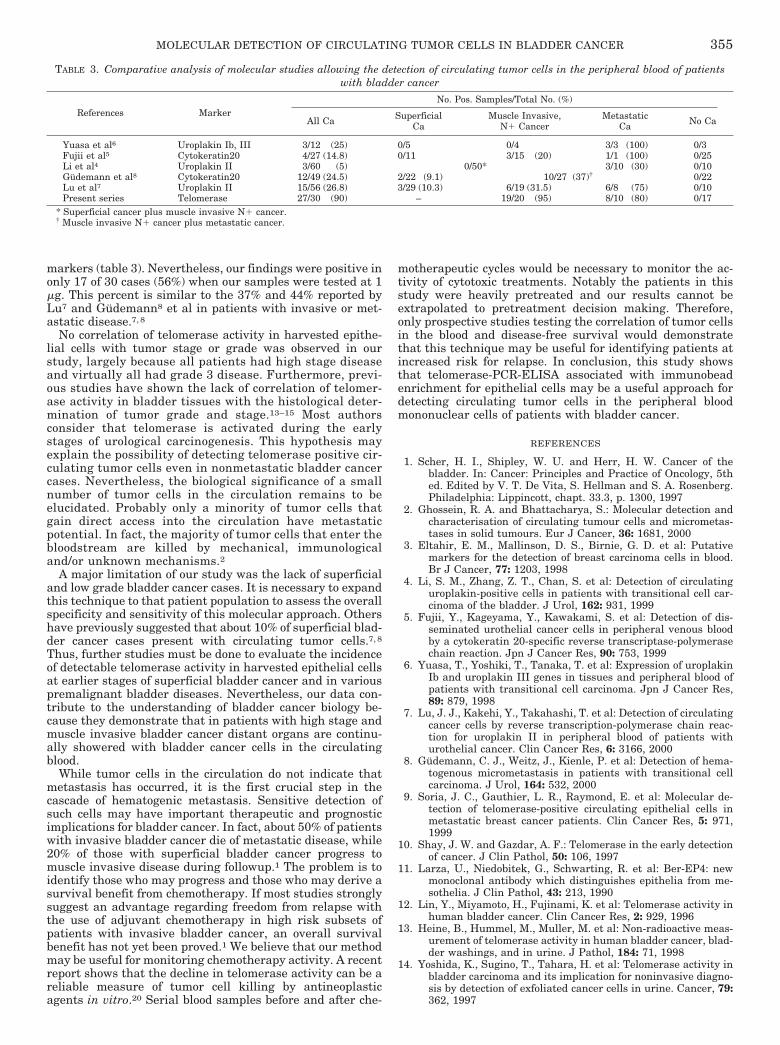
markers (table 3). Nevertheless, our findings were positive inonly 17 of 30 cases (56%) when our samples were tested at 1�g. This percent is similar to the 37% and 44% reported byLu7 and Gudemann8 et al in patients with invasive or met-astatic disease.7, 8
No correlation of telomerase activity in harvested epithe-lial cells with tumor stage or grade was observed in ourstudy, largely because all patients had high stage diseaseand virtually all had grade 3 disease. Furthermore, previ-ous studies have shown the lack of correlation of telomer-ase activity in bladder tissues with the histological deter-mination of tumor grade and stage.13–15 Most authorsconsider that telomerase is activated during the earlystages of urological carcinogenesis. This hypothesis mayexplain the possibility of detecting telomerase positive cir-culating tumor cells even in nonmetastatic bladder cancercases. Nevertheless, the biological significance of a smallnumber of tumor cells in the circulation remains to beelucidated. Probably only a minority of tumor cells thatgain direct access into the circulation have metastaticpotential. In fact, the majority of tumor cells that enter thebloodstream are killed by mechanical, immunologicaland/or unknown mechanisms.2
A major limitation of our study was the lack of superficialand low grade bladder cancer cases. It is necessary to expandthis technique to that patient population to assess the overallspecificity and sensitivity of this molecular approach. Othershave previously suggested that about 10% of superficial blad-der cancer cases present with circulating tumor cells.7, 8
Thus, further studies must be done to evaluate the incidenceof detectable telomerase activity in harvested epithelial cellsat earlier stages of superficial bladder cancer and in variouspremalignant bladder diseases. Nevertheless, our data con-tribute to the understanding of bladder cancer biology be-cause they demonstrate that in patients with high stage andmuscle invasive bladder cancer distant organs are continu-ally showered with bladder cancer cells in the circulatingblood.
While tumor cells in the circulation do not indicate thatmetastasis has occurred, it is the first crucial step in thecascade of hematogenic metastasis. Sensitive detection ofsuch cells may have important therapeutic and prognosticimplications for bladder cancer. In fact, about 50% of patientswith invasive bladder cancer die of metastatic disease, while20% of those with superficial bladder cancer progress tomuscle invasive disease during followup.1 The problem is toidentify those who may progress and those who may derive asurvival benefit from chemotherapy. If most studies stronglysuggest an advantage regarding freedom from relapse withthe use of adjuvant chemotherapy in high risk subsets ofpatients with invasive bladder cancer, an overall survivalbenefit has not yet been proved.1 We believe that our methodmay be useful for monitoring chemotherapy activity. A recentreport shows that the decline in telomerase activity can be areliable measure of tumor cell killing by antineoplasticagents in vitro.20 Serial blood samples before and after che-
motherapeutic cycles would be necessary to monitor the ac-tivity of cytotoxic treatments. Notably the patients in thisstudy were heavily pretreated and our results cannot beextrapolated to pretreatment decision making. Therefore,only prospective studies testing the correlation of tumor cellsin the blood and disease-free survival would demonstratethat this technique may be useful for identifying patients atincreased risk for relapse. In conclusion, this study showsthat telomerase-PCR-ELISA associated with immunobeadenrichment for epithelial cells may be a useful approach fordetecting circulating tumor cells in the peripheral bloodmononuclear cells of patients with bladder cancer.
REFERENCES
1. Scher, H. I., Shipley, W. U. and Herr, H. W. Cancer of thebladder. In: Cancer: Principles and Practice of Oncology, 5thed. Edited by V. T. De Vita, S. Hellman and S. A. Rosenberg.Philadelphia: Lippincott, chapt. 33.3, p. 1300, 1997
2. Ghossein, R. A. and Bhattacharya, S.: Molecular detection andcharacterisation of circulating tumour cells and micrometas-tases in solid tumours. Eur J Cancer, 36: 1681, 2000
3. Eltahir, E. M., Mallinson, D. S., Birnie, G. D. et al: Putativemarkers for the detection of breast carcinoma cells in blood.Br J Cancer, 77: 1203, 1998
4. Li, S. M., Zhang, Z. T., Chan, S. et al: Detection of circulatinguroplakin-positive cells in patients with transitional cell car-cinoma of the bladder. J Urol, 162: 931, 1999
5. Fujii, Y., Kageyama, Y., Kawakami, S. et al: Detection of dis-seminated urothelial cancer cells in peripheral venous bloodby a cytokeratin 20-specific reverse transcriptase-polymerasechain reaction. Jpn J Cancer Res, 90: 753, 1999
6. Yuasa, T., Yoshiki, T., Tanaka, T. et al: Expression of uroplakinIb and uroplakin III genes in tissues and peripheral blood ofpatients with transitional cell carcinoma. Jpn J Cancer Res,89: 879, 1998
7. Lu, J. J., Kakehi, Y., Takahashi, T. et al: Detection of circulatingcancer cells by reverse transcription-polymerase chain reac-tion for uroplakin II in peripheral blood of patients withurothelial cancer. Clin Cancer Res, 6: 3166, 2000
8. Gudemann, C. J., Weitz, J., Kienle, P. et al: Detection of hema-togenous micrometastasis in patients with transitional cellcarcinoma. J Urol, 164: 532, 2000
9. Soria, J. C., Gauthier, L. R., Raymond, E. et al: Molecular de-tection of telomerase-positive circulating epithelial cells inmetastatic breast cancer patients. Clin Cancer Res, 5: 971,1999
10. Shay, J. W. and Gazdar, A. F.: Telomerase in the early detectionof cancer. J Clin Pathol, 50: 106, 1997
11. Larza, U., Niedobitek, G., Schwarting, R. et al: Ber-EP4: newmonoclonal antibody which distinguishes epithelia from me-sothelia. J Clin Pathol, 43: 213, 1990
12. Lin, Y., Miyamoto, H., Fujinami, K. et al: Telomerase activity inhuman bladder cancer. Clin Cancer Res, 2: 929, 1996
13. Heine, B., Hummel, M., Muller, M. et al: Non-radioactive meas-urement of telomerase activity in human bladder cancer, blad-der washings, and in urine. J Pathol, 184: 71, 1998
14. Yoshida, K., Sugino, T., Tahara, H. et al: Telomerase activity inbladder carcinoma and its implication for noninvasive diagno-sis by detection of exfoliated cancer cells in urine. Cancer, 79:362, 1997
TABLE 3. Comparative analysis of molecular studies allowing the detection of circulating tumor cells in the peripheral blood of patientswith bladder cancer
References MarkerNo. Pos. Samples/Total No. (%)
All Ca SuperficialCa
Muscle Invasive,N� Cancer
MetastaticCa No Ca
Yuasa et al6 Uroplakin Ib, III 3/12 (25) 0/5 0/4 3/3 (100) 0/3Fujii et al5 Cytokeratin20 4/27 (14.8) 0/11 3/15 (20) 1/1 (100) 0/25Li et al4 Uroplakin II 3/60 (5) 0/50* 3/10 (30) 0/10Gudemann et al8 Cytokeratin20 12/49 (24.5) 2/22 (9.1) 10/27 (37)† 0/22Lu et al7 Uroplakin II 15/56 (26.8) 3/29 (10.3) 6/19 (31.5) 6/8 (75) 0/10Present series Telomerase 27/30 (90) – 19/20 (95) 8/10 (80) 0/17
* Superficial cancer plus muscle invasive N� cancer.† Muscle invasive N� cancer plus metastatic cancer.
MOLECULAR DETECTION OF CIRCULATING TUMOR CELLS IN BLADDER CANCER 355

15. Kinoshita, H., Ogawa, O., Kakehi, Y. et al: Detection of telom-erase activity in exfoliated cells in urine from patients withbladder cancer. J Natl Cancer Inst, 89: 724, 1997
16. Kavaler, E., Landman, J., Chang, Y. et al: Detecting humanbladder carcinoma cells in voided urine samples by assayingthe presence of telomerase activity. Cancer, 82: 708,1997
17. Weng, N. P., Levine, B. L., June, C. H. et al: Regulated expres-sion of telomerase activity in human T lymphocyte develop-ment and activation. J Exp Med, 183: 2471, 1996
18. Gross, H. J., Verwer, B., Houck, D. et al: Model study detectingbreast cancer cells in peripheral blood mononuclear cells at fre-quencies as low as 10�7. Proc Natl Acad Sci USA, 92: 537, 1995
19. Hardingham, J. E., Kotasek, D., Farmer, B. et al: Immunobead-PCR: a technique for the detection of circulating tumor cellsusing immunomagnetic beads and the polymerase chain reac-tion. Cancer Res, 53: 3455, 1993
20. Faraoni, I., Turriziani, M., Masci, G. et al: Decline in telomeraseactivity as a measure of tumor cell killing by antineoplasticagents in vitro. Clin Cancer Res, 3: 579, 1997
MOLECULAR DETECTION OF CIRCULATING TUMOR CELLS IN BLADDER CANCER356