the multiple lim domain-containing adaptor protein hic-5
TRANSCRIPT
The Multiple LIM Domain-Containing Adaptor ProteinHic-5 Synaptically Colocalizes and Interacts with theDopamine Transporter
Ana M. Carneiro,1,2 Susan L. Ingram,3 Jean-Martin Beaulieu,1 Ava Sweeney,1 Susan G. Amara,3Sheila M. Thomas,4 Marc G. Caron,1 and Gonzalo E. Torres1
1Howard Hughes Medical Institute, Department of Cell Biology, Duke University Medical Center, Durham, North Carolina27710, 2Department of Biochemistry and Immunology, Universidade Federal de Minas Gerais Belo Horizonte, Brazil,3Howard Hughes Medical Institute, Vollum Institute, Oregon Health Sciences University, Portland, Oregon 97201, and4Cancer Biology Program, Beth Israel Deaconess Medical Center/Harvard Medical School, Boston, Massachusetts02215
The Na�/Cl�-dependent dopamine transporter (DAT) is criticalin terminating dopaminergic transmission by removing thetransmitter away from the synapse. Several lines of evidencesuggest that transporter-interacting proteins may play a role inDAT function and regulation. In this report, using the yeasttwo-hybrid system, we have identified a novel interaction be-tween DAT and the multiple Lin-11, Isl-1, and Mec-3 (LIM)domain-containing adaptor protein Hic-5. This association in-volves the N-terminal portion of the intracellular tail of DAT andthe LIM region of Hic-5. In human embryonic kidney 293 cells,Hic-5 colocalizes with DAT at polarized sites and reduces DATuptake activity through a mechanism involving a decrease inthe cell-surface levels of the transporter. A fragment of Hic-5containing the LIM domains is sufficient to bind DAT but lacksthe ability to inhibit transporter activity. In addition, the LIM
fragment prevents the effect of the full-length Hic-5 on DATlocalization and function. In the brain, Hic-5 protein is ex-pressed in the cerebral cortex, hippocampus, hypothalamus,cerebellum, and striatum, suggesting a role for this protein inthe nervous system. The association of the endogenous Hic-5and DAT proteins was confirmed biochemically by coimmuno-precipitation from brain striatal extracts. Moreover, immuno-staining of rat midbrain neurons in culture revealed a presyn-aptic colocalization of Hic-5 and DAT. Because Hic-5 has beenshown to interact with several signaling molecules, includingthe nonreceptor protein tyrosine kinases focal adhesion kinaseand Fyn, this raises the possibility that this adaptor protein maylink DAT to intracellular signaling pathways.
Key words: dopamine transporter; Hic-5; adaptor; interac-tion; colocalization; uptake; LIM domain
Dopaminergic neurotransmission plays a major role in the mod-ulation of locomotor activity, neuroendocrine secretion, andemotion (Sealfon and Olanow, 2000). Signaling is initiated withthe release of dopamine (DA) from presynaptic terminals fol-lowed by activation of presynaptic and postsynaptic DA receptors.A critical step that determines the duration and intensity of DAactions is the reuptake of the neurotransmitter back into thesynaptic terminal for subsequent release. This action is achievedby the Na�/Cl�-dependent plasma membrane dopamine trans-porter (DAT) (Giros and Caron, 1993). Functional changes inDAT may be involved in the pathogenesis of attention deficithyperactivity disorder, schizophrenia, and/or neurodegenerativedisorders such as Parkinson’s disease (Bannon et al., 1998). Inaddition, drugs of abuse, such as cocaine and amphetamine,exhibit their reinforcing actions at least in part by interacting withDAT (Amara and Sonders, 1998). The importance of DAT in
controlling DA transmission was demonstrated recently in vivo.Deletion of the DAT gene in mice results in profound behavioraland neurochemical changes, including hyperlocomotor activity,increased dopamine receptor responsiveness, and sensitization topsychostimulants (Giros et al., 1996; Jones et al., 1998; Gainetdi-nov et al., 1999).
Molecular cloning of genes encoding DAT and other membersof the Na�/Cl�-dependent transporter family, such as the nor-epinephrine transporter (NET), serotonin transporter (SERT),and glycine and GABA transporters, reveals a highly conservedprimary structure (for review, see Masson et al., 1999). Theirpredicted topology suggests the presence of 12 transmembranedomains with intracellular N and C termini. The DAT possessesconsensus sequences for phosphorylation by protein kinases (Gi-ros et al., 1991; Kilty et al., 1991; Shimada et al., 1991), suggestingthat phosphorylation might play a role in the regulation of DATfunction. Indeed, several studies have shown that regulation ofkinase activity in striatal synaptosomal preparations and heterol-ogous cell systems affects DAT uptake activity (for review, seeZahniser and Doolen, 2001). This effect is believed to result as aconsequence of a rapid redistribution of transporter from the cellmembrane (Pristupa et al., 1998; Daniels and Amara, 1999; Me-likian and Buckley, 1999; Doolen and Zahniser, 2001). However,neither the identity of the specific kinases involved in the down-regulation of DAT nor the direct substrate for phosphorylationhas been identified. It has been proposed that transporter-
Received March 29, 2002; revised June 3, 2002; accepted June 5, 2002.This work was supported by National Institutes of Health Grants NS-19576
(M.G.C.) and DA-14150 (G.E.T.) A.M.C. is a recipient of a fellowship fromCompanha de Aperfeicoamento de Pessoal de Nıvel Superior, Brazil. J.-M.B. issupported by a Human Frontiers Science Program fellowship. S.G.A. and M.G.C.are Investigators of the Howard Hughes Medical Institute. We thank members of theCaron lab for helpful discussions. We also thank Drs. Laura Bohn and Richard T.Premont for reading this manuscript.
Correspondence should be addressed to Dr. Marc G. Caron, Department of CellBiology, Duke University Medical Center, Room 489, CARL Building, ResearchDrive, Durham, NC 27710. E-mail: [email protected] © 2002 Society for Neuroscience 0270-6474/02/227045-10$15.00/0
The Journal of Neuroscience, August 15, 2002, 22(16):7045–7054
interacting proteins might regulate directly or indirectly thekinase-dependent effect on DAT activity (Blakely et al., 1998).This mechanism has been demonstrated for the GABA trans-porter, where PKC downregulates transporter function by mod-ulating the association of syntaxin with the transporter (Beckmanet al., 1998).
Transporter-associated proteins may also play a broader role intransporter stability, location, and/or function. Lee et al. (2001)have shown recently that DAT activity might be affected by adirect interaction between the C terminus of DAT (CDAT) and�-synuclein. DAT function is also regulated by the interactionbetween DAT and the postsynaptic density-95/discs large/zonaoccludens-1 (PDZ) domain-containing protein PKC-interactingprotein-1 (PICK1) (Torres et al., 2001). Deletion of the PDZ-binding site in DAT impairs the targeting of the mutated trans-porter to neuronal processes in cultured neurons. Thus, thesearch for DAT-interacting proteins has opened a new and prom-ising opportunity for a better understanding of the mechanismsinvolved in the regulation of transporter function. Here, we haveidentified the focal adhesion protein Hic-5 as a DAT-interactingprotein. Hic-5 interacts directly with DAT both in vitro and invivo. In the brain, Hic-5 is expressed in dopaminergic neurons,where it colocalizes and coimmunoprecipitates with DAT. Ourfindings suggest that Hic-5 may play an important role as anadaptor protein in the regulation of DAT function.
MATERIALS AND METHODSYeast two-hybrid screening. A yeast two-hybrid screening of a human brainlibrary using the intracellular C terminus of DAT as bait was performedas described previously (Torres et al., 2001). Automated sequencingidentified �20 clones encoding partial sequences of the open readingframe of Hic-5. Fragments expressing the C termini of DAT, NET(residues 575–617), or SERT (residues 594–630) or the N terminus ofDAT (NDAT) (residues 1–60) were subcloned into pAS2.1 and trans-formed in pG694a yeast cells with a Hic-5 clone fused to pGAD10 usingthe LiAc method. Transformation efficiency was verified by growingtransformed yeast cells in leucine- and tryptophan-deficient medium,whereas specific interactions between each fragment and Hic-5 wereverified by the growth of yeast colonies in leucine-, tryptophan-, andadenine-free medium after 2–5 d of incubation at 30°C.
Cell culture and transfections. Human embryonic kidney 293 (HEK293)cells were maintained in MEM supplemented with 10% FBS and 50U/ml gentamicin (Invitrogen, Grand Island, NJ). Cells were transfectedusing the Ca2PO4 precipitation method with 10 �g of total DNA. After16–20 hr at 37°C with the Ca2PO4–DNA mixture, transfected cells wereincubated with fresh MEM and allowed to grow for an additional 48 hr.
Western blot analysis. Protein samples were prepared by incubation oftransfected HEK293 cells with radioimmunoprecipitation assay (RIPA)buffer (in mM): 100 Tris, 150 NaCl, 1 EDTA, 1% Triton X-100, 0.1%SDS, and 1% deoxycholic acid, pH 7.4, for 30 min at 4°C followed bycentrifugation at 14,000 rpm for 10 min. Alternatively, C57BL/6 micewere killed, and brain tissues were dissected and homogenized in RIPAbuffer. Samples were analyzed on 10% polyacrylamide gels and electro-blotted to nitrocellulose membranes using the Novex precast gel (Wads-worth, OH) and transfer system (Invitrogen, Carlsbad, CA). Hic-5 wasdetected using a mouse monoclonal antibody raised against the proline-rich region of Hic-5 (BD Transduction Laboratories, San Diego, CA)followed by a secondary HRP-linked anti-mouse antibody (AmershamBiosciences, Uppsala, Sweden). DAT was detected using a rat monoclo-nal antibody raised against the N-terminal domain of human DAT(Chemicon International Inc., Temecula, CA) and a secondary goatHRP-conjugated anti-rat antibody (Jackson ImmunoResearch, WestGrove, PA). Membranes were blocked in 5% milk in TBS-T (in mM): 10Tris, pH 7.4, 100 NaCl, and 0.05% Tween 20, for 30 min at roomtemperature and incubated with the appropriate primary antibody. Theprimary antibodies were used at a 1:1000 dilution for 1 hr at roomtemperature for samples from HEK293 cells or at 1:500 for 18–20 hr at4°C for samples from brain extracts. The membranes were then washedthree times with TBS-T, followed by incubation with secondary antibod-
ies for 1 hr. Immunoreactive bands were detected with the ECL system(Amersham Biosciences).
Glutathione S-transferase fusion protein precipitations. GlutathioneS-transferase (GST) constructs containing the entire C-terminal domainof DAT (residues 575–620) or NET (residues 575–617) were amplifiedby PCR using Taq polymerase (Fisher, Pittsburgh, PA) containing 10%of Vent polymerase (New England Biolabs Inc., Beverly, MA). Small 10aa fragments from the C-terminal region of DAT were generated byannealing complementary designed primers (Sigma Genosys, The Wood-lands, TX). These constructs were fused in frame into the GST fusionvector PGEX-4T-1 (Amersham Biosciences, Piscataway, NJ) and verifiedby automated sequencing. GST-fused fragments were expressed in BLX-Blue bacterial cells and isolated with glutathione–agarose beads (Am-ersham Biosciences). Aliquots containing GST fusion fragments werethen incubated with either 500 �g of protein lysates from transfectedHEK293 cells or 1 mg of brain lysate. Mixtures were incubated with GSTbeads (Amersham Biosciences), washed three times with RIPA buffer,and prepared for Western blotting as described previously.
Immunoprecipitations. Lysates from transfected HEK293 cells or mousetissue were prepared using RIPA buffer. Immunoprecipitations fromHEK293 cell lysates were performed using either anti-hemagglutinin (HA)or anti-myc monoclonal antibodies (Roche Diagnostics Corp., Indian-apolis, IN). Aliquots of the cleared lysates (500 �g of protein) wereincubated with 15 �g of the appropriate antibody at 4°C for 16–18 hr.Protein A Sepharose beads (Amersham Biosciences) were then addedand incubated for 2 hr at 4°C. The beads were washed three times withRIPA buffer and prepared for Western blotting. Immunoprecipitationsusing brain lysates were performed using 1 mg of total protein and 1 �gof the anti-Hic-5 antibody. Samples were analyzed by Western blotting asdescribed above.
Transport measurements. Uptake experiments in transfected HEK293cells were performed as described previously (Giros et al., 1994). Briefly,48 hr after transfections, cells were incubated at 37°C for 5 min in 250 �lof uptake buffer (in mM): 5 Tris base, 7.5 HEPES, 120 NaCl, 5.4 KCl, 1.2CaCl2, 1.2 MgSO4, 1 ascorbic acid, and 5 glucose, pH 7.4, containing 20nM [ 3H]DA (31.6 Ci /mmol) and increasing concentrations of cold DA,ranging from 100 to 30 �M. Cells were then washed with 500 �l ofNaCl-free uptake buffer and solubilized for 1 hr in 400 �l of 1% SDS.The amount of radioactivity incorporated into the cells was measured byscintillation counting. Data are presented as mean � SEM. Statisticalsignificance was determined by unpaired Student’s t test with a signifi-cance criterion of p � 0.05.
Cell-surface biotinylation. Transfected HEK293 cells were washed withPBS and incubated for 40 min at 4°C with 1 mg/ml sulfosuccinimidyl2-(biotinamido)ethyl-1, 3-dithiopropionate (sulfo-NHS-SS-biotin; Pierce,Rockford, IL) in (in mM): 150 NaCl, 2 CaCl2, and 10 triethanolamine, pH7.8, followed by incubation with 0.1 M glycine in PBS for 10 min. After twowashes with PBS, cells were lysed in 1 ml of RIPA buffer for 1 hr at 4°C,scraped, and centrifuged for 10 min at 4°C. Two aliquots of the supernatantwere collected; 5 �l were used for total protein normalization in Westernblots, and 500 �l were incubated with 25 �l of 50% avidin beads (Pierce) at4°C for 16–18 hr. The beads were washed three times with PBS andprepared for Western blotting using the anti-DAT antibody.
Immunocytochemistry and confocal microscopy. Transiently transfectedHEK293 cells were plated on glass coverslips and grown for 48 hr at37°C. Cells were washed with PBS and fixed with 4% paraformaldehydefor 10 min at 4°C. After three washes with PBS, the cells were blocked inPBS solution containing 5% goat serum, 1% BSA, and 0.05% TritonX-100; Sigma-Aldrich Co., Irvine, CA) for 1 hr at room temperature.Coverslips were incubated for 1 hr with a rat anti-DAT antibody and/ora rabbit anti-Hic-5 antibody (Matsuya et al., 1998), both at a dilution of1:1000. After three washes with PBS, cells were incubated with FITC-tagged anti-rabbit or Texas Red-conjugated anti-rat secondary antibodies(Jackson ImmunoResearch), both at a 1:200 dilution for 1 hr. Coverslipswere then washed in PBS and mounted onto glass slides with Vectashieldmounting medium (Vector Laboratories, Inc., Burlingame, CA). Neuro-nal cultures were prepared as described previously (Rayport et al., 1992).Briefly, Sprague Dawley rat pups (2–4 d of age) were anesthetized byintraperitoneal injection of ketamine HCl (3 mg/pup). Ventral midbrainswere dissected and incubated in a dissociation medium under constantoxygenation for 2 hr, followed by trituration with a fire-polished Pasteurpipette in glial medium (MEM, 10% FBS, 0.45% D-glucose, 5 pg/mlinsulin, 0.5 mM glutamine, 100 U/ml penicillin, and 100 �g/ml strepto-mycin). Dissociated cells were pelleted by centrifugation, plated on 24well plates containing glass coverslips, and allowed to grow for 5–7 d.
7046 J. Neurosci., August 15, 2002, 22(16):7045–7054 Carneiro et al. • Interaction of DAT with the LIM Domain Protein Hic-5

Cells were then fixed in 4% paraformaldehyde for 10 min, washed inPBS, and incubated in blocking solution (4% goat serum, 1% BSA, and0.2% Triton X-100 in PBS) for 30 min followed by incubation withprimary antibodies at a 1:500 dilution in 0.25% BSA and 0.2% TritonX-100 in PBS for 16–18 hr at 4°C. Secondary antibodies were added at a1:200 dilution and incubated for 2 hr at room temperature. Coverslipswere mounted on glass slides, and the samples were visualized using alaser scanning confocal microscope (Zeiss, Thornwood, NY) at 498 nmfor FITC and 585 nm for Texas Red.
RESULTSIn vitro interaction between DAT and Hic-5To search for DAT-interacting proteins, we used the yeast two-hybrid system to screen a human brain cDNA library using theintracellular C-terminal domain of DAT as bait. From �20 mil-lion transformants screened, �20 positive clones were found toencode partial sequences of the open reading frame of Hic-5, a461 aa protein highly homologous to the focal adhesion proteinpaxillin. Hic-5 was first identified as a TGF-�-inducible gene andproposed initially to function as a transcription factor because ofthe presence of four double zinc-finger LIM domains located inthe C-terminal half of the protein (Shibanuma et al., 1994, 1997).Although the function of Hic-5 is still not fully understood, itshigh homology with paxillin, as well as its presence in focaladhesion structures, suggests the involvement of Hic-5 as anadaptor protein regulating focal adhesion dynamics (Thomas etal., 1999).
We examined the specificity of the DAT and Hic-5 interactionby testing in the yeast two-hybrid system the ability of Hic-5 tobind the C termini of the monoamine transporters NET andSERT. As shown in Figure 1A, Hic-5 also interacts with theintracellular tails of NET and SERT in yeast, although the inter-action with SERT is weaker when compared with that obtainedwith either DAT or NET. To limit the possibility of a false-positive interaction, we used the intracellular N-terminal domainof DAT as a control. No interaction between the N terminus ofDAT and Hic-5 was detected in yeast (Fig. 1A).
To confirm the interaction of the C terminus of DAT withHic-5, we performed a GST fusion protein precipitation assayusing the complete C terminus of DAT fused to GST (GST-CDAT) (Fig. 1B). Results from precipitation experiments withGST beads using mouse whole-brain lysates demonstrate theassociation between GST-CDAT and Hic-5 under conditions inwhich GST alone was not able to associate with Hic-5. In addition,the C terminus of NET fused to GST (GST-CNET) also interactswith Hic-5 from brain lysates (Fig. 1B). Thus, these findingsdemonstrate a physical association between the tail of mono-amine transporters and the endogenous Hic-5.
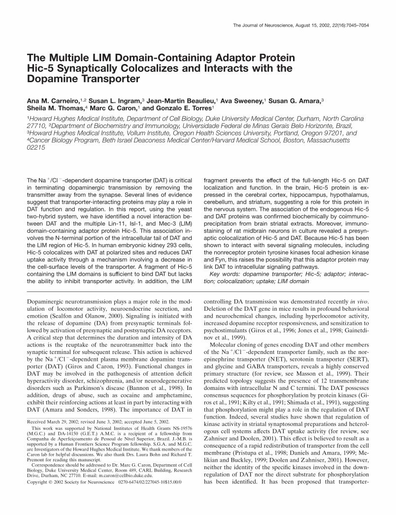
Interaction between the full-length Hic-5 and DAT inmammalian cellsWe subsequently examined whether the full-length DAT interactswith Hic-5 in mammalian cells by coexpressing the HA-taggedhuman DAT and the myc-tagged Hic-5 in HEK293 cells. NeitherHic-5 nor DAT were detected in mock-transfected cells as re-vealed with the anti-Hic-5 and the anti-DAT antibodies, respec-tively (Fig. 2A,B). Immunoprecipitation with the anti-myc anti-body results in the coprecipitation of HA-DAT only when bothproteins were coexpressed in HEK293 cells (Fig. 2A). In addi-tion, immunoprecipitation of HA-DAT also results in the copre-cipitation of Hic-5-myc when both tagged proteins are coex-pressed in HEK293 cells (Fig. 2B), demonstrating that theassociation between the full-length DAT and Hic-5 proteins isindependent of which protein is first pulled down.
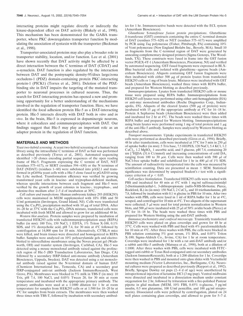
Mapping the interacting domains of the DAT andHic-5 interactionHic-5 contains multiple domains that have been shown to beresponsible for protein–protein interactions. The N-terminal halfcontains four leucine-rich domains (LD) that mediate the inter-action between Hic-5 and several focal adhesion proteins, includ-ing the focal adhesion kinase (FAK) (Fujita et al., 1998) and celladhesion kinase � (CAK�)/Pyk2 (Matsuya et al., 1998; Osada etal., 2001). In contrast, the C-terminal half contains four LIMdomains, each one consisting of two zinc-finger motifs. Thesedomains are responsible for the interaction between Hic-5 andprotein tyrosine phosphatase-proline serine threonine (PTP-PEST) (Nishiya et al., 1999) and the glucocorticoid receptor(Yang et al., 2000). To determine which region in Hic-5 mediatesthe interaction with DAT, we used two myc-tagged Hic-5 frag-ments corresponding to the LD domain-containing N-terminalhalf (residues 1–210; NH-myc) or the multiple LIM domain-containing C-terminal half (residues 211–461; COOH-myc).These constructs were expressed with DAT in HEK293 cells andassayed for protein–protein interaction by coimmunoprecipita-tion experiments using the anti-myc antibody. In cells coexpress-ing COOH-myc and DAT, immunoprecipitation with the anti-myc antibody resulted in coprecipitation of DAT. COOH-myc
Figure 1. The C-terminal domain of DAT interacts with Hic-5 in vitro. A,Interaction between the tail of monoamine transporters and Hic-5.pG694a yeast cells were transformed with pAS2.1 and pGAD10/Hic-5(1), pAS2.1/CDAT and pGAD10 (2), pAS2.1/NDAT and pGAD10/Hic-5 (3), pAS2.1/CDAT and pGAD10/Hic-5 (4), pAS2.1/CNET andpGAD10/Hic-5 (5), or pAS2.1/CSERT and pGAD10/Hic-5 (6). Positivetransformant yeast cells were selected in media lacking tryptophan,leucine, and adenine. CDAT, CNET, and CSERT represent the C ter-minus of DAT, NET, and SERT, respectively, whereas NDAT representsthe N terminus of DAT. B, GST fusion protein precipitation assay usingthe complete C terminus of DAT or NET fused to GST. Aliquotscontaining GST fusion fragments were incubated with 1 mg of mousewhole-brain lysate and analyzed by Western blot using a polyclonalanti-Hic-5 antibody.
Carneiro et al. • Interaction of DAT with the LIM Domain Protein Hic-5 J. Neurosci., August 15, 2002, 22(16):7045–7054 7047
was as efficient as the full-length myc-tagged Hic-5 at coprecipi-tating DAT (Fig. 3A). In contrast, no association was detectedwhen DAT was coexpressed with NH-myc, indicating that themultiple LIM domain-containing C terminus of Hic-5 mediatesthe interaction between Hic-5 and DAT.
Next, we mapped the residues within the intracellular tail ofDAT involved in the interaction with Hic-5. Six 10 aa GST-fusedfragments covering the last 60 residues from the C terminus ofDAT (Fig. 3B) were generated and tested for their ability tointeract with Hic-5 in precipitation experiments. The strongestinteraction was observed between Hic-5 and fragment 2 (residues571–580), whereas a much weaker interaction was observed whenusing fragment 1 (residues 561–570) or fragment 3 (residues581–590) (Fig. 3B). No interaction was detected when any of thethree fragments corresponding to the last 30 aa of DAT were usedin the GST precipitation with GST fusion proteins. Together,these results indicate that the interaction between DAT and Hic-5is mediated by the multiple LIM domain-containing half of Hic-5and the membrane-proximal portion of the intracellular tailof DAT.
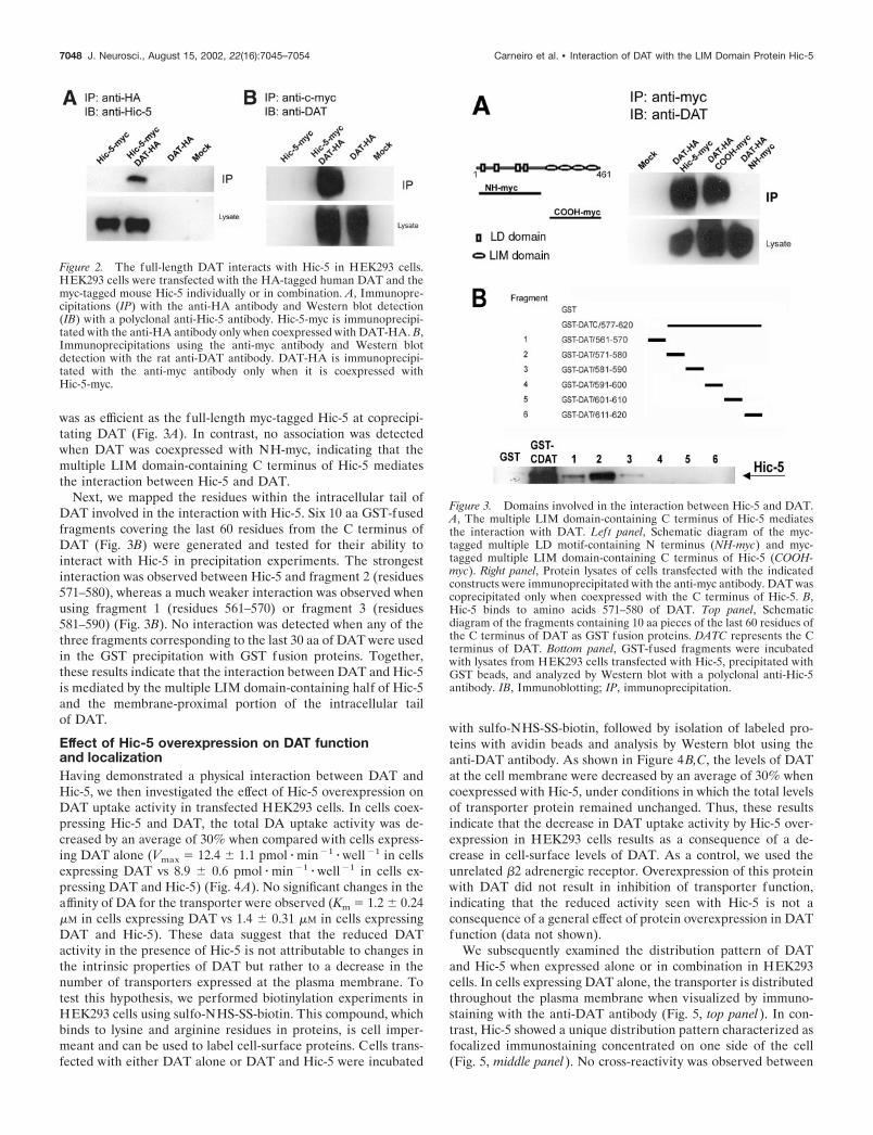
Effect of Hic-5 overexpression on DAT functionand localizationHaving demonstrated a physical interaction between DAT andHic-5, we then investigated the effect of Hic-5 overexpression onDAT uptake activity in transfected HEK293 cells. In cells coex-pressing Hic-5 and DAT, the total DA uptake activity was de-creased by an average of 30% when compared with cells express-ing DAT alone (Vmax � 12.4 � 1.1 pmol � min�1 � well�1 in cellsexpressing DAT vs 8.9 � 0.6 pmol � min�1 � well�1 in cells ex-pressing DAT and Hic-5) (Fig. 4A). No significant changes in theaffinity of DA for the transporter were observed (Km � 1.2 � 0.24�M in cells expressing DAT vs 1.4 � 0.31 �M in cells expressingDAT and Hic-5). These data suggest that the reduced DATactivity in the presence of Hic-5 is not attributable to changes inthe intrinsic properties of DAT but rather to a decrease in thenumber of transporters expressed at the plasma membrane. Totest this hypothesis, we performed biotinylation experiments inHEK293 cells using sulfo-NHS-SS-biotin. This compound, whichbinds to lysine and arginine residues in proteins, is cell imper-meant and can be used to label cell-surface proteins. Cells trans-fected with either DAT alone or DAT and Hic-5 were incubated
with sulfo-NHS-SS-biotin, followed by isolation of labeled pro-teins with avidin beads and analysis by Western blot using theanti-DAT antibody. As shown in Figure 4B,C, the levels of DATat the cell membrane were decreased by an average of 30% whencoexpressed with Hic-5, under conditions in which the total levelsof transporter protein remained unchanged. Thus, these resultsindicate that the decrease in DAT uptake activity by Hic-5 over-expression in HEK293 cells results as a consequence of a de-crease in cell-surface levels of DAT. As a control, we used theunrelated �2 adrenergic receptor. Overexpression of this proteinwith DAT did not result in inhibition of transporter function,indicating that the reduced activity seen with Hic-5 is not aconsequence of a general effect of protein overexpression in DATfunction (data not shown).
We subsequently examined the distribution pattern of DATand Hic-5 when expressed alone or in combination in HEK293cells. In cells expressing DAT alone, the transporter is distributedthroughout the plasma membrane when visualized by immuno-staining with the anti-DAT antibody (Fig. 5, top panel). In con-trast, Hic-5 showed a unique distribution pattern characterized asfocalized immunostaining concentrated on one side of the cell(Fig. 5, middle panel). No cross-reactivity was observed between
Figure 2. The full-length DAT interacts with Hic-5 in HEK293 cells.HEK293 cells were transfected with the HA-tagged human DAT and themyc-tagged mouse Hic-5 individually or in combination. A, Immunopre-cipitations (IP) with the anti-HA antibody and Western blot detection(IB) with a polyclonal anti-Hic-5 antibody. Hic-5-myc is immunoprecipi-tated with the anti-HA antibody only when coexpressed with DAT-HA. B,Immunoprecipitations using the anti-myc antibody and Western blotdetection with the rat anti-DAT antibody. DAT-HA is immunoprecipi-tated with the anti-myc antibody only when it is coexpressed withHic-5-myc.
Figure 3. Domains involved in the interaction between Hic-5 and DAT.A, The multiple LIM domain-containing C terminus of Hic-5 mediatesthe interaction with DAT. Left panel, Schematic diagram of the myc-tagged multiple LD motif-containing N terminus (NH-myc) and myc-tagged multiple LIM domain-containing C terminus of Hic-5 (COOH-myc). Right panel, Protein lysates of cells transfected with the indicatedconstructs were immunoprecipitated with the anti-myc antibody. DAT wascoprecipitated only when coexpressed with the C terminus of Hic-5. B,Hic-5 binds to amino acids 571–580 of DAT. Top panel, Schematicdiagram of the fragments containing 10 aa pieces of the last 60 residues ofthe C terminus of DAT as GST fusion proteins. DATC represents the Cterminus of DAT. Bottom panel, GST-fused fragments were incubatedwith lysates from HEK293 cells transfected with Hic-5, precipitated withGST beads, and analyzed by Western blot with a polyclonal anti-Hic-5antibody. IB, Immunoblotting; IP, immunoprecipitation.
7048 J. Neurosci., August 15, 2002, 22(16):7045–7054 Carneiro et al. • Interaction of DAT with the LIM Domain Protein Hic-5
the rat anti-DAT and the rabbit anti-Hic-5 antibodies. In cellscoexpressing DAT and Hic-5, the distribution pattern of DATchanged dramatically and resembled that of Hic-5 (Fig. 5, bottompanel). The strongest DAT signal was found on one side of thecell, where it colocalized with Hic-5.
To examine the specificity of the Hic-5 and DAT interaction,we tested whether a fragment containing the multiple LIM do-main of Hic-5 (COOH-myc) was able to compete with Hic-5 for
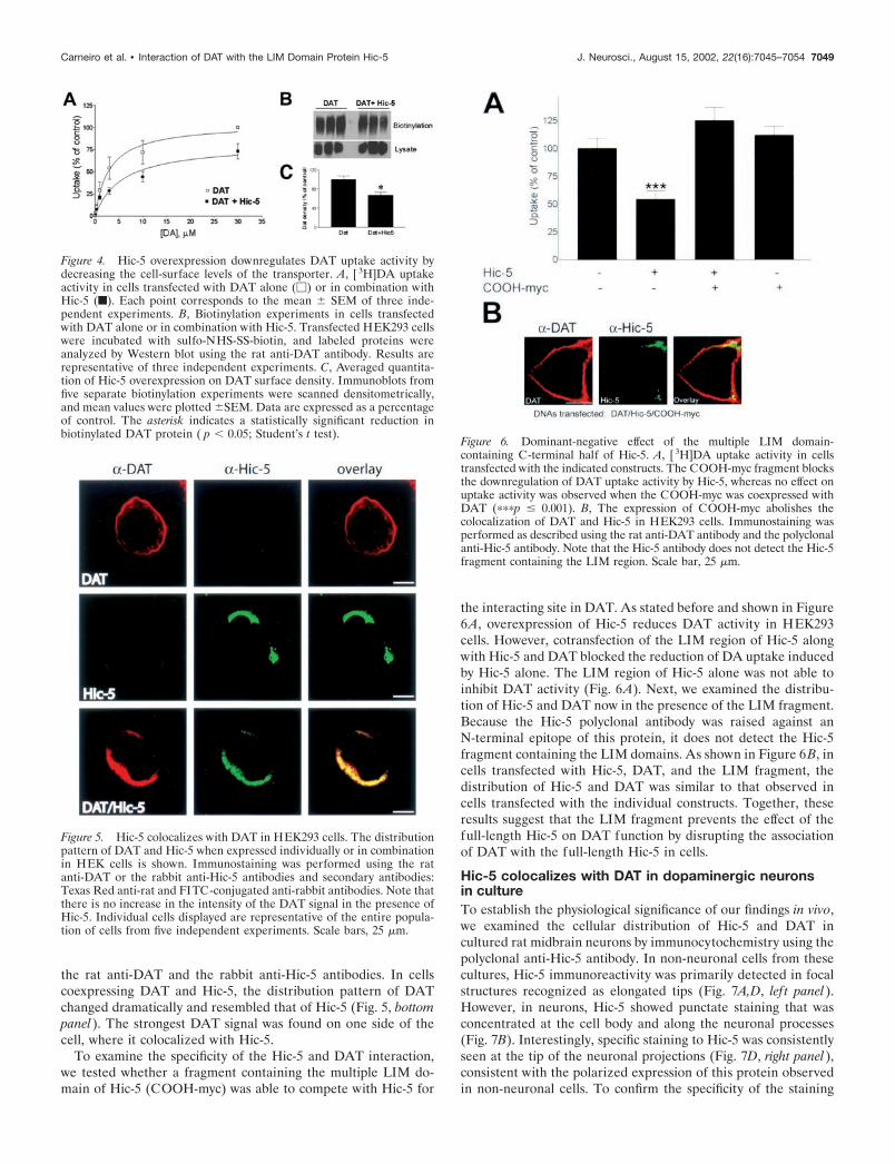
the interacting site in DAT. As stated before and shown in Figure6A, overexpression of Hic-5 reduces DAT activity in HEK293cells. However, cotransfection of the LIM region of Hic-5 alongwith Hic-5 and DAT blocked the reduction of DA uptake inducedby Hic-5 alone. The LIM region of Hic-5 alone was not able toinhibit DAT activity (Fig. 6A). Next, we examined the distribu-tion of Hic-5 and DAT now in the presence of the LIM fragment.Because the Hic-5 polyclonal antibody was raised against anN-terminal epitope of this protein, it does not detect the Hic-5fragment containing the LIM domains. As shown in Figure 6B, incells transfected with Hic-5, DAT, and the LIM fragment, thedistribution of Hic-5 and DAT was similar to that observed incells transfected with the individual constructs. Together, theseresults suggest that the LIM fragment prevents the effect of thefull-length Hic-5 on DAT function by disrupting the associationof DAT with the full-length Hic-5 in cells.
Hic-5 colocalizes with DAT in dopaminergic neuronsin cultureTo establish the physiological significance of our findings in vivo,we examined the cellular distribution of Hic-5 and DAT incultured rat midbrain neurons by immunocytochemistry using thepolyclonal anti-Hic-5 antibody. In non-neuronal cells from thesecultures, Hic-5 immunoreactivity was primarily detected in focalstructures recognized as elongated tips (Fig. 7A,D, lef t panel).However, in neurons, Hic-5 showed punctate staining that wasconcentrated at the cell body and along the neuronal processes(Fig. 7B). Interestingly, specific staining to Hic-5 was consistentlyseen at the tip of the neuronal projections (Fig. 7D, right panel),consistent with the polarized expression of this protein observedin non-neuronal cells. To confirm the specificity of the staining
Figure 4. Hic-5 overexpression downregulates DAT uptake activity bydecreasing the cell-surface levels of the transporter. A, [ 3H]DA uptakeactivity in cells transfected with DAT alone (�) or in combination withHic-5 (f). Each point corresponds to the mean � SEM of three inde-pendent experiments. B, Biotinylation experiments in cells transfectedwith DAT alone or in combination with Hic-5. Transfected HEK293 cellswere incubated with sulfo-NHS-SS-biotin, and labeled proteins wereanalyzed by Western blot using the rat anti-DAT antibody. Results arerepresentative of three independent experiments. C, Averaged quantita-tion of Hic-5 overexpression on DAT surface density. Immunoblots fromfive separate biotinylation experiments were scanned densitometrically,and mean values were plotted �SEM. Data are expressed as a percentageof control. The asterisk indicates a statistically significant reduction inbiotinylated DAT protein ( p � 0.05; Student’s t test).
Figure 5. Hic-5 colocalizes with DAT in HEK293 cells. The distributionpattern of DAT and Hic-5 when expressed individually or in combinationin HEK cells is shown. Immunostaining was performed using the ratanti-DAT or the rabbit anti-Hic-5 antibodies and secondary antibodies:Texas Red anti-rat and FITC-conjugated anti-rabbit antibodies. Note thatthere is no increase in the intensity of the DAT signal in the presence ofHic-5. Individual cells displayed are representative of the entire popula-tion of cells from five independent experiments. Scale bars, 25 �m.
Figure 6. Dominant-negative effect of the multiple LIM domain-containing C-terminal half of Hic-5. A, [ 3H]DA uptake activity in cellstransfected with the indicated constructs. The COOH-myc fragment blocksthe downregulation of DAT uptake activity by Hic-5, whereas no effect onuptake activity was observed when the COOH-myc was coexpressed withDAT (���p � 0.001). B, The expression of COOH-myc abolishes thecolocalization of DAT and Hic-5 in HEK293 cells. Immunostaining wasperformed as described using the rat anti-DAT antibody and the polyclonalanti-Hic-5 antibody. Note that the Hic-5 antibody does not detect the Hic-5fragment containing the LIM region. Scale bar, 25 �m.
Carneiro et al. • Interaction of DAT with the LIM Domain Protein Hic-5 J. Neurosci., August 15, 2002, 22(16):7045–7054 7049
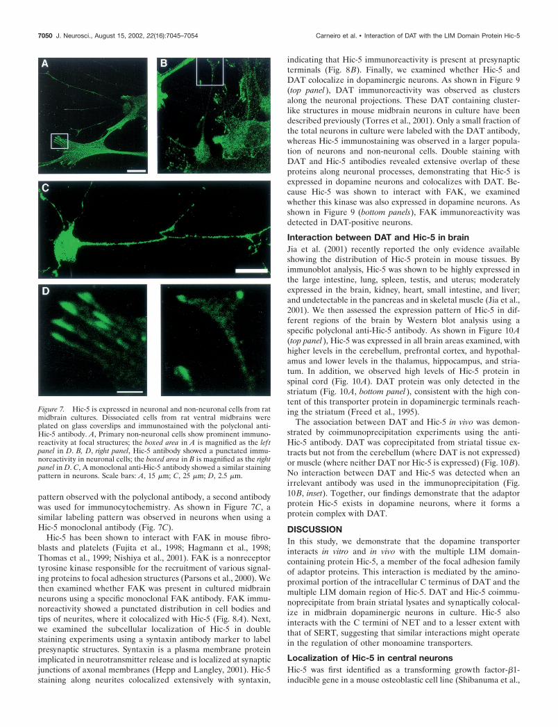
pattern observed with the polyclonal antibody, a second antibodywas used for immunocytochemistry. As shown in Figure 7C, asimilar labeling pattern was observed in neurons when using aHic-5 monoclonal antibody (Fig. 7C).
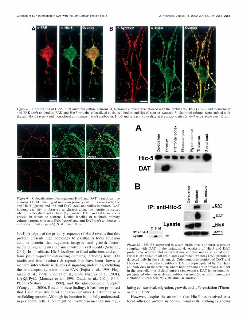
Hic-5 has been shown to interact with FAK in mouse fibro-blasts and platelets (Fujita et al., 1998; Hagmann et al., 1998;Thomas et al., 1999; Nishiya et al., 2001). FAK is a nonreceptortyrosine kinase responsible for the recruitment of various signal-ing proteins to focal adhesion structures (Parsons et al., 2000). Wethen examined whether FAK was present in cultured midbrainneurons using a specific monoclonal FAK antibody. FAK immu-noreactivity showed a punctated distribution in cell bodies andtips of neurites, where it colocalized with Hic-5 (Fig. 8A). Next,we examined the subcellular localization of Hic-5 in doublestaining experiments using a syntaxin antibody marker to labelpresynaptic structures. Syntaxin is a plasma membrane proteinimplicated in neurotransmitter release and is localized at synapticjunctions of axonal membranes (Hepp and Langley, 2001). Hic-5staining along neurites colocalized extensively with syntaxin,
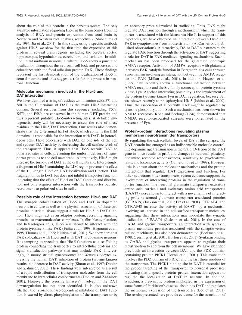
indicating that Hic-5 immunoreactivity is present at presynapticterminals (Fig. 8B). Finally, we examined whether Hic-5 andDAT colocalize in dopaminergic neurons. As shown in Figure 9(top panel), DAT immunoreactivity was observed as clustersalong the neuronal projections. These DAT containing cluster-like structures in mouse midbrain neurons in culture have beendescribed previously (Torres et al., 2001). Only a small fraction ofthe total neurons in culture were labeled with the DAT antibody,whereas Hic-5 immunostaining was observed in a larger popula-tion of neurons and non-neuronal cells. Double staining withDAT and Hic-5 antibodies revealed extensive overlap of theseproteins along neuronal processes, demonstrating that Hic-5 isexpressed in dopamine neurons and colocalizes with DAT. Be-cause Hic-5 was shown to interact with FAK, we examinedwhether this kinase was also expressed in dopamine neurons. Asshown in Figure 9 (bottom panels), FAK immunoreactivity wasdetected in DAT-positive neurons.
Interaction between DAT and Hic-5 in brainJia et al. (2001) recently reported the only evidence availableshowing the distribution of Hic-5 protein in mouse tissues. Byimmunoblot analysis, Hic-5 was shown to be highly expressed inthe large intestine, lung, spleen, testis, and uterus; moderatelyexpressed in the brain, kidney, heart, small intestine, and liver;and undetectable in the pancreas and in skeletal muscle (Jia et al.,2001). We then assessed the expression pattern of Hic-5 in dif-ferent regions of the brain by Western blot analysis using aspecific polyclonal anti-Hic-5 antibody. As shown in Figure 10A(top panel), Hic-5 was expressed in all brain areas examined, withhigher levels in the cerebellum, prefrontal cortex, and hypothal-amus and lower levels in the thalamus, hippocampus, and stria-tum. In addition, we observed high levels of Hic-5 protein inspinal cord (Fig. 10A). DAT protein was only detected in thestriatum (Fig. 10A, bottom panel), consistent with the high con-tent of this transporter protein in dopaminergic terminals reach-ing the striatum (Freed et al., 1995).
The association between DAT and Hic-5 in vivo was demon-strated by coimmunoprecipitation experiments using the anti-Hic-5 antibody. DAT was coprecipitated from striatal tissue ex-tracts but not from the cerebellum (where DAT is not expressed)or muscle (where neither DAT nor Hic-5 is expressed) (Fig. 10B).No interaction between DAT and Hic-5 was detected when anirrelevant antibody was used in the immunoprecipitation (Fig.10B, inset). Together, our findings demonstrate that the adaptorprotein Hic-5 exists in dopamine neurons, where it forms aprotein complex with DAT.
DISCUSSIONIn this study, we demonstrate that the dopamine transporterinteracts in vitro and in vivo with the multiple LIM domain-containing protein Hic-5, a member of the focal adhesion familyof adaptor proteins. This interaction is mediated by the amino-proximal portion of the intracellular C terminus of DAT and themultiple LIM domain region of Hic-5. DAT and Hic-5 coimmu-noprecipitate from brain striatal lysates and synaptically colocal-ize in midbrain dopaminergic neurons in culture. Hic-5 alsointeracts with the C termini of NET and to a lesser extent withthat of SERT, suggesting that similar interactions might operatein the regulation of other monoamine transporters.
Localization of Hic-5 in central neuronsHic-5 was first identified as a transforming growth factor-�1-inducible gene in a mouse osteoblastic cell line (Shibanuma et al.,
Figure 7. Hic-5 is expressed in neuronal and non-neuronal cells from ratmidbrain cultures. Dissociated cells from rat ventral midbrains wereplated on glass coverslips and immunostained with the polyclonal anti-Hic-5 antibody. A, Primary non-neuronal cells show prominent immuno-reactivity at focal structures; the boxed area in A is magnified as the lef tpanel in D. B, D, right panel, Hic-5 antibody showed a punctated immu-noreactivity in neuronal cells; the boxed area in B is magnified as the rightpanel in D. C, A monoclonal anti-Hic-5 antibody showed a similar stainingpattern in neurons. Scale bars: A, 15 �m; C, 25 �m; D, 2.5 �m.
7050 J. Neurosci., August 15, 2002, 22(16):7045–7054 Carneiro et al. • Interaction of DAT with the LIM Domain Protein Hic-5
1994). Analysis of the primary sequence of Hic-5 reveals that thisprotein presents high homology to paxillin, a focal adhesionadaptor protein that regulates integrin- and growth factor-mediated signaling mechanisms involved in cell motility (Schaller,2001). In fibroblasts, Hic-5 localizes to focal adhesions and con-tains protein–protein-interacting domains, including four LIMmotifs and four leucine-rich repeats that have been shown tomediate interactions with several signaling molecules, includingthe nonreceptor tyrosine kinase FAK (Fujita et al., 1998; Hag-mann et al., 1998; Thomas et al., 1999; Nishiya et al., 2001),CAK�/Pyk2 (Matsuya et al., 1998; Osada et al., 2001), PTP-PEST (Nishiya et al., 1999), and the glucocorticoid receptor(Yang et al., 2000). Based on these findings, it has been proposedthat Hic-5 regulates focal adhesion dynamics functioning as ascaffolding protein. Although its function is not fully understood,in peripheral cells, Hic-5 might be involved in mechanisms regu-
lating cell survival, migration, growth, and differentiation (Thom-as et al., 1999).
However, despite the attention that Hic-5 has received as afocal adhesion protein in non-neuronal cells, nothing is known
Figure 8. Localization of Hic-5 in rat midbrain culture neurons. A, Neuronal cultures were stained with the rabbit anti-Hic-5 ( green) and monoclonalanti-FAK (red) antibodies. FAK and Hic-5 proteins colocalized at the cell bodies and tips of neurites (arrow). B, Neuronal cultures were stained withthe anti-Hic-5 ( green) and monoclonal anti-syntaxin (red) antibodies. Hic-5 and syntaxin colocalize at presynaptic sites (arrowheads). Scale bars, 15 �m.
Figure 9. Colocalization of endogenous Hic-5 and DAT in rat dopamineneurons. Double labeling of midbrain primary culture neurons with theanti-Hic-5 ( green) and the anti-DAT (red) antibodies is shown. DATimmunoreactivity is observed as clusters along the neurite processeswhere it colocalizes with Hic-5 (top panels). DAT and FAK are coex-pressed in dopamine neurons. Double labeling of midbrain primaryculture neurons with anti-FAK ( green) and anti-DAT (red) antibodies isalso shown (bottom panels). Scale bars, 10 �m.
Figure 10. Hic-5 is expressed in several brain areas and forms a proteincomplex with DAT in the striatum. A, Analysis of Hic-5 and DATproteins by Western blot in several mouse brain areas and spinal cord.Hic-5 is expressed in all brain areas examined, whereas DAT protein isdetected only in the striatum. B, Coimmunoprecipitation of DAT andHic-5 with the anti-Hic-5 antibody. DAT is coprecipitated by the Hic-5antibody only in the striatum, where both proteins are expressed, but notin the cerebellum or skeletal muscle (Sk. muscle). DAT is not immuno-precipitated when an irrelevant antibody is used (inset). IP, Immunopre-cipitation; C, cerebellum; S, striatum, M, muscle.
Carneiro et al. • Interaction of DAT with the LIM Domain Protein Hic-5 J. Neurosci., August 15, 2002, 22(16):7045–7054 7051
about the role of this protein in the nervous system. The onlyavailable information regarding Hic-5 in the brain comes from theanalysis of RNA and protein expression from total brain byNorthern and Western blot analysis, respectively (Shibanuma etal., 1994; Jia et al., 2001). In this study, using a specific antibodyagainst Hic-5, we show for the first time the expression of thisprotein in several brain regions, including the cerebral cortex,hippocampus, hypothalamus, cerebellum, and striatum. In addi-tion, in rat midbrain neurons in culture, Hic-5 shows a punctatedlocalization throughout the neuronal cell body and processes andcolocalizes with the focal adhesion protein FAK. These findingsrepresent the first demonstration of the localization of Hic-5 incentral neurons and thus suggest a role for this protein in neu-ronal function.
Molecular mechanism involved in the Hic-5 andDAT interactionWe have identified a string of residues within amino acids 571 and580 in the C terminus of DAT as the main Hic-5-interactingdomain. Several residues from this sequence, including Y578,K579, and F580, are conserved in the human NET protein andthus represent putative Hic-5-interacting sites. A detailed mu-tagenesis study will be necessary to assess the role of theseresidues in the Hic-5/DAT interaction. Our results also demon-strate that the C-terminal half of Hic-5, which contains the LIMdomains, is responsible for the interaction with DAT. In heterol-ogous cells, Hic-5 colocalizes with DAT on one side of the celland reduces DAT activity by decreasing the cell-surface levels ofthe transporter. Thus, it appears that Hic-5 recruits DAT topolarized sites in cells, preventing the uniform delivery of trans-porter proteins to the cell membrane. Alternatively, Hic-5 mightincrease the turnover of DAT at the cell membrane. Interestingly,a fragment of Hic-5 containing the LIM region prevents the effectof the full-length Hic-5 on DAT localization and function. Thisfragment binds to DAT but does not inhibit transporter function,indicating that the effect of Hic-5 on DAT function and distribu-tion not only requires interaction with the transporter but alsorecruitment to polarized sites in cells.
Possible role of the interaction between Hic-5 and DATThe synaptic colocalization of Hic-5 and DAT in dopamineneurons in culture as well as the physical association of these twoproteins in striatal tissue suggest a role for Hic-5 in DAT func-tion. Hic-5 might act as an adaptor protein, recruiting signalingproteins to macromolecular complexes. In fibroblasts, platelets,and heterologous cells, Hic-5 was shown to interact with theprotein tyrosine kinase FAK (Fujita et al., 1998; Hagmann et al.,1998; Thomas et al., 1999; Nishiya et al., 2001). We show here thatFAK colocalizes with Hic-5 and with DAT in dopamine neurons.It is tempting to speculate that Hic-5 functions as a scaffoldingprotein connecting the transporter to intracellular proteins andsignal transduction pathways that may include FAK. Interest-ingly, in mouse striatal synaptosomes and Xenopus oocytes ex-pressing the human DAT, inhibition of protein tyrosine kinasesresults in a decrease in DAT activity (Simon et al., 1997; Doolenand Zahniser, 2001). These findings were interpreted as a resultof a rapid redistribution of transporter molecules from the cellmembrane to intracellular compartments (Doolen and Zahniser,2001). However, the tyrosine kinase(s) involved in the DATdownregulation has not been identified. It is also unknownwhether the tyrosine kinase-dependent inhibition of DAT func-tion is caused by direct phosphorylation of the transporter or by
an accessory protein involved in trafficking. Thus, FAK mightregulate DAT function through a mechanism in which the trans-porter is associated with the kinase via Hic-5. In support of thishypothesis, we have observed an interaction between Hic-5 andFAK in synaptosomes from mouse striatum (A. Carneiro, unpub-lished observations). Alternatively, DA or DAT substrates mightregulate FAK function through the activation of DAT, suggestinga role for DAT in FAK-mediated signaling mechanisms. Such amechanism has been proposed for the glutamate ionotropicAMPA receptor. Activation of AMPA receptors with glutamateincreases FAK catalytic function in Bergmann glia cells througha mechanism involving an interaction between the AMPA recep-tor and FAK (Millan et al., 2001). In addition, Hayashi et al.(1999) have recently shown a functional interaction betweenAMPA receptors and the Src-family nonreceptor protein tyrosinekinase Lyn. Another interesting possibility is the involvement ofthe protein tyrosine kinase Fyn in DAT regulation, because Fynwas shown recently to phosphorylate Hic-5 (Ishino et al., 2000).Thus, the association of Hic-5 with DAT might be regulated bytyrosine phosphorylation. Such a mechanism has been shown forNMDA receptors. Kohr and Seeburg (1996) demonstrated thatNMDA receptor-associated currents were potentiated in thepresence of Fyn.
Protein–protein interactions regulating plasmamembrane neurotransmitter transportersBy regulating the extracellular levels of DA at the synapse, theDAT protein has emerged as an indispensable molecule control-ling dopaminergic transmission in the brain. Deletion of the DATgene in mice results in profound changes in neuronal plasticity,dopamine receptor responsiveness, sensitivity to psychostimu-lants, and locomotor activity (Gainetdinov et al., 1999). However,little is known about the molecular mechanisms and the proteininteractions that regulate DAT expression and function. Forother neurotransmitter transporters, recent evidence supports theinvolvement of interacting proteins in the regulation of trans-porter function. The neuronal glutamate transporters excitatoryamino acid carrier-1 and excitatory amino acid transporter-4(EAAT4) were shown to interact with a novel family of interact-ing proteins termed glutamate transporter associated proteins(GTRAPs) (Jackson et al., 2001; Lin et al., 2001). GTRAP41 andGTRAP48 increase the activity of EAAT4 by a mechanisminvolving an increase in the cell-surface transporter molecules,suggesting that these interactions may modulate the synapticlocalization of EAAT4 (Jackson et al., 2001). In the case ofGABA and glycine transporters, an interaction with syntaxin,plasma membrane proteins associated with the synaptic vesiclerelease machinery, has also been demonstrated (Beckman et al.,1998; Geerlings et al., 2001; Horton et al., 2001). Syntaxin bindingto GABA and glycine transporters appears to regulate theirredistribution to and from the cell membrane. We have identifiedpreviously an interaction between DAT and the PDZ domain-containing protein PICK1 (Torres et al., 2001). This associationinvolves the PDZ domain of PICK1 and the last three residues ofthe transporter. The PICK1 binding site in DAT is essential forthe proper targeting of the transporter to neuronal processes,indicating that a specific protein–protein interaction appears toregulate the localization of DAT in neurons. In addition,synuclein, a presynaptic protein implicated in the expression ofsome forms of Parkinson’s disease, also binds DAT and regulatesthe membrane expression of the transporter (Lee et al., 2001).The results presented here provide evidence for the association of
7052 J. Neurosci., August 15, 2002, 22(16):7045–7054 Carneiro et al. • Interaction of DAT with the LIM Domain Protein Hic-5
the adaptor protein Hic-5 with DAT. Such interaction could serveto regulate DAT integrity and/or location, in addition to modu-lating transporter properties.
It becomes evident that protein–protein interactions play amajor role in the function of DAT at least at three different levels.First, protein–protein interactions must be required during theearly stages of the cell biology of DAT (i.e., synthesis, assembly,and trafficking of the transporter through intracellular mem-branes). Second, the specific targeting of DAT to perisynapticmembranes of nerve terminals also suggests that specific protein–protein interactions must contain localization signals that targetthe transporter to specialized membrane compartments. We haveshown previously that the interaction of DAT with PICK1 ap-pears to regulate the localization of the transporter in neurons.Third, activation of several second-messenger systems affects thetrafficking of DAT to and from the cell membrane. In DAT-expressing cells treated with phorbol esters or protein tyrosinekinase inhibitors, transporters are rapidly redistributed from thecell surface to intracellular compartments (Zahniser and Doolen,2001). Thus, transporter-associated proteins must participate inthe internalization and/or recycling of DAT from or to the cellsurface.
The regulation of DAT in neurons may be far more complexthan anticipated. Given the significance of DAT in normal andabnormal brain function, the identification of interacting proteinsprovides new opportunities to dissect the accessory componentsinvolved in transporter function and regulation. Our data suggestthat Hic-5, a multiple LIM domain-containing protein, associateswith DAT and may function as an adaptor protein that links thetransporter to a macromolecular signaling complex. Future stud-ies should be aimed at examining the physiological implications ofthis linkage.
REFERENCESAmara SG, Sonders MS (1998) Neurotransmitter transporters as molec-
ular targets for addictive drugs. Drug Alcohol Depend 51:87–96.Bannon MJ, Sacchetti P, Granneman JG (1998) The dopamine trans-
porter: potential involvement in neuropsychiatric disorders. In: Psycho-pharmacology: the fourth generation of progress (Watson SJ, ed), pp3–7. Philadelphia: Lippincott Williams & Wilkins.
Beckman ML, Bernstein EM, Quick MW (1998) Protein kinase C reg-ulates the interaction between a GABA transporter and syntaxin 1A.J Neurosci 18:6103–6112.
Blakely RD, Ramamoorthy S, Schroeter S, Qian Y, Apparsundaram S,Galli A, DeFelice LJ (1998) Regulated phosphorylation and traffick-ing of antidepressant-sensitive serotonin transporter proteins. Biol Psy-chiatry 44:169–178.
Daniels GM, Amara SG (1999) Regulated trafficking of the humandopamine transporter. Clathrin-mediated internalization and lysoso-mal degradation in response to phorbol esters. J Biol Chem 274:35794–35801.
Doolen S, Zahniser NR (2001) Protein tyrosine kinase inhibitors alterhuman dopamine transporter activity in Xenopus oocytes. J PharmacolExp Ther 296:931–938.
Freed C, Revay R, Vaughan RA, Kriek E, Grant S, Uhl GR, Kuhar MJ(1995) Dopamine transporter immunoreactivity in rat brain. J CompNeurol 359:340–349.
Fujita H, Kamiguchi K, Cho D, Shibanuma M, Morimoto C, Tachibana K(1998) Interaction of Hic-5, a senescence-related protein, with focaladhesion kinase. J Biol Chem 273:26516–26521.
Gainetdinov RR, Jones SR, Caron MG (1999) Functional hyperdopam-inergia in dopamine transporter knock-out mice. Biol Psychiatry46:303–311.
Geerlings A, Nunez E, Lopez-Corcuera B, Aragon C (2001) Calcium-and syntaxin 1-mediated trafficking of the neuronal glycine transporterGLYT2. J Biol Chem 276:17584–17590.
Giros B, Caron MG (1993) Molecular characterization of the dopaminetransporter. Trends Pharmacol Sci 14:43–49.
Giros B, el Mestikawy S, Bertrand L, Caron MG (1991) Cloning andfunctional characterization of a cocaine-sensitive dopamine trans-porter. FEBS Lett 295:149–154.
Giros B, Wang YM, Suter S, McLeskey SB, Pifl C, Caron MG (1994)
Delineation of discrete domains for substrate, cocaine, and tricyclicantidepressant interactions using chimeric dopamine-norepinephrinetransporters. J Biol Chem 269:15985–15988.
Giros B, Jaber M, Jones SR, Wightman RM, Caron MG (1996) Hyper-locomotion and indifference to cocaine and amphetamine in micelacking the dopamine transporter. Nature 379:606–612.
Hagmann J, Grob M, Welman A, van Willigen G, Burger MM (1998)Recruitment of the LIM protein hic-5 to focal contacts of humanplatelets. J Cell Sci 111:2181–2188.
Hayashi T, Umemori H, Mishina M, Yamamoto T (1999) The AMPAreceptor interacts with and signals through the protein tyrosine kinaseLyn. Nature 397:72–76.
Hepp R, Langley K (2001) SNAREs during development. Cell TissueRes 305:247–253.
Horton N, Quick MW (2001) Syntaxin 1A up-regulates GABA trans-porter expression by subcellular redistribution. Mol Membr Biol18:39–44.
Ishino M, Aoto H, Sasaski H, Suzuki R, Sasaki T (2000) Phosphorylationof Hic-5 at tyrosine 60 by CAKbeta and Fyn. FEBS Lett 474:179–183.
Jackson M, Song W, Liu MY, Jin L, Dykes-Hoberg M, Lin CI, BowersWJ, Federoff HJ, Sternweis PC, Rothstein JD (2001) Modulation ofthe neuronal glutamate transporter EAAT4 by two interacting proteins.Nature 410:89–93.
Jia Y, Ransom RF, Shibanuma M, Liu C, Welsh MJ, Smoyer WE (2001)Identification and characterization of hic-5/ARA55 as an hsp27 bindingprotein. J Biol Chem 276:39911–39918.
Jones SR, Gainetdinov RR, Jaber M, Giros B, Wightman RM, Caron MG(1998) Profound neuronal plasticity in response to inactivation of thedopamine transporter. Proc Natl Acad Sci USA 95:4029–4034.
Kilty JE, Lorang D, Amara SG (1991) Cloning and expression of acocaine-sensitive rat dopamine transporter. Science 254:578–579.
Kohr G, Seeburg PH (1996) Subtype-specific regulation of recombinantNMDA receptor-channels by protein tyrosine kinases of the src family.J Physiol (Lond) 492:445–452.
Lee FJ, Liu F, Pristupa ZB, Niznik HB (2001) Direct binding andfunctional coupling of alpha-synuclein to the dopamine transportersaccelerate dopamine-induced apoptosis. FASEB J 15:916–926.
Lin CI, Orlov I, Ruggiero AM, Dykes-Hoberg M, Lee A, Jackson M,Rothstein JD (2001) Modulation of the neuronal glutamate trans-porter EAAC1 by the interacting protein GTRAP3–18. Nature410:84–88.
Masson J, Sagne C, Hamon M, El Mestikawy S (1999) Neurotransmittertransporters in the central nervous system. Pharmacol Rev 51:439–464.
Matsuya M, Sasaki H, Aoto H, Mitaka T, Nagura K, Ohba T, Ishino M,Takahashi S, Suzuki R, Sasaki T (1998) Cell adhesion kinase betaforms a complex with a new member, Hic-5, of proteins localized atfocal adhesions. J Biol Chem 273:1003–1014.
Melikian HE, Buckley KM (1999) Membrane trafficking regulates theactivity of the human dopamine transporter. J Neurosci 19:7699–7710.
Millan A, Aguilar P, Mendez JA, Arias-Montano JA, Ortega A (2001)Glutamate activates PP125(FAK) through AMPA/kainate receptors inBergmann glia. J Neurosci Res 66:723–729.
Nishiya N, Iwabuchi Y, Shibanuma M, Cote JF, Tremblay ML, Nose K(1999) Hic-5, a paxillin homologue, binds to the protein-tyrosine phos-phatase PEST (PTP-PEST) through its LIM 3 domain. J Biol Chem274:9847–9853.
Nishiya N, Tachibana K, Shibanuma M, Mashimo JI, Nose K (2001)Hic-5-reduced cell spreading on fibronectin: competitive effects be-tween paxillin and Hic-5 through interaction with focal adhesion ki-nase. Mol Cell Biol 21:5332–5345.
Osada M, Ohmori T, Yatomi Y, Satoh K, Hosogaya S, Ozaki Y (2001)Involvement of Hic-5 in platelet activation: integrin alphaIIbbeta3-dependent tyrosine phosphorylation and association with proline-richtyrosine kinase 2. Biochem J 355:691–697.
Parsons JT, Martin KH, Slack JK, Taylor JM, Weed SA (2000) Focaladhesion kinase: a regulator of focal adhesion dynamics and cell move-ment. Oncogene 19:5606–5613.
Pristupa ZB, McConkey F, Liu F, Man HY, Lee FJ, Wang YT, NiznikHB (1998) Protein kinase-mediated bidirectional trafficking and func-tional regulation of the human dopamine transporter. Synapse30:79–87.
Rayport S, Sulzer D, Shi WX, Sawasdikosol S, Monaco J, Batson D,Rajendran G (1992) Identified postnatal mesolimbic dopamine neu-rons in culture: morphology and electrophysiology. J Neurosci12:4264–4280.
Schaller MD (2001) Paxillin: a focal adhesion-associated adapter protein.Oncogene 20:6459–6472.
Sealfon SC, Olanow CW (2000) Dopamine receptors: from structure tobehavior. Trends Neurosci 23:34–40.
Shibanuma M, Mashimo J, Kuroki T, Nose K (1994) Characterization ofthe TGF beta 1-inducible hic-5 gene that encodes a putative novel zincfinger protein and its possible involvement in cellular senescence. J BiolChem 269:26767–26774.
Carneiro et al. • Interaction of DAT with the LIM Domain Protein Hic-5 J. Neurosci., August 15, 2002, 22(16):7045–7054 7053

Shibanuma M, Mochizuki E, Maniwa R, Mashimo J, Nishiya N, Imai S,Takano T, Oshimura M, Nose K (1997) Induction of senescence-likephenotypes by forced expression of hic-5, which encodes a novel LIMmotif protein, in immortalized human fibroblasts. Mol Cell Biol 17:1224–1235.
Shimada S, Kitayama S, Lin CL, Patel A, Nanthakumar E, Gregor P,Kuhar M, Uhl G (1991) Cloning and expression of a cocaine-sensitivedopamine transporter complementary DNA. Science 254:576–578.
Simon JR, Bare DJ, Ghetti B, Richter JA (1997) A possible role fortyrosine kinases in the regulation of the neuronal dopamine transporterin mouse striatum. Neurosci Lett 224:201–205.
Thomas SM, Hagel M, Turner CE (1999) Characterization of a focaladhesion protein, Hic-5, that shares extensive homology with paxillin.J Cell Sci 112:181–190.
Torres GE, Yao WD, Mohn AR, Quan H, Kim KM, Levey AI, StaudingerJ, Caron MG (2001) Functional interaction between monoamineplasma membrane transporters and the synaptic PDZ domain-containingprotein PICK1. Neuron 30:121–134.
Yang L, Guerrero J, Hong H, DeFranco DB, Stallcup MR (2000) Inter-action of the tau2 transcriptional activation domain of glucocorticoidreceptor with a novel steroid receptor coactivator, Hic-5, which local-izes to both focal adhesions and the nuclear matrix. Mol Biol Cell11:2007–2018.
Zahniser NR, Doolen S (2001) Chronic and acute regulation of Na�/C l�-dependent neurotransmitter transporters: drugs, substrates,presynaptic receptors, and signaling systems. Pharmacol Ther 92:21–55.
7054 J. Neurosci., August 15, 2002, 22(16):7045–7054 Carneiro et al. • Interaction of DAT with the LIM Domain Protein Hic-5