the nature and role of pigments of marine invertebrates† · significance of such pigments, the...
TRANSCRIPT

REVIEW www.rsc.org/npr | Natural Product Reports
The nature and role of pigments of marine invertebrates†
Wickramasinghe M. Bandaranayake
Received (in Cambridge, UK) 15th December 2005First published as an Advance Article on the web 14th March 2006DOI: 10.1039/b307612c
Covering: 1980 to 2005
Marine animals, especially those from tropical waters, are often brilliantly coloured, andbright colouration is widespread in both sessile and non-sessile invertebrates. These spectacular naturalcolours are common in species inhabiting shallow waters, and appear not only in animals exposedto bright light, but also in those living in dark areas where colours are visible only with artificialillumination. Marine organisms also show variation in colour with depth and geographical location,and display great variety in colour patterning. These colour characteristics are the result of severaldifferent processes, and serve various purposes – the distribution and function of pigments seems to varybetween invertebrate groups. In addition to playing an important role in how marine organisms interact,pigments may be involved in physiological processes. Although nitrogenous pigments predominate,marine organisms contain pigments belonging to all the major strutural classes of natural products,as well as some that are unique to the marine environment. This review discusses the nature andsignificance of such pigments, the chemical and biological processes involved, the factors responsiblefor and affecting bright colourations, as well as their evolution and speculation as to their function.
1 Introduction2 Distribution3 Historical background4 Pigments and metabolites of marine organisms4.1 Carotenoids and carotenoproteins4.2 Tetrapyrroles4.3 Melanins4.4 Omnochromes4.5 Phenols, pyrones, and quinones4.6 Adenochrome and arenicochrome4.7 Azulenes4.8 Hemovanadin and tunichromes4.9 Marine indoles4.10 Zoanthoxanthins4.11 Alkaloidal pigments4.12 Miscellaneous pigments5 Bioluminescence and biological fluorescence5.1 Bioluminescence5.2 Biological fluorescence6 Colour related to biological processes7 Colour related to genetics8 Photo-protective pigments9 Environmental influence on colour10 Chromatophores11 Adaptive colouration11.1 Aposematism11.2 Advertisement11.3 Crypsis
Australian Institute of Marine Science, Cape Ferguson, PMB No. 3,Townsville M.C., Queensland, 4810, Australia† This review article is dedicated to Professor Gerald Pattenden of theUniversity of Nottingham on the occasion of his 65th birthday.
11.4 Mimicry12 The defensive role of pigments13 Physiological activities of pigments14 Pigments and metal ion chelation15 Conclusion16 References
1 Introduction
Coral reefs, often referred to as the “rainforest of the ocean”,are among the most ancient, complex, and fascinating life formsthat still thrive on Earth. For reasons fully understood only bynature, colour explodes across coral reefs, making them Earth’smost vivid landscapes. The world’s coral reefs, especially thosefrom tropical waters, provide some of the most spectacular naturalcolour, and provide a pageant of great beauty. They are one of themost productive and diverse ecosystems in the world, providinghabitat and food for a staggering variety of marine organisms.1–3
Marine animals are not colourful only in the conventional sense,but also in their habits and life-styles. They have developednumerous “cunning” ways of capturing, avoiding or co-operatingwith their neighbours. Many animals live with corals withoutdoing them any apparent harm. These commensal organismsinclude a variety of flatworms, polychaete worms, shrimps, crabs,brittle stars, molluscs and fish. Commensal organisms are able tolive independently or with a variety of different corals. In somecases, the relationship is very specific: the commensal organismhas an obligatory association with a particular coral species, orgroup of species, and modifies its colour, behaviour and evenreproductive cycle accordingly.2 These organisms can undergochanges in shape as part of their everyday survival strategy. Othersare adept quick-change artists, bestowed with powers of mimicryto escape detection. In this almost weightless environment, the
This journal is © The Royal Society of Chemistry 2006 Nat. Prod. Rep., 2006, 23, 223–255 | 223

ocean, plants and animals have the freedom to adopt shapesthat would be unthinkable on land. Despite an appearance ofsimplicity, the tiny individuals that make up the reefs are some ofthe most complex in the living world. Others are primitive, suchas sponges, which are metazoana 500–700 million years old.
Light and colour play an important role in how marineorganisms interact. Bright colouration is widespread in both sessileand non-sessile invertebrates and these colours are common inspecies inhabiting shallow waters, and appear not only in animalsexposed to bright light, but also in those living in dark areas wherethere is little perceptible colour and where colours are visible onlywith artificial illumination. In biology, a pigment is any materialresulting in colour in plant or animal cells, and is the result ofselective absorption. A chemist on the other hand, may definepigments as those compounds with absorptions in the UV-A orUV-B regions, and most importantly in the visible region. Somebiological materials have so-called structural colours, which are theresult of selective reflection or iridescence, usually achieved withmulti-layer structures. Unlike structural colour, pigment colour isthe same from all viewing angles. Butterfly wings typically containstructural colour, although many of them contain pigments aswell. Because pigment colour is the result of selective absorption,there is no such thing as white pigment. A white object is simply adiffuse reflecting object, which does not contain any pigment.
Wickrama Bandaranayake obtained his B.Sc. (Hons.) degreefrom the University of Ceylon (Colombo, Sri Lanka), and hisPh.D. degree from the University of Cardiff (UK), funded by theCommonwealth Scholarship Grant Scheme. After completing hisPh.D. research in synthetic organic chemistry under the supervisionof Dr D. A. Whiting and the late Professor Leslie Crombie, atthe University of Nottingham (UK), he served as a member ofthe academic staff at the University of Peradeniya (Sri Lanka),and the University of New England (Armidale, New South Wales,Australia). In 1980 he joined the Australian Institute of MarineScience at Townville (Australia), where he is currently a member ofthe research staff. He spent his study leave periods with ProfessorG. Pattenden at the University of Nottingham (UK), undertakingsynthetic studies in natural product chemistry. His research interestsfocuses on the commercial development of a new class of syntheticsunscreens based on natural sunscreens identified from marineorganisms, marine ecotoxicology, the chemistry of mangroves, anddevelopment of novel dietary pigments as food supplements for theornamental fish and aquaculture industry.
Wickramasinghe M. Bandaranayake
Colour results from the reflection of different wavelengths ofvisible light, and it cannot be detected in total darkness. In theocean, wavelengths of light are selectively absorbed. In clearwaters, red light disappears first, at a depth of about 15 m –red colouration (e.g. human blood) looks brown or green atdepths greater than this. As depth increases, wavelengths slowlydisappear until only monotonic blue remains. In turbid water, thewavelengths are absorbed more quickly.4
The colour characteristics of living organisms serve variouspurposes, and are the visual result of three different processes:1
(i) The metabolic formation of natural pigments or the storageof ingested pigments, both consisting of coloured molecules orchemical pigments which reflect and transmit parts of visible light;
(ii) The formation of structural colours (non-pigmented struc-tures), which include laminations, striations, ridges, air bubbles,crystals, particles etc. which split light into its constituent coloursby reflection, scattering, and interference;
(iii) A combination of (i) and (ii).1
Natural pigments are normally products of complex biosyn-thetic pathways involving numerous enzymes. The chemical pig-ments resulting from metabolic processes can be broadly classifiedinto two classes: Those pigments directly responsible for animalcolours, and the coloured secondary metabolites, which may ormay not be directly involved with the visible colours of theorganisms. Colouration in marine invertebrates such as the corals,sponges and squirts, may be derived from photosynthetic pigmentsof symbiotic zooxanthellae, cyanobacteria, or prochlorons presentin the different tissues of the organism. Additionally, such coloura-tion may be due to pigments involved in physiological processes,such as photo-protection not directly related to predator–preyinteractions, due to storage of waste products or products ofdigestion (resulting from degradation of hormones, metabolitesetc., or caused by pleiotropic gene activity, cellular activity duringdevelopment, or random neutral mutations at the molecular level).Colouration can be due to physiological pigments that serve inprocesses such as heat absorption, uptake of oxygen, digestion, andmechanical support and wound repair.4–8 The intensity and patternof pigmentation depend on many factors such as food supply,depth, intensity of the ambient light, and geographic location.
Colours ranging between yellow, green, blue, brown, orange,red, purple and black can be seen in situ in marine invertebrates(Table 1). The rich colouration of sea fans and sea feathers(Gorgonacea), blue corals (Coenothecalia), and organ-pipe corals(Stolonifera) results from carotenoids or carotenoproteins in theirskeleton.1,9–11 However, the skeletons of most corals are white,made up of calcareous matter, and their colours are due to thepresence of photopigments of zooxanthellae in the polyps. A rareexample of an animal that deposits a silica rather than a limestoneskeleton is encountered in the sponge Geodia mesotriaena, whichcontains the silicon-containing chromoproteins geodiastatins Aand B.12 Is it possible that these metabolites play a role in theformation of these siliceous structures and the protection of thesponge surface from predators? The polyps of scleractinian coralsare colourless or semi-transparent.13 Chlorophyll-a, porphyrins(e.g. porphyrin C 1), peridinin 2 and carotenoids such as 3–6 arethe major pigments.14
When stressed (change in salinity, temperature, anthropogeniceffects etc.), as in coral “bleaching”, the organisms lose thezooxanthellae, and hence their colour. Zooxanthelate corals
224 | Nat. Prod. Rep., 2006, 23, 223–255 This journal is © The Royal Society of Chemistry 2006

are constrained to live near the ocean surface.3 Zooxanthellaein scleractinian corals are generally regarded as the speciesSymbiodinium microadriaticum.15,16 Different physiological, bio-logical, and genetic variations occur among varieties of thissymbiotic alga.16 Could the photopigments in this single speciesof zooxanthellae be different enough to account for the numerouscolour appearances of scleractinian corals? If so, what is thepigment corresponding to each particular colouration? Or is itjust the variation of the ratio between different pigments in thezooxanthellae that results in the colour differences? For example,as many as 13 distinct colour types have been recognised in thepopulation of Agaricia tenuifolia.17 If this much variation arisesin the single species Symbiodinium microadriaticum, it wouldbe interesting to know the nature of the variation in pigments
behind these colourful morphs. Kelmanson and Matz14 reasonthat the different colours presented by some stony corals andrelated organisms of class Anthozoa do not originate from thecomposition of the pigments, but are essentially determined by asequence of a single protein, homologous to the green fluorescentprotein (GFP). They propose that the colour variation in somecolonies of corals is not a true polymorphism, but rather amanifestation of phenotypic plasticity.
Marine organisms also display many examples of colourpatterns.18 The abundance and diversity of invertebrates livingin “harmony”, in tropical and temperate waters, suggest thatthe organisms have evolved protective or defence mechanismsagainst predation. These include behavioural (e.g. cryptic, noc-turnal), bathymetric and morphological adaptations, physical
This journal is © The Royal Society of Chemistry 2006 Nat. Prod. Rep., 2006, 23, 223–255 | 225

Tab
le1
The
met
abol
ites
pres
ent,
phys
ical
appe
aran
cean
dth
ege
ogra
phic
allo
cati
onof
som
em
arin
ein
vert
ebra
tes
Pig
men
tSp
ecie
s288–
291
Col
our
ofth
esp
ecie
s288–
291
Loc
atio
n288–
291
i.A
deno
chro
min
es26
a–c
Cep
halo
pod
Oct
opus
vulg
aris
Yel
low
-gre
en–
blue
–br
owni
sh-v
iole
tP
hilip
pine
s,P
alau
,FSM
ii.C
elen
amid
e11
0Sp
onge
Clio
nasp
.R
ich
yello
w,g
olde
n-ye
llow
and
oran
ge-y
ello
w,
and
brig
htor
ange
–vi
olet
-red
Wor
ld-w
ide
iii.C
elen
amid
e11
0Sp
onge
Clio
nace
lata
pB
righ
tor
ange
–vi
olet
-red
PN
G,P
alau
iv.C
lath
ridi
ne12
2Sp
onge
Leu
cett
asp
.W
hite
wit
ha
pale
shad
eof
viol
etA
ntar
ctic
,New
Cal
edon
ia,C
oral
Sea
v.C
liona
mid
e11
8Sp
onge
Clio
nace
lata
Bri
ght
oran
ge–
viol
et-r
edP
NG
,Pal
auvi
.Cor
allis
tins
124a
–eSp
onge
Cor
allis
tes
sp.
Lig
htbl
ueC
arib
bean
,GB
R,J
apan
,Med
iter
rane
an,N
ewC
aled
onia
,PN
Gvi
i.C
oral
listi
ne10
1D
emos
pong
eC
oral
liste
ssp
.G
rey
tobr
illia
ntye
llow
Car
ibbe
an,F
SM,J
apan
viii.
Eud
isto
min
109
Asc
idia
nE
udis
tom
asp
.G
reen
–br
owni
sh-v
iole
t–
brig
htvi
olet
(mau
ve)
Indo
nesi
aix
.Geo
dias
tati
ns(b
row
nish
-bla
ckpo
wde
rs)
Spon
geG
eord
iam
esot
riae
naY
ello
w–
brow
nC
osm
opol
itan
x.Is
onaa
mid
ines
102e
–fSp
onge
Leu
cett
asp
.L
ight
tan,
grey
ish-
whi
te–
oliv
e-gr
een.
Rar
ely
pink
Cor
alSe
a,S.
Cal
iforn
iaxi
.Kua
noni
amin
e37
Spon
geO
cean
appi
asp
.Y
ello
w–
oran
ge–
mau
ve–
viol
et-b
row
nA
tlan
tic,
GB
R,G
ulf
ofT
haila
nd,P
NG
,Pal
auxi
i.N
aam
idin
es10
2a–d
Spon
geL
euce
tta
sp.
Whi
tew
ith
apa
lesh
ade
ofvi
olet
Ant
arct
ic,N
ewC
aled
onia
,Cor
alSe
a,F
ijixi
ii.N
orde
rcit
in11
3Sp
onge
Ste
llett
asp
.W
hite
,gre
y,br
owni
shC
arib
bean
,New
Cal
edon
iaxi
v.O
roid
in10
8Sp
onge
Age
las
sp.
Bro
wni
sh-o
rang
e,or
ange
Car
ibbe
an,H
ondu
ras
xv.P
lank
inid
ine
45Sp
onge
Pla
kort
issp
.Y
ello
w,l
ight
brow
n–
redd
ish-
brow
n–
mau
veW
orld
-wid
exv
i.P
rodi
gios
in10
6(d
eep
red)
Pse
udom
onas
and
mar
ine
bact
eria
xvii.
Pyr
onaa
mid
ine
127
Spon
geL
euce
tta
sp.
Yel
low
Cos
mop
olit
anxv
iii.S
herm
ilam
ines
40a+
bT
unic
ate
Tri
dide
mnu
msp
.O
cean
blue
and
brill
iant
blue
PN
Gxi
x.T
ambj
amin
es79
a–f
Nud
ibra
nch
Tam
bje
mor
osa
Bla
ckIn
done
sia
xx.T
etra
pyrr
ole
13A
scid
ian
Sig
illin
acy
anea
Pal
evi
olet
–br
ight
blue
Indo
nesi
axx
i.C
hlor
in11
Tun
icat
esT
ridi
dem
num
cycl
ops
and
T.so
lidum
Bri
llian
tgr
een
PN
Gxx
ii.B
onel
lin12
3E
uchu
ria
wor
mB
onel
liavi
rdis
Dar
kgr
een
–gr
eeni
sh-b
lack
GB
Rxx
iii.C
hlor
ophy
llone
AM
ollu
sc(c
lam
)R
udit
apes
phili
ppin
arum
Ext
rem
ely
vari
able
.Cre
amto
yello
ww
ith
pink
and
brow
nba
nds
Japa
n,M
edit
erra
nean
xxiv
.Cyc
loph
eoph
orbi
deen
ol12
5D
emos
pong
eD
arw
inel
laox
eata
Bri
llian
tye
llow
Bla
ck,G
BR
,Jap
an,M
edit
erra
nean
,Red
Sea
xxv.
Dis
code
rmid
e12
9Sp
onge
Dis
code
rma
diss
olut
aB
rilli
ant
gree
nw
ith
brig
htye
llow
band
sC
arib
bean
xxvi
.Did
emni
mid
es92
a–c
Tun
icat
eD
idem
num
mam
bran
aceu
mG
reen
–or
ange
–da
rkre
d–
dark
purp
leG
BR
,Ind
ones
ia.M
icro
nesi
axx
vii.
Fas
capl
ysin
59(b
row
nish
-yel
low
)Sp
onge
Fasc
aply
sino
psis
reti
cula
taY
ello
wto
mod
erat
ely
viol
etG
uam
xxvi
ii.P
alin
urin
57(g
reen
ish-
yello
w)
Spon
geIr
cini
asp
inul
ata
Yel
low
–da
rkbr
own
GB
R,F
SM,P
alau
,Phi
llipi
nes,
PN
Gxx
ix.P
arag
raci
ne11
9A
ntho
zoan
Para
zoan
thus
grac
ilis
Gre
en–
brig
htre
dIn
done
sia,
PN
Gxx
x.A
deno
chro
me
(vio
let)
Cep
halo
pod
Oct
opus
vulg
aris
Yel
low
-gre
en–
blue
–br
owni
sh-v
iole
tP
hilip
pine
s,P
alau
,FSM
xxxi
.Cla
thri
dine
122
Spon
geC
lath
rina
clat
hrus
Fai
ntly
viol
etto
“col
ourl
ess”
Wor
ld-w
ide,
esp.
PN
Gxx
xii.
Eila
tin
120
(blu
e)T
unic
ate
Eud
isto
ma
sp.
Pur
ple
Red
Sea
xxxi
ii.Ir
on-c
onta
inin
gpr
otei
n(b
lue)
Asc
idia
nP
yura
stol
onife
raB
row
nish
-vio
let
PN
Gxx
xiv.
Tun
ichl
orin
121
(blu
e-gr
een)
Sea
hare
Dal
bella
auri
cula
ria
Var
iabl
ew
ith
spec
ksof
gree
nan
dbr
own
Car
ibbe
anxx
xv.T
unic
hlor
in12
1(b
lue-
gree
n)T
unic
ate
Tri
dide
mnu
mso
lidum
Oce
anbl
ueP
NG
xxxv
i.T
unic
hrom
eA
n-1
32a
(yel
low
)T
unic
ate
Tri
dide
mnu
mcy
clop
sB
rilli
ant
gree
nP
NG
xxxv
ii.T
unic
hrom
eA
n-2
32b
Asc
idia
nA
scid
ani
gra
Bri
ght
blue
–vi
olet
Indo
nesi
axx
xviii
.Tun
ichr
ome
Mm
-132
dT
unic
ate
Mol
gula
man
hatt
ensi
sB
righ
tbl
ue–
viol
etIn
done
sia
FSM
:Fed
erat
edSt
ates
ofM
icro
nesi
a;P
NG
:Pap
uaN
ewG
uine
a;G
BR
:Gre
atB
arri
erR
eef.
226 | Nat. Prod. Rep., 2006, 23, 223–255 This journal is © The Royal Society of Chemistry 2006

(e.g. shielding by external tissues or objects), extra-cellular struc-tural adaptations (e.g. presence of protective sheaths or mucus)and chemical strategies such as the sequestration of metabolitesharmful or distasteful to the predator. Theories abound as towhy secondary metabolites are produced by marine organisms.19,20
Colouration can serve in visual predator–prey relationships, andcan be aposematic (warning), cryptic/homochromic (camouflage),or aid in mimicry and transparency.21–26 This review article dis-cusses how marine invertebrates have adapted not only to survive,but to live in harmony in a hostile environment, and the evolution,nature, role and significance of some of the pigments present.
2 Distribution
The phylum Coelenterata which includes anemones, jellyfish,hard and soft corals, hydroids, sponges (Demospongiae), followedby molluscs (Mollusca) and echinoderms (Echinodermata) havesome of the most beautiful and spectacular colourations, andhave some of the most unusual colour patterns amongst marineinvertebrates.2,3,5,6,9,10,27 They exhibit the widest range of pigmentsin the marine biosphere. Most of these colourful marine organismsare sessile and permanently attached to a substrate.8
Sponges show a circumpolar distribution and have adapted toa diverse array of marine habitats.28 For example, these sessileorganisms may be found beneath the Antarctic sea-ice, in tropicaland temperate oceans, and (rarely) in fresh water; their size,shape and colour may also vary extensively. Some sponges arecryptic, living in secluded caves or niches, whereas others areexposed, often signalling their presence by bright and conspicuouscolours. Despite their bright colours, very little is known aboutthe chemistry of the pigments of sponges (Porifera), anemones,corals, jellyfish etc. (Cnidarians) and sea squirts (Ascidians).Sponges derive their colours from pigment granules located inthe amebocytes29 or from the symbionts. Pigments in cnidariansand ascidians may diffuse through the body wall or localisein spicules or skeletal materials. The spatial relationship of thepigments to hard parts, transparent areas or each other can resultin the lightening or intensification of colours, or intermediate hues.Among benthic cnidarians, brightly coloured species occur in theorder Hydrocorallina and most of the orders in the Anthozoa.Integumentary carotenoids in the brown and purple variants areassociated with proteins of higher molecular weight and lessersolubility. Nudibranchs, also known as sea slugs, are exclusivelymarine, and are some of the most beautiful and diverse creaturesin the ocean. They are essentially snails without shells, and theirname literally means “naked gill”. They have undergone completedetorsion of the visceral mass and have lost the protective shellcharacteristic of other gastropods.
3 Historical background
Until recently, the study of marine pigments was empirical, andoften the “chemistry” of the pigments was ill-defined.1,30 Therelative difficulty of collecting material, especially from deeperwaters, compounded by the problem of identification, lack ofavailable taxonomic literature and innovative analytical techniquesand physical methods, may have contributed to the study of naturalpigments in the earlier days being desultory and empirical. As aresult, some of the early work on pigmentation should be accepted
with caution.29 The use of fine purple dye has a long history,and has considerable antiquarian interest. The well-known andancient “Tyrian Purple”, first described by Reaumur in 1771,31
was made from the nasty-smelling (garlic-like) fluid collected fromdying muricid whelks, especially the mollusc Murex trunculus, andMitra and Purpura species. The Phoenicians were the first to createan industry for its extraction, and the Babylonians used the dyeextensively. Tyrian Purple was used as a dye for centuries untilit was identified as 6,6′-dibromindigotin (dibromoindigo) 7 andsynthesised.32,33
The Purple whelk is a heavy-bodied snail, often with a shellcovered by frilly ridges. The exposed area of the animal is smoothand the colour can vary from white, to purple, yellow, orange andothers. In the living Purple whelks, the purple exists in the form ofcolourless precursors which, when exposed to UV light (sunlight)are transformed into purple by sulfatases. Reviews by Kennedy,1
Wicksten,20,21 and Goto35 focus on pigments and bioluminescence.Some organisms produce fluorescent pigments, and the chemicalsresponsible were considered to be polycyclic quinines,36 or relatedto hypericin.37–39 Fontane40 isolated a red fluorescing pigment, anon-metallic, free porphyrin derived from chlorophyll, from thepyrolytic caecae of the black brittle star Ophiocomina nigra (Ophi-uroidea). MacMunn41 extracted red fluorescent “chlorophyll-like” pigments from sponges such as Hymeniacidon sanguinea,Grantia seriata and Hircinia (Ircinia) variabilis. They had aphaeophytin-like absorption spectrum and were thought to bederived from the chlorophyll of algal symbionts. Carotenoids andcarotenoproteins were considered to be the major pigmentation ofthe phyla Coelenterata (Cnidaria) (corals, jellyfish, hydras, and seaanemones), Annelida (segmented worms), Echiura (unsegmentedworms such as bonellies, burrow, spoon or sausage worms),Phoronidea (worm-like marine creatures), Priapuloidea Priapulidor penis worms), Sipunculoidea (peanut worms), Porifera (towhich sponges belong), and Crustacea.5–7,10,11,27,42–52 Krukenberg53
described the lipid-soluble nature of some of the yellow andred pigments present in certain sponges. Melanins are foundin molluscs in various guises and colours. Although Herring54
reported a biliprotein in the venomous jellyfish (Cnidaria) knownas the Portuguese Man of War (Physalia physalis), and thechromophore to be a bilatriene, Mollusca is the only phylum ofmarine invertebrates in which biliproteins have been detected.1
It has been suggested that the rich colouration of ScyphozoanMedusae (jellyfish), Gorgonacea, Coenothecalia and Stoloniferaresults from carotenoid or chromo-proteins, chlorophyll, bilinsor bilichromes, flavins, haematins, melanins, porphyrins (e.g. 1),pterins (e.g. 8) and quinone derivatives.1,10,48
This journal is © The Royal Society of Chemistry 2006 Nat. Prod. Rep., 2006, 23, 223–255 | 227

Bilins or bilichromes are metabolic products of porphyrins (e.g.the heme portion of haemoglobin), whose colouration can varybetween yellow, green, red, and brown. They are non-metalliccompounds arranged in linear, or chain, structures rather than inthe cyclic configuration of porphyrins. Haematin is a breakdownproduct of haemoglobin. Red and orange colours appear to bethe most frequent colours among Echinodermata, with a fewexceptions like the bright blue starfish Linckia laevigata (thePacific blue starfish) from the Great Barrier Reef and L. miliarisfrom coral reefs of the Malay archipelago.55 Pigments includecarotenoids (astaxanthin 3 is the most abundant carotenoid),carotenoproteins, flavin 9, melanins, porphyrins, and quinones.
However, carotenoids are comparatively sparse in cephalopods.It is interesting to note that within the colour varieties of theplumose anemone Metridium senile, the white varieties containedsome carotenoids, the brown varieties the smallest amountsof carotenoids (but significant amounts of melanin), and asexpected, the yellow, orange and red forms the greatest quantitiesof carotenoids.41,56–58 The hermit crab Eupagurus prideauxii isparasitised by ciliates Polyspira sp. and Gymnodiodes sp., whichtake up the blue carotenoproteins from the host. Carotenoids wereconsidered to be the predominant pigments responsible for mostvivid colouration.
The first reported observation of an invertebrate tetrapyrrolepigment was that of Moseley,59 who extracted a red pigmentfrom the anemones Discosoma and Actinia, the scyphozoansCassiopeia, and from the corals Ceratotrochus diadema and Fungiasymmetrica. This was later confirmed to be protoporphyrin 10, apigment found in the bathypelagic scyphozoans Atolla wyvillei.60,61
Bonelline, a bright green pigment first isolated by Sorby62 in1875 from the echiuroid Bonnellia viridis, is a “chlorin”, adihydroporphyrin 11.63 Tyrosinase has been found in the tissuefluids of the black sponge Suberites domuncula, the orange spongeTethya aurantia and the grey sponge Cydomium (Geodia) gigas,indicating that at least some of the dark colours (such as greysand blacks of sponges) may be due to melanin.64 The reddish,purple and brown stripes of the jellyfish Pelagia colorata are dueto melanins.65
Not all coloured coral skeletons contain carotenoids. Theskeleton of the brilliant blue Alcyonarian coral Heliopora coerulea
contains a bilichrome.66 The crystalline violet pigment namedcalliactin, isolated from the anemone Calliactis effoeta, hasa close chemical relationship to bile pigments.67,68 The onlyknown anthraquinone in the Annelida, a very unusual one, hasbeen characterised from the integument of the polychaete Hallaparthenopeia.69 It is proposed that the deep red skeletons ofthe Organ-pipe coral Tubipora musica and the Precious coralCorallium rubrum are not coloured by carotenoids but by ferrichydroxide bound in the form of a complex salt with aluminiumand calcium to a mucopolysaccharide. The presence of paleyellow pterin 8 (with a strong blue fluorescence) in the epidermis,and riboflavin in the dorsal integuments of Crustacea has beendemonstrated.70
Sea hares (Aplysia species) secrete offensive fluids from apurple gland, and the “purple” produced by Aplysia con-sists of chromo-proteins. The heterocyclic sesquiterpene bromo-compounds aplysin 12 and aplysinol show distant relationshipswith the precursors of the Tyrian Purple of muricid snails.71
The integument of the Crown-of-thorns starfish Acanthasterplanci contains two free naphthazarin derivatives, while twonaphthoquinones have been isolated from the purple heart urchin,Spatangus purpureus, and naphthazarin has been found as aprotein complex in the holothurian Polyckeira rufescens.5,6,10,46
4 Pigments and metabolites of marine organisms
In contrast to terrestrial plants and animals, marine invertebratesappear to have more nitrogenous pigments. Pigments include themore common and widely distributed classes, such as melanins,porphyrins, bilins, and pteridines, and exhibit groups of ni-trogenous pigments (e.g. omnochromes) which are peculiar tothem; the list can be extended to a miscellany of less commonmolecular types, including the indigoids recoverable from gas-tropod molluscs, the fluorescent zoanthoxanthins found in somecolonial anthozoans, and the iron(III)-chelating adenochromes ofcephalopods.34,114 Metabolites from marine organisms representall the major structural classes of natural products, which includealkaloids, peptides, polyketides, shikimate-derived metabolites, aswell as compounds of mixed biogenesis. Metabolites arising fromthese biogenetic pathways have been isolated from sponges, whilethe majority of coelenterate metabolites are terpenoid in origin.It is hypothesised that ecological pressures have led to novelbiosynthetic strategies in marine organisms. Some molluscs aredependant on dietary sources for their metabolites while someothers can chemically modify the ingested compounds.72 Anothercategory of molluscs has the ability to biosynthesise metabolitesde novo.73 Echinoderms have succeeded in developing metabolicpathways for “inserting” oxygen atoms into various positions ofthe rings of the carotenoids and also for transporting and storingpreviously oxygenated molecules by conjugation with fatty acidsand proteins. It is proposed that the presence of some pigments islinked to the biosynthetic activity of the symbiotic bacteria.74–76
228 | Nat. Prod. Rep., 2006, 23, 223–255 This journal is © The Royal Society of Chemistry 2006

This is likely, but hard to prove, as at most times the wholehost/symbiont assemblage is extracted as a whole.
4.1 Carotenoids and carotenoproteins
Carotenoids, to which can be attributed colours varying fromyellow to orange and red, are the most widespread class ofpigments found in nature. Animals, incapable of synthesisingcarotenoids de novo, obtain their primary supplies from plantsor from animals that have derived their carotenoids from ultimateplant sources, and transform them into other carotenoids. Manynovel carotenoids, e.g. tedanin 4, and geliodesaxanthin 5, an apo-carotenal, not encountered in terrestrial plants and animals, havebeen isolated from marine organisms.20,22,77–79 For example, a seriesof novel marine carotenoids has been obtained from sea squirts,80
the sea cucumbers of the order Dendrochirotida,81 sponges,20,29,82
and sea mat polyps (Zoanthus sp.).83 Carotenoid sulfates arequite common in marine organisms. Bjornland and co-workers84
isolated the first allenic–acetylenic carotenoid, gyroxanthindiester 6.78
Non-covalent association of carotenoids with proteins is com-mon among the marine invertebrates providing, yet again, amultitude of colours (blue, green, purple, red etc.). There are twomajor types of carotenoproteins: Type A, in which the carotenoid(chromogen) is associated stoichiometrically with a simple proteinor glycoprotein, and Type B, usually less stable, in which thecarotenoid is associated with a lipo (glyco) protein. Type A usuallyoccurs at the external surface, such as the carapace of Crustaceaand the skin of Echinodermata, while Type B is commonly foundin the eggs, ovaries, and blood. The observed colour of a boundprotein pigment depends on the binding status of the chromogenand the nature of the protein. The most frequently occurringcarotenoid in the combinations is astaxanthin 3. The extensivelystudied lobster pigments crustacyanin (the blue astaxanthinprotein of the carapace) and ovoverdin (the green astaxanthin–lipoglycoprotein of the ovary and eggs), typify the two typesof complexes. Laboratory experiments conducted with the seaurchin Pseudocentrotus depressus led to the conclusion that somecarotenoids play an important role in biological defence. They alsoplay a significant role as antioxidants85 and in the reproductiveprocesses of sea urchins86 and holothurians.22
4.2 Tetrapyrroles
Following the carotenoids, the next most abundant class ofpigments are the tetrapyrroles, which include haemoglobins,haematins and porphyrins.7,50,87 The primary role of tetrapyrrolesis their involvement in the processes of biological oxidation, theirsecondary role being their association with the pigmentation oftissues.34 In the echinoderms, porphyrins (e.g. 1) apparently man-ifest themselves in two ways: (i) via the dietary source, distributedin the hepatic caeca and integument, and (ii) as a prosthetic groupin respiratory pigments. The unusual blue pigment from thenudibranch Nembrotha kubaryana has also been isolated from amutant of the bacterium Serratia marcescens, blue marine ascid-ians and a blue bryozoan. Since nudibranchs are known to feedon ascidians and bryozoans, this tetrapyrrole pigment 13 presentin the organism is presumed to be diet-derived.88 Bile pigmentsare open-chain tetrapyrroles. The porphyrin pigments corallistinsoccur in high amounts in the nudibranch Corallistes.89,90
4.3 Melanins
The melanins are a broad group of pigments with greatly differentstructures that are responsible for dark, tan, and even yellowish orreddish pigmentations, and are derived from the aerobic oxidationof phenols. They are polymers of either or both of two monomermolecules: an indolequinone (e.g. 5,6-indolequinone) 14a anda dihydroxyindole carboxylic acid (e.g. 5,6-dihydroxyindole-2-carboxylic acid) 14b.
More broadly, melanin is any of the polyacetylene, polyaniline,or polypyrrole “blacks” or their mixed polymers. They exist in theplant, animal and protista kingdoms, where among other func-tions, they serve as pigments. Because melanin is an aggregate ofsmaller component molecules, there are a number of different typesof melanins with different proportions and bonding patterns ofthe component molecules. Nitrogen-free melanins or allomelaninsare found in higher plants, fungi and bacteria, and nitrogen-containing melanins occur widely in the animal kingdom. Thelatter class of pigments are subdivided into two groups: The blackand brown insoluble eumelanins derived from aerobic oxidationof tyrosine in the presence of tyrosinase,34 and the alkali-solublephaeomelanins ranging from yellow to reddish brown, which ariseby deviation of the eumelanin pathway through intervention ofcysteine and/or glutathione. Eumelanins are commonly found ina variety of biological systems ranging from integuments andbody fluids of various invertebrates to mammalian skin andeyes. Dark brown melanin (melanoprotein) is stored in highconcentration in the ink sac of the cuttlefish Sepia officianalisand Octopus bimaculatus,91–93 and widely distributed in the classesEchinoidea (sand dollars, heart and sea urchins), Holothuroidea(sea cucumbers), and Ophiuroidea (basket, brittle and snakestars) in the phylum Echinodermata.94 They may be polymersarising from repeated couplings of simple bi- or polyfunctionalmonomeric intermediates, or may be of high molecular weightbecause these intermediates have coupled with large molecules, e.g.proteins.91,95,96 Phaeomelanins, found in reddish hair and feathers,on the other hand, are very unusual polymers containing benzoth-iazole and tetrahydroisoquinoline ring systems. Common featuresof these pigments include high molecular weight, insolubility, andlack of well-defined physical properties and characteristics. Thesepigments act as UV-absorbing compounds.
4.4 Omnochromes
Omnochromes can broadly be subdivided into two groups: theyellow, orange, bright red, and brownish-violet, alkali-labileommatins (e.g. xanthommatin 15) of low molecular weight found
This journal is © The Royal Society of Chemistry 2006 Nat. Prod. Rep., 2006, 23, 223–255 | 229

in crustaceans, and the dark purple sulfur-containing omminsof high molecular weight, which usually occur as a mixture ofcompounds and are commonly found among crustaceans andcephalopods, and situated in the chromatophores. The darkeningor lightening of the Common shrimp Crangon vulgaris is dueto the movements of ommin granules in processes involving theskin chromatophores. They are also responsible for the rapid andspectacular colour changes in the skin of cephalopods and arebrought about by radial muscles attached to each pigment cell.Omnochromes occur in cells as melanosomes and are insolublein water and neutral solvents. They arise biogenetically fromtryptophan, occur associated with proteins, and are often foundin animals that also synthesise melanins.34
4.5 Phenols, pyrones, and quinones
Apart from the quinones of melanin synthesis, three kindsof variously substituted polyketide pigments, naphthoquinones,anthraquinones (e.g. 16) and naphthopyrans (e.g. 17) have beenisolated from many families of marine organisms.34,97 Crinoidea(feather stars) contain phenolic sulfates derived from naph-thopyrones. Comaparvin 18a and 6-methoxycomaparvin 18bfrom the feather star Comanthus parvicirrus timorensis occur asmonosulfates.98 Ubiquinone (Coenzyme Q10, CoQ10) 19a andubiquinol 19b are not conspicuous pigments, but are respiratoryenzymes. The readily oxidisable hydroxyhydroquinone occurs inthe sponge Axinella polypoides.99
Although naphthoquinones have been found in several classesof the marine phylum Echinodermata,98,100 until recently, an-thraquinones and naphthopyrones were confined to a single class,the Crinoids (sea lilies),101,102 and crinemodin 20 appears to beone of the commonest crinoid pigments.103 The sulfurous yellowcolour of the Red Sea sponge Verongia (Aplysina) aerophobais due to the quinoline hydroquinone uranidine 21, which isresponsible for the rapid blackening observed when the yellowsponge or the predator nudibranch is exposed to air. This colourchange is due to the pH increase in the damaged cells andaerial oxidation.29,104,105 The unusual sesquiterpenoid aminobenzo-quinone 3-methylaminoavarone, 22, is a red pigment isolated fromthe sponge Dysidea avara.106–108 One of the most unusual quinones,halenaquinone 23, comes from the tropical sponge Xestospongiaexigua, which is, at least, partly responsible for its dark yellowcolour.109
Simple bromophenols are abundant among marine organisms,and antimicrobial activity is the most common biological propertyobserved for these metabolites. In addition, some are antibiotics,anti-cancer agents, and alarm pheromones.110 Cadiolides A 24aand B 24b are found in the Indonesian ascidian Botryllus sp.111
The pigment isorhodoptilometrin present in the starfish Echinasterechinophorus and Henricia leviuscula is a hydroxyanthroquinone,and the pigment is present as the water-soluble sodium salt of the2′-O-sulfate. This is not unusual for crinoid pigments, which seem
to serve as fish antifeedants, and thus has a defensive functionin these starfish.111,112 Phenanthroperylenequinone pigments, thegymnochromes, gymnochrome A, B, and C (25a, 25b, 25c) andisogymnochrome B 25d, have been isolated from a Norfolk Ridgedeep water “living fossil” crinoid, Gymnochrinus richeri. It isproposed that these pigments represent a conserved trait fromJurassic crinoids, while the presence of non-brominated analoguesin extant land plants and ciliates may be rationalised by convergentevolution.113
230 | Nat. Prod. Rep., 2006, 23, 223–255 This journal is © The Royal Society of Chemistry 2006

4.6 Adenochrome and arenicochrome
Adenochrome is a non-proteinaceous pigment that occurs asgarnet-red inclusions in the glandular heart tissues of the Commonoctopus (Octopus vulgaris), and in one of the largest knownoctopods, the giant Pacific octopus, and the Two-spot O. bimac-ulatus. This red-violet iron-sequestering pigment, which confers acharacteristic purple colour to the branchial hearts, is a complexmixture of related peptides derived from glycine, and three novelisomeric catechol amino acids, adenochromines A, B, and C 26a–c, which account for the chelate formation with Fe(III).34,114 Thesecatechols are obviously acting as ligands, and have the potential tobind and transport iron(III) ions. These characteristic componentsarise biogenetically by addition of a hitherto unknown 5-thiol-L-histidine 27 to dopaquinone formed by tyrosinase oxidationof dopa 28 (3,4-dihydroxyphenylalanine). 5-Thiol-L-histidine 27,the parent amino acid of the yellow disulfide 29a, and thederivatives 29b and 29c are found in the unfertilised eggs of variousechinoderms: sea urchins (Paracentrotus lividus, Arbasia lixulaand Sphaerechinus granularis), starfish (Marthasterias glacialis
and Astropectan aurantiacus) and sea cucumber (Holothuriatubulosa).114 The removal of iron from the pigment results in lossof the absorption maximum at 505 nm with the formation of apale yellow substance, which is capable of binding with Fe(III) andregenerating the original pigment.69
The occurrence of an adenochrome-like pigment has beenreported from the marine bryozoan Bugula neritina. The pigmentis an acid–base indicator, changing from blue-purple (pH 2.4)through purple, wine-red to purple-blue (pH 11.0) and exhibits asharp absorption band at 545 nm (aqueous solution at pH 6.8)with a shoulder around 545 nm.114 The Common lugworm
This journal is © The Royal Society of Chemistry 2006 Nat. Prod. Rep., 2006, 23, 223–255 | 231

Arenicola marina, prized by anglers for bait, when found inenvironments with low oxygen levels, contains the pigmentarenicochrome, which is a derivative of benzopyrene. The nativepigment is a disulfate but the position of the sulfate has not beenunambiguously determined. Hydrolysis of the disulfated pigmentyields arenicochromine 30.114
4.7 Azulenes
The strikingly brilliant colours of the tropical marine fauna includemany shades of blue and purple. The molecular structure of bluemarine pigments was unknown before the isolation of guaiazulene31 from the gorgonian octacoral Euplexaura erecta. Subsequently,various shades of blue and purple colours in both shallow and thedeep-sea gorgonians have been attributed to other guaiazuleneand related sesquiterpenoid derivatives (guaianoid pigments).115
The blue colours may also be due to other pigments such astetrapyrroles88 and carotenoproteins.55
4.8 Hemovanadin and tunichromes
Certain tunicates of the Ascidiidae family (e.g. Phallusia mammi-lata) turn blue in death, due to the unusual composition of thegreenish ascidian blood. This is believed to contain relatively largeamounts of vanadium, sulfuric acid, and an unknown nitrogenoussubstance,116 located in specific cells (vanadocytes). When sepa-rated, the green vanadocytes release the vanadium compound asa reddish brown solution (Henze solution), which on exposure toatmospheric air, turns deep blue and becomes colourless in acidicmedia. Various hypotheses have been proposed but little is knownabout the chemistry of the vanadium compound except for theobservation that the green colour of ascidian blood is not dueto a vanadium(III) complex. Tunichromes, e.g. tunichromes An-1, An-2 and An-3, 32a–c, from Ascidia nigra, and tunichromesMm-1 and Mm-2, 32d–e, from the tunicate Molgula manhattensisare novel, yellow, polyphenolic tripeptides, first isolated from thevanadium-rich blood cells of the tunicate Ascidia nigra.117
4.9 Marine indoles
Except for Tyrian Purple, the isolation of marine indole alkaloidsis a rather recent endeavour.118 Tyrian Purple, Royal Purple,Purple of the Ancients and purpurin are all synonyms for anancient dye obtained from certain gastropod molluscs, including
the genera Murex and Purpura. This has been identified as 6,6′′-dibromoindigotin 7; indigotin 33 was also recognised as a majorcomponent of the dye. Tyrian Purple has been isolated from anacorn worm Ptychodera flava laysanica, together with two otherindigotin derivatives 34a and 34b.114
Simple brominated methylindoles are encountered in the pre-cursors of Tyrian Purple. The precursor present in the secretion ofthe hypobranchial gland and in the molested gland of a numberof species from the molluscan families Muricidae and Thaisidae,and the egg mass of the muricid Dicathais orbita, is 6-bromo-2-methylthioindoxyl-3-sulfate (tyrindoxyl sulfate) 35a. The sulfateester is hydrolysed by enzymatic action to produce tyrindoxyl35b and/or the corresponding keto tautomer. The mechanism ofproduction of the coloured antimicrobial metabolites and Tyrian
232 | Nat. Prod. Rep., 2006, 23, 223–255 This journal is © The Royal Society of Chemistry 2006

Purple in the hypobranchial glands and the egg mass of D. orbitais shown in Fig. 1.114 The presence of indole metabolites, bothsimple and complex, is reported from several species of sponges,gorgonians, and the acorn worms. However, indole metabolitesare rare among bryozoans. The odorous constituents of thehemichordate Ptychodera flava laysanica have been identified aschloro- and bromo-indole derivatives.114
4.10 Zoanthoxanthins
Most of the brilliant yellow or red colours of marine coelenterates,for example, sea anemones and stony corals, are due to carotenoidsor to carotenoproteins. Among the few known exceptions are somecolonial anthozoans of the order Zoanthidea (e.g. Parazoanthusand Epizoaanthus sp.) containing a novel group of yellow ni-trogenous pigments, the zoanthoxanthins 36.119–123 Distinguishing
features of these pigments include their basic nature, insolubilityin nonpolar solvents, stability to acids, alkalis and oxidisingagents, and intense blue or yellowish green fluorescence in UVlight. They were identified as diaminotetrazacyclopentazulenes at
various levels of N-methylation possessing either a linear or anangular skeleton.34,124,125
4.11 Alkaloidal pigments
Recently, a plethora of alkaloidal pigments have been isolated froma wide variety of marine organisms74,113,126–128 (see also Sections12–14). A large majority of metabolites have a fused tetra- orpentacyclic ring system. Interstingly, pyridoacridine alkaloids havebeen reported with almost equal frequency from both sponges andascidians;128 these organisms are often brightly coloured due tothe presence of this class of alkaloids, and the colours exhibitedby pyridoacridine alkaloids may vary depending on the pH. Thisphysicochemical property of these biologically active alkaloids iscorrelated with the presence of at least two basic nitrogens in thearomatic ring system.127 This class of alkaloids, which currentlyseem to be the largest group, is represented by the kuanoniamines,e.g. kuanoniamine A 37, 2-bromoleptoclinidinone 38 (kmax 355–365 nm), eilatin 39, and shermilamines A and B 40a+b. Manytoxic pyridoacridine alkaloids have been isolated from the genusCystodytes.128
The compounds 41a and 41b are commonly referred to aspyridoacridines. A review by Marshall and Barrows128 illustratesthe different structural features of the pyridoacridines, andsummarises the assays used, the biological activities and structure–activity relationships for analogues of the amphimedine 42 andascididemin 43 classes of pyridoacridines, and the mechanisms ofaction of their biological activities. The principal structural featureof these alkaloids is the core of a planar iminoquinone moietywhich can intercalate into DNA and cleave the DNA double helixor inhibit the action of topoisomerase II.128
The thinly encrusting dark purple colonial ascidian, Didemnumsp., the mollusc Lamellaria sp.,129 and the black sponge Dendrilla
Fig. 1 The formation of Tyrian Purple from the precursor tyrindoxyl sulfate produced in the hypobranchial glands of Dicathais orbita.
This journal is © The Royal Society of Chemistry 2006 Nat. Prod. Rep., 2006, 23, 223–255 | 233

cactos130 owe their dark colour to a series of polyaromaticalkaloids, the lamellarins, e.g. lamellarins E and T 44a and 44b.The plankinidines, e.g. plankinidine D 45 isolated from a Plakortissp. sponge are polycyclic heteroaromatic compounds representingthe first members of the pyrroloacridine class of marine alkaloids.Plankinidine D 45, a red-orange solid (kmax 328, 388, 436, 514nm), represents the first metabolite in the pyrroloacridine familyof compounds to be isolated from an ascidian.131
A novel alkaloid with a cyclised didemnimide carbon skeleton,46, is a deep purple pigment (kmax 487 nm) isolated from themangrove ascidian Didemnum conchyliatum.132 The green deep-seatunicate Aplidium meridianum is a source of novel indole alkaloidsrepresented by meridianin A, psammopemmin A 47, variolin B48, and isomeridianins C and G 49a+b.133
A yellow amorphous solid, topsentin 50, obtained from the Aus-tralian sponge Spongosorites genitrix is a bis(indole) alkaloid.76
Alkaloidal quinones have aroused considerable interest becauseof their antitumour properties. Microorganisms may elaboratethe majority of these pigments, and this is probably true forthe renierone and renieramycin groups, e.g. renierone and re-nieramycin G 51, although they were isolated from a marinesponge. Orange sponges are conspicuous inhabitants of the GreatBarrier Reef, Australia. Among them are Phakellia, Agelas andAxinella sp. However, red sponges are relatively rare. The brightred sponges Kirkpatrickia varialosa, found in the Antarctic, andCrella spinulata, an inhabitant of the Great Barrier Reef, are twoexceptions. Crella spinulata, along with Phakellia, Agelas andAxinella sp. have yielded bright yellow metabolites with diversechemical structures.134 While most of them were nitrogenouscompounds such as Z-axinohydantoin 52 and spongiacidins A andB 53a+b, the bright yellow solid benzylthiocrellidone 54 isolatedfrom C. spinulata is a novel thioether containing a dimedonemoiety.135,136
234 | Nat. Prod. Rep., 2006, 23, 223–255 This journal is © The Royal Society of Chemistry 2006

4.12 Miscellaneous pigments
Although sponges represent a remarkable source of new sesqui-, di-, and sesterterpenes and many triterpene saponins, only afew triterpene derivatives have been reported. Most of these arecolourless metabolites, with the exception of the isomalabaricanetriterpenoid 55, a bright yellow solid isolated from a speciesof the sponge Jaspis.137 The structurally related sesquiterpenoidquinones, ilimaquinone 56a and 5-epi-smenospongine 56b, wereisolated from the sponges Dactylospongia elegans and Petrosaspon-gia metachromia.138 Ilimaquinone is an amorphous powder, yellowwhen isolated from acidic media and violet when isolated fromalkaline media. The unusual molecules palinurin 57, isolated as agreenish-brown oil, and variabilin, a brown powder containing25 carbon atoms and displaying at the ends a b-substitutedfuran ring and a tetronic acid, are characteristic of a largenumber of metabolites isolated from marine sponges, especially thegenera Ircinia, Sarcrotragus and Psammocinia; they dominate themetabolite pattern of Ircinia variabilis. Chemical polymorphismstudies reveal that, while some specimens contain either palinurinor variabilin, others contain both sesterterpenoids.139
Meridianins, e.g. meridianin A 58, are brominated 3-(2-aminopyrimidine)indoles characterised from the green SouthAtlantic ascidian Aplidium meridianum.133 The yellowish-brownpigment fascaplysin 59, associated with the sponge Fascaplysinop-sis reticulata, is a non-carotenoid pigment of the bacteriumAlteromonas living in association with the sponge.140 The orangeanthrathiophene pigment 60 is from a bryozoan.141 The cytotoxic
sulfur-containing pale yellow alkaloid dendrodoine 61, isolatedfrom the tunicate Dendrodoa grossularia, is unique in contain-ing the first identified naturally occurring 1,2,4-thiadiazole ringsystem.114 This compound may well equally be classified as anindole or guanidine alkaloid. A series of bromoindoles, theeudistomidines, e.g. eudistomidine A 62a, isolated as a yellowgum and containing a unique oxathiazepine structural feature, hasbeen identified from the colonial Caribbean tunicate Eudistomaolivaceum. Eudistomin C 62b, isolated as yellow oil, showed verystrong inhibition of Herpes simplex virus type 1.114 Flavins, pterinsand pteridines are rare among marine invertebrates.
5 Bioluminescence and biological fluorescence
Both bioluminescence and biological fluorescence phenomena,found in fireflies, glow worms and a diverse array of marineorganisms, involve the emission of visible light from living species,but are distinctly separate processes.35,142–146 Bioluminescence is the
This journal is © The Royal Society of Chemistry 2006 Nat. Prod. Rep., 2006, 23, 223–255 | 235
emission of visible light either voluntarily or involuntarily, whereasfluorescence is the involuntary emission of light when an organismis subjected to ultraviolet light.
5.1 Bioluminescence
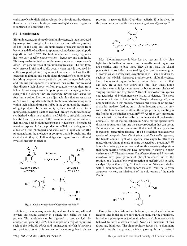
Bioluminescence, a subset of chemiluminescence, is light producedby an organism through a chemical reaction, and is the only sourceof light in the deep sea. Bioluminescent organisms range frombacteria and dinoflagellates to sponges, echinoderms, cephalopods(squid) and fish.35,142–144 The bioluminescence of every organismhas two very specific characteristics – frequency and amplitude.This may enable individuals of the same species to recognise eachother. Two general types of bioluminescence exist. The first type,only present in fish and squid, occurs when light is produced bycultures of photophores or symbiotic luminescent bacteria that theorganism maintains and manipulates through reflection or cover-ing. Many deep-sea species, particularly crustaceans, cephalopods,and fish, use photophores to illuminate their ventral surfaces andthus disguise their silhouettes from predators viewing them frombelow. In some organisms the photophores are simple glandularcups, while in others, they are elaborate devices with lenses forfocusing a colour filter, or an adjustable flap that serves as anon/off switch. Squid have both photophores and chromatophoreswithin their skin and can control both the colour and the intensityof light produced. In the second type, the chemical needed for aluminescent reaction is either acquired through the food chain, orsynthesised within the organism itself. Jellyfish, probably the mostbeautiful and spectacular of the bioluminescent marine animals,demonstrate both bioluminescence and iridescence. The chemicalreaction responsible for the production of light-bursts begins witha luciferin (the photogen) and ends with a light emitter (thephotagogikon), the molecule or complex that is brought into theexcited state (Fig. 2). Different types of organisms use differenttypes of luciferin molecules.
Fig. 2 Oxidative reaction of Cypridina luciferin.145
At times, the necessary reactants, luciferin, luciferase, salt, andoxygen, are bound together in a single unit called the photo-protein. This molecule can be triggered to produce light byanother ion, generally Ca2+. For example, an array of organismssuch as the hydroids Obelia and luminescent jellyfish Mitrocomause proteins, collectively known as calcium-regulated photo-
proteins, to generate light. Cypridina luciferin 63 is involved inthe bioluminescence of the crustacean Cypridina hilgendorfi.145
Most bioluminescence is blue for two reasons: firstly, bluelight travels furthest in water, and secondly, most organismsare sensitive only to blue light. They do not have the visualpigments to absorb the longer (red light) or shorter wavelengths.However, as with every rule, exceptions exist – some cnidarians,such as the jellyfish Aequorea, produce green bioluminescence.Each luminescent organism has a unique flash. Factors thatcan vary are colour, rise, decay, and total flash times. Someorganisms can emit light continuously, but most emit flashes ofvarying duration and brightness.146 One of the most advantageouscharacteristics of bioluminescence is that of defence. The mostcommon defensive technique is the “burglar alarm signal”, seenamong jellyfish. In this process, when a larger predator swims neara smaller predator feeding on its bioluminescent prey, the preyuses its bioluminescence to attract the larger predator, resulting inthe fleeing of the smaller predator!142–144 Another very importantcharacteristic that is enhanced by the luminescent ability of marineanimals is that of mating behaviour. Some marine species havedisperse populations, limiting the net reproduction that can occur.Bioluminescence is one mechanism that would allow a species toincrease its “perception distance”. It is believed that in at least twospecies of octopods, Japetella diaphana and Eledonella pygmaea,the female emits a light of a specific wavelength to attract itsmate, while avoiding the risk of being detected by a predator.142–144
It is a fascinating phenomenon and another amazing adaptationthat some marine organisms have developed to survive in theirenvironment.145 The protozoans Noctilluca miliaris and Pyrocystisnoctiluca have great powers of phosphorescence due to theproduction of oxyluciferin by the reaction of luciferin with oxygen,catalysed by luciferase (Fig. 2). Coelentarazine 64 is a metabolitewith a bioluminescent chromophore isolated from the jellyfishAequorea victoria, an inhabitant of the cold waters of the northPacific.
Except for a few fish and cephalopods, examples of biolumi-nescent lures in the sea are quite rare. In many marine organisms,including siphonophores (colonial hydrozoans), luminescence isbelieved to serve a defensive role. Nearly all members of thisgroup are luminous. The siphonophore Renna sp., a dominantpredator in the deep sea, twitches glowing lures to attract
236 | Nat. Prod. Rep., 2006, 23, 223–255 This journal is © The Royal Society of Chemistry 2006

fish, a rare phenomenon used for prey attraction among non-visual marine organisms. The lures also contain red fluorescentmaterial that shifts the wavelength of emitted light. Though redfluorescent substances have been noted in several marine phyla, redluminescence, so far, has only been observed in the rare scalelessmarine dragonflies (Stomiidae).35 Four main uses for an organismof bioluminescence have been hypothesised. It can be used to evadepredators (defense), attract prey, communicate within their speciesspecially in the dark (e.g. mating behaviour, courtship camouflage,etc.) and advertise.142–144,146
5.2 Biological fluorescence
In the phenomenon of fluorescence, light is absorbed at onewavelength and re-emitted at another wavelength; therefore or-ganisms “glow” only when light of a particular wavelength strikesthem. Fluorescence is fairly widespread in reef-building coralsand sea anemones, and they display a remarkable variety offluorescent colours. The jellyfish Aequorea also produces greenbioluminescence from small photo-organs located on its umbrella.The process is initiated by calcium ions released by the organism,which bind to a protein named aequorin, which then emitsblue light. The blue light is absorbed by another protein, Greenfluorescent protein (GFP), which in turn gives off green light. GFP,yellow under typical “room light”, absorbs ultraviolet light fromthe sun, and then emits it at as lower-energy green light.145
Fluorescent pigments may act as natural sunscreens, aid inphotosynthesis or serve as warning colouration.146 Some marineorganisms use colour-based visual signals to attract mates, warnrivals, or confuse predators. In an aquatic environment, colour-based visual signals may be unreliable because of variations inthe optical properties of water, such as water opacity and theangle of incident light. It has been proposed that fluorescencemay enhance colour in aquatic organisms. Some corals exhibitstriking fluorescent colouring, while some squids have fluorescentmarkings.146 The monogamous male Mantis shrimp, Lysiosquillinaglabriuscula, found in the western Atlantic ocean, when con-fronting predators or rivals, raises its head and thorax, spreadingits appendages so they appear larger. This posture also accentuatesthe yellow fluorescent patches on the organism. The wavelengthof this fluorescence (524 nm) transmits well through sea-water.147
6 Colour related to biological processes
Colonial ascidians display multiple body colours. Black, orange-red, violet, white, and yellow colours are due to pigmented bloodcells that exist particularly around a branchial siphon or onan atrial languet of individual zooids. These pigment cells aredistributed in the mesenchymal space or vascular lumen, and manyof them are loosely bound to the epithelium. The pigment cellshave a spherical shape with no dendrites, and contain many typesof granules; the pigmentary tissues contain carotenoids, pteridine,and purines. The main component of black and violet pigmentsis thought to be a melanin-like substance yet to be identified.Cephalopods, skilled in the art of colour change, which is usedfor camouflage or to startle and warn predators, have specialchromatophores connected to the nervous system in their skin.They are able to vary their colour and even create changing pat-terns by controlling the size of the cells by muscular contractions.
Haminea navicula, a sand-ploughing and -digging Mediterraneanand Atlantic cephalaspidean snail, is able to change the colourpattern of its body to adapt it to the surrounding sea-floor. Colourchanges occur by pigment migration in ramified epithelial andsubepithelial cells, by extension and contraction of melanophores.Within a few hours, some areas of the body can change frombeing totally “black” to nearly white.148 The erythrophores andmelanophores of the decapod Uca uruguayensis show a circadianrhythm of pigment translocation. The predominant erythrophoresdetermine the typical colouration of the species, which is redduring the day and beige at night. After eyestalk removal, theanimal becomes permanently red in colour.
The gastropod mollusc, the ormer, Haliotis tuberculata, hasthe appearance of typical abalones, characterised by a flattened,ear-shaped shell, covering and protecting the soft body parts,and with a large muscular foot which fills the shell opening.The epithelium where there is a high concentration of toxins isdenominated as the “foot side epithelium” and the epithelium ofthe foot in contact with the substrate is denominated as the “footsole epithelium”. Melanin and photosynthetic pigment granulesin the epidermal cells of the foot side epithelium give this skin itsappearance and colour, and the paralytic shellfish toxins (PSTs –saxitoxins and their derivatives) are located in specific cells of thefoot side epithelium, which are distinct from the epidermal cells.Conversely, the epidermal cells of the foot sole epithelium do nothave pigment granules, and PSTs have not been detected in thefoot sole epithelium.149
The anterior sensory vesicle of ascidian larvae is a single largevesicle in which lie two types of pigment cells, the anterior and pos-terior pigment cells, called the otolith and the ocellus respectively.Experimental results suggest that the otolith is used for gravitydetection, and that the ocellus is used for photoprotection.150 Itis suggested that the pigment cells in sea urchins (Echinoidea)embryos play a role in gastrulation.151
7 Colour related to genetics
The consistent range of colours and patterns in certain organisms,even in polymorphic species, is probably under genetic control, andthese colour patterns are not affected by environmental conditionsor the dietary source. Some sessile marine organisms show greatvariation in colour in the same habitat and locality.18,152–154 Someascidians show bands of silver derived from purine pigments,present in granules in blood cells, which are under geneticcontrol.5,6,8 Colour patterns may also be affected by processesoccurring during the development stage or may be due to randomneutral mutations at the molecular level.18,155 One-dimensionalmorphogenetic programmes usually control colour patterns onmollusc shells. In adult cypraeid gastropods (cowries), by com-parison, colour patterns are two-dimensional in morphogenesisand three-dimensional in structure. Visible patterns usually resultfrom the uneven thickness of a pigmented layer, rather than froma spatially uneven concentration of pigment.156
8 Photo-protective pigments
The biologically important solar UV-A and UV-B radiations havedeleterious effects (especially UV-B, due to its actinic nature) thatare manifested genetically, physiologically and photosynthetically,
This journal is © The Royal Society of Chemistry 2006 Nat. Prod. Rep., 2006, 23, 223–255 | 237

and are potentially damaging to many forms of life, includingmarine organisms. In addition to increases in temperature, UVradiation has been implicated in the phenomenon of coral“bleaching”. High levels of solar radiation affect photosynthesisof the symbionts, coral calcification, and survival of the organisms.However, the abundance of diversity of invertebrates in tropicalwaters suggests that protective or defence adaptations have evolvedto combat the harmful effects of UV radiation. Amongst thediversity of methods used by marine organisms to reduce damagecaused by UV radiation, the synthesis of UV-screening pigmentsis almost ubiquitous. Across diverse taxonomic groups of marineorganisms, there are several classes of compounds that absorbUV light and act as putative sunscreens. Mycosporine-like aminoacids (MAAs), with UV absorptions in the region 310–360 nm, arebelieved to be such a class of compounds.23,157–160 Mycosporines canbe considered to be Schiff bases (enaminoketones), and derivativesof aminocyclohexenone 65a or aminocyclohexenimine 65b rings.Being “colourless” compounds, some may not consider MAAsas pigments. Nageli and Schwenderer161 first described the browncolouration of some cyanobacteria, particularly in cyanobacterialmats. This colouration is now ascribed to scytonemin 66, whichis presumed to have a dedicated photo-protective as well as anantioxidant role. Scytonemin 66, with a bimodal UV absorptionat 252 and 370 nm (in vivo), is a lipid-soluble phenolic and indolicderivative produced in the sheaths of cyanobacteria.157,162–164
The UV-protective role of melanins is well established. Manymarine species possess the tyrosinase-mediated pathway to synthe-sise melanin, and in fact it occurs in a wide range of marine taxa.They can act as both optical filters and antioxidants.165 However,very little is known about the mechanisms, their efficiency or theirUV-protective role in aquatic taxa. Other compounds such asphlorotannins, sporopollenin (a polymer that constitutes the outerwalls of spores and pollen grains), coumarins, tridentatols (e.g.67) and polyphenolics have been implicated as UV protectants.159
Carotenoids are photopigments that have two major functions.Firstly, they are supplemental photosynthetic pigments thatabsorb light energy in the blue region. Secondly, they quenchoxygen free-radicals, thus acting indirectly as photo-protectivepigments.166–168 Our understanding of the range of UV-screeningcompounds in marine organisms is still in its infancy. The widedistribution of compounds such as MAAs suggests that a largenumber of compounds falling broadly into well-defined groupsmay be found in the future. Some may be represented in only
a few species. The oroidin alkaloids, which are various shadesof yellow and are represented by Z-axinohydantoin 52, sporan-giacidins B and D 53a+b,169 debromohymenialdisine 68,134,170 thio-compounds such as the tridentatols (e.g. 67 from a marine hydroid)and benzylthiocrellidone 54 from the sponge Crella spinulata,all absorbers of both UV-A and UV-B radiation, are a fewsuch examples.134–136,158,171 Many of these secondary metabolitesprobably have multiple functions. For example, tridentatols serveas allelopathic agents, antioxidants, and sunscreens.159,171–173
9 Environmental influence on colour
Evidence for environmental effects on colouration in marineinvertebrates is scarce. The colour of invertebrates varies accordingto depth, water temperature, food source, currents, geographiclocation, light attenuation and sedimentation, etc.18,174 For exam-ple, the carotenoid content in certain sea anemones decreasedwith depth.50 Species with photo-protective pigments appear“bleached” in darker and deeper locations and some spongesare less brilliant (i.e., they have reduced pigmentation) in shadedlocations than in well-lit areas.175,176 Photo-adaptation to decreas-ing flux generally results in a marked change in concentrationof photosynthetic pigments contained within the zooxanthellaeand shows three common trends. The first involves an increasein chlorophyll-a and the accessory pigments chlorophyll-c andperidinin with decreasing flux. The second is the relatively highconcentration of compounds with UV absorption over the rangeof 286–340 nm in corals from shallow reefs. The third trend isthe frequently higher concentrations of b-carotene and yellowxanthophylls in shallow-water corals.174,177
Colonies of the colonial ascidian–cyanophyte symbiosis Tri-didemnum solidum show morphological variation relative to thelight regime in which they live. Colonies growing in full sunlightare white, thicker, and heavily calcified, while the shaded coloniesare purple, thinner, and have a larger amount of phycoerythrin (anaccessory photosynthetic pigment with an absorption in the greenregion of the spectrum) relative to phycocyanin (a pigment thatabsorbs red light) in their symbiotic algae. The purple colourationof low-light colonies appears to be due primarily to the phycobilinpigment of the algae.178 This results in different colourations ofalgae growing under different lighting conditions. T. solidum fromthe Western Atlantic region is ocean-blue in appearance while T.cyclops, present in the tropical Pacific region (Papua New Guinea)is brilliant green. Perhaps the most remarkable adaptation shownby a coral to reduced radiant flux and different spectral qualityis that of the deep-water stony coral Leptoseris fragilis, whichhas a turquoise or green fluorescence and lives at depths of 100–150 m in the Red Sea. The efficient use of the available solarenergy is affected not only by the pigment changes in the symbioticalgae, but also by the fluorescent pigments sited within the coral
238 | Nat. Prod. Rep., 2006, 23, 223–255 This journal is © The Royal Society of Chemistry 2006

host tissues. Short-wavelength radiation, which would otherwisenot be absorbed by algal pigments, is absorbed by the coralpigments and fluoresced into longer wavelengths for harvesting bythe zooxanthellae. Such adaptation enables L. fragilis to colonisea habitat which is barren of all other symbiotic coral species.Furthermore, this mechanism is potentially present to some degreein all corals at all depths.179–182
Different species of fiddler crabs occupying a variety of intertidalniches along the Texas coast have adapted to a specific array ofphysical factors in the environment, and some aspects of theiradaptations are reflected in body colour. Interspecific differencesin morphological colourations are correlated with camouflage andsubstrate characteristics. Intraspecific colour variation is expressedthrough neurosecretion-mediated physiological change in cellularpigment distribution. Adaptation to a dark- or light-colouredbackground reveals different “secondary” chromo-motor capa-bilities for each species. In addition, pigments in melanophores,leucophores and erythrophores exhibit circadian rhythms ofdispersion and aggregation.183 Colouration is often uniform acrossvast geographic ranges, thus indicating a degree of genetic controlover the development and expression of colour patterns. There arealso species capable of matching a variety of host colour patterns,which indicates a degree of environmental influence.184 However,this raises the question of how host colouration is perceived andtranslated by newly settled juveniles.
Sodium cyanide, widely used for the capture of reef fishthroughout Southeast Asia, causes extensive fish mortality. Theexposure of corals to cyanide caused deformation of the symbioticzooxanthellae along with pigment loss, eventually resulting incoral mortality.185
10 Chromatophores
Chromatophores or pigment cells are colour-changing cells thatare directly innervated by central motor neurons and used mostnotably by chameleons as well as cephalopods such as squid,octopuses and cuttlefish. This behaviour is generated primarilyby chromatophores, which are composed of a single highlydeveloped chromatophore cell and numerous muscle, nerve, glialand sheath cells. These cells are contractile and contain vesiclesthat contain three different liquid pigments, and the three typesof chromatophore are characterised by the colour they carry.Erythrophores contain reddish pigments, such as carotenoids andpteridines. Melanophores contain black or brown pigments, themelanins. Xanthophores produce yellow pigments in the formof carotenoids. Leucophores and iridophores, unlike the abovetypes of chromatophores, are unable to produce pigments andtherefore are colourless. Cells carrying more than one pigmentare called compound chromatophores, and various hues are madepossible by the combination of different layers of chromatophores.Chromatophores are mainly located beneath the scales or in thedermis, but are sometimes seen superficially in the epidermis. Theyare responsible for the quick adaptation in colour required forcamouflaging.
The colours generated by the cells can be broken down intotwo categories: biochromes and schematochromes. Colours dueto microscopic, natural pigments that produce colours chemicallyare called biochromes. Their chemical composition is designedto absorb some colours of light and reflect others. The projected
colour is a combination of all the visible wavelengths of light thatare reflected by the pigment. Conversely, those colours producedby light reflections from a colourless surface and refractions by thetissues are named structural colours or schematochromes. Thesestructures act like prisms by refracting and scattering visible light,and ultimately reflect a certain combination of colours.
Colour changes involving biochromes and some changes in-volving schematochromes depend on the movement of pigmentswithin the colour cell. These short-term, rapid changes are calledphysiological colour changes, a phenomenon mostly found in fishand usually resulting from the animal’s response to an alterationin the surroundings. On the other hand, morphological colourchanges are long-term changes, occur during different life stagesof the animal, and are due to increases or decreases in the numberof chromatophores and a general alteration of pigmentation. Mostfish that appear to have green colouration on their scales actuallyhave a layer with yellow pigment and another layer on top thatscatters light and reflects a blue colour. To change their colour,the cells distort their form or size, stretching or contracting theirouter covering, and thus changing their transparency or opacity.Body patterning in unshelled cephalopod molluscs is the ability ofthese animals to create complex patterns on their skin. Pigmentgranules lie within the chromatophore cell in an intracellular sac,the cytoelastic sacculus, which has elastic walls. The contractionand retraction of the muscle cells causes the sacculus to changeits shape, bringing about a colour change in the organism.186 Thetremendous capacity of the chromatophore system in cephalopodskin to change its colour across space and time has allowed theseanimals to produce complex visual displays to conspecific, and het-erospecific targets.187 The chromatophores in crustaceans, whichhave fixed shape, bring about colour change through the dispersal(darkening) or aggregation (lightening) of granules within thecells. The aggregation and dispersal of colour pigments in chro-matophores seem to be under both hormonal and neural control.
11 Adaptive colouration
Adaptive colour patterns and variation in animals were at thecentre of early conceptual advances in evolutionary biology, andwere prominent in early thinking about evolution.188 Much of theliterature on adaptive colouration and colour patterns has beenparticularly concerned with predator–prey relationships. Part ofthe colour pattern is inherited from a common ancestor and partis due to convergent evolution, which can result from Batesian orMullerian mimicry.
11.1 Aposematism
The term aposematism (literally, “away signal”) is commonly usedas a synonym for warning colouration. It is the combinationof a conspicuous signal and an unprofitable trait in a preyspecies. Aposematism, a widespread trait in invertebrate taxa,and a well-documented phenomenon in nature, is the association,in a prey organism, of the presence of a warning signal withunprofitability to predators. Aposematism works by advertisingto potential predators, rather than by hiding or escaping fromthem. Aposmetism has benefits for predator and prey. Efficientinteractions between predator and prey in this context alloweach to pursue other activities such as mating, care for offsping
This journal is © The Royal Society of Chemistry 2006 Nat. Prod. Rep., 2006, 23, 223–255 | 239

and feeding. It is one of the oldest phenomena in evolutionarybiology and still a major puzzle to biologists. According tosome evolutionary biologists it is paradoxical that an associationbetween conspicuous signals and unprofitability could evolve.It would be difficult for conspicuous signals to evolve in preywith unprofitable traits because predators will quickly sample theprey and kill it, eliminating any genes for the conspicuous signalfrom the population. An alternative scenario is for unprofitabilityto evolve in prey with conspicuous colouration. However, thisis not likely because being conspicuous without any defenceoffers no protection and simply makes the prey more visibleto predators. On the other hand, without following exclusivepathways, unprofitability and conspicuous signals could evolvetogether. Much of the literature on adaptive colouration has beenparticularly concerned with the predator–prey relationship. It haslong been recognized that defended prey tend to be conspicuousand that aposematism arises because predators more readily learnto avoid attacking defended phenotypes when they are conspicu-ous. In order to fully understand aposematism there should bea clear picture of the evolutionary history behind the presentbehaviours.189 Many studies on aposematism have been descriptive,or conducted strictly from the ecological or behavioural points ofview. Aposematic colouration is common in most opisthobranchtaxa and is more predominant in tropical faunas than in temperateones, though at least one study disputed the role of pigments inaposematic colouration of tropical sponges.190,191,192 Resolving thedebate on the evolution of aposematism will require experimentsand observation of predation on aposomatic species in the wild.
Conspicuous signals are most often bright colours usuallypresented in banded or contrasting patterns. Bright colours mayadvertise to potential predators that a species is distasteful, toxic,unpalatable, vicious or difficult to capture. For a colour patternto be aposematic, predators must be able to see the colour or atleast detect contrasting patterns of shading. Conspicuous warningsignals of unprofitable prey are a defence against visual huntingpredators. Warning colours are usually, but not always, conspic-uous and consist of bright colours of red, yellow or white, oftenin combination with black, and are not intended to camouflage,but to make the animal more conspicuous. They work becausepredators learn to associate unprofitability with bright colourationand because strong signals are detectable and memorable.193
However, many species that can be considered defended are notvery conspicuous; they have weak warning signals.194
The co-occurrence of conspicuous colouration and noxiouscharacteristics suggests that aposematism is a common predatordeterrent in many coastal marine species. The organisms usewarning displays for defence and better survival, a strategycommonly found among nudibranchs (Opisthobranchia, Mol-lusca), particularly in many species of the nudibranch familiesChromodorididae and Phyllidiidae (colourful dorids or sea slugs).Colour has played a major role in the adaptive radiation andevolution of opisthobranch gastropods, which are among themost brilliantly coloured organisms inhabiting the world’s oceans.They have spectacular and obvious colour patterns for warningpotential predators, are known to have reduced or entirely losttheir shells independently within several distinct lineages, andevolved (or acquired from dietary sources) toxins that provideprotection from predation.105,195 Even though nudibranchs possessno protective shell and seem to be easy prey for fish or other
predators such as crabs, reports of predation (except by othernudibranchs) are virtually non-existent.196 They can also becoloured in such a way as to warn predators that conspicuouscolouration is often associated with the presence of toxic or defencesubstances. Studies on shell-less molluscs led to the theory that co-evolution with toxic foods had allowed them to lose their shellsover evolutionary time.74,126 Many chromodorid nudibranchs takedistasteful chemicals from the sponges they feed on and store themin special glands (repugnatorial glands), which are located aroundthe mantle edge. Contact with these antifeedant compounds isan extremely unpleasant dining experience and visual predatorswill quickly learn to avoid them by recognition of their distinctand bright colour patterns. Many aposematic species have evolvedto live in close proximity to others of their species; one possibleadvantage of this is that it makes the aposematic signal moreeffective by generating a greater aversion in predators. Spongescan be toxic to fish, and distasteful due to tough fibres, spiculesand toxic substances.197 Not only can organisms change theircolour to elude the enemy, they can also emit dye to detourthem.22,198 Even though no explanation other than aposematismhas been proposed for the colouration of marine gastropods that isconsistent with what is known of their biology, it should be notedthat no experiments on these organisms have conclusively demon-strated aposematism.199–201 The different categories of colour inopisthobranch molluscs have been reviewed.20,21,202
The possibility that bioluminescence functions as an aposematicsignal has been suggested.199 Bioluminescence in the Basket brittlestar, Ophiopsila riisei, functions as a facultative aposematic signal(to deter crab predators) and is common among generalisedgrazers, while more specialised predators often exhibit specialresemblance to their prey.
Toxicity or reduced palatability is due to a variety of organiccompounds, such as the isonitriles kalihinol A and B 69a+b,furans (e.g. furospongin 70), and inorganic compounds (e.g. sul-furic acid); most of these compounds are derivatives of chemicalsproduced by the prey. Only a few species manufacture toxins denovo.203–205 Laboratory feeding assays with fish and food pelletscontaining organic extracts of several species of demospongesfrom the reef, mangrove, and grass bed habitats indicated thatmost sponge species yielded deterrent extracts but there wasconsiderable inter- and intra-specific variability in deterrencysuggesting that, at least for some species, some individualsmay be chemically undefended. Reef sponges generally yieldedmore deterrent extracts than sponges from other habitats, and
240 | Nat. Prod. Rep., 2006, 23, 223–255 This journal is © The Royal Society of Chemistry 2006

there was no relationship between sponge colour and deterrency,suggesting that sponges are not aposematic.205,206 Feeding fish withbrightly coloured larvae, Young and Bingham207 addressed bothissues of chemical defence and aposematism. They and otherinvestigators173,208 have demonstrated how widespread aposema-tism might be among marine invertebrates, that rejection of larvaeby fish was chemically based, and that coloured (conspicuous)larvae are more likely to possess chemical defences.
11.2 Advertisement
Many species, including marine organisms, use colour-basedvisual signals to attract mates, warn rivals, or confuse predators.Often, bold designs accompany an unpleasant taste or dangerousvenom. Aposematic colouration works by advertising unpleasantqualities to potential predators. However, aposematism is not theonly reason for bright colouration of marine organisms – brightcolours also have other functions. For example, advertisement bybright colours or conspicuous patterns serves various purposesin coral reef organisms. Various life stages of organisms such asfish involve colour changes. Animals also change colour to signaldifferent moods. Some fish are pale when frightened and darkenin anger or in sexual excitement. The octopus can turn blackwhen in danger. Colouration also functions by differentiatingbetween sexes and aiding in selection of potential mates. Malesare mostly more brightly coloured than females – in some species,the colours and patterns are present only during the breedingseason in order to attract the female. Other courting rituals suchas intricate dances accompany these colour changes.199,200,202 Theability to change skin colour patterns allows squid to court mates,challenge rivals, and communicate with one another. A maleCaribbean reef squid, for example, splits into two patterns duringcourtship; its coppery right side is oriented toward the female,and its silvery left side is to ward off rival males.209 Becausephysical and chemical colouration is determined genetically, traitsare passed on from parent to offspring.
Many marine animal species are genetically polymorphic incolour. Visual selective predation is commonly suggested as amechanism for maintaining colour polymorphism in prey speciesand may, for example, result in correlations between colourfrequencies and the background colour. Such correlations arecommonly observed in some polymorphic gastropods.210
11.3 Crypsis
Many organisms in the ocean have the ability to hide or concealthemselves, a phenomenon referred to as crypsis or homochromy.Some organisms use methods of concealment to avoid beingdiscovered by potential predators and prey. There are threemain forms of crypsis: disruptive colouration, camouflage, andcolouration which mimics that of the host.
Disruptive colouration occurs when organisms are spotted,striped or have other types of patterns to make their outlinedifficult to distinguish. These patterns are useful to the marineorganism because they often blend in well with the surroundingsand aim to obscure the true form of the animal by partly blendingwith the background and distracting the attention of the viewer.211
Crypsis through background matching requires that the sizes andshapes of the pattern elements should closely resemble those of
the visual background.212 Animals like the octopus, cuttlefish andthe squid can change their colour depending on the situation thatthey are in. It is most often facilitated by the resemblance of theorganism to the background.
Another common method of crypsis or concealment is counter-shading (pelagic colouration). Countershading, the gradation ofcolour from dark on the dorsal (upper) side to light on the ventral(lower) side is generally considered to have the effect of makingorganisms difficult to detect. The mechanism that facilitates thisform of crypsis is often considered to be concealment of shadowscast on the body of the animal, but there is no conclusive empiricalevidence for this function. However, highly refined adaptations insome marine organisms to match the different background lightconditions against which they are set when viewed from differentaspects strongly suggest an adaptive advantage to countershadingin these environments.213
Although most transparent species reside in the pelagic zoneof the ocean, some benthic, neustonic (minute, surface-dwelling)organisms have transparent members. Among the transparentanimals are shrimp, octopuses, polychaete worms, the hydrozoanPortuguese Man-of-War, and copepods.214 Transparency is one ofthe few forms of camouflage, but it is a very successful methodin environments that offer no surfaces to match or hide behind,and appears to have evolved for passive crypsis.215 In the pelagicenvironment, the most prevalent form is complete transparency,achieved by reflection of most (or all) of the visible wavelength oflight, and counter-illumination by bioluminescence.216
In dermasal colouration, some species have similar coloursand designs that blend in with the bottom of the substrate. Yetsome others have the capacity to bear a general resemblance tovegetation, which is termed vegetal colouration. Organisms alsoshow colour resemblances to substrates that consists of stones,shells, and small algae, individually or in combination. Marineinvertebrates employ blue and yellow carotenoproteins for camou-flage or in mating rituals. Sometimes a symbiont can help the hostto camouflage. The luminescent symbiont Vibrio fischeri is housedin a complex light organ within the mantle cavity of the Hawaiianbobtail squid Euprymna scolopes, and the bioluminescence by thesymbiont helps the squid to camouflage itself.217
For many reasons and in many ways, some marine organismschange their colour and sometimes their shapes to disguisethemselves. Many animals, such as the decorator crab, Pederson’scleaning shrimp and the sea horse change their whole colour andappearance to hide from predators. The nudibranch Discodorisindecora shows a perfect camouflage on its prey, the dictyoceratidsponge Ircinia variabilis. The shape and colour of the nudibranchare remarkably similar to that of the sponge, which is widespreadin shallow waters of the Mediterranean Sea. While feeding onsponges, the nudibranchs sequester the effective chemical armouryof their prey, which is subsequently employed for their ownprotection.139,196
The cuttlefish Sepia officinalis can produce a variety of bodypatterns for camouflage on natural substrates, and they can blendinto the surroundings almost as fast as they can move.218,219 Onmixed light and dark gravel, they show disruptive body patternsfor camouflage.220,221 The recently discovered rare mimic octopusimpersonates the shape, posture and movements of various otheranimals. The octopus’ banded colouration copies the dramaticbanding of many dangerous or poisonous sea animals such as
This journal is © The Royal Society of Chemistry 2006 Nat. Prod. Rep., 2006, 23, 223–255 | 241

sea snakes, lionfish and flatfish.222 There has been considerabledebate regarding the function of coloured compounds in theshells of marine organisms. The red colouration of Calloria spp.(Brachiopoda) is due to a carotenoprotein that occurs within thecrystals of the shell. Equivalent pigments from mollusc shellsare considered to be a by-product of metabolism.223–225 Redcolouration permeates the body tissues of living brachiopods,as well as the shell.226 Such colouration in brachiopods as wellas in other taxa is thought to have a photo-protective function.Red pigmentation of animal shells may have a protective roleby warning potential predators that the brachiopod tissues maycontain toxin, or by functioning as camouflage.50,227 Many speciesof arboreal marine snails are polymorphic with regard to shellcolour, with morphs that correspond in both colour and frequencyto those predominant in the habitat. A combined effect of selectionfor crypsis and apostasy has been suggested as the most likelyreason.228 Sea hares (Family Aplysidae, Order Anaspidea) areherbivorous opisthibranch molluscs that acquire and store algalmetabolites that may act as chemical defences against predators.Like many other opisthobranchs, sea hares have multiple defensesagainst predation including physical, behavioural and chemicalstrategies. Physical and behavioural strategies include crypsisand movement, and crypsis is the most common and primarymeans for avoiding predators. One of the most common featuresof most sea hares is that the organism produces clouds ofreddish purple “ink” when disturbed, whose function is yet to bedetermined.88,202,203
11.4 Mimicry
In some geographical areas, groups of unrelated nudibranchs suchas the red-spotted chromodorid species found in south-easternAustralia, and polyclad flatworm species found in California, haveevolved very similar colour patterns, so that they share the task ofteaching the predator to leave the colour pattern alone. A numberof harmless snakes closely mimic the bright warning colouration ofthe coral snake – the most poisonous snake in the United States.The colour pattern of a few nasty distasteful species is copiedby other quite edible species. These species gain considerableprotection from predators that have learnt from experience toleave the distasteful species alone. This phenomenon where oneorganism (the mimic) evolves to resemble another species (themodel) such that these two organisms are confused by a thirdorganism (the receiver) for gaining some biological benefit is calledmimicry. The concept of disguise involves not only colour, but alsobody shape and behaviour. Warning colour and mimicry formpart of the general topic of defensive colouration. Mimicry inthe ocean is especially deceptive because of the capabilities oforganisms, such as molluscs, to grow appendages and go throughquick changes. An evolving mimic takes advantage of previouslyevolved communication signals and responses between organisms.To be successful and beneficial to the mimic, the model shouldbe an abundant species whose noxious characteristics have left alasting impression on predators.
There are four major forms of mimicry utilised by both preda-tor and prey: Batesian mimicry, Mullerian mimicry, aggressivemimicry and self-mimicry. Mimicry refers to the similaritiesbetween animal species; camouflage refers to an animal speciesresembling an inanimate object.
In Batesian mimicry, first described in 1852 by the Britishnaturalist Henry Bates, the mimicking organism has evolved somefeatures of a poisonous organism but is not poisonous itself – aBatesian mimic cheats.229 In the case of two unrelated, but similarlymarked families of Brazilian forest butterflies, one of which (themodel) was poisonous to birds and the other (the mimic) palatable,the latter survived because of their resemblance to the poisonousfamily. Mimics usually copy the aposematic colouration of themodel species. In this kind of mimicry, the mimicking organismhas evolved some features of a poisonous organism but is notpoisonous itself. This is essentially equivalent to camouflage.
In 1878, the German zoologist Fritz Muller proposed anexplanation to Bates’ paradox, in describing Mullerian mimicry.In Batesian mimicry, one species is distasteful and another one ispalatable, while in Mullerian mimicry both species are distasteful.While Bates had observed a resemblance among several unrelatedbutterflies all of which were inedible, Muller realised that theexplanation might lie in the advantage to one edible species inhaving a predator learn from another. Once the predator haslearned to avoid the particular conspicuous warning colourationwith which it had its initial contact, it would then avoid all othersimilarly patterned species, edible or inedible. Mullerian mimicsgain maximum protection when all individuals have the samesignal (signal standardisation). This tendency of inedible andnoxious species to evolve to have the same or similar warningsignals such as the same pattern of bright colours is called Mul-lerian mimicry.230 This form of mimicry refers to two unpalatablespecies that are mimics of each other with conspicuous warningor aposomatic colouration. Thus all the mimics share the benefitsof the colouration, since the predator, after a few bad experiences,will recognise the colouration of an unpalatable group.
In aggressive mimicry, the organism mimics a signal that isattractive or deceptive to its prey.202,229,231–234 Aggressive mimicryusually is seen in skin biters imitating cleaner fishes. Cleanersare not often preyed upon and other fishes that mimic theircolour patterns and behaviour can gain protection. The red andblack colouration of deep-sea marine organisms appears black atdepth, and black blends in, rendering these organisms invisible topredators.
In some cases, as in self-mimicry, colouration mimics a par-ticular region or microhabitat of the host. Self-mimicry is amisleading term for animals that have one body part that mimicsanother to increase survival during an attack or helps predatorsappear innocuous. Host mimicry may be further enhanced bymorphological or behavioural adaptations. In all these instances,a mimetic relationship is postulated.235 Reimchen reported the firstexample of gastropod–polychaete mimicry and presented evidencefor the overwhelming role of visual predators and natural selectionin the evolution of shell colour among intertidal prosobranchLittorinamariae.235 There are only a few examples of aggressiveand self mimicry in the marine environment.229
12 The defensive role of pigments
The high frequency of bioactive components in the primitivefilter-feeding sponges is interpreted as chemical defence againstenvironmental stress factors such as predation, infection, over-growth by fouling organisms or competition for space.236 Thesetoxic or deterrent sponge metabolites are found in habitats such
242 | Nat. Prod. Rep., 2006, 23, 223–255 This journal is © The Royal Society of Chemistry 2006

as coral reefs that are characterised by intense competition andfeeding pressure. Further support for the adaptive significanceof sponge constituents is derived from the observation thatsponges which grow in exposed places are usually more toxicthan those in unexposed places.237 Early studies concluded thatthe incidence of defensive chemicals was much higher at tropicalthan temperate latitudes. Tropical–temperate comparisons ledto a “latitudinal” hypothesis, suggesting an inverse correlationbetween the incidence of chemical defence in marine invertebratesand latitude. Whereas sponges or the symbionts associated withthem are believed to synthesise these metabolites, gastropodsand nudibranchs sequester this “chemical armoury” from theirprey, which is subsequently employed for their own protection.Some nudibranchs, however, have become independent of thisinterspecific flow of natural products and are able to accumulatedefensive compounds through de novo synthesis.238
Porifera (sponges) are the dominant phylum on the Antarcticbenthos, and members of this phylum have been shown toelaborate sea star feeding deterrents, inhibitors of fouling orinfectious organisms, and metabolites which mediate predation bymoult inhibition. Antarctic sponges have some unique attributes.They are remarkably diverse – some are related to deep-sea spongespecies, but some others to those from South America. Amongthe common members of the sponge community are severalconspicuously coloured species. Isodictya erinacea (polychaetesponge) and Dendrilla membranosa are bright yellow spongeslacking structural defences such as spicules, and the deep greendemosponge Latrunculia apicalis is not preyed upon by thespongivorous sea star Perknaster fuscus. This ecologically relevantsympatric sea star is a major predator of the sponges in thisenvironment.239–243 For example, the fast-growing, potentiallyspace-dominating sponge Mycale acerata is kept in check by thissea star. The soft-bodied, fire-red sponge Kirkpatrickia variolosa,a minor component of the diet of the sea star P. fuscus, is themost striking sponge in the Antarctic benthos. Suberites sp., witha yellow appearance, is a common Antarctic sponge. Asbestoplumahypogea (Carnivorous sponge), Cinachyra subterranean (Globularsponge), Cinachyra antarctica (Spiky sponge), Clathria nidificata(Orange dendritic sponge), Dendrilla antarctica (Cactus sponge),Iophon radiatus (Epibiotic sponge), Hexactinellida sp. (glasssponges), and Leucetta leptoraphis (Rubber sponge), are otherexamples of brilliantly coloured Antarctic sponges. The coloursof the Antarctic sponges can be white – light tan, bright yellow,greenish-yellow, green, dark brown, violet, or (in a few cases)pink. The pigments which bestow this bright colouration arethemselves various shades of orange, yellow and purple. Antarcticbenthic marine invertebrates have no apparent reason to bebrightly coloured, yet they are, suggesting that pigmentation couldnot be driven by aposematism.200,205,239,240,243 The presence of thisbright colouration is intriguing in a marine system where grazingpressure by visually oriented predators such as fish is generallylacking. In contrast to predictions that global patterns of predationshould select for an inverse correlation between latitude andchemical defences in sponges, such defences are not uncommonamong Antarctic sponges.26,241–243 The characterisation of chemicaldefences and the presence of bioactive pigments from most highlypigmented benthic Antarctic invertebrates raise the intriguingquestions of the role of colouration in such an ecosystem, andwhether evolutionary selection for, or retention of, pigmentation
has been driven by predation pressure.239,243 Colouration in thesesponges may be the result of relict pigments originally selectedfor aposematism or UV screening, yet conserved because of theirdefensive properties. Pigments in other marine organisms andterrestrial plants are almost exclusively employed in energy captureor to serve as antioxidants, but these are unlikely roles for pigmentsof benthic marine invertebrates. Conversely, there is little evidenceon whether pigments from tropical or temperate benthic marineinvertebrates may serve as antifeedants. The distribution of knownmetabolites in these organisms differs considerably from that ofthe general marine realm.239–243 Most of the secondary metabolitesdescribed from Antarctic organisms are not found in congenersfrom temperate and tropical regions. Most of them are vulnerableto predation, especially by sea stars. Recent laboratory studies haveindicated that these pigments in Antarctic sponges are themselvesbioactive feeding deterrents, and defend the organisms from preda-tors or fouling organisms.239,243 The hypothesis is supported by thebioactive properties of pigments present in them. Among the pig-ments characterised so far are the variolins, e.g. variolin B 48 fromKirkpatrickia variolosa, bearing a very unusual pyridopyrrolopy-rimidine ring system. This is a group of alkaloids represented bythe discorhabdins, e.g. discorhabdins A and C 71a+b (present inLatrunculia apicalis), suberitenones A and B 72a+b (the majormetabolites of Suberites sp.), and the yellow isoquinoline pigment73 from Dendrilla membranosa. The discorhabdins account forthe green colouration of L. apicalis,244 and discorhabdin C 71b isa potent mammalian cytotoxin, emphasising its bioactive nature.
The pigments variolins, discorhabdins, the isoquinoline metabo-lite from Dendrilla membranosa, and to a lesser extent, thesuberitenones, display ecologically relevant bioactivity, includingtube-foot retraction (indicative of feeding deterrence) and antimi-crobial activity. These pigments displayed bioactivity towards theircommon enemy, the sea star Perknaster fuscus, which lacks visualorientation.243,245 They serve as defensive agents, both towardpredators and potential infectious agents. Discorhabdin C isperhaps the strongest sea star deterrent studied to date.243 Seastars, the specialist predators of the Antarctic sponges, often feedby extruding their cardiac stomachs over their prey. Because ofthis unique stomach extension, feeding mechanisms and feedingassays typically used with other predators are less appropriatefor sea stars. The tube-feet are chemosensory, used by the sea
This journal is © The Royal Society of Chemistry 2006 Nat. Prod. Rep., 2006, 23, 223–255 | 243

star for, among other things, assessment of prey suitability –contact of the tube-feet with sponge extracts elicits a characteristicchemosensory response.243 It is generally accepted that tube-feetare a primary site for chemical reception in echinoderms andthat tube-foot chemoreception in sea stars is primarily a defensiveresponse.245 The quinoline pigment 73 present in D. membranosa isinhibitory towards a water-column bacterium.246 It is among thefew Antarctic sponges known to produce terpenes.243 Cytotoxicerinacean 74 has been isolated from Isodictya erinacea, and eco-logically relevant concentrations of p-hydroxybenzaldehyde fromthis bright yellow sponge are feeding inhibitors.239,243 The yellowpigment eribusinone 75, a tryptophan catabolite, has been isolatedfrom the sponge I. erinacea. Tryptophan catabolites are involvedin crustacean moult regulations. Moult regulation as a potentialmechanism of chemical defence in a marine realm has been demon-strated. Eribusinone 75 from I. erinacea was studied as a moultinhibitor due to its structural similarity to the crustacean moult in-hibitor 3-hydroxykynurenine 76. Preliminary feeding experimentsconducted with a carnivorous predator, the amphipod Orchomeneplebs, on a diet enriched with eribusinone, led to significant de-crease in moulting events and concomitant increase in mortality.243
The ovaries, eggs, embryos and larvae of some of these Antarcticbenthic invertebrates are unpalatable or chemically defended fromsea star, sea anemone and amphipod predators.241,242 In oneinteresting twist, it has been observed that a sponge pigmentmimicked a chemical involved in regulating moulting of anamphipod known to prey on the sponge. When the amphipodeats the sponge the chemical pigment prevents moulting and theamphipod eventually dies.239,241–243 Studies on Antarctic spongeshas thrown new insights into the functional role of pigmentsand the ability of sponges to sequester metabolites, and a newmechanism of chemical defence has been described.239,243
Chemicals released when organisms are attacked can serve tomark a location as dangerous to conspecifics. Many gastropodsleave a slime trail behind them as they move, which can allowfor easier location by conspecifics in search of mates. However,when sufficiently molested (as by a predator), the nudibranchNavanax inermis secretes a mixture of bright yellow chemicals,the navenones, (e.g. navenones A and B 77a+b) into its slimy trail,displays alarm pheromone properties and causes an avoidanceresponse in trail-following conspecifics.247,248 The lignarenones, e.g.lignarenones A and B 78a+b, isolated as pale yellow gums fromthe mantle of the mollusc Scaphander lignarius and structurallyrelated to the navenones, did not show similar properties.249
Tambja abdere, the greenish-coloured nudibranch with mottledpatches of yellow and bluish-ocre splattered through the body,sequester tambjamines A, B, C, E and F 79a–e (belonging to the4-methoxypyrrole family of natural products), from its bryozoanprey for the same purpose, and to deter fish. Holothuria atra,when handled, releases a burgundy-coloured toxin copiously intothe surrounding water, which is lethal enough to kill polychaetesand flatworms and immobilise cone snails (Conus).22,198 The red-coloured sponge Latrunculia magnifica found in the Red Sea,when squeezed, exuded a reddish fluid that caused fish to fleeimmediately from the vicinity, and when the juice was squeezedinto aquaria, fish (Gambusia affinis) died within a few minutes.Chemical analysis afforded the macrocyclic light yellow alkaloids,the latrunculins (e.g. latrunculins A and B 80a+b), which wereresponsible for the strong ichthyotoxicity of the sponge.196 Theunusual sesterterpenoids palinurin 57 and variabilin 81 are the
244 | Nat. Prod. Rep., 2006, 23, 223–255 This journal is © The Royal Society of Chemistry 2006

dominant metabolites in Ircinia variabilis. When disturbed, thesponge secretes copious amounts of a substance that contains largeamounts of both metabolites. The defensive role of palinurin andvariabilin is strongly supported by their anatomical location.139
13 Physiological activities of pigments
Several studies of the chemical defences of marine invertebratespoint to an increased interest in understanding the ecologicalrationale for the production of these compounds, and theirbiological activities. Pigments in marine invertebrates and theirdistribution and functions seem to differ between invertebrategroups. It is possible that some of the bright colours observedtoday are relics from the past, when the colours served an adaptivefunction that now has been lost due to changes in environmentalconditions, or extinction of predators that once were deterred bythe colour patterns. For example, alkaloids (especially the indoleand bis(indole) alkaloids comprising the yellow pigments fromsponges, ascidians and hard corals) play an important role as thedefence of the host animal against predation, and have showna broad spectrum of biological activity. It is proposed that thepresence of these pigments is linked to the biosynthetic activity ofthe symbiotic bacteria.74–76 Marine organisms, the nudibranchs inparticular, which are shell-less (and hence seemingly defenceless),and conspicuously coloured, have invariably been found to containcompounds identical or similar to those found in their diet.250
These compounds are accumulated on a functional basis, andit is possible that nudibranchs fractionate the best chemicaldefence compounds from their diet. This biological rationalesuggests that potent defence compounds have other uses. Indeed,the unusual blue tetrapyrrole pigment 13 from the nudibranchNembrotha kubaryana was found to be a potent antimicrobialagent. In addition, tambjamines A, B, C, E, and F 79a–eisolated from this organism showed antimicrobial, antitumour andimmunosuppressive activities.88 Several guaiazulenes and relatedsesquiterpenoids, well recognised for their blue and purple colours,have been reported to exhibit various bioactivities, includingantibacterial, antimicrobial, cytotoxic, antitumour, and immuno-regulating activities, as well as inhibitory activity against celldivision of fertilised sea urchin and ascidian eggs.115,251–253 Twonew carotenoids isolated from the sponge Phakellia stellidermashowed mild cytotoxicity against mouse leukemia cells.83 The redpigment fascaplysin 59 (a metabolite of Fascaplysinopsis sp.254),arcyriaflavin 82 and the related staurosporine aglycone 83 (fromEudistoma sp.,255 a tunicate), the pale yellow disulfide polycarpine84 (isolated from the tunicates Polycarpa clavata256 and P. au-rata257), the discorhabdins 71a+b and related pigments from thesponge Latrunculia apicalis,258 the meridianin alkaloids (e.g. 58from the tunicate Aplydium meridianum133), coscinamide alkaloids(e.g. 85 from the sponge Coscinoderma sp.133) and topsentinalkaloids (e.g. 50 from the sponge Spongosorites genitrix76) havebeen shown to be cytotoxic. The meridianins are a new family ofprotein kinase inhibitors.133
A marine gram-negative aerobic bacteria that produced cyto-toxic lemon-yellow chromopeptide pigments that inhibited thedevelopment of sea urchin eggs has also been isolated fromthe Australian sponge Fascaplysinopsis reticulata.259 The dis-corhabdins, e.g. 71a+b, are often cytotoxic, bear antitumour andantiviral properties, and have broad-spectrum antibiotic activity
toward sympatric bacteria. The unusual red pigment trikendiol86, isolated from the Axinellidae sponge Trikentrion loeve, is aninhibitor of HIV-1.260 The variolins, e.g. 48 from the Antarcticsponge Kirkpatrickia variolosa, are potent cytotoxins towards theP388 cell line.239,243 K. variolosa elaborates reserveritol triacetate 87,the only stilbene so far reported from a marine organism. Stilbenesfound in many plant genera are considered to be phytoalexins.
Among the ecologically relevant natural products, the yellowpigment eribusinone 75 from the Antarctic sponge Isodictaerinacea appears to be a tryptophan catabolite, and behaves muchas other such catabolites in regulating crustacean moulting.239,243
An unusual alkylated purine, with no currently understoodfunctional role, has also been isolated from the organism. An-tibiotic bromophenols are present predominantly in the distalregions of the polychaete worm Thelepus extensus, suggestinga role in fouling and/or infection control. Arcyriaflavin 82 andthe related staurosporine aglycone 83 (from the grey-colouredtunicate Eudistoma sp.), aaptamine 88 and isoaaptamine 89 (both
This journal is © The Royal Society of Chemistry 2006 Nat. Prod. Rep., 2006, 23, 223–255 | 245

pale brown in appearance, from the sponge Aaptos aaptos) areprotein kinase C inhibitors, as are the oroidin alkaloids Z-axinohydantoin 52 and debromo-Z-axinohydantoin 90, obtainedas minor metabolites from the sponge Stylotella aurantium (ag-minata).169 This organism fluoresces and its colour varies fromyellow to orange. Similarly, the spongiacidins B and D 53a+b(another group of metabolites from the oroidin group) from aHymeniacidon sp. sponge,130,170,203 and the rhopaladins 91a+b,bis-alkaloids from a blue translucent sea squirt, Rhopalaea sp.,are c-erbB-2 kinase and cyclin-dependent kinase 4 inhibitors.261
The ascidian Didemnum conchyliatum, which was collected fromsea grass blades in mangrove habitats, contained the indolealkaloids didemnimides 92a–c, which inhibit predation by fish,while Didemnum granulatum contained G2 cell cycle check-pointinhibitors.261–263
The insecticidal activity and cytotoxicity of kuanoniamines 37from a Micronesian sponge, Oceanapia sp., has been reported.264
The ascidian Pseudodistoma aureum contained the b-carbolineeudistomin 62b, a calmodulin antagonist also isolated from the tu-nicate Eudistoma glaucas.265 The pyridoacridine neoamphimedin93, a constituent of the sponge Xestospongia sp., is a topomeraseII inhibitor that catenates DNA.266 The tetramic acid glycosides,aurantosides A and B, 94a+b, isolated from the lithistid spongeSiliquariaspongia japonica possess antifungal activities,267 whilethe related tetramic acid glycosides rubrosides A and B 95a+b,present in the same organism, induce large intracellular vacuolesin 3Y1 rat fibroblasts.267
The Caribbean tube sponge Cribrochalina sp. contained theisoquinoline alkaloid cribrostatin 96, a minor antimicrobial agent,and a Suberea sp., a sponge, contained a cytotoxic pyrazin-2-(1H)-one, ma’edamine 97. A phosphodiesterase inhibitor fromEudistoma rigida has been named rigidin 98.268,269 DragmacidinB 99a and D 99b is an antiviral bromoindole alkaloid from aMediterranean sponge, Halicortex sp.
Keramidine 100, the antagonist of the serotonergic receptor ofthe rabbit aorta, is present in a species of Agelas,270 a sponge.The quinoline pigment 73 in Dendrilla membranosa (a pale brown– deep red sponge inhabiting the Antarctic and South GeorgeIsland) along with the alkaloids isolated, are likely to be respon-sible for much of the anti-predator, antibiotic and antibacterialactivities reported for the sponge.271,272 The bis(imidazole) alkaloidcorallistine 101 from the deep-water, lithsid sponge Corallistes
246 | Nat. Prod. Rep., 2006, 23, 223–255 This journal is © The Royal Society of Chemistry 2006
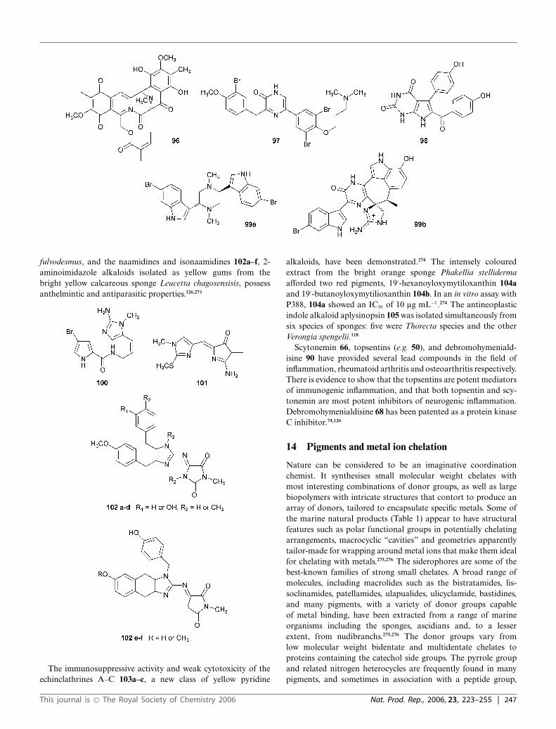
fulvodesmus, and the naamidines and isonaamidines 102a–f, 2-aminoimidazole alkaloids isolated as yellow gums from thebright yellow calcareous sponge Leucetta chagosensisis, possessanthelmintic and antiparasitic properties.126,273
The immunosuppressive activity and weak cytotoxicity of theechinclathrines A–C 103a–c, a new class of yellow pyridine
alkaloids, have been demonstrated.274 The intensely colouredextract from the bright orange sponge Phakellia stellidermaafforded two red pigments, 19′-hexanoyloxymytiloxanthin 104aand 19′-butanoyloxymytilioxanthin 104b. In an in vitro assay withP388, 104a showed an IC50 of 10 lg mL−1.274 The antineoplasticindole alkaloid aplysinopsin 105 was isolated simultaneously fromsix species of sponges: five were Thorecta species and the otherVerongia spengelii.118
Scytonemin 66, topsentins (e.g. 50), and debromohymeniald-isine 90 have provided several lead compounds in the field ofinflammation, rheumatoid arthritis and osteoarthritis respectively.There is evidence to show that the topsentins are potent mediatorsof immunogenic inflammation, and that both topsentin and scy-tonemin are most potent inhibitors of neurogenic inflammation.Debromohymenialdisine 68 has been patented as a protein kinaseC inhibitor.74,126
14 Pigments and metal ion chelation
Nature can be considered to be an imaginative coordinationchemist. It synthesises small molecular weight chelates withmost interesting combinations of donor groups, as well as largebiopolymers with intricate structures that contort to produce anarray of donors, tailored to encapsulate specific metals. Some ofthe marine natural products (Table 1) appear to have structuralfeatures such as polar functional groups in potentially chelatingarrangements, macrocyclic “cavities” and geometries apparentlytailor-made for wrapping around metal ions that make them idealfor chelating with metals.275,276 The siderophores are some of thebest-known families of strong small chelates. A broad range ofmolecules, including macrolides such as the bistratamides, lis-soclinamides, patellamides, ulapualides, ulicyclamide, bastidines,and many pigments, with a variety of donor groups capableof metal binding, have been extracted from a range of marineorganisms including the sponges, ascidians and, to a lesserextent, from nudibranchs.275,276 The donor groups vary fromlow molecular weight bidentate and multidentate chelates toproteins containing the catechol side groups. The pyrrole groupand related nitrogen heterocycles are frequently found in manypigments, and sometimes in association with a peptide group,
This journal is © The Royal Society of Chemistry 2006 Nat. Prod. Rep., 2006, 23, 223–255 | 247

for example, the deeply red-coloured pigments prodigiosin 106,metacycloprodigiosin 107 (containing a 4-methoxy-2,2′-bipyrrolemoiety), oroidin 108, tambjamines 79a–e, tetrapyrroles such as13, and the eudistomins (e.g. eudistomodin A 62a, eudistominB 109, and eudistomin C 62b). Eudistomins have the samecoordinating groups as the iron(II) chelator pyrimine isolated froma Pseudomonas species.276 The peptides and 3,4,5-trihydroxyphenylgroups are both found in celenamide E 110, and in tunichromes32a and 32b.
Adenochromines 26a–c have a particularly interesting array ofdonor groups with catechol, thioether, imidazole and amino acidgroupings.276 The thiazole ring is found in a range of aza-crowncyclic compounds, and the oxazolone ring in ulicyclamide andlissoclinamides. Other pigments such as 2-bromoleptoclinidinone38, ascididemin 43, tjipanazoles D 111 and J 112, shermilaminesA and B 40a+b, nordercitin 113, kuanoniamines A 37 and D 114,plankinidine D 45, anguibactin 115, dysidenin 116, pyrimine 117,clionamide 118, and bisimidazoles such as paragracin 119, all havethe potential to act as ligands.275,276
However, apart from the boron-containing aplasmomycins, e.g.aplasmomycin A 120 (which can also bind with alkali metals) froma marine microorganism, the tunichromes (powerful reducingagents towards vanadium(V) and iron(III)), Ni-containing eilatin
248 | Nat. Prod. Rep., 2006, 23, 223–255 This journal is © The Royal Society of Chemistry 2006

39, the blue-green pigment tunichlorin 121, the guanidine alkaloidclathridine 122 together with its zinc complex, geodiastatins A andB (brownish-black silicon-containing luminescent chromoproteinsfrom the sponge Geodia mesotriaena), the iron protein fromthe blood cells of the stolidobranch Pyura stolonifera,275,276 andthe ubiquitous magnesium-containing chlorophylls, examples ofmetal-containing complexes are rare.
In contrast, a large number of metal-free tetrapyrrole metabo-lites, including the pigments chlorophyllone A, bonellin 123,corallistins A–E 124a–e, and cyclopheophorbide enol 125 havebeen characterised. The cyclic peptide jasplakinolide (jaspamide)126 from Jaspis johnstoni, corallistine 101, naamidine A 102a,isonaamidine A 102b and pyronaamidine 127, structurally similarto clathridine (but devoid of the metal), have been identified fromdifferent sponges.89,90,275–277
These compounds are certainly capable of forming metal com-plexes. The apo-protein, ferreascidin, derived from the iron proteinfrom the blood cells of the stolidobranch Pyura stolonifera,276
complexes with iron to produce a blue pigment. The violetadenochromes, Fe3+-binding pigments from Octopus vulgaris, onremoval of the Fe3+ ion, produce the colourless adenochromines26a–c. These colourless desferri-adenochromes react with iron(III)chloride to form a blue precipitate. Both reactions appear to be pH-dependent, the reaction taking place at pH 4.5.34,114 It is well knownthat sea squirts assimilate metals such as vanadium, manganese,nickel and iron from sea-water. For example, the tunicate Ascidianigra accumulates vanadium to produce yellow pigments, thetunichromes, whereas Trididemnum solidum produces the nickel-containing blue-green pigment tunichlorin278 121, and Molgulamanhattensis sequesters iron.275 The Papua New Guinean shell-less mollusc Dolabella auricularia contains a series of green toblue-green chlorins (2,3-dihydroporphyrins) 11, one of which wasidentified as the nickel chelate tunichlorin, previously isolated fromT. solidum.278 Ascidians accumulate vanadium ions (up to 1 M)from sea-water, where vanadium is present in the +5 oxidationstate. In ascidian blood cells, however, the vanadium was foundto be in the +3 and/or +4 states. In Ascidia nigra, at least90% of the total vanadium is in the oxygen-sensitive +3 state.Some adenochromines from Ascidia nigra279 and T. solidum278
showed vanadium(III) (green) and vanadium(IV) (blue) oxidationlevels. These oxygen-sensitive pigments underwent pH-dependentreactions with vanadium(IV) and vanadium(V) to produce insol-uble green or green-black polynuclear complexes. During thisprocess vanadium vanadium(V) and vanadium(IV) were reducedto the +3 oxidation level.280,281 Eilatin 39, a 1,10-phenanthrolinederivative, complexes with nickel, ruthenium and osmium. Thenickel complex is blue, but the ruthenium complexes are green orred, depending on the ratio of ruthenium to eilatin. The fused-ring,pentacyclic, biologically active alkaloid 2-bromoleptoclinidone111, isolated from the Mediterranean ascidian Cystodytes del-lechiajei, possesses a bidentate chelating site which forms acomplex with ruthenium(II).275,282–284 Derivatives of patellamideA 128 reacts with copper(II) to yield compounds absorbing atwavelengths of 675 nm and 687 nm.286 The acyl tetramic acid,discodermide 129, a macrocyclic lactam from the deep-watersponges Discodermia dissoluta285 and D. polydiscus277 forms anorange-red complex with iron(III) chloride and a greenish-yellowcomplex with copper(III) acetate.275,285 The removal of Fe3+ resultsin loss of the absorption maximum with the formation of a paleyellow substance, which is capable of binding with Fe(III) andregenerating the original pigment. The known naamidines, novelzinc complexes of naamidine-type alkaloids A and G, and thefirst natural marine mixed-ligand zinc(II) metal complex 130 havebeen characterised from a calcareous sponge, Leucetta sp., of theCoral Sea. It is noteworthy that complexing with zinc did notbring about significant changes in the absorption maxima of theparent compounds, usually observed with other metabolite–metalchelates. The absorption maxima of the naamidines (kmax 385 nm),bis(naamidinato)zinc(II) complexes A:A (kmax 380 nm), G:G (kmax
386 nm), and A:G (kmax 390 and 373 nm) were very similar.287
In addition, the animals were snow-white to faintly brown inappearance.
Apart from common metal cations such as calcium, magnesium,potassium and sodium, (concentrations below 103 M), most other
This journal is © The Royal Society of Chemistry 2006 Nat. Prod. Rep., 2006, 23, 223–255 | 249
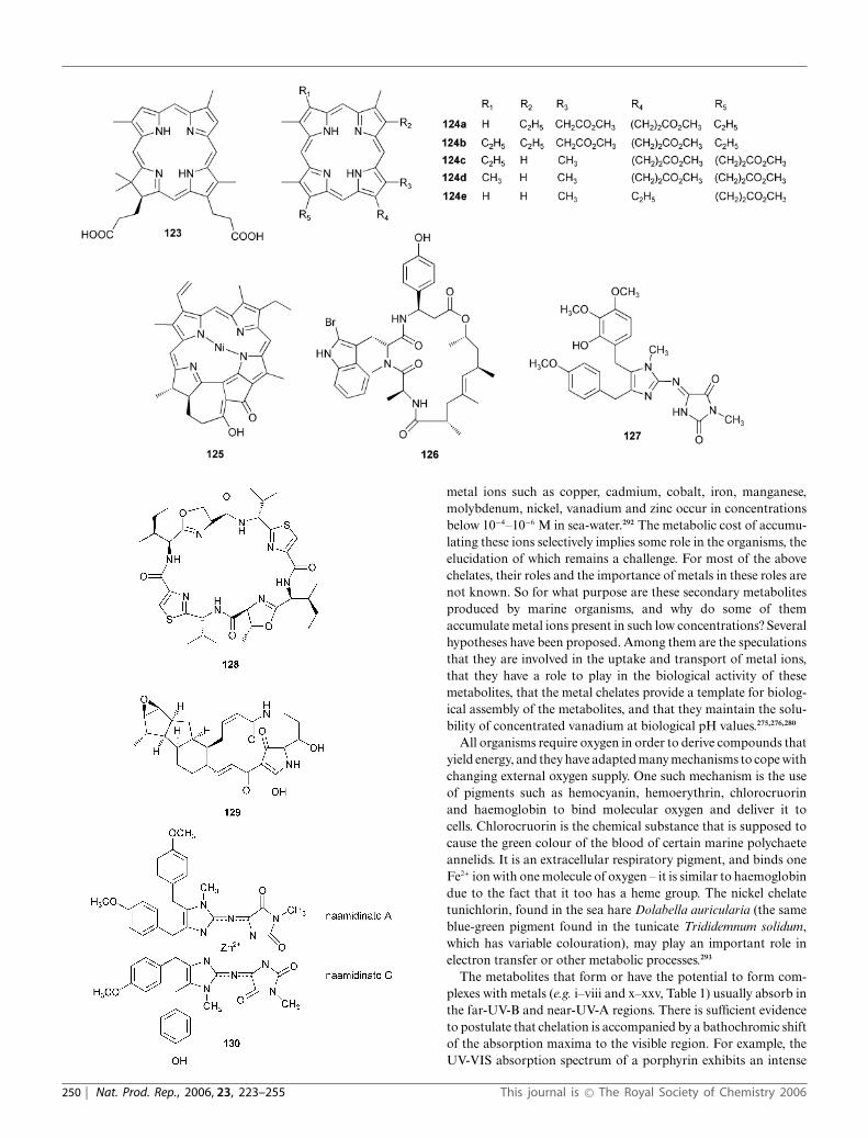
metal ions such as copper, cadmium, cobalt, iron, manganese,molybdenum, nickel, vanadium and zinc occur in concentrationsbelow 10−4–10−6 M in sea-water.292 The metabolic cost of accumu-lating these ions selectively implies some role in the organisms, theelucidation of which remains a challenge. For most of the abovechelates, their roles and the importance of metals in these roles arenot known. So for what purpose are these secondary metabolitesproduced by marine organisms, and why do some of themaccumulate metal ions present in such low concentrations? Severalhypotheses have been proposed. Among them are the speculationsthat they are involved in the uptake and transport of metal ions,that they have a role to play in the biological activity of thesemetabolites, that the metal chelates provide a template for biolog-ical assembly of the metabolites, and that they maintain the solu-bility of concentrated vanadium at biological pH values.275,276,280
All organisms require oxygen in order to derive compounds thatyield energy, and they have adapted many mechanisms to cope withchanging external oxygen supply. One such mechanism is the useof pigments such as hemocyanin, hemoerythrin, chlorocruorinand haemoglobin to bind molecular oxygen and deliver it tocells. Chlorocruorin is the chemical substance that is supposed tocause the green colour of the blood of certain marine polychaeteannelids. It is an extracellular respiratory pigment, and binds oneFe2+ ion with one molecule of oxygen – it is similar to haemoglobindue to the fact that it too has a heme group. The nickel chelatetunichlorin, found in the sea hare Dolabella auricularia (the sameblue-green pigment found in the tunicate Trididemnum solidum,which has variable colouration), may play an important role inelectron transfer or other metabolic processes.293
The metabolites that form or have the potential to form com-plexes with metals (e.g. i–viii and x–xxv, Table 1) usually absorb inthe far-UV-B and near-UV-A regions. There is sufficient evidenceto postulate that chelation is accompanied by a bathochromic shiftof the absorption maxima to the visible region. For example, theUV-VIS absorption spectrum of a porphyrin exhibits an intense
250 | Nat. Prod. Rep., 2006, 23, 223–255 This journal is © The Royal Society of Chemistry 2006

peak at about 420 nm followed by several weak peaks at longerwavelengths (from 500–650 nm). Reduction of one or more pyrrolerings and chelation with magnesium (as in chlorophyll-a) results ina strong absorption peak at approximately 650 nm.294 The yellowpigment tunichrome from the ascidian Ascidia nigra produces aninsoluble blue or green-black polymer complex with vanadium(indeed, some of these animals are blue or black in colour).
The colours of the corresponding organisms varied frombrilliant yellow to bright green and blue, shades of violet – some areeven black (Table 1). (However, these descriptive colours should betreated with caution, for marine invertebrates show variations incolour and morphology relative to the light regime of the habitat,depth, and the geographical location, etc.178). Is it then reasonableto postulate that the chelation of these “ligands” with metals bringsabout changes in the colour of the pigments (at least in some ofthe animals), that the colour of the organism is a reflection ofthe colour of the pigment–metal chelate, and that the chelation isdestroyed during the extraction/purification process?
15 Conclusion
The key to the existence of a species is its ability to survivelong enough to successfully reproduce and pass its genes on tothe next generation. During the evolution of animals, most ofthem developed defence mechanisms to improve their chances ofsurviving in a changing world. The accurate knowledge of factorsaffecting the survival of early life stages of marine invertebratesis critically important for the understanding of their populationdynamics and the evolution of their diverse reproductive andlife history characteristics. Chemical defence is an importantdeterminant of survival for larvae, juvenile as well as adult stages ofmany sessile benthic invertebrates, yet relatively little considerationhas been given to chemical defences at the early life stages.Information on the localisation of defensive secondary metabolitesin larvae and the fitness-related consequences of consuming evena small amount of toxic secondary metabolite underpin proposalsregarding the potential for Mullerian and Batesian mimicry tooccur among marine larvae.279
Coral reefs are among the oldest and largest living entities onearth, whose individual components live or die together, ratherlike a forest. Vast numbers of creatures live in them, including anestimated quarter of the world’s oceanic fish.295 There are manyamazing strategies, adaptations, and behaviours that marine plantsand animals have developed in order to be able to live and flourishin our greatest ecosystem, the ocean. From microscopic plants andanimals to some of the ocean’s behemoths such as the giant mantaray, these organisms reveal how each one fits into its own specialniche. The violent cuttlefish mating ritual ensures that only thestrongest will reproduce. Sea anemones might look beautiful tous but not to a shrimp captured by thousands of their miniatureharpoons, injected with poison and slowly devoured. Sea haresmight appear dull in comparison to their fellow sea slugs, themore flamboyant nudibranchs, but they can make huge matingchains, with each individual acting as male and female to differentneighbours. Sea hares live no more than a year, but what a year!Females of the humble-looking slipper limpet secrete a substanceinto the water that maintains the masculinity of nearby males. Ason land, the shape of a plant or animal will determine how andwhere they fit into the larger picture. In the ocean, as on land,
some creatures are built for speed, while others seldom move, orare in fact sessile. Others are still at the mercy of wind and currentfor transportation, and this exquisitely complex ecosystem is veryvulnerable to many environmental factors.
Our knowledge about the role and importance of colourationin marine organisms is in its infancy. The colour characteristicsof organisms serve various purposes and are the result of severaldifferent processes. It is likely that there is no one single reason whymarine invertebrates are coloured. It may be that the colours couldbe clues to physiological processes, developmental conditions,environmental interactions etc., or a combination of these factors.The number of alkaloidal pigments from marine organismsmentioned in this article is only a “drop in the ocean” comparedwith what is reported in the literature.74,113,126,236,239,261,275,277,296–300
These organisms produce a wide array of nitrogenous compoundswith diverse structures, yet very little is known of their roles inthese organisms.
Mimicry is a great example of evolution by natural selection,and has been celebrated for this ever since. But there are still manypuzzling aspects of mimicry that need to be studied – for example,the molecular nature of the genes involved, and whether randomdrift may initiate some of these changes. Carefully controlledfield and laboratory studies on aposematism are needed beforegeneral inferences can be adequately evaluated. Nevertheless, theexamples that are presently known are intriguing and stronglysuggestive that crypsis, aposematic colouration (both Batesianand Mullerian), are effective defensive strategies in the marineenvironment. In order to assess the functional role of pigments,bioassays that model ecological interactions, such as predator–prey and other survival-related relationships, must be employed.Opisthobranch molluscs, by virtue of the fact that they containdiverse and widespread toxins and exhibit an array of distinctand vibrant colour patterns, are ideal organisms for further studyand experimentation. Studies on adaptive colouration amonginvertebrates could show important differences in protectivemechanisms between and within taxa as well as between land,freshwater and marine habitats. With the coupling of innovativeanalytical techniques with physical methods (such as nuclearmagnetic resonance, mass spectrometry and X-ray crystallographywith tools of modern genetics and mechanistic biology) put to theservice of identifying complex pigments and metabolites, prepar-ing and isolating pigment–metal complexes, finding evidence formetal–metabolite interactions and carrying out adaptive studiesof colour pattern variation,296 we can look forward to a newera of understanding of this venerable aspect of evolutionarybiology.
16 References
1 G. Y. Kennedy, Adv. Mar. Biol., 1979, 16, 309.2 J. E. N. Veron, Corals of Australia and the Indo-Pacific, University of
Hawaii Press, Honolulu, 1986.3 J. E. N. Veron, Corals of the World, vols. 1–3, ed. M. Stafford-Smith,
Australian Institute of Marine Science, Queensland, Australia, 2000.4 H. Rossotti, Colour, Princeton University Press, Princeton, New
Jersey, 1985.5 D. L. Fox, Comp. Biochem. Physiol., B, 1976, 55, 137.6 D. L. Fox, Animal biochromes and structural colors (2nd edn),
University of California Press, Berkeley, 1976.7 A. E. Needham, The significance of zoochromes, Springer-Verlag, New
York, 1974..
This journal is © The Royal Society of Chemistry 2006 Nat. Prod. Rep., 2006, 23, 223–255 | 251

8 V. Pearse, J. Pearse, M. Buchsbaum and R. Buchsbaum, Livinginvertebrates, Blackwell Science Publications, Palo Alto, California,1987.
9 T. W. Goodwin, The Comparative Biochemistry of the Carotenoids,Chapman and Hall, London, 1952.
10 T. W. Goodwin, in Chemical Zoology, ed. M. Florkin and B. T. Scheer,Academic Press, New York/London, 1968, vol. 11, p. 149.
11 T. W. Goodwin, in Chemical Zoology, ed. M. Florkin and B. T. Scheer,Academic Press, New York/London, 1972, ch. 8.
12 G. R. Pettit, J. A. Rideout and J. A. Hasler, J. Nat. Prod., 1981, 44,588.
13 J. L. Sumich, in An Introduction to the Biology of Marine Life, ed.J. L. Sumich and I. Dubuque, Wm. C. Brown Publishers, 1980,p. 126.
14 I. V. Kelmanson and M. V. Matz, Mol. Biol. Evol., 2003, 20, 1133.15 P. G. Falkowski and Z. Dubinsky, Nature, 1981, 289, 172.16 R. J. Blank and R. K. Trench, Science, 1985, 229, 656.17 K. Rutzler and I. G. Macintyre, Contrib. Mar. Sci., 1982, 12, 9.18 T. A. Stephenson, Endeavour, 1947, 6, 152.19 V. J. Paul and M. P. Puglisi, Nat. Prod. Rep., 2004, 21, 189.20 A. Kelecom, An. Acad. Bras. Cienc., 2002, 74, 1.21 M. K. Wicksten, in ‘Adaptive coloration in invertebrates’, Proc. Symp.
Am. Soc. Zool., ed. M. K. Wicksten, Galveston, TX, USA, 1990,vol. 127.
22 W. M. Bandaranayake and A. Des Rocher, Mar. Biol., 1999, 133, 163.23 W. M. Bandaranayake, Nat. Prod. Rep., 1998, 15, 159.24 W. M. Bandaranayake and W. A. Wickramasinghe, Comp. Biochem.
Physiol., B, 1996, 113, 499.25 W. M. Bandaranayake, Trends Comp. Biochem. Physiol., 2002, 9, 229.26 J. B. McClintock, B. J. Baker and D. K. Steinberg, in Marine Chemical
Ecology, ed. J. B. McClintock and B. J. Baker, CRC Press, New York,2001, p. 195.
27 D. L. Fox and C. F. A. Pantin, Biol. Rev., 1944, 19, 121.28 P. Proksch, Toxicon, 1994, 32, 639.29 P. R. Bergquist, Sponges, Hutchinson and Co., London, 1978.30 R. H. Thompson, Chem. Br., 1978, 14, 133.31 R. A. de Reaumur, Mem. Acad. R. Sci., 1771, 118.32 P. Friedlander, Monatsh. Chem., 1907, 28, 991.33 P. Friedlander, Ber. Dtsch. Chem. Ges., 1909, 42, 765.34 G. Prota, in Marine Natural Products: Chemical and Biological
Perspectives, ed. P. J. Scheuer, Academic Press, New York, 1980,p. 141.
35 T. Goto, in Marine Natural Products: Chemical and BiologicalPerspectives, ed. P. Scheuer, Academic Press, New York, 1980, p. 179.
36 M. Fontaine, C. R. Hebd. Seances Acad. Sci., 1934, 180, 1369.37 V. Tartar, The Biology of Stentor, Pergamon Press, Oxford, 1961.38 A. C. Giese and R. M. Grainger, Photochem. Photobiol., 1970, 12,
489.39 E. Faure-Fremiet, Biol. Bull. (Woods Hole, MA, U. S.), 1936, 70, 353.40 A. R. Fontane, J. Mar. Biol. Assoc. U. K., 1962, 42, 9.41 C. A. MacMunn, Q. J. Microsc. Sci., 1890, 30, 51; C. A. MacMunn,
J. Physiol., 1899, 24, 1.42 M. I. Newbigin, Color in Nature, John Murray, London, 1898.43 M. Abeloos-Parize and R. Abeloos-Parize, C. R. Seances Soc. Biol.
Ses. Fil., 1926, 94, 560.44 J. Verne, Couleurs et Pigments des etres vivants, Armand Colin, Paris,
1930.45 E. Lederer, C. R. Seances Soc. Biol. Ses. Fil., 1933, 113, 1391.46 D. F. Cheesman, W. L. Lee and P. F. Zagalsky, Biol. Rev., 1967, 42,
132.47 B. C. L. Weedon, in Carotenoids: Occurrence of Carotenoids, ed.
O. Osler, Birkhauser Verlag, Basel/Stuttgart, 1971.48 G. Y. Kennedy, in Chemical Zoology, ed. M. Florkin and B. T. Scheer,
Academic Press, New York, 1969, ch. 10.49 A. Comfort, Biol. Rev., 1951, 26, 285.50 D. L. Fox and H. G. Vevers, The Nature of Animal Colors, Sidgwick
& Jackson, London, 1960.51 B. M. Gilchrist, Comp. Biochem. Physiol., 1968, 24, 123.52 D. L. Fox, in Biochemical and Biophysical Perspectives in Marine
Biology, ed. J. R. Sargent and D. Malins, Academic Press, NewYork/London, 1974, p. 1.
53 C. F. W. Krukenberg, Die Pigmente: ihre Eigenschaften, ihre Geneseund ihre Metamorphosen bei wirbellosen Tieren, Vergleichende physi-ologische Studien, Heidelberg, 1882, p. 92.
54 P. J. Herring, Comp. Biochem. Physiol., B, 1971, 39, 739.
55 P. F. Zagalsky, F. Haxo, S. Hertzberg and S. Liaaen-Jensen, Comp.Biochem. Physiol., B, 1989, 93, 339.
56 I. M. Heilbron, H. Jackson and R. N. Jones, Biochem. J., 1935, 29,1384.
57 D. L. Fox and C. F. A. Pantin, Philos. Trans. R. Soc. London, Ser. B,1941, 230, 415.
58 J. Roche, Bull. Soc. Chim. Biol., 1936, 18, 825.59 H. N. Moseley, Q. J. Microsc. Sci., 1877, 17, 1.60 P. J. Herring, Nature, 1972, 238, 276.61 G. Y. Kennedy and H. G. Vevers, Comp. Biochem. Physiol., B, 1976,
55, 117.62 H. C. Sorby, Q. J. Microsc. Sci., 1875, 15, 166.63 A. Pelter, J. A. Ballantine, V. Ferrito, V. Jaccarini, A. P. Psaila and P. J.
Schembri, J. Chem. Soc., Chem. Commun., 1976, 999.64 J. Cote, C. R. Seances Soc. Biol. Ses. Fil., 1903, 55, 137.65 D. L. Fox and N. Millott, Proc. R. Soc. London, Ser. B, 1954, 142, 392.66 R. Tixier, Ann. Inst. Oceanogr. (Monaco), 1945, 22, 343.67 M. Abeloos and G. Teissier, Bull. Soc. Zool. Fr., 1926, 51, 145.68 E. Lederer, Biol. Rev., 1940, 15, 273.69 G. Prota, S. Ito and G. Nardi, in Marine Natural Products Chemistry,
ed. J. Faulkner and W. H. Fenical, Plenum, New York, 1977, p. 45.70 E. Beerstechner, Vitam. Horm., 1950, 10, 69.71 S. Yamamura and Y. Hirata, Tetrahedron, 1963, 19, 1485.72 K. D. Barrow, in Marine Natural Products, ed. P. J. Scheuer, 1983,
p. 51.73 M. J. Garson, in Marine Chemical Ecology, ed. J. B. McClintock and
B. J. Baker, CRC Press, Boca Raton/London/New York, 2001, p. 71.74 D. J. Faulkner, Nat. Prod. Rep., 1991, 8, 97 (and references cited
therein).75 O. F. Smetanina, L. S. Schevchenko, T. A. Kuznetsova, V. V. Mikhailov
and G. B. Elyakov, in ‘Bridges of Science between North America andRussian Far East’, 45th Arctic Science Conference, Anchorage, AK,USA, 1994, paper no. 299.
76 J. Shin, Y. Seo, K. W. Cho, J. R. Rho and C. J. Sim, J. Nat. Prod., 1999,62, 647.
77 T. W. Goodwin, The Biochemistry of Carotenoids, Chapman Hall, NewYork, 1980, vol. 2.
78 H. Pfander, Key to Carotenoids, Birkhauser, Basel, 1987.79 S. Liaaen-Jensen, New J. Chem., 1990, 14, 747.80 T. Matsuno, M. Ookubo, T. Nishizawa and I. Shimizu, Chem. Pharm.
Bull., 1984, 32, 4309.81 T. Matsuno and M. Tsushima, Comp. Biochem. Physiol., B, 1995, 111,
597.82 A. Kitamura, J. Tanaka and T. Higa, J. Nat. Toxins, 1996, 5, 219.83 P. S. Parameswaran and C. T. Achuthankutty, Proc. Andhra Pradesh
Akad. Sci., 2005, 9, 135.84 T. Bjornland, A. Fiksdahl, T. Skjetne, J. Krane and S. Liaaen-Jensen,
Tetrahedron, 2000, 56, 9047.85 M. H. Gorden, Nat. Prod. Rep., 1996, 13, 265.86 T. Kawakami, M. Tsushima, Y. Katambi, M. Mine, A. Ishida and T.
Matsuno, J. Exp. Mar. Biol. Ecol., 1998, 226, 165.87 G. Y. Kennedy, Ann. N. Y. Acad. Sci., 1975, 244, 662.88 P. Karuso and P. J. Scheuer, Molecules, 2002, 7, 1.89 M. D’Ambrosio, A. Guerriero, C. Debitus, O. Ribes, B. Richer de
Forges and F. Pietra, Helv. Chim. Acta, 1989, 72, 1451.90 M. D’Ambrosio, A. Guerriero, C. Debitus, O. Ribes and F. Pietra,
Helv. Chim. Acta, 1993, 76, 1489.91 R. A. Nicolaus, Melanins, Hermann, Paris, 1968.92 R. A. Nicolaus, in Methodicum chimicum (Part 3), ed. F. Korte,
Academic Press, New York, 1978, p. 190.93 G. Prota, Med. Res. Rev., 1988, 8.94 D. L. Fox and T. S. Hopkins, in Physiology of Echinodermata,
ed. R. A. Boolootian, Interscience Publishers, New York, 1966,p. 277.
95 H. S. Mason, in Pigment cell growth, ed. M. Gordon, Academic Press,New York, 1953, p. 207.
96 M. Piattelli and R. A. Nicolaus, Tetrahedron, 1961, 15, 66.97 R. H. Thomson, Naturally Occurring Quinones, Chapman and Hall,
London, 1987.98 I. R. Smith and M. D. Sutherland, Aust. J. Chem., 1971, 24, 1487.99 G. Cimino, S. De Stefano and L. Minale, Comp. Biochem. Physiol., B,
1974, 47, 895.100 H. Singh, R. E. Moore and P. G. Scheuer, Experientia, 1967, 23, 624.101 K. A. Francesconi, Aust. J. Chem., 1980, 33, 2781.102 J. A. Rideout and M. D. Sutherland, Aust. J. Chem., 1985, 38, 793.
252 | Nat. Prod. Rep., 2006, 23, 223–255 This journal is © The Royal Society of Chemistry 2006

103 J. A. Rideout, N. B. Smith and M. D. Sutherland, Experientia, 1979,35, 1273.
104 D. M. Balitz, J. A. Bush, J. A. and W. T. Bradner, J. Antibiot., 1982,35, 259.
105 G. Cimino and G. Sodano, in Sponges in Space and Time, ed.R. W. M. Van Soest, T. M. G. Van Kempen and J. S. Breakman,Balkema, Rotterdam, 1994, p. 459.
106 L. Cariello, M. De Nicola Giudici and L. Zanetti, Comp. Biochem.Physiol., C, 1980, 65, 37.
107 Y. C. Shen, C. H. Lu, R. Chakraborty and Y. H. Kuo, Nat. Prod. Res.,2003, 17, 83.
108 G. Cimino, S. De Rosa and S. De Stefano, Experientia, 1982, 38, 896.109 D. M. Roll, P. J. Scheuer, G. K. Matsumoto and J. Clardy, J. Am.
Chem. Soc., 1983, 105, 6177.110 T. Higa, T. Fujiyama and P. J. Scheuer, Comp. Biochem. Physiol., B,
1980, 65, 525.111 C. J. Smith, R. L. Hettich, J. Jompa, A. Tahir, M. V. Buchanan and
C. M. Ireland, J. Org. Chem., 1998, 63, 4147.112 J. A. Rideout and M. D. Sutherland, Aust. J. Chem., 1981, 34, 2385.113 D. Laurent and F. Pietra, Chem. Biodiversity, 2004, 1, 539.114 C. Christophersen and U. Anthoni, Sulfur Rep., 1986, 4, 365 (and
references cited therein).115 Y. Seo, J. R. Rho, N. Geum, J. B. Yoon and J. Shin, J. Nat. Prod., 1996,
59, 985.116 D. A. Webb, J. Exp. Biol., 1939, 16, 499.117 I. G. Macara, G. C. McLeod and K. Kustin, Biochem. J., 1979, 181,
457.118 C. Christophersen, in Marine Natural Products: Chemical and Biolog-
ical Perspectives, ed. P. J. Scheuer, Academic Press, New York, 1983,p. 259.
119 J. T. Baker, Endeavour, 1974, 33, 11.120 L. Carriello, S. Crescenzi, G. Prota, F. Giordano and L. Mazzarella,
J. Chem. Soc., Chem. Commun., 1973, 99.121 L. Carriello, S. Crescenzi, G. Prota and L. Zanetti, Tetrahedron, 1974,
30, 4191.122 L. Carriello, M. De Nicola-Giudici, L. Zanetti and G. Prota, Proc.
Congr. Soc. Ital. Biol. Mar., 1977, 9, 171.123 L. Carriello, M. De Nicola-Giudici, S. Crescenzi and G. Prota,
Experientia, 1978, 34, 1460.124 C. Jimenez and P. Crews, J. Nat. Prod., 1993, 56, 9.125 H. Hoffmann and T. Lindel, Synthesis, 2003, 1753.126 D. J. Faulkner, Nat. Prod. Rep., 2002, 19, 1 (and references cited
therein).127 T. F. Molinski, Chem. Rev., 1993, 93, 1825.128 K. M. Marshall and L. R. Barrows, Nat. Prod. Rep., 2004, 21, 731.129 A. R. Carrol, B. F. Bowden and J. C. Coll, Aust. J. Chem., 1993, 46,
489.130 S. Urban, L. Hobbs, J. N. A. Hooper and R. J. Capon, Aust. J. Chem.,
1995, 48, 1491.131 P. W. Ford and B. S. Davidson, J. Nat. Prod., 1997, 60, 1051.132 H. C. Vervoort, W. Fenical and P. A. Keifer, J. Nat. Prod., 1999, 62,
389.133 M. Gompel, M. Leost, E. B. De, K. Joffe, L. Puricelli, L. H. Franco,
J. Palermo and L. Meijer, Bioorg. Med. Chem. Lett., 2004, 14, 1703.134 W. M. Bandaranayake, AIMS Rep., no. 18, 1994.135 H. W. Lam, P. A. Cooke, G. Pattenden, W. M. Bandaranayake and W.
Wickramasinghe, J. Chem. Soc., Perkin Trans., 1999, 847.136 W. M. Bandaranayake, G. Pattenden and W. A. Wickramasinghe,
Trends Comp. Biochem. Physiol., 2002, 9, 205.137 A. Zampella, M. V. D’Auria, C. Debitus and J. L. Menou, J. Nat.
Prod., 2000, 63, 943.138 J. H. Kwak, F. J. Schmitz and M. Kelly, J. Nat. Prod., 2000, 63, 1153.139 A. Marin, M. D. Lopez Belluga, G. Scognamiglio and G. Cimino,
J. Molluscan Stud., 1997, 63, 431.140 C. Jimenez, E. Quinoa and M. Adamczeski, J. Org. Chem., 1991, 56,
3403.141 T. Shindo, A. Sato, N. Kasanuki, K. Hasegawa, S. Sato, T. Iwata and
T. Hata, Experientia, 1993, 49, 177.142 S. H. D. Haddock, C. W. Dunn, P. R. Pugh and C. E. Schnitzler,
Science, 2005, 309, 263 (and references cited therein).143 B. H. Robinson, J. Mar. Biol. Assoc. U. K., 1992, 72, 463.144 A. K. Campbell and P. J. Herring, Mar. Biol., 1990, 104, 219.145 F. H. Johnson and O. Shimomura, in Methods in Enzymology,
ed. M. A. DeLuca, Academic Press, New York, 1978, vol. LVII,p. 331.
146 A. Salih, A. Larkum, G. Cox, M. Kuhl and O. Hoegh-Guldberg,Nature, 2000, 408, 850.
147 C. H. Mazel, T. W. Cronin, R. L. Caldwell and N. J. Marshall, Science,2004, 303, 51.
148 K. Edlinger, Malacologia, 1982, 22, 1.149 I. Bravo, J. M. Franco, A. Alonso, R. Dietrich and P. Molist, Mar.
Biol., 2001, 138, 709.150 M. Tsuda, D. Sakurai and M. Goda, J. Exp. Biol., 2003, 206, 1409.151 H. Takata and T. Kominami, Zool. Sci., 2002, 19, 1450.152 K. L. Gosner, A field guide to the Atlantic seashore, Houghton Mifflin,
Boston, 1979.153 E. C. Haderlie, C. Hand and W. G. Gladfelter, in Intertidal invertebrates
of California, ed. R. H. Morris, D. P. Abbott and E. C. Haderlie,Stanford University Press, Stanford, 1980, p. 40.
154 G. J. Bakus and D. P. Abbott, in Intertidal invertebrates of California,ed. R. H. Morris, D. P. Abbott and E. C. Haderlie, Stanford UniversityPress, Stanford, 1980, p. 21.
155 M. Kimura, Sci. Am., 1979, 24, 98.156 E. Savazzi, Lethaia, 1998, 31, 15.157 F. Garcia-Pichel, N. D. Sherry and R. W. Castenholz, Appl. Environ.
Microbiol., 1993, 59, 170.158 C. S. Cockell and J. Knowland, Biol. Rev., 1999, 74, 311.159 D. Karentz, in Marine Chemical Ecology, ed. J. B. McClintock and
B. J. Baker, CRC Press, New York, 2001, p. 481.160 W. C. Dunlap and J. M. Shick, J. Phycol., 1998, 34, 418.161 C. Nageli and S. Schwenderer, Das Mikroskop, W. Englemann,
Leipzig, 1877.162 F. Garcia-Pichel and R. W. Castenholz, J. Phycol., 1991, 27, 395.163 F. Garcia-Pichel, F. Wingard and R. W. Castenholz, Photochem.
Photobiol., 1992, 56, 17.164 P. J. Proteau, W. H. Gerwick, F. Garcia-Pichel and R. W. Castenholz,
Experientia, 1993, 49, 825.165 J. W. Porter and N. M. Targett, Biol. Bull., 1988, 175, 230.166 N. I. Krinsky, Pure Appl. Chem., 1979, 51, 649.167 A. J. Young, Physiol. Plant., 1991, 83, 702.168 B. Demmig-Adams and W. W. Adams, Annu. Rev. Plant Physiol. Plant
Mol. Biol., 1992, 43, 599.169 A. D. Patil, A. J. Freyer, L. Killmer, G. Hofmann and R. K. Johnson,
Nat. Prod. Lett., 1997, 9, 201.170 I. Kitagawa, M. Kobayashi, K. Kitanaka, M. Kido and Y. Kyogoku,
Chem. Pharm. Bull., 1983, 31, 2321.171 N. Lindquist, E. Lobkovsky and J. Clardy, Tetrahedron Lett., 1996,
9131.172 N. L. Lindquist, US Pat. 5705146, 1998.173 N. Lindquist, M. E. Hay and W. Fenical, Ecol. Monogr., 1992, 62, 547.174 W. M. Bandaranayake, 2005, unpublished results.175 R. Riedl, Eine Monographie, Paul Farey, Hamburg, 1966.176 G. L. Voss, Seashore life of Florida and the Caribbean, E. A. Seemann,
Miami, Florida, 1976.177 E. A. Titlyanov, M. G. Shaposhnikova and V. I. Zvalinsky, Photosyn-
thetica, 1980, 14, 413.178 R. R. Olson, Biol. Bull., 1986, 170, 62.179 D. J. Barnes and B. E. Chalker, in Ecosystems of the World: Coral
Reefs, ed. Z. Dubinsky, Elsevier, New York, 1990, p. 109.180 B. E. Brown, in Advances in Marine biology, ed. J. H. S. Blaxter and
A. J. Southward, Academic Press, London/Sydney/New York, 1997,vol. 31.
181 D. Schlichter and H. W. Fricke, Naturwissenschaften, 1990, 77, 447.182 P. Kaiser, D. Schlichter and H. W. Fricke, Mar. Biol., 1993, 117, 45.183 C. L. Thurman, ‘Adaptive coloration in invertebrates’, Proc. Symp.
Am. Soc. Zool., ed. M. K. Wicksten, Galveston, TX, USA, 1990,p. 109.
184 D. L. Zmarzly, in ‘Adaptive coloration in invertebrates’, Proc. Symp.Am. Soc. Zool., ed. M. K. Wicksten, Galveston, TX, USA, 1990,pp. 69–76.
185 J. M. Cervino, R. L. Hayes, M. Honovich, T. J. Goreau, S. Jones andP. J. Rubec, Mar. Pollut. Bull., 2003, 46(5), 573.
186 R. A. Cloney and E. Florey, Z. Zellforsch., 1968, 89, 250.187 J. A. Mather and D. L. Mather, J. Zool., 2004, 263, 1, 89.188 C. R. Darwin, The Origin of Species, Modern Library, 1859.189 C. Haerlin and M. Haerlin, Evol. Ecol., 2003, 17(1), 197.190 C. S. Loban and P. J. Harrison, Seaweed Ecology and Physiology,
Cambridge University Press, Cambridge, 1994.191 T. Guilford and J. Cuthill, Evolution, 1991, 45, 449.192 B. Chanas and J. R. Pawlik, Mar. Ecol.: Prog. Ser., 1995, 127, 195.
This journal is © The Royal Society of Chemistry 2006 Nat. Prod. Rep., 2006, 23, 223–255 | 253

193 G. Gamberale and S. Tullberg, Proc. R. Soc. London, 1998, 265, 889.194 J. A. Endler and J. Mappes, Am. Nat., 2004, 163(4), 532.195 G. Cimino, A. Spinella and G. Sodano, Tetrahedron Lett., 1989, 30,
3589.196 P. Proksch, Toxicon, 1994, 32, 639.197 G. J. Bakus, Science, 1981, 211, 497.198 G. J. Bakus, Mar. Biol., 1968, 2, 23.199 M. S. Grober, in ‘Adaptive coloration in invertebrates’, ed.
M. K. Wicksten, Proc. Symp. Am. Soc. Zool., ed. M. K. Wicksten,Galveston, TX, USA, 1990, p. 77.
200 T. Guilford and I. Cuthill, Evolution, 1991, 45, 449.201 G. Rosenberg, Evolution, 1991, 45, 451.202 T. M. Gosliner and D. W. Behrens, in ‘Adaptive coloration in
invertebrates’, ed. M. K. Wicksten, Proc. Symp. Am. Soc. Zool., ed.M. K. Wicksten, Galveston, TX, USA, 1990, pp. 127–145.
203 D. J. Faulkner and M. T. Ghiselin, Mar. Ecol.: Prog. Ser., 1983, 13,295.
204 D. Mebs, J. Chem. Ecol., 1985, 11, 713.205 D. P. Pisut and J. R. Pawlik, J. Exp. Mar. Biol. Ecol., 2002, 270, 203.206 J. B. McClintock, D. Swenson, H. Trapido-Rosenthal and L. Bang-
hart, J. Chem. Ecol., 1997, 23, 1607.207 C. M. Young and B. L. Bingham, Mar. Biol., 1987, 96, 539.208 J. B. McClintock and J. Vernon, Mar. Biol., 1990, 105, 491.209 R. Hanlon, Natl. Geogr., 2004, 206(2), 30.210 A. Ekendahl, J. Exp. Mar. Biol. Ecol., 1998, 222, 239.211 R. Bedini, M. G. Canali and A. Bedini, A, Cah. Biol. Mar., 2003, 4,
375.212 S. Merilaita, Biol. Sci., 1998, 265(1401), 1059.213 G. D. Ruxton, M. P. Speed and D. J. Kelly, Anim. Behav., 2004, 68(3),
445.214 W. M. Bandaranayake and P. Gentien, Comp. Biochem. Physiol., B,
1982, 72, 409.215 S. Johnsen, Biol. Bull., 2001, 201, 301.216 M. J. McFall-Ngai, Am. Zool., 1990, 30, 175.217 B. W. Jones and M. K. Nishiguchi, Mar. Biol., 2004, 144, 1151.218 A. Barbosa, C. F. Florio, C. Chiao and R. T. Hanlon, Bull. Mar. Biol.,
2004, 207, 154.219 C. Chiao, E. J. Kelman and R. T. Hanlon, Bull. Mar. Biol., 2004, 207,
153.220 C. Chiao, E. J. Kelman and R. T. Hanlon, Bull. Mar. Biol., 2005, 208,
7.221 L. Litman and R. T. Hanlon, Bull. Mar. Biol., 2004, 207, 154.222 M. Norman, Nature Aust., 2002, 27, 38.223 P. H. Kelly and C. T. Swan, J. Paleontol., 1988, 61, 83.224 M. Cusack, G. B. Curry, H. Clegg, G. Abbott and H. Armstrong,
Philos. Trans. R. Soc. London, Ser. B, 1991, 333, 359.225 M. Cusack, G. B. Curry, H. Clegg, G. Abbott and H. Armstrong,
Comp. Biochem. Physiol., B, 1992, 102, 93.226 M. James, A. D. Ansell and G. B. Curry, Mar. Biol., 1991, 111, 411.227 C. W. Thayer and R. A. Allmon, in Brachiopods Through Time, ed.
D. I. Mackinnon, D. E. Lee and J. D. Campbell, Balkema, Rotterdam,1991, p. 253.
228 S. C. McKiliup and R. V. McKiliup, Biol. J. Linn. Soc., 2002, 77, 367.229 O. G. Davidson, Enchanted Braid: Coming to terms with Nature on the
Coral Reefs, John Wiley and Sons, New York, 1998, p. 85.230 D. Owen, Survival of the wild: Camouflage and mimicry, University of
Chicago Press, Chicago, 1980, pp. 146–149.231 M. Ferrari, Colors for survival: Mimicry, Thomasson-Grant Publish-
ing, Charlottesville, VA, 1992, p. 144.232 A. S. Bull, Creatures, corals and colors in American seas, Audubon
Park Press, New Orleans, LA, 1990.233 M. Joron and J. Mallet, Trends Ecol. Evol., 1998, 13, 461.234 J. Mallet and M. Joron, Annu. Rev. Ecol. Syst., 1999, 30, 201.235 T. E. Reimchen, Biol. J. Linn. Soc., 1989, 36, 97.236 D. J. Faulkner, Nat. Prod. Rep., 1983, 9, 323 (and references cited
therein).237 J. R. Pawlik, Chem. Rev., 1993, 93, 1911.238 D. J. Faulkner, in Ecological Roles of Marine Natural Products, ed.
V. J. Paul, Ithaca, Comstock, 1992, p. 119.239 C. D. Amsler, K. B. Iken, J. B. McClintock and B. J. Baker, in Marine
Chemical Ecology, ed. J. B. McClintock and B. J. Baker, CRC Press,New York, 2001, p. 195.
240 J. J. Stachowicz, in Marine Chemical Ecology, ed. J. B. McClintock,B. J. Baker, CRC Press, Boca Raton/London/New York, 2001,p. 157.
241 J. McClintock, Those Amazing Sponges (online), retreived 1st Septem-ber 2005 from www.antarctica.uab.edu/Templates/Journal.aspx/pid=39, The University of Alabama in Antarctica, 2004.
242 J. B. McClintock, C. D. Amsler, B. J. Baker and R. W. M. van Soest,Integr. Comp. Biol., 2005, 45, 359.
243 C. D. Amsler, J. B. McClintock and B. J. Baker, Am. Zool., 2001, 41,17 (and references cited therein).
244 F. B. Furrow, C. D. Amsler, C. D. McClintock and B. J. Baker, Mar.Biol., 2003, 143, 443.
245 N. B. Perry, L. Ettouati, M. Litaudon, J. W. Blunt, M. G. H. Munro,S. Parkin and H. Hope, Tetrahedron, 1994, 50, 3987.
246 A. Yang, B. J. Baker, J. E. Grimwade, A. C. Leonard and J. B.McClintock, J. Nat. Prod., 1995, 58, 1596.
247 A. D. Davis and A. L. Butler, J. Exp. Mar. Biol. Ecol., 1989, 127, 189.248 G. Cimino, A. Spinella and G. Sodano, Tetrahedron Lett., 1989, 30,
3589.249 P. F. Sale, Oceanogr. Mar. Biol., 1980, 18, 367.250 P. Karuso, in Bioorganic and Marine Chemistry, ed. P. J. Scheuer,
Springer-Verlag, Berlin, 1985, p. 31.251 M. Ochi, K. Kataoka, A. Tatsukawa, H. Kotsuki and K. Shibata,
Chem. Lett., 1993, 2003.252 V. M. Dembitsky, T. A. Gloriozova and V. V. Poroikov, Mini-Rev.
Med. Chem., 2005, 5, 319.253 K. R. Gustafson, N. Oku and D. J. Milanowski, Curr. Med. Chem.,
2004, 3, 233.254 D. M. Roll, C. M. Ireland, H. S. M. Lu and G. B. Elyakov, J. Org.
Chem., 1988, 53, 3276.255 P. A. Horton, R. E. Longley, O. J. McConnell and L. M. Ballas,
Experientia, 1994, 50, 843.256 H. Kang and W. Fenical, Tetrahedron Lett., 1996, 37, 2369.257 S. A. Abas, M. B. Hossain, D. Van der Helm, F. J. Schmitz, M. Laney,
R. Cabuslay, R. and R. C. Schatzman, J. Org. Chem., 1996, 61, 2709.258 Z. Ding, K. Chichak and J. W. Lown, Curr. Med. Chem., 1999, 6, 1.259 P. Ivanova, L. S. Shevchenko, T. Sawabe, A. M. Lysenko, V. I.
Svetashev, N. M. Gorshkova, M. Satomi, R. Christen and V. V.Mikhailov, Int. J. Syst. Evol. Microbiol., 2002, 52, 263.
260 A. Loukaci and M. Guyot, Tetrahedron Lett., 1994, 35, 6869.261 U. Pindur and T. Lamster, Curr. Med. Chem., 2001, 8, 1681.262 R. G. S. Bernlinck, R. Britton, E. Piers, L. Lim, M. Roberge, R. M.
da Rocha and R. J. Anderson, J. Org. Chem., 1998, 63, 9850.263 H. C. Vervoort, S. E. Richard-Gross, W. Fenical, A. Y. Lee and J.
Clardy, J. Org. Chem., 1997, 62, 1486.264 C. Eder, P. Schupp, P. Proksch, V. Wray, K. Steube, C. E. Muller, W.
Frobenius, M. Herderich and R. W. M. van Soest, J. Nat. Prod., 1998,61, 301.
265 R. A. Davis, A. R. Carroll and R. J. Quinn, J. Nat. Prod., 1998, 61,959.
266 F. S. De Guzman, B. Carte, N. Troupe, D. J. Faulkner, M. K. Harper,G. P. Concepcion, G. C. Mangalindan, S. S. Matsumoto, L. R. Barrowsand C. M. Ireland, J. Org. Chem., 1999, 64, 1400.
267 N. U. Sata, S. Wada, S. Matsunaga, S. Watabe, R. W. M. van Soes andN. Fusetani, J. Nat. Prod., 1999, 64, 2331.
268 J. Kobayashi, H. Nakamura, Y. Ohizumi and Y. Hirata, TetrahedronLett., 1986, 27, 1191.
269 J. Kobayashi, J. Cheng, Y. Kikuchi, M. Ishibashi, S. Yamamura, Y.Ohizumi, T. Ohta and S. Nozoe, Tetrahedron Lett., 1990, 31, 4617.
270 H. Nakamura, Y. Ohizumi, J. Kobayashi and Y. Hirata, TetrahedronLett., 1984, 25, 2475.
271 T. F. Molinski and D. J. Faulkner, J. Org. Chem., 1987, 52, 296.272 J. B. McClintock and J. J. Gauthier, Antarctic Sci., 1992, 179.273 Y. Kashmen, S. Carmely, D. Blasberger, S. Hirsh and D. Green, Pure
Appl. Chem., 1989, 61, 517.274 T. Higa, J. Tanaka, A. Kitamura, T. Koyama, M. Takahashi and T.
Uchida, Pure Appl. Chem., 1994, 66, 2227.275 J. P. Michael and G. Pattenden, Angew. Chem., Int. Ed. Engl., 1993,
32, 1 (and references cited therein).276 J. Hawkins, Pure Appl. Chem., 1988, 60, 1267 (and references cited
therein).277 A. Bewley and D. J. Faulkner, Angew. Chem., 1998, 110, 2280.278 G. R. Pettit, D. Kantoci, D. L. Doubek, B. E. Tucker, W. E. Pettit and
R. M. Schroll, J. Nat. Prod., 1993, 56, 1981.279 R. C. Bruening, E. M. Oltz, J. Furukawa, K. Nakanishi and K. Kustin,
J. Nat. Prod., 1986, 49, 193.280 D. E. Ryan, N. D. Ghatlia, A. E. McDermott, N. J. Turro and K.
Nakanishi, J. Am. Chem. Soc., 1992, 114, 9659.
254 | Nat. Prod. Rep., 2006, 23, 223–255 This journal is © The Royal Society of Chemistry 2006

281 D. E. Ryan, K. B. Grant and K. Nakanishi, Biochemistry, 1996, 35,8640.
282 Q. Ding, K. Chichak and J. W. Lown, Curr. Med. Chem., 1999, 6, 1.283 E. C. Glazer and Y. Tor, Angew. Chem., 2002, 114, 4194.284 S. D. Bergman, D. Gut, M. Kol, C. Sabatini, A. Barbieri and F.
Barigelletti, Inorg. Chem., 2005, 44, 7943.285 S. P. Gunasekera, M. Gunasekara and P. McCarthy, J. Org. Chem.,
1991, 56, 4830.286 P. V. Bernhardt, P. Comba, D. P. Fairlie, L. R. Gahan, G. R. Hanson
and L. Lotzbeyer, Chem. Eur. J., 2002, 8, 1527.287 I. Mancini, G. Guella, C. Debitus and F. Pietra, Helv. Chim. Acta,
1995, 78, 1178.288 L. Colin and C. Arneson, Tropical Pacific Invertebrates, Coral Reef
Press, California, USA, 1995.289 M. Norman, Cephalopods: A world guide, Conchbooks, Hackenheim,
Germany, 2000.290 J. N. A. Hooper and F. Wiedenmayer, ‘Porifera’, in Zoological
catalogue of Australia, ed. A. Wells, CSIRO, Melbourne, Australia,1994, vol. 12.
291 Systema Porifera, ed. J. N. A. Hooper and R. W. M. Van Soest,Kluwer Academic/Plenum Publishers, New York, 2002, vols. 1and 2.
292 R. A. Horne, Marine Chemistry: The structure of water andthe chemistry of the hydrosphere, Wiley-Interscience, New York,1969.
293 N. Lindquist, J. Chem. Ecol., 2002, 28, 1987.294 H. J. Kim, Ph.D. dissertation, 2005, Department of Chemistry, North
Carolina State University, Raleigh, USA.295 J. Wallace, Coral reefs: Exploring the world below, Friedman/Fairfax,
New York, 1995.296 E. M. Antunes, B. R. Copp, M. T. Davies-Coleman and T. Samaai,
Nat. Prod. Rep., 2005, 22, 62.297 R. A. Manderville, Curr. Med. Chem.: Anti-Cancer Agents, 2001, 1,
195.298 S. Hibino and T. Choshi, Nat. Prod. Rep., 2002, 19, 148.299 N. L. Thakur and W. E. G. Muller, Curr. Sci., 2004, 86, 1506.300 Bioactive Compounds from Natural Sources, ed. C. Tringali, Taylor
and Francis, New York, 2001.
This journal is © The Royal Society of Chemistry 2006 Nat. Prod. Rep., 2006, 23, 223–255 | 255