the necessity of the neuro- retinal visual · pdf filethe necessity of the neuro-retinal...
TRANSCRIPT

5/14/2017
1
THE NECESSITY OF THE NEURO-RETINAL RIM
Presented by Kelly A. Malloy, OD
June 24, 2017
Nothing to Disclose
THE OPTIC NERVE EVALUATION
• HISTORY • VISUAL ACUITY
• VISUAL FIELD DEFECT • COLOR VISION
• CONTRAST SENSITIVITY • AFFERENT PUPILLARY DEFECT
• DISC APPEARANCE • INTRAOCULAR PRESSURE
OPTIC DISC FEATURES
• NEURO-RETINAL RIM
• COLOR
• CUP
• VESSELS
• DISC MARGINS
STRUCTURE and FUNCTION
Nasal fibers Macular
fibers
LEFT DISC
distal
proximal
LEFT VISUAL
FIELD
distal
proximal
macular nasal
TEMPORAL
FIELD
NASAL
DISC
• NEURO RETINAL RIM / OPTIC DISC ASSESSMENT
– NEURO RETINAL RIM LOSS
– NEURO RETINAL RIM PALLOR
– NEURO RETINAL RIM EDEMA
Normal NRR Size / Thickness
• Disc area
– I > S > N > T
• Elevation

5/14/2017
2
NEURO RETINAL RIM LOSS
NEURO RETINAL RIM LOSS
• Neuro retinal rim thinning (cupping)
• Glaucomatous
• Non-Glaucomatous
CAUSES OF CUPPING
• GLAUCOMA
• NON-GLAUCOMA – ISCHEMIA
• NAION (10%)
• AAION (50%)
– COMPRESSION {orbital, sellar}(6%)
– INFLAMMATION
– TRAUMA
– HEREDITARY / MITOCHONDRIAL
What Differentiates Glaucomatous
From Non-Glaucomatous
Cupping?
DISC APPEARANCE (Trobe et al 1980)
“-OMA”
• PALE rim (94% specific)
GLAUCOMA
• PINK rim (87% specific)
• Polar notching
• Rim obliteration
• Rim sharpening
• Peripapillary halo
IS CUPPING ALWAYS A SIGN OF GLAUCOMA?

5/14/2017
3
IS CUPPING ALWAYS A SIGN OF GLAUCOMA? IS CUPPING ALWAYS A SIGN OF GLAUCOMA?
Non-glaucomatous
cupping can present
WITHOUT pallor!!
Especially true with
sellar lesions.
Normal Tension Glaucoma
• Diagnosis of Exclusion • Many things can mimic NTG • If IOP has never been elevated, need to rule out other causes • Especially if there is optic disc pallor and the VF does not match the
ONH, additional work-up is needed
– Lab testing to r/o infectious, inflammatory, nutritional etiologies – Neuro-imaging to r/o mass, abnormal enhancement – Carotid ultrasound to r/o stenosis – If progressive vision loss, lumbar puncture may be warranted
Possible Labs for Optic Neuropathy • CBC with differential and platelet count • C-reactive protein (inflammation) • ESR (inflammation) • Lyme titer (if + get Western blot IgG and IgM) • ANA with reflex titer (auto-immune disease) • ACE (sarcoid) • RPR (syphilis) - If (+) RPR, LP done to confirm neuro-syphils • FTA-ABS (syphilis) • Vitamin B 12 (nutritional) • Folic acid (nutritional) • Methylmalonic acid (nutritional) • Homocysteine (nutritional / inflammatory) • SPEP (Serum Protein Electrophoresis) (possible malignancy)
GLAUCOMA versus “-OMA”
• Reduced VA
• Reduced color vision
• APD is likely
• disc / field NOT match
• Rim pallor
• Normal VA
• Normal
• APD less likely
• disc / field DO match
• Rim defect
Non-Glaucomatous Optic Neuropathy

5/14/2017
4
NEURO RETINAL RIM PALLOR
• Patterns
• Laterality
• Associated Findings
• Causes
• Work-Up
Development of Pallor Takes about 1 month to develop
Initial Presentation 26 days later
Neuro-Retinal Rim Pallor
• Pathologic NRR = PALE • Degeneration / atrophy of axons • Pallor can ONLY occur in lesions
from the Lateral Geniculate Nucleus (LGN) FORWARD
• Ganglion cell axons synapse in
LGN • Lesions posterior to LGN cannot
cause pallor
CAUSE OF OPTIC ATROPHY?
• DISC MARGINS (sharp or blurred?)
• COLOR & NERVE FIBER LAYER
(patterns of pallor?)
• VESSELS
(shunts, peripapillary arterioles, attenuation, embolus?)
WHAT CAUSED THE OPTIC ATROPHY? WHAT CAUSED THE OPTIC ATROPHY?

5/14/2017
5
ASCENDING OPTIC ATROPHY (Disease Within the Eye)
• PAPILLITIS
• CHRONIC ATROPHIC PAPILLEDEMA
• PERIARTERITIS
• CRAO
• AION WHAT CAUSED THE OPTIC ATROPHY?
DESCENDING OPTIC ATROPHY
(Disease Behind the Eye)
• RETROBULBAR DISEASE
• OPTIC NEURITIS
• TOXIC
• TRAUMA
• COMPRESSION
PATTERNS OF DISC PALLOR
• DIFFUSE
• SEGMENTAL
– Temporal
– Wedge shaped
– Altitudinal
– Band / Bow-tie (optic tract lesion)
Nasal fibers Macular
fibers
LEFT DISC
distal
proximal
LEFT VISUAL
FIELD
distal
proximal
macular nasal
TEMPORAL
FIELD
NASAL
DISC

5/14/2017
6
PATTERNS OF PALLOR AND NERVE FIBER LAYER LOSS
PAPILLOMACULAR BUNDLES TOXIC / NUTRITIONAL
HEREDITO/DEGENERATIVE
DEMYELINATING
COMPRESSIVE
ARCUATE BUNDLES glaucoma
ischemia
NASAL BUNDLES CHIASM AND RETROCHIASM
CAUSE OF OPTIC ATROPHY?
• DISC MARGINS (sharp or blurred?)
• COLOR & NERVE FIBER LAYER (patterns of pallor?)
• VESSELS (shunts, peripapillary arterioles, attenuation?)
OPTOCHOROIDAL (optociliary) SHUNTS
LARGE VEINS CONNECTING THE CHOROIDAL AND
RETINAL CIRCULATION AT THE OPTIC NERVE HEAD
Optochoroidal Shunt Vessels
• Pre-existing channels
• Dilate in response to chronic obstruction of CRV
• Shunt venous flow to choroidal circulation
• Can be congenital (single) – RARE – only 5% (work-up)
• Acquired (usually multiple) in 95% - to edge of disc
ACQUIRED OPTOCHOROIDAL SHUNTS
• CHRONIC PAPILLEDEMA
• OPTIC NERVE MENINGIOMA / GLIOMAS, SPHENOID WING MENINGIOMA
• CRVO (nonischemic)
• ADVANCED GLAUCOMA
PAPILLEDEMA CONGENITAL
CRVO POAG

5/14/2017
7
ACQUIRED OPTOCHOROIDAL SHUNTS
• If the patient has no history or clinical findings of CHRONIC PAPILLEDEMA, CRVO, or GLAUCOMA, then we must rule-out a:
– MENINGIOMA of the optic nerve sheath or sphenoid wing
(neuro-imaging indicated)
OPTIC DISC FEATURES
• NEURO-RETINAL RIM
• CUP
• COLOR
• VESSELS
• DISC MARGINS
• NEURO RETINAL RIM / OPTIC DISC ASSESSMENT
– NEURO RETINAL RIM LOSS
– NEURO RETINAL RIM PALLOR
– NEURO RETINAL RIM EDEMA
DISC DILEMMA
• IS THIS DISC EDEMA?
PSEUDOPAPILLEDEMA
• Hypoplastic optic discs
• Optic disc drusen
• Other types of anomalous optic discs
– Usually most elevated nasally
OPTIC NERVE HYPOPLASIA • PALE, PINK, or BLURRED
• “DOUBLE-RING” SIGN
• VESSELS LOOK TOO LARGE
• TILTED / ELEVATED
• SEGMENTAL or PROFOUND

5/14/2017
8
OPTIC NERVE HYPOPLASIA (Skarf & Hoyt 1984)
• IF BILATERAL IN CHILD
– 46% DEVELOPMENTAL DELAY
– DECREASED GROWTH HORMONE
– HYPOTHYROIDISM
• NEED ENDOCRINE WORK-UP
• POSSIBLE NEUROLOGIC WORK-UP
OPTIC DISC DRUSEN
DISC DRUSEN vs. PAPILLEDEMA
or
DISC DRUSEN and PAPILLEDEMA
DISC DRUSEN
• MARGINS
• NRR
• NFL
• CUP
• VESSELS
• Scalloped, Blurred
• Central elevation,
Lumpy / bumpy
• Nasal light reflexes
• Small/ no cup
• Central origin,
Trifurcations
OCT and B-scan
use in
differentiating
papilledema
from pseudo-
papilledema
A/B scan with 30 degree test
• To confirm subtle edema
• To differentiate papilledema from pseudopapilledema
• To prove need for more invasive work-up, especially in an asymptomatic patient
A
B
BOTH pt A and B
Young Women
above ideal body
weight
Only symptom is
occasional
headaches
(-) SVP

5/14/2017
9
Results of A/B SCAN with 30 degree test
PATIENT B
primary gaze 30 degrees lateral
Right Eye: 2.5 mm 2.5 mm
Left Eye: 2.5 mm 2.5 mm
Results of A/B SCAN with 30 degree test
PATIENT A
Right Eye: 4.8 mm 3.1 mm
Left Eye: 5.5 mm 2.7 mm
primary gaze 30 degrees lateral
PAPILLEDEMA
• MARGINS
• NRR
• NFL
• CUP
• VESSELS
• Blurred (ISNT rule)
• Edema extends out into
NFL
• Temp Paton’s lines
• Present
• Obscured at margin, no
SVP
ACUTE PAPILLEDEMA
CHRONIC PAPILLEDEMA
Can Progress to Chronic Atrophic Papilledema Features of edema
• Axoplasmic stasis in pre-laminar optic nerve
• Obscuration of retinal vessels coursing over the disc margin
• Paton’s lines temporally
• Extruded axoplasm (in chronic papilledema)
• Can have hemorrhages

5/14/2017
10
Pattern of Edema • Corresponds with
NFL thickness • Superior, Inferior >
Nasal > Temp • Superior and
Inferior NFL swell first
• Last to swell is
Temporal NFL
Modified Frisén Papilledema Scale
Grade 0
Grade 1
Grade 2
Grade 4
Grade 3
Grade 5
Papilledema (Increased Intra-cranial pressure)
• Headache • Nausea • Vomiting • Diplopia (Abduction deficit – CN VI) • Pulsatile tinnitus • Transient Visual Obscurations (TVOs)
• Last few seconds (uni or bi-lateral) • Transient ONH ischemia
• Optic Disc Edema
– Bilateral/Asymmetric (anatomic difference in lamina) – RARELY Unilateral
Papilledema Work-Up
• MRI (r/o mass)
• MRV (r/o venous sinus thrombosis)
• LP - only after MRI (r/o meningitis / infection)
• Could be caused by mass of spine
• Idiopathic Intracranial Hypertension is a Diagnosis of Excusion
DISC DILEMMA
• IF THIS IS UNILATERAL DISC EDEMA, WHAT IS THE CAUSE?

5/14/2017
11
NAION AION OPTICNEURITIS
AGE 55-65 65-75 (60+) 20-40
FEMALE/MALE
= 3:1 3:1
RACE LESS INAA
LESS INAA
SEVEREVA in EAA
AGE
HEMORRHAGE
NAION AION OPTIC NEURITIS
HEMORRHAGE YES (72%) YES/ NO RARE
DISC EDEMA
Diffuse 75% Focal 25% Pink Lux perf Art Vasoc ?
Diff edema
Pallid (50%)
Diff edema Pink
AAION NAION
Optic Neuritis
NAION AION ONVA 49%>20/64
34%<20/20070%<20/20021% = NLP
20/20-NLP20/50
NADIR Days (3.4 )29% progress> 30days
Minutes todays
Days (4.7)
VISUAL LOSS PAIN
NAION AION OPTIC NEURITIS
PAIN RARE 10%
YES 78.8%
Precedes visual loss 92.2%
Transient Monocular Blindness
NO YES NO

5/14/2017
12
NAION PREDISPOSING FACTORS
• SMALL, CROWED OPTIC DISC (DISC AT RISK)
• HYPERTENSION
• DIABETES
• CHOLESTEROL
• GENETIC
• SLEEP APNEA
TIME
GCA SYMPTOMS
• > 50 YEARS OLD • HEADACHE • TENDER TEMPORAL ARTERY • MYALGIA/ ARTHRALGIA • FEVER • WEIGHT LOSS (Deceased Appetite) • JAW CLAUDICATION
• MALAISE
Types of Optic Neurtis
• Perioptic Neuritis
• Neuro-Retinitis
• Papillitis
• Retrobulbar Optic
Neuritis
PERIOPTIC NEURITIS
• SWOLLEN DISC
• ASYMPTOMATIC PATIENT
• GOOD VISUAL FUNCTION
• EXUDATIVE
• SARCOID, SYPHILIS
NEURORETINITIS
• ACUTE VISUAL LOSS
• DISC SWELLING
• PERIPAPILLARY EXUDATIVE RD
• MACULAR STAR IN 2 WEEKS
• EXCELLENT PROGNOSIS
• NOT RELATED TO MS!
Optic disc edema
first
Followed about 2
weeks later
by macular star
5/14/2017
13
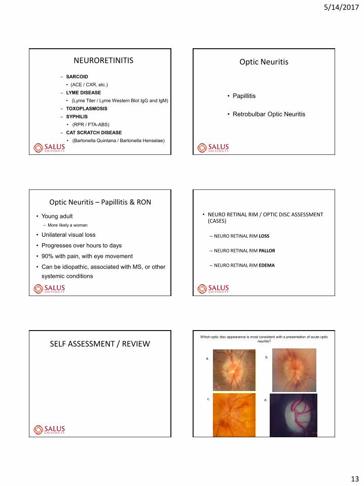
NEURORETINITIS
– SARCOID
• (ACE / CXR, etc.)
– LYME DISEASE
• (Lyme Titer / Lyme Western Blot IgG and IgM)
– TOXOPLASMOSIS
– SYPHILIS
• (RPR / FTA-ABS)
– CAT SCRATCH DISEASE
• (Bartonella Quintana / Bartonella Henselae)
Optic Neuritis
• Papillitis
• Retrobulbar Optic Neuritis
Optic Neuritis – Papillitis & RON
• Young adult
– More likely a woman
• Unilateral visual loss
• Progresses over hours to days
• 90% with pain, with eye movement
• Can be idiopathic, associated with MS, or other
systemic conditions
• NEURO RETINAL RIM / OPTIC DISC ASSESSMENT (CASES)
– NEURO RETINAL RIM LOSS
– NEURO RETINAL RIM PALLOR
– NEURO RETINAL RIM EDEMA
SELF ASSESSMENT / REVIEW
Which optic disc appearance is most consistent with a presentation of acute optic
neuritis?
a. b.
c. d.

5/14/2017
14
This patient LEAST likely has?
a. Idiopathic intracranial hypertension
b. Optic disc drusen
c. Transverse venous sinus thrombosis
d. Meningitis
This patient has a prominent spontaneous venous
pulsation in the left eye, and no visual complaints.
What is the most likely diagnosis?
a. Papilledema
b. Buried optic disc drusen
c. Optic neuritis
d. Ischemic optic neuropathy
This patient most likely has?
a. Idiopathic intracranial hypertension
b. Sub-dural hematoma
c. Optic neuritis
d. Arteritic anterior ischemic optic neuropathy
This patient most likely has?
a. Giant cell artertitis
b. Sarcoid
c. Syphilis
d. Papilledema
This patient LEAST likely has?
a. Vitamin B 12 deficiency / folate deficiency
b. Pituitary adenoma
c. Multiple sclerosis
d. Bilateral occipital lobe infarcts
CASE 1

5/14/2017
15
46 year old woman
• Noticed decreased vision OU x 3 months
– Getting progressively worse
• Color vision is altered –blue and orange haze
• Denies eye pain or headache
• Sys Hx: Asthma
• Past ocular hx: unremarkable (past records retrieved – VA was normal one year prior)
• Social Hx:
• Smoking 2 packs of cigarettes per day x 30 years
• Drinks heavily (“alcoholic” x 2 years)
• Admits to poor nutritional habits associated with her alcohol use
• BCVA: OD 20/60 and OS 20/50
• Color 14/14 OD and 14/14 OS
• PERRL (-) APD
• CF: full OU
• HVF: general reduction of sensitivity and reduced foveal threshold bilaterally
• SLE and IOP normal OU
PAPILLOMACULAR BUNDLES TOXIC / NUTRITIONAL
HEREDITO/DEGENERATIVE
DEMYELINATING
COMPRESSIVE
Labs for Optic Neuropathy • CBC with differential and platelet count • C-reactive protein (inflammation) • ESR (inflammation) • Lyme titer (if + get Western blot IgG and IgM) • ANA with reflex titer (auto-immune disease) • ACE (sarcoid) • RPR (syphilis) • FTA-ABS (syphilis) • Vitamin B 12 (nutritional) • Folic acid (nutritional) • Methylmalonic acid (nutritional) • Homocysteine (nutritional / inflammatory) • SPEP (Serum Protein Electrophoresis) (possible malignancy)

5/14/2017
16
Labs for Optic Neuropathy • CBC with differential and platelet count • C-reactive protein (inflammation) • ESR (inflammation) • Lyme titer (if + get Western blot IgG and IgM) • ANA with reflex titer (auto-immune disease) • ACE (sarcoid) • RPR (syphilis) • FTA-ABS (syphilis) • Vitamin B 12 (nutritional) • Folic acid (nutritional) • Methylmalonic acid (nutritional) • Homocysteine (nutritional / inflammatory) • SPEP (Serum Protein Electrophoresis) (possible malignancy)
Lab Results
• Low folic acid at 2.94 (normal > 5.4)
• Low normal vitamin B 12 at 464 (normal 200-1100)
• Normal methylmalonic acid
• Elevated homocysteine at 53 (normal up to 13.9)
Homocysteine • Amino acid in the blood • Increased with vitamin B12 or folate deficiency, genetic causes
or renal disease • Related to greater risk of cardiovascular disease, stroke,
peripheral vascular disease • Elevated levels = atherosclerosis • Folic acid and vitamins B6 and B 12 break down homocysteine
in the blood • A 3umol/L drop in homocysteine = 16% less chance of heart
attack, 24% less chance of stroke, and 25% less chance of DVT • Levels above 14 umol increase risk of Alzheimer’s by 150%
Methylmalonic acid
• Early indicator of (occult) vitamin B 12 deficiency, or renal insufficiency
• Level is elevated when abnormal
• Methylmalonyl co A succinyl coA » vit B12
If both MMA and Homocysteine elevated = Vit B 12 Def
If MMA normal and Homocysteine elevated = Folate Def
Either can cause megaloblastic anemia
Toxic / Nutritional Optic Neuropathy
• Started on 1 mg Folic acid po qd
• Began taking multivitamins
• Need to d/c alcohol use, and improve eating habits
• Hematology consult requested to see if additional treatment is needed
– IM/IV folic acid
– High dose Thiamine
• Pt refused alcohol support groups / counseling
• Gave pt phone number for alcohol rehabilitation program

5/14/2017
17
CASE 2
62 year-old woman
• Referred because of optochoroidal shunt vessels
• History of glaucoma
– Using Travatan and Azopt OU
• No eyecare x 1.5 months due to lack of insurance
– PCP did refill drops during that time
• Chief complaint – Blur with prolonged reading
– Rare headaches from lack of sleep
• Systemic History: – Diabetes
– Hypertension
– Hypercholesterolemia
– Rheumatoid Arthritis
• BCVA: OD 20/20 OS 20/20
• Color: OD 12/14 OS 14/14
• PERRLA (-) RAPD
• Normal ocular motility
• SLE: only mild lens changes
• TA: OD 17 mm Hg OS 18 mm Hg
• BP: 142/80

5/14/2017
18
• Pt is of Haitian descent
– Came to US 6 years ago
• She recalls that in Haiti about 20 years ago
– Decrease in vision in OS
– Unsure of diagnosis, but remembers being told about some bleeding in the eye
CASE 3
73 year-old man
• CC: Has noticed a gradual decrease in vision OS • Occasional headaches, longstanding, stable, attributes
to sinus issues • Systemic History
• Diabetes • Hypertension • Hypercholesterolemia • Hyperthyroidism - uncontrolled
• Ocular History • Normal Tension Glaucoma x 7 years • Cataract Surgery OU

5/14/2017
19
Exam Findings
• VA: 20/25 OD, 20/40 OS
• Color: 6/14 OD, 7/14 OS
• PERRLA (+) RAPD OS
• EOMs: no restrictions
• No ptosis or proptosis
Exam Findings
• Normal slit lamp exam
• IOP: 17 OD, 16 OS
• BP: 169/95
• DFE: 0.6 x 0.6 cupping OD
O.75 x 0.75 cupping OS
• (-) edema
OD OS OD OS
RAPD, dyschromatopsia and pallor can only occur from lesions anterior to the LGN.
NRR pallor is NOT consistent with glaucoma!!
Assessment: Bitemporal hemianopia, suspect structural abnormality in area of suprasellar cistern
Plan: Order MRI of brain with and without contrast
Order pituitary gland function tests (prolactin, growth hormone, cortisol, TSH, FSH, LH, and IGF-1)

5/14/2017
20
Pituitary Macroadenoma Patient
underwent trans-spenoidal resection.
1 Month Follow-Up
• BCVA 20/20 OD
20/25 OS
• Improvement in Visual Field
• Under the care of endocrinologist
Normal Tension Glaucoma • Diagnosis of Exclusion • Many things can mimic NTG • Especially if there is optic disc pallor and the VF
does not match the ONH, additional work-up is needed
– Lab testing to r/o infectious, inflammatory, nutritional – Neuro-imaging to r/o mass, abnormal enhancement
Labs for Optic Neuropathy • CBC with differential and platelet count • C-reactive protein (inflammation) • ESR (inflammation) • Lyme titer (if + get Western blot IgG and IgM) • ANA with reflex titer (auto-immune disease) • ACE (sarcoid) • RPR (syphilis) • FTA-ABS (syphilis) • Vitamin B 12 (nutritional) • Folic acid (nutritional) • Methylmalonic acid (nutritional) • Homocysteine (nutritional / inflammatory) • SPEP (Serum Protein Electrophoresis) (possible malignancy)
CASE 4
18 year-old girl
• CC: diplopia at D and in R gaze
– Onset 2 ½ months prior
• Headache, constant
– Onset 2 ½ months prior
– No improvement with OTC medications
• Pain in right eye

5/14/2017
21
• Gave birth about 3 months prior
• Began noticing symptoms around that time
• Patient thinks symptoms began after getting epidural anesthesia
• Patient has “not felt right” since
• Very tired, “ wants to sleep a lot”
• S/ P 3 Pregnancies • Full term birth January 2006
• Elevated blood pressure
• Terminated Pregnancy • Premature birth (at 34 weeks) October 2007
• Systemic Hx: Otherwise unremarkable • Medications: Depo Provera
(medroxyprogesterone IM) • Report history of “lazy eye” (R lid droop) • Denies any other health problems • Denies trauma
Examination Results
• VA: OD 20/20 OS 20/20
• Color (Ishihara): OD 14/14 OS 14/14
– No red desaturation
• Pupils: PERRL (-) APD
• CF: Full OU
• Exophthalmometry: OD 17 mm OS 18 mm
• Normal anterior segment exam
• IOP: OD 12 mm Hg OS 12 mm Hg
Ocular Motility
OD OS
100
100
100
100
100
100 100 20eso 40eso 8eso
70
NEGATIVE FORCED DUCTION TEST
5/14/2017
22
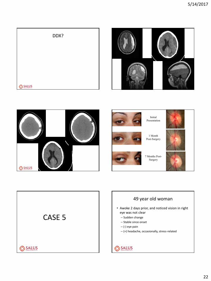
DDX?
1 Month
Post-Surgery
7 Months Post-
Surgery
Initial
Presentation
CASE 5
49 year old woman
• Awoke 2 days prior, and noticed vision in right eye was not clear
– Sudden change
– Stable since onset
– (-) eye pain
– (+) headache, occasionally, stress-related

5/14/2017
23
• SYSTEMIC HEALTH: – Diabetes x 20 years (last HbA1c: 11%)
– Hypertension x 30 years
– Hypercholesterolemia
– Asthma
– Sleep Apnea (CPAP broken)
– Depression
– s/p right mastectomy for breast cancer 7 years prior • Radiation and chemotherapy
• MEDICATIONS:
– HCTZ
– Lotrel
– Novalog
– Metformin
– Naproxen
– Singulair
• OCULAR HISTORY:
– Unremarkable
• SOCIAL HISTORY:
– Unremarkable
• BCVA: OD 20/1000 and OS 20/20 • Color 10/14 OD (with EF) and 14/14 OS • 50% reduced red saturation • 50% reduced brightness sense • Pupils – isocoric, 1.5 log RAPD OD
• Confrontation Fields: – OD- inferior nasal defect extending into inferior
temporal and superior nasal quadrants – OS- possible mild inferior nasal defect
• HVF: poor reliability, significant VF loss OD • No ptosis or proptosis • Normal ocular motility examination
Poor reliability

5/14/2017
24
• Slit Lamp Exam: Mild lens changes
• TONOMETRY: OD-12 mm Hg, OS- 13 mm Hg
• BP: 170/100
• Pulse: 69 bpm
• Average RNFL thickness:
• OD: 404um • OS: 105 um
DDX • NA-AION
– Consistent Features: • Sudden onset
• Disc edema with hemorrhages
• Disc at risk in fellow eye
– Risk factors: • Hypertension
• Diabetes
• Sleep apnea
• Scheduled pt to see endocrinologist
• Scheduled for sleep study (needs new CPAP machine)
Follow-Up 2 months later
• Since last visit, increased medication for HTN and DM
• Some improvement in vision

5/14/2017
25
Avg NFL thickness: OD: 146 um OS 98 um
1 year later…..
• Patient missed previously scheduled follow-ups
• Returns now due to vision loss in left eye!
• Blur OS x 6 days
• Went to ER – BP was high – Increased dosage of Clonidine
– Pt put back on aspirin • Discontinued prior to breast reconstruction after breast
cancer
• Prior to loss of vision – Not taking aspirin
– Not wearing CPAP (says it is too tight)
• Labs done a few days ago
– HbA1c: 11.6%
– Cholesterol: 215
– Triglycerides: 208
– Pt states difficult to take care of herself because she is under a lot of stress
• BCVA: OD 20/200 and OS 20/125 • Color 11/14 OD and 4/14 OS • Pupils – isocoric, 0.6-0.9 log RAPD OS • Humphrey VF: unreliable • Normal efferent function
5/14/2017
26
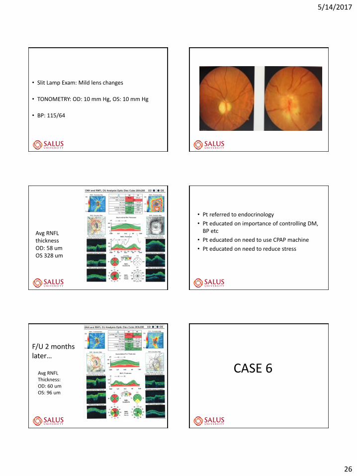
• Slit Lamp Exam: Mild lens changes
• TONOMETRY: OD: 10 mm Hg, OS: 10 mm Hg
• BP: 115/64
Avg RNFL thickness OD: 58 um OS 328 um
• Pt referred to endocrinology
• Pt educated on importance of controlling DM, BP etc
• Pt educated on need to use CPAP machine
• Pt educated on need to reduce stress
F/U 2 months later…
Avg RNFL Thickness: OD: 60 um OS: 96 um
CASE 6

5/14/2017
27
40 year old asymptomatic woman
• Hx of keratoconus
• Came in for CL eval, and anterior uveitis was noted
• On Pred Forte qid OS and Nevanac (NSAID prodrug) qid OS
• Photosensitivity – stable x years
• Denies other symptoms
• Systemic Hx:
– Hypercholesterolemia – no meds
– S/p removal of axillary lymph nodes (infxn?)
– S/p 2 full term uncomplicated pregnancies
– Asthma
– Meds: Advair, Combivent, Zyrtec, Claritin
• VA: OD 20/25 OS 20/60
• Color: 13/14 OD and 0/14 OS
• PERRL (+) >1.8 log RAPD OS
• No ptosis or proptosis
• Normal ocular motility exam
• SLE: only few residual cells
• TA: OD 18 mmHg OS 21 mm Hg
• Normal neurologic exam
Labs for Optic Neuropathy • CBC • C-reactive protein • ESR • Platelet count • Lyme titer (if + get Western blot IgG and IgM) • ANA with reflex titer • ACE • RPR & FTA-ABS • Vitamin B 12 • Folic acid • Methylmalonic acid • SPEP
5/14/2017
28
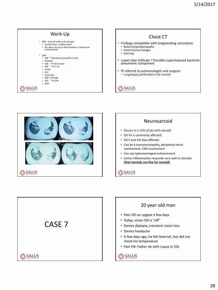
Work-Up
• MRI – brain & orbits w & w/o gad • Arnold Chiari I malformation • No other structural abnormalities or abnormal
enhancement
• Labs • CBC **elevated eosinophils 11.6% • Platelets • ESR **H 85 mm/hr • CRP **H 1.22 • Folate • B12 • Lyme titer • RPR / FTA-ABS • ACE **H 254 • ANA
Chest CT • Findings compatible with longstanding sarcoidosis • Bulky lymphadenopathy • Parenchymal changes • Scarring
• Lower lobe infiltrate ? Possible superimposed bacterial pneumonic component
• Pt referred to pulmonologist and surgeon • Lung biopsy performed (+) for sarcoid
Neurosarcoid
• Occurs in 5-15% of pts with sarcoid
• CN VII is commonly affected
• CN II and VIII also affected
• Can be a mononeuropathy, peripheral nerve involvement, CNS involvement
• Can see leptomeningeal enhancement
• Active inflammation responds very well to steroids (Oral steroids are fine for sarcoid)
CASE 7
20 year-old man
• Pain OD on upgaze x few days
• Today, vision OD is “off”
• Denies diplopia, transient vision loss
• Denies headache
• A few days ago, he felt feverish, but did not check his temperature
• Fam HX: Father dx with Lupus in 20s

5/14/2017
29
• BCVA: OD 20/50 and OS 20/20
• Color 14/14 OD and 14/14 OS
• Pupils – pharm dilated
• CF: full OU
• HVF: essentially normal OU
• SLE and IOP normal OU
• BP: 104/70
• Temp: 98.8 degrees
Right Eye Left Eye
Humphrey Visual FIeld: At initial presentation. Note only slightly enlarged blind spot in the right eye, and fairly preserved central visual field.
Initial Presentation
Right Eye Left Eye
• Pt denies any rashes (only when asked)
• He does admit to a scratch by a cat (kitten) several weeks ago (only when asked)
• A few weeks ago, his right eyelid was swollen
• Pt has several scars on his forehead, above right eye, and on his nose
Labs Ordered – told to have done today! • CBC • C-reactive protein • ESR • Platelet count • Lyme titer (if + get Western Blot Lyme IgG and IgM) • ANA with reflex titer • ACE • RPR • FTA-ABS • Bartonella Quintana titer • Bartonella Henselae titer
Follow-up 5 days later
• Pt notes a spot in right vision, that is getting larger
• Reduced central vision
• Since last visit, has had chills and fever
• Has also had headache
• Decreased appetite
• Unable to work – doesn’t feel right
• Labs not done until 2 days ago – not complete

5/14/2017
30
Lab results (so far)
• ANA ( + ) titer and pattern not yet known
• Lyme titer is ( + ) WB IgG (-), IgM (+)
• ACE slightly elevated at 70
• ESR: 44
• CRP: pending
• Bartonella titers: pending
• BCVA: OD 20/200 and OS 20/20
• Color 1/14 OD and 14/14 OS
• Pupils – trace RAPD OD
• CF: very large blindspot OD
• HVF: large blindspot OD – extending past fixation and superiorly out to 10 degrees
• SLE and IOP normal OU (-) cells
Right Eye Left Eye
Humphrey Visual Field: 6 days after initial presentation. Note significant increase in blind spot in the right eye. The left field was unreliable due to patient fatigue.
Follow-Up: 6 days after initial presentation
Right Eye Left Eye
Follow-Up: 6 days after initial presentation
Right Eye Left Eye
• Need to r/o Lyme, sarcoid, auto-immune disease
• Ds DNA (-)
• Repeat ACE (-), CXR (-)
• LP (-) for Lyme, sarcoid

5/14/2017
31
• Bartonella titers
• Bartonella Quintana (-)
• Bartonella Hensalea (+)
• (+) IgG > 1:2560
• (+) IgM > 1:800
• DX: Cat-scratch Disease
Follow-Up: 15 days after initial presentation
Right Eye Left Eye
Neuro-retinitis
• Cat-scratch
• Sarcoid
• Syphilis
• Lyme
• Toxo
• NOT typical in MS
• Treatment:
– Antibiotics
» Doxycycline (pt vomited every time he took this medication)
» Rifampin
» Bactrim prescribed in place of Doxycycline
» Pt then switched to Azithromycin by Infectious Disease
Cat Scratch Disease
Typically transmitted by a kitten (by a scratch or a lick)
Only a minority of the exposures to B. Henselae result in cat-scratch disease. The ability of the cat to transmit the disease is transient
Most cases occur in fall / early summer - related to kitten births and flea infestations
80% of cases occur in patients under age 21
Starts with local infection, then lymphadenopathy, and rarely progresses – eg. Neuroretinitis, etc.

5/14/2017
32
Cat Scratch Disease
– Treatment / Response:
– Excellent prognosis - Most cases are self-limiting and fully resolve, even when involving the CNS
– Drugs of choice – Bactrim, Gentamicin, Ciprofloxacin, Rifampin, Azithromycin
Humphrey Visual Field: 34 days after initial presentation. Note reduction in blindspot size in the right eye
corresponding with a reduction in optic disc edema. A central scotoma persists in the right eye, related to the
macular star and the reduced visual acuity.
Right Eye Left Eye
Follow-Up: 34 days after initial presentation
Right Eye Left Eye
Follow-Up: 34 days after initial presentation
Right Eye Left Eye
CASE 8
28 year old woman
• 2 weeks prior, noticed blurry vision OS
• Blur persists
• No pain in the left eye
• 3 days prior, noticed pain on eye movements OD
• 1 day prior noticed decreased vision inferior OD
• She is now having difficulty functioning

5/14/2017
33
• OTHER SYMPTOMS:
– Weakness and tingling left thigh
• Several episodes over past few years
• Though to be sciatica
• Thought to be related to being over-weight – Lost 30 pounds, but symptom still recurred
• SYSTEMIC HEALTH:
– asthma
• MEDICATIONS:
– Albuterol, Advair
• OCULAR HISTORY:
– Unremarkable
• SOCIAL HISTORY:
– unremarkable
• BCVA: OD 20/25 and OS 20/40 • Color 14/14 OD and 10/14 OS • 25% reduced red saturation OS • 25% reduced brightness sense OS • Pupils – pharm dilated
• Normal ocular motility exam
• Slit Lamp Exam: normal OU
• TONOMETRY: OD-16 mm Hg, OS- 16 mm Hg
• BP: 132/92

5/14/2017
34
DDX
• Optic Neuritis OD, Optic Neuropathy OS
• ? Demyelinating Disease
• WORK-UP
– Lab testing
– MRI brain and orbits with contrast
– MRI spine
– Possible lumbar puncture
• TREATMENT:
• IV steroids x 3 days
RESULTS • MRI brain and orbits – mild enhancement of posterior
right optic nerve
• MRI spine: lesion at C3 level, but no brain lesions
• Pt diagnosed with Multiple Sclerosis at hospital
FOLLOW-UP
• Eye pain returns (was better on steroids)
• Severe headaches
• Leg paresthesias recur (better on steroids)
• BCVA: OD 20/25 and OS 20/25
• Color 14/14 OD and 10/14 OS
• Pupils – equivocal RAPD OS
• Normal ocular motility exam

5/14/2017
35
• Because vision did not improve significantly after steroid treatment, and there was spine involvement but no brain involvement, need to consider:
• NMO (neuro-myelitis optica or Devic’s disease)
• Order NMO antibody testing (NMO-IgG Ab)
• Also test for other causes of optic neuritis
• (ANA, Lyme, ACE, FTA-ABS, etc)
• NMO (AQP4) antibody was POSITIVE
• Positive ANA, positive Sjogren’s antibodies
• Pt started on Azathioprine
• Then put on Rituximab
– some improvement in field of right eye
5 WEEKS LATER
Neuromyelitis optica is, like Multiple Sclerosis an inflammatory, demyelinating syndrome of the central Nervous system
Wingerchuk DM, Hogancamp WF, O’Brien PC, Weinshenker BG. The clinical course of neuromyelitis optica (Devic’s syndrome). Neurology 1999; 53: 1107–14.
Neuro Myelitis Optica (NMO)
preferentially affects the -optic nerve (optic neuritis) -spinal cord (myelitis)
Neuromyelitis Optica (NMO) (Devic’s Disease)
Multiple Sclerosis
affects the CNS -optic nerve and -spinal cord -brain
Wingerchuk DM, Hogancamp WF, O’Brien PC, Weinshenker BG. The clinical course of neuromyelitis optica (Devic’s syndrome). Neurology 1999; 53: 1107–14.
Within 5 years of disease onset, more than 50% of patients with relapsing neuromyelitis optica are blind in one or both eyes or require ambulatory help.
Dean M Wingerchuk(et al (The Lancet Neurology, 2007, 6:805-15)

5/14/2017
36
The detection of neuromyelitis optica immunoglobulin G (NMO-IgG), an autoantibody, in the serum of patients with neuromyelitis optica, distinguishes neuromyelitis optica from other demyelinating disorders NMO-IgG binds to aquaporin 4, which is the main channel that regulates water homoeostasis in the central nervous system
Lennon VA, Wingerchuk DM, Kryzer TJ, et al. A serum
autoantibody marker of neuromyelitis optica: distinction from
multiple sclerosis. Lancet 2004; 364: 2106–12.
Using typical MS treatments for NMO can cause NMO exacerbation!
Interferon-beta Natalizumab Fingolimod
CASE 9 ANY QUESTIONS?
THANK YOU.