the nervous systemmsbeelsscience.weebly.com/uploads/3/8/0/0/38003353/... · structure and functions...
TRANSCRIPT

N E U R O N S A N D T H E N E R V O U S S Y S T E M
The Nervous System

Structure and Functions System
A physical organ system
The main cell of the Nervous System is:
The NEURON!
Organs of the nervous system form two subsystems
Central Nervous System (CNS)
Peripheral Nervous System (PNS)
The PNS delivers information to the CNS and carries messages from the CNS to other organs via NERVES

Nerves
Nerves
Collections of neurons that are joined together by connective tissue.
Responsible for transferring impulses (a message carried by neurons) from receptors to CNS and back to effectors.

CNS & PNS
The CNS is made up of:
The Brain
The Spinal Cord
The PNS is made up of
All nervous tissue outside of the CNS
Sensory Neurons
Motor Neurons (sensory & autonomic)

Central Nervous System (CNS)
Brain Spinal Cord
Peripheral Nervous System (PNS)
Sensory Neurons Motor Neurons
Somatic Nervous System
• Voluntary movements via skeletal muscles
Autonomic Nervous System
• Organs, Smooth muscles
Sympathetic
- “Fight-or-Flight” responses Parasympathetic
- maintenance
Break It DOWN! The Nervous System

Divisions of the Autonomic Nervous System
The Sympathetic division of the nervous system prepares the body for action
The Parasympathetic division returns the body to a resting state

Neurons
Made up of a cell body and branches called dendrites and axons
Dendrites receive messages from other neurons and send them to the cell body
Axons carry messages away from the cell body
A message carried by a neuron is called an impulse
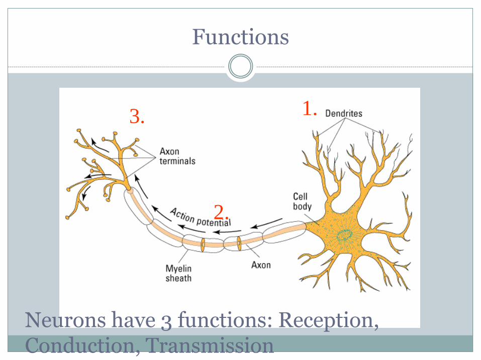
Functions
Neurons have 3 functions: Reception, Conduction, Transmission
1. 3.
2.

Types of Neurons
1. Sensory Neurons Neurons located near receptor organs (skin, eyes, ears).
Function: receive incoming stimuli from the environment.
2. Motor Neurons Neurons located near effectors (muscles and glands)
Function: Carry impulses to effectors to initiate a response.
3. Interneurons Neurons that relay messages between other neurons such as sensory and motor neurons. (Found most often in Brain and Spinal chord).

Types of Neurons

e.g., skin
e.g., muscle
Gray’s Anatomy 38 1999
Sensory Vs. Motor Sensory Nerve Neurons that send signals from the senses, skin, muscles,
and internal organs to the CNS
Motor Nerve Neurons that transmit commands from the CNS to the
muscles, glands, and organs

The Withdrawal Reflex

Parts of a Neuron
Dendrite Fine hair-like extensions on the end of a neuron. Function: receive incoming stimuli.
Receive messages from other neurons and send them to the cell body
Cell Body or Soma The control center of the neuron. Function: Directs impulses from the
dendrites to the axon. Nucleus Control center of the Soma. Function: Tells the soma what to do.

Neuron Structure

Extra Extra!
Dendrite The bushy, branching extensions of a
neuron that receive messages and conduct impulses toward the cell body
Axon The extension of a neuron, ending in
branching terminal fibers, through which messages are sent to other neurons or to muscles or glands

Parts of a Neuron Continued
Axon Pathway for the nerve impulse (electrical message) from the soma to the opposite end of the neuron.
Carries messages away from the cell body
Myelin Sheath An insulating layer around an axon. Made up of Schwann cells.
Nodes of Ranvier Gaps between Schwann cells.
Function: Conduction of the impulse. (Situation where speed of an impulse is greatly increased by the message ‘jumping’ the gaps in an axon).


Myelin Sheath & Nodes of Ranvier
Myelin Sheath Fatty material made by glial cells Insulates the axon Allows for rapid movement of
electrical impulses along axon Multiple sclerosis is a breakdown of
myelin sheath Nodes of Ranvier: gaps in myelin sheath where
action potentials are transmitted Speed of neural impulse Ranges from 2 – 200+ mph

Neurons
Axon of another neuron
Cell Body Dendrites
Axon
Myelin Sheath
Dendrites of another neuron