the nervous system. functions of the nervous system sensory input – gathering information to...
Post on 19-Dec-2015
213 views
TRANSCRIPT
The Nervous System

Functions of the Nervous SystemFunctions of the Nervous System
Sensory input – gathering information To monitor changes occurring inside and
outside the body
Changes = stimuli
Integration To process and interpret sensory input and
decide if action is needed

Functions of the Nervous SystemFunctions of the Nervous System
Motor output
A response to integrated stimuli
The response activates muscles or glands
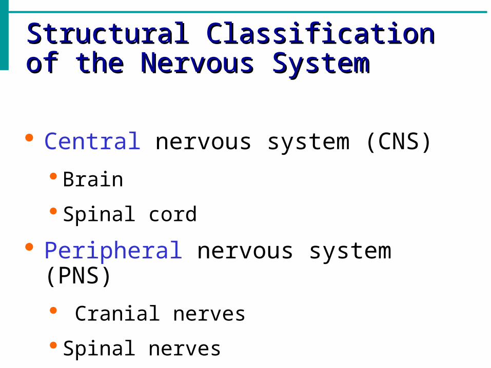
Structural Classification of the Structural Classification of the Nervous SystemNervous System
Central nervous system (CNS)
Brain
Spinal cord
Peripheral nervous system (PNS)
Cranial nerves
Spinal nerves

Functional Classification is Functional Classification is concerned only with PNSconcerned only with PNS
1-Sensory (afferent) division
Nerve fibers that carry information to the central nervous system
Figure 7.1

Functional Classification of the Functional Classification of the Peripheral Nervous SystemPeripheral Nervous System
2-Motor (efferent) division
Nerve fibers that carry impulses away from the central nervous system ( to Muscles &Glands)
Figure 7.1

Motor (efferent) division has two subdivisions
1-Somatic nervous system = voluntary, it controls skeletal muscles
skeletal muscle reflexes are involuntary
2-Autonomic nervous system = involuntary, it controls smooth & cardiac muscles & glands
This also is divided into sympathetic & parasympathetic

Organization of the Nervous SystemOrganization of the Nervous System
Figure 7.2
Histology of Nervous TissueHistology of Nervous Tissue
• Despite the complexity of the nervous system, there are only two functional cell types
• Neurons - excitable nerve cells that transmit electrical signals
• Neuroglia (glial) cells - supporting cells

Nervous Tissue: NeuronsNervous Tissue: Neurons
Neurons = nerve cells
Cells specialized to transmit messages
Major regions of neurons
Cell body – nucleus and metabolic center of the cell
Processes – fibers that extend from the cell body

Anatomy of neuron (Nerve cell)Anatomy of neuron (Nerve cell)
Differ structurally but have common Differ structurally but have common features,all features,all
have have A Cell body
with Nucleus, the usual organelles
Except centrioles
One or more processes
Figure 7.4a

Neuron AnatomyNeuron Anatomy
Extensions outside the cell body Dendrites –
conduct impulses toward the cell body
Axons – conduct impulses away from the cell body
Figure 7.4a

Axons and Nerve ImpulsesAxons and Nerve Impulses
Axons end in axonal terminals
Axonal terminals contain vesicles with neurotransmitters
Axonal terminals are separated from the next neuron (neuroneural junction) by a gap called Synaptic cleft (Synapse)

Nerve Fiber CoveringsNerve Fiber Coverings--Most long nerve fibers are coveredMost long nerve fibers are covered
with a whitish, fatty material calledwith a whitish, fatty material called
Myelin with waxy appearance. It Myelin with waxy appearance. It
insulates the fiber &Increases insulates the fiber &Increases
transmission ratetransmission rate
-Axons outside CNS are wrapped -Axons outside CNS are wrapped
by Schwann Cellsby Schwann Cells
Figure 7.5

Neuron Cell Body LocationNeuron Cell Body Location
Most are found in the central nervous system
Gray matter – cell bodies and unmyelinated fibers
Nuclei – clusters of cell bodies within the white matter of the central nervous system
Ganglia – collections of cell bodies outside the central nervous system
White matter- collection of myelinated fibers (Tracts)

Functional Classification of NeuronsFunctional Classification of Neurons1-Sensory (afferent) neurons
Carry impulses from the sensory receptors
Cutaneous sense organs
Proprioceptors – detect stretch or tension in muscles and tendons and joints
2-Motor (efferent) neurons
Carry impulses from the central nervous system to muscles and glands ,their cell bodies are always in CNS

Functional Classification of NeuronsFunctional Classification of Neurons
3-Interneurons (association neurons)
Their cell bodies are always found in CNS
Connect sensory and motor neurons in neural pathways

Neuron ClassificationNeuron Classification
Figure 7.6

Structural Classification of NeuronsStructural Classification of Neurons
Multipolar neurons – many extensions from the cell body
Figure 7.8a

Structural Classification of NeuronsStructural Classification of Neurons
Bipolar neurons – one axon and one dendrite
Figure 7.8b

Structural Classification of NeuronsStructural Classification of Neurons
Unipolar neurons – have a short single process leaving the cell body which is very short ,divides almost immediately
Figure 7.8c

Functional Properties of NeuronsFunctional Properties of Neurons
Irritability – ability to respond to stimuli
Conductivity – ability to transmit an impulse
The plasma membrane at rest is polarized
Fewer positive ions are inside the cell than outside the cell
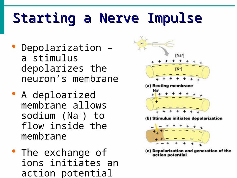
Starting a Nerve ImpulseStarting a Nerve Impulse
Depolarization – a stimulus depolarizes the neuron’s membrane
A deploarized membrane allows sodium (Na+) to flow inside the membrane
The exchange of ions initiates an action potential in the neuron
The Action PotentialThe Action Potential
If the action potential (nerve impulse) starts, it is propagated over the entire axon
Potassium ions rush out of the neuron after sodium ions rush in, which repolarizes the membrane
The sodium-potassium pump restores the original configuration This action requires ATP

Nerve Impulse PropagationNerve Impulse Propagation
The impulse continues to move toward the cell body
Impulses travel faster when fibers have a myelin sheath
Figure 7.9c–e

Continuation of the Nerve Impulse Continuation of the Nerve Impulse between Neuronsbetween Neurons
Impulses are able to cross the synapse to another nerve
Neurotransmitter is released from a nerve’s axon terminal
The dendrite of the next neuron has receptors that are stimulated by the neurotransmitter
An action potential is started in the dendrite

How Neurons Communicate at How Neurons Communicate at SynapsesSynapses
Figure 7.10

The Reflex ArcThe Reflex Arc
Reflex – rapid, predictable, and involuntary responses to stimuli
Reflex arc – direct route from a sensory neuron, to an interneuron, to an effector
Ref. arch have a minimum 5 elements
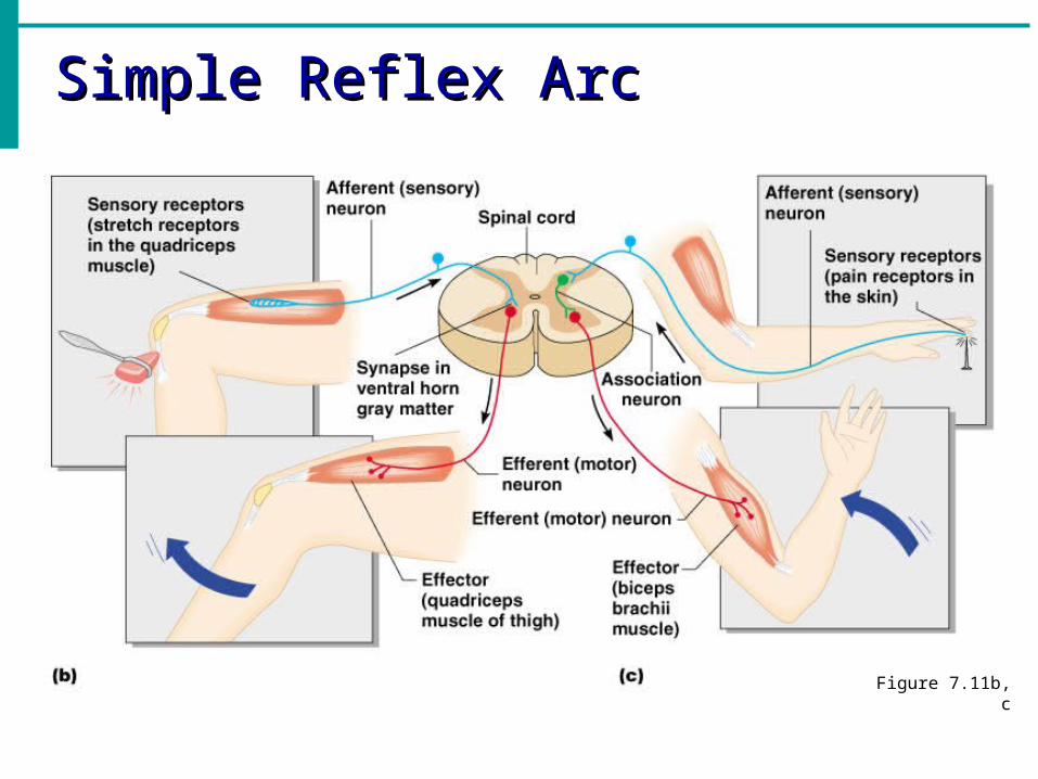
Simple Reflex ArcSimple Reflex Arc
Figure 7.11b, c

Types of Reflexes and RegulationTypes of Reflexes and Regulation
Autonomic reflexes eg.
Salivary gland secretion
Heart and blood pressure regulation
Changes in size of the pupil
Digestive system regulation
Somatic reflexes
Activation of skeletal muscles
Central Nervous System (CNS)Central Nervous System (CNS)
CNS develops from the embryonic neural tube
By the fourth week the anterior end begins to expand and brain formation begins, The rest of the tube becomes the spinal cord
The central canal becomes enlarged in 4 regions of the brain to form ventricles:
-Four chambers within the brain
-Filled with cerebrospinal fluid

Regions of the BrainRegions of the Brain
Cerebral hemispheres
Diencephalon
Brain stem
CerebellumFigure 7.12

Cerebral Hemispheres (Cerebrum)Cerebral Hemispheres (Cerebrum)
Paired (left and right) superior parts of the brain
Include more than half of the brain mass
Figure 7.13a

Layers of the CerebrumLayers of the Cerebrum
Gray matter
Outer layer
Composed mostly of neuron cell bodies
Figure 7.13a

Layers of the CerebrumLayers of the Cerebrum
White matter
Fiber tracts inside the gray matter
Example: corpus callosum connects hemispheres
Figure 7.13a

Layers of the CerebrumLayers of the Cerebrum
Basal nuclei – internal islands of gray matter
Figure 7.13a

Cerebral Hemispheres (Cerebrum)Cerebral Hemispheres (Cerebrum)
The surface is made of ridges (gyri) and grooves (sulci)
Figure 7.13a

Specialized Area of the CerebrumSpecialized Area of the Cerebrum
Cerebral areas involved in special senses
Gustatory area (taste)
Visual area
Auditory area
Olfactory area

Specialized Area of the CerebrumSpecialized Area of the Cerebrum
Interpretation areas of the cerebrum
Speech/language region
Language comprehension region
General interpretation area

Specialized Area of the CerebrumSpecialized Area of the Cerebrum
Figure 7.13c

DiencephalonDiencephalon
Sits on top of the brain stem
Enclosed by the cerebral hemispheres
Made of three parts Thalamus
Hypothalamus
Epithalamus
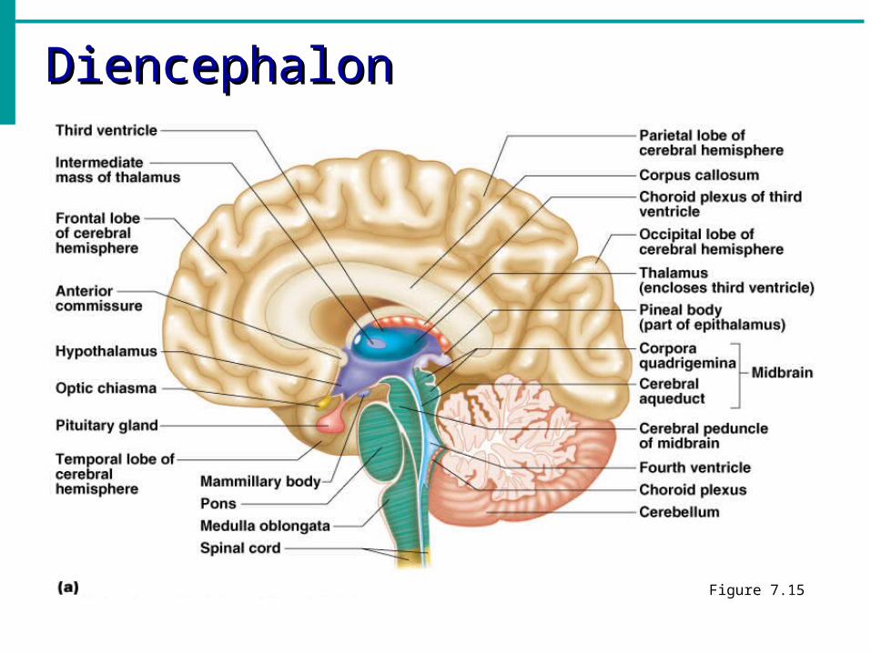
DiencephalonDiencephalon
Figure 7.15

ThalamusThalamus
Surrounds the third ventricle
The relay station for sensory impulses (except olfaction)
Transfers impulses to the correct part of the cortex for localization and interpretation

HypothalamusHypothalamus
Under the thalamus
Important autonomic nervous system center Helps regulate body temperature
Controls water balance
Regulates metabolism
HypothalamusHypothalamus
An important part of the limbic system (emotions)
The pituitary gland is attached to the hypothalamus

EpithalamusEpithalamus
Forms the roof of the third ventricle
Houses the pineal body (an endocrine gland)
Includes the choroid plexus – forms cerebrospinal fluid

Brain StemBrain Stem
Attaches to the spinal cord
Parts of the brain stem Midbrain
Pons
Medulla oblongata

Brain StemBrain Stem
Figure 7.15a

MidbrainMidbrain
Mostly composed of tracts of nerve fibers
Reflex centers for vision and hearing

PonsPons
The bulging center part of the brain stem
Mostly composed of fiber tracts
Includes nuclei involved in the control of breathing

Medulla OblongataMedulla Oblongata The lowest part of the brain stem Merges into the spinal cord Includes important fiber tracts Contains important control centers
Heart rate control Blood pressure regulation Breathing Swallowing Vomiting
Reticular FormationReticular Formation
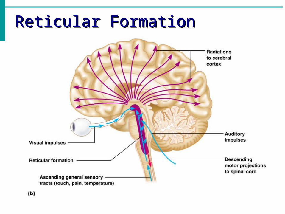
Diffuse mass of gray matter along the brain stem
Involved in motor control of visceral organs
Reticular activating system plays a role in awake/sleep cycles and consciousness
Reticular FormationReticular Formation

CerebellumCerebellum
Two hemispheres with convoluted surfaces
Provides involuntary coordination of body movements

CerebellumCerebellum

Protection of the Central Nervous Protection of the Central Nervous SystemSystem
Scalp and skin
Skull and vertebral column
Meninges
Figure 7.16a
Protection of the Central Nervous Protection of the Central Nervous SystemSystem
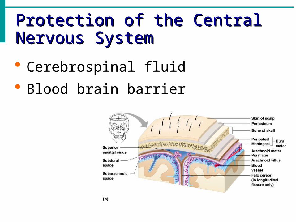
Cerebrospinal fluid
Blood brain barrier

MeningesMeninges
• Three connective tissue membranes lie external to the CNS – dura mater, arachnoid mater, and pia mater
• Functions of the meninges
• Cover and protect the CNS
• Protect blood vessels and enclose venous sinuses
• Contain cerebrospinal fluid (CSF)
• Form partitions within the skull

MeningesMeninges
Figure 12.24a

Dura MaterDura Mater
• Leathery, strong meninx composed of two fibrous connective tissue layers
• The two layers separate in certain areas and form dural sinuses
• Double-layered external covering
• Periosteum – attached to surface of the skull
• Meningeal layer – outer covering of the brain
• Folds inward in several areas
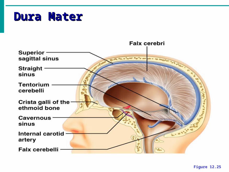
Dura MaterDura Mater
Figure 12.25

Arachnoid MaterArachnoid Mater
• The middle meninx, which forms a loose brain covering
• It is separated from the dura mater by the subdural space
• Beneath the arachnoid is a wide subarachnoid space filled with CSF and large blood vessels

Arachnoid Mater
Figure 12.24a

Pia MaterPia Mater
• Deep meninx composed of delicate connective tissue that clings tightly to the brain

Cerebrospinal FluidCerebrospinal Fluid
Similar to blood plasma composition
Formed by the choroid plexus in epithalamus
Forms a watery cushion to protect the brain
Circulated in arachnoid space, ventricles, and central canal of the spinal cord

Ventricles and Location of the Ventricles and Location of the Cerebrospinal FluidCerebrospinal Fluid
Figure 7.17a

Blood Brain BarrierBlood Brain Barrier Includes the least permeable capillaries
of the body
Excludes many potentially harmful substances
Useless against some substances Fats and fat soluble molecules Respiratory gases Alcohol Nicotine Anesthesia

Traumatic Brain InjuriesTraumatic Brain Injuries Concussion
Slight brain injury No permanent brain damage
Contusion Nervous tissue destruction occurs Nervous tissue does not regenerate
Cerebral edema Swelling from the inflammatory response May compress and kill brain tissue

Cerebrovascular Accident (CVA)Cerebrovascular Accident (CVA)
Commonly called a stroke
The result of a ruptured blood vessel supplying a region of the brain
Brain tissue supplied with oxygen from that blood source dies
Loss of some functions or death may result

Alzheimer’s DiseaseAlzheimer’s Disease
Progressive degenerative brain disease
Mostly seen in the elderly, but may begin in middle age
Structural changes in the brain include abnormal protein deposits and twisted fibers within neurons
Victims experience memory loss, irritability, confusion and ultimately, hallucinations and death

Spinal CordSpinal Cord
Extends from the medulla oblongata to the region of T12
Below T12 is the cauda equina (a collection of spinal nerves)
Enlargements occur in the cervical and lumbar regions
Figure 7.18
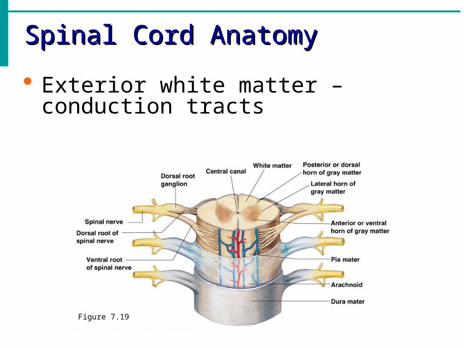
Spinal Cord AnatomySpinal Cord Anatomy
Exterior white matter – conduction tracts
Figure 7.19

Spinal Cord AnatomySpinal Cord Anatomy
Internal gray matter - mostly cell bodies Dorsal (posterior) horns
Anterior (ventral) horns
Figure 7.19

Spinal Cord AnatomySpinal Cord Anatomy
Central canal filled with cerebrospinal fluid
Figure 7.19

Peripheral Nervous SystemPeripheral Nervous System
Nerves and ganglia outside the central nervous system
Nerve = bundle of neuron fibers
Neuron fibers are bundled by connective tissue
Classification of NervesClassification of Nerves
Mixed nerves – both sensory and motor fibers
Afferent (sensory) nerves – carry impulses toward the CNS
Efferent (motor) nerves – carry impulses away from the CNS

Cranial NervesCranial Nerves
12 pairs of nerves that mostly serve the head and neck
Numbered in order, front to back
Most are mixed nerves, but three are sensory only
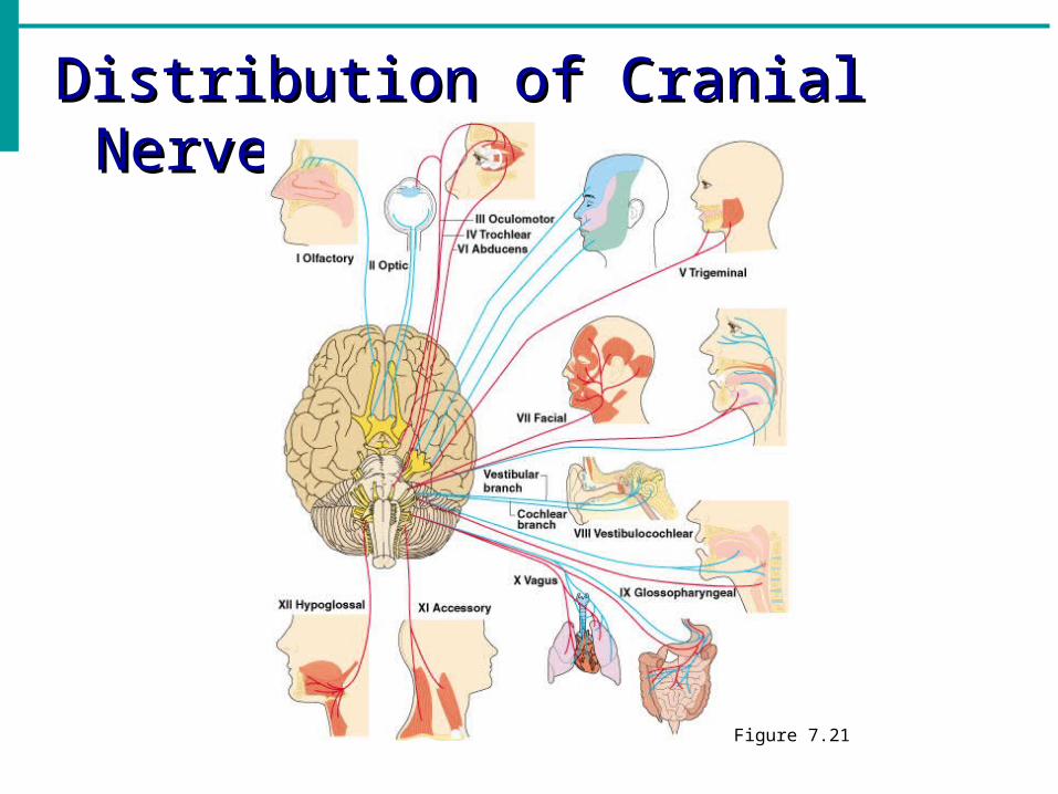
Distribution of Cranial NervesDistribution of Cranial Nerves
Figure 7.21

Cranial NervesCranial Nerves
I Olfactory nerve – sensory for smell
II Optic nerve – sensory for vision
III Oculomotor nerve – motor fibers to eye muscles
IV Trochlear – motor fiber to eye muscles

Cranial NervesCranial Nerves
V Trigeminal nerve – sensory for the face; motor fibers to chewing muscles
VI Abducens nerve – motor fibers to eye muscles
VII Facial nerve – sensory for taste; motor fibers to the face
VIII Vestibulocochlear nerve – sensory for balance and hearing

Cranial NervesCranial Nerves
IX Glossopharyngeal nerve – sensory for taste; motor fibers to the pharynx
X Vagus nerves – sensory and motor fibers for pharynx, larynx, and abdominal viscera
XI Accessory nerve – motor fibers to neck and upper back
XII Hypoglossal nerve – motor fibers to tongue

Spinal NervesSpinal Nerves
There is a pair of spinal nerves at the level of each vertebrae for a total of 31 pairs
Spinal nerves are formed by the combination of the ventral and dorsal roots of the spinal cord
Spinal nerves are named for the region from which they arise

Spinal NervesSpinal Nerves
Figure 7.22a
Autonomic Nervous SystemAutonomic Nervous System
The involuntary part of the peripheral nervous system
Consists of motor nerves only
Divided into two divisions
Sympathetic division
Parasympathetic division
Differences Between Somatic and Differences Between Somatic and Autonomic Nervous SystemsAutonomic Nervous Systems
Nerves Somatic – one motor neuron
Autonomic – preganglionic and postganglionic nerves
Effector organs Somatic – skeletal muscle
Autonomic – smooth muscle, cardiac muscle,and glands

Differences Between Somatic and Differences Between Somatic and Autonomic Nervous SystemsAutonomic Nervous Systems
Neurotransmitters
Somatic – always use acetylcholine
Autonomic – use acetylcholine, epinephrine, or norepinephrine

Figure 7.24

Anatomy of the Sympathetic Anatomy of the Sympathetic Division (thoracolumber)Division (thoracolumber)
Slide 7.70
Originates from T1 through L2
Ganglia are at the sympathetic trunk (near the spinal cord)
Short pre-ganglionic neuron and long postganglionic neuron transmit impulse from CNS to the effector
Norepinephrine and epinephrine are neurotransmitters to the effector organs

Sympathetic PathwaysSympathetic Pathways
Slide 7.71
Figure 7.26

Anatomy of the Parasympathetic Anatomy of the Parasympathetic Division (craniosacral)Division (craniosacral)
Originates from the brain stem and S1 through S4
Terminal ganglia are at the effector organs
Always uses acetylcholine as a neurotransmitter

Anatomy of the Autonomic Nervous Anatomy of the Autonomic Nervous SystemSystem
Figure 7.25

Autonomic FunctioningAutonomic Functioning
Sympathetic – “fight-or-flight”
Response to unusual stimulus
Takes over to increase activities
Remember as the “E” division = exercise, excitement, emergency, and embarrassment

Autonomic FunctioningAutonomic Functioning
Parasympathetic – housekeeping activities
Conserves energy
Maintains daily necessary body functions
Remember as the “D” division - digestion, defecation, and diuresis


Development Aspects of the Development Aspects of the Nervous SystemNervous System
The nervous system is formed during the first month of embryonic development
Any maternal infection can have extremely harmful effects
The hypothalamus is one of the last areas of the brain to develop

Development Aspects of the Development Aspects of the Nervous SystemNervous System
No more neurons are formed after birth, but growth and maturation continues for several years
The brain reaches maximum weight in
Young adults