the nervous system. introduction ftwo organ systems, the nervous and endocrine systems, coordinate...
TRANSCRIPT

The Nervous SystemThe Nervous System
IntroductionIntroduction
Two organ systems, the nervous and endocrine systems, coordinate organ system activity.
The nervous system provides swift but brief responses to stimuli
The endocrine system adjusts metabolic operations and directs long-term changes
Two organ systems, the nervous and endocrine systems, coordinate organ system activity.
The nervous system provides swift but brief responses to stimuli
The endocrine system adjusts metabolic operations and directs long-term changes
The Nervous System
The Nervous System
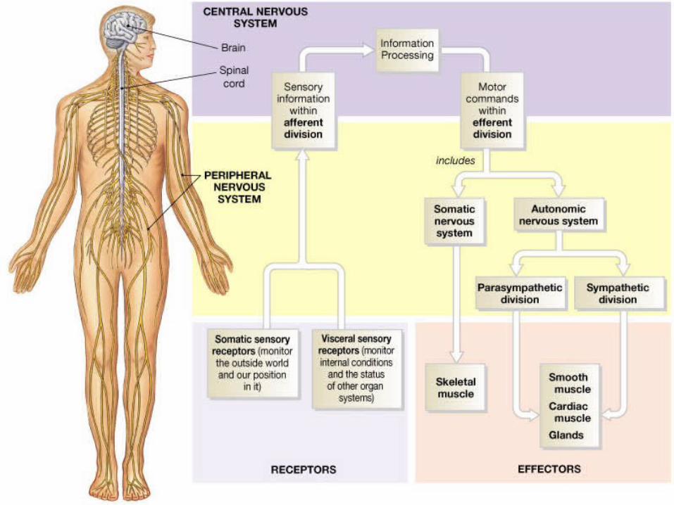
The nervous system includes al the neural tissue in the body.
Its anatomical divisions include the central nervous system (CNS) (the brain and spinal cord
And the peripheral nervous system (PNS) (all neural tissue outside the CNS)
The nervous system includes al the neural tissue in the body.
Its anatomical divisions include the central nervous system (CNS) (the brain and spinal cord
And the peripheral nervous system (PNS) (all neural tissue outside the CNS)
Functionally it can be divided into an afferent division which brings sensory information to the CNS
And an efferent division which carries motor commands to muscles and glands
Functionally it can be divided into an afferent division which brings sensory information to the CNS
And an efferent division which carries motor commands to muscles and glands
The efferent division includes the somatic nervous system (voluntary control over skeletal muscle contractions
And the autonomic nervous system (involuntary regulation of smooth muscle, cardiac muscle, and glandular activity.
The efferent division includes the somatic nervous system (voluntary control over skeletal muscle contractions
And the autonomic nervous system (involuntary regulation of smooth muscle, cardiac muscle, and glandular activity.
8-18-1
Cellular Organization in
Neural Tissue
Cellular Organization in
Neural TissueThere are two types of cells in neural tissueNeurons, which are responsible for information transfer and processing
Neuroglia, which provide a supporting framework and act as phagocytes.
There are two types of cells in neural tissueNeurons, which are responsible for information transfer and processing
Neuroglia, which provide a supporting framework and act as phagocytes.
NeuronsNeurons
Sensory neurons: form the afferent division of the PNS and deliver information to the CNS
Motor neurons: stimulate or modify the activity of a peripheral tissue, organ or organ system
Sensory neurons: form the afferent division of the PNS and deliver information to the CNS
Motor neurons: stimulate or modify the activity of a peripheral tissue, organ or organ system
Interneurons: may be located between sensory and motor neurons; they analyze sensory inputs and coordinate motor outputs.
Interneurons: may be located between sensory and motor neurons; they analyze sensory inputs and coordinate motor outputs.
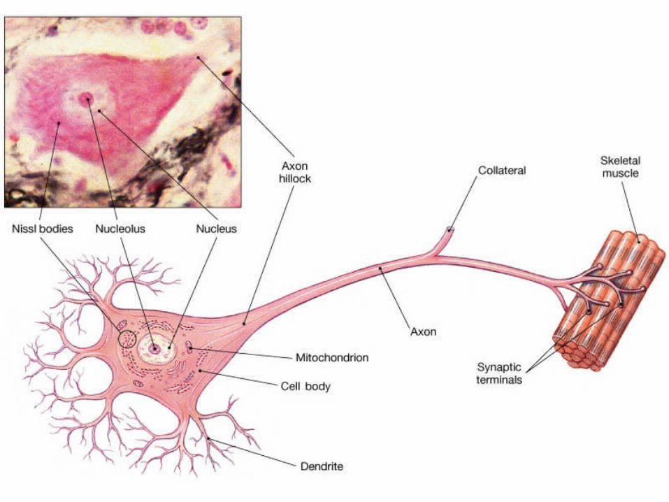
A typical neuron has a cell body, an axon, and several branching dendrites and synaptic terminals
A typical neuron has a cell body, an axon, and several branching dendrites and synaptic terminals
8-28-2
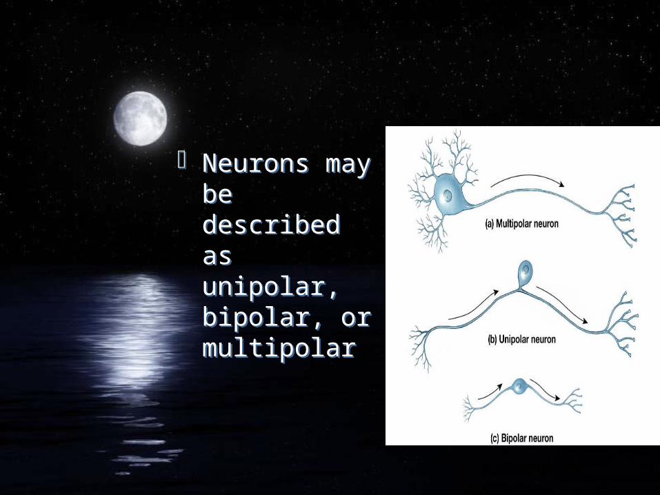
Neurons may be described as unipolar, bipolar, or multipolar
Neurons may be described as unipolar, bipolar, or multipolar
8-38-3
NeurogliaNeuroglia
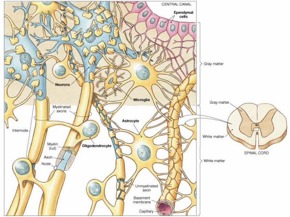
Four typesAstrocytes: largest and most numerous
Oligodendrocytes: myelinate CNS neurons
Microglia: phagocytic white glod cells
Ependymal: produce and help circulate cerebral spinal fluid
Four typesAstrocytes: largest and most numerous
Oligodendrocytes: myelinate CNS neurons
Microglia: phagocytic white glod cells
Ependymal: produce and help circulate cerebral spinal fluid
8-48-4
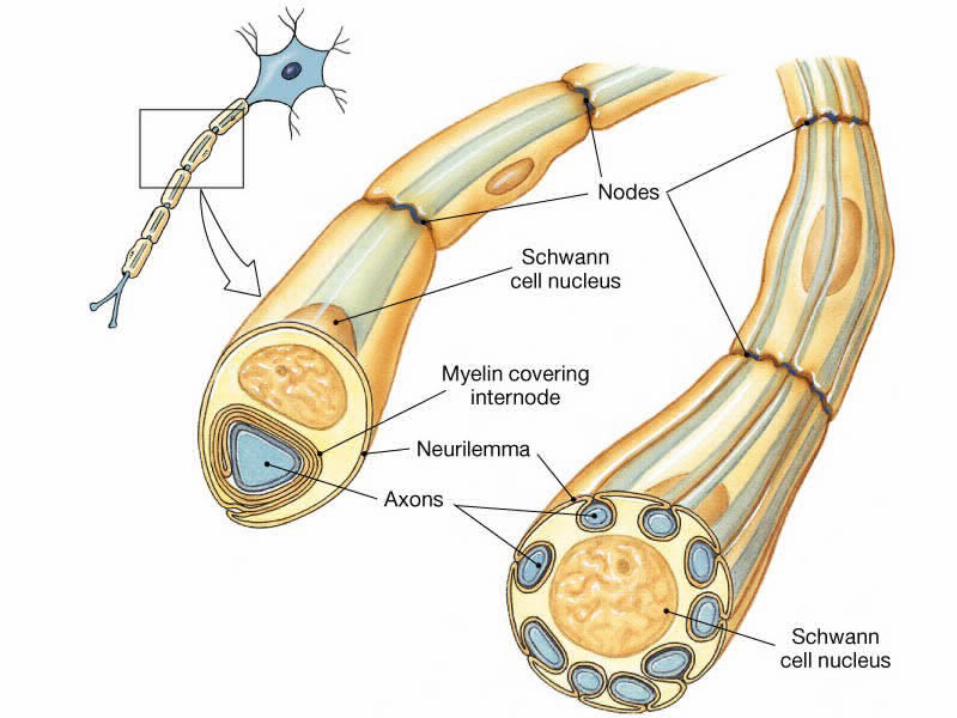
Nerve cell bodies in the PNS are clustered into ganglia.
Their axons are covered by myelin wrappings of Schwann cells.
Nerve cell bodies in the PNS are clustered into ganglia.
Their axons are covered by myelin wrappings of Schwann cells.
8-58-5
Anatomical organizationAnatomical
organizationIn the CNS, a collection of neuron cell bodies that share a particular function is called a center
A center with a a discrete anatomical boundary is called a nucleus.
Portions of the brain surface are covered by a thick layer of gray matter called the neural cortex
In the CNS, a collection of neuron cell bodies that share a particular function is called a center
A center with a a discrete anatomical boundary is called a nucleus.
Portions of the brain surface are covered by a thick layer of gray matter called the neural cortex
The white matter of the CNS contains bundles of axons, or tracts that share common origins, destinations and functions.
Tract in the spinal cord form larger groups called columns
The white matter of the CNS contains bundles of axons, or tracts that share common origins, destinations and functions.
Tract in the spinal cord form larger groups called columns

8-68-6
Neuron Function: Membrane
Potential
Neuron Function: Membrane
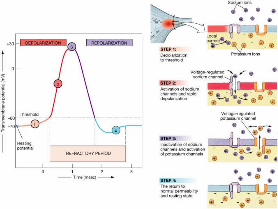
PotentialThe resting potential, or membrane potential of an undisturbed nerve cell, is due to a balance between the rate of sodium ion entry and potassium ion loss and to the sodium-potassium exchange pump
The resting potential, or membrane potential of an undisturbed nerve cell, is due to a balance between the rate of sodium ion entry and potassium ion loss and to the sodium-potassium exchange pump
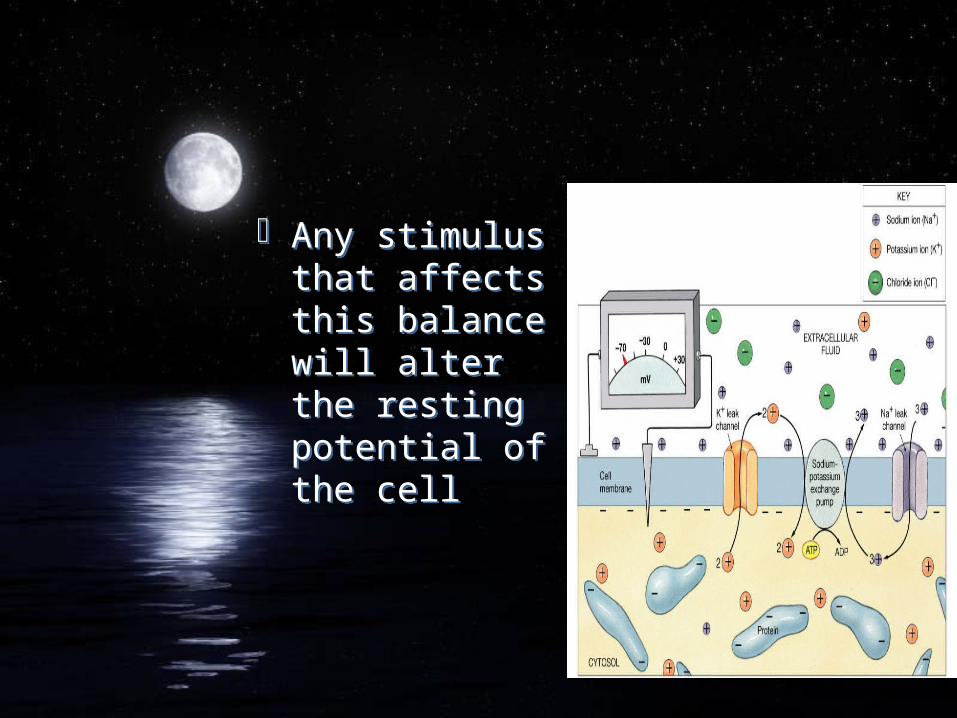
Any stimulus that affects this balance will alter the resting potential of the cell
Any stimulus that affects this balance will alter the resting potential of the cell
8-78-7
An action potential appears when the membrane depolarizes to a level known as the threshold.
Steps include; opening of sodium channels and opening of potassium channels, return to normal permeability
An action potential appears when the membrane depolarizes to a level known as the threshold.
Steps include; opening of sodium channels and opening of potassium channels, return to normal permeability
8-88-8
Propagation of an Action Potential
Propagation of an Action Potential
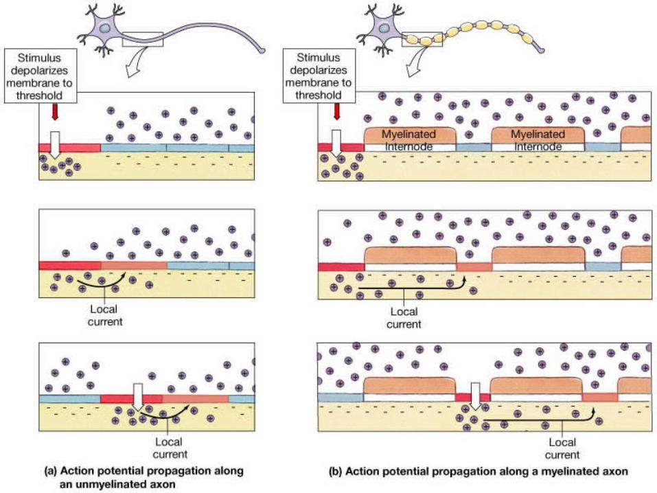
In continuous propagation, an action potential spread across the entire excitable membrane surface in a series of small steps.
During saltatory propagation, the action potential appears to leap from node to node.
In continuous propagation, an action potential spread across the entire excitable membrane surface in a series of small steps.
During saltatory propagation, the action potential appears to leap from node to node.
8-98-9

Action potentialAction potential
Neural Communication
Neural Communication
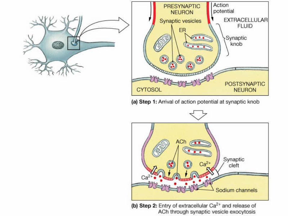
A synapse is a site where intercellular communication occurs through the release of chemicals called neurotransmitters.
A synapse where neurons communicate with other cell types is a neuroeffector junction.
A synapse is a site where intercellular communication occurs through the release of chemicals called neurotransmitters.
A synapse where neurons communicate with other cell types is a neuroeffector junction.
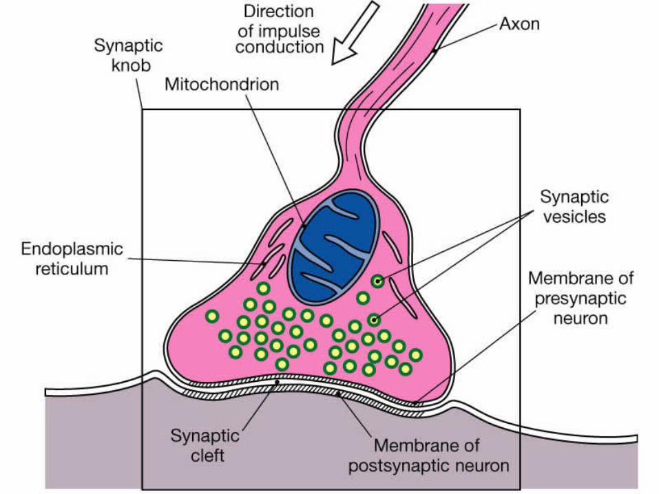
Structure of a Synapse
Structure of a Synapse
Neural communication moves from the presynaptic neuron to the postsynaptic neuron over the synaptic cleft
Neural communication moves from the presynaptic neuron to the postsynaptic neuron over the synaptic cleft
8-108-10
Synaptic function and
Neurotransmitters
Synaptic function and
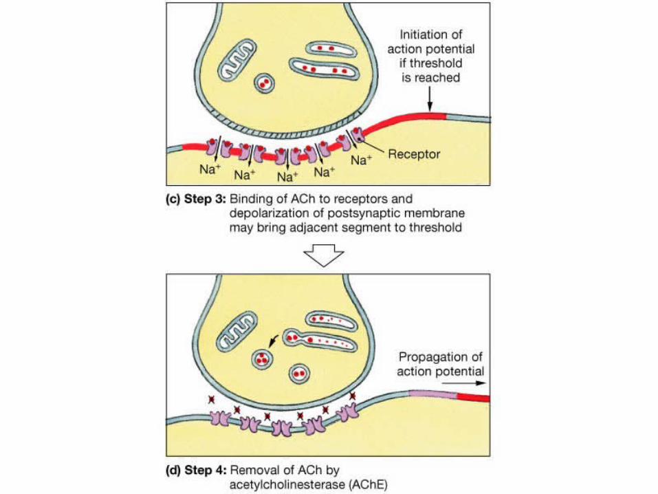
NeurotransmittersCholinergic synapses release the neurotransmitter acetylcholine (Ach)
Ach is broken down in the synaptic cleft by the enzyme acetylcholinesterase (AChE)
Cholinergic synapses release the neurotransmitter acetylcholine (Ach)
Ach is broken down in the synaptic cleft by the enzyme acetylcholinesterase (AChE)
Other neurotransmitters
Other neurotransmittersNorepinephrine-excitesDopamine-inhibitsGABA -inhibitsSerotonin-inhibits
Norepinephrine-excitesDopamine-inhibitsGABA -inhibitsSerotonin-inhibits
8-11, table 8-18-11, table 8-1
neurotransmitters neurotransmitters