the nervous system - mt. san antonio collegeinstruction2.mtsac.edu/crexach/physiology/pdf physio...
TRANSCRIPT
The Nervous SystemThe Nervous SystemThe Nervous System
Dr. Carmen E. Dr. Carmen E. RexachRexachPhysiologyPhysiology
MtSACMtSAC Biology DepartmentBiology Department
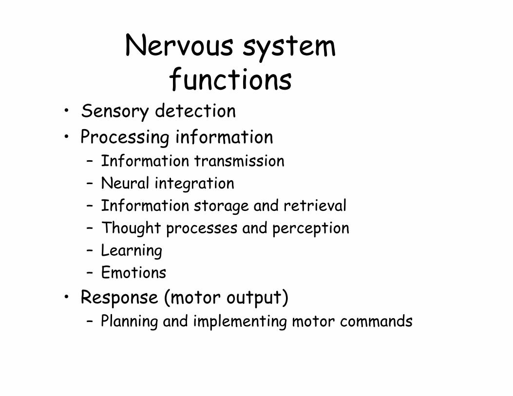
Nervous system functions
• Sensory detection• Processing information
– Information transmission– Neural integration– Information storage and retrieval– Thought processes and perception– Learning– Emotions
• Response (motor output) – Planning and implementing motor commands
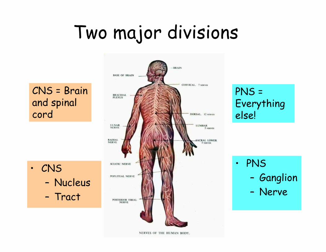
Two major divisions
• PNS– Ganglion– Nerve
• CNS– Nucleus– Tract
CNS = Brain and spinal cord
PNS = Everything else!
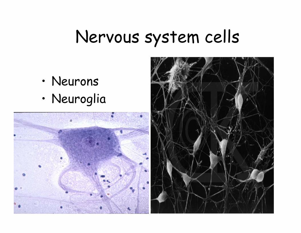
Nervous system cells
• Neurons• Neuroglia
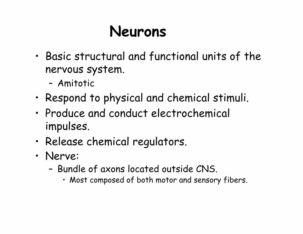
Neurons• Basic structural and functional units of the
nervous system.– Amitotic
• Respond to physical and chemical stimuli.• Produce and conduct electrochemical
impulses.• Release chemical regulators.• Nerve:
– Bundle of axons located outside CNS.• Most composed of both motor and sensory fibers.

Anatomy of a Neuron• Cell body (perikaryon)
– “Nutrition center.”– Cell bodies are clustered
• ‘Nuclei’ in CNS• ‘Ganglia’ in PNS
• Dendrites– Provide receptive area.– Transmit electrical impulses to cell body.
• Axon– Conducts impulses away from cell body.
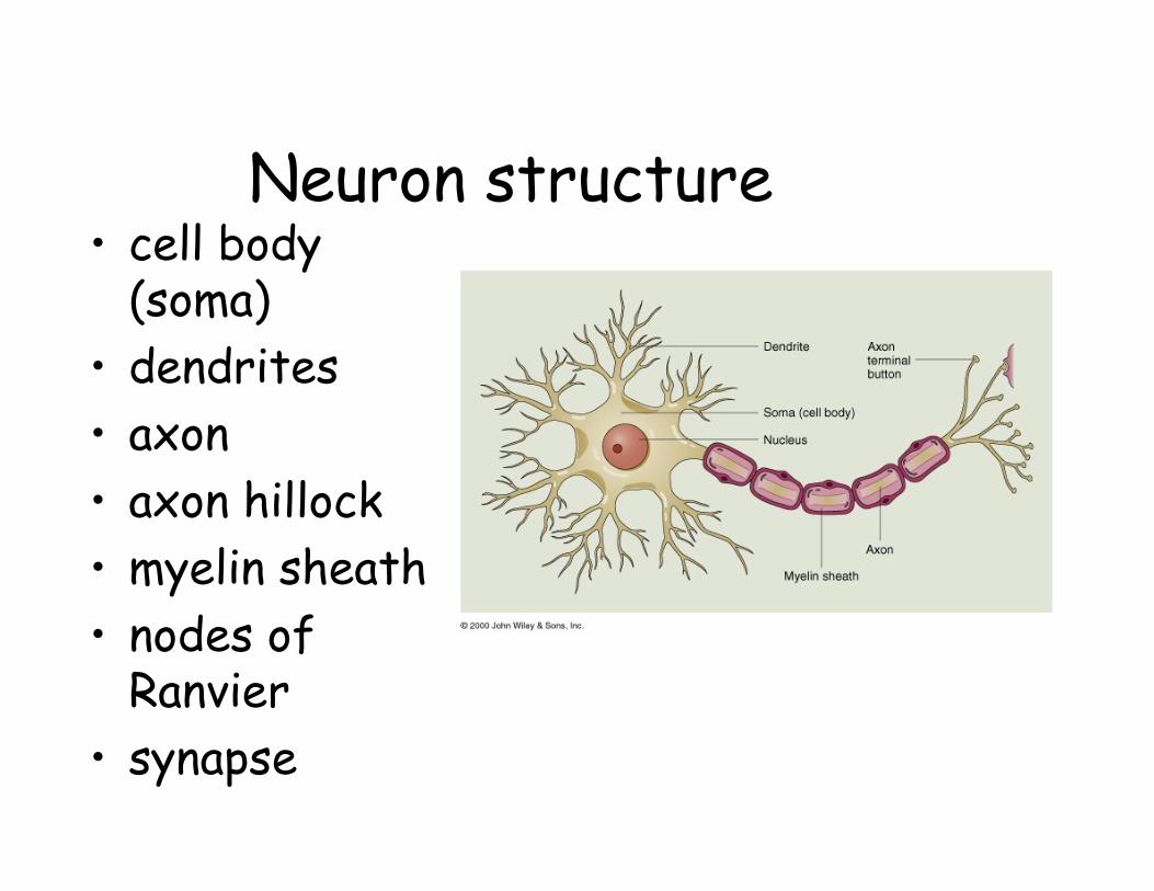
Neuron structure• cell body
(soma)• dendrites• axon• axon hillock• myelin sheath• nodes of
Ranvier• synapse
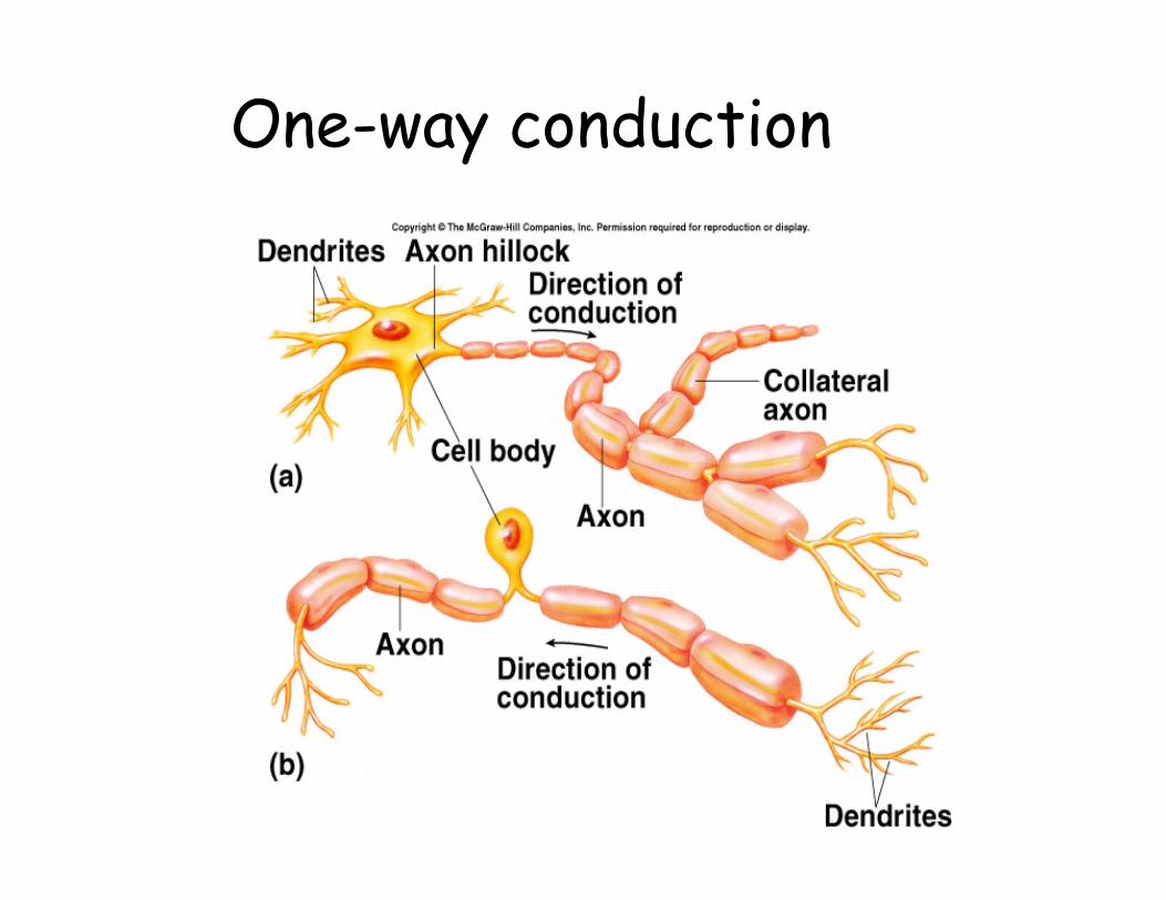
One-way conduction

Neuroglia = “nerve glue”
• Astrocytes• Ependymal cells• Microglia• Oligodendrocytes• Schwann cells
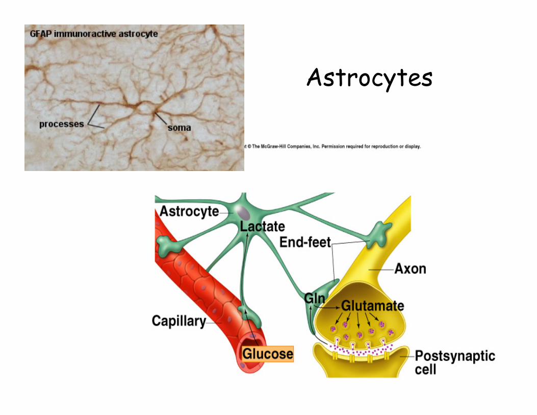
Astrocytes• Most numerous glial cell• Regulate microenvironment of neurons in
CNS– Blood brain barrier– Buffer extracellular environment
• Metabolize neurotransmitter substances and uptake K+
• Mechanical support for CNS tissues• Trigger synapse formation by secreting
signaling proteins called thrombospondins(Science, 21 Nov 2003)
Astrocytes
Blood-Brain Barrier• Capillaries in brain do not have pores
between adjacent endothelial cells.– Joined by tight junctions.
• Molecules within brain capillaries moved selectively through endothelial cells by:– Diffusion.– Active transport.– Endocytosis.– Exocytosis.

Ependymal cells• Epithelium that lines CNS• Specialized ependymal cells of choroid plexus
secrete components of CSF• Function as neural stem cells: can divide,
progeny can differentiate
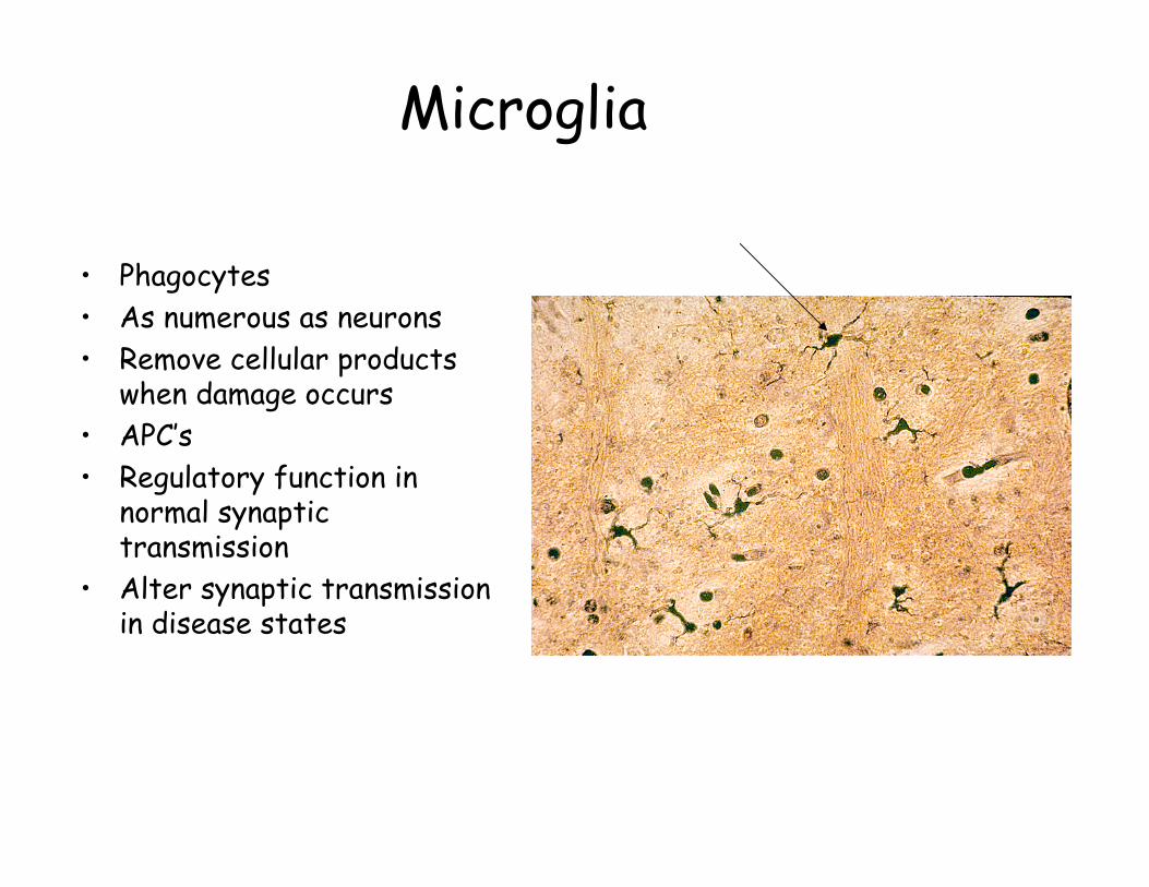
Microglia
• Phagocytes• As numerous as neurons• Remove cellular products
when damage occurs• APC’s• Regulatory function in
normal synaptic transmission
• Alter synaptic transmission in disease states
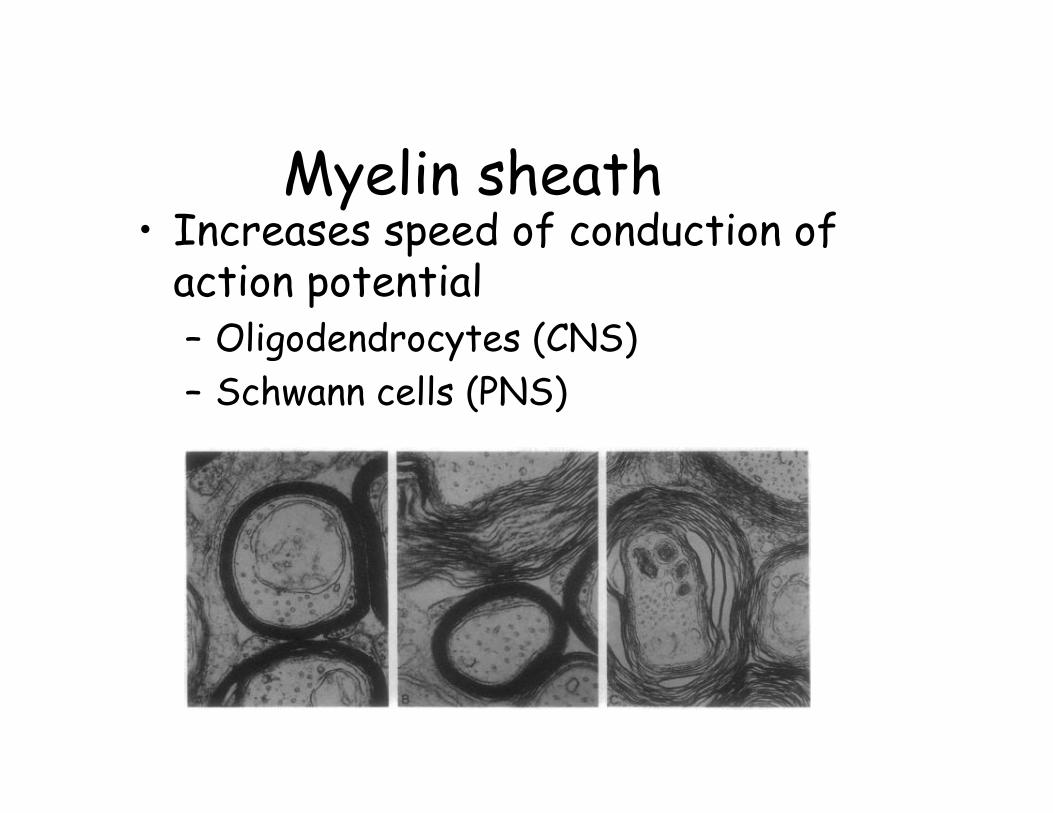
Myelin sheath• Increases speed of conduction of
action potential– Oligodendrocytes (CNS)– Schwann cells (PNS)

Oligodendrocytes
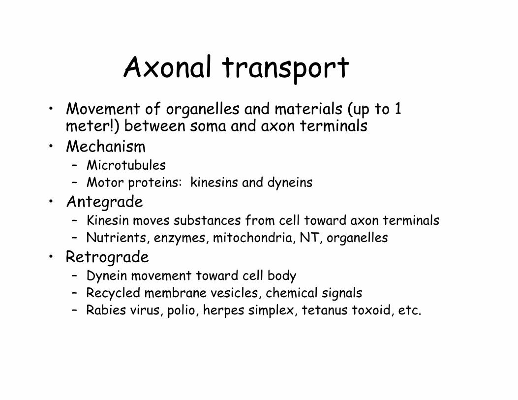
Axonal transport• Movement of organelles and materials (up to 1
meter!) between soma and axon terminals• Mechanism
– Microtubules– Motor proteins: kinesins and dyneins
• Antegrade– Kinesin moves substances from cell toward axon terminals– Nutrients, enzymes, mitochondria, NT, organelles
• Retrograde– Dynein movement toward cell body– Recycled membrane vesicles, chemical signals– Rabies virus, polio, herpes simplex, tetanus toxoid, etc.
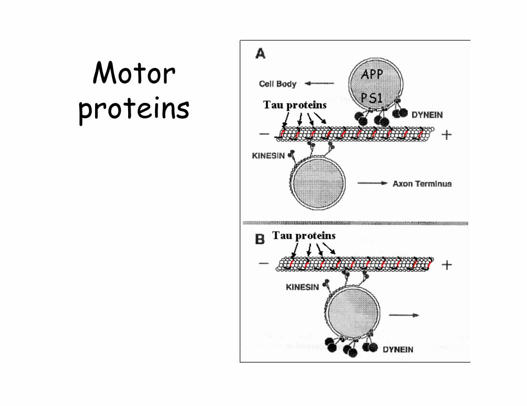
Motor proteins
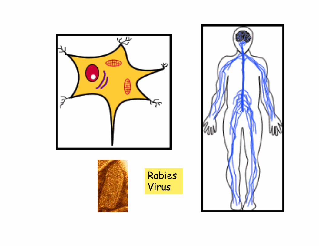
Rabies Virus
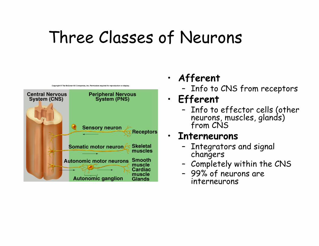
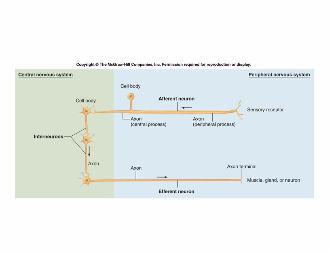
Three Classes of Neurons
• Afferent– Info to CNS from receptors
• Efferent– Info to effector cells (other
neurons, muscles, glands) from CNS
• Interneurons– Integrators and signal
changers– Completely within the CNS– 99% of neurons are
interneurons
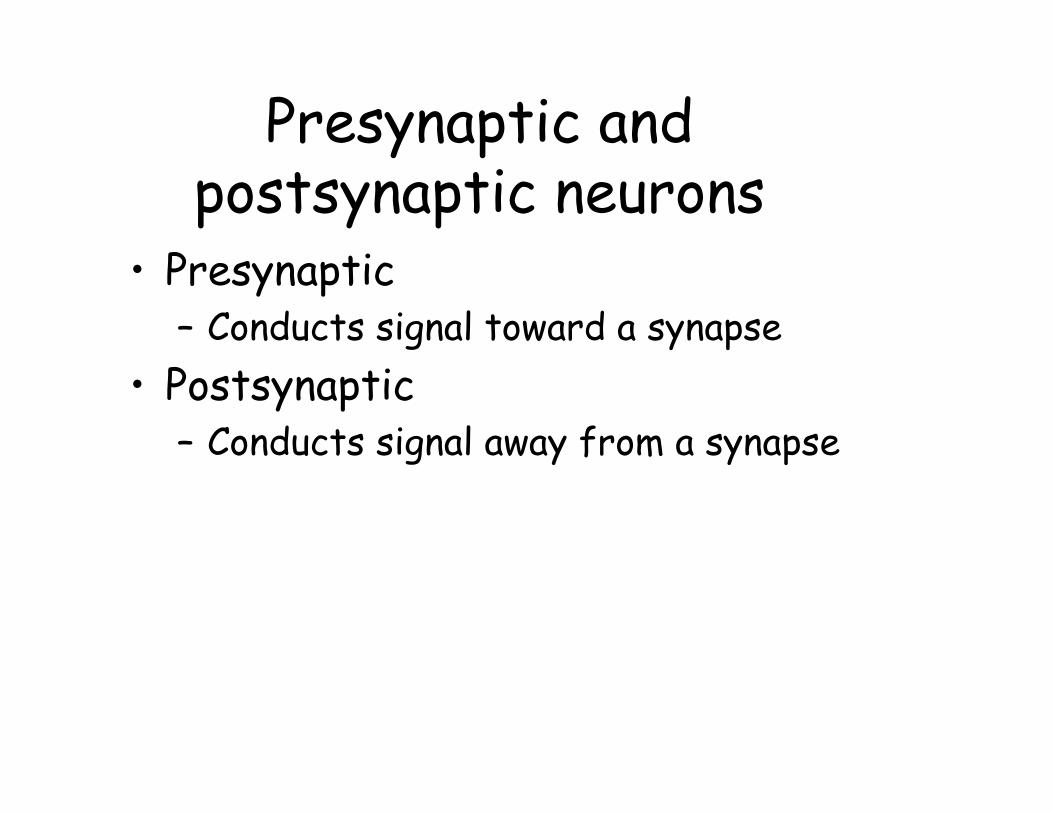
Presynaptic and postsynaptic neurons
• Presynaptic– Conducts signal toward a synapse
• Postsynaptic– Conducts signal away from a synapse
Neural development• Differentiation of neural cells from embryonic stem
cells– Division and differentiation of neural cells during fetal
development results in increased susceptibility to drugs, alcohol, etc.
• Development of axon by extension of growth cone• Receptors and neurotrophic factors determine
neural pathways• Apoptosis of 50-70% of neurons• Neuron precursors complete division before birth
– New synaptic connections continue to form and degenerate throughout life
When axon is damaged: Axonal reaction
• Occurs when axon is transected• Damage control and repair results in
increased protein synthesis = chromatolysis
• Disintegration of axon distal to transection• Viability of myelin sheath maintained• sprout formation & growth cones
– possible in PNS, less likely in CNS
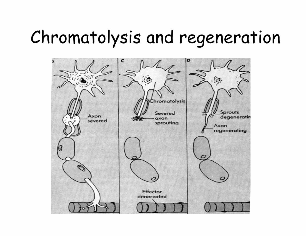
Chromatolysis and regeneration
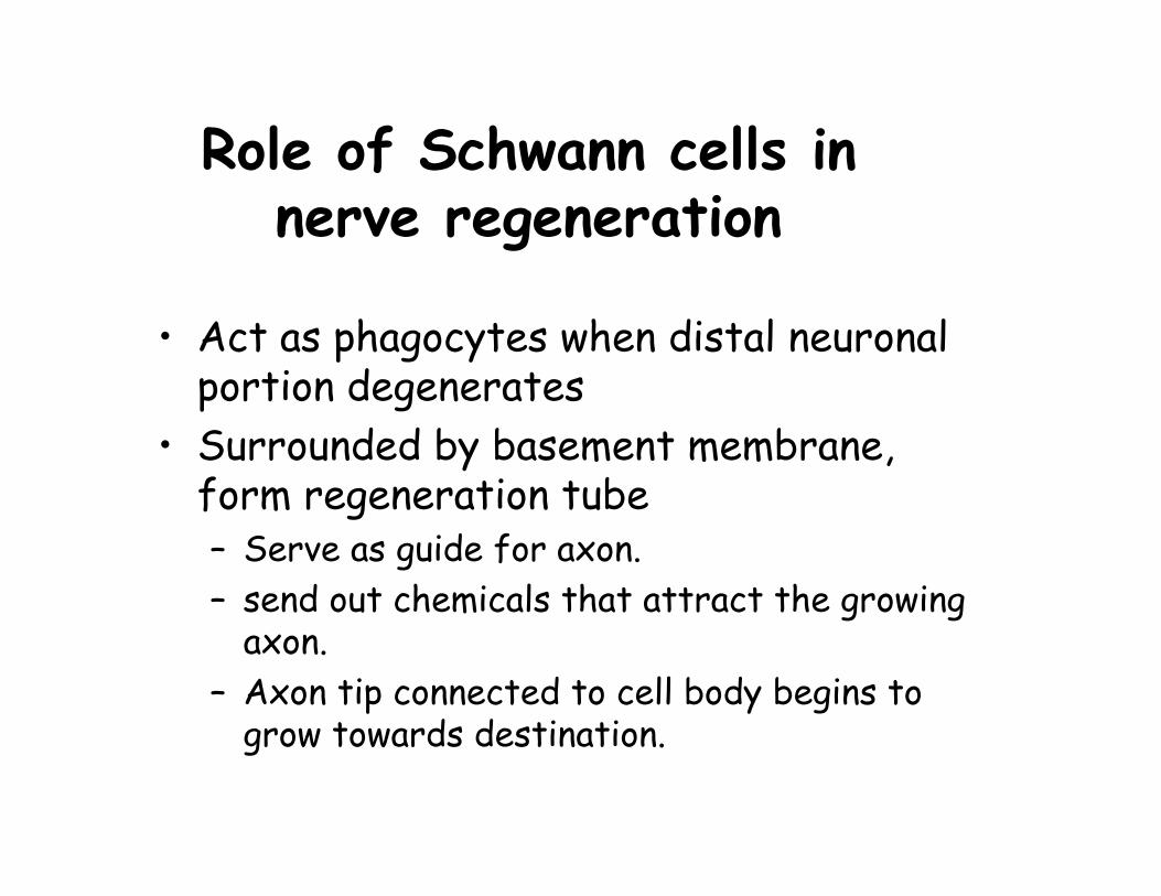
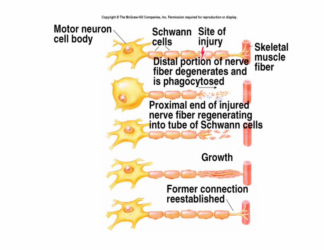
Role of Schwann cells in nerve regeneration
• Act as phagocytes when distal neuronal portion degenerates
• Surrounded by basement membrane, form regeneration tube– Serve as guide for axon.– send out chemicals that attract the growing
axon.– Axon tip connected to cell body begins to
grow towards destination.

Action potentials and graded potentials
Action potentials and Action potentials and graded potentialsgraded potentials
Electricity: the basics!
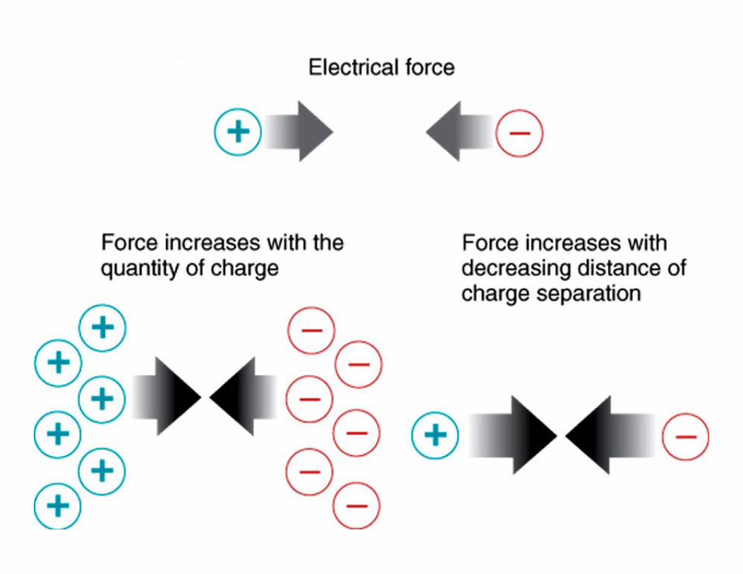
• Opposite charges attract• Electrical force of attraction
– increases with quantity of charge – decreases with distance between charges
• Charges separated across plasma membrane– Electrical potential = potential difference (mV)– Current = movement of electrical charge– Ohm’s law = I =V/R (I=current, v=voltage,
r=resistance)• Plasma membrane (lipids) have high electrical
resistance• Water has low electrical resistance (cytosol, ECF)
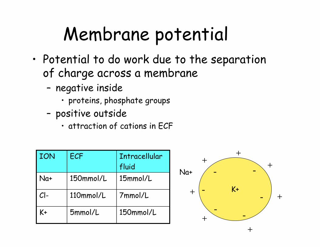
Membrane potential• Potential to do work due to the separation
of charge across a membrane– negative inside
• proteins, phosphate groups– positive outside
• attraction of cations in ECF
K+
- -
-
--
-
+
+
+
+
+
+
+
150mmol/L5mmol/LK+
7mmol/L110mmol/LCl-
15mmol/L150mmol/LNa+
Intracellular fluid
ECFION
Na+
Resting membrane potential
• The potential difference across a membrane when the cell is in an unstimulated state
• Range: -40 to -90 mV in neurons• Determined by:
– Differences in ion concentrations in intracellular and extracellular compartments
– permeability of the membrane to each ion
Excitable membranes
• Cells with excitable or irritable membranes can change their membrane potential when stimulated
• Mechanism– movement of ions by changing membrane
permeability• Neurons can use this to transmit impulses
down axon• Muscle cells can use this to initiate
contraction.
Equilibrium potential• Remember that we have to consider
not only the concentration gradient, but also the electrical gradient
• Equilibrium potential is a theoretical voltage produced across a cell membrane with the movement of only one ion
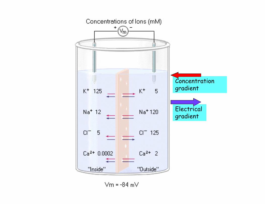
Concentration gradient
Electrical gradient
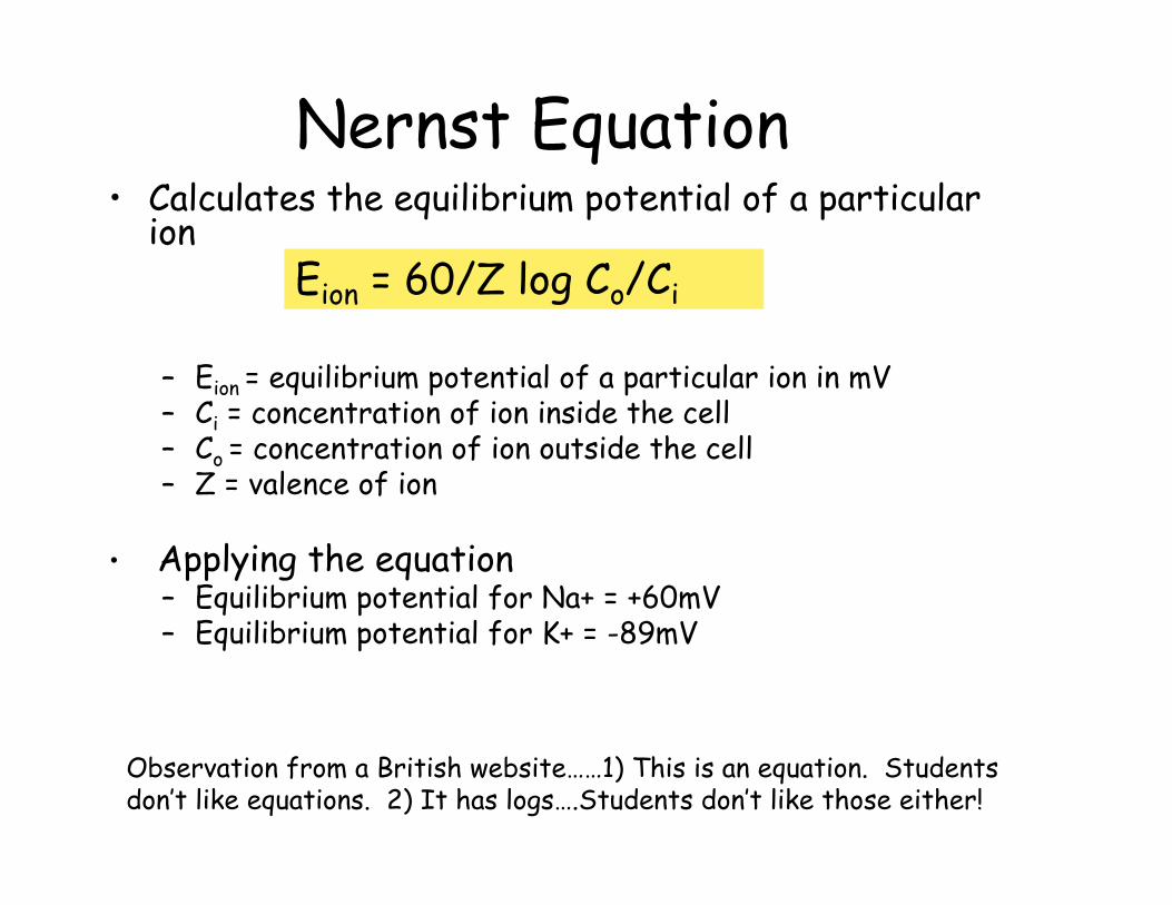
Nernst Equation• Calculates the equilibrium potential of a particular
ion
– Eion = equilibrium potential of a particular ion in mV– Ci = concentration of ion inside the cell– Co = concentration of ion outside the cell– Z = valence of ion
• Applying the equation– Equilibrium potential for Na+ = +60mV– Equilibrium potential for K+ = -89mV
Eion = 60/Z log Co/Ci
Observation from a British website……1) This is an equation. Students don’t like equations. 2) It has logs….Students don’t like those either!
Understanding equilibrium potential
• The cell is filled with fixed anions• If K+ diffuse into the cell unimpeded, equilibrium
would be reached when the electrical attraction for the anions inside the cell and the diffusion gradient were equal in magnitude but opposite in direction
• The number of K+ ions inside the cell at equilibrium would be greater than the number outside, but not enough to neutralize the fixed anions and the cell would have a net voltage of approx -70mV, the resting membrane potential
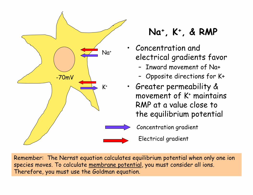
Na+, K+, & RMP
• Concentration and electrical gradients favor– Inward movement of Na+– Opposite directions for K+
• Greater permeability & movement of K+ maintains RMP at a value close to the equilibrium potentialConcentration gradient
Electrical gradient
Na+
K+
Remember: The Nernst equation calculates equilibrium potential when only one ion species moves. To calculate membrane potential, you must consider all ions. Therefore, you must use the Goldman equation.
-70mV
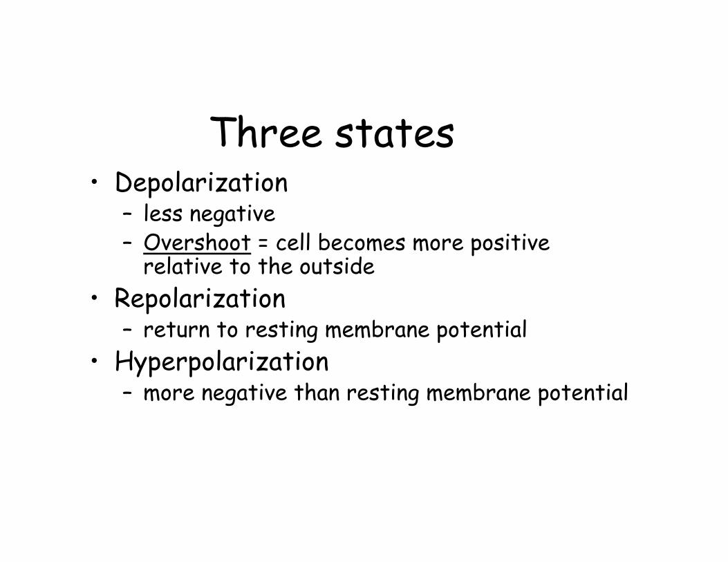
Three states• Depolarization
– less negative– Overshoot = cell becomes more positive
relative to the outside• Repolarization
– return to resting membrane potential• Hyperpolarization
– more negative than resting membrane potential
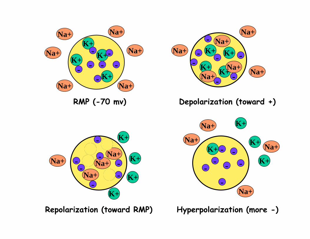
RMP (-70 mv) Depolarization (toward +)
Repolarization (toward RMP) Hyperpolarization (more -)
K+
K+
K+K+
K+
K+ K+K+
K+K+
K+
K+
K+K+
K+
K+
Na+
Na+ Na+
Na+ Na+
Na+Na+
Na+
Na+Na+
Na+
Na+
Na+
Na+
Na+
Na+
-
--
--
- -
-
- --
-
-
-
--
-
-
- --
-
-
-
-
- --
K+
K+
K+
K+
Na+
Na+
Na+Na+
Graded Potentials• Changes in membrane potential• Variable magnitude
– Depolarization or hyperpolarization• Short distances• “fizzle out”
– Conducted decrementally• Examples:
– Receptor potential– Synaptic potential– Pacemaker potential
Action potential • Definition: All-or-none event in an axon or
muscle fiber in which the polarity of the membrane potential is rapidly reversed and reestablished
• Characteristics:– Large voltage changes– Rapid (1-4msec/ap)– Occurs in cells with excitable membranes
• Nerve cells• Muscle cells• Some others (some endocrine, immune, reproductive…not
all!)
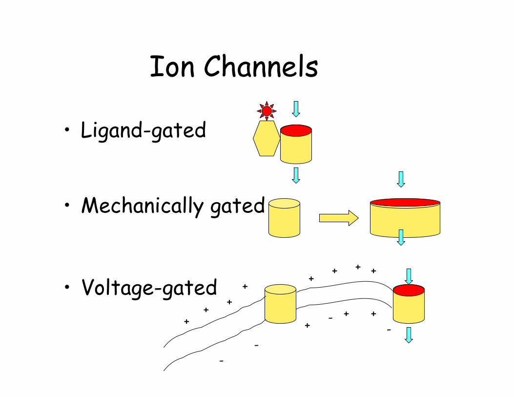
Ion Channels
• Ligand-gated
• Mechanically gated
• Voltage-gated+
+
++
+
+
++
--
--
+
+
+
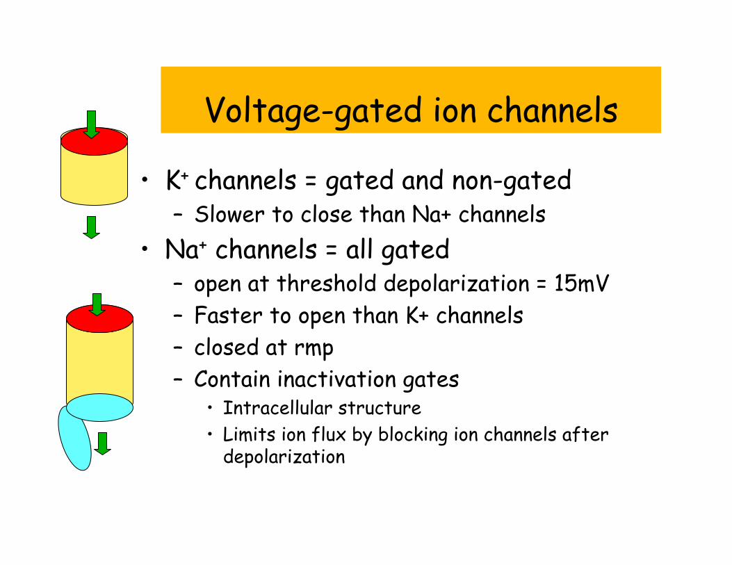
Voltage-gated ion channels
• K+ channels = gated and non-gated– Slower to close than Na+ channels
• Na+ channels = all gated– open at threshold depolarization = 15mV– Faster to open than K+ channels– closed at rmp– Contain inactivation gates
• Intracellular structure• Limits ion flux by blocking ion channels after
depolarization
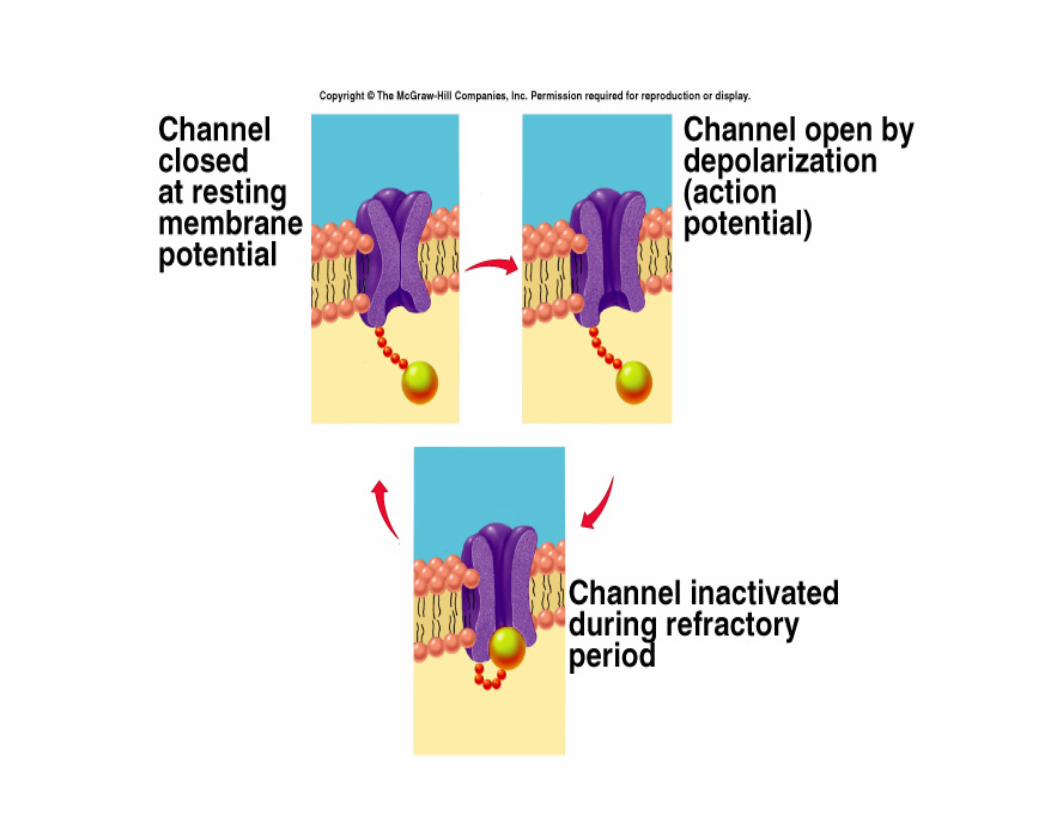
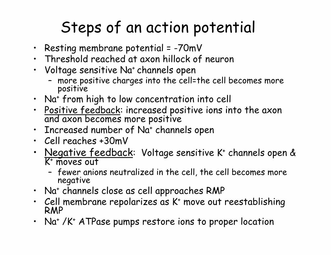
Steps of an action potential• Resting membrane potential = -70mV• Threshold reached at axon hillock of neuron• Voltage sensitive Na+ channels open
– more positive charges into the cell=the cell becomes more positive
• Na+ from high to low concentration into cell• Positive feedback: increased positive ions into the axon
and axon becomes more positive• Increased number of Na+ channels open• Cell reaches +30mV• Negative feedback: Voltage sensitive K+ channels open &
K+ moves out– fewer anions neutralized in the cell, the cell becomes more
negative• Na+ channels close as cell approaches RMP• Cell membrane repolarizes as K+ move out reestablishing
RMP• Na+ /K+ ATPase pumps restore ions to proper location
Na+ & K+ concentration• Relative concentration of Na+/K+ remains
unchanged during a/p– small number of ions actually move– huge number of Na+/K+ATPase pumps
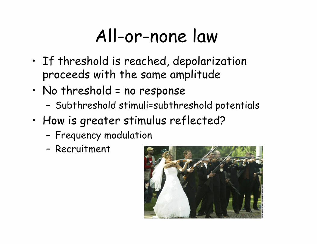
All-or-none law• If threshold is reached, depolarization
proceeds with the same amplitude• No threshold = no response
– Subthreshold stimuli=subthreshold potentials• How is greater stimulus reflected?
– Frequency modulation– Recruitment
Effects of anesthesia
• Block action potentials• Local anesthetics block voltage-gated
Na+ channels– Novocain– lidocaine
• Fugu– Tetrodotoxin produced in ovaries
Graded potentials received by sensory neurons cannot be sent to the CNS.

Fugu is a delicacy in Japan & the Philippines. Because of the toxin, sushi chefs preparing fugu in some countries must be licensed.
Refractory period• Prevents an action potential from
beginning before the first is complete– absolute refractory period
• Na+ channels already open• Na+ channels in inactive state
– relative refractory period• Follows absolute refractory period • Stronger than normal stimulus required
• Limits number of a/p a nerve can produce in a given time period
• Ensures that a/p move down axon
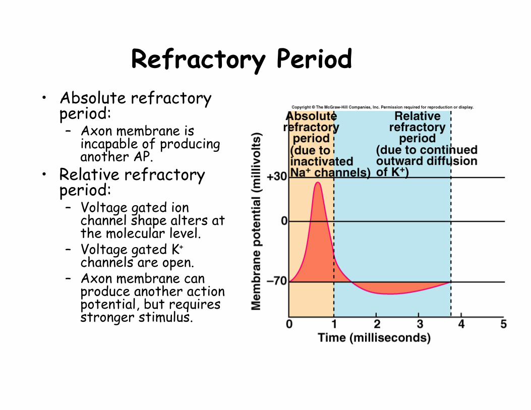
Refractory Period• Absolute refractory
period:– Axon membrane is
incapable of producing another AP.
• Relative refractory period:– Voltage gated ion
channel shape alters at the molecular level.
– Voltage gated K+
channels are open.– Axon membrane can
produce another action potential, but requires stronger stimulus.
Conduction of nerve impulse• Propagation of nerve impulse by
incremental depolarization of axon segments– Ions leak to next segment, initiating a/p– Unidirectional due to refractory period
• Allows for impulse to travel length of the axon and trigger release of neurotransmitter
• Speed of impulse related to diameter of axon and myelination status
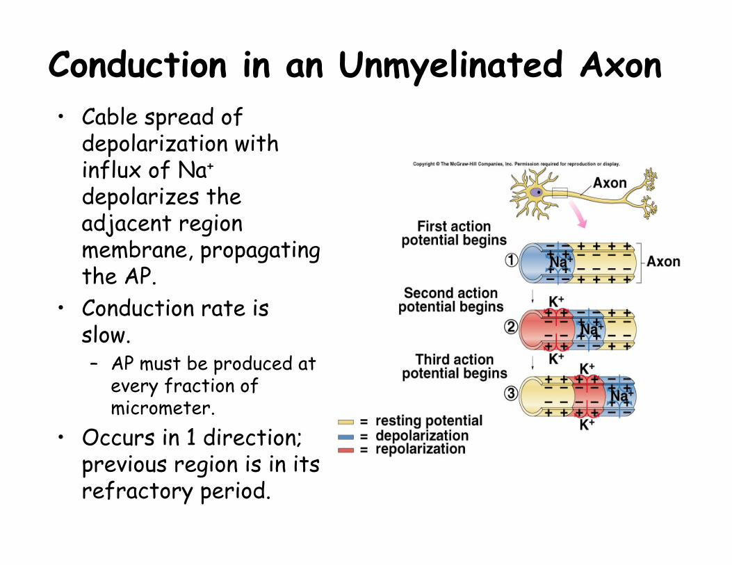
Conduction in an Unmyelinated Axon• Cable spread of
depolarization with influx of Na+
depolarizes the adjacent region membrane, propagating the AP.
• Conduction rate is slow.– AP must be produced at
every fraction of micrometer.
• Occurs in 1 direction; previous region is in its refractory period.

Myelination of axons• Unmeylinated axons
– depolarization of each segment
– small unmyelinated axon = 1.0m/sec
• Myelinated axons– saltatory conduction– nodes of Ranvier and Na+
channel concentration– thick, myelinated axon =
100m/sec
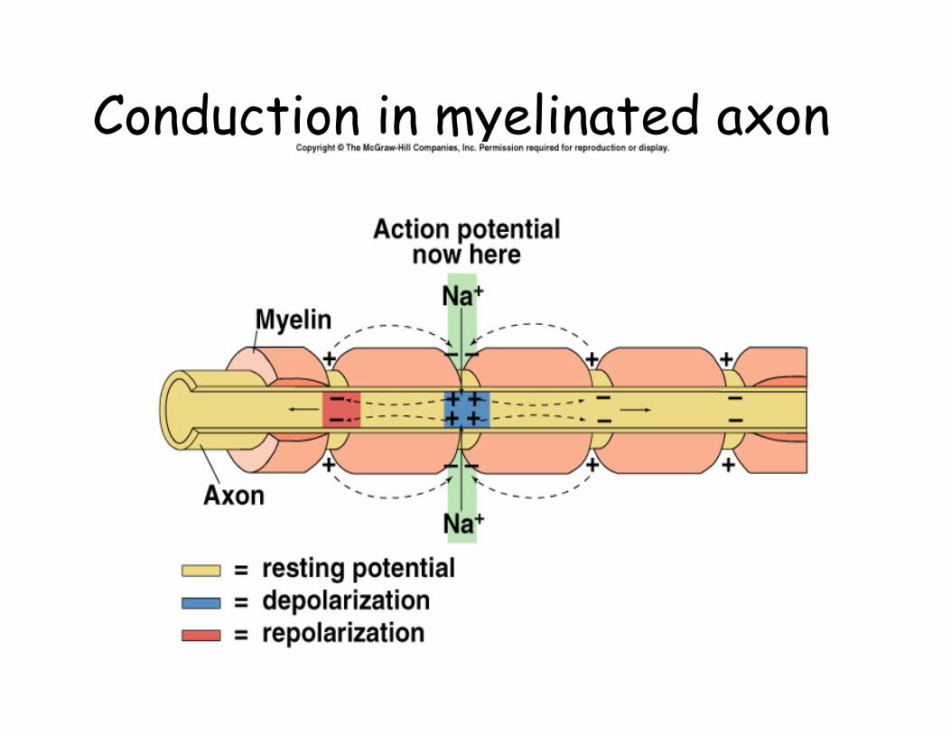
Conduction in myelinated axon
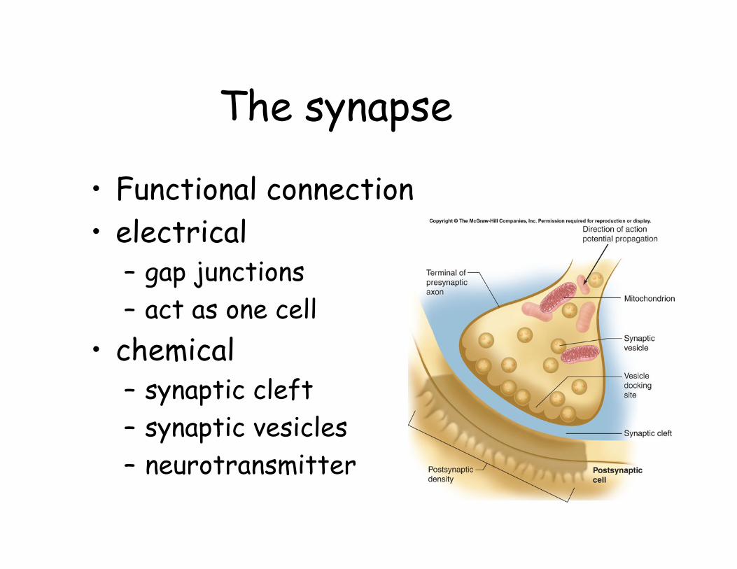
The synapse
• Functional connection• electrical
– gap junctions– act as one cell
• chemical– synaptic cleft– synaptic vesicles– neurotransmitter

Electrical Synapse• Impulses can be
regenerated without interruption in adjacent cells.
• Gap junctions:– Adjacent cells
electrically coupled through a channel.
– Each gap junction is composed of 12 connexin proteins.
• Examples:– Smooth and cardiac
muscles, brain, and glialcells.
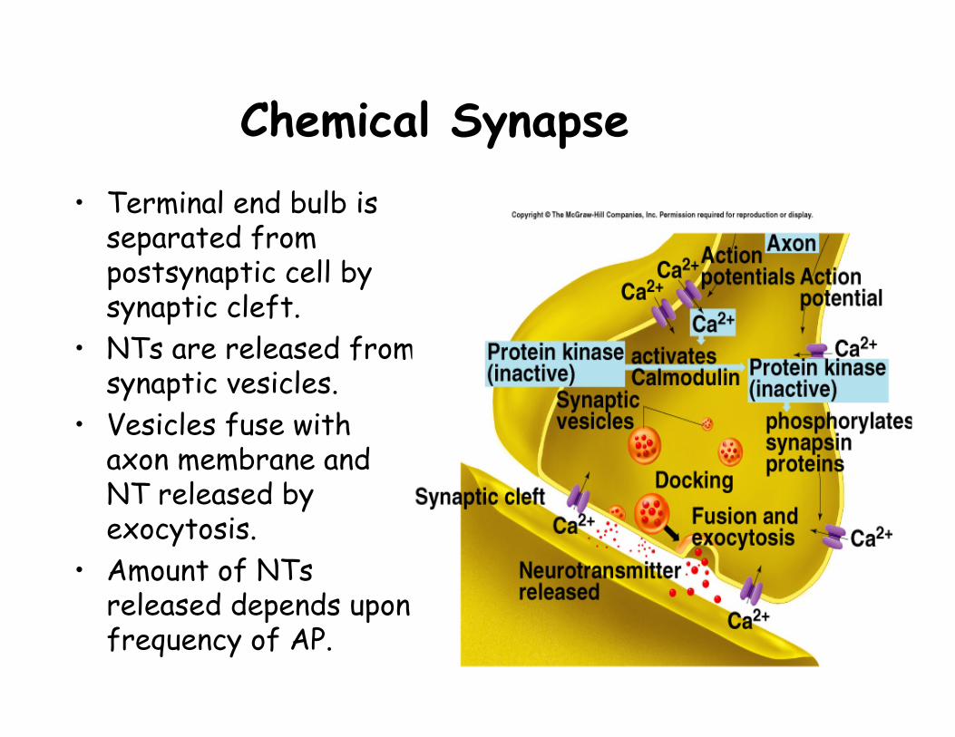
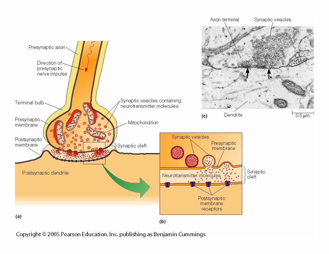
Chemical Synapse• Terminal end bulb is
separated from postsynaptic cell by synaptic cleft.
• NTs are released from synaptic vesicles.
• Vesicles fuse with axon membrane and NT released by exocytosis.
• Amount of NTsreleased depends upon frequency of AP.
Release of neurotransmitter• Action potential arrives at the terminal end bulb of
presynaptic neuron• voltage-regulated Ca++ channels open• Ca++ influx activates calmodulin• activated calmodulin activates a protein kinase• synapsin phosphorylation• fusion of vesicles with presynaptic membrane &
release of neurotransmitter• NT binds to receptors on postsynaptic membrane• opening of channels with chemically regulated gates
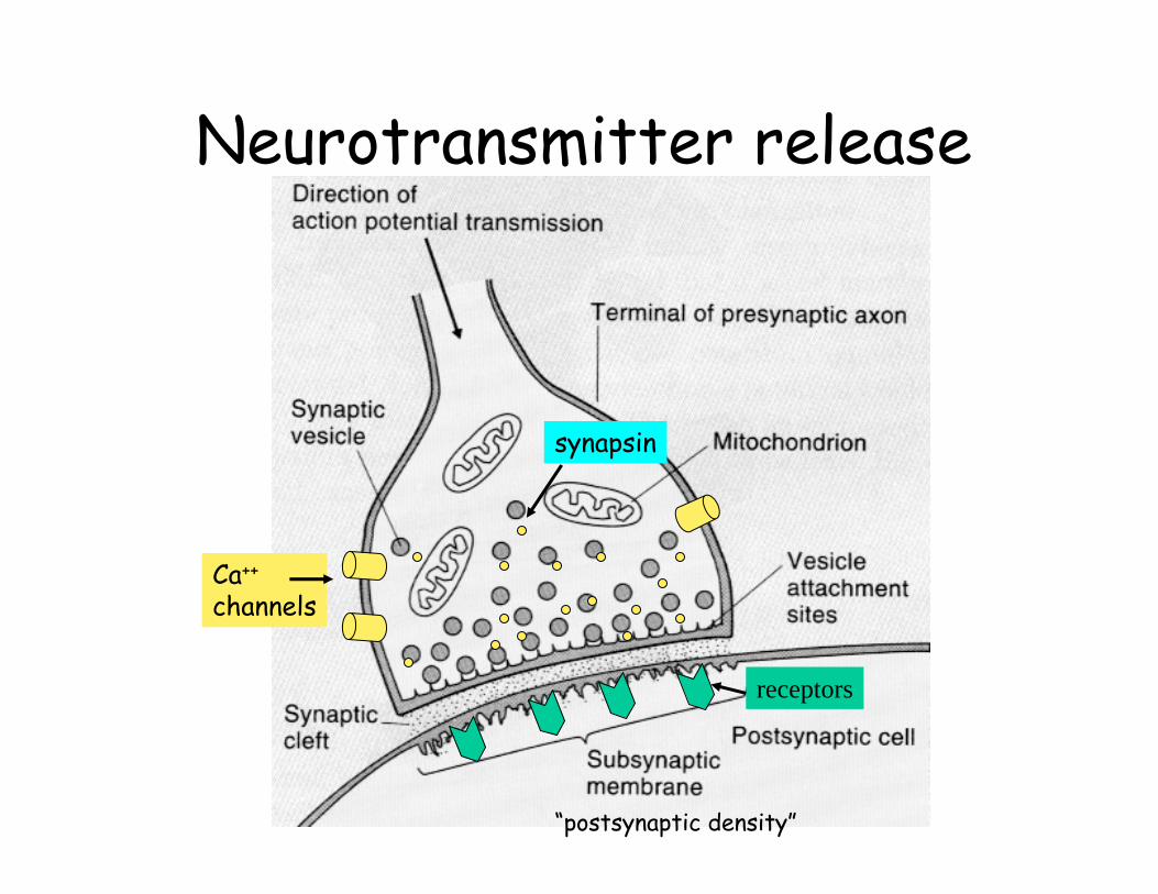
Neurotransmitter release
synapsin
Ca++
channels
receptors
“postsynaptic density”
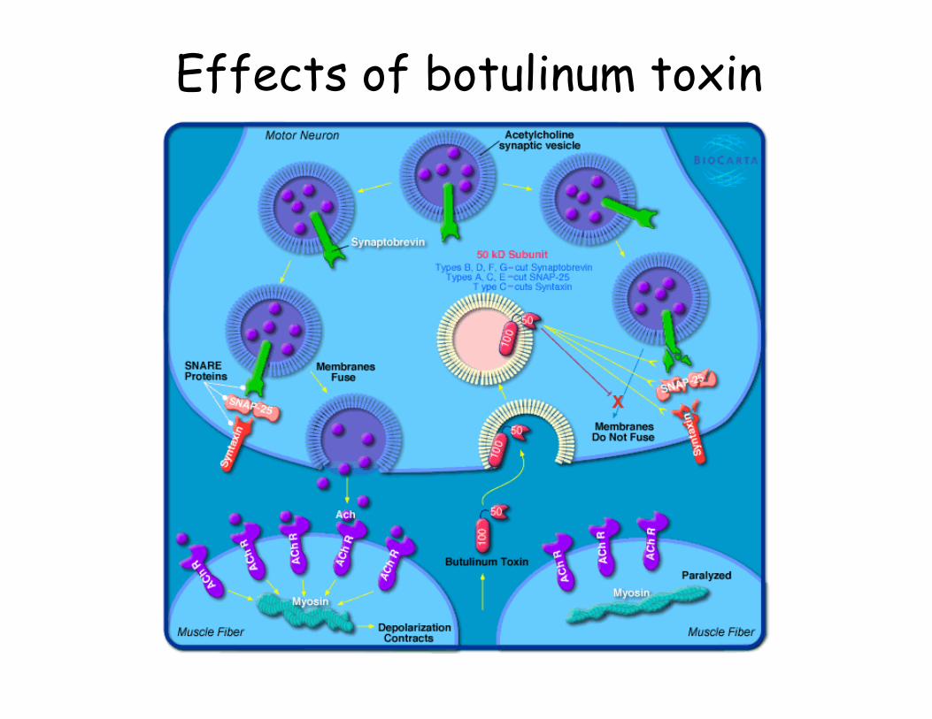
Effects of botulinum toxin
Results of NT binding on post synaptic membrane
• EPSP = excitatory postsynaptic potential– slight depolarization– closer to threshold
• IPSP = inhibitory postsynaptic potential– slight hyperpolarization– farther from threshold
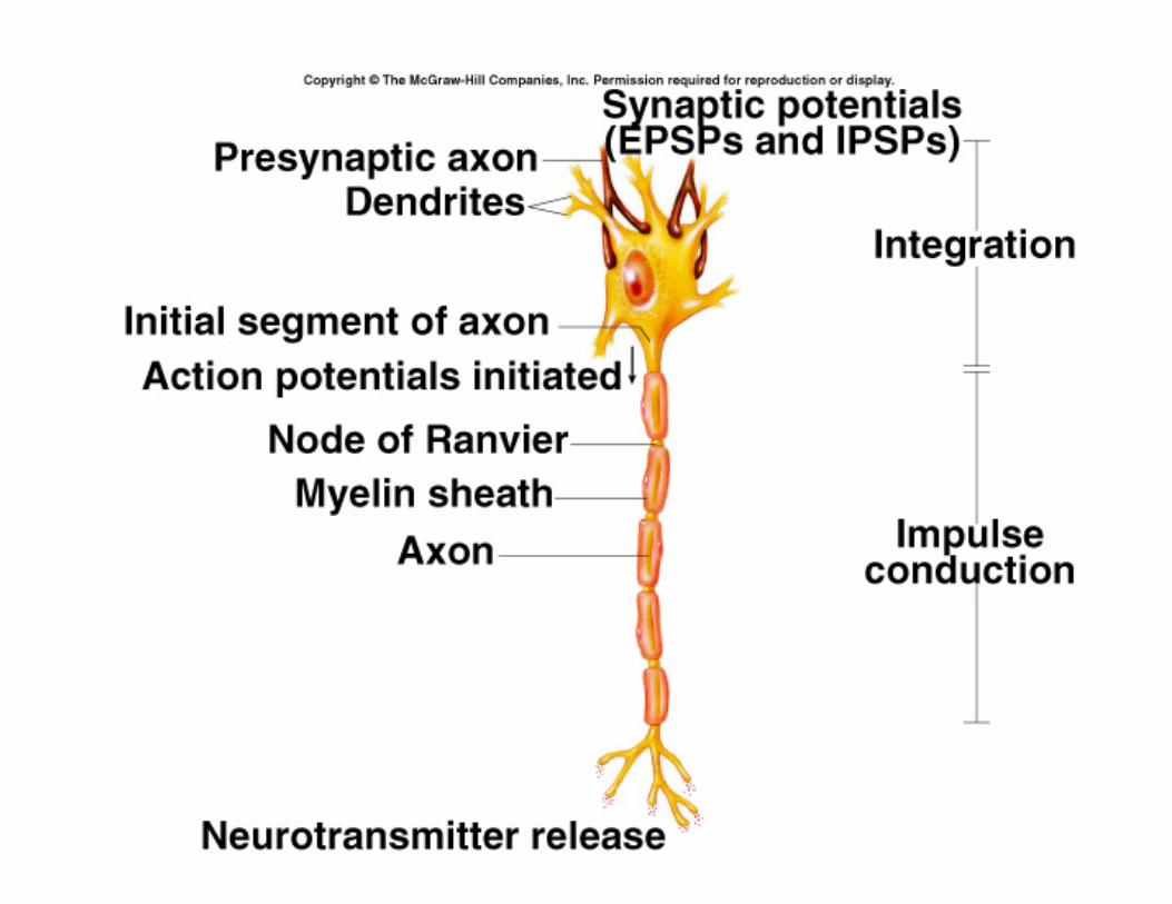

Synaptic integrationSpatial summationTemporal summation
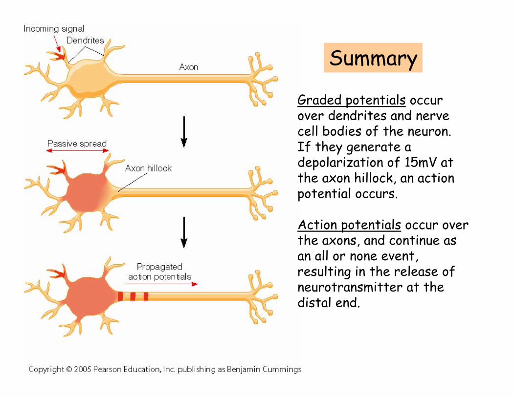
Graded potentials occur over dendrites and nerve cell bodies of the neuron. If they generate a depolarization of 15mV at the axon hillock, an action potential occurs.
Action potentials occur over the axons, and continue as an all or none event, resulting in the release of neurotransmitter at the distal end.
Summary
Clinical application: Epilepsy• Published August 2004• Two possible triggers for epileptic
seizures– Axonal sprouting
• New axons can sprout from an axon and form new connections (this is normal)
• In this case, they form connections with a neighboring dendrite, resulting in continuous stimulation
– Dendritic echo• Graded potential moves back up dendrite• Causes loss of inhibitory A currents
– Special currents that block A-type K+ channels– Result: Stronger echo that can stimulate the same
neuron again, resulting in a new action potential

NeurotransmittersNeurotransmittersNeurotransmitters
General categories• Acetylcholine• Biogenic amines
– Tyrosine precursor (catecholamines)• epinephrine• norepinephrine• dopamine
– Tryptophan precursor• seratonin, melatonin
–
• Amino acid derivatives– Excitatory (glutamate)– Inhibitory (GABA)
• Neuropeptides– Ex) endogenous opioids
• Miscellaneous– NO– Purines (ex: ATP)
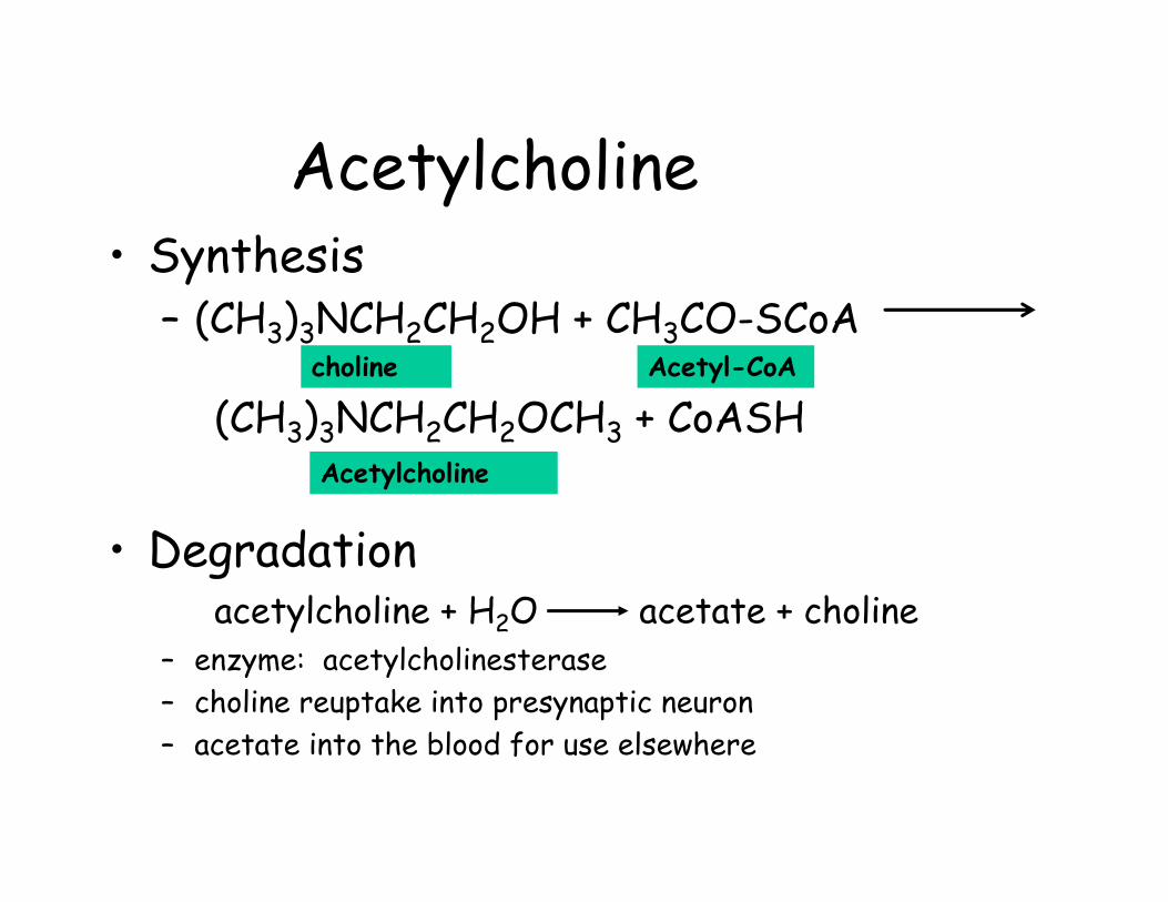
Acetylcholine• Synthesis
– (CH3)3NCH2CH2OH + CH3CO-SCoA
(CH3)3NCH2CH2OCH3 + CoASH
• Degradationacetylcholine + H2O acetate + choline
– enzyme: acetylcholinesterase– choline reuptake into presynaptic neuron– acetate into the blood for use elsewhere
choline Acetyl-CoA
Acetylcholine
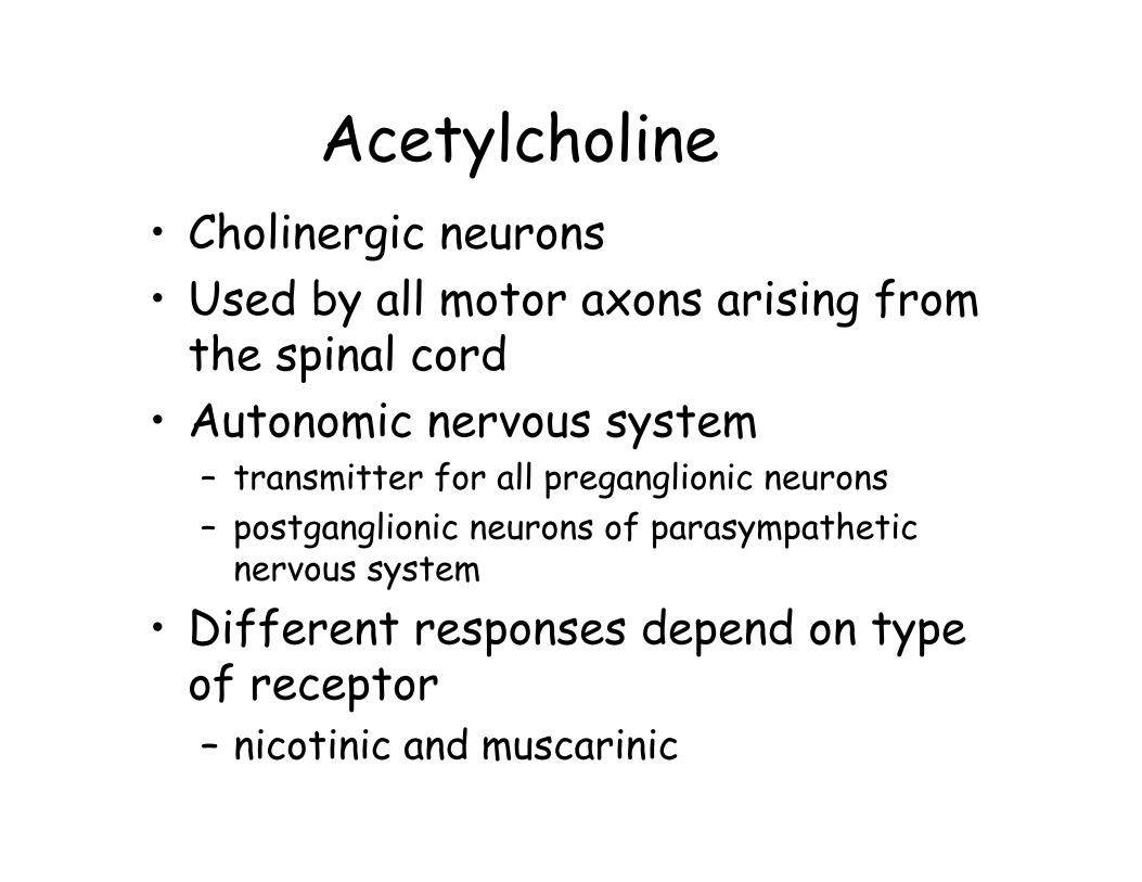
Acetylcholine• Cholinergic neurons• Used by all motor axons arising from
the spinal cord • Autonomic nervous system
– transmitter for all preganglionic neurons– postganglionic neurons of parasympathetic
nervous system
• Different responses depend on type of receptor– nicotinic and muscarinic
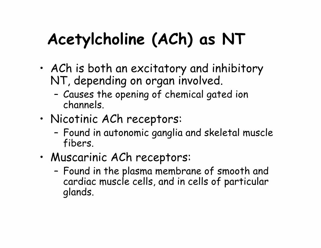
Acetylcholine (ACh) as NT
• ACh is both an excitatory and inhibitory NT, depending on organ involved.– Causes the opening of chemical gated ion
channels.• Nicotinic ACh receptors:
– Found in autonomic ganglia and skeletal muscle fibers.
• Muscarinic ACh receptors:– Found in the plasma membrane of smooth and
cardiac muscle cells, and in cells of particular glands.
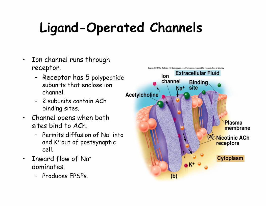
Ligand-Operated Channels
• Ion channel runs through receptor.– Receptor has 5 polypeptide
subunits that enclose ion channel.
– 2 subunits contain AChbinding sites.
• Channel opens when both sites bind to ACh.– Permits diffusion of Na+ into
and K+ out of postsynaptic cell.
• Inward flow of Na+
dominates.– Produces EPSPs.
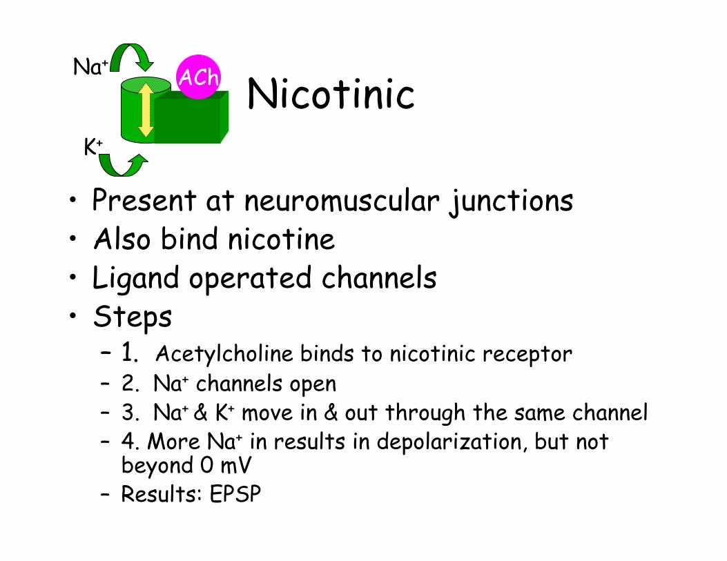
Nicotinic
• Present at neuromuscular junctions• Also bind nicotine• Ligand operated channels• Steps
– 1. Acetylcholine binds to nicotinic receptor– 2. Na+ channels open– 3. Na+ & K+ move in & out through the same channel– 4. More Na+ in results in depolarization, but not
beyond 0 mV – Results: EPSP
Na+
K+
ACh
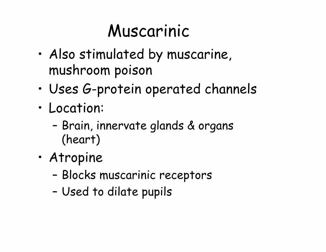
Muscarinic• Also stimulated by muscarine,
mushroom poison• Uses G-protein operated channels• Location:
– Brain, innervate glands & organs (heart)
• Atropine– Blocks muscarinic receptors– Used to dilate pupils
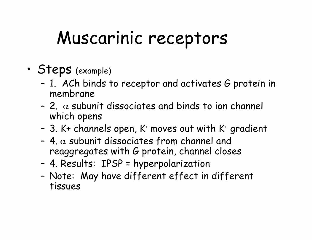
Muscarinic receptors• Steps (example)
– 1. ACh binds to receptor and activates G protein in membrane
– 2. α subunit dissociates and binds to ion channel which opens
– 3. K+ channels open, K+ moves out with K+ gradient– 4. α subunit dissociates from channel and
reaggregates with G protein, channel closes– 4. Results: IPSP = hyperpolarization– Note: May have different effect in different
tissues
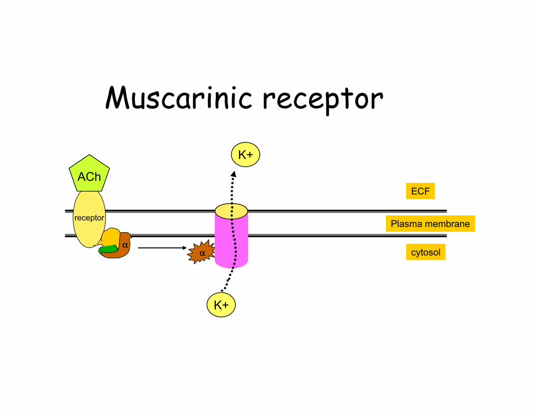
Muscarinic receptor
receptor
α
ECF
Plasma membrane
cytosolα
K+
K+
ACh
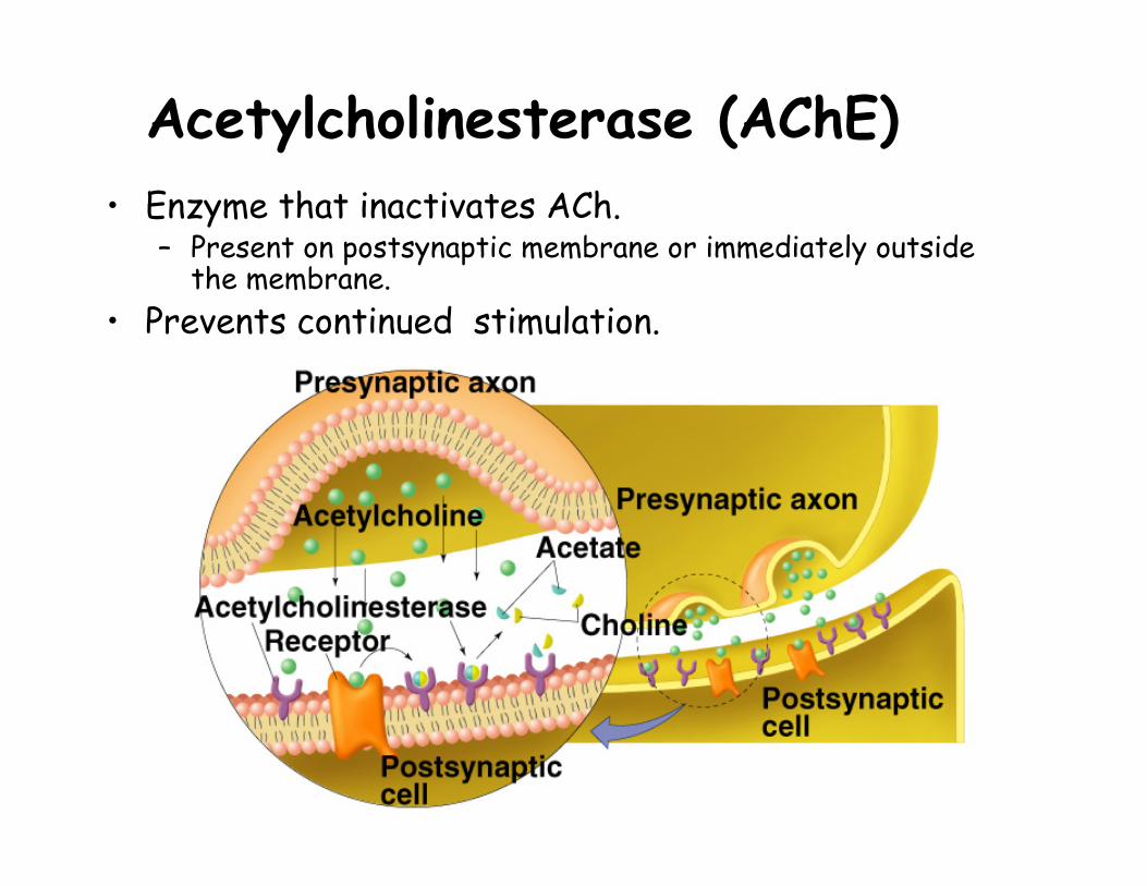
Acetylcholinesterase (AChE)• Enzyme that inactivates ACh.
– Present on postsynaptic membrane or immediately outside the membrane.
• Prevents continued stimulation.
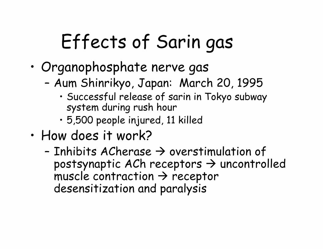
Effects of Sarin gas• Organophosphate nerve gas
– Aum Shinrikyo, Japan: March 20, 1995• Successful release of sarin in Tokyo subway
system during rush hour• 5,500 people injured, 11 killed
• How does it work?– Inhibits ACherase overstimulation of
postsynaptic ACh receptors uncontrolled muscle contraction receptor desensitization and paralysis
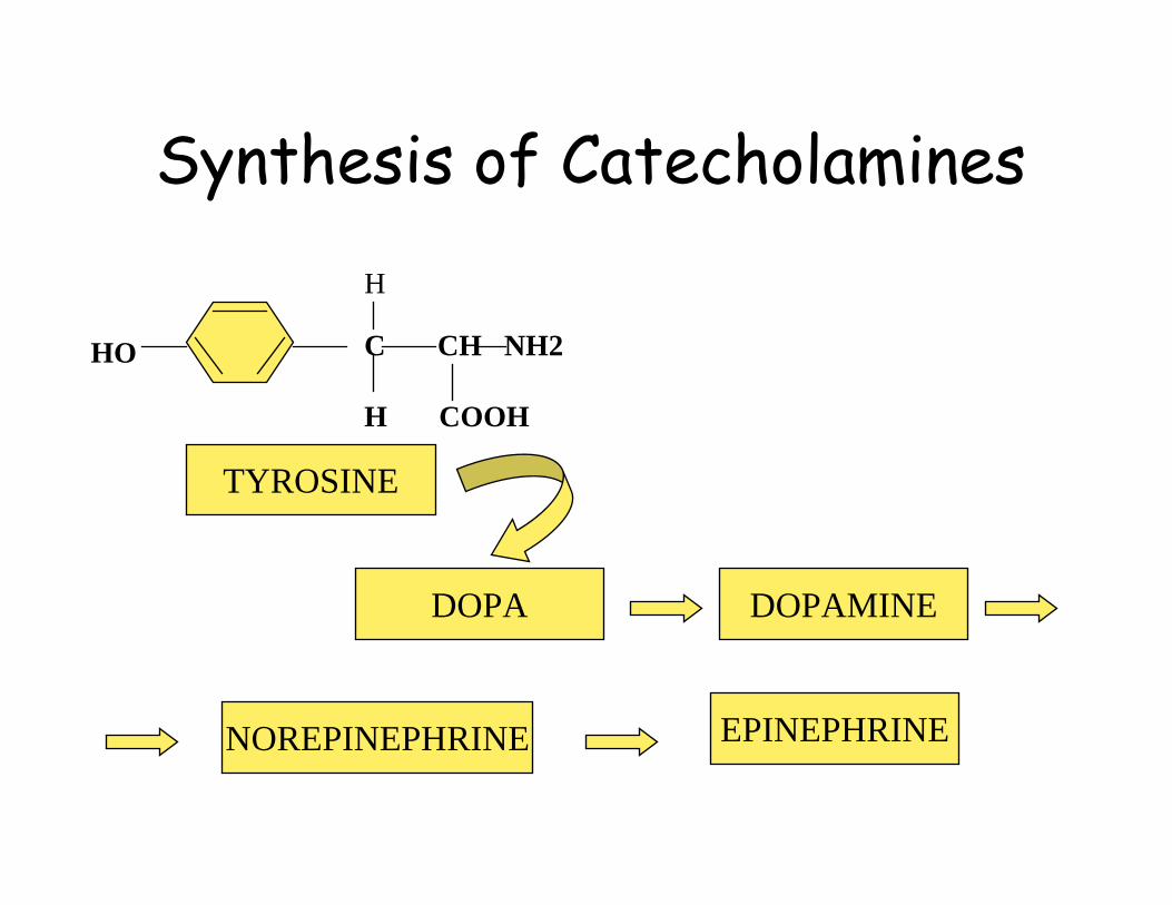
Synthesis of Catecholamines
HO C CH NH2
H COOH
H
TYROSINE
DOPA DOPAMINE
NOREPINEPHRINE EPINEPHRINE
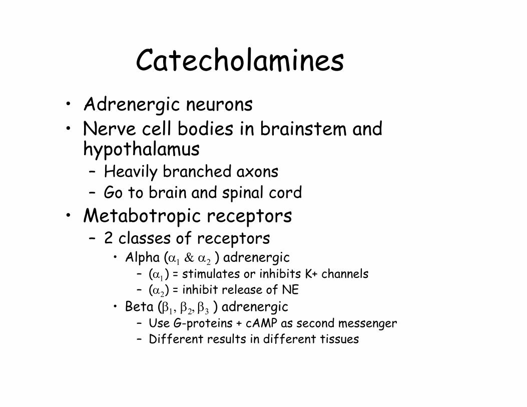
Catecholamines• Adrenergic neurons• Nerve cell bodies in brainstem and
hypothalamus– Heavily branched axons– Go to brain and spinal cord
• Metabotropic receptors– 2 classes of receptors
• Alpha (α1 & α2 ) adrenergic– (α1) = stimulates or inhibits K+ channels– (α2) = inhibit release of NE
• Beta (β1, β2, β3 ) adrenergic– Use G-proteins + cAMP as second messenger– Different results in different tissues

Catecholamines• Mechanism: second messenger (cAMP)
– Norepinephrine binds to G-protein receptor
– (α) subunit binds to adenylate cyclase– ATP converted to cAMP + 2Pi– cAMP activates protein kinase– proteins phosphorylated– Results: Ion channels are opened
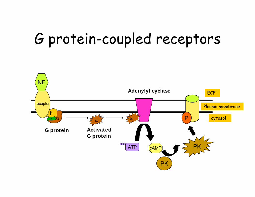
G protein-coupled receptors
receptor
αβγ
Adenylyl cyclase
ATP cAMP
G protein ActivatedG protein
α α
ECF
Plasma membrane
cytosol
NE
PK
PK
P
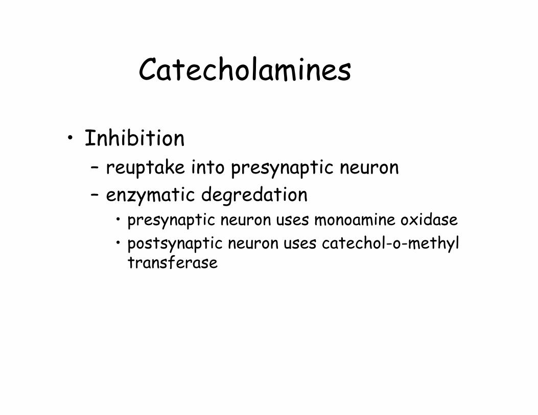
Catecholamines
• Inhibition– reuptake into presynaptic neuron– enzymatic degredation
• presynaptic neuron uses monoamine oxidase• postsynaptic neuron uses catechol-o-methyl
transferase
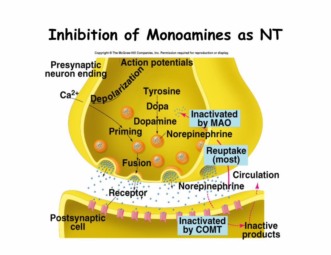
Inhibition of Monoamines as NT

Dopamine
• Found as NT only in the CNS• can be inhibitory or excitatory• Degeneration of neurons in
substantia nigra producing dopamine results in Parkinson’s disease
• Mediated by administration of L-dopa
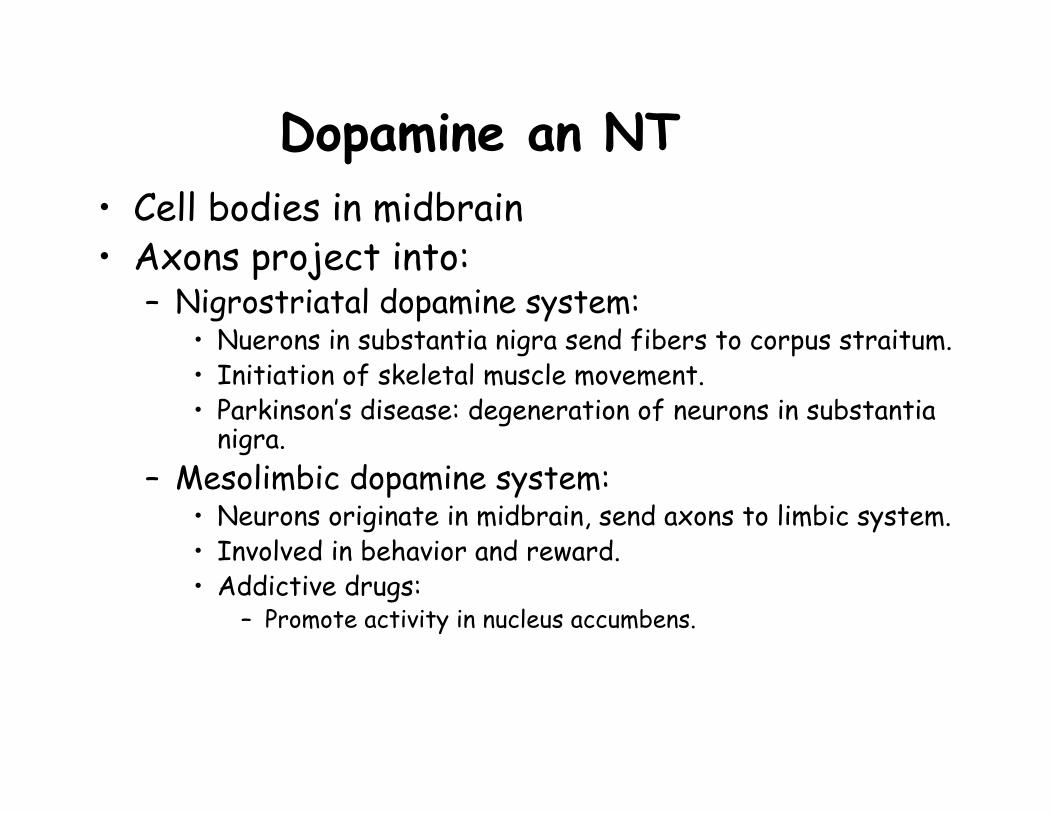
Dopamine an NT• Cell bodies in midbrain• Axons project into:
– Nigrostriatal dopamine system:• Nuerons in substantia nigra send fibers to corpus straitum. • Initiation of skeletal muscle movement.• Parkinson’s disease: degeneration of neurons in substantia
nigra.– Mesolimbic dopamine system:
• Neurons originate in midbrain, send axons to limbic system.• Involved in behavior and reward.• Addictive drugs:
– Promote activity in nucleus accumbens.
Seratonin• Precursor: tryptophan• Neuromodulator
– In muscles = excitatory effect– In pathways mediating sensation = inhibitory
• Low levels associated with depression– SSRI’s =medications which inhibit the reuptake
of seratonin (Prozac, Paxil, Zoloft)• degraded in presynaptic neuron by oxidative
deamination
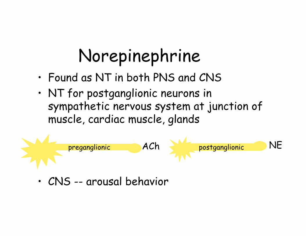
Norepinephrine• Found as NT in both PNS and CNS• NT for postganglionic neurons in
sympathetic nervous system at junction of muscle, cardiac muscle, glands
• CNS -- arousal behavior
preganglionic postganglionicACh NE
Amino Acids• Glutamic acid (glutamate)
– Most common NT in CNS at excitatory synapses– Long-term potentiation
• Involved in learning and memory• Aspartic acid (aspartate)• Glycine
– Primary inhibitory NT from interneurons in brain and spinal cord
– Hyperpolarize or stabilize membrane potential by increasing permeability to Cl-
– Impaired by strychnine• Gamma (γ)-aminobutyric acid (GABA)

γ-aminobutyric acid (GABA)• Major inhibitory NT produced in CNS• recycled by astrocytes
– taken up and converted to glutamine– transported to presynaptic neurons– converted to GABA in inhibitory neurons
• Receptors for GABA also bind benzodiazepines (Xanax, Valium)– Hyperpolarizes by increasing permeability to Cl-
• deficiency = Huntington’s disease– loss of striatal neurons producing NT
Effects of GABA• Role changes from excitatory to inhibitory
as brain matures– Inhibitory
• Regulation of muscle tone– Excitatory in neurogenesis
• GABA-ergic drugs have anti-anxiety, anti-convulsvie effects
• Affected by wide variety of drugs• Defects in signaling lead to:
– Epilepsy, schizophrenia, hypertonia, addiction, anxiety disorder

Neuropeptides• Synthesized as larger peptides
– enzymatic cleavage• mediate sensory and emotional responses• examples
– CCK = satiety– substance P– endogenous opioids
• Types – Beta (β)-endorphins– enkephalins– dynorphins
• Receptors bind opiates (morphine, codiene)

Nitric oxide• Precursor: L-arginine• roles
– relaxation of smooth muscle in blood vessels
– kills bacteria in macrophage– NT using cGMP as second messenger
• PNS = smooth muscle relaxation• CNS = memory and learning

Endogenous Cannabinoids– Bind to the same receptor as THC
• tetrahydrocannabinol = active ingredient in cannabis
– Regulate probability of NT release in amygdala, basal ganglia, hippocampus, cerebellum
– Synchronize timing between uterine receptivity and implantation of embryo
– Retrograde signaling• ECS released from postsynaptic neuron• Binds to presynaptic membrane supprssing NT
release temporarily• Function: long term potentiation & memory
Carbon monoxide– Stimulate production of cGMP within
neurons.– Promotes odor adaptation in olfactory
neurons.– May be involved in neuroendocrine
regulation in hypothalamus.
Neuromodulators• Modify postsynaptic
response to NT• Alter synthesis, release,
reuptake, metabolism of NT• Different receptor
characteristics than NT– Large and growing list of
chemicals– Slower action (minutes to days)– Often influence change in
metabolism • Use G-proteins + second
messengers
Drugs that affect Neural Control
• Botulinum toxin: Inhibits release of Ach• Curare: prevents interaction of Ach w/ its
receptor • Α-Bungarotoxin: Binds to Ach receptor• Tetrodotoxin: Blocks voltage gated Na+
channels• Nerve gas: Inhibits Ach-esterase• Strychnine: Prevents IPSPs in spinal cord that
inhibit contraction of antagonistic muscles