the neurogenic potential of astrocytes is regulated by
TRANSCRIPT
The Neurogenic Potential of Astrocytes Is Regulatedby Inflammatory Signals
Alessandro Michelucci1,2 & Angela Bithell3 & Matthew J. Burney1 &
Caroline E. Johnston1& Kee-Yew Wong4 & Siaw-Wei Teng4 & Jyaysi Desai1 &
Nigel Gumbleton3& Gregory Anderson1
& Lawrence W. Stanton4&
Brenda P. Williams1 & Noel J. Buckley5
Received: 6 March 2015 /Accepted: 8 June 2015 /Published online: 4 July 2015# The Author(s) 2015. This article is published with open access at Springerlink.com
Abstract Although the adult brain contains neural stem cells(NSCs) that generate new neurons throughout life, theseastrocyte-like populations are restricted to two discrete niches.Despite their terminally differentiated phenotype, adult paren-chymal astrocytes can re-acquire NSC-like characteristics fol-lowing injury, and as such, these ‘reactive’ astrocytes offer analternative source of cells for central nervous system (CNS)repair following injury or disease. At present, the mechanismsthat regulate the potential of different types of astrocytes arepoorly understood. We used in vitro and ex vivo astrocytes toidentify candidate pathways important for regulation of
astrocyte potential. Using in vitro neural progenitor cell(NPC)-derived astrocytes, we found that exposure of morelineage-restricted astrocytes to either tumor necrosis factoralpha (TNF-α) (via nuclear factor-κB (NFκB)) or the bonemorphogenetic protein (BMP) inhibitor, noggin, led to re-acquisition of NPC properties accompanied by transcriptomicand epigenetic changes consistent with a more neurogenic,NPC-like state. Comparative analyses of microarray data fromin vitro-derived and ex vivo postnatal parenchymal astrocytesidentified several common pathways and upstream regulatorsassociated with inflammation (including transforming growthfactor (TGF)-β1 and peroxisome proliferator-activated recep-tor gamma (PPARγ)) and cell cycle control (including TP53)as candidate regulators of astrocyte phenotype and potential.We propose that inflammatory signalling may control the nor-mal, progressive restriction in potential of differentiating as-trocytes as well as under reactive conditions and representfuture targets for therapies to harness the latent neurogeniccapacity of parenchymal astrocytes.
Keywords Inflammation . Astrocytes . Neural stem cells .
Noggin . NFκB . Epigenetic
Introduction
Astrocytes were historically seen as support cells in the centralnervous system (CNS) and in fact discharge multiple func-tions including regulation of energy metabolism, calcium sig-nalling, synaptic transmission and mediating inflammatoryresponses. This view has been expanded by the recognitionthat astrocytes are also highly heterogeneous [1–3]. Interest-ingly, neurogenic adult neural stem cells (aNSCs) that residein two niches (the subventricular zone (SVZ) of the lateralventricles and the subgranular zone (SGZ) of the dentate gyrus
Alessandro Michelucci and Angela Bithell contributed equally to thiswork.
Electronic supplementary material The online version of this article(doi:10.1007/s12035-015-9296-x) contains supplementary material,which is available to authorized users.
* Angela [email protected]
* Noel J. [email protected]
1 Institute of Psychiatry, Centre for the Cellular Basis of Behaviour,The James Black Centre, King’s College London, 125 ColdharbourLane, London SE5 9NU, UK
2 Luxembourg Centre for Systems Biomedicine, University ofLuxembourg, Campus Belval, 7, Avenue des Hauts-Fourneaux,L-4362 Esch-Belval, Luxembourg
3 School of Pharmacy, Department of Pharmacology, The HopkinsBuilding, University of Reading, Whiteknights, Reading RG6 6AP,UK
4 Genome Institute of Singapore, 60 Biopolis Street, #02-01, GenomeBuilding, Singapore 138672, Singapore
5 Department of Psychiatry,Warneford Hospital, University of Oxford,Warneford Lane, Oxford OX3 7JX, UK
Mol Neurobiol (2016) 53:3724–3739DOI 10.1007/s12035-015-9296-x
[4, 5]) have an astrocyte-like phenotype [6]. Even more in-triguingly, there is a growing body of evidence that someparenchymal astrocytes have a latent neurogenic capacity.These observations underlie the need to identify regulatorypathways that govern the ability of an astrocyte to expressNSC properties.
Early evidence for the neurogenic potential of parenchymalastrocytes came from observations that immature astrocytesfrom neonatal mouse adopt a radial glia-like phenotype whencultured with embryonic day 14 (E14) cortical cells [7], whilstastrocytes from embryonic or neonatal brain can formmultipotent neurospheres [8]. Furthermore, forced expressionof neurogenic transcription factors including Mash1, Ngn2 orDlx2 is capable of converting postnatal parenchymal astro-cytes to functional neurons [9–12]. Collectively, these studiesshow that immature parenchymal astrocytes have a latent neu-rogenic potential that can be realised by manipulation of in-trinsic transcriptional programmes or the cellular milieu. Im-portantly, astrocytes from more mature brain after postnatalday 10 (P10) are not capable of generating neurospheres, in-dicating that the ability to dedifferentiate is a unique propertyof immature astrocytes. Intriguingly, under inflammatory con-ditions or following injury, mature parenchymal astrocytescan become reactive and re-acquire more immature or neuralprogenitor cell (NPC)-like properties [6, 13, 14]. Furthermore,reactive astrocytes isolated from adult cortex can give rise tomultipotent neurospheres [15]. These observations underliethe relevance of understanding the latent neurogenic capacityof astrocytes for regenerative medicine strategies designed torecruit astrocytes into repair of damaged brain. Reactive as-trocytes are, by definition, present at the site of injury andtherefore offer an advantage over niche aNSCs that may residefar from the injury site. Although these studies clearly dem-onstrate the latent neurogenic capacity of some parenchymalastrocytes, the signalling pathways and mechanisms that reg-ulate the reprogramming of astrocytes to NPC-like states ordirectly to neurons remain largely unknown [16].
The drive to identify genes for ‘stemness’ was initially ledby transcriptome studies [17, 18], but recently, the idea hasemerged that epigenetic signatures (including post-translational modification to histones and DNA methylation)can provide an indicator of cellular potential [19–24]. Indeed,epigenetic reprogramming is key to the generation of inducedpluripotent stem cells (iPSCs). Whilst we are beginning tounravel the role of epigenetics in NSCs and neuronal differ-entiation, understanding of its contribution to maturation andreactivation of astrocytes is poor.
Our aim was to explore the transcriptome and epigeneticprofile of different astrocyte populations to identify molecularsignatures of astrocyte plasticity. To do this, we used homo-geneous populations of NPC-derived astrocytes that show adifferential ability to revert to an NPC-like state. We identifiedtwo factors: the pro-inflammatory cytokine, tumor necrosis
factor alpha (TNF-α), known to play a role in reactive gliosisand NPC proliferation [25–27] and the BMP antagonist, nog-gin, as key regulators that govern reprogramming of astro-cytes to NPCs. We also show that changes in epigenetic pro-files accompany changes in cellular potential. Importantly, bycomparing in vitro and ex vivo astrocyte transcriptomes, weprovide evidence that there are likely to be common pathwaysand regulators responsible for astrocyte identity and potentialin normal and injured brain that include pro- and anti-inflammatory signalling. Further, we find that astrocyte poten-tial is also reflected in their intrinsic epigenetic signatures.These data add to a growing repository that aids identificationof regulatory pathways involved in maintenance or re-acquisition of neurogenic NPC potential that may allow re-cruitment of parenchymal astrocytes in repair strategies fortreatment of brain injury and degeneration.
Materials and Methods
Mice
All UK animal handling and procedures were performed ac-cording to the UK Animals (Scientific Procedures) Act, 1986under Home Office licence. All animal procedures in Luxem-bourg were performed according to Federation of EuropeanLaboratory Animal Science Associations (FELASA) guide-lines for the use of animals in research. Transgenic mice weregenotyped using standard protocols and specific primers(Table S1).
Cell Culture
Cell Line
The CTX12 cell line is a conditionally immortalised mouseNPC line derived from E12-5 mouse cortex that has beengenerated in-house at King’s College London by Dr. Bithelland Dr. Williams. Cells were grown on poly-D-lysine (PDL)/laminin-coated plastic (Sigma) in modified Sato’s medium(modified Sato’s medium: Dulbecco’s modified Eagle’s medi-um (DMEM)/F12 supplemented with 5.6 mg/ml glucose,100 μg/ml bovine serum albumin (BSA), 16 μg/ml putres-cine, 60 ng/ml progesterone, 400 ng/ml L-thyroxine,300 ng/ml 3,3′,5-triiodothyronine, 5 μg/ml insulin, 5 μg/mlapo-transferrin, 5 ng/ml sodium selenite, 1× glutamine, 1×pen/strep) containing 10 ng/ml fibroblast growth factor 2(FGF2), 20 ng/ml epidermal growth factor (EGF) and100 nM 4-hydroxytamoxifen (4-OHT). Medium was changedevery 2–3 days and cells passaged using trypsin-EDTA andtrypsin inhibitor (Sigma). For astrocyte differentiation,CTX12 cells were plated at 0.5×10⁵ cells/cm2 and cultured inmodified Sato’s medium with 10 % foetal bovine serum (FBS)
Mol Neurobiol (2016) 53:3724–3739 3725

or 20 ng/ml bone morphogenetic protein 4 (BMP4) (Peprotechand R&D Systems). Where indicated, cells were treated withadditional factors: noggin (500 ng/ml), TNF-α (50 ng/ml)(Peprotech) and JSH-23 nuclear factor-κB (NFκB) ActivationInhibitor II (JSH-23, 10 μM, Santa Cruz).
Primary Cells
Cortices were isolated from P21 Swiss Webster and collectedin calcium/magnesium-free Hank’s balanced salt solution(HBSS), trypsinised and DNAseI treated (50 μg/ml, Sigma)for 20 min at 37 °C and mechanically dissociated into a ho-mogenous cell suspension [28]. Following washes and centri-fugation, mixed glial cells were plated onto PDL (Sigma) inDMEM/F12 (Invitrogen) with 100 U/ml penicillin/100 mg/mlstreptomycin (Sigma) and 10 % FBS (Biosera). Once conflu-ent, cultures were shaken overnight at 180 rpm to removemicroglia and oligodendrocytes and treated for 4–7 days with20 μM cytosine arabinoside (AraC) to kill remaining dividingcells and obtain essentially pure astrocytes (>98 %), deter-mined by immunofluorescence using anti-glial fibrillary acid-ic protein (GFAP) (Millipore) and anti-S100β (Dako) for as-trocytes, anti-Iba1 (Biocare, microglia), anti-O4 (Sigma, oli-godendrocytes) and TuJ1 (Covance, neurons).
Preparation of Mouse Forebrain Cell Suspensionsand Fluorescence-Activated Cell Sorting (FACS)of Astrocytes
Different developmental stages (P4, P10 or P21) of forebrainsfrom Aldh1l1-EGFP (Fthfd) transgenic mice (GenSat/MMRRC) were collected in calcium/magnesium-free HBSS.Tissue was diced and papain digested at 33 °C for 90 min(20 U/ml, Sigma) in dissociation buffer [EBSS (Sigma), D(+)-glucose 22.5 mM, NaHCO3 26 mM and DNaseI 125 U/mlwith EDTA 0.5 mM and L-cysteine–HCl 1 mM (Sigma)] andwashed 3× in dissociation buffer with BSA (1.0 mg/ml, Sig-ma) and trypsin inhibitor (1.0 mg/ml, Sigma) before mechan-ical dissociation through 5 ml and fire-polished Pasteur pi-pettes to a single cell suspension. Cells were pelleted, resus-pended in cold phosphate-buffered saline (PBS) with DNaseIat 1×10⁶ cell/ml, passed through a 70-μm filter and 7-aminoactinomycin D (7-AAD, Sigma) added. FITC-posi-tive/PE-Cy5-negative cells were sorted. FACS was performedusing a FACSAria I SORP running FACSDiva6.3 software(BD Biosciences).
Immunocytochemistry
Cells were fixed for 10 min in 4 % paraformaldehyde,permeabilised and incubated with primary antibodies in 1×PBS with 10 % normal serum. Primary antibodies used wereanti-Olig2 (1:500; Millipore), anti-GFAP (1:400; Millipore),
anti-Ki67 (1:1,000; Abcam), anti-Nestin (1:1,000; Abcam),anti-Sox2 (1:200; Santa Cruz), anti-O4 (Sigma, 1:200), anti-Iba1 (1:200, Biocare), TuJ1 (1:1,000, Covance), anti-GFP(1:2,000, Abcam), anti-bromodeoxyuridine (BrdU) (1:250,Abcam) and anti-NFκB-p65 (1:500; Abcam). Primary anti-bodies were visualised using specific AlexaFluor secondaryantibodies (Molecular Probes), and nuclei were counter-stained with 4′,6-diamidino-2-phenylindole (DAPI). Cover-slips were mounted in Prolong Gold anti-fade mounting me-dium (Molecular Probes) and analysed using ZeissAxioImager Z1 microscopes and AxioVision software.
BrdU Labelling
Cells were pulsed with BrdU (100 μM, Sigma) for 24 h, fixedwith 4 % paraformaldehyde and treated with 2 N HCl for45min and 0.1M borax (pH 8.5) for 15min before processingfor immunocytochemistry (above).
RNA Isolation, Microarray Hybridisation and DataAnalysis
Mouse Ref-8 Expression BeadChips (Illumina), washed andscanned with Illumina BeadStation according to the Illuminaprotocols. Raw data were analysed in R using BeadArray andthe Limma package. Ingenuity Pathway Analysis was used toperform pathway analysis on geneset data (IPA, IngenuitySystems Inc. at www.ingenuity.com).
RNA Isolation and Reverse-Transcription PCR (RT-PCR)
Total RNAwas purified from cells using the Qiagen RNeasyMini Kit (Qiagen) as per manufacturer’s instructions. Firststrand cDNA was synthesised from 1 to 2 μg of total RNAusing M-MLV reverse transcriptase (Promega). RT-PCR wascarried out on the Chromo4 System (Bio-Rad) using primerslisted in Table S1. PCR conditions were as follows: 3 min at95 °C and 40 cycles of 10 s at 95 °C, 30 s at 60 °C and 30 s at72 °C followed by 10-s 70–95° melt curves. All experimentsincluded three no-template controls and were performed onthree biological replicates with three technical replicates foreach sample and normalised to GAPDH. Results wereanalysed using Opticon Monitor software (Bio-Rad), and rel-ative gene expression levels were calculated using the Pfafflmethod [29].
3726 Mol Neurobiol (2016) 53:3724–3739
Cells were pelleted and lysed with TRIzol reagent(Invitrogen). Total RNAwas further purified using an RNeasyMini Kit (Qiagen). Biological replicates were prepared formicroarrays using an Illumina TotalPrep RNA AmplificationKit (Ambion) and amplified RNAs hybridised on Sentrix®
Chromatin Immunoprecipitation (ChIP) Analysis
ChIP was performed as described previously [30] and detailedhere briefly. Cells were crosslinked with 1 % formaldehyde inPBS, quenched with 125 mM glycine and washed 3× withcold PBS (containing protease inhibitors) before centrifuga-tion and lysis in lysis buffer [5 mM PIPES pH 8.0, 85 mMKCl, 0.5 % NP-40] for 30 min on ice. Pelleted nuclei wereresuspended in shearing buffer [50 mM Tris pH 8.1, 10 mMEDTA, 0.1 % sodium dodecyl sulphate (SDS), 0.5 % sodiumdeoxycholate] and sonicated in a Bioruptor (Diogenode) withsufficient cycles (30 s on, 30 s off) to obtain an average chro-matin shear size of 200–500 bp. Ten micrograms of pre-cleared chromatin was immunoprecipitated in modified RIPAbuffer [140 mM NaCl, 10 mM Tris pH 7.5, 1 mM EDTA,0.5 mM EGTA, 1 % TX-100, 0.01 % SDS, 0.1 % sodiumdeoxycholate] with specific antibodies (2 μg), protease inhib-itors and pre-blockedmagnetic protein G beads (ActiveMotif)at 4 °C. Following washes [2×Wash Buffer 1–20mMTris pH8.1, 50 mMNaCl, 2 mMEDTA, 1 % TX-100, 0.1 % SDS; 1×Wash Buffer 2–10 mM Tris pH 8.1, 150 mM NaCl, 1 mMEDTA, 1 % NP-40, 1 % sodium deoxycholate, 250 mM LiCl;2× TE], the chromatin was eluted [0.1 M NaHCO3, 1 % SDS]and de-crosslinked for 4 h at 65 °C with RNase and 200 mMNaCl then treated with proteinase K for 2 h at 42 °C. ChIPDNA was purified using a QIAquick PCR purification kit(Qiagen, according to manufacturer’s instructions). ChIP-qPCR was performed using ChIP DNA with promoter-specific primers (see Table S1). Controls included non-specific IgG and H3 ChIPs and ChIP-qPCR with primers fornon-specific regions of genomic DNA where enrichment isnot expected. Enrichment was analysed using a standard curveto quantitate, and data were normalised to total H3 (unlessspecified otherwise). qPCRs were run using an iCycler andMyiQ software (Bio-Rad).
The antibodies used were as follows: anti-H3 (rabbit IgG,AbCam), anti-H3K4me3 (rabbit IgG, AbCam; rabbit serum,Active Motif) and anti-H3K27me3 (rabbit IgG, Upstate) withrabbit IgG as a non-specific negative control. For primer se-quences, see Table S1.
Results
Generation of Phenotypically Distinct Astrocytesfrom NPCs
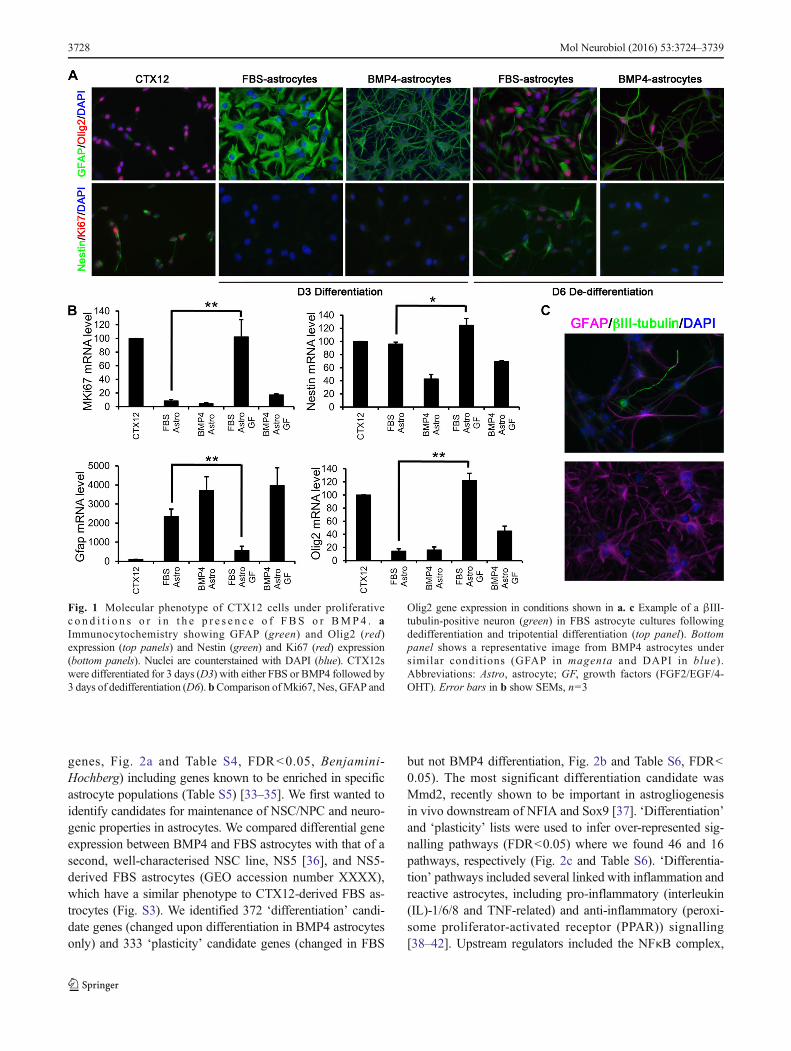
The mouse NPC line, CTX12, grown as a monolayer in thepresence of EGF, FGF2 and 4-OHT (collectively ‘GFs’) ex-presses classic NPC markers including Nestin and Sox2(Figs. 1a, b and S1A) and can differentiate into both neuronsand astrocytes (Fig. S1B, C). CTX12s cultured without GFs inthe presence of FBS or BMP4 for 3 days led to astrocyte
differentiation. In both conditions, CTX12 cells ceased prolif-eration (loss of Ki67), down-regulated Nestin and Olig2 andup-regulated GFAP (Fig. 1a, b). However, each condition gen-erated homogeneous astrocytes with distinct morphology:FBS astrocytes were flatter with few processes whilst BMP4astrocytes were more ramified and stellate (Fig. 1a). To testwhether either population retained plasticity in an NPC-permissive environment, FBS/BMP4 were removed and re-placed by GFs for 3 days (‘dedifferentiation’ conditions).FBS astrocytes became morphologically NPC-like, re-entered cell cycle, down-regulated GFAP and up-regulatedNestin and Olig2 (Fig. 1a, b). In contrast, few BMP4 astro-cytes became proliferative, and morphology and gene expres-sion were relatively unchanged (Fig. 1a, b). We subjecteddedifferentiated cells to a tripotential differentiation paradigm[31] to test their ability to generate neurons and glia. BothβIII-tubulin-positive neurons and GFAP-positive astrocyteswere readily obtainable from FBS astrocytes, but almost nonewere generated from BMP4 astrocytes (Figs. 1c and S1D).Thus, only FBS astrocytes re-acquire an NPC state followingdedifferentiation, suggesting that they differ in their neurogen-ic potential from the BMP4 astrocytes.
Identification of a Common Set of Astrocyte-RegulatedGenes and Pathways
To identify pathways or factors that regulate astrocyte differ-entiation and potential in our cellular model, we performedmicroarray analysis on CTX12s, FBS and BMP4 astrocytes(GEO accession number, with the authors). We identified~8,000 probes with significantly changed expression upondifferentiation from NPCs to FBS or BMP4 astrocytes (6,326and 6,256 genes, respectively, false discovery rate (FDR)<0.05, Benjamini-Hochberg [32], Tables 1 and Table S2, qPCRvalidation in Fig. S2). We first focussed on changes andenriched pathways common to both astrocyte populations fol-lowing differentiation. Classical astrocyte markers were highlyup-regulated including GFAP, Aqp4 and Slc39a12 [33]. Path-way analysis (IPA, Ingenuity Systems, Inc.) following FBS orBMP4 differentiation revealed a remarkable overlap in themostsignificant results, including cell cycle and proliferation-associated functions (Table S3), several of which are knownto be enriched in astrocytes [33]. IPA also permits predictionof upstream regulators and their activation state, with severalcommon to BMP4 and FBS astrocytes (relative to CTX12)including activation of TP53 and NFκB-related regulators andinhibition of MYC (Table S3).
Identification of a Set of Differentially RegulatedAstrocyte Genes
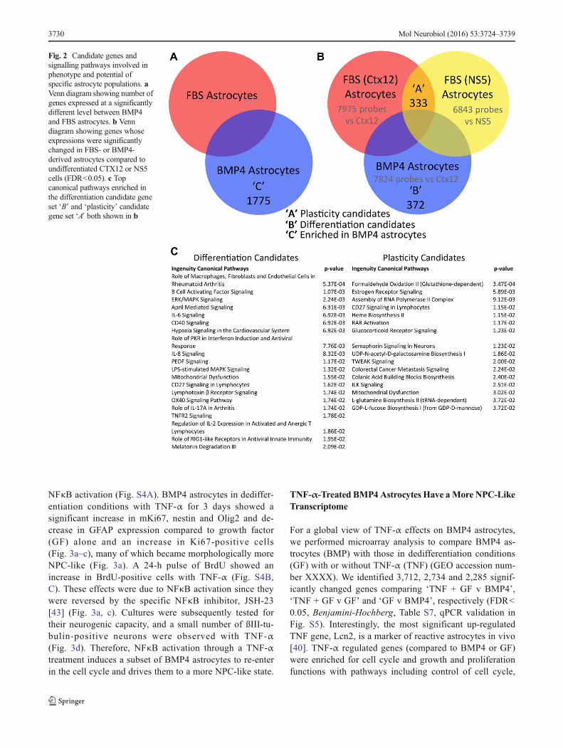
Next, we focussed on genes differentially expressed betweenBMP4 and FBS astrocytes and identified 1,775 probes (1,579
Mol Neurobiol (2016) 53:3724–3739 3727
genes, Fig. 2a and Table S4, FDR<0.05, Benjamini-Hochberg) including genes known to be enriched in specificastrocyte populations (Table S5) [33–35]. We first wanted toidentify candidates for maintenance of NSC/NPC and neuro-genic properties in astrocytes. We compared differential geneexpression between BMP4 and FBS astrocytes with that of asecond, well-characterised NSC line, NS5 [36], and NS5-derived FBS astrocytes (GEO accession number XXXX),which have a similar phenotype to CTX12-derived FBS as-trocytes (Fig. S3). We identified 372 ‘differentiation’ candi-date genes (changed upon differentiation in BMP4 astrocytesonly) and 333 ‘plasticity’ candidate genes (changed in FBS
but not BMP4 differentiation, Fig. 2b and Table S6, FDR<0.05). The most significant differentiation candidate wasMmd2, recently shown to be important in astrogliogenesisin vivo downstream of NFIA and Sox9 [37]. ‘Differentiation’and ‘plasticity’ lists were used to infer over-represented sig-nalling pathways (FDR<0.05) where we found 46 and 16pathways, respectively (Fig. 2c and Table S6). ‘Differentia-tion’ pathways included several linked with inflammation andreactive astrocytes, including pro-inflammatory (interleukin(IL)-1/6/8 and TNF-related) and anti-inflammatory (peroxi-some proliferator-activated receptor (PPAR)) signalling[38–42]. Upstream regulators included the NFκB complex,
Fig. 1 Molecular phenotype of CTX12 cells under proliferativec o n d i t i o n s o r i n t h e p r e s e n c e o f FBS o r BMP4 . aImmunocytochemistry showing GFAP (green) and Olig2 (red)expression (top panels) and Nestin (green) and Ki67 (red) expression(bottom panels). Nuclei are counterstained with DAPI (blue). CTX12swere differentiated for 3 days (D3) with either FBS or BMP4 followed by3 days of dedifferentiation (D6). bComparison ofMki67, Nes, GFAP and
Olig2 gene expression in conditions shown in a. c Example of a βIII-tubulin-positive neuron (green) in FBS astrocyte cultures followingdedifferentiation and tripotential differentiation (top panel). Bottompanel shows a representative image from BMP4 astrocytes undersimilar conditions (GFAP in magenta and DAPI in blue).Abbreviations: Astro, astrocyte; GF, growth factors (FGF2/EGF/4-OHT). Error bars in b show SEMs, n=3
3728 Mol Neurobiol (2016) 53:3724–3739
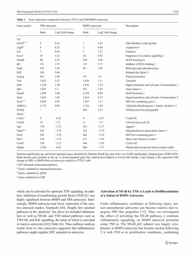
which can be activated by upstream TNF signalling. In addi-tion, inhibition of transforming growth factor (TGF)-β1 washighly significant between BMP4 and FBS astrocytes. Inter-estingly, BMP4 astrocytes had lower expression of the reac-tive astrocyte marker, Serpina3n [40]. Despite few enrichedpathways in the ‘plasticity’ list, these too included inflamma-tion as well as NFκB- and TNF-linked pathways such asTWEAK and ILK signalling, the latter of which is activatedin reactive astrocytes [40] (Table S6). Thus, pathway analysisresults from in vitro astrocytes suggested that inflammatorypathways might regulate NPC potential in astrocytes.
Activation of NFκB by TNF-α Leads to Dedifferentiationof a Subset of BMP4 Astrocytes
Under inflammatory conditions or following injury, ma-ture parenchymal astrocytes can become reactive and re-acquire NPC-like properties [15]. Thus, we investigatedthe effect of activating the NFκB pathway, a commoninflammatory signalling, on BMP4 astrocyte potentialusing TNF-α. The NFκB p65 subunit was largely cyto-plasmic in BMP4 astrocytes but became nuclear following2 h with TNF-α in proliferative conditions, confirming
Table 1 Gene expression comparison between CTX12 and FBS/BMP4 astrocytes
Gene symbol FBS astrocytes BMP4 astrocytes Description
Rank Log2 fold change Rank Log2 fold change
Up
GFAPa, b 5 8.54 1 9.85 Glial fibrillary acidic protein
Aqp4b 6 8.53 2 8.64 Aquaporin 4
Clu 7 8.04 5 7.27 Clusterin
Socs3 33 5.23 34 4.92 Suppressor of cytokine signalling 3
Smad6 88 4.25 254 2.96 MAD homolog 6
Id2 151 3.75 112 3.75 Inhibitor of DNA binding 2
Pygb 191 3.39 99 3.89 Brain glycogen phosphorylase
Klf2 262 3.04 – – Krüppel-like factor 2
Nanog 835 1.88 731 1.9 Nanog homeobox
Vim 1,033 1.67 1,626 1.11 Vimentin
Stat1 1,229 1.50 1,070 1.54 Signal transducer and activator of transcription 1
Jak1 1,467 1.3 932 1.65 Janus kinase 1
Smad1 1,849 1.06 2,134 0.84 MAD homolog 1
Stat3 1,854 1.05 2,414 0.72 Signal transducer and activator of transcription 3
Sox2a, c 2,059 0.95 1,027 1.57 SRY-box-containing gene 2
Aldh1L1 2,174 0.88 1,126 1.49 Aldehyde dehydrogenase 1 family, member L1
S100β – – 464 2.33 S100 protein beta polypeptide
Down
Ccnb1c 4 −8.41 4 −8.27 Cyclin B1
Cdc20 14 −7.2 11 −7.3 Cell division cycle 20
Jag1 176 −3.75 253 −3.17 Jagged 1
Olig2a, b 183 −3.70 162 −3.79 Oligodendrocyte transcription factor 2
Sox8 238 −3.39 462 −2.22 SRY-box-containing gene 8
Hes5 374 −2.74 611 −1.92 Hairy and enhancer of split 5
Ccnd2 530 −2.27 444 −2.29 Cyclin D2
Pdgfra 1,816 −0.91 860 −1.57 Platelet-derived growth factor receptor alpha
Selected significantly up- and down-regulated genes identified by Illumina BeadChip array from over 6,000 significantly changed genes (FDR<0.05).Rank denotes gene position in the up- or down-regulated gene lists ordered from highest to lowest fold change. Log2 change is the expression foldchange in FBS- or BMP4-derived astrocytes relative to CTX12 cells
ChIP chromatin immunoprecipitationa Genes validated by immunofluorescencebGenes validated by qPCRcGenes validated by ChIP
Mol Neurobiol (2016) 53:3724–3739 3729
NFκB activation (Fig. S4A). BMP4 astrocytes in dediffer-entiation conditions with TNF-α for 3 days showed asignificant increase in mKi67, nestin and Olig2 and de-crease in GFAP expression compared to growth factor(GF) alone and an increase in Ki67-positive cells(Fig. 3a–c), many of which became morphologically moreNPC-like (Fig. 3a). A 24-h pulse of BrdU showed anincrease in BrdU-positive cells with TNF-α (Fig. S4B,C). These effects were due to NFκB activation since theywere reversed by the specific NFκB inhibitor, JSH-23[43] (Fig. 3a, c). Cultures were subsequently tested fortheir neurogenic capacity, and a small number of ßIII-tu-bulin-positive neurons were observed with TNF-α(Fig. 3d). Therefore, NFκB activation through a TNF-αtreatment induces a subset of BMP4 astrocytes to re-enterin the cell cycle and drives them to a more NPC-like state.
TNF-α-Treated BMP4Astrocytes Have aMore NPC-LikeTranscriptome
For a global view of TNF-α effects on BMP4 astrocytes,we performed microarray analysis to compare BMP4 as-trocytes (BMP) with those in dedifferentiation conditions(GF) with or without TNF-α (TNF) (GEO accession num-ber XXXX). We identified 3,712, 2,734 and 2,285 signif-icantly changed genes comparing ‘TNF + GF v BMP4’,‘TNF + GF v GF’ and ‘GF v BMP4’, respectively (FDR<0.05, Benjamini-Hochberg, Table S7, qPCR validation inFig. S5). Interestingly, the most significant up-regulatedTNF gene, Lcn2, is a marker of reactive astrocytes in vivo[40]. TNF-α regulated genes (compared to BMP4 or GF)were enriched for cell cycle and growth and proliferationfunctions with pathways including control of cell cycle,
Fig. 2 Candidate genes andsignalling pathways involved inphenotype and potential ofspecific astrocyte populations. aVenn diagram showing number ofgenes expressed at a significantlydifferent level between BMP4and FBS astrocytes. b Venndiagram showing genes whoseexpressions were significantlychanged in FBS- or BMP4-derived astrocytes compared toundifferentiated CTX12 or NS5cells (FDR<0.05). c Topcanonical pathways enriched inthe differentiation candidate geneset ‘B’ and ‘plasticity’ candidategene set ‘A’ both shown in b
3730 Mol Neurobiol (2016) 53:3724–3739

p53, cancer-related pathways and TNF-related pathways(Table S8), consistent with a change in proliferative po-tential. Predicted upstream regulators included TNF andNFκB, activation of MYC, WNT and other pro-inflammatory molecules (including IFNG, IL1/17,Table S8). One of the few predicted regulators in the‘GF v BMP’ dataset was sonic hedgehog (SHH) (activat-ed), also in the TNF-α dataset, which may reflect theroles of pro-inflammatory molecules and SHH signallingin astrocyte acquisition of NSC properties [44, 45]. Inter-estingly, in dedifferentiation conditions (GF or TNF +
GF), Ascl1, encoding the proneural protein Mash1, wasup-regulated whilst members of Notch (Notch1, 4), Id(Id1-3), Stat (Stat2) and Hes (Hes1, 5) families weredown-regulated, particularly in TNF + GF conditions.This is consistent with a neurogenic NPC-like capabilitysince many are involved in the neurogenic–gliogenicswitch, including several regulated by BMP signalling[46]. Taken together, these transcriptomic results highlightthat the expression levels of several NPC-associatedgenes/pathways are more highly re-activated in the pres-ence of TNF-α when compared to the corresponding
Fig. 3 A subset of BMP4 astrocytes treated with TNF-α re-enter the cellcycle and re-acquire NPC characteristics. a Immunocytochemistry onBMP4 astrocytes cultured in dedifferentiation conditions for 3 daysalone (GF), in the presence of TNF-α (+GF + TNFα) or with TNF-αand JSH23 (+GF + TNFα + JSH23). JSH23 was added 30 min beforeculturing astrocytes in dedifferentiation conditions in the presence ofTNF-α. Top row shows GFAP (green) and Olig2 (red), and bottom rowshows Nestin (green) and Ki67 (red). Nuclei were counterstained withDAPI (blue). b Percentage of Ki67-positive cells in cultures shown in a. c
Gene expression levels of MKi67, Nestin (Nes), GFAP and Olig2 inBMP4 astrocytes in conditions shown in (a, b). Data are expressed aspercentage of expression relative to D3 BMP4 astrocytes. d βIII-tubulin-positive neurons (green) and GFAP-positive astrocytes (magenta) withDAPI-labelled nuclei (blue) in BMP4 astrocyte cultures followingdedifferentiation with TNF-α followed by tripotential differentiation.For b and c, n=3. Scale bars in a and d: 20 μm. P values in b and c:*p<0.05, **p<0.01 (Student’s t test), error bars show SEMs
Mol Neurobiol (2016) 53:3724–3739 3731

BMP4-derived astrocytes under normal or dedifferentia-tion conditions.
Mimicking CNS Damage Promotes NPC Propertiesin BMP4-Derived Astrocytes
In keeping with down-regulation of BMP-regulated genes,our analysis predicted inhibition of BMP itself under dediffer-entiation conditions with and without TNF-α. This suggeststhat BMP4 astrocytes produce endogenous BMPs and thattheir inhibition is required for dedifferentiation. Interestingly,reactive astrocytes adjacent to penetrating CNS injuries inboth spinal cord and brain up-regulate the BMP inhibitor,noggin [47], and BMP secreted from blood endothelial cellscan induce reversible quiescence of NSC/NPCs in vitro, re-versible with noggin [48]. We thus tested whether noggincould induce re-acquisition of NPC properties in BMP4 astro-cytes. This led to a change from astrocyte to NPC-like mor-phology and a significant increase in proliferation (Fig. 4a, b),concomitant with down-regulation of GFAP and up-regulation of mKi67, Nestin and Olig2 (Fig. 4c) and down-regulation of genes enriched in parenchymal astrocytes, in-cluding Aqp4, Thrsp and Ngef (Table S5), to levels similarto dedifferentiated FBS-derived astrocytes (Fig. S6). Noggin-
treated dedifferentiated astrocytes were also neurogenic, witha small number of ßIII-tubulin-positive neurons (Fig. 4d).
Histone Modifications at Key Promoters DistinguishBetween Astrocyte Types
Differentiation, including acquisition and maintenance of amore restricted fate, is known to be associated with epigeneticchanges [20, 21, 24, 49]. The ability of FBS astrocytes todedifferentiate compared to BMP4 astrocytes may reflect in-trinsic epigenetic as well as transcriptional differences, includ-ing histone modifications in promoters associated with active(e.g. H3K9ac and H3K4me3) or repressed (H3K27me3) chro-matin states [21, 50].
To look specifically at histone modifications associatedwith cellular potential, we used ChIP-qPCR to examine rela-tive levels of H3K4me3 and H3K27me3 at selected gene pro-moters: Ccnb1 (positive cell cycle regulator), Nestin (NPC-specific), Olig2 (neural-specific bHLH factor) and Sox2(NSC/NPC marker). H3K4me3 was significantly decreasedat Ccnb1, Nestin and Sox2 in BMP4 astrocytes compared toCTX12s and FBS astrocytes, whilst H3K27me3 increased atCcnb1, Nestin and Olig2 in both astrocytes, correspondingwith its down-regulation and cell cycle exit (Fig. 5a). Similar
Fig. 4 Inhibition of BMP signalling leads to re-acquisition of NPCcharacteristics in BMP4 astrocytes. a Immunocytochemistry comparingKi67 (red) and GFAP (green) expression in BMP4 astrocytes indedifferentiation conditions with and without noggin (+GF + nogginand +GF, respectively). Noggin was added simultaneously with GF.Nuclei are counterstained with DAPI (blue). b Percentage of Ki67-positive cells in the conditions shown in a. c Gene expression levels ofMki67, Nes, GFAP and Olig2 in the conditions shown in a. Expression
levels are shown as percentage in +GF + noggin conditions relative to +GF conditions (100 %). d βIII-tubulin-positive neurons (green) andGFAP-positive astrocytes (magenta) with DAPI-labelled nuclei (blue)in BMP4 astrocyte cultures following dedifferentiation with nogginfollowed by tripotential differentiation. For b and c, n=3. Scale bars ina and d, 20 μm. P values in b and c: *p<0.05, **p<0.01, ***p<0.001(Student’s t test), error bars show SEMs
3732 Mol Neurobiol (2016) 53:3724–3739
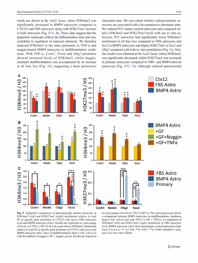
trends are shown at the Ascl1 locus, where H3K4me3 wassignificantly decreased in BMP4 astrocytes compared toCTX12s and FBS astrocytes along with H3K27me3 increasein both astrocytes (Fig. S7A, B). These data suggest that theepigenetic landscape reflects the differentiation state and maycontribute to regulation of astrocyte plasticity. We thereforeanalysed H3K4me3 at the same promoters in TNF-α andnoggin-treated BMP4 astrocytes in dedifferentiation condi-tions. With TNF-α, Ccnb1, Nestin and Olig2 promotersshowed increased levels of H3K4me3, whilst noggin-mediated dedifferentiation was accompanied by an increaseat all four loci (Fig. 5b), suggesting a more permissive
chromatin state. We next asked whether cultured primary as-trocytes are associated with a less permissive chromatin state.We cultured P21 mouse cortical astrocytes and compared rel-ative H3K4me3 and H3K27me3 levels with our in vitro as-trocytes. P21 astrocytes had significantly lower H3K4me3enrichment at all four loci compared to FBS astrocytes andSox2 in BMP4 astrocytes and higher H3K27me3 at Sox2 andOlig2 compared with both in vitro populations (Fig. 5c). Sim-ilar results were obtained at the Ascl1 locus, where H3K4me3was significantly decreased, whilst H3K27me3 was increasedin primary astrocytes compared to FBS- and BMP4-derivedastrocytes (Fig. S7C, D). Although cultured parenchymal
Fig. 5 Epigenetic comparison of phenotypically distinct astrocytes. aH3K4me3 (left) and H3K27me3 (right) enrichment relative to totalH3 at specific gene promoters in CTX12 cells (grey), FBS astrocytes(red) and BMP4 astrocytes (blue). Results are expressed as a percentageof levels in CTX12 (100 %). b Left graph shows H3K4me3 enrichmentrelative to total H3 at specific gene promoters in CTX12 cells (grey) andBMP4 astrocytes after 3 days of dedifferentiation alone (+GF, yellow) orwith the addition of noggin (+GF + noggin, green). Results are expressed
as a percentage of levels in CTX12 (100%). The right-hand graph showsa comparison between BMP4 astrocytes in dedifferentiation conditionsalone (+GF, yellow) and with TNF-α (+GF + TNFα). c Comparison ofH3K4me3 (left) and H3K27me3 (right) enrichment in FBS astrocytes(red), BMP4 astrocytes (dark blue) and primary cortical astrocytes (lightblue). For a–c, n=3. *p<0.05, **p<0.01, ***p<0.001 (Student’s t test),and error bars show SEMs
Mol Neurobiol (2016) 53:3724–3739 3733
astrocytes may represent a more immature/reactive phenotypethat non-cultured counterparts [33], our data are consistentwith the restricted state of later postnatal astrocytes and sug-gest a more restricted state in BMP4 versus FBS astrocytes.Thus, astrocyte plasticity and neurogenic potential arereflected at the chromatin level.
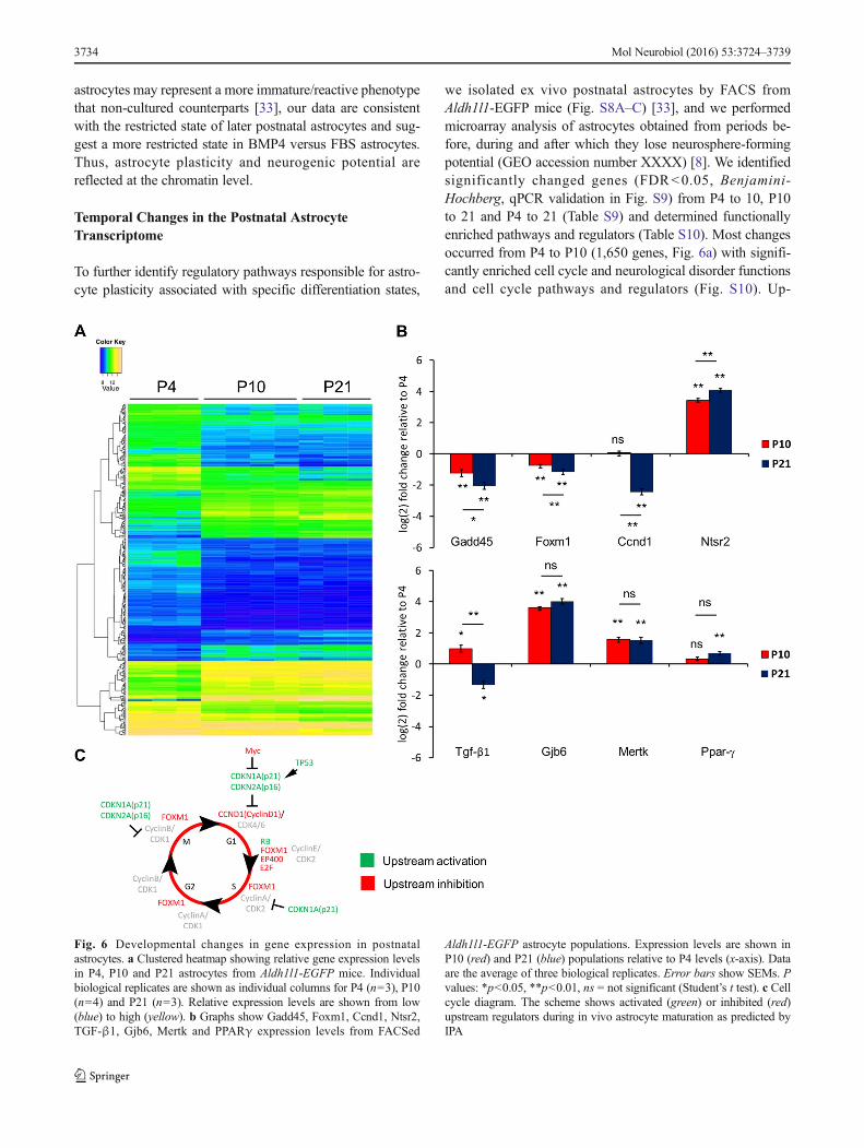
Temporal Changes in the Postnatal AstrocyteTranscriptome
To further identify regulatory pathways responsible for astro-cyte plasticity associated with specific differentiation states,
we isolated ex vivo postnatal astrocytes by FACS fromAldh1l1-EGFP mice (Fig. S8A–C) [33], and we performedmicroarray analysis of astrocytes obtained from periods be-fore, during and after which they lose neurosphere-formingpotential (GEO accession number XXXX) [8]. We identifiedsignificantly changed genes (FDR<0.05, Benjamini-Hochberg, qPCR validation in Fig. S9) from P4 to 10, P10to 21 and P4 to 21 (Table S9) and determined functionallyenriched pathways and regulators (Table S10). Most changesoccurred from P4 to P10 (1,650 genes, Fig. 6a) with signifi-cantly enriched cell cycle and neurological disorder functionsand cell cycle pathways and regulators (Fig. S10). Up-
Fig. 6 Developmental changes in gene expression in postnatalastrocytes. a Clustered heatmap showing relative gene expression levelsin P4, P10 and P21 astrocytes from Aldh1l1-EGFP mice. Individualbiological replicates are shown as individual columns for P4 (n=3), P10(n=4) and P21 (n=3). Relative expression levels are shown from low(blue) to high (yellow). b Graphs show Gadd45, Foxm1, Ccnd1, Ntsr2,TGF-β1, Gjb6, Mertk and PPARγ expression levels from FACSed
Aldh1l1-EGFP astrocyte populations. Expression levels are shown inP10 (red) and P21 (blue) populations relative to P4 levels (x-axis). Dataare the average of three biological replicates. Error bars show SEMs. Pvalues: *p<0.05, **p<0.01, ns = not significant (Student’s t test). c Cellcycle diagram. The scheme shows activated (green) or inhibited (red)upstream regulators during in vivo astrocyte maturation as predicted byIPA
3734 Mol Neurobiol (2016) 53:3724–3739
regulated genes also included Ntsr2, Gjb6 and Mertk(Fig. 6b), all highly enriched in mature in vivo astrocytes[33]. The top pathway identified was cell cycle regulation ofreplication. Interestingly, this was enriched in TNF + GF genesets from BMP4 astrocytes, where many genes down-regulated from P4 to P10 showed up-regulation with TNF-αin vitro (Fig. S9). Signalling involving janus tyrosine kinase(JAK) family kinases and IL-6 revealed down-regulation ofseveral genes by P10 that were significantly lower in BMP4versus FBS astrocytes (Fig. S11). P10–P21 analysis (357genes) again showed enrichment for cell cycle-associatedpathways (including GADD45) and P4–P21 analysis (2,732genes) identified additional pathways, some of which wereidentified in BMP4 astrocytes exposed to TNF-α(Tables S10 and S9). These data suggest common pathwaysin vitro and ex vivo that regulate astrocyte phenotype, poten-tial and differentiation. We therefore interrogated upstreamregulators to identify regulators of astrocyte differentiationand NSC potential.
P4–P10 upstream regulators included PPARG (and PPARGC1A), TP53 and CDKN2A activation and MYC, E2F1,EP400 and vascular endothelial growth factor (VEGF) inhibi-tion (Table S10) with additional regulators from P10-P21 in-cluding FOXM1 (inhibition). Analysing P4–P21 also predict-ed CDKN1A activation and TGF-β1 and CCND1 inhibition.Broadly, these data support progressive cell cycle arrest(Fig. 6c) accompanied by changes consistent with inhibitionof inflammatory signalling and identified other candidateswith potential roles during early postnatal stages.
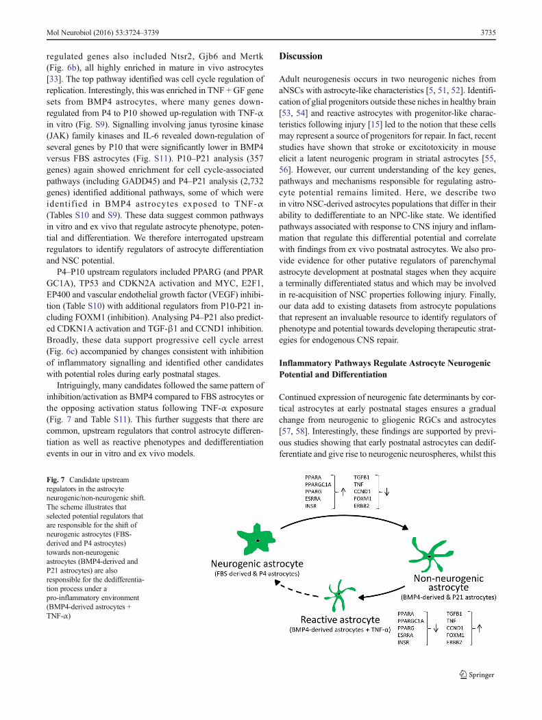
Intriguingly, many candidates followed the same pattern ofinhibition/activation as BMP4 compared to FBS astrocytes orthe opposing activation status following TNF-α exposure(Fig. 7 and Table S11). This further suggests that there arecommon, upstream regulators that control astrocyte differen-tiation as well as reactive phenotypes and dedifferentiationevents in our in vitro and ex vivo models.
Discussion
Adult neurogenesis occurs in two neurogenic niches fromaNSCs with astrocyte-like characteristics [5, 51, 52]. Identifi-cation of glial progenitors outside these niches in healthy brain[53, 54] and reactive astrocytes with progenitor-like charac-teristics following injury [15] led to the notion that these cellsmay represent a source of progenitors for repair. In fact, recentstudies have shown that stroke or excitotoxicity in mouseelicit a latent neurogenic program in striatal astrocytes [55,56]. However, our current understanding of the key genes,pathways and mechanisms responsible for regulating astro-cyte potential remains limited. Here, we describe twoin vitro NSC-derived astrocytes populations that differ in theirability to dedifferentiate to an NPC-like state. We identifiedpathways associated with response to CNS injury and inflam-mation that regulate this differential potential and correlatewith findings from ex vivo postnatal astrocytes. We also pro-vide evidence for other putative regulators of parenchymalastrocyte development at postnatal stages when they acquirea terminally differentiated status and which may be involvedin re-acquisition of NSC properties following injury. Finally,our data add to existing datasets from astrocyte populationsthat represent an invaluable resource to identify regulators ofphenotype and potential towards developing therapeutic strat-egies for endogenous CNS repair.
Inflammatory Pathways Regulate Astrocyte NeurogenicPotential and Differentiation
Continued expression of neurogenic fate determinants by cor-tical astrocytes at early postnatal stages ensures a gradualchange from neurogenic to gliogenic RGCs and astrocytes[57, 58]. Interestingly, these findings are supported by previ-ous studies showing that early postnatal astrocytes can dedif-ferentiate and give rise to neurogenic neurospheres, whilst this
Fig. 7 Candidate upstreamregulators in the astrocyteneurogenic/non-neurogenic shift.The scheme illustrates thatselected potential regulators thatare responsible for the shift ofneurogenic astrocytes (FBS-derived and P4 astrocytes)towards non-neurogenicastrocytes (BMP4-derived andP21 astrocytes) are alsoresponsible for the dedifferentia-tion process under apro-inflammatory environment(BMP4-derived astrocytes +TNF-α)
Mol Neurobiol (2016) 53:3724–3739 3735

capacity declines during the second postnatal week [8]. Con-ceptually, functional specialisation is likely to require cell cy-cle exit and acquisition of a postmitotic status rendering astro-cytes refractory to mitogens [59]. However, under inflamma-tory conditions or following injury, mature parenchymal as-trocytes can become reactive and re-acquire immature orNPC-like properties [15, 60]. Nevertheless, signalling path-ways that modulate astrogliosis with respect to time after in-jury and the type of damage are complex and not fully known[16, 61].
Here, we have used BMP4- and FBS-derived astrocytes toidentify regulators that control the balance between astrocytequiescence and maintenance of latent neurogenic potential.We identified a likely role for pro-inflammatory signalling,showing that NFκB activation by TNF-α can facilitate there-acquisition of NPC properties. Indeed, our results showingthat TNF-α treatment of BMP4-derived astrocytes modulatesseveral Notch members (such as a decrease in Notch1 expres-sion levels), are in agreement with the recent discovery thatattenuated Notch1 signalling is necessary for neurogenesis bystriatal astrocytes [55]. Several other signalling molecules in-cluding SHH and VEGF were also candidate regulators,which is interesting in light of recent evidence for the likelyimportance of signals from the vasculature for reactive astro-cyte proliferation [62] and that SHH is necessary and suffi-cient to induce NSC-like properties in astrocytes [45]. It is alsoknown that during brain injury, pro-inflammatory molecules,such as IL-1 and TNF-α, are produced [63] and activate SHHsignalling and subsequent reactive gliosis in astrocytes [44]. Itis intriguing to speculate that our in vitro model may recapit-ulate the action of a pro-inflammatory niche environment toactivate SHH and VEGF in BMP4 astrocytes, leading to re-acquisition of NPC properties in a sub-population of cells.This is supported by our ex vivo parenchymal astrocyte ex-pression data before and after the ‘neurogenic’ period withcandidate upstream regulators including VEGF (inhibition).SHH signalling was previously reported as a significantlyenriched pathway in postnatal astrocytes [33]. Our ex vivodata are also consistent with the hypothesis that SHH, VEGFand inflammatory-associated signalling may be involved incontrol of NSC properties in parenchymal, non-niche astro-cytes during normal development and reactive gliosis.
Other pro- and anti-inflammatory pathway candidates iden-tified included TGF-β1 and PPAR signalling, respectively.Based on our expression analyses, a more restricted (non-neurogenic) astrocyte phenotype correlates with inhibition ofTGF-β1 and activation of PPAR (particularly PPARγ), andboth of these pathways are amongst those highly enriched inpostnatal astrocytes [33]. PPAR proteins (α, δ and γ) arenuclear hormone receptors with roles including regulation ofinflammation in CNS disorders and following injury [39].PPARα agonists can inhibit glial activation by lipopolysac-charides (LPS) by inhibiting astrocyte (and microglial)
induction of TNF-α, IL-1β and IL-6, and PPARγ can inhibitNFκB and JAK/STAT signalling [38, 39]. PPARγ agonistscan also affect the proliferation and differentiation of NSCs[64] and thus appear to be bona fide candidates for furtherinvestigation. TGF-β1 is a known mediator of inflammationwhose expression is increased in the CNS in association withmany disorders [42]. Exposure of postnatal parenchymal as-trocytes to TGF-β1 in vitro causes widespread gene expres-sion changes including up-regulation of the reactive astrocytemarker Lcn2, enrichment of the PPARα/RXRα activationpathway and association with immune or inflammation sig-nalling (including NFκB and TNF) when used in combinationwith LPS and IFNγ [42]. Together, these data lend weight tothe possibility of a cross-regulatory role between pro- andanti-inflammatory regulators that may be common to normalastrocyte differentiation (accompanied by loss of NSC prop-erties) and re-acquisition of these properties following injury.Moreover, we have shown the validity of our in vitro systemfor identification of physiologically relevant candidatepathways.
Changes in Cell Cycle Regulators Occur in the Postnatal‘Neurogenic Window’
We identified few pathways or regulators linked with cellcycle as differentially represented between our two in vitroastrocytes; indeed, many were shared by both upon differen-tiation. For example, both showed inhibition of FOXM1 andactivation of FOXO3—forkhead transcription factors with re-ciprocally antagonist actions implicated in many cancers andin cardiomyocyte proliferation [65, 66]. Furthermore, FOXO3is also involved in regulation of NSCs, preventing prematureneuronal differentiation [67, 68]. FOXM1 was significantlydown-regulated during astrocyte maturation (from P4 toP21). Despite the heterogeneity of in vivo astrocyte popula-tions, our ex vivo array data revealed a significant role for cellcycle-associated genes and regulators during maturation. Thiswas particularly evident during the P4–P10 transition, afterwhich the neurogenic capacity of parenchymal astrocytes islost [8]. This included down-regulation of genes that havebeen previously shown to be decreased at P17 (Mcm2/5/6,Tgif2 and Uhrf1 [33]). Our observation that Stat3 and Jak2are down-regulated between P4 and P10 (and are lower inBMP4 versus FBS astrocytes) implicate JAK kinases/IL-6signalling in regulation of astrocyte plasticity. This is support-ed by their up-regulation during induction of astrogliosis [69].
Endogenous BMP Signalling Is Able to Maintain In VitroAstrocyte Quiescence
Following CNS injury, reactive astrocytes can up-regulatenoggin expression [47]. Exogenous noggin in our in vitromodel increased proliferation and acquisition of a more
3736 Mol Neurobiol (2016) 53:3724–3739

NPC-like phenotype in BMP4 astrocytes, showing that en-dogenous BMP signalling may regulate their proliferativeand neurogenic properties. Indeed, increased levels of secretedBMPs upon loss of p21 in adult NSCs lead to their prematuredifferentiation into mature astrocytes [70]. However, expres-sion of Bmp and noggin was not significantly different be-tween BMP4 and FBS astrocytes (except Bmp1 is lower inBMP4 astrocytes). Serum is often used as a proxy for BMPs;however, different levels of BMPs can affect different cellularresponses. For example, low BMP2 levels increase prolifera-tion of embryonic NPCs, whilst higher levels induce differen-tiation [71]. Interestingly, this is due to the differential activa-tion status of the BMP receptor BMPR1B (relative toBMPR1A), which at higher levels leads to cell cycle arrest(and apoptosis or terminal differentiation). Indeed, BMPR1Aand 1B have directly opposing roles (positive and negative,respectively) in regulating astrogliosis in vivo [72]. BMP4astrocytes express twofold higher Bmpr1b and significantlylower Bmpr1a than FBS astrocytes, a process that is reversedfollowing dedifferentiation with TNF-α. Therefore, BMP4and FBS astrocytes may have different responses to BMPsignalling, and future work is required to test whether thisexplains noggin effects on astrocyte potential.
The Epigenetic Profile of Astrocytes Correlates with TheirPotential
The chromatin landscape acts as a cellular memory that deter-mines and permits long-lasting transcriptional programmesthroughout development and differentiation [73]. Changes inH3K4me3 and H3K27me3 are coordinately controlled atgenes activated or repressed, respectively, during differentia-tion [74, 75]. We have demonstrated that FBS astrocytes havemore permissive chromatin at cell cycle- and NPC-related lociwhen compared to BMP4 astrocytes, correlating with theirneurogenic potential. Moreover, TNF-α or noggin exposureinduces the dedifferentiation of a subset of BMP4 astrocytes,and this is accompanied by epigenetic changes consistent withincreased cellular potential. Of note, the importance of epige-netic regulation during neocortical development has been re-cently investigated, showing that the polycomb group com-plex (PcG) restricts neurogenic competence of NPCs and pro-motes the transition of NPC fate from neurogenic toastrogliogenic [76]. In this context, other studies in our grouphave shown that NSC-derived astrocytes retain an active epi-genetic signature at promoters of neural lineage-specificgenes, even though they are not expressed (unpublished data).
Our studies show that changes from neurogenic NPCs andastrocytes to non-neurogenic astrocytes are reflected at thetranscriptional and epigenetic level. Further work will explorethe relative contribution of identified inflammatory pathwaysand changes to the epigenome to astrocyte potential. Our dataalso add to existing datasets from astrocyte populations that
represent an invaluable resource to identify regulators of phe-notype and potential towards developing therapeutic strategiesfor endogenous CNS repair.
Acknowledgments We thank Prof. Paul Heuschling for the laboratoryand assistance with Aldh1L1-EGFP mice in Luxembourg as well as Dr.Tony Heurtaux and Annegrät Daujeumont for technical assistance withbrain dissections and FACS, respectively.
Compliance with Ethical Standards This work was supported by theWellcome Trust, GIS, Fonds National de la Recherche Luxembourg(AFR PDR-09-003).
Conflict of interest The authors declare that they have no competinginterests.
All UK animal handling and procedures were performed according tothe UK Animals (Scientific Procedures) Act, 1986 under Home Officelicence. All animal procedures in Luxembourg were performed accordingto FELASA guidelines for the use of animals in research.
Open Access This article is distributed under the terms of theCreative Commons Attribution 4.0 International License (http://creativecommons.org/licenses/by/4.0/), which permits unrestricteduse, distribution, and reproduction in any medium, provided you giveappropriate credit to the original author(s) and the source, provide a linkto the Creative Commons license, and indicate if changes were made.
References
1. Zhang Y, Barres BA (2010) Astrocyte heterogeneity: an underap-preciated topic in neurobiology. Curr Opin Neurobiol 20:588–594
2. Molofsky AV, Krenick R, Ullian EM, Tsai HH, Deneen B,Richardson WD, Barres BA, Rowitch DH (2012) Astrocytes anddisease: a neurodevelopmental perspective. Genes Dev 26:891–907
3. Anderson MA, Ao Y, Sofroniew MV (2014) Heterogeneity of re-active astrocytes. Neurosci Lett 565:23–29
4. Doetsch F (2003) A niche for adult neural stem cells. Curr OpinGenet Dev 13:543–550
5. Doetsch F, Caille I, Lim DA, Garcia-Verdugo JM, Alvarez-BuyllaA (1999) Subventricular zone astrocytes are neural stem cells in theadult mammalian brain. Cell 97:703–716
6. Robel S, Berninger B, Gotz M (2011) The stem cell potential ofglia: lessons from reactive gliosis. Nat Rev Neurosci 12:88–104
7. Hunter KE, Hatten ME (1995) Radial glial cell transformation toastrocytes is bidirectional: regulation by a diffusible factor in em-bryonic forebrain. Proc Natl Acad Sci U S A 92:2061–2065
8. Laywell ED, Rakic P, Kukekov VG, Holland EC, Steindler DA(2000) Identification of a multipotent astrocytic stem cell in theimmature and adult mouse brain. Proc Natl Acad Sci U S A 97:13883–13888
9. Berninger B, Costa MR, Koch U, Schroeder T, Sutor B, Grothe B,Götz M (2007) Functional properties of neurons derived fromin vitro reprogrammed postnatal astroglia. J Neurosci Off J SocNeurosci 27:8654–8664
10. Heinrich C, Blum R, Gascon S, Masserdotti G, Tripathi P, SánchezR, Tiedt S, Schroeder T et al (2010) Directing astroglia from thecerebral cortex into subtype specific functional neurons. PLoS Biol8, e1000373
Mol Neurobiol (2016) 53:3724–3739 3737
11. Heinrich C, Gascon S,Masserdotti G, Lepier A, Sanchez R, Simon-Ebert T, Schroeder T, Götz M et al (2011) Generation of subtype-specific neurons from postnatal astroglia of the mouse cerebral cor-tex. Nat Protoc 6:214–228
12. Blum R, Heinrich C, Sanchez R, Lepier A, Gundelfinger ED,Berninger B, Götz M (2011) Neuronal network formation fromreprogrammed early postnatal rat cortical glial cells. Cereb Cortex21:413–424
13. Pekny M, Nilsson M (2005) Astrocyte activation and reactivegliosis. Glia 50:427–434
14. Sofroniew MV, Vinters HV (2010) Astrocytes: biology and pathol-ogy. Acta Neuropathol 119:7–35
15. Buffo A, Rite I, Tripathi P, Lepier A, Colak D, Horn AP, Mori T,Götz M (2008) Origin and progeny of reactive gliosis: a source ofmultipotent cells in the injured brain. Proc Natl Acad Sci U S A105:3581–3586
16. Buffo A, Rolando C, Ceruti S (2010) Astrocytes in the damagedbrain: molecular and cellular insights into their reactive responseand healing potential. Biochem Pharmacol 79:77–89
17. Ivanova NB, Dimos JT, Schaniel C, Hackney JA, Moore KA,Lemischka IR (2002) A stem cell molecular signature. Science298:601–604
18. Ramalho-Santos M, Yoon S, Matsuzaki Y, Mulligan RC, MeltonDA (2002) "Stemness": transcriptional profiling of embryonic andadult stem cells. Science 298:597–600
19. Bernstein BE, Mikkelsen TS, Xie X, Kamal M, Huebert DJ, Cuff J,Fry B, Meissner A et al (2006) A bivalent chromatin structuremarks key developmental genes in embryonic stem cells. Cell125:315–326
20. Meissner A, Mikkelsen TS, Gu H,Wernig M, Hanna J, SivachenkoA, Zhang X, Bernstein BE et al (2008) Genome-scale DNA meth-ylation maps of pluripotent and differentiated cells. Nature 454:766–770
21. Mikkelsen TS, KuM, Jaffe DB, Issac B, Lieberman E, GiannoukosG, Alvarez P, Brockman W et al (2007) Genome-wide maps ofchromatin state in pluripotent and lineage-committed cells. Nature448:553–560
22. Azuara V, Perry P, Sauer S, Spivakov M, Jørgensen HF, John RM,Gouti M, Casanova M et al (2006) Chromatin signatures of plurip-otent cell lines. Nat Cell Biol 8:532–538
23. Spivakov M, Fisher AG (2007) Epigenetic signatures of stem-cellidentity. Nat Rev Genet 8:263–271
24. Burney MJ, Johnston C, Wong KY, Teng SW, Beglopoulos V,Stanton LW, Williams BP, Bithell A et al (2013) An epigeneticsignature of developmental potential in neural stem cells and earlyneurons. Stem Cells 31:1868–1880
25. Widera D, Mikenberg I, Elvers M, Kaltschmidt C, Kaltschmidt B(2006) Tumor necrosis factor alpha triggers proliferation of adultneural stem cells via IKK/NF-kappaB signaling. BMC Neurosci 7:64
26. Peng H,Whitney N,WuY, Tian C, Dou H, Zhou Y, Zheng J (2008)HIV-1-infected and/or immune-activated macrophage-secretedTNF-alpha affects human fetal cortical neural progenitor cell pro-liferation and differentiation. Glia 56:903–916
27. Wu JP, Kuo JS, Liu YL, Tzeng SF (2000) Tumor necrosis factor-alpha modulates the proliferation of neural progenitors in thesubventricular/ventricular zone of adult rat brain. Neurosci Lett292:203–206
28. McCarthy KD, de Vellis J (1980) Preparation of separate astroglialand oligodendroglial cell cultures from rat cerebral tissue. J CellBiol 85:890–902
29. Pfaffl MW, Horgan GW, Dempfle L (2002) Relative expressionsoftware tool (REST) for group-wise comparison and statisticalanalysis of relative expression results in real-time PCR. NucleicAcids Res 30, e36
30. Soldati C, Bithell A, Conforti P, Cattaneo E, Buckely NJ (2011)Rescue of gene expression by modified REST decoy oligonucleo-tides in a cellular model of Huntington’s disease. J Neurochem 116:415–425
31. Glaser T, Pollard SM, Smith A, Brüstle O (2007) Tripotential dif-ferentiation of adherently expandable neural stem (NS) cells. PLoSOne 2, e298
32. Benjamini Y, Hochberg Y (1995) Controlling the false discoveryrate—a practical and powerful approach to multiple testing. J R StatSoc Ser B Methodol 57:289–300
33. Cahoy JD, Emery B, Kaushal A, Foo LC, Zamanian JL,Christopherson KS, Xing Y, Lubischer JL et al (2008) A tran-scriptome database for astrocytes, neurons, and oligodendrocytes:a new resource for understanding brain development and function.J Neurosci Off J Soc Neurosci 28:264–278
34. Beckervordersandforth R, Tripathi P, Ninkovic J, Bayam E, LepierA, Stempfhuber B, Kirchhoff F, Hirrlinger J et al (2010) In vivo fatemapping and expression analysis reveals molecular hallmarks ofprospectively isolated adult neural stem cells. Cell Stem Cell 7:744–758
35. Lovatt D, Sonnewald U, Waagepetersen HS, Schousboe A, He W,Lin JH, Han X, Takano T et al (2007) The transcriptome and met-abolic gene signature of protoplasmic astrocytes in the adult murinecortex. J Neurosci 27:12255–12266
36. Conti L, Pollard SM, Gorba T, Reitano E, Toselli M, Biella G, SunY, Sanzone S et al (2005) Niche-independent symmetrical self-renewal of a mammalian tissue stem cell. PLoS Biol 3, e283
37. Kang P, Lee HK, Glasgow SM, Finley M, Donti T, Gaber ZB,Graham BH, Foster AE et al (2012) Sox9 and NFIA coordinate atranscriptional regulatory cascade during the initiation ofgliogenesis. Neuron 74:79–94
38. Xu J, Chavis JA, Racke MK, Drew PD (2006) Peroxisomeproliferator-activated receptor-alpha and retinoid X receptor ago-nists inhibit inflammatory responses of astrocytes. J Neuroimmunol176:95–105
39. Bright JJ, Kanakasabai S, Chearwae W, Chakraborty S (2008)PPAR regulation of inflammatory signaling in CNS diseases.PPAR Res 2008:658520
40. Zamanian JL, Xu L, Foo LC, Nouri N, Zhou L, Giffard RG, BarresBA (2012) Genomic analysis of reactive astrogliosis. J Neurosci 32:6391–6410
41. Drew PD, Xu J, Storer PD, Chavis JA, Racke MK (2006)Peroxisome proliferator-activated receptor agonist regulation of gli-al activation: relevance to CNS inflammatory disorders.Neurochem Int 49:183–189
42. Hamby ME, Coppola G, Ao Y, Geschwind DH, Khakh BS,Sofroniew MV (2012) Inflammatory mediators alter the astrocytetranscriptome and calcium signaling elicited by multiple G-protein-coupled receptors. J Neurosci 32:14489–14510
43. Koo JW, Russo SJ, Ferguson D, Nestler EJ, Duman RS (2010)Nuclear factor-kappaB is a critical mediator of stress-impairedneurogenesis and depressive behavior. Proc Natl Acad Sci U S A107:2669–2674
44. Amankulor NM, Hambardzumyan D, Pyonteck SM, Becher OJ,Joyce JA, Holland EC (2009) Sonic hedgehog pathway activationis induced by acute brain injury and regulated by injury-relatedinflammation. J Neurosci 29:10299–10308
45. Sirko S, Behrendt G, Johansson PA, Tripathi P, Costa M, Bek S,Heinrich C, Tiedt S et al (2013) Reactive glia in the injured brainacquire stem cell properties in response to sonic hedgehog[corrected]. Cell Stem Cell 12:426–439
46. Sauvageot CM, Stiles CD (2002) Molecular mechanisms control-ling cortical gliogenesis. Curr Opin Neurobiol 12:244–249
47. Hampton DW, Steeves JD, Fawcett JW, RamerMS (2007) Spinallyupregulated noggin suppresses axonal and dendritic plasticity fol-lowing dorsal rhizotomy. Exp Neurol 204:366–379
3738 Mol Neurobiol (2016) 53:3724–3739
48. Mathieu C, Sii-Felice K, Fouchet P, Etienne O, Haton C,MabondzoA, Boussin FD, MouthonMA (2008) Endothelial cell-derived bonemorphogenetic proteins control proliferation of neural stem/progenitor cells. Mol Cell Neurosci 38:569–577
49. Mohn F, Weber M, Rebhan M, Roloff TC, Richter J, Stadler MB,Bibel M, Schübeler D (2008) Lineage-specific polycomb targetsand de novo DNA methylation define restriction and potential ofneuronal progenitors. Mol Cell 30:755–766
50. Bernstein BE, Meissner A, Lander ES (2007) The mammalian epi-genome. Cell 128:669–681
51. Alvarez-Buylla A, Garcia-Verdugo JM, Tramontin AD (2001) Aunified hypothesis on the lineage of neural stem cells. Nat RevNeurosci 2:287–293
52. Seri B, Garcia-Verdugo JM,McEwen BS, Alvarez-Buylla A (2001)Astrocytes give rise to new neurons in the adult mammalian hippo-campus. J Neurosci Off J Soc Neurosci 21:7153–7160
53. Dimou L, Simon C, Kirchhoff F, Takebayashi H, Götz M (2008)Progeny of Olig2-expressing progenitors in the gray and white mat-ter of the adult mouse cerebral cortex. J Neurosci 28:10434–10442
54. Rivers LE, Young KM, Rizzi M, Jamen F, Psachoulia K, Wade A,Kessaris N, Richardson WD (2008) PDGFRA/NG2 glia generatemyelinating oligodendrocytes and piriform projection neurons inadult mice. Nat Neurosci 11:1392–1401
55. Magnusson JP, Göritz C, Tatarishvili J, Dias DO, Smith EM,Lindvall O, Kokaia Z, Frisén J (2014) A latent neurogenic programin astrocytes regulated by Notch signaling in the mouse. Science346:237–241
56. Nato G, Caramello A, Trova S, Avataneo V, Rolando C, Taylor V,Buffo A, Peretto P et al (2015) Striatal astrocytes produceneuroblasts in an excitotoxic model of Huntington’s disease.Development 142:840–845
57. Hochstim C, Deneen B, Lukaszewicz A, Zhou Q, Anderson DJ(2008) Identification of positionally distinct astrocyte subtypeswhose identities are specified by a homeodomain code. Cell 133:510–522
58. Sakurai K, Osumi N (2008) The neurogenesis-controlling factor,Pax6, inhibits proliferation and promotes maturation in murine as-trocytes. J Neurosci Off J Soc Neurosci 28:4604–4612
59. Costa MR, Gotz M, Berninger B (2010) What determines neuro-genic competence in glia? Brain Res Rev 63:47–59
60. Buffo A, VoskoMR, Erturk D, Hamann GF, Jucker M, Rowitch D,Götz M (2005) Expression pattern of the transcription factor Olig2in response to brain injuries: implications for neuronal repair. ProcNatl Acad Sci U S A 102:18183–18188
61. Burda JE, Sofroniew MV (2014) Reactive gliosis and the multicel-lular response to CNS damage and disease. Neuron 81:229–248
62. Bardehle S, Kruger M, Buggenthin F, Schwausch J, Ninkovic J,Clevers H, Snippert HJ, Theis FJ et al (2013) Live imaging ofastrocyte responses to acute injury reveals selective juxtavascularproliferation. Nat Neurosci 16:580–586
63. Lozano D, Gonzales-Portillo GS, Acosta S, de la Pena I, Tajiri N,Kaneko Y, Borlongan CV (2015) Neuroinflammatory responses to
traumatic brain injury: etiology, clinical consequences, and thera-peutic opportunities. Neuropsychiatr Dis Treat 11:97–106
64. Cimini A, Ceru MP (2008) Emerging roles of peroxisomeproliferator-activated receptors (PPARs) in the regulation of neuralstem cells proliferation and differentiation. Stem Cell Rev 4:293–303
65. Sengupta A, Kalinichenko VV, Yutzey KE (2013) FoxO1 andFoxM1 transcription factors have antagonistic functions in neonatalcardiomyocyte cell-cycle withdrawal and IGF1 gene regulation.Circ Res 112:267–277
66. Myatt SS, Lam EW (2007) The emerging roles of forkhead box(Fox) proteins in cancer. Nat Rev Cancer 7:847–859
67. Renault VM, Rafalski VA, Morgan AA, Salih DA, Brett JO, WebbAE, Villeda SA, Thekkat PU et al (2009) FoxO3 regulates neuralstem cell homeostasis. Cell Stem Cell 5:527–539
68. Webb AE, Pollina EA, Vierbuchen T, Urbán N, Ucar D, LeemanDS, Martynoga B, Sewak M et al (2013) FOXO3 shares commontargets with ASCL1 genome-wide and inhibits ASCL1-dependentneurogenesis. Cell Rep 4:477–491
69. Sriram K, Benkovic SA, Hebert MA, Miller DB, O’Callaghan JP(2004) Induction of gp130-related cytokines and activation ofJAK2/STAT3 pathway in astrocytes precedes up-regulation of glialfibrillary acidic protein in the 1-methyl-4-phenyl-1,2,3,6-tetrahydropyridinemodel of neurodegeneration: key signaling path-way for astrogliosis in vivo? J Biol Chem 279:19936–19947
70. Porlan E, Morante-Redolat JM, Marques-Torrejon MA, Andreu-Agulló C, Carneiro C, Gómez-Ibarlucea E, Soto A, Vidal A et al(2013) Transcriptional repression of Bmp2 by p21(Waf1/Cip1)links quiescence to neural stem cell maintenance. Nat Neurosci16:1567–1575
71. Panchision DM, Pickel JM, Studer L, Lee SH, Turner PA, HazelTG, McKay RD (2001) Sequential actions of BMP receptors con-trol neural precursor cell production and fate. Genes Dev 15:2094–2110
72. Sahni V, Mukhopadhyay A, Tysseling V, Hebert A, Birch D,Mcguire TL, Stupp SI, Kessler JA (2010) BMPR1a and BMPR1bsignaling exert opposing effects on gliosis after spinal cord injury. JNeurosci 30:1839–1855
73. Orford K, Kharchenko P, LaiW, DaoMC,WoehunskyDJ, Ferro A,Janzen V, Park PJ et al (2008) Differential H3K4 methylation iden-tifies developmentally poised hematopoietic genes. Dev Cell 14:798–809
74. Boyer LA, Mathur D, Jaenisch R (2006) Molecular control ofpluripotency. Curr Opin Genet Dev 16:455–462
75. Lee TI, Jenner RG, Boyer LA, Guenther MG, Levine SS, KumarRM, Chevalier B, Johnstone SE et al (2006) Control of develop-mental regulators by Polycomb in human embryonic stem cells.Cell 125:301–313
76. Hirabayashi Y, Suzki N, Tsuboi M, Endo TA, Toyoda T, Shinga J,Koseki H, Vidal M et al (2009) Polycomb limits the neurogeniccompetence of neural precursor cells to promote astrogenic fatetransition. Neuron 63:600–613
Mol Neurobiol (2016) 53:3724–3739 3739