the neurology exam & clinical pearls · –rapid screen for cognitive impairment ... there is a...
TRANSCRIPT

1
THE NEUROLOGY Exam &
Clinical Pearls
Gaye McCafferty, RN, MS, NP-BC, MSCS, SCRNNPANYS-SPHP Education DayTroy, New YorkApril 7, 2018
Objectives
I. Describe the core elements of the neurology exam
II. List clinical pearls of the neuro exam
Neurology Exam
General Physical Exam Mental Status Cranial Nerves Motor Exam Reflex Examination Sensory Exam Coordination Gait and Station

2
General Systemic Physical Exam
Head Trauma Dysmorphism
Neck Tone Thyromegaly Bruits
General Systemic Physical Exam
Cardiovascular Heart rate, rhythm, murmur; peripheral pulses, JVD
Pulmonary Breathing pattern, cyanosis, Mallampati airway
General AppearanceHygiene, grooming, weight (signs of self neglect)
Funduscopic Exam
MSOffice1
Mental Status
Level of Consciousness AwakeDrowsy Somnolent Comatose

Slide 5
MSOffice1 , 6/14/2009

3
Orientation & Attention
OrientationTimePlacePerson
Orientation & Attention
Attention Digit Span-have the patient repeat a series of numbers,
start with 3 or 4 in a series and increase until the patient makes several mistakes.
Then explain that you want the numbers backwards. Normal-seven forward, five backward
Hint; use parts of telephone numbers you know
Memory Immediate recall and attention Tell the patient you want him to remember a name
and address– Jim Green– 20 Woodlawn Road, ChicagoNote how many errors are made in repeating it and
how many times you have to repeat it before it is repeated correctly.
Normal: Immediate registration

4
Memory
Short-term memory About 5 minutes after asking the patient to
remember the name and address, ask him to repeat it.
Long –term memory Test factual knowledge
Dates of WWII Name a president who was shot dead
Memory
Mini-Mental State Exam– 30 items
Mini-Cog– Rapid Screen for Cognitive Impairment– A Composite of 3 item recall and clock
drawing– Takes about 5 minutes to administer
Mini-Cog
Mini-Cog
Recall 0 Recall 1-2 Recall 3
Abnormal Clock Normal Clock
Demented
Demented
Non-demented
Non-demented

5
Memory Visuospatial Skills Clock drawing, figure copying
Judgment Insight, thought content
Mood anxiety, depression, manic, labile mood, flat
affect
Language
Naming Repetition Spontaneous Speech Comprehension
Language
Dysarthria– Generic term that applies to motor speech
disorders that reflect muscular weakness, incoordination, slowness, excess or variable speed of movement of muscles of respiration, phonation or articulation.
– Dysarthria is a defect of the physiology of motor speech

6
Language
Prosody Loquacious Circuitous Soft, monotone speech
Cranial Nerves
I Olfactory Sense of smell in each nostril Causes of Anosmia of both nostrils
Blocked nasal passages, trauma, relative loss with aging, Parkinson's disease
Unilateral Anosmia Blocked nostril, unilateral frontal lesion
(rare)
.
Cranial Nerves
II Optic Visual acuity Visual Fields Defects in Color vision Afferent Pupillary Defect-swinging flashlight test
III Oculomotor Pupillary Response Moves eyes Elevates upper lid

7
Cranial Nerves
IV Trochlear Moves eyes downward and inward (superior
oblique) VI Abducens Abducts the eyes (lateral rectus)
V Trigeminal (3 Divisions) Feels touch, pain and temperature Mastication Corneal reflex
Cranial Nerves VII Facial
– Closes eyelids, muscles of facial expression, nasolabial folds
– Facial asymmetry Bell’s palsy LMN Bell’s Phenomenon Stroke
Cranial Nerves
VIII Acoustic AUDITORY (Cochlear Branch)
– Rub your fingers together, if one side is reduced; perform Weber & Rinne’s tests
– 516 Hz Tuning Fork– Weber Test--ask which ear it is louder in– Conductive deafness→ deaf ear– Sensorineural deafness→ good ear

8
Cranial Nerves
VIII Acoustic–AUDITORY (Cochlear Branch)
Rinne Test in Deaf EarConductive Deafness BC>ACSensorineural Deafness AC>BC
Cranial Nerves
Gait Nystagmus Caloric test
VIII Vestibular
Cranial NervesIX Glossopharyngeal Moves pharyngeal muscles Secretes saliva Sensory posterior 1/3 tongue, pharynx
and middle earX Vagus Muscles of Palate, Pharynx and
larynx

9
Cranial Nerves
IX Spinal AccessorySternocleidomastoid MusclesTrapezius
XII HypoglossalMoves Tongue
Motor Exam
StrengthUpper and Lower Extremities
Pronator DriftBulk & ToneAbnormal Movements
Motor Exam
Strength (0-5) Scale– 0 No movement– 1 Flicker– 2 Moves with gravity eliminated– 3 Moves against gravity, but no resistance– 4- Slight movement against resistance– 4 Moderate movement against resistance– 4+ Submaximal movement against resistance– 5 Normal Power

10
Functional Weakness The weakness is not in a distribution that can
be understood on an anatomical basis
When there are no changes in reflex or tone The movements are very variable and power
erratic
There is a difference between apparent power of moving a limb voluntarily and when power is being tested
Motor Exam
Motor Exam- Upper Extremities
Deltoids/Biceps C5 Wrist Extensors C6 Triceps C7 Hand Intrinsincs, finger flexors C8
Motor Exam- Lower Extremities Hip Flexion (psoas) L1-L2 Hip Abductors (gluteus med & min) L4-L5 Hip Adductors (adductors) L2-L3 Knee Extension (quads) L3-L4 Knee Flexion (hamstrings) L5-S1 Ankle Dorsiflexion(tibialis ant) L4-L5 Ankle Plantarflexion (gastrocnemius) S1 Foot Inversion (tibialis post) L4-L5 Foot Eversion (Peroneus Longus & brevis) L5-S1 Big Toe Extension (EHL) L5

11
Motor Exam
Pronator Drift Bulk & Tone Look for wasting Compare the right and left sides Cogwheeling, flaccidity, spasticity
Abnormal Movements Tremor, tic, dystonia, chorea, myoclonia,
asterixis
Motor Exam -Tremor
Reflex Examination
Reflexes (0-4)0 No response1+Minimal or diminished response2+ Average or Normal3+More brisk than average; may be normal for that patient or
indicative of disease4+Very Brisk, hyperactive, muscle undergoes repeated
contractions or clonus; often indicative of disease
Jendrassik Manuever

12
Reflexes
C5 Biceps C6 Brachioradialis C7 Triceps L4 Patellar S1 Achilles
Grasp Reflex: Palmar stimulation results in a grasp reflex (dementia, bifrontal brain impairment)
Snout Reflex: Puckering of lips in response to gentle percussion in the oral region (dementia, bifrontal brain impairment)
Sucking Reflex: Sucking movements of lips in response to stimulating lips, tongue or palate (dementia, bifrontal brain impairment)
Rooting Reflex: Stimulation of lips results in head deviating to direction of stimuli (dementia, bifrontal brain impairment)
Pathological Reflexes
Palmomental Reflex: Ipsilateral contraction of the chin following scratching stimulation of the thenar area (palm) of the hand. (May be found in unaffected people, but common in dementia)
Glabellar Reflex (Myerson’s sign) Blinking of the eyes each time the area between the eyes is tapped. Normally, the patient blinks only the first few times tapping is initiated. (parkinsonism)
Pathological Reflexes

13
Pathological Reflexes
Babinski’s Reflex Stimulation of the plantar surface of the foot with a moderately sharp object, (from heel to toe along the lateral aspect), is followed by dorsiflexion of the toes, especially the great toe, and separation or fanning of the toes. (positive Babinski is related to disease of the cortical spinal tract at any level from the motor cortex through the descending pathways.
Hoffman’s sign Clonus
Sensory 5 Modalities Light touch Posterior columns Vibration Posterior columns Proprioception Posterior columns Pinprick Spinothalamic tract Temperature Spinothalamic tract Use 128 Hz tuning fork
Sensory
Graphesthesia Stereogenesis DSS Two point discrimination

14
Cerebellum
Finger to Nose Test– Patient can complete quickly and accurately-Normal– Pt develops a tremor as his finger approaches the target
Intention tremor
Repeated Movements– Disorganization of the movements of the hands and elbows,
taking wider excursions than expected Cerebellar incoordination
– Disorganization of the tapping the hand, then turning it over Dysdiadochokinesia
Heel-Shin
Gait & Station
Can they get out of the chair with their arms crossed?
Station: narrow vs wide Posture: straight or stooped Stride: Short, shuffling Festinating gait Heel, toe and tandem Arm swing: Present or absent Steppage gait: Foot drop Scissoring gait: Spastic paraparesis
Gait & Station Hemiplegic: Stroke, MS Gait Apraxia: usually with frontal lobe pathology,
NPH, Stroke, Dementia Functional Gait: variable, may be inconsistent with
the rest of the exam, worse when watched. Romberg: ONLY positive if the patient falls, or if you
have to catch them to keep from falling– Can occur with posterior column lesion in spinal cord, B12
deficiency, tabes dorsalis, degen spinal cord disease, peripheral neuropathy
Retropulsion Non-neurological Gaits-Antalgic

15
Headaches
FACT OR FICTION
There are two kinds of headaches: migraines and non-migraneous
Headaches
FACT OR FICTION
Headache medication can cause headaches
Headaches
Migraine Muscle Tension headache Analgesic Over Use Headache Red Flags

16
Headaches-Migraine
28 million Americans suffer from disabling headaches
17% of women have migraines 8% of men have migraines Diagnosis is made based on the
headache’s characteristics and associated symptoms
Migraine headache
Common Migraine– No aura
Classical Migraine- With Aura
Focal neurologic symptoms that precede, accompany or rarely follow an attack.
Migraine Auras
Develops over 5-20 minutes Lasts less than 60 minutes Can involve visual, sensorimotor,
language or brainstem disturbance. Most common is visual

17
Migraine Characteristics
Unilateral Throbbing or pulsating Aggravated by activity Photophobia Phonophobia Osmophobia Nausea and +/- Vomiting
Other Types of Migraines
Complicated Migraine– Attacks with major neuro deficit, which can
outlasts the headache by 1-2 days
– Ophthalmoplegic and Basilar, generally in childhood; accompanied by occipital lobe or brainstem signs: Diplopia, Bilateral VF abnormalities, Ataxia, Dysarthria,
CN abnormalities, Sensory and motor impairments
Migraine Headaches Triggers
Hormonal changes
Foods
Stress

18
Migraine Headaches Triggers Sensory Stimulus
Physical Factors
Changes in Environment
Medications
Migraine Treatment
Abortive Therapy– Acetaminophen, NSAIDS, Triptans,
antiemetics– Opiods are of limited use
Prevention– Beta Blockers– TCAs– Calcium Channel Blockers– Anticonvulsants
Cluster Headache
Affects less than 1% of the population
Five times morecommon in men
One of the most painful types of headaches

19
Cluster HeadacheSigns and Symptoms
Sharp, burning, and penetrating, pain
Restlessness
Teary eye and stuffed nose
Red, flushed face
Swelling around the eye on the affected side
Reduced pupil size
Drooping eyelid
Cluster Period Characteristics
Generally last from two to twelve weeks
Seasonal
During a cluster, headaches occur daily
A single attack can last 45 to 90 minutes
Clock-like regularity
Episodic or Chronic
Causes of Cluster Headaches
Alcohol
Cigarettes
Sleep apnea
Disruption of normal sleep patterns
Increased sensitivity of nerve pathways

20
Treatment of Cluster Headaches There is NO cure
Oxygen
Medications
Long Term Prevention
Tension Headache
Most Common Type of Headache
The exact cause is unknown
Some researchers hypothesize that these headaches are due to chemical changes
Characteristics
Tension in the scalp, neck, and shoulder muscles
Difficulty sleeping
Fatigue
Irritability
Loss of appetite
Difficulty concentrating

21
Triggers / Aggravators
Stress
Depression and anxiety
Lack of sleep or changes in sleep
Skipping meals
Poor posture
Working in awkward positions or holding one position for a long period of time
Triggers / Aggravators
Occasionally, hormone changes related to menstruation, pregnancy, menopause, or hormone replacement therapy
Medications to treat other conditions such as depression or hypertension
Overuse of headache medications
Treatment of Tension Type Headaches Acute Therapy:
- Over the counter medication
- Prescription medication

22
Prevention ofTension Type Headaches
Preventative Medications– TCA
– Anticonvulsants
– Tizanidine
Lifestyle changes
Physical Therapy
Lifestyle Changes and Self-Care Healthy lifestyle
Stress management
Regular exercise
Muscle relaxation
Good posture
Complementary and alternative medicine
Rebound Headaches
“Medication Overuse Headaches”
Pain relievers offer quick relief for occasional headaches
THERE IS A LIMIT!!!

23
Characteristics
Occur daily
Can awaken you in the morning and continue throughout the day
Can be dull, achy, throbbing, or pounding
Treatment
It will get worse before it gets better
Break the cycle
RED FLAGS
Headache that begins after Age 50– Temporal arteritis, mass lesion
Sudden Onset Headache– SAH, bleed in to a mass or AVM– “worst headache of my life”
Accelerating Pattern of Headaches– Mass lesion, SDH, Medication Overuse

24
RED FLAGS
New Onset HA in patient with Cancer or HIV– Meningitis, abscess, mets
Headache with systemic illness (fever, rash, stiff neck)– Meningitis, encephalitis, Lyme, systemic infection
Brudzinski’s (hips flex with chin on chest) Kernig’s (neck pain when knee extended with hips flexed)
RED FLAGS
Focal Neurological Symptoms– Mass lesion, AVM, Stroke
Papilloedema– Mass lesion, pseudotumor, meningitis
Dizziness
Nonspecific term– Vertigo (rotational sensation)– Syncope
Light headed Impending fainting sensation Dimming of Vision
– Disequilibrium

25
Vestibular Disorders
Vertigo is a symptom Nystagmus is a sign
– If the vertigo is peripheral, nystagmus is present
– At the time of the exam, if the patient is c/o vertigo, but no nystagmus is seen consider a central origin
Peripheral Vertigo
Vertigo is accompanied by nausea and vomiting
Vestibular Neuronitis– Repetitive attacks of Peripheral Vertigo
without auditory dysfunction Labryninthitis
– Severe vertigo without autonomic sxs, accompanied by otitis or viremia
Peripheral Vertigo
BPPV– Confirmed when symptom of vertigo and
horizontal nystagmus are reproduced by Nylen-Barany or Dix-Hallpike Maneuver
– Caused by debris (otoliths) in the semicircular canals
– Is turning over in bed in a certain direction a trigger?
– Liberatory (Epley) maneuver

26
Peripheral Vertigo
Other Causes– Meniere’s Disease– Acoustic Neuroma
Treatment– Meclizine 12.5-25mg every 6 hours
Central Vertigo
VBI– Cerebral ischemia producing vertigo is in
the vertebral basilar distribution, therefore; carotid ultrasound is NOT indicated
– Sudden Onset of dizziness with vomiting, disequilibrium and truncal ataxia is commonly seen with cerebellar hemorrhage or infarction
Central VertigoWallenberg Syndrome
Vertigo is often seen with infarction in the lateral brainstem
Associated signs:– Ipsilateral face pain– Diplopia, Dysphagia, Dysphonia
27
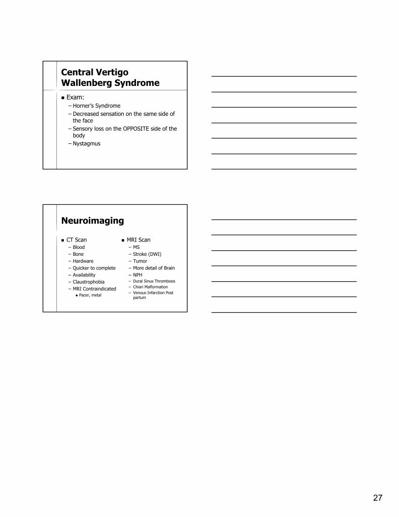
Central VertigoWallenberg Syndrome Exam:
– Horner’s Syndrome– Decreased sensation on the same side of
the face– Sensory loss on the OPPOSITE side of the
body– Nystagmus
Neuroimaging
CT Scan– Blood– Bone– Hardware– Quicker to complete– Availability– Claustrophobia– MRI Contraindicated
Pacer, metal
MRI Scan– MS– Stroke (DWI)– Tumor– More detail of Brain– NPH– Dural Sinus Thrombosis– Chiari Malformation– Venous Infarction Post
partum