the nordic expert group for criteria documentation of ... · expert group for criteria...
TRANSCRIPT

arbete och hälsa | vetenskaplig skriftserie
isbn 91-7045-687-9 issn 0346-7821
nr 2003:11
The Nordic Expert Group for Criteria Documentationof Health Risks from Chemicals and The Dutch Expert
Committee on Occupational Standards
132. FormaldehydeAnton Wibowo
National Institute for Working Life
Nordic Council of Ministers

ARBETE OCH HÄLSAEditor-in-chief: Staffan MarklundCo-editors: Marita Christmansson, Birgitta Meding,Bo Melin and Ewa Wigaeus Tornqvist
© National Institut for Working Life & authors 2003
National Institute for Working LifeS-113 91 StockholmSweden
ISBN 91–7045–687–9ISSN 0346–7821http://www.arbetslivsinstitutet.se/Printed at Elanders Gotab, Stockholm
Arbete och Hälsa
Arbete och Hälsa (Work and Health) is ascientific report series published by theNational Institute for Working Life. Theseries presents research by the Institute’sown researchers as well as by others, bothwithin and outside of Sweden. The seriespublishes scientific original works, disser-tations, criteria documents and literaturesurveys.
Arbete och Hälsa has a broad target-group and welcomes articles in differentareas. The language is most often English,but also Swedish manuscripts arewelcome.
Summaries in Swedish and English as wellas the complete original text are availableat www.arbetslivsinstitutet.se/ as from1997.

Preface
An agreement has been signed by the Dutch Expert Committee on OccupationalStandards (DECOS) of the Health Council of the Netherlands and the NordicExpert Group for Criteria Documentation of Health Risks from Chemicals (NEG).The purpose of the agreement is to write joint scientific criteria documents, whichcould be used by the national regulatory authorities in both the Netherlands and inthe Nordic countries.
The document on health effects of formaldehyde was written by Anton Wibowo,Coronel Institute, Academic Medical Centre, University of Amsterdam, theNetherlands, and has been reviewed by DECOS as well as by NEG.
The joint document is published separately by DECOS and NEG. The NEGversion presented herein has been adapted to the requirements of NEG and theformat of Arbete och Hälsa. The editorial work and technical editing has beencarried out by Anna-Karin Alexandrie, and Jill Järnberg, scientific secretary ofNEG, at the National Institute for Working Life in Sweden.
We acknowledge the Nordic Council of Ministers for its financial support ofthis project.
G.J. Mulder G. JohansonChairman ChairmanDECOS NEG

Abbreviations
ACGIH American Conference of Governmental Industrial HygienistsCI confidence intervalCNS central nervous systemCRR combined relative riskEPA United States Environmental Protection AgencyFEV1 forced expiratory volume in one secondFEV3 forced expiratory volume in three secondsFVC forced vital capacityIARC International Agency for Research on CancerIHF Industrial Health FoundationIPCS International Programme on Chemical SafetyLOAEL lowest observed adverse effect levelMAK maximale ArbeitsplatzkonzentrationNIOSH National Institute for Occupational Safety and HealthNOAEL no observed adverse effect levelOR odds ratioOSHA Occupational Safety and Health AssociationRD50 concentration associated with a 50% decrease in respiratory rateSMR standard mortality ratioSPIR standardised proportionate incidence ratioSRR standardised rate ratioTLV threshold limit valueTWA time weighted averageWHO World Health Organisation

Contents
Abbreviations
1. Introduction 1
2. Identity, properties and monitoring 12.1 Identity and chemical properties 12.2 Physical characteristics 12.3 Validated analytical methods 2
2.3.1 Environmental exposure monitoring 22.3.2 Biological exposure monitoring 2
3. Sources 33.1 Natural sources 33.2 Man-made sources 3
3.2.1 Production 33.2.2 Uses 3
4. Exposure 34.1 General population 34.2 Working population 4
5. Kinetics 55.1 Absorption 55.2 Distribution and biotransformation 65.3 Elimination 75.4 Possibilities for biological monitoring 75.5 Summary 7
6. Effects 86.1 Observation in man 8
6.1.1 Odour 86.1.2 Sensory irritation 86.1.3 Rhinitis 106.1.4 Effects on pulmonary function in healthy and asthmatic subjects 106.1.5 Sensitisation 126.1.6 Toxicity due to acute and short-term exposures 156.1.7 Epidemiological studies 15
6.2 Animal experiments 276.2.1 Sensory irritation 276.2.2 Airway reactivity 276.2.3 Sensitisation 286.2.4 Acute cytotoxic effects on nasal epithelium 286.2.5 Toxicity during short-term exposure 296.2.6 Toxicity due to long-term exposure and carcinogenicity 326.2.7 Genotoxicity 326.2.8 Mechanism of formaldehyde nasal carcinogenesis 346.2.9 Reproductive toxicity 36

6.2.10 Other studies 376.3 Summary 37
6.3.1 Human studies 376.3.2 Animal studies 40
7. Existing guidelines, standards and evaluations 427.1 General population 427.2 Working population 427.3 Evaluations of standards 42
7.3.1 The Netherlands 427.3.2 United States 437.3.3 Germany 447.3.4 Sweden 447.3.5 IARC / WHO 447.3.6 European Union 45
8. Hazard assessment 458.1 Assessment of the health hazard 458.2 Groups at extra risk 488.3 Scientific basis for an occupational exposure limit 48
9. Summary 49
10. Summary in Swedish 50
11. References 51
12. Data 58
Appendix 1 59
Appendix 2 65
Appendix 3 74
1
1. Introduction
Formaldehyde is a colourless gas at room temperature and normal atmosphericpressure. It is flammable, reactive and polymerises readily at room temperature. Itforms explosive mixtures with air and oxygen at atmospheric pressure. Thesubstance occurs naturally in the environment and is produced physiologically bymammalian cells during metabolism.
Formaldehyde is used as a raw material in chemical reactions, and as anintermediate in the manufacture of numerous products. It has also a medicalapplication as a disinfectant and is used as a preservative in various consumerproducts.
A criteria document on formaldehyde was written for the Nordic Expert Groupfor Documentation of Occupational Exposure Limits (NEG) in 1982 (66).
The present document is a co-production between NEG and the Dutch Expertcommittee on Occupational Standards (DECOS) hereafter called the committees,and the document is an up-date of the previus DECOS publication from 1987(34).
2. Identity, properties and monitoring
2.1 Identity and chemical properties
Chemical formula: CH2O (HCHO)CAS registry number: 50-00-0RTECS registry number: LP 8925000UN number: 1198, 2209, 2213EC numbers: 605-001-01 (sol 5% to < 25%)
605-001-02 (sol 1% to < 5%)605-001-005 (sol ≥ 25%)
IUPAC name: methanalCommon synonyms: formaldehyde, methylene oxide,
oxymethylene, methylaldehyde, oxomethaneCommon names forsolutions of formaldehyde: formalin, formol
2.2 Physical characteristics (27, 59)
Relative molecular mass: 30.03Boiling point: -20°CMelting point: -92°CRelative density (water=1): 0.8Solubility in water: miscibleRelative vapour density (air = 1): 1.08Flash point: flammable gas, 60°C

2
Auto-ignition temperature: 300°CExplosive limits: 7-73 vol% in airVapour pressure: 0.2 kPa at 20°C, 101.3 kPa at -19°C,
52.6 kPa at -33°CConversion factors: 1 ppm = 1.2 mg/m3
(25°C, 1066 mbar) 1 mg/m3 = 0.83 ppm
Formaldehyde is a colourless gas at room temperature and normal atmosphericpressure. It is flammable, reactive and readily polymerises at room temperature. Itforms explosive mixtures with air and oxygen at atmospheric pressure.
Formaldehyde is present in aqueous solutions as a hydrate and tends topolymerise. At room temperature, and a formaldehyde content of 30% and more,the polymers precipitate and render the solution turbid. Under atmosphericconditions, formaldehyde is readily photo-oxidised in sunlight to carbon dioxide.
2.3 Validated analytical methods
2.3.1 Environmental exposure monitoringThe most widely used methods for the determination of formaldehyde are basedon photometric measurements. The sampling method depends on the medium inwhich formaldehyde is to be determined.
The International Programme on Chemical Safety/World Health Organisation(IPCS/WHO) reported a number of different methods for determination offormaldehyde, using spectrophotometric, colorimetric, fluorometric, highperformance liquid chromatographic, polarographic, gas chromatographic,infrared, and visual analytical methods (59). On each method the analyticalsensitivity was reported.
Formaldehyde in air may be collected in an absorbing medium by diffusion(passive sampling). Aqueous or 50% 1-propanol solutions are also used forformaldehyde sampling. For active sampling, aqueous solutions and solutionscontaining sulphite, 3-methyl-2-benzothiazolene hydrazine, chromotropic acid or2,4-dinitrophenylhydrazine are generally used as the absorbing solution. Forpassive sampling sodium bisulphite, triethanolamine and 2,4-dinitrophenyl-hydrazine are used and sorbents such as silica gel, aluminium oxide and activatedcarbon, sometimes specially treated, may be useful for taking samples at theworkplace.
2.3.2 Biological exposure monitoringUntil present, biological monitoring methods for exposure to formaldehyde havenot been fully examined. Considering the critical effects and the target organsbiological monitoring seems to be irrelevant.

3
3. Sources
3.1 Natural sources
Formaldehyde is naturally formed in the troposphere during the oxidation ofhydrocarbons.
Formaldehyde is one of the volatile compounds formed in the early stages ofdecomposition of plant residues in the soil.
3.2 Man-made sources
The most important man-made source of formaldehyde is automotive exhaustfrom engines not fitted with catalytic converters.
3.2.1 ProductionFormaldehyde is produced by oxidising methanol using two different procedures:(a) oxidation with silver crystals or silver nets at 600-720°C, and (b) oxidationwith iron molybdenum oxides at 270-380°C. Formaldehyde can be produced as aby-product of hydrocarbon oxidation processes.
In 1992 worldwide formaldehyde production was estimated to be 12 milliontonnes. Major formaldehyde producing countries in 1990 were the United Statesand Japan with 3 million and 1.5 million tonnes, respectively. Other productionnumbers were: Germany 680 000; China 467 000; Sweden 244 000; Finland48 000 and Denmark 3 000 tonnes (58).
3.2.2 UsesFormaldehyde is an inexpensive starting material for a number of chemicalreactions, and a large number of products are made using formaldehyde as a base.
As an intermediate product, formaldehyde is used in the manufacture ofparticleboard, fibreboard, plywood, paper treatment, textile treatment, mouldingcompounds, surface coatings, foam, plywood adhesive, insulation, foundrybinders, phenolic thermosetting, resin curing agents, explosives, lubricants,automobile applications, plumbing components, alkyd resins, synthetic lubricants,tall oil esters, foundry resins and controlled release fertilisers.
Furthermore, formaldehyde has medical applications as a preservative anddisinfectant and it is used as a preservative in various consumer products.
4. Exposure
4.1 General population
The possible sources of exposure to formaldehyde of the general population aretobacco smoke, automobile emissions, building and insulating materials, foodproducts, cosmetics, household cleaning agents, medicinal products, and innature (59). Routes of exposure are inhalation, ingestion and dermal absorption.

4
The IPCS/WHO made the following estimation on the contribution of variousatmospheric environments to the total formaldehyde intake by inhalation of anindividual (Table 1) (59).
Guicherit and Schulting reported an average concentration of 7.4 µg/m3 (0.006ppm) of formaldehyde in the ambient air of Terschelling Island, Delft andRotterdam, the Netherlands, in the 1980s (45).
The IPCS/WHO estimated that smoking 20 cigarettes per day would lead to anaverage daily intake of 1 mg formaldehyde per day (59). Formaldehyde producedby cigarettes may also mean considerable exposure for non-smokers throughpassive smoking. The more so since it has been reported that the effects ofgaseous formaldehyde are potentiated by smoke particles and aerosols.
4.2 Working population
Exposure to formaldehyde in the workplace can be caused by either the produc-tion or handling of this compound or products containing it. Concentrations offormaldehyde in occupational settings in the United States were reported by theICPS/WHO (59), these are presented in Appendix 1.
The following represents more recent occupational exposure data.Akbar-Khanzadeh et al. reported concentrations ranging from 0.08 to 3.53
mg/m3 (0.07-2.94 ppm) formaldehyde in a gross anatomy laboratory of theMedical College in Ohio, United States (3). The 8-hour time weighted average(TWA) exposure of 31.7% of the subjects working in the laboratory exceeded theaction level of 0.6 mg/m3 (0.5 ppm) set by the Occupational Safety and HealthAssociation (OSHA).
The mean concentration of formaldehyde in area samples of an anatomylaboratory in Singapore was 0.6 mg/m3 (0.5 ppm) with a range of 0.5-0.7 mg/m3
Table 1. Contribution of various atmospheric environments to the total formaldehydeintake by inhalation of an individual (59).
Source Average intake (mg/day)
Ambient air (10% of the time) 0.02
Indoor air, home (65% of the time)prefabricated (particle board) 1-10conventional home 0.5-2
Workplace air (25% of the time)without occupational exposurea
0.2-0.8occupational exposure to 1 mg/m3 5environmental tobacco smoke 0.1-1.0
Smoking (20 cigarettes/day) 1.0
a Assuming the normal formaldehyde concentration in conventional buildings.

5
(0.4-0.6 ppm). The mean of personal samples was 0.9 mg/m3 (0.74 ppm) with arange of 0.5-1.4 mg/m3 (0.41-1.20 ppm) during a session of 2.5 hours (28).
Kilburn et al. reported 0.24-6.0 mg/m3 (0.2-5 ppm) formaldehyde levels in theworkplace air by area sampling in 10 representative histology laboratories in LosAngeles, United States, in 1983 (64). The sampling duration was not reported.The levels were highest during selection of tissue samples for processing.
Kriebel et al. reported formaldehyde exposures in the breathing zone rangingfrom 0.59-1.12 mg/m3 (0.49 to 0.93 ppm) with a geometric mean of 0.88 mg/m3
(0.73 ppm) during a clinical anatomy laboratory course at the University ofMassachusetts in the United States (67).
Suruda et al. studied 29 mortician students who were taking a course inembalming (105). During an 85-day study period, the subjects performed anaverage of 62.9 embalmings and had average cumulative formaldehyde exposuresof 14.8 ppm⋅hour, with an average air concentration of 1.68 mg/m3 (1.4 ppm)during embalming. Since the average time spent embalming was 125 minutes,formaldehyde exposures calculated as an 8-hour TWA were 0.40 mg/m3 (0.33ppm).
Mean levels of 8-hour TWA exposure to formaldehyde ranged from about 0.09mg/m3 (0.08 ppm) in the sawmill and shearing-press departments to 0.39 mg/m3
(0.32 ppm) in the warehouse area of a plywood factory in Italy (10).Herbert et al. examined the concentrations of formaldehyde from particles and
vapour at five sampling sites in an oriented strand board plant in Canada (54). Inthe manufacture they used wood fibre derived from Aspen trees bonded by phenolformaldehyde. The highest total concentration of formaldehyde was 0.32 mg/m3
(0.27 ppm) recorded at the preheat conveyor. The lowest was 0.08 mg/m3 (0.07ppm) recorded at the saw line. The samples were collected for 21 hours conti-nuously at the sites.
5. Kinetics
5.1 Absorption
There are limited human data regarding absorption of formaldehyde throughinhalation. Under normal conditions, absorption is expected to occur in the upperrespiratory tract (nasal passages in obligate nose-breathers; trachea and bronchi inoral breathers).
From animal data absorption of formaldehyde through the upper respiratorytract is estimated to be 100% as concluded from the removal of formaldehydefrom the air (59). Detailed studies on the distribution of 14C-formaldehyde in therat nasal cavities have confirmed that it is absorbed primarily in the upperrespiratory system.
Another study investigated the retention of formaldehyde gas in the nasalpassages of anaesthetised male rats exposed in a nose-only system to

6
14C-formaldehyde at 2.4-60 mg/m3 (2-50 ppm) for 30 minutes. More than 93% ofthe substance was retained, regardless of airborne concentrations.
Loden performed an in vitro experiment to study the permeability of humanskin to formaldehyde using excised skin in a flow-through diffusion cell (70). Therate of resorption was determined by measuring the amount of substance found inthe receptor fluid beneath the skin at steady state. The resorption rates offormaldehyde were: from a concentrated solution of formalin, 319 mg/cm2 perhour, from a solution of 10% formalin1 in phosphate buffer, 16.7 mg/cm2 perhour. The fact that formaldehyde induces denaturation of the skin proteins mayhave influenced the absorption of the compound.
5.2 Distribution and biotransformation
The IPCS/WHO cited a study on rats, which were exposed by inhalation for 6hours to 18 mg/m3 (15 ppm) 14C-formaldehyde (59). The distribution ofradioactivity in the tissues was determined. The highest concentrations occurred inthe oesophagus, followed by the kidneys, liver, intestines, and lungs.
There are no data available on the distribution of formaldehyde in the humanbody. The mean formaldehyde concentration in human blood after inhalatoryexposure to 2.3 mg/m3 (1.9 ppm) formaldehyde vapour during 40 minutes wasapproximately 2.61 ± 0.14 mg/100 ml. However, no statistical difference wasfound with pre-exposure levels (59). No increases in blood concentrations offormaldehyde were detected in rats or human beings exposed to formaldehydethrough inhalation due to rapid metabolism.
The overall metabolism of formaldehyde is summarised in Figure 1. Ofimportance are the oxidation of formaldehyde into formic acid and carbondioxide, the reaction with glutathione, and the covalent linkage with proteins andnucleic acids.
proteins and labile methyl groups andnucleic acids one carbon metabolism
formaldehyde formic acid CO2
urine as sodium salt
Figure 1. Overall metabolism of formaldehyde (65).
1 Formalin is defined as 37% formaldehyde in water containing 10-15% methanol

7
Formaldehyde is an endogenous metabolite in mammalian systems and it israpidly metabolised to formate, which is partially incorporated via normalmetabolic pathways into the one-carbon pool of the body or further oxidised tocarbon dioxide.
5.3 Elimination
After absorption formaldehyde is rapidly metabolised to formate or enters the one-carbon pool to be incorporated into other molecules. Besides this, there are twopathways of final elimination, via exhalation or renal elimination. There are nohuman data available on the elimination of formaldehyde, but the IPCS/WHOreported that 81% of subcutaneously administered 14C-formaldehyde to rats wasfound again as carbon dioxide and a small amount in choline (59).
5.4 Possibilities for biological monitoring
At present there are no biological monitoring methods available to determine themagnitude of past exposure to formaldehyde.
There have been a number of cytologic and cytogenetic studies of formaldehydeexposure in man. These studies examined nasal and buccal cells and bloodlymphocytes of occupationally exposed workers and unexposed control volun-teers. These studies will be evaluated in the respective chapters.
5.5 Summary
Under normal conditions it is expected that formaldehyde in ambient air isabsorbed through inhalation in the upper respiratory tract. In animals absorptionhas been found to be 100%. From in vitro experiments using human skin, it isestimated that the absorption of a concentrated solution of formalin through theskin amounted to 319 mg/cm2 per hour.
After inhalation of radioactive formaldehyde by the rat the radioactivity isdistributed in the tissues, with the highest concentration in the oesophagus,followed by the kidney, liver, intestines, and lung. Retention in the nasal passageof the rat is estimated at 93% of the dose, regardless of airborne concentrations.
Formaldehyde is an endogenous metabolite in mammalian systems and it israpidly metabolised to formate, which is partially incorporated via normalmetabolic pathways into the one-carbon pool of the body or further oxidised tocarbon dioxide. There are two pathways for elimination: via exhalation and via thekidneys.
There are no biological monitoring methods at present to determine themagnitude of past exposure to formaldehyde.

8
6. Effects
6.1 Observation in man
Only a selection of the most adequate human studies from the review ofPaustenbach et al. is discussed in this chapter (92).
6.1.1 OdourAt high concentrations, e.g. 6-12 mg/m3 (5-10 ppm), formaldehyde has a distinctand pungent odour. The odour of formaldehyde is detectable and/or recognisableby most individuals at concentrations around 1.2 mg/m3 (1 ppm) (59). The odourthreshold (i.e. the concentration at which a group of observers can detect theodour in 50% of the presentations) of formaldehyde ranges from 0.06 to 0.22mg/m3 (0.05-0.18 ppm).
6.1.2 Sensory irritationFor most odorous irritants, the trigeminal nerve has a higher threshold than theolfactory nerve. However, when the formaldehyde concentration is increased,sensory irritation is first experienced in the eyes, then the odour is perceived, andfinally nasal irritation occurs (59).
SurveysAkbar-Khanzadeh et al. studied 34 workers employed in a gross anatomylaboratory in Toledo, Unites States (3). They were exposed to formaldehyde at(TWA) concentrations ranging from 0.08 to 3.53 mg/m3 (0.07-2.94 ppm)(duration of exposure not described). More than 94% of the subjects were exposedto formaldehyde concentrations exceeding 0.36 mg/m3 (0.3 ppm). By more than70% of the exposed subjects, irritation of the eyes (88%) and nose (74%) werereported.
Kriebel et al. investigated students exposed to formaldehyde during a clinicalanatomy laboratory course when dissecting cadavers for 3 hours per week over a10-week period (67). Formaldehyde exposures in the breathing zone ranged from0.59-1.12 mg/m3 (0.49-0.93 ppm), with a geometric mean of 0.88 mg/m3 (0.73ppm). Symptoms of irritation increased strongly during the day, and the effectswere stronger at the beginning than at the end of the semester. The prevalence ofsymptoms at the start of the laboratory session ranged from 15% for cough to 46%for nose irritation. At the end of the session the prevalences were 20 and 67,respectively. The average increase in symptoms prevalence from beginning to endof laboratory session was greatest for eye irritation, with an increase of 43%. Nostatistical analyses were reported.
Wilhelmsson and Holmström performed a cross-sectional study on 66employees of a formaldehyde producing plant in Sweden to determine whetherchronic exposure to formaldehyde often causes symptoms by direct irritation(120). The workers were exposed almost exclusively to formaldehyde. Meanduration of exposure was 10 years (range 1-36 years). Thirty-six community

9
clerks served as a reference group. The exposure level of the exposed group asmeasured by personal sampling was between 0.05 to 0.60 mg/m3 (0.04-0.50 ppm)formaldehyde, with a mean of 0.26 mg/m3 (0.22 ppm). The reference group wasexposed to an average concentration of 0.09 mg/m3 (0.07 ppm) formaldehyde overthe year. From a (not specified) questionnaire, it appeared that 67% of the exposedgroup experienced general nasal discomfort compared to 25% of the referencegroup (p<0.001). Nasal discomfort strictly connected to the workplace occurred in53% of the exposed group and in 3% of the reference group (p<0.001). However,the questionnaire was not published. Therefore, the committees are of the opinionthat this study might only suggest that after long-term occupational exposure (0.26mg/m3 formaldehyde), more than 50% of the exposed workers complained ofnasal discomfort, which was attributed to their occupation.
Liu et al. studied the irritant effects associated with formaldehyde exposure inmobile homes in California (69). Week-long integrated formaldehyde concent-rations were measured in summer (663 mobile homes with 1 394 residents) andwinter (523 mobile homes with 1 096 residents), using passive monitors while themobile home residents continued their normal activities. The concentrationsvaried from below the detection limit (0.0012 mg/m3) to 0.55 mg/m3. Irritanteffects were found to be significantly associated with formaldehyde exposure aftercontrolling for age, sex, smoking status, and chronic illnesses. Effects includedcomplaints of burning/tearing eyes, stinging/burning skin, fatigue, and sleepingproblems in summer and burning/tearing eyes, chest pain, dizziness, sleepingproblems, and sore throat in winter. For the three weekly ranges of formaldehydeexposure that were distinguished (less than 8.4 mg/m3·hour, between 8.4-14.4mg/m3·hour, more than 14.4 mg/m3·hour), the percentages of people withburning/tearing eyes in the summer increased from 13.3% to 17.1% and then to21.4%. In winter, percentages increased from 10.8% to 14.7% and then to 20.6%.
Controlled human studiesWeber-Tschopp et al. exposed healthy volunteers to increasing concentrations offormaldehyde from 0.036 to 4.8 mg/m3 (0.03-4 ppm) (116). Thirty-three subjectswere continuously exposed for 35 minutes and 48 subjects were exposed for 1.5minute. The irritating effects were determined by the eye-blinking rate of theindividuals. The authors found that the irritating effects increased as a function ofthe formaldehyde concentration. The irritation threshold of formaldehyde wasplaced in the range between 1.2 and 2.4 mg/m3 (1 and 2 ppm). The authorssuggested that adaptation to the irritation occurred after a few minutes in subjectsafter prolonged exposure to formaldehyde.
Bender et al. studied eye irritation in groups of volunteers (n= 5-28 per group)exposed to 0, 0.42, 0.67, 0.84, 1.08 and 1.2 mg/m3 (0, 0.35, 0.56, 0.7, 0.9 and 1.0ppm) formaldehyde for 6 minutes (12). The authors reported that the subjectivemeasurements of eye irritation may be affected by a variety of psychological andphysiological factors, such as air flow over the eyes, dust particles, length of sleepthe previous night, etc. In spite of the large variation in response time, there wasstill a significant relationship between formaldehyde concentration and time to

10
detection of response. The authors concluded that eye irritation occurred atexposure concentrations of 0.42-1.1 mg/m3 (0.35-0.9 ppm) formaldehyde. Theresponse was slight until a concentration of 1.2 mg/m3 (1 ppm) was reached.
Andersen and Mølhave conducted a study in which 16 healthy subjects (5smokers) were exposed to 0.29, 0.48, 0.97 or 1.92 mg/m3 (0.24, 0.4, 0.81 or 1.6ppm) formaldehyde for 5 hours (4). The purpose of the study was to determine theconcentration at which eye irritation occurred. Nineteen percent of the respon-dents reported eye irritation at 0.29 mg/m3 (0.24 ppm). Discomfort increasedduring the first 2 hours of exposure up to 0.97 mg/m3 (0.81 ppm); then irritationstabilised for the remaining 3 hours. A decrease in discomfort was observed at1.92 mg/m3 (1.6 ppm), indicating acclimatisation. After 5 hours of exposure, 38%of the subjects had no complaints at 1.92 mg/m3 (1.6 ppm), and 63% had nodiscomfort at 0.97 mg/m3 (0.81 ppm). This study illustrates the relatively widevariation in individual susceptibility to irritation from formaldehyde.
6.1.3 RhinitisPazdrak et al. tried to characterise the nature of formaldehyde induced nasalresponse consisting of symptoms of rhinitis and changes in nasal lavage fluid (93).Eleven healthy subjects and 9 patients with specific skin sensitisation wereprovoked in an experimental chamber with formaldehyde at a concentration of0.48 mg/m3 (0.4 ppm) for 2 hours. Nasal lavage was performed prior to andimmediately after provocation, and 4 and 8 hours later. It was found that theprovocation caused transient symptoms of rhinitis and prolonged changes in nasalwashing. There were increases in the relative number of eosinophils, and inalbumin and total protein levels in the nasal fluid, 4 and 8 hours after provocation.No difference was found between the healthy subjects and patients. These dataconfirm the irritant effects of inhaled formaldehyde and might suggest that inhaledformaldehyde is capable of inducing non-specific inflammatory changes at aconcentration of 0.48 mg/m3 (0.4 ppm).
6.1.4 Effects on pulmonary function in healthy and asthmatic subjectsWitek Jr et al. evaluated the respiratory effects in asthmatics after exposure toformaldehyde (123). Fifteen asthmatic volunteers were exposed in a double-blindmanner to room air or 2.4 mg/m3 (2 ppm) formaldehyde for 40 minutes. Theseexposures were repeated on a separate day during moderate exercise (450kpm/minutes) for 10 minutes. Pulmonary function was assessed by using partialand maximal flow volume curves. The following parameters were determined:vital capacity, residual volume, total lung capacity, forced expiratory volume inone second (FEV1), forced vital capacity (FVC), peak expiratory flow rate, andmaximal flow at 50% of vital capacity. No significant airway obstruction orairway resistance was noted in this group during and immediately after exposure.However, bad odour, sore throat, and eye irritation were common duringexposure, but the symptoms were infrequent afterwards. No delayed broncho-constriction was detected with measurements of peak expiratory flow.

11
The results of this study were substantiated by Sauder et al. (98). In their studyon 9 non-smoking asthmatic volunteers, they also found no significant changes inthe pulmonary function [FVC, FEV1, mean forced expiratory flow during themiddle half of the FVC (25-75%), specific airway conductance or functionalresidual capacity] or airway reactivity when the volunteers were exposed to 3.6mg/m3 (3 ppm) formaldehyde vapour for 3 hours. However, there was asignificant increase in nose and throat irritation at the 30th minute and eyeirritation at the 60th and 180th minutes of exposure.
Harving et al. studied the possible effects of acute formaldehyde exposure onthe lung function of asthmatic subjects. They exposed 15 non-smoking asthmaticsubjects, with documented bronchial hyperresponsiveness, to 0.08, 0.12 or 0.85mg/m3 formaldehyde for 90 minutes (47). All except one subject requiredbronchodilator therapy and none were using methylxanthines or corticosteroids.Exposure occurred in a climate chamber and the protocol was double blind. Nocontrol group was used in this experiment. Lung function tests were carried outbefore the exposure period and repeated near the end. The results showed nosignificant changes in the FEV1, functional residual capacity, airway resistance,specific airway resistance, and flow-volume curves during formaldehydeexposure. Furthermore, histamine challenge performed immediately afterformaldehyde exposure showed no evidence of changes in bronchial hyper-reactivity. No late reactions were registered during the first 14-16 hours afterexposure. There was no association of subjective ratings of symptoms, if any, withincreasing exposure. The rating of symptoms did not differ significantly when thethree exposure levels were compared. The results of this study suggest that theexposure levels of formaldehyde used were of minor, if any, importance in theemergence of pulmonary symptoms in asthmatic subjects.
Chia et al. examined 150 first-year medical students exposed to formaldehydeduring dissection of cadavers in a gross anatomy laboratory (28). As a referencegroup they used 189 third- and fourth-year medical students matched for sex,ethnic group, and age. The mean concentration of formaldehyde in the area was0.60 mg/m3 (0.50 ppm) and the mean concentration of personal samples was 0.89mg/m3 (0.74 ppm). The latter had a range of 0.49 to 1.44 mg/m3 (0.41-1.20 ppm).No differences were found in FEV1 and FVC among 22 randomly selected maleand female subjects, when the measurements were compared between the first dayafter two weeks vacation and after the dissection period. Significant differences,however, were observed in the exposed group for symptoms of decreased abilityto smell, eye irritation, and dry mouth in comparison with the reference group.
Herbert et al. performed a cross-sectional study on 99 workers employed in themanufacture of oriented strand board (54). The reference group consisted of 165unexposed workers from a petroleum industry. Both groups were investigatedusing questionnaires, spirometry and skin prick tests to common environmentalantigens. Environmental monitoring showed dust levels with a mean of 0.27mg/m3. The mass mean aerodynamic diameter of the particles was 2.5 mm. Theconcentration of formaldehyde was between 0.08 and 0.32 mg/m3 (0.07-0.27 ppm)in the strand board factory. Lung function tests showed significant differences

12
between strand board workers and workers from the petroleum industry in theFEV1/FVC ratio and reductions of FEV1 (p=0.044) and FVC (p=0.022) during theshift work. Also, the strand board workers complained of self-reported asthma andof lower respiratory tract symptoms significantly more frequent than the oilworkers. The prevalence of atopy did not differ between both groups. Lungfunction was significantly better in the strand board workers who had nosymptoms, compared with symptomatic workers. Since the complaints of self-reported asthma and of lower respiratory tract symptoms by the exposed groupoccurred at rather low concentrations of formaldehyde and dusts, the authorsconcluded that the effects may have been related to small particles containingformaldehyde that penetrated deep into the airways.
Horvath et al. surveyed 109 workers (exposed to formaldehyde from 1 to 20years) for symptoms of respiratory tract irritation (57). Estimates of the exposureranged from 0.2 to 3.5 mg/m3 (0.17-2.93 ppm) (mean 0.83 mg/m3 (0.69 ppm)).The percentage of the exposed workers reporting respiratory irritation wassignificantly higher than in the non-exposed group (n=264).
6.1.5 SensitisationRespiratory tract sensitisationGrammer et al. evaluated the immunological response to formaldehyde exposurein a group of 37 workers in a cross-sectional study (44). The durations ofemployment were not reported. Concentrations of formaldehyde in air sampling inseveral work areas at various times ranged from 0.004 to 0.087 mg/m3
(0.003-0.073 ppm) as TWAs. The workers were also exposed to phenol andorganic solvents. A clinical assessment included review of a summary of medicalhistory, physical examination, chest X-ray films, and pulmonary function studies.Serologic assessment was made with an enzyme linked immunosorbent assay forIgE and IgG to formaldehyde-human serum albumin. It was found that none of theworkers had IgE or IgG antibodies to formaldehyde-human serum albumin or animmunologically mediated respiratory or ocular disease caused by formaldehyde.
Thrasher et al. studied four groups of patients with long-term inhalationexposure to formaldehyde consisting of (1) mobile home residents, (2) officeworkers who had worked in a new office building, (3) subjects who had movedfrom mobile homes for at least one year, and (4) subjects who had worked in jobswith possible exposure to formaldehyde (110). All patients in this study hadsought continuous medical attention because of multiple complaints involving thecentral nervous system (CNS). They were compared with a group of students whohad been exposed to formaldehyde for 13 hours per week for 28 weeks whilestudying anatomy. No measurements of formaldehyde in air were performed.When compared to the controls it was found that the patients had significantlyhigher autoantibodies and antibody titers and B-cell titers to formaldehyde-humanserum albumin.
Sixty-three practising pathologists in Alberta, Canada, were studied regardingatopy and sensitivity to formaldehyde (97). Serum samples were assayed for totalIgE levels and the presence of IgE with specificity towards formaldehyde.

13
Twenty-nine of the subjects (46%) had a history of atopy that was confirmed in 12by either IgE levels or a positive radio-allergosorbent test. Twenty-nine (46%)complained of formaldehyde sensitivity. In this study, none of the pathologistshad allergen-specific IgEs directed against formaldehyde, and there was noevidence of a tendency for atopic subjects to be more prone to sensitivity toformaldehyde. However, the authors confirmed that this might have been relatedto the deliberate reduction in exposure by individuals experiencing adverseeffects.
A case-report was described by Grammer et al. (43). The subject was a workerwith clinical symptoms compatible with bronchospasm caused by formaldehydeexposure. An enzyme linked immunosorbent assay showed that the worker hadpositive IgE and IgG titers to formaldehyde-human serum albumin. The workerhad a positive intracutaneous test for formaldehyde-human serum albumin. Thecutaneous reactivity could be transferred to a rhesus monkey through the worker’sserum. The worker had a negative metacholine challenge at 25 mg/ml andnegative formaldehyde inhalation challenges at 0.36, 1.2, 3.6, and 6 mg/m3 (0.3, 1,3, and 5 ppm) for 20 minutes. The authors concluded that the worker’s symptomswere probably not caused by immunologically mediated asthma. Based on theirexperience, they stated that immunologically mediated asthma caused byformaldehyde is extremely rare, if it exists at all.
In 1991, Bardana Jr and Montanaro made an extensive review and analysis ofthe immunological effects of formaldehyde (11). They concluded that formal-dehyde is capable of acting as a respiratory irritant. But according to the authorsof the review, there is no consistent evidence indicating that formaldehyde is arespiratory sensitiser. Formaldehyde does not induce transient or permanentbronchial hyperreactivity, which has been associated with e.g. exposure to ozoneor nitrogen dioxide. Almost the same conclusions were drawn by IPCS/WHO(59). They commented that there are a few case-reports of asthma-like symptomscaused by formaldehyde, but none of these demonstrated a sensitisation effect(neither Type I nor Type IV) and the symptoms were considered to be due toirritation.
Garrett et al. studied a group of 148 children (age 7-14), 53 of whom wereasthmatic, in houses in Australia between March 1994 and February 1995 (42).The mean indoor formaldehyde exposure level was 15.8 mg/m3 and an associationbetween formaldehyde exposure and atopy [odds ratio (OR) 1.4; 95% confidenceinterval (CI): 0.98-2.00] was observed. The committees noted, however, thepotential selection bias in this study.
Skin sensitisationAccording to the IPCS/WHO skin sensitisation by formaldehyde has been shownonly by direct skin contact with formaldehyde solutions in concentrations of 20 g/l(2%) and higher (59). The lowest patch test challenge concentration in an aqueoussolution reported to produce a reaction in sensitised persons was 0.05% formal-dehyde.

14
Flyvholm and Menné interviewed 11 patients with eczema and a positive patchtest to formaldehyde (40). All patients used one or more products containingformaldehyde or formaldehyde releasers. Sources of exposure were cosmetics andpersonal care products, dishwashing liquids, water-based paints, photographicproducts etc.
Liden et al. reported absence of specific IgE antibodies in allergic contactsensitivity to formaldehyde (68). They studied 23 patients with positiveepicutaneous test reactions to formaldehyde, recruited from dermatologicdepartments in Sweden. The patients were between 21-74 years old and 19 werewomen. The tests had been performed 6 months to 10 years before inclusion in thestudy. On re-testing, 15 showed a positive reaction. Eight patients showed atopicdiathesis, and 8 had a history of ongoing atopic dermatitis. In the radio-allergo-sorbent test only 2 non-atopic patients had specific IgE antibodies to formalde-hyde. In cellular infiltrates from biopsies of epicutaneous test sites cells reactivewith monoclonal antibodies against IgE were found in positive and in negativeformalin tests, both in atopics and non-atopics, as well as in control biopsies fromnon-lesional skin. Double immunofluorescence staining experiments showed thatIgE occurred on Langerhans cells. The proportion of IgE-positive cells correlatedto the level of serum IgE, but not to atopy. These cells were also found in theepidermis and in the dermis of non-atopic patients. The authors concluded thatthis study did not support the hypothesis that specific IgE antibodies are active inthe pathogenesis of contact sensitivity to formaldehyde, neither in atopic nor innon-atopic patients.
Cronin performed an investigation in the St John Dermatology Center inLondon to determine the prevalence of formaldehyde sensitivity and to establishwhether there is a significant correlation between formaldehyde sensitivity andhand eczema (33). The study spanned 6 years, from 1984 to 1989. In this period atotal of 4 553 men were patch tested with a 1% aqueous solution of formaldehyde.The prevalence of sensitisation was approximately 2-3% each year. During these6 years, 98 men (2.2%) were sensitised. During the same period 6 479 womenwere patch tested with a 1% aqueous solution of formaldehyde. The prevalence ofsensitisation was remarkably constant at approximately 4% each year. Duringthese 6 years 235 women (3.6%) showed a positive reaction and 117 women wereprimarily sensitised by formaldehyde, of whom 61 (52%) had hand eczema. Ofthis group 2% was occupationally exposed and 88% domestic.
In their review Bardane and Montanaro pointed out that the threshold forinduction of delayed hypersensitivity contact dermatitis has not been determinedprecisely (11). The frequency of allergic contact dermatitis to formaldehyde wasestimated by the authors to range between 3% and 6% in the general population.Cross reactivity with other aldehydes has not yet been demonstrated; glutar-aldehyde does not cross react. Formaldehyde has also been reported to causecontact urticaria, but the mechanism of action has never been clearly demon-strated.

15
6.1.6 Toxicity due to acute and short-term exposuresNo cases of death from formaldehyde inhalation have been published (59).
The IPCS/WHO summarised the clinical features of formaldehyde intoxicationincluding weakness, headache, abdominal pain, vertigo, anaesthesia, anxiety,burning sensation in the nose and throat, thirst, clammy skin, central nervoussystem depression, coma, convulsions, cyanosis, diarrhoea, dizziness, dysphagia,irritation and necrosis of mucous membranes and gastrointestinal tract, vomiting,hoarseness, nausea, pallor, shock, and stupor (59).
Effects on the respiratory system caused by high formaldehyde concentrationsare pneumonia, dyspnoea, wheezing, laryngeal and pulmonary oedema, broncho-spasm, coughing of frothy fluid, respiratory depression, obstructive tracheo-bronchitis, laryngeal spasm, and sensation of substernal pressure.
Acute ingestion may cause renal injury (dysuria, anuria, pyuria, and haemat-uria) and leads to an increase in formate levels in the urine.
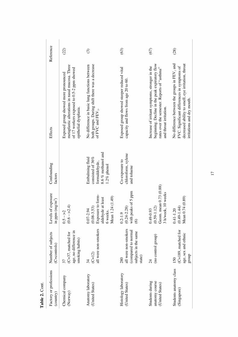
6.1.7 Epidemiological studiesCross-sectional morbidity studiesA summary of cross-sectional morbidity studies of workers occupationallyexposed to formaldehyde is presented in Table 2.
From these studies it may be concluded that symptoms of irritation of the upperrespiratory tract already occurred after acute exposure to levels below 1.2 mg/m3
(1 ppm) formaldehyde. After exposure for a few hours decreases of the FEV1 andFVC have been observed.
Of interest are the cross-sectional morbidity studies performed by Wilhelmssonand Holmström (120), Herbert et al. (54), and Boysen et al. (22).
The study by Wilhelmsson and Holmström (120) on 66 workers occupationallyexposed to formaldehyde during formaldehyde production is described in section6.1.2. Beside irritation, the authors were also interested in whether chronicexposure affected exposed people through hyperreactivity in atopic persons,through formaldehyde-induced hyperreactivity in non-atopic persons, or throughimmunologically mediated, immediate Type I reactions to formaldehyde itself.Among the 53% of the exposed workers experiencing nasal discomfort throughhyperreactivity, atopics were not significantly overrepresented. Two workers withoccasional occupational nasal discomfort, and sensitised by long-term inhalation,had a positive radio-allergosorbent test for formaldehyde. Of the occupationallyexposed group 20% experienced general eye problems. The frequency in thecontrol group was 0%. Thirty-six percent of the exposed group had dermato-logical problems such as eczema or itching, while the corresponding frequencyamong the control group was 11%. The authors concluded that in certaincircumstances formaldehyde can induce an IgE-mediated Type I reaction in thenose, but in most cases the annoying nasal symptoms are caused by formaldehydeinduced hyperreactivity, which can cause problems in about 50% of a populationexposed to formaldehyde at an average level of 0.26 mg/m3 (0.22 ppm). Anotherinteresting finding was that atopics run approximately the same risk of sufferingfrom this hyperreactivity as non-atopics. However, these results were obtained

16
Tab
le 2
. Cro
ss-s
ectio
nal m
orbi
dity
stu
dies
of
wor
kers
occ
upat
iona
lly e
xpos
ed to
for
mal
dehy
de.
Fact
ory
or p
rofe
ssio
ns(c
ount
ry)
Num
ber
of s
ubje
cts
(C=
cont
rols
)L
evel
s of
exp
osur
ein
ppm
(m
g/m
3 )C
onfo
undi
ngfa
ctor
sE
ffec
tsR
efer
ence
Air
plan
e pr
oduc
tion
(Uni
ted
Stat
es)
37 (no
cont
rol g
roup
)0.
003-
0.07
3(0
.004
-0.0
88)
Co-
expo
sure
to p
heno
lan
d or
gani
c so
lven
ts14
wor
kers
with
irri
tant
syn
drom
e. N
one
ofth
em h
ad r
espi
rato
ry o
r oc
ular
dis
ease
that
was
imm
unol
ogic
ally
med
iate
d.
(44)
Plyw
ood
fact
ory
(Ita
ly)
15 (C=
15, m
atch
ed f
orag
e an
d se
x)
0.08
-0.3
2(0
.09-
0.39
)C
o-ex
posu
re to
woo
ddu
sts
(0.2
3-0.
73 m
g/m
3 )
Hig
her
freq
uenc
y of
mic
ronu
clea
ted
cells
inna
sal r
espi
rato
ry c
ells
. Chr
onic
infl
amm
atio
nof
the
nasa
l muc
osa.
Hig
her
freq
uenc
y of
squa
mou
s m
etap
lasi
a ce
lls.
(10)
Form
alde
hyde
prod
ucin
g pl
ant
(Sw
eden
)
66 (
36%
sm
oker
s)(C
=36
, 28%
smok
ers)
0.04
-0.5
0(0
.05-
0.60
)m
ean
0.22
(0.2
6)
53%
of
expo
sed
grou
p ha
d na
sal d
isco
mfo
rt(3
% in
con
trol
gro
up).
33%
of
expo
sed
grou
pha
d ge
nera
l low
er r
espi
rato
ry tr
act d
isco
mfo
rt(C
=1%
). 2
0% o
f ex
pose
d gr
oup
had
eye
prob
lem
s (C
=0%
).
(120
)
Ori
ente
d st
rand
boar
d m
anuf
actu
re(C
anad
a)
99 (C=
165)
0.07
-0.2
7(0
.08-
0.32
)D
ust l
evel
0.2
7 m
g/m
3
with
mas
s m
ean
aero
-dy
nam
ic d
iam
eter
2.5
µm
Sign
ific
ant l
ower
FE
V1/
FVC
, and
cro
ss-s
hift
redu
ctio
n of
FE
V1 an
d FV
C. E
leva
ted
repo
rts
of “
asth
ma”
and
hig
her
freq
uenc
y of
low
erre
spir
ator
y tr
act s
ympt
oms.
No
diff
eren
ce in
atop
y.
(54)
Pape
r m
ill(I
ndia
)22 (C
=27
)0.
025
8-ho
ur T
WA
(0.0
3)
Exp
osed
sub
ject
s sh
owed
mor
e re
spir
ator
ysy
mpt
oms
and
com
plai
nts
pert
aini
ng to
gast
roin
test
inal
, mus
culo
skel
etal
and
card
iova
scul
ar s
yste
ms.
No
diff
eren
ce in
hem
atol
ogy.
(102
)
17
Tab
le 2
. Con
t.
Fact
ory
or p
rofe
ssio
ns(c
ount
ry)
Num
ber
of s
ubje
cts
(C=
cont
rols
)L
evel
s of
exp
osur
ein
ppm
(m
g/m
3 )C
onfo
undi
ngfa
ctor
sE
ffec
tsR
efer
ence
Che
mic
al c
ompa
ny(N
orw
ay)
37 (C=
37, m
atch
ed f
orag
e, n
o di
ffer
ence
insm
okin
g ha
bits
)
0.5
– >
2(0
.6 –
>2.
4)E
xpos
ed g
roup
sho
wed
mor
e pr
onou
nced
met
apla
stic
alte
ratio
ns in
nas
al m
ucos
a. T
hree
of 1
7 w
orke
rs e
xpos
ed to
0.5
-2 p
pm s
how
edep
ithel
ial d
yspl
asia
.
(22)
Ana
tom
y la
bora
tory
(Uni
ted
Stat
es)
34 (C=
12)
all w
ere
non-
smok
ers
0.07
-2.9
4(0
.08-
3.53
)E
xpos
ure
to f
orm
-al
dehy
de a
t lea
st6
wee
ks.
Mea
n 1.
24 (
1.49
)
Em
balm
ing
flui
dco
nsis
ted
of 3
6%fo
rmal
dehy
de,
8.6
% m
etha
nol a
nd1.
2% p
heno
l
No
diff
eren
ce in
bas
ic lu
ng f
unct
ions
bet
wee
nbo
th g
roup
s. D
urin
g sh
ift t
here
was
a d
ecre
ase
of F
VC
and
FE
V3.
(3)
His
tolo
gy la
bora
tory
(Uni
ted
Stat
es)
280
all w
ere
non-
smok
ers
(com
pare
d to
nor
mal
subj
ects
in th
e sa
me
stat
e)
0.2-
1.9
(0.2
4-2.
28)
with
pea
ks o
f 5
ppm
(6)
Co-
expo
sure
toch
loro
form
, xyl
ene
and
tolu
ene
Exp
osed
gro
up s
how
ed s
teep
er r
educ
ed v
ital
capa
city
and
flo
ws
from
age
20
to 6
0.(6
3)
Stud
ents
dur
ing
anat
omy
cour
se(U
nite
d St
ates
)
24 (no
cont
rol g
roup
)0.
49-0
.93
(0.5
9-1.
12)
Geo
m. m
ean
0.73
(0.
88)
3 h/
wee
k, 1
0 w
eeks
Incr
ease
of
irri
tant
sym
ptom
s, s
tron
ger
in th
ebe
ginn
ing.
Dec
line
in th
e pe
ak e
xpir
ator
y fl
owra
tes
over
the
sem
este
r. R
epor
ts o
f “a
sthm
a”an
d th
roat
irri
tatio
n.
(67)
Stud
ents
ana
tom
y cl
ass
(Sin
gapo
re)
150
(C=
189,
mat
ched
for
age,
sex
and
eth
nic
grou
p
0.41
-1.2
0(0
.49-
1.44
)M
ean
0.74
(0.
89)
No
diff
eren
ce b
etw
een
the
grou
ps in
FE
V1 a
ndFV
C. S
igni
fica
nt d
iffe
renc
es in
sym
ptom
s of
decr
ease
d ab
ility
to s
mel
l, ey
e ir
rita
tion,
thro
atir
rita
tions
and
dry
mou
th.
(28)

18
from a not published questionnaire and therefore the results are of limited use.The cross-sectional study by Herbert et al. (54) on workers employed in a
manufacture of oriented strand board is described in section 6.1.4. The workersshowed reduced lung functions and complained more of self-reported asthma andof lower respiratory tract symptoms compared to the reference group.
Boysen et al. (22) reported on a study on nasal biopsies of 37 workersoccupationally exposed to formaldehyde (chemical company producingformaldehyde and formaldehyde resin). The workers were exposed for more than5 years, and they were compared to 37 age-matched controls. The level ofexposure of the exposed group ranged from 0.6 to more than 2.4 mg/m3
formaldehyde. The two groups did not differ as to other environmental influences,smoking habits, and previous nasal disease. The authors found that the degree ofmetaplasia of the nasal mucosa cells was more pronounced among the exposedworkers than among the controls. Three cases of dysplasia out of 17 workers(18%), all of the squamous type, were observed in the formaldehyde group (zerocases in the control group). These workers had been exposed daily to formalde-hyde concentrations ranging from 0.6 mg/m3 to more than 2.4 mg/m3 for morethan 22 years. According to the committees the study, however, is too small todraw any conclusions. Since only a small area of the nasal mucosa can beexamined histologically, the number of dysplastic lesions found can not beexpected to reflect the real prevalence of dysplasia and therefore the committeesare of the opinion that the real prevalence of dysplasia could even be higher.
Longitudinal/prospective morbidity studiesNunn et al. followed a group of 164 workers exposed daily to formaldehydeduring the production of urea-formaldehyde resin, together with 129 workers notexposed to formaldehyde, for 6 years (87). Exposure was classified as high(TWA more than 2.4 mg/m3), medium (0.72-2.4 mg/m3) or low (0.12-0.6 mg/m3).Twenty-five % of the workers had high exposure during several periods and 17%moderate exposure. The annual assessment included lung function testing. Theproportion of self-reported respiratory symptoms was similar in the two groups.The initial FEV1 was within 0.5 litre of the predicted value (by age and height) in65% of the exposed and 59% of the unexposed workers, and more than 0.5 litrebelow the predicted value in 9% of the exposed and 11% of the unexposedworkers. The mean decline in FEV1 was 42 ml/year in the exposed group and 41ml/year in the unexposed group. The authors found no association between therate of decline and indices of exposure to formaldehyde in the exposed group. Ininterpreting these results it is important to assess any possible bias in the conductof the study. Workers with adverse respiratory effects from exposure to highconcentrations of formaldehyde may have left employment so that only“survivors” are included in the study (healthy worker effect).
The effect of low-level exposure to formaldehyde on oral, nasal, andlymphocytic biological markers were studied prospectively by Suruda et al. in agroup of 29 mortician students who were about to take a course in embalming(105). During the 85-day study period the subjects performed an average of 69
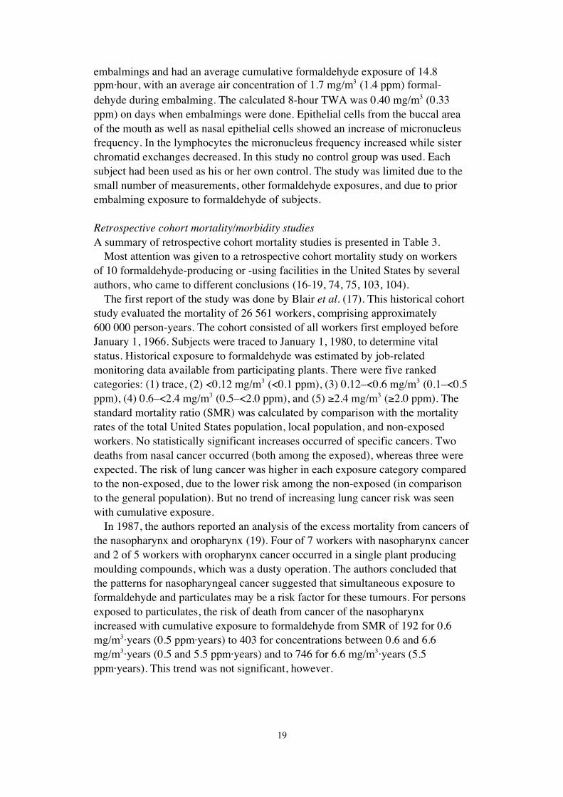
19
embalmings and had an average cumulative formaldehyde exposure of 14.8ppm⋅hour, with an average air concentration of 1.7 mg/m3 (1.4 ppm) formal-dehyde during embalming. The calculated 8-hour TWA was 0.40 mg/m3 (0.33ppm) on days when embalmings were done. Epithelial cells from the buccal areaof the mouth as well as nasal epithelial cells showed an increase of micronucleusfrequency. In the lymphocytes the micronucleus frequency increased while sisterchromatid exchanges decreased. In this study no control group was used. Eachsubject had been used as his or her own control. The study was limited due to thesmall number of measurements, other formaldehyde exposures, and due to priorembalming exposure to formaldehyde of subjects.
Retrospective cohort mortality/morbidity studiesA summary of retrospective cohort mortality studies is presented in Table 3.
Most attention was given to a retrospective cohort mortality study on workersof 10 formaldehyde-producing or -using facilities in the United States by severalauthors, who came to different conclusions (16-19, 74, 75, 103, 104).
The first report of the study was done by Blair et al. (17). This historical cohortstudy evaluated the mortality of 26 561 workers, comprising approximately600 000 person-years. The cohort consisted of all workers first employed beforeJanuary 1, 1966. Subjects were traced to January 1, 1980, to determine vitalstatus. Historical exposure to formaldehyde was estimated by job-relatedmonitoring data available from participating plants. There were five rankedcategories: (1) trace, (2) <0.12 mg/m3 (<0.1 ppm), (3) 0.12–<0.6 mg/m3 (0.1–<0.5ppm), (4) 0.6–<2.4 mg/m3 (0.5–<2.0 ppm), and (5) ≥2.4 mg/m3 (≥2.0 ppm). Thestandard mortality ratio (SMR) was calculated by comparison with the mortalityrates of the total United States population, local population, and non-exposedworkers. No statistically significant increases occurred of specific cancers. Twodeaths from nasal cancer occurred (both among the exposed), whereas three wereexpected. The risk of lung cancer was higher in each exposure category comparedto the non-exposed, due to the lower risk among the non-exposed (in comparisonto the general population). But no trend of increasing lung cancer risk was seenwith cumulative exposure.
In 1987, the authors reported an analysis of the excess mortality from cancers ofthe nasopharynx and oropharynx (19). Four of 7 workers with nasopharynx cancerand 2 of 5 workers with oropharynx cancer occurred in a single plant producingmoulding compounds, which was a dusty operation. The authors concluded thatthe patterns for nasopharyngeal cancer suggested that simultaneous exposure toformaldehyde and particulates may be a risk factor for these tumours. For personsexposed to particulates, the risk of death from cancer of the nasopharynxincreased with cumulative exposure to formaldehyde from SMR of 192 for 0.6mg/m3·years (0.5 ppm·years) to 403 for concentrations between 0.6 and 6.6mg/m3·years (0.5 and 5.5 ppm·years) and to 746 for 6.6 mg/m3·years (5.5ppm·years). This trend was not significant, however.

20
Tab
le 3
. A s
umm
ary
of r
etro
spec
tive
coho
rt m
orta
lity
stud
ies
of w
orke
rs o
ccup
atio
nally
exp
osed
to f
orm
alde
hyde
.
Fact
orie
s or
occu
patio
ns(c
ount
ry)
Est
imat
ion
ofex
posu
reC
hara
cter
istic
s of
coh
ort
Res
ults
Ref
eren
ce
10 f
orm
alde
hyde
prod
uctio
n an
d us
efa
cilit
ies
(Uni
ted
Stat
es)
Bas
ed o
n jo
b tit
les.
Usi
ng a
vaila
ble
mon
itori
ng d
ata
from
part
icip
atin
g pl
ants
.5
rank
ed c
ateg
orie
sof
exp
osur
e.
26 5
61 w
orke
rs (
appr
ox. 6
00 0
00pe
rson
-yea
rs).
Fol
low
-up
1966
to19
80.
Com
pari
son
with
US
popu
latio
n,lo
cal p
opul
atio
n an
d no
n-ex
pose
dw
orke
rs. I
nfor
mat
ion
on s
mok
ing
habi
ts w
as n
ot a
vaila
ble.
No
sign
ific
ant e
xces
ses
for
spec
ific
can
cers
. SM
Rs
for
canc
er o
f th
ere
spir
ator
y sy
stem
are
112
(95
% C
I: 9
7-12
8) f
or w
hite
men
, 121
(95%
CI:
52-
238)
for
whi
te w
omen
, 68
(95%
CI:
34-
124)
for
bla
ckm
en.
The
re is
no
tren
d of
incr
easi
ng lu
ng c
ance
r ri
sk w
ith c
umul
ativ
eex
posu
re le
vel.
Mor
talit
y fr
om c
ance
r of
the
nasa
l cav
ity w
as n
otex
cess
ive.
The
pat
tern
of
naso
phar
ynge
al c
ance
r su
gges
ts th
at s
imul
tane
ous
expo
sure
to f
orm
alde
hyde
and
“pa
rtic
ulat
es”
may
be
a ri
sk f
acto
rfo
r th
is tu
mou
r.
(17-
19)
Aut
omot
ive
iron
fou
ndry
(Uni
ted
Stat
es)
Bas
ed o
n jo
b tit
les,
4 ca
tego
ries
(hig
h, m
ediu
m, l
owan
d no
ne).
3 92
9 w
orke
rs. F
ollo
w-u
p pe
riod
1960
-198
9.C
ompa
riso
n w
ith U
S po
pula
tion
and
non-
expo
sed
wor
kers
(n
= 2
032
).Sm
okin
g st
atus
asc
erta
ined
in 6
5.4%
of e
xpos
ed a
nd 5
5.1%
of
the
unex
pose
d co
hort
.
No
asso
ciat
ion
betw
een
form
alde
hyde
exp
osur
e an
d de
aths
fro
mm
alig
nant
or
non-
mal
igna
nt d
isea
se o
f th
e re
spir
ator
y sy
stem
.SM
Rs
for
canc
er o
f bu
ccal
cav
ity a
nd p
hary
nx: e
xpos
ed w
orke
rs13
1 (9
5% C
I: 4
8-28
6); u
nexp
osed
wor
kers
169
(95
% C
I: 5
4-39
5).
SMR
s fo
r ca
ncer
of
trac
hea,
bro
nchu
s an
d lu
ng: e
xpos
ed w
orke
rs12
0 (9
5% C
I: 8
9-15
8); u
nexp
osed
wor
kers
119
(95
% C
I: 8
4-16
3).
(5, 6
)
Che
mic
al a
ndpl
astic
indu
stry
(Uni
ted
Kin
gdom
)
Bas
ed o
n jo
b tit
les,
4 ca
tego
ries
(hig
h, m
oder
ate,
low
and
back
grou
nd).
7 66
0 m
en f
irst
em
ploy
ed b
efor
e19
65, a
nd 6
357
men
fir
st e
mpl
oyed
afte
r 19
64 (
tota
l 14
017)
. Fol
low
-up
until
198
9.C
ompa
riso
n w
ith d
eath
rat
es f
rom
Eng
land
and
Wal
es, a
lso
loca
l rat
es.
The
re w
ere
no d
eath
s fr
om c
ance
r of
nas
opha
rynx
(ex
pect
ed 1
.3).
Am
ong
earl
ier
grou
p of
wor
kers
ther
e w
as n
o su
gges
tion
of a
tren
din
mor
talit
y du
e to
lung
can
cer
with
incr
easi
ng e
xpos
ure.
The
hig
hex
posu
re g
roup
, how
ever
, did
hav
e th
e hi
ghes
t SM
R (
124,
95%
CI:
107-
144)
, whi
ch w
as la
rgel
y du
e to
dat
a fr
om o
ne f
acto
ry. T
here
was
no
rela
tion
betw
een
mor
talit
y fr
om lu
ng c
ance
r an
d cu
mul
ativ
edo
se.
(41)

21
In 1990, the same authors again performed additional analyses to determinewhether the association with formaldehyde may have occurred in a subgroup ofthe cohort and/or to identify other occupational risk factors that might have beeninvolved (18). This report includes only 20 714 white men, the race-sex group thathad an excess of lung cancer. Cumulative exposure was used to assess total dose.The SMRs and standardised rate ratios (SRRs) were estimated. The authors foundthat, in general, the relative risk for lung cancer (both SMRs and SRRs) 20 ormore years after first exposure did not rise with increasing exposure to formal-dehyde. There was a lack of consistency among the various plants for risk of lungcancer. Mortality from lung cancer was more strongly associated with exposure toother substances, including phenol, melanine, urea, and wood dust than withexposure to formaldehyde.
In 1992, Marsh et al. (75) performed an additional analysis from the same datacollected from Blair et al. (17) by using regression analysis of lung cancermortality. There were 242 lung cancer deaths in the cohort of 20 067 white maleworkers. SMRs were computed by plant, age, calendar time, and job type forseveral time-dependent formaldehyde exposures, including formaldehydeexposures in the presence of twelve selected co-exposures to other agents. A 1.6-fold increase in lung cancer risk was found (significant with p<0.01), beginningapproximately 16-20 years after first employment. For workers who were neverco-exposed to any of the ten other agents associated with increased lung cancerrisk, an inverse relation was found between the estimated lung cancer risk ratiosand (cumulative) formaldehyde exposure.
Two years later the same authors (74) performed an enlarged and updatedinvestigation on one of the plants from the study of Blair et al. (17), whichrevealed an excess of nasopharyngeal cancer (4 cases). The cohort consisted of7 359 workers first employed between the plant start-up in 1941 and 1984. Vitalstatus was determined on December 31, 1984 for 96% of the cohort and deathcertificates were obtained for 93% of 1531 deaths. The statistical analyses focusedon 6 039 white males for the 1945-1984 period. SMRs were calculated based onboth United States and local county death rates. A significantly increased SMR(550 by local comparison) was found for nasopharyngeal cancer based on thesame 4 cases found earlier. But when the workers were divided into long-term andshort-term employed workers, there were no significant excesses or deficits in themortality of long-term workers (n=2 590). In contrast, the short-term workers(n=3 449) had significantly elevated SMRs for total mortality, ischemic heartdisease, non-malignant respiratory disease, and accidents, and for cancers of thelung, skin, and CNS. The authors claimed that these increases are difficult tointerpret due to the brief employment of the workers. The results provided littleevidence that the risk of lung cancer and nasopharyngeal cancer was associatedwith formaldehyde exposure alone or in combination with particulate or pigmentexposures.
In 1994, Sterling and Weinham (103), using the same data from Blair et al.(17), compared the more exposed to less exposed workers to compute relativerisks for respiratory and lung cancers using a multiple, log-linear model,

22
incorporating factors for job type, cumulative exposure, length of exposure, andage. Models were fit for all workers, all males, all workers less than 65 years ofage, and for all males less than 65 years of age. The results showed that whileonly at high levels of cumulative exposure a significant elevation in relative lungcancer risk was observed, trend analyses of the coefficients of log-linear modelsindicated a significant trend of increasing risk with increasing formaldehydeexposure.
Shortly after this publication, Blair and Stewart (16) stated that it is unclear whythe results from Sterling and Weinham’s calculations were different from thoseperformed by others using other approaches which failed to note an exposure-response gradient. Blair and Stewart noted that apparently the authors had notconsidered exposures other than formaldehyde in their analyses and Blair andStewart disagreed with their conclusions for several reasons: (1) the exposure-response gradient was not confirmed by others, (2) the findings differed fromthose of other major studies on formaldehyde in several countries, and (3) therewas a stronger linkage between lung cancer and exposures to agents other thanformaldehyde than with formaldehyde itself.
In 1995, Sterling and Weinham replied to the comments (104). They acknow-ledged that there were a number of crucial procedural differences between Blair etal. and theirs. Their analysis showed a trend in relative lung and respiratory cancerrisks with increasing cumulative exposure; Blair’s did not. Besides, trend analysisby Blair et al. was performed on white males and on white male wage earners, andtheirs on all employees and all males. Sterling and Weinham attributed Blair’sfailure to find such a trend to failing to adequately adjust for the “healthy workereffect”, to restricting their analysis to white males and white male hourly workersonly, and to possible misclassification bias due to their use of less preciseexposure computations.
Hansen and Olsen studied the risk of cancer morbidity in Denmark during1970-1984 from standardised proportionate incidence ratios (SPIR) among men in265 companies in which formaldehyde was used (46). The longest employmenthad been held since 1964, at least 10 years before diagnosis of cancer. A total of126 347 men with cancer, born between 1897 and 1964, were identified in thefiles of the nationwide Danish Cancer Registry. Individual employment historieswere established for the patients through comprehensive data linkage withSupplementary Pension Fund. Only 91 182 male cancer cases (72.2%) were foundin the files of the latter, of the rest no record of employment was found. Theresults did not show an association between formaldehyde exposure and lungcancer (SPIR 1.0; 95% CI: 0.9-1.1). However, significantly elevated risks werefound for cancers of the colon (SPIR 1.2; 95% CI: 1.1-1.4), kidney (SPIR 1.3;95% CI: 1.0-1.6), and sinonasal cavities (SPIR 2.3; 95% CI: 1.3-4.0). Forsinonasal cancer, a relative risk of 3.0 (95% CI: 1.4-5.7) was found among bluecollar workers with no probable exposure to wood dust, the major confounder.The authors concluded that formaldehyde may increase the risk of sinonasalcancer in humans. Because of the rarity of nasopharyngeal cancer, it was notpossible to evaluate the risk in this study. According to the committees there are

23
some serious shortcomings in this study. First, the exposure classification wasbased on the unusual criterion of having been employed at a company thatannually used over one kilogram of formaldehyde per employee. Clearly, only asmall proportion of these employees had been exposed to formaldehyde.Secondly, job histories were only collected for exposed cases and not for exposedcontrols. Thus, an actual comparison of job histories between cases and controls isnot possible. In addition, several of the job histories of the 13 “exposed” casesprovided no evidence for formaldehyde exposure. For instance it is quite unlikelythat a representative of a glue manufacturing company had been exposed toformaldehyde.
Case-control studiesPartanen et al. performed a nested case-control study in a woodworker cohort inFinland (91). The cohort consisted of all male production workers who enteredand were employed for at least a year in these plants between January 1944 andDecember 1965. Cases (n=136) of respiratory cancers were newly diagnosedamong the cohort members between 1957 and 1982. Three controls (408 in all)were individually matched to each case according to year of birth. The study sizewas determined prior to the start in such a way that an OR of at least 2 would bedetected for respiratory cancer and formaldehyde exposure at an alpha of 0.05(one-sided) and a power of 0.8. The occupational exposure of the cases rangedfrom less than 0.12 to 3.6 mg/m3 (0.1-3 ppm) formaldehyde. The results showedthat the most relevant figure was the OR adjusted for both vital status andsmoking with provision for a latency period of at least 10 years. This OR was 1.4(95% CI: 0.4-4.1), which did not differ significantly from unity (=1). The OR forlung cancer was near unity. The number of cases exposed to repeated peakexposures to formaldehyde was small, and no excess risk was observed. Nosignificant exposure-response relationship was observed.
Luce et al. conducted a case-control study of cancer of the nose and paranasalsinuses in France (71). There were 207 histologically confirmed cases, whichwere diagnosed between January 1986 and February 1988. The controls wereobtained from two sources, the first being hospital controls consisting of patientswith cancers at other sites, matched for age and sex (control to case ratio 3:2), andthe second coming from a list provided by the cases, matched in sex, age andresidence (n = 233). Occupational exposure to formaldehyde and 14 othersubstances was assessed by an occupational hygienist, and the levels of exposurecategorised into low, medium and high. The results indicated that the ORestimates for formaldehyde exposure and squamous cell carcinomas of nasalcavities among males, adjusted for exposure to wood dust and glues, did notsignificantly differ; the highest OR was below 1.5. The ORs decreased when theduration and the cumulative levels of exposure increased. This study confirmedthe association between nasal adenocarcinoma and exposure to wood dust. Theauthors suggested that interaction between formaldehyde and wood dust isplausible, since the action of wood dust, by impairing the nasal mucosa, mightenhance the effect of formaldehyde.

24
Recently, Andjelkovich et al. reported a nested case-control study in the UnitedStates to identify the determinants of lung cancer mortality in a cohort of 8 147male foundry workers among whom an excess of lung cancer deaths was observedpreviously (7). This study consisted of 220 lung cancer deaths that occurred in thiscohort between 1950 and 1989. Both living and dead controls, matched on raceand attained age, were selected in the ratio 10:1 (n = 2 200). Smoking history wasobtained for about 71% of the study objects. The formaldehyde exposures werecategorised into high, medium, low, and none. The same was done for silicaexposure. The results showed that cigarette smoking was a strong predictor oflung cancer mortality. Neither exposure to formaldehyde nor silica, noremployment in any of the six major work areas within the foundry indicated anassociation with lung cancer.
A population-based case-control study on cases of bladder cancer was carriedout in Montreal, Canada by Siemiatycki et al. (100). Between 1979 and 1986, 484persons with pathologically confirmed cases of bladder cancer and 1 879 controlswith cancers at other sites were interviewed, as well as a series of 533 controls ofthe general population. The job histories of the subjects were evaluated by a teamof chemist/hygienists for exposure to 294 workplace chemicals, and informationon relevant non-occupational compounds was obtained. One of the substances,which showed no evidence of an association, was formaldehyde. The estimatedOR for “non-substantial” exposure to formaldehyde was 1.2 (95% CI: 0.9-1.6)and for “substantial” exposure was 1.2 (95% CI: 0.7-2.0). The results wereadjusted for age, ethnicity, socio-economic status, smoking, coffee, and status ofthe respondent.
From these case-control studies the committees conclude that no clear relationscan be found between occupational exposure to formaldehyde and cancer of therespiratory tract, including cancers of the nose, paranasal sinuses, the lung, andbladder cancer.
Meta-analysis studiesThree meta-analyses of the carcinogenicity data have been published (15, 31, 90).The committees decided to use these data as a starting point for the evaluation ofthe carcinogenicity and completed with more recent epidemiological studies (ifrelevant), which were not discussed in the meta-analysis. The first two meta-analyses took similar approaches to analysing the data.
Blair et al. performed a meta-analysis of 30 epidemiological studies to evaluatecancer risks associated with formaldehyde exposure (15). In some studiesexcesses were reported for: leukaemia and cancers of the nasal cavities,nasopharynx, lung, and brain. However, no consistent pattern emerged for anygiven cancer across the 30 studies. Inconsistencies among and within studiesimpeded assigning formaldehyde a convincing causal role for the excesses of lungcancer found among industrial workers. The authors divided the exposed groupsinto two categories: the professionals, like embalmers, anatomists, pathologistsand funeral professionals, and the industrial workers, subjects employed in theproduction of formaldehyde, formaldehyde resins, formaldehyde adhesives,

25
paraform, and alcoforms. In the analyses, the observed and expected numberswere summed for studies of professional and industrial groups separately to createcombined relative risk (CRR) estimates. The summation approach weighs therisks estimates by study size. The authors found that among the professionals,significant excesses occurred for leukaemia (CRR 1.6, p<0.05), brain cancer(CRR 1.5, p<0.05), and colon cancer (CRR 1.3, p<0.05). Fewer deaths from lungcancer occurred among the professionals (CRR 0.9, p<0.05). In contrast to theprofessionals, industrial workers did not show elevated mortality from leukaemia(CRR 1.1) or brain cancer (CRR 0.9). A small but significant excess of lungcancer (CRR 1.1, p<0.05) was seen among industrial workers. A non-significantincrease was observed for nasopharyngeal cancer (CRR 1.2), nasal cavity cancer(CRR 1.1), and bladder cancer (CRR 1.1). The risk of nasal cancer was evaluatedby exposure level or duration. The results showed no exposure-related responsegradient. On the other hand, for nasopharyngeal cancer, the CRR values rose to2.1 in the high-exposure category (higher than 6.6 mg/m3·year cumulativeexposure), a trend which was significant. The authors concluded that: (1) a causalassociation between exposure to formaldehyde and lung cancer could not beentirely discounted; (2) a causal role for formaldehyde is most probable forcancers of the nasopharynx; (3) the association with nasal cancer is plausible, butsomehow less persuasive than that for nasopharyngeal cancer; (4) the absence ofexcesses for leukaemia and cancers of colon and brain among industrial workerssuggests that the association seen among professional workers may not be due toformaldehyde.
Partanen (90) also performed a meta-analysis using the same sources as Blair etal. (15) with some updating. The overlaps between the studies were removed, asin the earlier study. The aggregated risk ratios were estimated as aggregatedobserved-to-expected ratios, and the 95% CIs were set for the risk ratio values.The main difference between the earlier (original) analysis and the reanalysis wasthe selection of the input values. In the reanalysis, of both sinonasal and naso-pharyngeal cancers, a significant increase was associated with “substantial”exposure category (risk ratios 1.7 for sinonasal cancers and 2.7 for nasopha-ryngeal cancers, respectively). Neither an increased risk nor an exposure-responserelation was suggested by the aggregated data for the combined category oforopharynx, hypopharynx, lip, tongue, salivary glands, and mouth cancer.Analyses for lung cancer showed a decreased risk for professionals (aggregatedrisk ratios = 0.3 and 1.0), for industrial workers the aggregated risk ratio was 1.1(95% CI: 1.0-1.2). Further analyses for industrial workers alone showed anaggregated risk ratio of 1.2 for “low-medium” exposure and 1.1 for “substantial”exposure. The authors concluded that it did remain unlikely that workplaceexposures to formaldehyde pose any substantial lung cancer hazard amonghumans. On the other hand, an exposure-response gradient was revealed onsinonasal cancer; risk in the category of substantial exposure was significantlyelevated. However, according to both committees, in this meta-analysis theauthors did not correct for the unreported studies in which no cases of nasal

26
cancers were found. This method must have led to an overestimation of theoverall relative risk for nasopharyngeal cancer.
Collins et al. reported a review of 47 epidemiological studies in which thecarcinogenic risk after occupational exposure to formaldehyde was studied (31).These 47 studies included studies of industrial cohorts of exposed workers, ofexposed medical specialists, and exposed embalmers case-control studies. Aftercorrection for underreporting a meta relative risk of 1.0 for nasal cancer wasfound in the cohort studies and a relative risk of 1.3 for the case-control studies.The authors concluded that the available studies do not support a causal relationbetween formaldehyde exposure and nasopharyngeal cancer. In addition to theliterature review the investigators provide four factors that explain the discrepancywith the two earlier positive literature reviews. Since this well conducted (morerecent) meta-analysis includes more studies (positive and negative) than theprevious literature reviews and the exposure potential for jobs included in thegeneral population case-control studies was evaluated, the committees givepreference to the review of Collins et al. over the earlier reviews.
In 2000, Vaughan et al. published a case-control study at five cancer registriesin the Unites States (113). Cases (n=196) with nasopharyngeal cancer diagnosedbetween 1987-1993 and controls (n=244) were questioned. The authors concludedthat the results of this study support the hypothesis that occupational exposure toformaldehyde, but not to wood dust, increases the risk of nasophryngeal cancer(specific for squamous cell carcinomas). However, no actual exposures toformaldehyde were measured, the authors used self-reported occupationalhistories for assessing exposure concentration. Thus, misclassification wasinevitable.
Finally, the committees conclude that although a small number of studiesproduce limited evidence for the association between nasopharyngeal cancer andexposure to formaldehyde, the overall total body of epidemiological data does notsupport a causal relationship for a nasal cancer risk at the experienced exposurelevels.
GenotoxicitySeveral studies were identified that described the positive and negative genotoxiceffects after exposure to formaldehyde.
Ying et al. studied the frequency of micronuclei in the cells of nasal mucosa,oral mucosa, and in lymphocytes of 25 students exposed to formaldehyde (127).The concentration of formaldehyde was 0.508 ± 0.299 mg/m3. A higher frequencyof micronuclei was observed in nasal and oral exfoliative cells but not in lympho-cytes. In 1999, Ying et al. evaluated the effects of formaldehyde on peripherallymphocytes of 23 non-smoking students (128). No significant difference wasreported between lymphocyte proliferation and sister chromatid exchange.
Vasudeva et al. examined the effect of formaldehyde in the incidence ofchromosome aberrations in peripheral blood lymphocytes of 30 medical studentsexposed to concentrations of less than 1.2 mg/m3 (112). There was no differencein incidence of chromosomal aberrations between the exposed and control group.

27
He et al. examined human peripheral lymphocytes of 13 students exposed toformaldehyde (3.17 mg/m3) for abnormalities (48). Lymphocytes of 10 students ofthe same school without formaldehyde exposure served as controls. The micro-nuclei rate (6.38 ± 2.5, p<0.01), chromosome aberration rate (5.92 ± 2.4, p<0.01),and sister chromatid exchange rate (3.15 ± 1.57, p<0.05) in the exposed group wasincreased.
In conclusion, evidence for genotoxic potential of formaldehyde in humansexposed to occupational levels is insufficient and conflicting.
6.2 Animal experiments
6.2.1 Sensory irritationKane and Alarie exposed Swiss Webster mice for 10 minutes to concentrations offormaldehyde ranging from 0.62 to 13.4 mg/m3 (0.52-11.2 ppm) to evaluatesensory irritation after single exposures (61). The concentration associated with a50% decrease in respiratory rate (RD50) appeared to be 3.6 ± 0.34 mg/m3
(3.0 ± 0.28 ppm).Wood and Coleman studied the irritant properties of formaldehyde in mice
(n=8) by observing their behaviour (124). The animals were initially trained toterminate exposure to ammonia by poking their nose five times into a conicalsensor. In this experiment, mice were exposed to a series of concentrations from1.2 to 12 mg/m3 (1-10 ppm) formaldehyde for a maximum of 60 seconds followedby a 60 seconds washout period; this cycle was repeated 25 times per session. Asthe concentration of formaldehyde increased, the time span after which theanimals terminated their exposure shortened. This study showed that formalde-hyde was aversive to mice at concentrations, which approximate those at whichhumans reported sensory irritation.
6.2.2 Airway reactivityAdult male Cynomolgus monkeys (n=9) exposed to an average of 3.1 mg/m3 (2.6ppm) formaldehyde for 10 minutes showed significant pulmonary functiondeficits immediately after the challenge (14). The design of this experimentincluded a pre-exposure metacholine challenge to determine whether responses toformaldehyde were associated with pre-existing bronchial hyperreactivity. Asignificant increase of the average pulmonary flow resistance was observed 2, 5,and 10 minutes after formaldehyde challenge.
The hyperreactivity of the respiratory smooth muscle after exposure toformaldehyde was studied by Swiecichowski et al. (109). Groups of 5-7 guineapigs were exposed to (I) 1, 4, 11.3, or 37.3 mg/m3 (0.86, 3.4, 9.4, or 31.1 ppm)formaldehyde for 2 hours, or to (II) 0.1, 0.4, 0.7 or 1.3 mg/m3 (0.11, 0.31, 0.59 or1.05 ppm) formaldehyde for 8 hours. The airway reactivity was assessed beforeexposure to formaldehyde and 1 and 24 hours after exposure, using in vivo and invitro methods. The authors found that the specific pulmonary resistance andairway reactivity (to infused acetylcholine) increased with increasing form-aldehyde exposure. Formaldehyde exposure caused bronchoconstriction and

28
hyperreactivity at lower concentrations when exposure was extended from 2 to 8hours. Exposure to concentrations of formaldehyde higher than 0.37 mg/m3 for 8hours was sufficient to produce a significant increase in airway reactivity, whilesimilar effects after 2 hours exposure only occurred at concentrations above 11mg/m3. Formaldehyde exposure also heightened airway smooth muscle respon-siveness to acetylcholine or carbachol in vitro. These effects occurred with noevidence of epithelial damage or inflammation up to 4 days after formaldehydeexposure. From this study the committees conclude that the no observed adverseeffect level (NOAEL) for airway reactivity in guinea pigs is 0.13 mg/m3 (0.11ppm) formaldehyde vapour.
6.2.3 SensitisationHilton et al. studied the sensitising property of formaldehyde (56). They reportedthat the compound elicited strong positive responses in three independentmethods: the guinea pig maximisation test (n=10), the guinea pig occluded patchtest of Buehler (n=10), and the mouse local lymph node assay (n=4). In contrast,formaldehyde was negative in the mouse IgE test (n=6), which is a novelpredictive test method for assessment of respiratory sensitisation potential. Theauthors concluded that, although formaldehyde is a potent contact allergen, itlacks a significant potential to cause sensitisation of the respiratory tract.
Boman et al. (21) studied the potency of contact allergens, including formal-dehyde, by using the guinea pig maximisation test (21). For each chemical fivegroups of 5 animals each were treated intradermally with concentrations per groupreduced with increments of a factor three from the highest concentration thatcould be applied intradermally. Two of the five groups were treated topically withthe highest non-irritating concentration and the three other groups with a 100times lower concentration. All groups were challenged and re-challenged with thehighest non-irritating concentration. For each chemical a vehicle control groupwas included for comparison. Measurements were performed in two differentlaboratories. A highly significant dose response relationship was obtained and thecurves were similar at both laboratories and corresponded well with earlierreported test results supporting that multidose design gives reproducible results.
6.2.4 Acute cytotoxic effects on nasal epitheliumIn vitro experiments have been performed by Colizzo et al. to study the alterationsof specific ciliated epithelial cell surface components after exposure to formal-dehyde levels, which decreased respiratory ciliary function (30). In this experi-ment, bovine trachea was exposed to 0, 16, 33, and 66 mg formaldehyde per cm2
epithelial surface for 30 minutes. The results showed that the axoneme proteins(i.e. part of the cilia) decreased with increased formaldehyde concentrations andthe biotinylated proteins proportionally increased. Membrane fractions showedlittle change in protein. The data suggest that increasing formaldehyde exposurereduced both extractable ciliary axonemes and detergent-soluble surfacecomponents.

29
Bhalla et al. investigated the distribution of epithelial cells over the turbinates inthe rat nasal cavity and their injury following exposure to formaldehyde in a nose-only manner (13). Rats were exposed to either purified air or to 12 mg/m3 (10ppm) formaldehyde for a period of 4 hours. Changes were seen in the variousregions of the turbinates in the form of ciliary destruction and cell separation(especially in the naso- and maxilloturbinates), cellular swelling (throughout theturbinate), mucous release by the goblet cells (in the naso turbinate), and in somecases pores on the cell surface or between adjacent cells (evident in the meates).The authors concluded that the degree of deleterious effects of formaldehyde onthe nasal epithelia of rats is dependent upon cell type and location.
6.2.5 Toxicity during short-term exposureMajor short-term inhalation toxicity studies of formaldehyde in experimentalanimals are summarised in Table 4.
The critical effects of short-term exposure to airborne formaldehyde inexperimental animals are damage to and increased proliferation of the nasalepithelium. The histopathological changes range from slight hyperplasia andsquamous cell metaplasia of the ciliated and non-ciliated respiratory epithelium inspecific areas (found at low effective exposure concentrations, i.e. 2.4-3.6 mg/m3)to severe rhinitis, necrosis, and extensive hyper/metaplasia of major portions ofthe nasal epithelium (found at exposure concentrations of about 7.2 mg/m3 andhigher). Substantial increases in epithelial cell turnover rates occur in rats atexposure concentrations of 7.2 mg/m3 and higher. Marginally and only transientlyincreased cell turnover rates have occasionally been found at levels of 0.6-2.4mg/m3.
Table 4 shows that the majority of NOAELs are between 1.2 and 2.4 mg/m3
(1-2 ppm). Table 4 also reveals that in all studies with a NOAEL of 1.2 mg/m3
(1 ppm) the lowest observed adverse effect level (LOAEL) is higher than 2.4mg/m3 (2 ppm), indicating a steep dose-response relation (it is possible that inthese studies a NOAEL of 2.4 mg/m3 might have been obtained if this exposureconcentration would have been included in these experiments). However,occasionally (107, 108, 129) increased cell proliferation has been found atexposure levels of 0.6 or 1.2 mg/m3 (0.5 or 1 ppm), while the findings ofWoutersen et al. (125) turned out to be inconclusive with respect to 1.2 mg/m3
(1 ppm) being a NOAEL or a LOAEL.

30
Tab
le 4
. Sho
rt-t
erm
inha
latio
n to
xici
ty s
tudi
es o
f fo
rmal
dehy
de in
rat
s, m
ice
or m
onke
ys.
Stud
y de
sign
NO
AE
L(m
g/m
3 )L
OA
EL
(mg/
m3 )
Cri
tical
eff
ect
Ref
eren
ce
Gro
ups
of 5
0 m
ale
and
50 f
emal
e ra
ts e
xpos
ed to
;0,
0.3
, 1 o
r 3
ppm
(0,
0.3
6, 1
.2 o
r 3.
6 m
g/m
3 );6
h/da
y; 5
day
s/w
eek;
13
wee
ks.
Sate
llite
gro
ups
of 5
mal
es a
nd 5
fem
ales
exp
osed
toth
e sa
me
conc
entr
atio
ns f
or 3
day
s or
13
wee
ks.
1.2
(13-
wee
k st
udy)
0.36
(3-d
ay s
tudy
)
3.6
(13-
wee
k st
udy)
1.2
(3-d
ay s
tudy
)
His
topa
thol
ogic
al c
hang
es in
nas
al c
avity
and
incr
ease
d ep
ithel
ial c
ell p
rolif
erat
ion
in n
asal
cavi
ty (
13-w
eek
stud
y).
Incr
ease
d ep
ithel
ial c
ell p
rolif
erat
ion
in n
asal
cavi
ty (
3-da
y st
udy)
.
(129
)
Gro
ups
of 1
0 m
ale
rats
exp
osed
to;
0, 0
.3, 1
.1 o
r 3.
1 pp
m (
0, 0
.36,
1.3
or
3.7
mg/
m3 );
22 h
/day
for
3 c
onse
cutiv
e da
ys
1.3
3.7
His
topa
thol
ogic
al c
hang
es a
nd in
crea
sed
epith
elia
l cel
l pro
lifer
atio
n in
the
nasa
l cav
ity.
(95)
Gro
ups
of 5
-6 m
ale
rats
exp
osed
to;
0, 1
, 3.2
or
6.4
ppm
(0,
1.2
, 3.8
or
7.7
mg/
m3 );
6 h/
day,
3 c
onse
cutiv
e da
ys
1.2
3.8
His
topa
thol
ogic
al c
hang
es a
nd in
crea
sed
epith
elia
l cel
l pro
lifer
atio
n in
the
nasa
l cav
ity.
(26)
Gro
ups
of 2
5 m
ale
rats
exp
osed
to e
ither
0, 1
or
2 pp
m(0
, 1.2
or
2.4
mg/
m3 )
for
8 h/
day
(“co
ntin
uous
exp
osur
e”)
or to
2 o
r 4
ppm
(2.
4 or
4.8
mg/
m3 )
for
8 30
-min
. exp
osur
epe
riod
s se
para
ted
by 3
0-m
in. i
nter
vals
(“i
nter
mitt
ent
expo
sure
”); 5
day
s/w
eek;
13
wee
ks
2.4
4.8
His
topa
thol
ogic
al c
hang
es a
nd in
crea
sed
epith
elia
l cel
l pro
lifer
atio
n in
the
nasa
l cav
ity;
squa
mou
s m
etap
lasi
a w
ith b
asal
cel
l hyp
erpl
asia
in n
asal
epi
thel
ium
.
(122
)
Gro
ups
of 1
0 m
ale
rats
exp
osed
to;
0, 6
, or
12 m
g/m
3 (0,
5, 1
0 pp
m);
for
8 h/
day
(“co
ntin
uous
exp
osur
e”)
or to
;10
or
20 p
pm (
12 o
r 24
mg/
m3 );
for
8 30
-min
. exp
osur
e pe
riod
s se
para
ted
by 3
0-m
in. i
nter
vals
(“in
term
itten
t exp
osur
e”);
5 d
ays/
wee
k; 4
wee
ks
6H
isto
path
olog
ical
eff
ects
and
incr
ease
d ce
lltu
rnov
er r
ates
, squ
amou
s m
etap
lasi
a w
ith c
ellu
lar
hype
rpla
sia,
min
imal
to m
oder
ate
rhin
itis.
In
rats
with
the
sam
e da
ily c
umul
ativ
e do
se, t
he e
ffec
tsw
ere
grea
ter
in r
ats
expo
sed
inte
rmitt
ently
to th
ehi
gher
con
cent
ratio
n.
(121
)
Gro
ups
of 6
mal
e ra
ts e
xpos
ed to
;0,
0.6
, 2.4
, 7.1
or
17.3
mg/
m3 (
0, 0
.5, 2
, 5.9
or
14.4
ppm
);6
h/da
y, 5
day
s/w
eek;
1, 2
, 4, 9
or
14 d
ays
2.4
7.1
His
topa
thol
ogic
al e
ffec
ts in
nas
al c
avity
.In
hibi
tion
of m
ucoc
iliar
y cl
eara
nce.
(83)

31
Tab
le 4
. con
t.
Stud
y de
sign
NO
AE
L(m
g/m
3 )L
OA
EL
(mg/
m3 )
Cri
tical
eff
ect
Ref
eren
ce
Gro
ups
of 1
0 m
ale
rats
exp
osed
to;
0, 0
.7, 2
, 5.9
, 10.
5 or
14.
5 pp
m(0
, 0.8
, 2.4
, 7.1
, 12.
6 or
17.
4 m
g/m
3 );6
h/da
y; 5
day
s/w
eek
for
11 w
eeks
2.4
7.1
His
topa
thol
ogic
al c
hang
es a
nd in
crea
sed
epith
elia
l cel
l pro
lifer
atio
n in
the
nasa
l cav
ity.
(24)
Gro
ups
of 3
mal
e R
hesu
s m
onke
ys e
xpos
ed to
;0
or 6
ppm
(0
or 7
.2 m
g/m
3 );6
h/da
y; 5
day
s/w
eek
for
1 or
6 w
eeks
7.2
His
topa
thol
ogic
al c
hang
es a
nd in
crea
sed
epith
elia
l cel
l pro
lifer
atio
n in
upp
er r
espi
rato
rytr
act.
(80)
Gro
ups
of 3
6 m
ale
rats
exp
osed
to;
0, 0
.7, 2
, 6.2
, 9.9
or
14.8
ppm
(0, 0
.8, 2
.4, 7
.4, 1
1.9
or 1
7.8
mg/
m3 );
6 h/
day;
5 d
ays/
wee
k fo
r 1,
4 o
r 9
days
or
6 w
eeks
2.4
7.4
His
topa
thol
ogic
al c
hang
es a
nd n
asal
epi
thel
ial
cell
necr
osis
, neu
trop
hil i
nfilt
ratio
n, e
pith
elia
lhy
perp
lasi
a, s
quam
us m
etap
lasi
a, in
crea
sed
cell
prol
ifer
atio
n.
(79)
Rat
s an
d m
ice
(no.
and
sex
not
spe
cifi
ed)
expo
sed
to;
0, 0
.6, 2
.4, 7
.2, 1
8 m
g/m
3
(0, 0
.5, 2
, 6, 1
5 pp
m);
6 h/
day;
3 d
ays
mic
e: 7
.2m
ice:
18
rats
: pe
rman
ent
effe
cts
at 7
.2, a
ndtr
ansi
ent e
ffec
ts a
t0.
6 an
d 2.
4.
Incr
ease
d ep
ithel
ial c
ell p
rolif
erat
ion
in n
asal
cavi
ty.
(107
, 108
)
Gro
ups
of 1
0 m
ale
rats
exp
osed
to;
0, 0
.1, 1
or
9.4
ppm
(0,
0.1
2, 1
.2 o
r 11
.3 m
g/m
3 );6
h/da
y; 5
day
s/w
eek
for
13 w
eeks
1.2
11.3
His
topa
thol
ogic
al c
hang
es in
nas
al c
avity
(rhi
nitis
, hyp
erpl
asia
and
met
apla
sia)
.(8
)
Gro
ups
of 1
0 m
ale
and
10 f
emal
e ra
ts e
xpos
ed to
;0,
1.2
, 11.
6 or
23.
8 m
g/m
3 (0,
1, 9
.7 o
r 19
.8 p
pm);
6 h/
day,
5 d
ays/
wee
k; 1
3 w
eeks
1.2
how
ever
, dou
btfu
lac
cord
ing
to a
utho
rs
11.6
His
topa
thol
ogic
al e
ffec
ts in
nas
al c
avity
.(1
25)

32
6.2.6 Toxicity due to long-term exposure and carcinogenicityMajor long-term inhalation toxicity and/or carcinogenicity studies in rats and miceare summarised in Table 5.
Critical effects of long-term inhalation exposure to formaldehyde includeinflammatory, degenerative and regenerative changes of the nasal mucosa, andsquamous cell carcinomas of the nasal respiratory epithelium. The non-neoplasticnasal changes range from a minimal degree of hyperplasia and squamous cellmetaplasia of the nasal respiratory epithelium (occasionally seen at concentrationsof approximately 2.4 mg/m3 or lower) to rhinitis, necrosis and extensive restora-tive hyperplasia and metaplasia of the nasal respiratory epithelium invariably seenat concentrations of about 7.2 to 18 mg/m3 (6-15 ppm). High incidences ofsquamous cell carcinomas have been found in rats at exposure levels of 12 mg/m3
(10 ppm) or higher.In most long-term studies, a NOAEL of 1.2 or 2.4 mg/m3 have been reported
(Table 5). However, in one long-term study in rats 2.4 mg/m3 (2 ppm) appeared tobe a LOAEL (62) and in another long-term rat study a LOAEL as low as 0.36mg/m3 (0.3 ppm) was reported (60).
6.2.7 GenotoxicityThe mutagenic properties of formaldehyde have been investigated in many testsystems. A summary as presented by the International Agency for Research onCancer (IARC) and WHO is shown in Appendix 2 (58).
After the appearance of the IARC/WHO document (58), more data on thegenotoxicity of formaldehyde have been published. Vock et al. studied theinduction of DNA double-strand breaks in cultured human lung epithelial cells bypulse-field gel electrophoresis, and the viability was evaluated by the MTT(dimethylthiazol-diphenyltetrazolium bromide) cytotoxicity test (115). Theyreported induction of DNA double-strand breaks by formaldehyde when cellviability was reduced to less than 60% of the control values, indicating that DNAdouble-strand breaks were the consequence of extragenomic damage and viabilityloss.
Merk and Speit studied formaldehyde induced DNA-protein cross-links in V79Chinese hamster cells (77). They observed that formaldehyde, parallel to theinduction of cytotoxicity, induced significant numbers of DNA-protein cross-links, sister chromatid exchanges, and micronuclei in the same range ofconcentrations. In contrast, treatment of V79 cells with formaldehyde did notinduce gene mutations in the HPRT test, even after variations of the treatmentprotocol. The authors concluded that formaldehyde induced DNA-protein cross-links seem to be related to cytotoxicity and clastogenicity, but do not lead to theformation of gene mutations in mammalian cells.
In an in vivo experiment Casanova et al. reported covalent binding offormaldehyde to DNA in the respiratory tract of rhesus monkeys (25). The DNA-protein cross-links were formed after exposure by inhalation (head only) to 0.8,2.4 or 7.2 mg/m3 (0.7, 2 or 6 ppm) formaldehyde for 6 hours (n=3 per group).

33
Tab
le 5
. Lon
g-te
rm in
hala
tion
toxi
city
and
/or
stud
ies
of f
orm
alde
hyde
in r
ats
and
mic
e.
Stud
y de
sign
NO
AE
L(m
g/m
3 )L
OA
EL
(mg/
m3 )
Maj
or e
ffec
tR
efer
ence
Gro
ups
of 3
2 m
ale
rats
exp
osed
to;
0, 0
.3, 2
or
15 p
pm (
0, 0
.36,
2.4
or
18 m
g/m
3 )6
h/da
ys; 5
day
s/w
eek;
28
mon
ths
0.36
His
topa
thol
ogic
al c
hang
es in
nas
al c
avity
. The
eff
ect s
een
at 0
.36
mg/
m3 w
as n
ot s
tatis
tical
ly s
igni
fica
ntly
dif
fere
nt f
rom
that
in th
eco
ntro
ls b
ut w
as n
ever
thel
ess
cons
ider
ed f
orm
alde
hyde
rel
ated
by
the
auth
ors
due
to a
cle
ar d
ose-
resp
onse
rel
atio
nshi
p fo
r th
ese
nasa
lfi
ndin
gs.
Nas
al s
quam
ous
cell
carc
inom
a: 1
3/29
rat
s ex
pose
d to
18
mg/
m3 .
(60)
Gro
ups
of 1
19-1
20 m
ale
and
120
fem
ale
rats
, and
119-
120
mal
e an
d 12
0-12
1 fe
mal
e m
ice
expo
sed
to;
0, 2
.0, 5
.6 a
nd 1
4.3
ppm
(0,
2.4
, 6.7
or
17.2
mg/
m3 )
6 h/
day;
5 d
ays/
wee
k; u
p to
24
mon
ths
mice:
2.4
mice:
6.7
rats:
2.4
His
topa
thol
ogic
al c
hang
es in
nas
al c
avity
. Nas
al s
quam
ous
cell
carc
inom
a: 2
/17
mal
e m
ice
expo
sed
to 1
7.2
mg/
m3 a
nd k
illed
at 2
4m
onth
s. N
asal
squ
amou
s ce
ll ca
rcin
oma:
51/
117
mal
e an
d 52
/115
fem
ale
rats
exp
osed
to 1
7.2
mg/
m3 . N
asal
pol
ypoi
d ad
enom
a: 1
/232
,8/
236,
6/2
35 a
nd 5
/232
rat
s ex
pose
d to
0, 2
.4, 6
.7 o
r 17
.2 m
g/m
3 , res
p.
(62)
Gro
ups
of a
ppro
xim
atel
y 90
-150
mal
e ra
ts e
xpos
ed to
;0,
0.7
, 2, 6
, 10
or 1
5 pp
m (
0, 0
.8, 2
.4, 7
.2, 1
2 or
18
mg/
m3 )
6 h/
day;
5 d
ays/
wee
k; u
p to
24
mon
ths
2.4
7.2
His
topa
thol
ogic
al c
hang
es in
nas
al c
avity
and
incr
ease
d pr
olif
erat
ion
ofna
sal e
pith
elia
l cel
ls. N
asal
squ
amou
s ce
ll ca
rcin
oma:
20/
90 a
nd69
/147
rat
s ex
pose
d to
12
and
18 m
g/m
3 , res
p.
(81)
Gro
ups
of 3
0 m
ale
rats
exp
osed
to;
0, 0
.1, 1
or
9.2
ppm
(0,
0.1
2, 1
.2 o
r 11
mg/
m3 )
6 h/
day;
5 d
ays/
wee
k fo
r 3
mon
ths
and
then
obs
erve
d fo
r a
furt
her
25 m
onth
s
1.2
11H
isto
path
olog
ical
cha
nges
in n
asal
cav
ity. N
o na
sal t
umou
rs.
(126
)
Gro
ups
of 1
0 m
ale
rats
exp
osed
to; 0
, 0.1
, 1 o
r 9.
4 pp
m(0
, 0.1
2, 1
.2 o
r 11
.3 m
g/m
3 ); 6
h/d
ay; 5
day
s/w
k; 5
2 w
eeks
1.2
11.3
His
topa
thol
ogic
al c
hang
es in
nas
al c
avity
. No
nasa
l tum
ours
.(8
)
Gro
ups
of 3
0 m
ale
rats
exp
osed
to;
0, 0
.1 o
r 9.
8 pp
m (
0, 0
.12
or 1
1.8
mg/
m3 )
6 h/
day;
5 d
ays/
wee
k; 2
8 m
onth
s
1.2
11.8
His
topa
thol
ogic
al c
hang
es in
nas
al c
avity
. No
nasa
l tum
ours
.(1
26)
Thr
ee g
roup
s of
90-
100
mal
e ra
ts e
xpos
ed to
;0,
0 (
colo
ny c
ontr
ols)
or
14.2
ppm
(0,
0 o
r 17
.5 m
g/m
3 )6
h/da
y; 5
day
s/w
eek
for
life
17.5
His
topa
thol
ogic
al c
hang
es in
nas
al c
avity
. Nas
al s
quam
ous
cell
carc
inom
a: 3
8/10
0 ra
ts e
xpos
ed to
17.
5 m
g/m
3 .(9
9)

34
Odeigah performed two short-term in vivo mutagenicity tests (sperm headabnormality and dominant lethal mutation assays) in isogenic strains of albino rats(88). Five daily intraperitoneal injections of formaldehyde resulted in a statisti-cally significant increase of sperm head abnormalities at doses of 0.125-0.500mg/kg body weight. The frequency of dominant lethal mutations in female ratssired by males exposed to formaldehyde was significantly higher than in thecontrol group.
In summary, no adequate data are available on genetic effects of formaldehydein humans. Formaldehyde is comprehensively genotoxic in a variety of experi-mental systems, ranging from bacteria to rodents. Formaldehyde given byinhalation or gavage to rats in vivo induced chromosomal aberrations in lung cells,micronuclei in the gastrointestinal tract, and sperm-head anomalies. Formalde-hyde induced DNA-protein cross-links, DNA single-strand breaks, chromosomalaberrations, sister chromatid exchanges, and gene mutations in human cells invitro. It induced cell transformation, chromosomal aberrations, sister chromatidexchanges, DNA strand breaks, DNA-protein cross-links and gene mutations inrodent cells in vitro. Administration of formaldehyde to Drosophila melanogasterin the diet induced lethal and visible mutations, deficiencies, duplications,inversions, translocations, and crossing-over in spermatogonia. Formaldehydeinduced mutations, gene conversion, DNA strand breaks and DNA-protein cross-links in fungi, and mutations and DNA damage in bacteria. Inhalation of formal-dehyde leads to formation of DNA-protein cross-links in the nasal respiratorymucosa of rats and monkeys. The formation of DNA-protein cross-links is asublinear function of the formaldehyde concentration in inhaled air from 0.86 to18.4 mg/m3 (0.71-15.27 ppm), and the yield of DNA-protein cross-links at a giveninhaled concentration is approximately an order of magnitude lower in monkeysthan in rats. There is no detectable accumulation of DNA-protein cross-linksduring repeated exposures.
6.2.8 Mechanism of formaldehyde nasal carcinogenesisFrom the above data it is clear that formaldehyde is a highly cytotoxic, genotoxiccarcinogen capable of inducing nasal carcinomas in rats and possibly in mice. Thenasal toxicity of formaldehyde is characterised by inhibition of mucociliaryfunction (83), reaction with small proteins present in nasal mucus (20), reactionwith glutathione followed by detoxification by formaldehyde dehydrogenase (53),and, when biotransformation is overwhelmed or even inactivated, rhinitis,degeneration and necrosis followed by regenerative hyperplasia and metaplasia ofthe respiratory epithelium (82, 108). These distinct toxic effects have beeninvariably found in rats after short- and long-term exposure to concentrations ofabout 2.4 mg/m3 (2 ppm) and higher (8, 62, 81, 83, 99, 125, 126).
Formaldehyde appears to be a direct-acting genotoxicant capable of inducingDNA-protein cross-links in nasal respiratory epithelium of experimental animalsfollowing inhalation exposure (“local genotoxicity”) (72). Cross-linking of DNAwith proteins might be expected to lead to DNA damage during cell replication,and potential mechanisms for such effects have been reviewed by Heck et al. (51).

35
A series of studies has clearly demonstrated a strong deviation from linearity ofthe formation of DNA-protein cross-links in the nasal epithelium of rats (23, 50,52). One of the reasons for this non-linearity is inactivation of formaldehyde byglutathione, which apparently is much more effective at low (1.2-2.4 mg/m3) thanat high (7.2-18 mg/m3) formaldehyde concentrations (51, 52).
High incidences of nasal carcinomas have been found in rats following long-term exposure to concentrations of 12 mg/m3 (10 ppm) or higher (60, 62, 81, 99).These tumour data and the aforementioned toxicity data demonstrate thatexposure levels causing nasal tumours also cause rhinitis, necrosis and epithelialhyperplasia and metaplasia of the nasal mucosa. Moreover, in a study on the moreprecise localisation of the formaldehyde induced nasal tumours in rats, Morgan etal. showed that tumours invariably occurred at locations of mucociliary inhibition,and epithelial hyperplasia and metaplasia (84). The dose-response curve for nasaltumours is very steep and extremely non-linear, while its shape appears to corres-pond with that of the dose-response curves for DNA-protein cross-links, inhibitionof the mucociliary function, increased cell proliferation, and hyperplasia andmetaplasia of the nasal respiratory epithelium. Obviously, an association existsbetween the cytotoxic, genotoxic, and carcinogenic effects (82). In other words,the steep non-linear dose-response curve for nasal tumours – indicating a morethan proportionate decrease in cancer incidence at low concentrations – is mostprobably due to the fact that defence mechanisms of the nose (mucociliaryclearance, detoxification by dehydrogenase, DNA repair) are very effective at lowconcentrations, but can be overwhelmed and inactivated at high concentrations;consequently, cell and tissue damage and finally tumours occur at high concen-trations only.
These data and considerations suggest that the induction of nasal carcinomas byformaldehyde requires long-term exposure to levels that cause considerabledamage to the nasal epithelium followed by restorative hyperplasia. Thisincreased cell replication and subsequent cycles of DNA synthesis, provoked bylong-term exposure to formaldehyde, may strongly enhance the likelihood ofrelevant DNA damage, and moreover, may strongly enhance the progression ofinitiated/preneoplastic cells to cancer. This also means that formaldehyde inconcentrations not leading to tissue damage most probably cannot act as acomplete carcinogen (causing initiation, promotion, and progression), and as aresult is very unlikely to induce cancer by itself. Therefore, it is concluded thatcytotoxic effects of formaldehyde play a highly significant, if not an essential role,in the formation of nasal tumours by formaldehyde. This conclusion is stronglysupported by the results of a long-term inhalation study, in which male rats with aseverely damaged or undamaged nasal mucosa were exposed to 0, 0.12, 1.2 or 12mg/m3 (0, 0.1, 1.0 or 10 ppm) formaldehyde for 6 hours/day, 5 days/week, duringeither 28 months or 3 months followed by a non-exposure, observation period of25 months (126). The damage to the nasal mucosa was induced by bilateralintranasal electrocoagulation. Treatment related nasal tumours (squamous cellcarcinomas) only occurred in the 12 mg/m3 group of rats with a damaged nasalmucosa and exposed to formaldehyde for 28 months. Obviously, severe damage to

36
the nasal mucosa in combination with prolonged exposure to a relatively highcytotoxic concentration of formaldehyde leads to tumour formation. In this study,12 mg/m3 formaldehyde induced extensive and severe hyperplasia and metaplasiain the intact nasal mucosa, but no tumours. Clearly, for tumour formation“drastic” conditions seem to be required: severe damage plus a relatively highconcentration (dose) of formaldehyde (38, 39).
6.2.9 Reproductive toxicityIn 1987, DECOS concluded in its previous document on formaldehyde that, basedon studies available at that time, formaldehyde had not been demonstrated tocause adverse reproductive outcomes, even though foetotoxicity but not teratoge-nic effects had been observed, following administration of high doses of a knownprecursor of formaldehyde (hexamethylene tetramine). Therefore, it wassuggested that additional studies in this field should be conducted. In 1989, theIPCS/WHO concluded that animal experiments did not show any evidence of theembryo, it being unusually sensitive to formaldehyde, and there was no informa-tion to show that formaldehyde was teratogenic in rodents when administeredorally or applied dermally in non-toxic amounts to the dams (59). Furthermore,the data did not provide any evidence indicating that formaldehyde caused terataat exposure concentrations that were not toxic for the adult.
Saillenfait et al. studied the reproductive toxicity of formaldehyde in Sprague-Dawley rats (96). Groups of 25 pregnant rats were exposed by inhalation to 0, 6,12, 24 or 48 mg/m3 (0, 5, 10, 20 or 40 ppm) formaldehyde, 6 hours/day, from day6 to 20 of gestation. No effect was found on embryonic or foetal lethality, norsignificant alterations in the external, visceral or skeletal appearances of thefoetuses. Significant concentration related reduction of foetal body weightoccurred at 24 and 48 mg/m3 (20 and 40 ppm). Maternal toxicity was observed at48 mg/m3 (40 ppm), as indicated by reduction of body weight and body weightgain.
Martin exposed groups of 25 mated rats by (whole-body) inhalation to 2.4, 6.0or 12 mg/m3 (2, 5 or 10 ppm) formaldehyde 6 hours/day, from day 6 to day 15 ofgestation (76). Two control groups were used. The pregnancy rate in all groupswas at least 80%. At the highest dose (12 mg/m3) there was a significant decreasein maternal food consumption and body weight gain. Pregnancy parameters,including numbers of corpora lutea, implantation sites, live foetuses andresorptions, foetal weights, sex ratios, and preimplantation and postimplantationlosses were unaffected by the treatment. The overall incidences of litters andfoetuses with major malformations, minor external and visceral anomalies, andminor skeletal anomalies were not affected by treatment with formaldehyde.There was no evidence of maternal toxicity at 2.4 and 6 mg/m3 (2 and 5 ppm)exposure levels. At the 6 and 12 mg/m3 (5 and 10 ppm) dose levels, an apparentlysignificant concentration related decrease in ossification was detected in the foetalbones of the pelvic girdle, which was associated with larger litter sizes withdecreased foetal weights in both these groups. Also the slightly lower foetalweights were considered to be due to the larger litter sizes.

37
Recently, Majumber and Kumar reported inhibitory effects of formaldehyde onthe reproductive system of male rats (73). In their experiment, adult male ratswere treated intraperitoneally with formaldehyde at a dose of 10 mg/kg bodyweight per day given for 30 days. After the exposure period they found a fall intissue protein contents of the epididymis and prostate, while these were notaffected in testes and seminal vesicles. On the other hand, the DNA content hadsignificantly decreased only in the testes and prostate of treated rats compared tocontrol rats. The sperm count had decreased by 50% in treated rats. The spermviability was also significantly affected and only 30% of viable sperms in thetreated group were motile as compared to 86% in the control group. The authorsalso performed an in vitro study in which equal volumes of sperm suspension ofnormal rats and different concentrations of formaldehyde were mixed andincubated at ambient temperature for different time intervals. In this study, 80%sperms were viable over a period of 1 hour in the control group. At concentrationsof 5 ng/ml formaldehyde only 50% spermatozoa were viable over a period of 30minutes. At 500 ng/ml formaldehyde 50% spermatozoa were viable over a periodof 6 minutes and at 2.5 mg/ml the effect was profound and instantaneous, andsperm viability dropped to zero within 10 minutes. Clearly, direct contact of highconcentrations of formaldehyde with sperm affected sperm viability.
From the data the committees conclude that there is no evidence that formal-dehyde may induce teratogenicity or may affect reproduction by inhalationexposure.
6.2.10 Other studiesVargova et al. studied the immunotoxicity of formaldehyde in male rats (111).The animals were exposed to doses of 0, 20, 40, and 80 mg/kg body weight perday by oral administration (gastric tube) for 28 days. The body weights of ratsexposed to the highest dose were slightly decreased. The lymph node weightswere significantly increased, but the cellularity of lymphoid organs was notinfluenced after 28 days of exposure to formaldehyde. There was a dosedependent reduction of antibody response (IgG and IgM) at doses of 20, 40, and80 mg/kg body weight per day. However, there was no significant reduction of thespleen cells producing IgM antibodies in exposed rats. The hepatocytes of theexposed animals showed increased cytoplasmic vacuolisation. Histochemistryrevealed narrowing of the thymus-dependent zone in the spleen.
6.3 Summary
The odour threshold of formaldehyde varies from 0.06 to 0.22 mg/m3 (0.05-0.18ppm) (59).
6.3.1 Human studiesSensory irritationSensory irritation in man is first (at low concentrations) experienced in the eyes,then (at higher concentrations) the odour of formaldehyde is perceived, and finally

38
nasal and throat irritation occur (59). After long-term occupational exposure to anaverage concentration of 0.26 mg/m3 (0.22 ppm) formaldehyde (range 0.05-0.6mg/m3) more than 50% of the workers complained of nasal discomfort (120).However, in this (not well controlled) study the questionnaire used was notpublished. From cross-sectional morbidity studies it appeared that symptoms ofirritation of the upper respiratory tract may occur after acute exposure to formal-dehyde levels below 1.2 mg/m3 (1 ppm) (28, 102, 120). Also from controlledstudies in volunteers it appeared that at exposure levels for a short period lowerthan 1.2 mg/m3 (1 ppm) sensory irritation may still occur in a substantialpercentage of exposed individuals (4, 12). In one study (4), 19% of the exposedpersons reported eye irritation at an exposure level of 0.29 mg/m3 (0.24 ppm).
RhinitisTransient rhinitis has been found in volunteers exposed to 0.48 mg/m3 (0.4 ppm)formaldehyde for 2 hours (93). A cross-sectional study on workers exposed toformaldehyde levels between 0.6 and 2.4 mg/m3 (0.5-2 ppm) for more than 22years revealed that 3 of 37 workers (18%) showed epithelial dysplasia in nasalbiopsies; in all 3 cases the dysplasia was of the squamous type (22).
Pulmonary functionNo changes in pulmonary function have been found in humans exposed toformaldehyde concentrations up to 3.6 mg/m3 (3 ppm) (47, 98, 123).
SensitisationThere is no consistent evidence of formaldehyde being capable of sensitising therespiratory tract. Under certain circumstances formaldehyde induced an IgE-mediated Type I reaction in the nose, but in most cases the annoying nasalsymptoms were caused by formaldehyde induced hyperreactivity (120). Aninteresting finding was that atopics run approximately the same risk of sufferingfrom this hyperreactivity as non-atopics (120). Formaldehyde did not inducetransient or permanent bronchial hyperreactivity (11). Symptoms of the lowerrespiratory tract, like decreases of lung function parameters, were suggested to berelated to exposure of workers to respirable particles containing formaldehydepenetrating deep into the airways (54).
Skin sensitisation by formaldehyde has been shown only by direct skin contactwith formaldehyde solutions in concentrations higher than 2% (59). The thresholdfor induction of delayed hypersensitivity contact dermatitis has not been deter-mined precisely. Formaldehyde induced allergic contact dermatitis has beenestimated to occur in 3 to 6% of the population. Formaldehyde has also beenreported to cause contact urticaria, but the mechanism is unknown (11).
Carcinogenic effectsAn extensive retrospective cohort mortality study consisting of 26 561 workersfrom 10 formaldehyde-producing or -using facilities in the United States showedno statistically significant excess for specific cancers (17). There was no trend of

39
rising lung cancer risk with increasing levels of cumulative exposure toformaldehyde. Further analysis showed that 4 of 7 workers with nasopharynxcancer and 2 of 5 workers with oropharynx cancer occurred in a single plantproducing moulding compounds, which was a dusty operation (19). The authorssuggested that simultaneous exposure to formaldehyde and particulates may be arisk factor for these tumours. Using the same data, other authors calculated therewas a 1.6-fold increase in lung cancer risk beginning approximately 16-20 yearsafter first employment (75). For workers who were never co-exposed to any of theten substances associated with increased lung cancer risk, the cumulativeformaldehyde exposure was inversely related with the estimated lung cancer riskratios. An update of the investigation by the same authors provided little evidencethat the risk of lung cancer and nasopharyngeal cancer was associated either withformaldehyde exposure alone or in combination with particulate or pigmentexposures (74). At the same time, other authors using a different statisticaltechnique on the same data concluded that only high levels of cumulativeexposure showed a significant elevation in relative lung cancer risk (103). Trendanalysis indicated a significant trend of increasing risk of lung cancer andrespiratory cancer with increasing formaldehyde exposure.
These results have been strongly opposed by the original investigators (16) whocommented that Sterling et al. (103) apparently had not considered exposuresother than formaldehyde in their analysis. Differences in the outcome might havebeen attributable to differences in the target population confounded by the healthyworker effect (104).
A cancer morbidity study showed that formaldehyde may increase the risk forsinonasal cancer in humans (46). Because of the rarity of nasopharyngeal cancer,it was not possible to evaluate the risks. There were some serious shortcomings inthis study. Various case-control studies have been performed using end-points as:respiratory cancer, cancer of the nose and paranasal sinuses, lung cancer, andbladder cancer. In these studies no firm relationships could be found betweenoccupational exposure to formaldehyde and these cancers.
A meta-analysis of 30 epidemiological studies indicated no exposure-relatedresponse gradient for CRR on nasal cancer (15). On the other hand, onnasopharyngeal cancer the CRR value rose to 2.1 in the highest exposurecategory; the trend was significant. The results of this study were substantiated byanother meta-analysis (90). However, in this meta-analysis the authors did notcorrect for the unreported studies in which no cases of nasal cancers were found.It is likely that this may have caused an overestimation of the true relative risk. Ina recent meta-analysis of 47 epidemiological studies a correction for underrepor-ting was made (31). Relative risks for nasal cancers in cohort and case-controlstudies were 1.0 and 1.3, respectively. The authors concluded that these studies donot support a causal relation between formaldehyde exposure and nasopharyngealcancer.
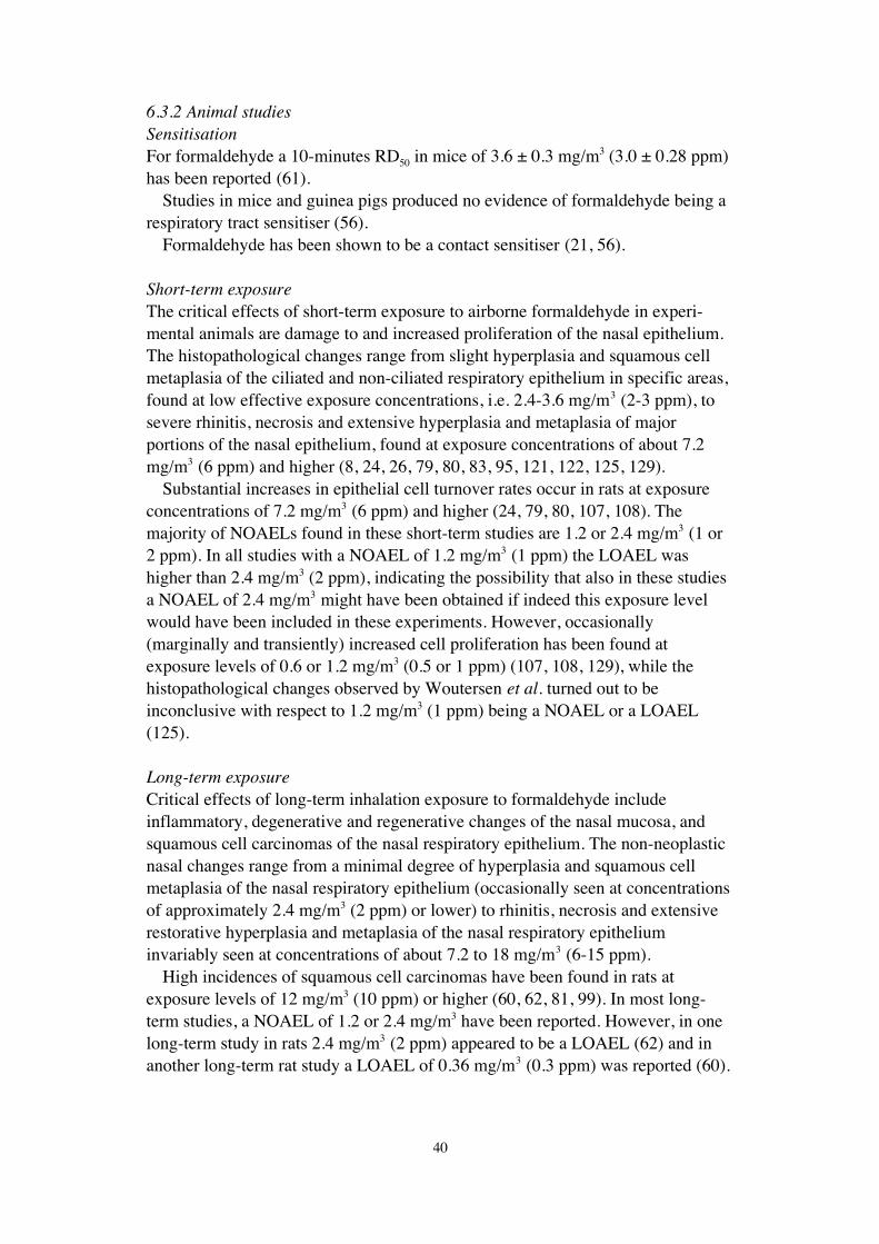
40
6.3.2 Animal studiesSensitisationFor formaldehyde a 10-minutes RD50 in mice of 3.6 ± 0.3 mg/m3 (3.0 ± 0.28 ppm)has been reported (61).
Studies in mice and guinea pigs produced no evidence of formaldehyde being arespiratory tract sensitiser (56).
Formaldehyde has been shown to be a contact sensitiser (21, 56).
Short-term exposureThe critical effects of short-term exposure to airborne formaldehyde in experi-mental animals are damage to and increased proliferation of the nasal epithelium.The histopathological changes range from slight hyperplasia and squamous cellmetaplasia of the ciliated and non-ciliated respiratory epithelium in specific areas,found at low effective exposure concentrations, i.e. 2.4-3.6 mg/m3 (2-3 ppm), tosevere rhinitis, necrosis and extensive hyperplasia and metaplasia of majorportions of the nasal epithelium, found at exposure concentrations of about 7.2mg/m3 (6 ppm) and higher (8, 24, 26, 79, 80, 83, 95, 121, 122, 125, 129).
Substantial increases in epithelial cell turnover rates occur in rats at exposureconcentrations of 7.2 mg/m3 (6 ppm) and higher (24, 79, 80, 107, 108). Themajority of NOAELs found in these short-term studies are 1.2 or 2.4 mg/m3 (1 or2 ppm). In all studies with a NOAEL of 1.2 mg/m3 (1 ppm) the LOAEL washigher than 2.4 mg/m3 (2 ppm), indicating the possibility that also in these studiesa NOAEL of 2.4 mg/m3 might have been obtained if indeed this exposure levelwould have been included in these experiments. However, occasionally(marginally and transiently) increased cell proliferation has been found atexposure levels of 0.6 or 1.2 mg/m3 (0.5 or 1 ppm) (107, 108, 129), while thehistopathological changes observed by Woutersen et al. turned out to beinconclusive with respect to 1.2 mg/m3 (1 ppm) being a NOAEL or a LOAEL(125).
Long-term exposureCritical effects of long-term inhalation exposure to formaldehyde includeinflammatory, degenerative and regenerative changes of the nasal mucosa, andsquamous cell carcinomas of the nasal respiratory epithelium. The non-neoplasticnasal changes range from a minimal degree of hyperplasia and squamous cellmetaplasia of the nasal respiratory epithelium (occasionally seen at concentrationsof approximately 2.4 mg/m3 (2 ppm) or lower) to rhinitis, necrosis and extensiverestorative hyperplasia and metaplasia of the nasal respiratory epitheliuminvariably seen at concentrations of about 7.2 to 18 mg/m3 (6-15 ppm).
High incidences of squamous cell carcinomas have been found in rats atexposure levels of 12 mg/m3 (10 ppm) or higher (60, 62, 81, 99). In most long-term studies, a NOAEL of 1.2 or 2.4 mg/m3 have been reported. However, in onelong-term study in rats 2.4 mg/m3 (2 ppm) appeared to be a LOAEL (62) and inanother long-term rat study a LOAEL of 0.36 mg/m3 (0.3 ppm) was reported (60).
41
GenotoxicityNo adequate data were available on genetic effects of formaldehyde in humans.Formaldehyde has been investigated for genotoxic properties in many test systems(58). It is comprehensively genotoxic in a variety of experimental systems,ranging from bacteria to rodents in vivo. Formaldehyde given by inhalation orgavage to rats induced chromosomal aberrations in lung cells, micronuclei ingastro-intestinal tract cells, and sperm-head anomalies. Inhalation of formal-dehyde leads to formation of DNA-protein cross-links in the nasal respiratoryepithelium of rats and monkeys. The formation of DNA-protein cross-links is asub-linear function of the formaldehyde concentration in inhaled air from 0.86 to18.4 mg/m3 (0.71-15.3 ppm), and the yield of DNA-protein cross-links at a giveninhaled concentration is approximately an order of magnitude lower in monkeysthan in rats. There is no detectable accumulation of DNA-protein cross-linksduring repeated exposures (58). In V79 Chinese hamster cells, formaldehydeinduced DNA-protein cross-links, sister chromatid exchanges, and micronuclei,but no gene mutations, in concentrations similar to those inducing cytotoxicity,suggesting that formaldehyde induced DNA-protein cross-links are related tocytotoxicity and clastogenicity (77). In cultured human lung epithelial cells, DNAdouble-strand breaks were induced by formaldehyde only when cell viability wasreduced to 60%, indicating that the double-strand breaks were caused byextragenomic damage and viability loss (115). Recio suggested that the nasalinflammation and proliferation induced by formaldehyde exposure may contributeto the induction of genetic alterations through a variety of mechanisms includinggeneration of reactive oxygen species, alterations in nucleotide pools, free radicalformation, and clonal expansion with further mutation of genetically altered cells(94).
With respect to the mechanism underlying the nasal carcinogenicity offormaldehyde in rats, there is a large body of data suggesting an associationbetween the cytotoxic, genotoxic, and carcinogenic effects of formaldehyde (29,38, 39, 51, 53, 82, 84, 126). The steep non-linear dose-response curve for nasaltumours – indicating a disproportionate decrease in carcinoma incidence at lowconcentrations – is most probably due to the fact that defence mechanisms of thenose (mucociliary clearance, detoxification by dehydrogenase, DNA repair) arevery effective at low concentrations, but can be overwhelmed and inactivated athigh concentrations; consequently, cell and tissue damage and finally tumoursoccur at high concentrations only. This also means that formaldehyde inconcentrations not leading to tissue damage most probably cannot act as acomplete carcinogen (causing initiation, promotion and progression), and as aresult is very unlikely to induce cancer by itself.
In several animal studies, inhalation of formaldehyde was not found to affectreproduction.

42
7. Existing guidelines, standards and evaluations
7.1 General population
The following recommendation was forwarded by IPCS/WHO (59): “Theformaldehyde air concentration allowed in living, sleeping and working roomsshould not be higher than 0.12 mg/m3 (0.1 ppm), in order to minimise the risk ofrepeated or continuous low concentration exposure to formaldehyde”.
Using a linear-at-low-dose extrapolation, the United States EnvironmentalProtection Agency (EPA) developed an upper-limit unit-risk estimate of1.6 x 10-2 /ppm continuous exposure to formaldehyde, in 1987 (32). This approachwas based solely on the dose-response for formaldehyde induced tumourformation, assuming a five-stage model for carcinogenesis. The EPA subsequentlychanged its risk estimate using a three-stage model, resulting in an upper-limit riskof 6.1 x 10⁻³/ppm formaldehyde exposure. In 1991, the EPA further revised its riskestimate using the levels of DNA-protein cross-links in the rat and monkey as anindicator of delivered formaldehyde dose. The use of this information in a two-stage model, based upon the goodness-of-fit of the data, yielded an upper-limitunit risk estimate of 3.3 x 10⁻⁴/ppm. Thus, the use of mechanistic informationresulted in a 50-fold reduction in the estimation of carcinogenic risk to humansfrom formaldehyde.
In 2002, the WHO/IPCS has published a review on formaldehyde (CICAD)(119). They concluded that based on studies in both animals and humans,formaldehyde is weakly genotoxic, with good evidence of an effect at site ofcontact. Epidemiological studies taken as a whole do not provide strong evidencefor a causal association between formaldehyde and human cancer, although thepossibility of increased respiratory cancer cannot be excluded. Therefore, basedprimarily upon data derived from laboratory studies, the inhalation of formal-dehyde under conditions that induce cytotoxicity and sustained regenerativeproliferation is considered to present a carcinogenic hazard to humans.
7.2 Working population
Occupational exposure standards in various countries are shown in Table 6.
7.3 Evaluations of standards
7.3.1 The NetherlandsIn 1981, DECOS concluded that formaldehyde is a proven genotoxic carcinogenin experimental animals and that the induction of cancer in humans could not beexcluded (117). DECOS estimated that exposure to 0.1 mg/m3 or 0.5 mg/m3
formaldehyde, 8 hours/day, 5 days/week for 40 years with a life span of 75 years,would result in maximal cancer risks of 1:40 000 and 1:10 000, respectively.
In 1987, however, DECOS updated the previous document and concluded thatan occupational exposure limit not exceeding 1.2 mg/m3 (1 ppm) formaldehyde,

43
Table 6. Occupational exposure standards in various countries.
Country-organisation
Occupationalexposure limit
Averagingtime
Notea Year ofadoptionb
Referencec
ppm mg/m3
The Netherlands 1 1.5 8 h 1986 (78)2 3.0 15 min
Germany 0.30 0.37 8 h 4d
SensUnknown (35)
Great Britain 2 2.5 8 h Unknown (49)2 2.5 15 min
Sweden 0.5 0.6 8 h Sens 1987 (106)1.0 1.2 Ceiling Carc
Denmark 0.3 0.4 Ceiling Carc Unknown,before 1994
(9)
Finland 0.3 0.37 8 h Unknown (101)1 1.2 Ceiling
Norway 0.5 0.6 8 h Sens 1984 (36)1.0 1.2 Ceiling Carc
Iceland 0.3 0.4 8 h Sens 1999 (114)1.0 1.2 Ceiling
United States–ACGIH 0.3 0.37 Ceiling Group A2
d 1992 (2)–OSHA 0.75 0.9 8 h Unknown (89)–NIOSH 0.016 0.02 8 h Carc Unknown (85)
0.10 0.12 15 min
European Union Carc Unknown (86)Cat 3
a. Sens, substance can cause sensitisation.Carc, classification of carcinogenic properties.
b. Year that this limit was officially adopted.c. Reference to the most recent official publication of occupational exposure limits.d. Genotoxicity playing no or at most a minor part.
15 minutes TWA, virtually should not constitute an increased nasal cancer risk(34). From studies in rats DECOS concluded that at subcytotoxic levels the risk ofinduction of nasal cancer appears to be negligibly small.
7.3.2 United StatesIn 1989, the American Conference of Governmental Industrial Hygienists(ACGIH) revised their assessment on formaldehyde. The proposed threshold limitvalue (TLV) for formaldehyde was 0.37 mg/m3 (0.3 ppm) as a ceiling, with anotation “suspected human carcinogen” (A2) (1). In the opinion of the ACGIH thisTLV as a ceiling should reduce the risk of sensory irritation for workers handlingformaldehyde or formaldehyde containing products. They also advised to reduceformaldehyde workplace exposure to the lowest possible level in view of the

44
reported dose-dependent carcinogenic effect in rats and mice, and the inadequateepidemiological data on the cancer risk in man.
In 1992, OSHA responded to a remand by the Unites States Court of Appealsfor the DC Circuit (89). The final amendments lowered the permissible exposurelevel for formaldehyde from 1 ppm as an 8-hour TWA to an 8-hour TWA of 0.75ppm (0.9 mg/m3). It should be noted that the former standard had been challengedin Unites States Court by both industry and labour. Four unions had challengedthe standard as being insufficiently protective. They contended that the formerpermissible exposure limit was not low enough to eliminate all significant risk ofharm, from both cancer and from formaldehyde irritant effects.
The National Institute for Occupational Safety and Health (NIOSH) recommen-ded an exposure limit of 0.02 mg/m3 (0.016 ppm) (8-hour TWA) and a 0.12mg/m3 (0.1 ppm) 15 minutes limit (85).
7.3.3 GermanyIn 2000, Deutsche Forschungsgemeinschaft set a “Maximale Arbeitsplatz-konzentration” (MAK) value of 0.37 mg/m3 (0.3 ppm) for formaldehyde.Formaldehyde is classified in category 4, which contains substances withcarcinogenic potential, for which genotoxicity plays no or at most a minor part.No contribution to human cancer risk is expected at the MAK-value. Theclassification is supported especially by evidence that increases in cellularproliferation or changes in cellular differentiation are important in the mode ofaction. To characterise the cancer risk, the manifold mechanism contributing tocarcinogenesis and their characteristic dose-time response relationships are takeninto consideration. Furthermore, formaldehyde is classified in germ cellmutagenicity category 5. A risk of damage to developing embryos or foetuses isnot to be expected at concentrations below the MAK value. Thereforeformaldehyde is classified in group C for compounds which may influencepregnancy.
7.3.4 SwedenThe most current consensus report for formaldehyde by the National Board ofOccupational Safety and Health was dated 25-8-1982. It was concluded that thebasis for occupational exposure standards should be the irritating effects offormaldehyde on the respiratory organs and eyes. Formaldehyde has been shownto be carcinogenic in animal studies. Epidemiological studies provide inadequateevidence and cannot be used to assess carcinogenic effects on man.
7.3.5 IARC / WHOThe most recent evaluation by IARC on formaldehyde concluded that there waslimited evidence in humans for the carcinogenicity of formaldehyde (58). There issufficient evidence in experimental animals for the carcinogenicity of formalde-hyde. The overall evaluation was that formaldehyde is probably carcinogenic tohumans (Group 2A).

45
7.3.6 European UnionThe European Union has classified the carcinogenic effects of formaldehyde incategory 3 (substances which cause concern for man owing to possiblecarcinogenic effects but in respect of which the available information is notadequate for making a satisfactory assessment) (Appendix 3).
8. Hazard assessment
8.1 Assessment of the health hazard
Formaldehyde occurs naturally in the environment and is produced physiologi-cally by mammalian cells during metabolism. It has been used by man for over acentury in a variety of products and activities. The human cell can rapidlydetoxify lower levels of formaldehyde.
Airborne formaldehyde exposures can occur as vapour or as particles (solids ormists) or as a combination of both. The relative intensities of vapour and particleexposures vary with the industry and the job activities. The anatomic site of tissuecontact as well as the intensity of exposure depend on the physical form of thecompound. Inhaled formaldehyde vapour is usually efficiently removed by thenose, mouth, and trachea, but in analogy with sulphur dioxide, some vapourprobably penetrates into the lower airways with mouth breathing during modera-tely heavy or heavy work (55). Inhaled particles containing formaldehyde aredeposited in the respiratory system as a function of their aerodynamic character-ristics and, given an appropriate aerodynamic diameter, may result in exposuresdeep within the respiratory tract. The biological behaviour of formaldehydedeposited in particulate form is unknown.
From the toxicological data base on formaldehyde it is evident that the criticaleffects of formaldehyde are sensory irritation, and cytotoxicity induced regene-rative hyperplasia (increased cell proliferation/increased cell turnover rates) andmetaplasia of the nasal respiratory epithelium accompanied by nasal carcinomasin rats and possibly in mice after long-term exposure to high cytotoxicconcentrations.
Symptoms of formaldehyde exposure in humans start with sensory irritationfirst experienced in the eyes, followed by perception of the odour, and thenirritation of the nose and throat, accompanied by discomfort, lachrymation,sneezing, coughing, nausea and dyspnoea. A panel of independent expertsconvened by the Industrial Health Foundation (IHF) studied all available data onsensory irritation related to formaldehyde exposure (92). This IHF-panelconcluded that for most persons eye irritation does not occur until at least 1.2mg/m3 (1.0 ppm) formaldehyde. This panel also observed that from controlledstudies in volunteers, it appears that moderate to severe eye, nose and throatirritation does not occur for most individuals until exposure concentrations offormaldehyde exceed 2.4-3.6 mg/m3 (2.0-3.0 ppm). The panel further concludedthat an occupational exposure limit of less than 0.6 mg/m3 (0.5 ppm) may be

46
needed to prevent sensory irritation in a diverse working population, and thereforerecommended for formaldehyde an occupational exposure limit of 0.36 mg/m3
(0.3 ppm) as an 8-hour TWA with a ceiling value of 1.2 mg/m3 (1.0 ppm) (92).However, according to the committees (DECOS and NEG) the database onsensory irritation of formaldehyde reveals that at lower exposure levels sensoryirritation may still occur in substantial percentages of exposed individuals. Forinstance, in a not well-documented study, more than 50% of occupationallyexposed workers complained of nasal discomfort after long-term exposure to anaverage concentration of 0.26 mg/m3 (0.22 ppm; range 0.05-0.6 mg/m3 or 0.04-0.5ppm) (120). Moreover, from a controlled study in volunteers it appeared that 19%(n=3) of the exposed subjects (n=16) reported eye irritation at an exposureconcentration of 0.29 mg/m3 (0.24 ppm) (4). However, according to the IHF-panelsuch a response is often considered of doubtful toxicological significance becauseirritation responses of 15-20% may be obtained in unexposed volunteers as well(92).
In experimental animals, irritation of eyes, nose, throat, and lungs wereobserved at exposure concentrations higher than 2.4 mg/m3 (2.0 ppm). Kane andAlarie determined in mice a 10-minutes RD50 for formaldehyde of 3.6±0.43mg/m3 (3.0±0.28 ppm) (61). Compared to humans, experimental animals seem tobe less sensitive to stimulation of the trigeminal nerve by formaldehyde. More-over, in view of the wealth of reliable data on sensory irritation in humans, theirritation data on formaldehyde in experimental animals are considered ofsecondary importance in terms of both hazard identification and risk assessment.
Overall, weighing the total body of data on sensory irritation, the committeesestimate that 0.3 mg/m3 (0.25 ppm) formaldehyde is the lowest exposureconcentration at which sensory irritation may occur in low but significantpercentages of exposed workers
Nasal carcinomas in rats have only been found at high, cytotoxic exposureconcentrations causing rhinitis, necrosis and regenerative hyperplasia andsquamous metaplasia of the nasal respiratory epithelium (60, 62, 81, 99). Thecrucial role of tissue damage followed by hyperplasia and metaplasia of the nasalrespiratory epithelium in formaldehyde carcinogenesis has been demonstrated in aconvincing way (38, 39, 126) and has meanwhile been widely recognised (92,118) and should therefore be included in human cancer risk assessment (29).Despite differences in anatomy and physiology of the nose between rats andhumans, the upper respiratory tract defence systems are similar in both species(82). It is, therefore, reasonable to conclude that the response of the respiratorytract to formaldehyde will be qualitatively similar in rats and humans. If inhumans exposure of formaldehyde were to be accompanied by recurrent tissuedamage at the site of contact, formaldehyde may be assumed to have carcinogenicpotential in man. Correspondingly, if the respiratory tract tissue is not recurrentlyinjured, exposure of humans to relatively low non-cytotoxic levels offormaldehyde can be assumed to be associated with a negligible cancer risk.
The committees observe that the majority of short- and long-term inhalationtoxicity studies with formaldehyde in experimental animals reveal a NOAEL of

47
1.2 or 2.4 mg/m3 (1 or 2 ppm). However, in one 24-month inhalation study in rats,2.4 mg/m3 (2 ppm) formaldehyde (lowest level tested) induced mild squamousmetaplasia of the epithelium lining the nasal turbinates (62). Moreover, a 13-weekinhalation toxicity study with formaldehyde, 1.2 mg/m3 (1 ppm), in rats wasinconclusive with respect to its effects on the nasal respiratory epithelium (125).Furthermore, in two short-term inhalation studies in rats, slightly (and onlytransiently) increased cell proliferation of the respiratory epithelium was seen in aspecific area of the nasal mucosa at formaldehyde exposure concentrations of 0.6or 1.2 mg/m3 (0.5 or 1 ppm) (107, 108, 129). Finally, in one recently publishedlong-term inhalation toxicity/carcinogenicity study on formaldehyde in rats (60), alow incidence of hyperplasia with or without squamous metaplasia of the nasalrespiratory epithelium was found at 0.36 mg/m3 (0.3 ppm). This low incidence(4/32) was not statistically significantly different from that in controls (0/32), butwas nevertheless considered toxicologically relevant (i.e. formaldehyde induced)because there was a clear dose-response relationship with the increased incidencesat the higher exposure levels being statistically significantly different from that incontrols.
The data in humans are less clear. Three meta-analyses of epidemiologicalstudies have shown inconsistent results. In two of them a significant relationbetween exposure to formaldehyde and nasopharyngeal cancer risk was observed.The association between formaldehyde exposure and nasal cancer was ambiguous(15, 90). However, according to the committees in these meta-analyses the authorsdid not correct for the unreported studies in which no cases of nasal cancers werefound. This most likely led to an overestimation of the overall relative risk ofnasopharyngeal cancer. In the third, more recent, published meta-analysis, relativerisks of 1.0 and 1.3 were found for nasal cancer in cohort and case-control studies,respectively (31). In this meta-analysis a correction was made for underreporting.Moreover, the authors evaluated the exposure potential for jobs included in thegeneral population case-control studies. The authors concluded that there was nosupport for a causal relation between formaldehyde exposure and nasopharyngealcancer. The committees (both DECOS and NEG) endorse this conclusion andfurther conclude that the currently available epidemiological database does notprovide support for a nasal cancer risk at the exposure levels lower than 0.3mg/m3. Also from the epidemiological database it seems unlikely that exposure toformaldehyde affects lung cancer risk (5, 6, 15, 18, 41, 46, 90). Overall, bothcommittees conclude that the currently available epidemiological database onformaldehyde does not provide evidence for a respiratory tract cancer risk at theexperienced exposure levels. In correspondence to the previous evaluation offormaldehyde by DECOS in 1987 (34), the committees endorse the conclusionfrom 1987 that with prevention of cytotoxicity, carcinogenic effects will notoccur.

48
8.2 Groups at extra risk
Allergic dermal sensitisation to formaldehyde in man occurs in 3 to 6% of thegeneral population. It is not possible to identify individuals with elevated risk forallergic sensitisation a priori with a simple screening test. Skin sensitisationconstitutes a health risk in workers occupationally exposed to formaldehyde.
8.3 Scientific basis for an occupational exposure limit
The critical effect of formaldehyde is sensory irritation (LOAEL 0.25 ppm).Another concern is cytotoxicity induced regenerative hyperplasia and metaplasiaof the nasal respiratory epithelium accompanied by nasal carcinomas in rats andpossibly in mice after long-term exposure to high cytotoxic concentrations.
High, cytotoxic formaldehyde vapour concentrations (≥10 ppm) can inducenasal cancer in rats. A large body of data suggests an association between thecytotoxic, genotoxic and carcinogenic effects of formaldehyde. Tissue damagefollowed by hyperplasia and metaplasia of the nasal respiratory epithelium has acrucial role in formaldehyde carcinogenesis. Thus, formaldehyde in non-cytotxicconcentrations not leading to tissue damage most probably cannot act as acomplete carcinogen. However, if human exposure of formaldehyde is accompa-nied by recurrent tissue damage at the site of contact, formaldehyde may beassumed to have a carcinogenic potential in man.
Formaldehyde is a skin sensitiser.

49
9. Summary
Wibowo, Anton. The Nordic Expert Group for Criteria Documentation of HealthRisks from Chemicals and the Dutch Expert Committee on OccupationalStandards. 132. Formaldehyde. Arbete och Hälsa 2003;11:1-76.
Formaldehyde is a colourless, flammable, reactive gas, which readily polymerises,and forms explosive mixtures with air and oxygen. It is used as a raw material inchemical reactions, and as an intermediate in the manufacture of numerousproducts. It has also a medical application as a disinfectant or preservative.
Inhaled formaldehyde is almost completely absorbed in the upper respiratorytract in rodents. Formaldehyde is a normal metabolite in mammalians and israpidly metabolised to formate, which may be further oxidised to carbon dioxide.Final elimination occurs via exhalation and via the kidneys.
The target organs of formaldehyde vapour are the eyes, nose, and throat. Theeffects of concern are sensory irritation and cytotoxicity-induced regenerativehyperplasia and metaplasia of the nasal respiratory epithelium accompanied bynasal carcinomas in rats. Weighting the total body of data 0.25 ppm formaldehydeis the LOAEL at which sensory irritation may occur in a low but significantpercentage of exposed workers. The majority of short- and long-term inhalationanimal studies reveal a NOAEL of 1-2 ppm. However, in a few studies slighthistopathological changes of the nasal respiratory epithelium were observed at0.3-2 ppm.
Formaldehyde is genotoxic in a variety of experimental systems, includingrodents in vivo. There is overwhelming evidence that high, cytotoxic formal-dehyde vapour concentrations (≥10 ppm) can induce nasal cancer in rats. A largebody of data suggests an association between the cytotoxic, genotoxic, andcarcinogenic effects of formaldehyde. The crucial role of tissue damage followedby hyperplasia and metaplasia of the nasal respiratory epithelium in formaldehydecarcinogenesis has been demonstrated in a convincing way. Thus, formaldehydein non-cytotxic concentrations most probably cannot act as a complete carcinogen.However, if human exposure to formaldehyde is accompanied by recurrent tissuedamage at the site of contact, formaldehyde may be assumed to have carcinogenicpotential in man.
Formaldehyde-induced allergic contact dermatitis has been estimated to occurin 3-6% of the population, and skin sensitisation by direct skin contact has beeninduced with formaldehyde solutions. There is no consistent evidence offormaldehyde being a respiratory sensisitiser.
Key words: allergy, cancer, contact dermatitis, cytotoxicity, formaldehyde, geno-toxicity, irritation, nasal, occupational exposure limit, review, risk assessment

50
10. Summary in Swedish
Wibowo, Anton. The Nordic Expert Group for Criteria Documentation of HealthRisks from Chemicals and the Dutch Expert Committee on OccupationalStandards. 132. Formaldehyde. Arbete och Hälsa 2003;11:1-76.
Formaldehyd är en färglös, lättantändlig, reaktiv gas som lätt polymeriserar ochbildar explosiva blandningar med luft och syre. Ämnet används som råmaterial ikemiska reaktioner och som intermediär vid tillverkning av ett stort antalprodukter. Det har även medicinsk användning som desinfektionsmedel ochkonserveringsmedel.
Inandad formaldehyd absorberas nästan fullständigt i de övre luftvägarna.Formaldehyd är en normal ämnesomsättningsprodukt hos däggdjur och omvand-las snabbt till format (myrsyra) som kan oxideras vidare till koldioxid. Slutligeliminering sker via utandning och via njurarna.
Målorgan för formaldehydånga är ögon, näsa och hals. Effekter av betydelseär sensorisk irritation samt cytotoxiskt inducerad regenerativ hyperplasi ochmetaplasi i nässlemhinnan, följt av näscancer hos råtta. Sammantaget bedöms0,25 ppm vara den lägsta nivå (LOAEL) vid vilken sensorisk irritation uppträderhos en liten men signifikant andel av exponerade arbetare. Merparten inhalations-studier, såväl kort- som långtidsstudier, rapporterar en icke-effektnivå (NOAEL)på 1-2 ppm. I några studier har man dock observererat små histopatologiskaförändringar av nässlemhinnan vid 0,3-2 ppm.
Formaldehyd är genotoxiskt i en rad experimentella system inklusive gnagare invivo. Det finns överväldigande bevis för att höga, cytotoxiska formaldehyd-koncentrationer (≥10 ppm) kan inducera näscancer hos råtta. En mängd data pekarpå ett samband mellan de celltoxiska, genotoxiska och carcinogena effekterna avformaldehyd. Vävnadsskada följt av hyperplasi och metaplasi i näsans respirato-riska epitel spelar en avgörande roll i carcinogenesen vid formaldehydexponering,vilket har visats på ett övertygande sätt. Formaldehyd i icke-cytotoxiskaconcentrationer är därför troligen inte en komplett carcinogen. Om människorexponeras för formaldehyd med upprepad vävnadsskada vid kontaktstället somföljd kan formaldehyd emellertid antas ha carcinogen potential.
Formaldehyd-inducerat allergiskt kontakteksem beräknas upträda hos 3-6% avbefolkningen och hudsensibilisering har inducerats via direkt hudkontakt medformaldehydlösningar. Det finns inga entydiga bevis för att formaldehyd kan gesensibilisering via inandning.
Nyckelord: allergi, cancer, kontakteksem, cytotoxicitet, formaldehyd, geno-toxicitet, gränsvärden, irritation, näs-, riskbedömning, översikt

51
11. References
1. ACGIH. Threshold limit values for chemical substances and physical agens. Cincinatti:American Conference of Governmental Industrial Hygienists Inc., 1989.
2. ACGIH. Documentation of the threshold limit values for chemical substances and physicalagens. Cincinatti: American Conference of Governmental Industrial Hygienists Inc., 2001.
3. Akbar-Khanzadeh F, Vaquerano MU, Akbar-Khanzadeh M, Bisesi MS. Formaldehydeexposure, acute pulmonary response, and exposure control options in a gross anatomylaboratory. Am J Ind Med 1994;26:61-75.
4. Andersen I, Mølhave L. Controlled human studies with formaldehyde. In: Gibson JE, ed.Formaldehyde toxicity. Washington: Hemisphere Publishing Corporation, 1983:154-165.
5. Andjelkovich DA, Janszen DB, Brown MH, Richardson RB, Miller FJ. Mortality of ironfoundry workers: IV. Analysis of a subcohort exposed to formaldehyde. J Occup EnvironMed 1995;37:826-837.
6. Andjelkovich DA, Janszen DB, Conolly RB, Miller FJ. Formaldehyde exposure notassociated with cancer of the respiratory tract in iron foundry workers: a synopsis of CIITepidemiologic findings. CIIT activities 1995;15:1-9.
7. Andjelkovich DA, Shy CM, Brown MH, Janszen DB, Levine RJ, Richardson RB. Mortalityof iron foundry workers. III. Lung cancer case-control study. J Occup Med 1994;36:1301-1309.
8. Appelman LM, Woutersen RA, Zwart A, Falke HE, Feron VJ. One-year inhalation toxicitystudy of formaldehyde in male rats with a damaged or undamaged nasal mucosa. J ApplToxicol 1988;8:85-90.
9. Arbejdstilsynet. Exposure limit values for substances and materials. C.0.1. Arbejdstilsynet,2002.
10. Ballarin C, Sarto F, Giacomelli L, Bartolucci GB, Clonfero E. Micronucleated cells in nasalmucosa of formaldehyde-exposed workers. Mutat Res 1992;280:1-7.
11. Bardana EJ, Jr., Montanaro A. Formaldehyde: an analysis of its respiratory, cutaneous, andimmunologic effects. Ann Allergy 1991;66:441-452.
12. Bender JR, Mullin LS, Graepel GJ, Wilson WE. Eye irritation response of humans toformaldehyde. Am Ind Hyg Assoc J 1983;44:463-465.
13. Bhalla DK, Mahavni V, Nguyen T, McClure T. Effects of acute exposure to formaldehyde onsurface morphology of nasal epithelia in rats. J Toxicol Environ Health 1991;33:171-188.
14. Biagini RE, Moorman WJ, Knecht EA, Clark JC, Bernstein IL. Acute airway narrowing inmonkeys from challenge with 2.5 ppm formaldehyde generated from formalin. Arch EnvironHealth 1989;44:12-17.
15. Blair A, Saracci R, Stewart PA, Hayes RB, Shy C. Epidemiologic evidence on therelationship between formaldehyde exposure and cancer. Scand J Work Environ Health1990;16:381-393.
16. Blair A, Stewart P. Comments on the Sterling and Weinham analysis of data from theNational Cancer Institute Formaldehyde study. Am J Ind Med 1994;25:603-606.
17. Blair A, Stewart P, O'Berg M, Gaffey W, Walrath J, Ward J, Bales R, Kaplan S, Cubit D.Mortality among industrial workers exposed to formaldehyde. J Natl Cancer Inst1986;76:1071-1084.
18. Blair A, Stewart PA, Hoover RN. Mortality from lung cancer among workers employed informaldehyde industries. Am J Ind Med 1990;17:683-699.
19. Blair A, Stewart PA, Hoover RN, Fraumeni JF, Jr., Walrath J, O'Berg M, Gaffey W. Cancersof the nasopharynx and oropharynx and formaldehyde exposure. J Natl Cancer Inst1987;78:191-193.

52
20. Bogdanffy MS, Randall HW, Morgan KT. Biochemical quantitation and histochemicallocalization of carboxylesterase in the nasal passages of the Fischer-344 rat and B6C3F1mouse. Toxicol Appl Pharmacol 1987;88:183-194.
21. Boman A, Dyring-Jacobsen S, Klastrup S, Svendsen O, Wahlberg JE. Potency evaluation ofcontact allergens. Dose-response studies using the guinea pig maximization test. Departmentof Dermatology, National Institute of Occupational Health, Solna, Sweden. TemaNord,1996:570.
22. Boysen M, Zadig E, Digernes V, Abeler V, Reith A. Nasal mucosa in workers exposed toformaldehyde: a pilot study. Br J Ind Med 1990;47:116-121.
23. Casanova M, Heck Hd A. Further studies of the metabolic incorporation and covalent bindingof inhaled [3H]- and [14C]formaldehyde in Fischer-344 rats: effects of glutathione depletion.Toxicol Appl Pharmacol 1987;89:105-121.
24. Casanova M, Morgan KT, Gross EA, Moss OR, Heck HA. DNA-protein cross-links and cellreplication at specific sites in the nose of F344 rats exposed subchronically to formaldehyde.Fundam Appl Toxicol 1994;23:525-536.
25. Casanova M, Morgan KT, Steinhagen WH, Everitt JI, Popp JA, Heck HD. Covalent bindingof inhaled formaldehyde to DNA in the respiratory tract of rhesus monkeys: pharmaco-kinetics, rat-to-monkey interspecies scaling, and extrapolation to man. Fundam Appl Toxicol1991;17:409-428.
26. Cassee FR, Groten JP, Feron VJ. Changes in the nasal epithelium of rats exposed byinhalation to mixtures of formaldehyde, acetaldehyde, and acrolein. Fundam Appl Toxicol1996;29:208-218.
27. Chemiekaartenboek.12e editie. NIA-TNO, Samson HD Tjeenk Willink, Alphen aan de Rijn,1997.
28. Chia SE, Ong CN, Foo SC, Lee HP. Medical students' exposure to formaldehyde in a grossanatomy dissection laboratory. J Am Coll Health 1992;41:115-119.
29. CIIT. Formaldehyde: hazard characterization and dose-response assessment forcarcinogenicity by the route of inhalation. Revised edition. Chemical Industry Institute ofToxicology, Research Triangle Park, NC: CIIT publications, September 1999.
30. Colizzo F, Krantz MJ, Fish JE, Hastie AT. Ciliated respiratory epithelial surface changesafter formaldehyde exposure. J Toxicol Environ Health 1992;35:221-234.
31. Collins JJ, Acquavella JF, Esmen NA. An updated meta-analysis of formaldehyde exposureand upper respiratory tract cancers. J Occup Environ Med 1997;39:639-651.
32. Conaway CC, Whysner J, Verna LK, Williams GM. Formaldehyde mechanistic data and riskassessment: endogenous protection from DNA adduct formation. Pharmacol Ther1996;71:29-55.
33. Cronin E. Formaldehyde is a significant allergen in women with hand eczema. ContactDermatitis 1991;25:276-282.
34. DECOS. Health based recommended occupational exposure limits for formaldehyde.Directorate-General of Labour, RA 3/87. Dutch Expert Committee on OccupationalStandards, 1987.
35. Deutsche Forschungsgemeinschaft. MAK- und BAT-Werte List. Weinheim: Wiley-VCH.,2002.
36. Direktoratet for arbeidstilsynet. Administrative normer for forurensning i arbeidsatmosfære.Veiledning til arbeidsmiljøloven. Arbeidstilsynet, Bestillingsnr. 361, 2001.
37. ECETOC. Formaldehyde and human cancer risk. Technical report no. 165. Brussels:European Centre for Ecotoxicology and Toxicology for Chemicals, May 1995.
38. Feron VJ, Wilmer JW, Woutersen RA, Zwart A. Inhalation toxicity and carcinogenicity offormaldehyde in animals: significance for assessment of human health risk. In: Mohr, ed. inchief. Assessment of Inhalation Hazards. Berlin: Springer-Verlag, 1989:131-138.

53
39. Feron VJ, Woutersen RA. Role of tissue damage in nasal carcinogenisis. In: Feron VJ,Bosland MC, eds. Nasal carcinogenisis in rodents: relevance to human health risk. PudokWageningen, 1989:76-84.
40. Flyvholm MA, Menné T. Allergic contact dermatitis from formaldehyde. A case studyfocusing on sources of formaldehyde exposure. Contact Dermatitis 1992;27:27-36.
41. Gardner MJ, Pannett B, Winter PD, Cruddas AM. A cohort study of workers exposed toformaldehyde in the British chemical industry: an update. Br J Ind Med 1993;50:827-834.
42. Garrett MH, Hooper MA, Hooper BM, Rayment PR, Abramson MJ. Increased risk of allergyin children due to formaldehyde exposure in homes. Allergy 1999;54:330-337.
43. Grammer LC, Harris KE, Cugell DW, Patterson R. Evaluation of a worker with possibleformaldehyde-induced asthma. J Allergy Clin Immunol 1993;92:29-33.
44. Grammer LC, Harris KE, Shaughnessy MA, Sparks P, Ayars GH, Altman LC, Patterson R.Clinical and immunologic evaluation of 37 workers exposed to gaseous formaldehyde. JAllergy Clin Immunol 1990;86:177-181.
45. Guicherit R, Schulting FL. The occurrence of organic chemicals in the atmosphere of TheNetherlands. Sci Total Environ 1985;43:193-219.
46. Hansen J, Olsen JH. Formaldehyde and cancer morbidity among male employees inDenmark. Cancer Causes Control 1995;6:354-360.
47. Harving H, Korsgaard J, Pedersen OF, Mølhave L, Dahl R. Pulmonary function andbronchial reactivity in asthmatics during low-level formaldehyde exposure. Lung1990;168:15-21.
48. He JL, Jin LF, Jin HY. Detection of cytogenetic effects in peripheral lymphocytes of studentsexposed to formaldehyde with cytokinesis-blocked micronucleus assay. Biomed Environ Sci1998;11:87-92.
49. Health and Safety Executive. Occupational exposure limits 2000. EH 40/2000. Health andSafety Executive, 2000
50. Heck Hd A, Casanova M. Isotope effects and their implications for the covalent binding ofinhaled [3H]- and [14C]formaldehyde in the rat nasal mucosa. Toxicol Appl Pharmacol1987;89:122-134.
51. Heck Hd A, Casanova M, Starr TB. Formaldehyde toxicity--new understanding. Crit RevToxicol 1990;20:397-426.
52. Heck Hd A, Casanova M, Steinhagen WH, Everitt JI, Morgan KT, Popp JA. Formaldehydetoxicity-DNA-protein cross-links studies in rats and non humans primates. Feron VJ, BoslandMC, eds. Nasal carcinogenisis in rodents: relevance to human health risk. PudokWageningen, 1989:159-164.
53. Heck Hd A, Casanova-Schmitz M. Biochemical toxicology of formaldehyde. Rev BiochemToxicol 1984;6:155-189.
54. Herbert FA, Hessel PA, Melenka LS, Yoshida K, Nakaza M. Respiratory consequences ofexposure to wood dust and formaldehyde of workers manufacturing oriented strand board.Arch Environ Health 1994;49:465-470.
55. Higginson J, Jensen OM, Kinlen L, Kirsten W, MacMahon B, Matanoski G, Smith T,Thomas D. Epidemiology of chronic occupational exposure to formaldehyde: report of the adhoc panel on health aspects of formaldehyde. Toxicol Ind Health 1988;4:77-90.
56. Hilton J, Dearman RJ, Basketter DA, Scholes EW, Kimber I. Experimental assessment of thesensitizing properties of formaldehyde. Food Chem Toxicol 1996;34:571-578.
57. Horvath EP, Jr., Anderson H, Jr., Pierce WE, Hanrahan L, Wendlick JD. Effects offormaldehyde on the mucous membranes and lungs. A study of an industrial population.JAMA 1988;259:701-707.
58. IARC. Wood dust and formaldehyde. In: IARC Monographs on the evaluation of carcino-genic risks to humans. Volume 62. Lyon, France: International Agency for Research onCancer, 1995.

54
59. IPCS/WHO. Formaldehyde. In: Environmental Health Criteria 89. Geneva: InternationalProgramme on Chemical Safety, World Health Organisation, 1989.
60. Kamata E, Nakadate M, Uchida O, Ogawa Y, Suzuki S, Kaneko T, Saito M, Kurokawa Y.Results of a 28-month chronic inhalation toxicity study of formaldehyde in male Fisher-344rats. J Toxicol Sci 1997;22:239-254.
61. Kane LE, Alarie Y. Sensory irritation to formaldehyde and acrolein during single andrepeated exposures in mice. Am Ind Hyg Assoc J 1977;38:509-522.
62. Kerns WD, Pavkov KL, Donofrio DJ, Gralla EJ, Swenberg JA. Carcinogenicity offormaldehyde in rats and mice after long-term inhalation exposure. Cancer Res1983;43:4382-4392.
63. Kilburn KH, Warshaw R, Thornton JC. Pulmonary function in histology technicianscompared with women from Michigan: effects of chronic low dose formaldehyde on anational sample of women. Br J Ind Med 1989;46:468-472.
64. Kilburn KH, Warshaw RH. Neurobehavioral effects of formaldehyde and solvents onhistology technicians: repeated testing across time. Environ Res 1992;58:134-146.
65. Kitchens JF, Casner RE, Edwards GS, Harward WE, Macri BJ. Investigation of selectedpotential environmental contaminants: formaldehyde. Washington DC, US EnvironmentalProtection Agency, 1976.
66. Kolmodin-Hedman B, Nordman H. Nordic Expert Group for Documentation of OccupationalExposure Limits. 37. Formaldehyde. Arbete och Hälsa 1982;27:1-46. Arbetarskyddsverket,Solna.
67. Kriebel D, Sama SR, Cocanour B. Reversible pulmonary responses to formaldehyde. A studyof clinical anatomy students. Am Rev Respir Dis 1993;148:1509-1515.
68. Liden S, Scheynius A, Fischer T, Johansson SG, Ruhnek-Forsbeck M, Stejskal V. Absence ofspecific IgE antibodies in allergic contact sensitivity to formaldehyde. Allergy 1993;48:525-529.
69. Liu KS, Huang FY, Hayward SB, Wesolowski J, Sexton K. Irritant effects of formaldehydeexposure in mobile homes. Environ Health Perspect 1991;94:91-94.
70. Loden M. The in vitro permeability of human skin to benzene, ethylene glycol,formaldehyde, and n-hexane. Acta Pharmacol Toxicol (Copenh) 1986;58:382-389.
71. Luce D, Gerin M, Leclerc A, Morcet JF, Brugere J, Goldberg M. Sinonasal cancer andoccupational exposure to formaldehyde and other substances. Int J Cancer 1993;53:224-231.
72. Ma TH, Harris MM. Review of the genotoxicity of formaldehyde. Mutat Res 1988;196:37-59.
73. Majumder PK, Kumar VL. Inhibitory effects of formaldehyde on the reproductive system ofmale rats. Indian J Physiol Pharmacol 1995;39:80-82.
74. Marsh GM, Stone RA, Esmen NA, Henderson VL. Mortality patterns among chemical plantworkers exposed to formaldehyde and other substances. J Natl Cancer Inst 1994;86:384-386.
75. Marsh GM, Stone RA, Henderson VL. Lung cancer mortality among industrial workersexposed to formaldehyde: a Poisson regression analysis of the National Cancer InstituteStudy. Am Ind Hyg Assoc J 1992;53:681-691.
76. Martin WJ. A teratology study of inhaled formaldehyde in the rat. Reprod Toxicol1990;4:237-239.
77. Merk O, Speit G. Significance of formaldehyde-induced DNA-protein crosslinks formutagenesis. Environ Mol Mutagen 1998;32:260-268.
78. Ministerie van Sociale Zaken en Werkgelegenheid. De nationale MAC lijst 2002.
79. Monticello TM, Miller FJ, Morgan KT. Regional increases in rat nasal epithelial cellproliferation following acute and subchronic inhalation of formaldehyde. Toxicol ApplPharmacol 1991;111:409-421.

55
80. Monticello TM, Morgan KT, Everitt JI, Popp JA. Effects of formaldehyde gas on therespiratory tract of rhesus monkeys. Pathology and cell proliferation. Am J Pathol1989;134:515-527.
81. Monticello TM, Swenberg JA, Gross EA, Leininger JR, Kimbell JS, Seilkop S, Starr TB,Gibson JE, Morgan KT. Correlation of regional and nonlinear formaldehyde-induced nasalcancer with proliferating populations of cells. Cancer Res 1996;56:1012-1022.
82. Morgan KT. A brief review of formaldehyde carcinogenesis in relation to rat nasal pathologyand human health risk assessment. Toxicol Pathol 1997;25:291-307.
83. Morgan KT, Gross EA, Patterson DL. Distribution, progression, and recovery of acuteformaldehyde-induced inhibition of nasal mucociliary function in F-344 rats. Toxicol ApplPharmacol 1986;86:448-456.
84. Morgan KT, Jiang XZ, Starr TB, Kerns WD. More precise localization of nasal tumorsassociated with chronic exposure of F-344 rats to formaldehyde gas. Toxicol Appl Pharmacol1986;82:264-271.
85. NIOSH Pocket guide to chemical hazards. Washington: U.S. Department of Health andHuman Services, National Institute for Occupational Safety and Health, 2000.
86. Nordic Council of Ministers. European Chemicals Bureau. The N-class database onenvironmental hazard classification (online). Last update: April 2002.[http://www.kemi.se/nclass/default.asp].
87. Nunn AJ, Craigen AA, Darbyshire JH, Venables KM, Newman Taylor AJ. Six year follow upof lung function in men occupationally exposed to formaldehyde. Br J Ind Med 1990;47:747-752.
88. Odeigah PG. Sperm head abnormalities and dominant lethal effects of formaldehyde in albinorats. Mutat Res 1997;389:141-148.
89. OSHA. Occupational exposure to formaldehyde. Federal register; 57: 22290-22338 (presentas computer disc). Occupational Safety and Health Administration, 1992.
90. Partanen T. Formaldehyde exposure and respiratory cancer--a meta-analysis of theepidemiologic evidence. Scand J Work Environ Health 1993;19:8-15.
91. Partanen T, Kauppinen T, Hernberg S, Nickels J, Luukkonen R, Hakulinen T, Pukkala E.Formaldehyde exposure and respiratory cancer among woodworkers--an update. Scand JWork Environ Health 1990;16:394-400.
92. Paustenbach D, Alarie Y, Kulle T, Schachter N, Smith R, Swenberg J, Witschi H, HorowitzSB. A recommended occupational exposure limit for formaldehyde based on irritation. JToxicol Environ Health 1997;50:217-263.
93. Pazdrak K, Gorski P, Krakowiak A, Ruta U. Changes in nasal lavage fluid due toformaldehyde inhalation. Int Arch Occup Environ Health 1993;64:515-519.
94. Recio L. Oncogene and tumor suppressor gene alterations in nasal tumors. Mutat Res1997;380:27-31.
95. Reuzel PG, Wilmer JW, Woutersen RA, Zwart A, Rombout PJ, Feron VJ. Interactive effectsof ozone and formaldehyde on the nasal respiratory lining epithelium in rats. J ToxicolEnviron Health 1990;29:279-292.
96. Saillenfait AM, Bonnet P, de Ceaurriz J. The effects of maternally inhaled formaldehyde onembryonal and foetal development in rats. Food Chem Toxicol 1989;27:545-548.
97. Salkie ML. The prevalence of atopy and hypersensitivity to formaldehyde in pathologists.Arch Pathol Lab Med 1991;115:614-616.
98. Sauder LR, Green DJ, Chatham MD, Kulle TJ. Acute pulmonary response of asthmatics to3.0 ppm formaldehyde. Toxicol Ind Health 1987;3:569-578.
99. Sellakumar AR, Snyder CA, Solomon JJ, Albert RE. Carcinogenicity of formaldehyde andhydrogen chloride in rats. Toxicol Appl Pharmacol 1985;81:401-406.

56
100. Siemiatycki J, Dewar R, Nadon L, Gerin M. Occupational risk factors for bladder cancer:results from a case-control study in Montreal, Quebec, Canada. Am J Epidemiol1994;140:1061-1080.
101. Sosiaali-ja terveysministeriö. HTP-arvot 2002, Työsuojelusäädöksiä 3, 2002.
102. Srivastava AK, Gupta BN, Bihari V, Gaur JS, Mathur N, Awasthi VK. Clinical studies ofemployees in a sheet-forming process at a paper mill. Vet Hum Toxicol 1992;34:525-527.
103. Sterling TD, Weinkam JJ. Mortality from respiratory cancers (including lung cancer) amongworkers employed in formaldehyde industries. Am J Ind Med 1994;25:593-602; discussion603-606.
104. Sterling TD, Weinkam JJ. Comments on the Blair and Stewart comments on the Sterling andWeinkam analysis of data from the National Cancer Institute Formaldehyde Study. Am J IndMed 1995;27:301-305.
105. Suruda A, Schulte P, Boeniger M, Hayes RB, Livingston GK, Steenland K, Stewart P,Herrick R, Douthit D, Fingerhut MA. Cytogenetic effects of formaldehyde exposure instudents of mortuary science. Cancer Epidemiol Biomarkers Prev 1993;2:453-460.
106. Swedish National Board of Occupational Safety and Health. Occupational exposure limitvalues, 2000 (AFS 2000:3).
107. Swenberg JA, Gross EA, Randall H. W. Localization and quantitation of cell proliferationfollowing exposure to nasal irritants. In: Barrow CS, ed. Toxicology of the nasal passage.Washington, New York and London: Hemisphere Publishing Corporation, 1986:291-300.
108. Swenberg JA, Gross EA, Randall HW, Barrow CS. The effect of formaldehyde exposure oncytotoxicity and cell proliferation. In: Clary JJ, Gibson JE, Waritz RS, eds. FormaldehydeToxicology, epidemiology, mechanisms. New York and Basel: Marcel-Dekker Inc., 1983:225-236.
109. Swiecichowski AL, Long KJ, Miller ML, Leikauf GD. Formaldehyde-induced airwayhyperreactivity in vivo and ex vivo in guinea pigs. Environ Res 1993;61:185-199.
110. Thrasher JD, Broughton A, Madison R. Immune activation and autoantibodies in humanswith long-term inhalation exposure to formaldehyde. Arch Environ Health 1990;45:217-223.
111. Vargova M, Wagnerova J, Liskova A, Jakubovsky J, Gajdova M, Stolcova E, Kubova J,Tulinska J, Stenclova R. Subacute immunotoxicity study of formaldehyde in male rats. DrugChem Toxicol 1993;16:255-275.
112. Vasudeva N, Anand C. Cytogenetic evaluation of medical students exposed to formaldehydevapor in the gross anatomy dissection laboratory. J Am Coll Health 1996;44:177-179.
113. Vaughan TL, Stewart PA, Teschke K, Lynch CF, Swanson GM, Lyon JL, Berwick M.Occupational exposure to formaldehyde and wood dust and nasopharyngeal carcinoma.Occup Environ Med 2000;57:376-384.
114. Vinnueftirlit Ríkisins. Reglur og leidbeiningar um mengunarmörk og adgerdir til ad draga úrmengun á vinnustödum, 1999.
115. Vock EH, Lutz WK, Ilinskaya O, Vamvakas S. Discrimination between genotoxicity andcytotoxicity for the induction of DNA double-strand breaks in cells treated with aldehydesand diepoxides. Mutat Res 1999;441:85-93.
116. Weber-Tschopp A, Fischer T, Grandjean E. Reizwirkung des formaldehyds (HCHO) auf denMenschen. Int Arch Occup Environ Health 1977;39:207-218.
117. WGD. Rapport inzake formaldehyde. RA 4/81. Voorburg: Werkgroep van Deskundigen vande Nationale MAC-commissie, 1981.
118. WHO. Air quality guidelines for Europe. 2nd ed. WHO Regional Publications, EuropeanSeries, No. 91. Copenhagen: World Health Organisation Regional Office for Europe, 2000.
119. WHO. Concise Internationale Chemical Assessment Document 40, Formaldehyde. Geneva:World Health Organisation, 2002.
120. Wilhelmsson B, Holmström M. Possible mechanisms of formaldehyde-induced discomfort inthe upper airways. Scand J Work Environ Health 1992;18:403-407.

57
121. Wilmer JW, Woutersen RA, Appelman LM, Leeman WR, Feron VJ. Subacute (4-week)inhalation toxicity study of formaldehyde in male rats: 8-hour intermittent versus 8-hourcontinuous exposures. J Appl Toxicol 1987;7:15-16.
122. Wilmer JW, Woutersen RA, Appelman LM, Leeman WR, Feron VJ. Subchronic (13-week)inhalation toxicity study of formaldehyde in male rats: 8-hour intermittent versus 8-hourcontinuous exposures. Toxicol Lett 1989;47:287-293.
123. Witek TJ, Jr., Schachter EN, Tosun T, Beck GJ, Leaderer BP. An evaluation of respiratoryeffects following exposure to 2.0 ppm formaldehyde in asthmatics: lung function, symptoms,and airway reactivity. Arch Environ Health 1987;42:230-237.
124. Wood RW, Coleman JB. Behavioral evaluation of the irritant properties of formaldehyde.Toxicol Appl Pharmacol 1995;130:67-72.
125. Woutersen RA, Appelman LM, Wilmer JW, Falke HE, Feron VJ. Subchronic (13-week)inhalation toxicity study of formaldehyde in rats. J Appl Toxicol 1987;7:43-49.
126. Woutersen RA, van Garderen-Hoetmer A, Bruijntjes JP, Zwart A, Feron VJ. Nasal tumoursin rats after severe injury to the nasal mucosa and prolonged exposure to 10 ppm formal-dehyde. J Appl Toxicol 1989;9:39-46.
127. Ying CJ, Yan WS, Zhao MY, Ye XL, Xie H, Yin SY, Zhu XS. Micronuclei in nasal mucosa,oral mucosa and lymphocytes in students exposed to formaldehyde vapor in anatomy class.Biomed Environ Sci 1997;10:451-455.
128. Ying CJ, Ye XL, Xie H, Yan WS, Zhao MY, Xia T, Yin SY. Lymphocyte subsets and sister-chromatid exchanges in the students exposed to formaldehyde vapor. Biomed Environ Sci1999;12:88-94.
129. Zwart A, Woutersen RA, Wilmer JW, Spit BJ, Feron VJ. Cytotoxic and adaptive effects in ratnasal epithelium after 3-day and 13-week exposure to low concentrations of formaldehydevapour. Toxicology 1988;51:87-99.

58
12. Data
A number of reviews on formaldehyde were the starting point in searchingliterature on the health effects to formaldehyde (32, 37, 58, 59, 92).
Unless otherwise indicated, data were derived from these documents. Dataconsidered to be critical were evaluated by reviewing the original publications. Inaddition, literature was retrieved from the on-line databases Medline (starting at1966) and Mbase (from 1988 onwards) prior to January 1997, and from NIOSH-TIC and HSE-line from 1996 backwards.
In addition, Toxline and Medline were searched for studies published betweenJanuary 1997 and October 1999. Those studies that were considered relevant tothe conclusion of the committee were included in the document. A final searchwas performed in October 2002. Studies published between October 1999 andOctober 2002 were no reason for the committees to adjust their conclusions andare therefore not included in this document.
Appendix 1
Formaldehyde monitoring data in occupational settings (59).
Appendix 2
Genetic and related effects of formaldehyde (58).
Appendix 3
Classification of substances with respect to carcinogenicity (guideline 93/21/EEGof the European Community).
Submitted for publication October 13, 2003.
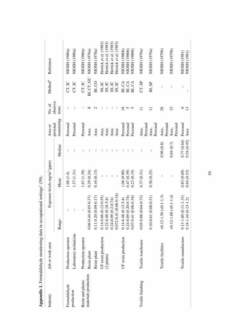
59
App
endi
x 1.
For
mal
dehy
de m
onito
ring
dat
a in
occ
upat
iona
l set
tings
a (59
).
Exp
osur
e le
vels
mg/
m3 (p
pm)
Indu
stry
Job
or w
ork
area
Ran
geM
ean
Med
ian
Are
a or
pers
onal
mon
itori
ng
No.
of
obse
rva-
tions
Met
hodb
Ref
eren
ce
Prod
uctio
n op
erat
or–
1.68
(1.
4)–
Pers
onal
–C
T, I
CN
IOSH
(19
80a)
Form
alde
hyde
prod
uctio
nL
abor
ator
y te
chni
cian
–1.
57 (
1.31
)–
Pers
onal
–C
T, I
CN
IOSH
(19
80a)
Prod
uctio
n op
erat
or–
1.67
(1.
39)
–Pe
rson
al –
CT
, IC
NIO
SH (
1980
a)
Res
in p
lant
0.06
-0.4
4 (0
.05-
0.37
)0.
29 (
0.24
)–
Are
a 8
BI,
CT
, GC
NIO
SH (
1976
a)
Res
in p
lant
0.11
-0.2
0 (0
.09-
0.17
)0.
16 (
0.13
)–
Are
a 2
BI,
CO
NIO
SH (
1978
a)
UF
resi
n pr
oduc
tion
0.14
-0.6
6 (0
.12-
0.55
)–
–A
rea
–SS
, IC
Her
rick
et a
l. (1
983)
Res
in a
nd p
last
icm
ater
ials
pro
duct
ion
(2 p
lant
s)0.
22-6
.48
(0.1
8-5.
4)–
–A
rea
–SS
, IC
Her
rick
et a
l. (1
983)
0.24
-0.8
9 (0
.2-0
.74)
––
Are
a –
SS, I
CH
erri
ck e
t al.
(198
3)0.
072-
0.41
(0.
06-0
.34)
––
Are
a –
SS, I
CH
erri
ck e
t al.
(198
3)
UF
resi
n pr
oduc
tion
0.14
-6.4
8 (0
.12-
5.4)
1.08
(0.
90)
–Pe
rson
al18
BI,
CA
NIO
SH (
1980
b)0.
24-0
.89
(0.2
0-0.
74)
0.47
(0.
39)
–Pe
rson
al 5
BI,
CA
NIO
SH (
1980
b)0.
07-0
.41
(0.0
6-0.
34)
0.23
(0.
19)
–Pe
rson
al 5
BI,
CA
NIO
SH (
1980
b)
Tex
tile
fini
shin
gT
extil
e w
areh
ouse
0.05
-0.8
8 (0
.04-
0.73
)0.
37 (
0.31
)–
Are
a,Pe
rson
al11
CT
, SP
NIO
SH (
1979
a)
0.10
-0.6
1 (0
.08-
0.51
)0.
30 (
0.25
)–
Are
a,Pe
rson
al11
BI,
SP
NIO
SH (
1979
a)
Tex
tile
faci
litie
s<
0.12
-1.5
6 (<
0.1-
1.3)
–0.
96 (
0.8)
Are
a,Pe
rson
al28
–N
IOSH
(19
79b)
<0.
12-1
.68
(<0.
1-1.
4)–
0.84
(0.
7)A
rea,
Pers
onal
15–
NIO
SH (
1979
b)
Tex
tile
man
ufac
ture
0.13
-1.6
0 (0
.11-
1.33
)0.
83 (
0.69
)0.
77 (
0.64
)Pe
rson
al 6
–N
IOSH
(19
81)
0.18
-1.4
4 (0
.15-
1.2)
0.64
(0.
53)
0.54
(0.
45)
Are
a13
–N
IOSH
(19
81)

60
App
endi
x 1.
Con
t.
Exp
osur
e le
vels
mg/
m3 (p
pm)
Indu
stry
Job
or w
ork
area
Ran
geM
ean
Med
ian
Are
a or
pers
onal
mon
itori
ng
No.
of
obse
rva-
tions
Met
hodb
Ref
eren
ce
Clo
thin
g pr
oduc
tion
Perm
anen
t pre
ss0.
18-0
.46
(0.1
5-0.
38)
0.37
(0.
31)
–A
rea
9B
I, I
US
DH
EW
(19
66)
0-3.
24 (
0-2.
7)0.
89 (
0.74
)–
Are
a32
BI,
IU
S D
HE
W (
1968
)
War
ehou
se0.
13-0
.68
(0.1
1-0.
57)
0.47
(0.
39)
0.44
(0.
37)
Pers
onal
13–
NIO
SH (
1979
a)0.
05-0
.23
(0.0
4-0.
19)
0.14
(0.
12)
0.18
(0.
15)
Are
a 9
–N
IOSH
(19
79a)
Sew
ing
mac
hine
0.61
-1.0
9 (0
.51-
0.91
)0.
86 (
0.72
)0.
85 (
0.71
)Pe
rson
al16
–N
IOSH
(19
79a)
oper
ator
s0.
36-2
.16
(0.3
-1.8
)1.
44 (
1.2)
1.44
(1.
2)Pe
rson
al41
–N
IOSH
(19
79a)
Clo
thin
g pr
esse
rs0.
006-
1.14
(0.
005-
0.95
)0.
08 (
0.07
)0.
065
(0.0
54)
Pers
onal
40–
NIO
SH (
1976
a)
All
wor
kers
–1.
2-3.
0–
Are
a–
–N
IOSH
(19
79b)
Plyw
ood
part
icle
-bo
ard
prod
uctio
n(1
-2.5
)
0.01
-0.3
(0.
008-
0.25
)0.
14 (
0.12
)–
Are
a11
BI,
CA
Her
rick
et a
l. (1
983)
Woo
d fu
rnitu
rem
anuf
actu
rePa
rtic
le b
oard
vene
erin
g1.
08-7
.68
(0.9
-6.4
)3.
30 (
2.75
)–
Are
a–
BI,
CA
Her
rick
et a
l. (1
983)
0.24
-0.6
6 (0
.2-0
.55)
0.48
(0.
40)
–A
rea
9B
I, C
AH
erri
ck e
t al.
(198
3)0.
24-3
.0 (
0.2-
2.5)
0.84
(0.
70)
–A
rea
13B
I, C
AH
erri
ck e
t al.
(198
3)
Plas
tic m
ould
ing
Inje
ctio
n m
ould
0.01
-0.1
2 (0
.01-
0.1)
0.04
4 (0
.037
)–
Pers
onal
9C
AN
IOSH
(19
73a)
Are
a sa
mpl
es0.
01-0
.64
(0.0
1-0.
53)
0.24
(0.
20)
–A
rea
8C
AN
IOSH
(19
73a)
Ope
rato
rs<
2.4
(<2)
<2.
4 (<
2)<
2.4
(<2)
Pers
onal
28D
TN
IOSH
(19
73a)
Nea
r gr
inde
r ho
pper
2.4-
4.8
(2-4
)3.
6 (3
)3.
6 (3
)A
rea
3D
TN
IOSH
(19
73a)
0.12
-0.8
4 (0
.1-0
.7)
0.37
(0.
31)
0.24
(0.
2)Pe
rson
al28
–N
IOSH
(19
76a)
Sand
mou
ldpr
oduc
tion
ND
-1.3
2 (N
D-1
.1)
0.20
(0.
17)
0.12
(0.
1)A
rea
29–
NIO
SH (
1976
a)
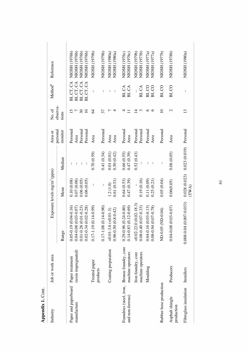
61
App
endi
x 1.
Con
t.
Exp
osur
e le
vels
mg/
m3 (p
pm)
Indu
stry
Job
or w
ork
area
Ran
geM
ean
Med
ian
Are
a or
pers
onal
mon
itor
No.
of
obse
rva-
tions
Met
hodb
Ref
eren
ce
0.05
-0.1
9 (0
.04-
0.16
)0.
10 (
0.08
)–
Pers
onal
15B
I, C
T, C
AN
IOSH
(19
76b)
Pape
r an
d pa
perb
oard
man
ufac
ture
Pape
r tr
eatm
ent
(res
in im
preg
nate
d)0.
04-0
.08
(0.0
3-0.
07)
0.07
(0.
06)
–A
rea
7B
I, C
T, C
AN
IOSH
(19
76b)
0.01
-0.2
8 (0
.01-
0.23
)0.
06 (
0.05
)–
Pers
onal
30B
I, C
T, C
AN
IOSH
(19
76b)
0.02
-0.3
4 (0
.02-
0.28
)0.
06 (
0.05
)–
Pers
onal
10B
I, C
T, C
AN
IOSH
(19
76b)
Tre
ated
pap
erpr
oduc
ts0.
17-1
.19
(0.1
4-0.
99)
–0.
70 (
0.59
)A
rea
64–
NIO
SH (
1979
b)
0.17
-1.0
8 (0
.14-
0.90
)–
0.41
(0.
34)
Pers
onal
37–
NIO
SH (
1979
b)
Coa
ting
prep
arat
ion
<0.
01-3
.6 (
<0.
01-3
)1.
2 (1
.0)
0.01
(0.
01)
Are
a 7
–N
IOSH
(19
80a)
0.96
-0.5
0 (0
.8-0
.42)
0.61
(0.
51)
0.50
(0.
42)
Are
a 4
–N
IOSH
(19
80a)
0.29
-0.9
6 (0
.24-
0.80
)0.
64 (
0.53
)0.
66 (
0.55
)Pe
rson
al 4
BI,
CA
NIO
SH (
1976
c)Fo
undr
ies
(ste
el, i
ron,
and
non-
ferr
ous)
Bro
nze
foun
dry,
cor
em
achi
ne o
pera
tors
0.14
-0.8
3 (0
.12-
0.69
)0.
47 (
0.39
)0.
47 (
0.39
)A
rea
11B
I, C
AN
IOSH
(19
76c)
<0.
02-2
2.0
(0.0
2-18
.3)
–0.
52 (
0.43
)Pe
rson
al14
–N
IOSH
(19
79b)
Iron
fou
ndry
, cor
em
achi
ne o
pera
tors
0.08
-0.4
0 (0
.07-
0.33
)0.
19 (
0.16
)–
Pers
onal
3B
I, C
AN
IOSH
(19
73b)
Mou
ldin
g0.
04-0
.16
(0.0
3-0.
13)
0.11
(0.
09)
–Pe
rson
al 6
BI,
CO
NIO
SH (
1977
a)0.
08-0
.94
(0.0
7-0.
78)
0.25
(0.
21)
–A
rea
6B
I, C
ON
IOSH
(19
77a)
Rub
ber
hose
pro
duct
ion
ND
-0.0
5 (N
D-0
.04)
0.05
(0.
04)
–Pe
rson
al10
BI,
CO
NIO
SH (
1977
b)
Prod
ucer
s0.
04-0
.08
(0.0
3-0.
07)
0.06
(0.0
5)0.
06 (
0.05
)A
rea
2B
I, C
ON
IOSH
(19
78b)
Asp
halt
shin
gle
prod
uctio
n
Fibr
egla
ss in
sula
tion
Inst
alle
rs0.
008-
0.04
(0.
007-
0.03
3)0.
028
(0.0
23)
0.02
3 (0
.019
)Pe
rson
al13
–N
IOSH
(19
80a)
(TW
A)

62
App
endi
x 1.
Con
t.
Exp
osur
e le
vels
mg/
m3 (p
pm)
Indu
stry
Job
or w
ork
area
Ran
ge M
ean
Med
ian
Are
a or
pers
onal
mon
itori
ng
No.
of
obse
rva-
tions
Met
hodb
Ref
eren
ce
0.08
-2.4
(0.
07-2
)–
––
–IC
Her
rick
et a
l. (1
983)
0.96
-1.9
2 (0
.8-1
.6)
1.26
(1.
05)
–A
rea
36
BI,
CA
NIO
SH (
1979
b)0.
36-3
.72
(0.3
-3.1
)1.
73 (
1.44
)–
Are
a 3
0C
T, I
CN
IOSH
(19
79b)
Ure
a-Fo
rmal
dehy
defo
am in
sula
tion
deal
ing
and
inst
alla
tion
Subu
rban
sho
ppin
gce
ntre
insu
late
d w
ithU
F fo
am<
0-6.
36 (
<0-
5.3)
1.87
(1.
56)
–A
rea
16
DT
NIO
SH (
1979
b)
Fert
ilize
rm
anuf
actu
ring
0.24
-2.2
8 (0
.2-1
.9)
1.08
(0.
9)–
Are
a,Pe
rson
al 1
1–
NIO
SH (
1979
b)
Mus
hroo
m f
arm
ing
<0.
61-1
2+ (
<0.
51-1
0+)
3.22
(2.
68)
–A
rea
12
DT
NIO
SH (
1980
b)N
D-3
.24
(ND
-2.7
)–
–Pe
rson
al
3C
T, I
CN
IOSH
(19
80b)
ND
-5.9
2 (N
D-4
.93)
––
Are
a
3C
T, I
CN
IOSH
(19
80b)
0.1-
6.3
(0.0
9-5.
26)
0.89
(0.
74)
–A
rea
187
CA
Ker
foot
and
Moo
ney
(197
5)Fu
nera
l hom
esE
mba
lmer
s
0.24
-4.7
9 (0
.20-
3.99
)1.
32 (
1.1)
0.65
(0.
54)
Are
a,Pe
rson
al
8C
TN
IOSH
(19
80c)
1.56
-4.7
2 (1
.30-
3.99
)3.
24 (
2.7)
2.99
(2.
49)
Are
a,Pe
rson
al
5C
TN
IOSH
(19
80c)
Path
olog
yA
utop
sy r
oom
0.07
-9.5
(0.
06-7
.9)
5.76
(4.
8)–
Are
a 1
0B
I, C
AC
ovin
o (1
979)
2.64
-9.5
(2.
20-7
.9)
5.22
(4.
35)
–A
rea
6
–N
IOSH
(19
79b)
Bio
logy
teac
hing
Lab
orat
ory
3.30
-17.
76 (
2.75
-14.
8)9.
96 (
8.3)
–A
rea
8
BI,
CA
US
EPA
(19
81)
Hos
pita
lL
abor
ator
y2.
64-2
.76
(2.2
-2.3
)2.
70 (
2.25
)–
Pers
onal
2
BI
Bla
de (
1983
)2.
28 (
1.9)
––
Pers
onal
1
CT
Bla
de (
1983
)2.
64 (
2.2)
2.40
(2)
–A
rea
2
CT
Bla
de (
1983
)

63
App
endi
x 1.
Con
t.
Exp
osur
e le
vels
mg/
m3 (p
pm)
Indu
stry
Job
or w
ork
area
Ran
ge M
ean
Med
ian
Are
a or
pers
onal
mon
itori
ng
No.
of
obse
rva-
tions
Met
hodb
Ref
eren
ce
Gov
erm
ent
Lab
orat
ory
2.88
(2.
4)–
–Pe
rson
al1
CT
Bla
de (
1983
)0.
96 (
0.8)
––
Are
a1
CT
Bla
de (
1983
)
Hos
pita
lD
ialy
sis
unit
ND
-1.0
8 (N
D-0
.90)
0.50
(0.
42)
–A
rea
9C
TB
lade
(19
83)
0.32
-0.7
6 (0
.27-
0.63
)0.
49 (
0.41
)–
Pers
onal
5C
TB
lade
(19
83)
0.05
-0.6
0 (0
.04-
0.05
)0.
61 (
0.51
)–
Are
aC
EA
Bla
de (
1983
)
Ani
mal
dis
sect
ion
Lab
orat
ory
<0.
46-1
.25
(<0.
38-1
.04)
––
Pers
onal
15C
AB
lade
(19
83)
0.06
-0.4
8 (0
.05-
0.40
)0.
18 (
0.15
)–
Are
a6
BI
Bla
de (
1983
)0.
13-0
.22
(0.1
1-0.
29)
0.22
(0.1
8)–
Are
a3
CE
AB
lade
(19
83)
Gar
men
t man
ufac
turi
ng(3
pla
nts)
<0.
17-0
.76
(<0.
14-0
.63)
0.28
-0.4
0(0
.23-
0.33
)–
Pers
onal
40C
TB
lade
(19
83)
<0.
04-0
.48
(<0.
03-0
.40)
0.23
-0.3
1(0
.19-
0.26
)–
Are
a43
CT
Bla
de (
1983
)
0.04
-0.4
8 (0
.03-
0.40
)0.
25 (
0.21
)–
Are
a43
BI
Bla
de (
1983
)0.
06-1
.34
(0.0
5-1.
2)0.
55 (
0.46
)–
Are
a42
CE
AB
lade
(19
83)
0.05
-1.9
2 (0
.04-
1.6)
0.66
(0.
55)
–Pe
rson
al3
BI
Bla
de (
1983
)C
hem
ical
man
ufac
turi
ng0.
04-0
.52
(0.0
3-0.
43)
0.20
(0.
17)
–A
rea
5B
IB
lade
(19
83)
Gla
ss m
anuf
actu
ring
0.50
(0.
42)
0.50
(0.
42)
–Pe
rson
al1
CT
Bla
de (
1983
)0.
54-0
.80
(0.4
5-0.
64)
0.65
(0.
54)
–A
rea
2C
TB
lade
(19
83)
Hos
pita
l wor
k0.
44-0
.88
(0.3
7-0.
73)
0.66
(0.
56)
–A
rea
2B
IB
lade
(19
83)

64
App
endi
x 1.
Con
t.
Exp
osur
e le
vels
mg/
m3 (p
pm)
Indu
stry
Job
or w
ork
area
Ran
ge M
ean
Med
ian
Are
a or
Pers
onal
mon
itori
ng
No.
of
obse
rva-
tions
Met
hodb
Ref
eren
ce
<0.
30-1
.02
(<0.
25-0
.85)
0.66
(0.
55)
–Pe
rson
al10
CA
Bla
de (
1983
)Pa
rafo
rmal
dehy
depa
ckag
ing
0.34
-4.0
8 (0
.28-
3.4)
1.40
(1.
17)
–A
rea
8C
EA
Bla
de (
1983
)
0.02
-0.1
4 (0
.02-
0.12
)0.
07 (
0.06
)–
Are
a39
BI
Bla
de (
1983
)O
ffic
e w
ork
(3 lo
catio
ns)
<0.
05 (
<0.
04)
<0.
05 (
<0.
04)
–A
rea
9C
TB
lade
(19
83)
Aut
opsy
roo
ms
Res
iden
t–
1.90
c (1.
58)
–Pe
rson
al10
CA
Mak
ar e
t al.
(197
5)
Path
olog
ist
–1.
50c (
1.24
)–
Pers
onal
9C
AM
akar
et a
l. (1
975)
Tec
hnic
ian
–0.
68c (
0.57
)–
Pers
onal
2C
AM
akar
et a
l. (1
975)
Ass
ista
nts
0.16
-16.
28 (
0.13
-13.
57)
0.86
(0.
72)
–A
rea
23C
AM
akar
et a
l. (1
975)
a. F
rom
: Con
sens
us W
orks
hop
of F
orm
alde
hyde
(19
84).
b. A
bbre
vatio
ns f
or a
naly
tical
pro
cedu
re: B
I, b
isul
phite
impi
nger
s; C
A, c
hrom
otro
pic
acid
pro
cedu
re; C
EA
, CE
A in
stru
men
ts M
odel
555
; CO
, col
orim
etri
c an
alys
is;
CT
, cha
rcoa
l tub
es; D
T, D
raeg
er tu
bes;
GC
, gas
cro
mat
ogra
phy;
IC
, ion
cro
mat
ogra
phy;
SP,
spe
ctro
phot
omet
ric
proc
edur
e; S
S, s
olid
sor
bent
s.c.
Ave
rage
.

65
App
endi
x 2.
Gen
etic
and
rel
ated
eff
ects
of
form
alde
hyde
(58
).
Res
ulta
Dos
ebR
efer
ence
Tes
t sys
tem
With
out
exog
enou
sm
etab
olic
syst
em
With
exog
enou
sm
etab
olic
syst
em
(LE
D/H
ID)
*M
isin
corp
orat
ion
of D
NA
bas
es in
to s
ynth
etic
pol
ynuc
leot
ides
in
vitr
o+
030
Snyd
er &
Van
Hou
ten
(198
6)PR
BPr
opha
ge in
duct
ion,
SO
S re
pair
test
, DN
A s
tran
d br
eaks
, cro
ss-l
inks
or
rela
ted
dam
age
+0
0.00
75K
uyke
ndal
l & B
ogda
nffy
(19
92)
PRB
Prop
hage
indu
ctio
n , S
OS
repa
ir te
st, D
NA
str
and
brea
ks, c
ross
-lin
ksor
rel
ated
dam
age
+0
20L
e C
urie
ux e
t al.
(199
3)
EC
BE
sche
rich
ia c
oli,
DN
A s
tran
d br
eaks
, cro
ss-l
inks
or
rela
ted
dam
age;
DN
A r
epai
r+
060
0W
ilkin
s &
Mac
Leo
d (1
976)
EC
BE
sche
rich
ia c
oli,
DN
A s
tran
d br
eaks
, cro
ss-l
inks
or
rela
ted
dam
age;
DN
A r
epai
r+
060
Pove
renn
y et
al.
(197
5)
EC
DE
sche
rich
ia c
oli p
olA
/W31
110-
P347
8, d
iffe
rent
ial t
oxic
ity (
spot
test
)+
010
Lei
fer
et a
l. (1
981)
EC
LE
sche
rich
ia c
oli K
12 K
S160
-KS6
6 po
lAI,
dif
fere
ntia
l tox
icity
+0
60Po
vere
nny
et a
l. (1
975)
EC
KE
sche
rich
ia c
oli K
12, f
orw
ard
or r
ever
se m
utat
ion
+0
60Z
ijlst
ra (
1989
)E
CK
Esc
heri
chia
col
i K12
, for
war
d or
rev
erse
mut
atio
n+
018
.8G
rave
s et
al.
(199
4)E
CK
Esc
heri
chia
col
i K12
, for
war
d or
rev
erse
mut
atio
n+
012
0C
rosb
y et
al.
(198
8)SA
FSa
lmon
ella
typh
imur
ium
, for
war
d m
utat
ion
++
10T
emch
aroe
n &
Thi
lly (
1983
)SA
OSa
lmon
ella
typh
imur
ium
TA
100,
rev
erse
mut
atio
n(+
)0
25M
arne
tt et
al.
(198
5)SA
OSa
lmon
ella
typh
imur
ium
TA
100,
rev
erse
mut
atio
n–
–30
Goc
ke e
t al.
(198
1)SA
OSa
lmon
ella
typh
imur
ium
TA
100,
rev
erse
mut
atio
n–
+16
.6H
awor
th e
t al.
(198
3)SA
OSa
lmon
ella
typh
imur
ium
TA
100,
rev
erse
mut
atio
n(+
)+
30 (
toxi
c ab
ove
125 µ
g/pl
ate)
Con
nor
et a
l. (1
983)
SAO
Salm
onel
la ty
phim
uriu
m T
A10
0, r
ever
se m
utat
ion
+0
7.5
Tak
ahas
hi e
t al.
(198
5)SA
OSa
lmon
ella
typh
imur
ium
TA
100,
rev
erse
mut
atio
n+
+c
4.5
Pool
et a
l. (1
984)
SAO
Salm
onel
la ty
phim
uriu
m T
A10
0, r
ever
se m
utat
ion
+0
9.3
O´D
onov
an &
Mee
(19
93)

66
App
endi
x 2.
Con
t
Res
ulta
Dos
ebR
efer
ence
Tes
t sys
tem
With
out
exog
enou
sm
etab
olic
syst
em
With
exog
enou
sm
etab
olic
syst
em
(LE
D/H
ID)
SAO
Salm
onel
la ty
phim
uriu
m T
A10
0, r
ever
se m
utat
ion
(+)
+3
Schm
id e
t al.
(198
6)SA
2Sa
lmon
ella
typh
imur
ium
TA
102,
rev
erse
mut
atio
n+
010
Mar
nett
et a
l. (1
985)
SA2
Salm
onel
la ty
phim
uriu
m T
A10
2, r
ever
se m
utat
ion
+0
10L
e C
urie
ux e
t al.
(199
3)SA
2Sa
lmon
ella
typh
imur
ium
TA
102,
rev
erse
mut
atio
n+
035
.7O
´Don
ovan
& M
ee (
1993
)SA
4Sa
lmon
ella
typh
imur
ium
TA
104,
rev
erse
mut
atio
n+
010
Mar
nett
et a
l. (1
985)
SA5
Salm
onel
la ty
phim
uriu
m T
A15
35, r
ever
se m
utat
ion
––
30G
ocke
et a
l. (1
981)
SA5
Salm
onel
la ty
phim
uriu
m T
A15
35, r
ever
se m
utat
ion
––
50H
awor
th e
t al.
(198
3)SA
5Sa
lmon
ella
typh
imur
ium
TA
1535
, rev
erse
mut
atio
n0
–c9
Pool
et a
l. (1
984)
SA5
Salm
onel
la ty
phim
uriu
m T
A15
35, r
ever
se m
utat
ion
–0
143
O´D
onov
an &
Mee
(19
93)
SA7
Salm
onel
la ty
phim
uriu
m T
A15
37, r
ever
se m
utat
ion
––
30G
ocke
et a
l. (1
981)
SA7
Salm
onel
la ty
phim
uriu
m T
A15
37, r
ever
se m
utat
ion
––
50H
awor
th e
t al.
(198
3)SA
7Sa
lmon
ella
typh
imur
ium
TA
1537
, rev
erse
mut
atio
n–
014
3O
´Don
ovan
& M
ee (
1993
)SA
8Sa
lmon
ella
typh
imur
ium
TA
1538
, rev
erse
mut
atio
n–
–30
Goc
ke e
t al.
(198
1)SA
8Sa
lmon
ella
typh
imur
ium
TA
1538
, rev
erse
mut
atio
n–
014
3O
´Don
ovan
& M
ee (
1993
)SA
9Sa
lmon
ella
typh
imur
ium
TA
98, r
ever
se m
utat
ion
+0
5M
arne
tt et
al.
(198
5)SA
9Sa
lmon
ella
typh
imur
ium
TA
98, r
ever
se m
utat
ion
––
30G
ocke
et a
l. (1
981)
SA9
Salm
onel
la ty
phim
uriu
m T
A98
, rev
erse
mut
atio
n–
(+)
16.6
Haw
orth
et a
l. (1
983)
SA9
Salm
onel
la ty
phim
uriu
m T
A98
, rev
erse
mut
atio
n–
(+)
30 (
toxi
c ab
ove
100 µ
g/pl
ate)
Con
nor
et a
l. (1
983)
SA9
Salm
onel
la ty
phim
uriu
m T
A98
, rev
erse
mut
atio
n0
(+)c
3Po
ol e
t al.
(198
4)SA
9Sa
lmon
ella
typh
imur
ium
TA
98, r
ever
se m
utat
ion
+0
17.9
O´D
onov
an &
Mee
(19
93)
SAS
Salm
onel
la ty
phim
uriu
m T
A97
, rev
erse
mut
atio
n+
05
Mar
nett
et a
l. (1
985)
SAS
Salm
onel
la ty
phim
uriu
m (
othe
r m
isce
llane
ous
stra
ins)
, rev
erse
mut
atio
n–
–10
0 (t
oxic
at 2
50µ
g/m
l)C
onno
r et
al.
(198
3)

67
App
endi
x 2.
Con
t.
Res
ulta
Dos
ebR
efer
ence
Tes
t sys
tem
With
out
exog
enou
sm
etab
olic
syst
em
With
exog
enou
sm
etab
olic
syst
em
(LE
D/H
ID)
EC
WE
sche
rich
ia c
oli W
P2 u
vrA
, rev
erse
mut
atio
n+
015
Tak
ahas
hi e
t al.
(198
5)E
CW
Esc
heri
chia
col
i WP2
uvr
A(p
KM
101)
, rev
erse
mut
atio
n+
017
.9O
´Don
ovan
& M
ee (
1993
)E
C2
Esc
heri
chia
col
i WP2
, rev
erse
mut
atio
n+
01.
2N
ishi
oka
(197
3)E
C2
Esc
heri
chia
col
i WP2
(pK
M10
1), r
ever
se m
utat
ion
+0
35.7
O´D
onov
an &
Mee
(19
93)
EC
2E
sche
rich
ia c
oli W
P2, r
ever
se m
utat
ion
+0
60T
akah
ashi
et a
l. (1
985)
EC
RE
sche
rich
ia c
oli (
othe
r m
isce
llane
ous
stra
ins)
, rev
erse
mut
atio
n+
090
0Pa
nfilo
va e
t al.
(196
6)E
CR
Esc
heri
chia
col
i (ot
her
mis
cella
neou
s st
rain
s), r
ever
se m
utat
ion
+0
80D
emer
ec e
t al.
(195
1)E
CR
Esc
heri
chia
col
i (ot
her
mis
cella
neou
s st
rain
s), r
ever
se m
utat
ion
+0
30T
akah
ashi
et a
l. (1
985)
SSB
Sacc
haro
myc
es s
peci
es, D
NA
str
and
brea
ks, c
ross
-lin
ks o
r re
late
dda
mag
e+
099
0M
agan
a-Sc
hwen
cke
(197
8)
SSB
Sacc
haro
myc
es s
peci
es, D
NA
str
and
brea
ks, c
ross
-lin
ks o
r re
late
dda
mag
e+
050
0M
agan
a-Sc
hwen
cke
& E
kert
(197
8)SS
BSa
ccha
rom
yces
spe
cies
, DN
A s
tran
d br
eaks
, cro
ss-l
inks
or
rela
ted
dam
age
+0
500
Mag
ana-
Schw
enck
e &
Mou
stac
chi (
1980
)SC
HSa
ccha
rom
yces
cer
evis
iae,
gen
e co
nver
sion
+0
540
Cha
net e
t al.
(197
5)SC
HSa
ccha
rom
yces
cer
evis
iae,
hom
ozyg
osis
by
mito
tic r
ecom
bina
tion
orge
ne c
onve
rsio
n+
018
.5Z
imm
erm
ann
& M
ohr
(199
2)
NC
FN
euro
spor
a cr
assa
, for
war
d m
utat
ion
+0
100
de S
erre
s et
al.
(198
8)N
CR
Neu
rosp
ora
cras
sa, r
ever
se m
utat
ion
–0
732
Dic
key
et a
l. (1
949)
NC
RN
euro
spor
a cr
assa
, rev
erse
mut
atio
n+
037
500
Jens
en e
t al.
(195
2)N
CR
Neu
rosp
ora
cras
sa, r
ever
se m
utat
ion
–0
300
Köl
mar
k &
Wes
terg
aard
(19
53)
PLM
Plan
ts (
othe
r), m
utat
ion
+0
0.0
Aue
rbac
h et
al.
(197
7)

68
App
endi
x 2.
Con
t.
Res
ulta
Dos
ebR
efer
ence
Tes
t sys
tem
With
out
exog
enou
sm
etab
olic
syst
em
With
exog
enou
sm
etab
olic
syst
em
(LE
D/H
ID)
DM
GD
roso
phil
a m
elan
ogas
ter,
gen
etic
cro
ssin
g ov
er o
r re
com
bina
tion
+27
00R
atna
yake
(19
70)
DM
GD
roso
phil
a m
elan
ogas
ter,
gen
etic
cro
ssin
g ov
er o
r re
com
bina
tion
+42
0A
lder
son
(196
7)D
MG
Dro
soph
ila
mel
anog
aste
r, g
enet
ic c
ross
ing
over
or
reco
mbi
natio
n+
1260
Sobe
ls &
van
Ste
enis
(19
57)
DM
XD
roso
phil
a m
elan
ogas
ter,
sex
-lin
ked
rece
ssiv
e le
thal
mut
atio
ns+
420
Ald
erso
n (1
967)
DM
XD
roso
phil
a m
elan
ogas
ter,
sex
-lin
ked
rece
ssiv
e le
thal
mut
atio
ns(+
)19
40R
atna
yake
(19
68)
DM
XD
roso
phil
a m
elan
ogas
ter,
sex
-lin
ked
rece
ssiv
e le
thal
mut
atio
ns+
2380
Rat
naya
ke (
1970
)D
MX
Dro
soph
ila
mel
anog
aste
r, s
ex-l
inke
d re
cess
ive
leth
al m
utat
ions
+19
40A
uerb
ach
& M
oser
(19
53)
DM
XD
roso
phil
a m
elan
ogas
ter,
sex
-lin
ked
rece
ssiv
e le
thal
mut
atio
ns+
1080
Kap
lan
(194
8)D
MX
Dro
soph
ila
mel
anog
aste
r, s
ex-l
inke
d re
cess
ive
leth
al m
utat
ions
+42
0K
han
(196
7)D
MX
Dro
soph
ila
mel
anog
aste
r, s
ex-l
inke
d re
cess
ive
leth
al m
utat
ions
+27
0St
umm
-Teg
etho
ff (
1969
)D
MX
Dro
soph
ila
mel
anog
aste
r, s
ex-l
inke
d re
cess
ive
leth
al m
utat
ions
+12
60So
bels
& v
an S
teen
is (
1957
)D
MH
Dro
soph
ila
mel
anog
aste
r, h
erita
ble
tran
sloc
atio
n+
2700
Rat
naya
ke (
1970
)D
MH
Dro
soph
ila
mel
anog
aste
r, h
erita
ble
tran
sloc
atio
n+
420
Kha
n (1
967)
DM
LD
roso
phil
a m
elan
ogas
ter,
dom
inan
t let
hal m
utat
ion
+19
40A
uerb
ach
& M
oser
(19
53)
DM
LD
roso
phil
a m
elan
ogas
ter,
dom
inan
t let
hal m
utat
ion
+14
00Sr
ám (
1970
)*
Cae
norh
abdi
tis
eleg
ans,
rec
essi
ve le
thal
mut
atio
n+
700
John
sen
& B
ailli
e (1
988)
DIA
DN
A s
tran
d br
eaks
, cro
ss-l
inks
or
rela
ted
dam
age,
ani
mal
cel
ls in
vit
ro+
06
Ros
s &
Shi
pley
(19
80)
DIA
DN
A s
tran
d br
eaks
, cro
ss-l
inks
or
rela
ted
dam
age,
ani
mal
cel
ls in
vit
ro+
03.
75R
oss
et a
l. (1
981)
DIA
DN
A s
tran
d br
eaks
, cro
ss-l
inks
or
rela
ted
dam
age,
ani
mal
cel
ls in
vit
ro+
022
.5D
emko
wic
z-D
obrz
ansk
i &C
asto
ngua
y (1
992)
DIA
DN
A s
tran
d br
eaks
, cro
ss-l
inks
or
rela
ted
dam
age,
ani
mal
cel
ls in
vit
ro+
07.
5O
´Con
nor
& F
ox (
1987
)G
9HG
ene
mut
atio
n, C
hine
se h
amst
er V
79 c
ells
, hpr
t loc
us+
09
Gra
fstr
öm e
t al.
(199
3)

69
App
endi
x 2.
Con
t.
Res
ulta
Dos
ebR
efer
ence
Tes
t sys
tem
With
out
exog
enou
sm
etab
olic
syst
em
With
exog
enou
sm
etab
olic
syst
em
(LE
D/H
ID)
SIC
Sist
er c
hrom
atid
exc
hang
e, C
hine
se h
amst
er c
ells
in v
itro
+0
1O
be &
Bee
k (1
979)
SIC
Sist
er c
hrom
atid
exc
hang
e, C
hine
se h
amst
er c
ells
in v
itro
++
3.2
Nat
araj
an e
t al.
(198
3)SI
CSi
ster
chr
omat
id e
xcha
nge,
Chi
nese
ham
ster
cel
ls in
vit
ro+
+1.
8B
asle
r et
al.
(198
5)C
ICC
hrom
osom
al a
berr
atio
ns, C
hine
se h
amst
er c
ells
in v
itro
++
6.5
Nat
araj
an e
t al.
(198
3)C
ICC
hrom
osom
al a
berr
atio
ns, C
hine
se h
amst
er c
ells
in v
itro
+0
18Is
hida
te e
t al.
(198
1)T
CM
Cel
l tra
nsfo
rmat
ion,
C3H
10T
1/2
mou
se c
ells
+d
00.
5R
agan
& B
orei
ko (
1981
)D
IHD
NA
str
and
brea
ks, c
ross
-lin
ks o
r re
late
d da
mag
e, h
uman
cel
ls in
vit
ro+
024
Forn
ace
et a
l. (1
982)
DIH
DN
A s
tran
d br
eaks
, cro
ss-l
inks
or
rela
ted
dam
age,
hum
an c
ells
in v
itro
+0
1.5
Cra
ft e
t al.
(198
7)D
IHD
NA
str
and
brea
ks, c
ross
-lin
ks o
r re
late
d da
mag
e, h
uman
cel
ls in
vit
ro+
03
Gra
fstr
öm e
t al.
(198
6)D
IHD
NA
str
and
brea
ks, c
ross
-lin
ks o
r re
late
d da
mag
e, h
uman
cel
ls in
vit
ro+
03
Snyd
er &
van
Hou
ten
(198
6)D
IHD
NA
str
and
brea
ks, c
ross
-lin
ks o
r re
late
d da
mag
e, h
uman
cel
ls in
vit
ro+
03
Sala
dino
et a
l. (1
985)
DIH
DN
A s
tran
d br
eaks
, cro
ss-l
inks
or
rela
ted
dam
age,
hum
an c
ells
in v
itro
+0
3G
rafs
tröm
et a
l. (1
984)
DIH
DN
A s
tran
d br
eaks
, cro
ss-l
inks
or
rela
ted
dam
age,
hum
an c
ells
in v
itro
+0
12G
rafs
tröm
(19
90)
UIH
Uns
ched
uled
DN
A s
ynth
esis
, hum
an b
ronc
hial
epi
thel
ial c
ells
in v
itro
–0
3 (>
0.1
mm
ol/L
was
leth
al)
Doo
little
et a
l. (1
985)
GIH
Gen
e m
utat
ion,
hum
an c
ells
in v
itro
+0
3G
rafs
tröm
et a
l. (1
985)
GIH
Gen
e m
utat
ion,
hum
an c
ells
in v
itro
+0
3.9
Gol
dmac
her
& T
hilly
(19
83)
GIH
Gen
e m
utat
ion,
hum
an c
ells
in v
itro
+0
0.9
Cra
ft e
t al.
(198
7)G
IHG
ene
mut
atio
n, h
uman
cel
ls in
vit
ro+
04.
5C
rosb
y et
al.
(198
8)G
IHG
ene
mut
atio
n, h
uman
cel
ls in
vit
ro+
04.
5L
iber
et a
l. (1
989)
GIH
Gen
e m
utat
ion,
hum
an c
ells
in v
itro
+0
3G
rafs
tröm
(19
90)
RIH
DN
A r
epai
r ex
clus
ive
of u
nsch
edul
ed D
NA
syn
thes
is, h
uman
cel
ls in
vitr
o+
06
Gra
fstr
öm e
t al.
(198
4)

70
App
endi
x 2.
Con
t.
Res
ulta
Dos
ebR
efer
ence
Tes
t sys
tem
With
out
exog
enou
sm
etab
olic
syst
em
With
exog
enou
sm
etab
olic
syst
em
(LE
D/H
ID)
SHL
Sist
er c
hrom
atid
exc
hang
e, h
uman
lym
phoc
ytes
in v
itro
+0
5.4
Obe
& B
eek
(197
9)SH
LSi
ster
chr
omat
id e
xcha
nge,
hum
an ly
mph
ocyt
es in
vit
ro+
05
Kre
iger
& G
arry
(19
83)
SHL
Sist
er c
hrom
atid
exc
hang
e, h
uman
lym
phoc
ytes
in v
itro
++
3.75
Schm
id e
t al.
(198
6)C
HF
Chr
omos
omal
abe
rrat
ions
, hum
an f
ibro
blas
ts in
vit
ro+
060
Lev
y et
al.
(198
3)C
HL
Chr
omos
omal
abe
rrat
ions
, hum
an ly
mph
ocyt
es in
vit
ro+
010
Mir
etsk
aya
& S
hvar
tsm
an (
1982
)C
HL
Chr
omos
omal
abe
rrat
ions
, hum
an ly
mph
ocyt
es in
vit
ro+
+7.
5Sc
hmid
et a
l. (1
986)
CH
LC
hrom
osom
al a
berr
atio
ns, h
uman
lym
phoc
ytes
in v
itro
+0
3.75
Dre
sp &
Bau
chin
ger
(198
8)D
VA
DN
A-p
rote
in c
ross
-lin
ks, r
at c
ells
in v
ivo
+1.
5 in
hal.
6hC
asan
ova-
Schm
itz e
t al.
(198
4b)
DV
AD
NA
-pro
tein
cro
ss-l
inks
, rat
cel
ls in
viv
o(+
)1.
5 in
hal.
6hL
arn
et a
l. (1
985)
DV
AD
NA
-pro
tein
cro
ss-l
inks
, rat
cel
ls in
viv
o+
0.25
inha
l. 3h
Hec
k et
al.
(198
6)D
VA
DN
A-p
rote
in c
ross
-lin
ks, r
at c
ells
in v
ivo
+0.
25 in
hal.
3hC
asan
ova
& H
eck
(198
7)D
VA
DN
A-p
rote
in c
ross
-lin
ks, r
at c
ells
in v
ivo
+0.
08 in
hal.
6hC
asan
ova
et a
l. (1
989)
DV
AD
NA
-pro
tein
cro
ss-l
inks
, rhe
sus
mon
key
nasa
l tur
bina
te c
ells
in v
ivo
+0.
05 in
hal.
6hH
eck
et a
l. (1
989)
DV
AD
NA
-pro
tein
cro
ss-l
inks
, rhe
sus
mon
key
nasa
l tur
bina
te c
ells
in v
ivo
+0.
05 in
hal.
6hC
asan
ova
et a
l. (1
991)
*D
NA
-pro
tein
cro
ss-l
inks
, rat
trac
heal
impl
ant c
ells
in v
ivo
+2
mg/
ml i
nstil
.C
osm
a et
al.
(198
8)SV
ASi
ster
chr
omat
id e
xcha
nge,
rat
cel
ls in
viv
o–
3.9
inha
l. 6h
/d x
5K
liger
man
et a
l. (1
984)
*M
icro
nucl
eus
indu
ctio
n, n
ewt (
Ple
urod
eles
wal
tl)
in v
ivo
–5 µ
g/m
l, 12
dSi
boul
et e
t al.
(198
4)M
VM
Mic
ronu
cleu
s in
duct
ion,
mou
se in
viv
o–
25 ip
x 1
Nat
araj
an e
t al.
(198
3)M
VM
Mic
ronu
cleu
s in
duct
ion,
mou
se in
viv
o–
30 ip
x 1
Goc
ke e
t al.
(198
1)M
VR
Mic
ronu
cleu
s in
duct
ion,
rat
(ga
stro
inte
stin
al tr
act)
in v
ivo
+20
0 po
x 1
Mig
liore
et a
l. (1
989)
CB
AC
hrom
osom
al a
berr
atio
ns, m
ouse
bon
e-m
arro
w c
ells
in v
ivo
–25
ip x
1N
atar
ajan
et a
l. (1
983)
CB
AC
hrom
osom
al a
berr
atio
ns, r
at b
one-
mar
row
cel
ls in
viv
o+
0.07
inha
l. 4h
/d, 4
mon
thK
itaev
a et
al.
(199
0)

71
App
endi
x 2.
Con
t.
Res
ulta
Dos
ebR
efer
ence
Tes
t sys
tem
With
out
exog
enou
sm
etab
olic
syst
em
With
exog
enou
sm
etab
olic
syst
em
(LE
D/H
ID)
CB
AC
hrom
osom
al a
berr
atio
ns, r
at b
one-
mar
row
cel
ls in
viv
o–
3.9
inha
l. 6h
/d x
5,
8 w
eeks
Dal
las
et a
l. (1
992)
CL
AC
hrom
osom
al a
berr
atio
ns, r
at le
ukoc
ytes
in v
ivo
–3.
9 in
hal.
6h/d
x 5
Klig
erm
an e
t al.
(198
4)C
CC
Chr
omos
omal
abe
rrat
ions
, mou
se s
perm
atoc
ytes
trea
ted
in v
ivo,
sper
mat
ocyt
es o
bser
ved
–50
ip x
1Fo
ntig
nie-
Hou
brec
hts
(198
1)
CV
AC
hrom
osom
al a
berr
atio
ns, m
ouse
spl
een
cells
in v
ivo
–25
ip x
1N
atar
ajan
et a
l. (1
983)
CV
AC
hrom
osom
al a
berr
atio
ns, r
at p
ulm
onar
y la
vage
cel
ls in
viv
o+
3.9
inha
l. 6h
/d x
5D
alla
s et
al.
(199
2)G
VA
Gen
e m
utat
ion,
rat
cel
ls in
viv
o (p
53 p
oint
mut
atio
ns in
nas
alca
rcin
omas
)+
3.9
inha
l. 6h
/d, 2
year
sR
ecio
et a
l. (1
992)
MST
Mou
se s
pot t
est
–3.
9 in
hal.
6h/d
x 3
Jens
en &
Coh
r (1
983)
(A
bstr
act)
DL
MD
omin
ant l
etha
l mut
atio
ns, m
ouse
(+)
50 ip
x 1
Font
igni
e-H
oubr
echt
s (1
981)
DL
MD
omin
ant l
etha
l mut
atio
ns, m
ouse
–20
ip x
1E
pste
in e
t al.
(197
2)D
LR
Dom
inan
t let
hal m
utat
ions
, rat
(+)
0.2
inha
l. 4h
/d x
120
Kita
eva
et a
l. (1
990)
DL
MD
omin
ant l
etha
l mut
atio
ns, m
ouse
–20
ip x
1E
pste
in &
Sha
fner
(19
68)
MV
HM
icro
nucl
eus
form
atio
n, h
uman
lym
phoc
ytes
in v
ivo
(+)
0.06
e inha
l. 8h
-TW
ASu
ruda
et a
l. (1
993)
MV
HM
icro
nucl
eus
form
atio
n, h
uman
cel
ls (
bucc
al e
pith
eliu
m)
in v
ivo
+0.
06e in
hal.
8h-T
WA
Suru
da e
t al.
(199
3)M
VH
Mic
ronu
cleu
s fo
rmat
ion,
hum
an c
ells
(na
sal e
pith
eliu
m)
in v
ivo
–0.
06e in
hal.
8h-T
WA
Suru
da e
t al.
(199
3)M
VH
Mic
ronu
cleu
s fo
rmat
ion,
hum
an c
ells
(na
sal e
pith
eliu
m)
in v
ivo
+0.
06e in
hal.
8h-T
WA
Bal
lari
n et
al.
(199
2)SL
HSi
ster
chr
omat
id e
xcha
nge,
hum
an ly
mph
ocyt
es in
viv
o–
0.5
inha
l. 8h
-TW
AT
hom
son
et a
l. (1
984)
SLH
Sist
er c
hrom
atid
exc
hang
e, h
uman
lym
phoc
ytes
in v
ivo
–0.
5 in
hal.
8h-T
WA
Bau
chin
ger
& S
chm
id (
1985
)SL
HSi
ster
chr
omat
id e
xcha
nge,
hum
an ly
mph
ocyt
es in
viv
o+
0.2
inha
l. 8h
-TW
AY
ager
et a
l. (1
986)
SLH
Sist
er c
hrom
atid
exc
hang
e, h
uman
lym
phoc
ytes
in v
ivo
–0.
06e in
hal.
8h-T
WA
Suru
da e
t al.
(199
3)
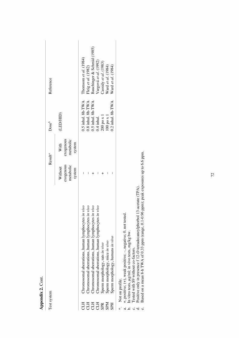
72
App
endi
x 2.
Con
t.
Res
ulta
Dos
ebR
efer
ence
Tes
t sys
tem
With
out
exog
enou
sm
etab
olic
syst
em
With
exog
enou
sm
etab
olic
syst
em
(LE
D/H
ID)
CL
HC
hrom
osom
al a
berr
atio
ns, h
uman
lym
phoc
ytes
in v
ivo
–0.
5 in
hal.
8h-T
WA
Tho
mso
m e
t al.
(198
4)C
LH
Chr
omos
omal
abe
rrat
ions
, hum
an ly
mph
ocyt
es in
viv
o–
0.8
inha
l. 8h
-TW
AFl
eig
et a
l. (1
982)
CL
HC
hrom
osom
al a
berr
atio
ns, h
uman
lym
phoc
ytes
in v
ivo
+0.
5 in
hal.
8h-T
WA
Bau
chin
ger
& S
chm
id (
1985
)C
LH
Chr
omos
omal
abe
rrat
ions
, hum
an ly
mph
ocyt
es in
viv
o–
0.4
inha
l.V
argo
vá e
t al.
(199
2)SP
RSp
erm
mor
phol
ogy,
rat
s in
viv
o+
200
po x
1C
assi
dy e
t al.
(198
3)SP
MSp
erm
mor
phol
ogy,
mic
e in
viv
o–
100
po x
1W
ard
et a
l. (1
984)
SPH
Sper
m m
orph
olog
y, h
uman
s in
viv
o–
0.2
inha
l. 8h
-TW
AW
ard
et a
l. (1
984)
*.
Not
on
prof
ile.
a.
+, p
ositi
ve; (
+),
wea
k po
sitiv
e; –
, neg
ativ
e; 0
, not
test
ed.
b.
In v
itro
test
s, µ
g/m
l; in
viv
o te
sts,
mg/
kg b
w.
c.
Tes
ted
with
S9
with
out c
o-fa
ctor
s.d.
Po
sitiv
e on
ly in
pre
senc
e f
12-O
-tet
rade
cano
ylph
orbo
l 13-
acet
ate
(TPA
).e.
B
ased
on
a m
ean
8-h
TW
A o
f 0.
33 p
pm (
rang
e, 0
.1-0
.96
ppm
); p
eak
expo
sure
s up
to 6
.6 p
pm.

73
App
endi
x 2.
Sum
mar
y ta
ble
of g
enet
ic a
nd r
elat
ed e
ffec
ts o
f fo
rmal
dehy
de.
Non
-mam
mal
ian
syst
ems
Mam
mal
ian
syst
ems
Pro-
Low
er P
lant
s
Ins
ects
In v
itro
In v
ivo
kary
otes
euka
ryot
esA
nim
al c
ells
Hum
an c
ells
Ani
mal
Hum
an
DG
DR
GA
DG
CR
GC
AD
GS
MC
AT
ID
GS
MC
AT
ID
GS
MC
DL
AD
SM
CA
++
++
++1
++
+ +
+1 +
+
+1 +
++
+
+
–1
+1
??
? ?1
?
A, a
neup
loid
y; C
, chr
omos
omal
abb
erat
ions
; D, D
NA
dam
age;
DL
, dom
inan
t let
hal m
utat
ion;
G, g
ene
mut
atio
n; I
, inh
ibiti
on o
f in
terc
ellu
lar
com
mun
icat
ion;
M, m
icro
nucl
ei; R
, mito
tic r
ecom
bina
tion
and
gene
con
vers
ion;
S, s
iste
r ch
rom
atid
exc
hang
e; T
, cel
l tra
nsfo
rmat
ion.
In c
ompl
etin
g th
e ta
ble,
the
follo
win
g sy
mbo
ls in
dica
te th
e co
ncen
sus
of th
e W
orki
ng g
roup
with
reg
ard
to th
e re
sults
for
eac
h en
d-po
int:
+
cons
ider
ed to
be
posi
tive
for
the
spec
ific
end
-poi
nt a
nd le
vel o
f bi
olog
ical
com
plex
ity.
+1
cons
ider
ed to
be
posi
tive,
but
onl
y on
e va
lid s
tudy
was
ava
ilabl
e to
the
Wor
king
gro
up.
– co
nsid
ered
to b
e ne
gativ
e.–1
cons
ider
ed to
be
nega
tive,
but
onl
y on
e va
lid s
tudy
was
ava
ilabl
e to
the
Wor
king
gro
up.
? co
nsid
ered
to b
e eq
uivo
cal o
r in
conc
lusi
ve (
e.g.
ther
e w
ere
cont
radi
ctor
y re
sults
fro
m d
iffe
rent
labo
rato
ries
; the
re w
ere
conf
ound
ing
expo
sure
s; th
e re
sults
wer
eeq
uivo
cal.

74
Appendix 3
Classification of substances with respect to carcinogenicity
Guideline 93/21/EEG of the European Community
4.2 Criteria for classification, indication of danger, choice of risk phrases
4.2.1 Carcinogenic substances
For the purpose of classification and labelling, and having regard to the current stateof knowledge, such substances are divided into three categories:
Category 1:
Substances known to be carcinogenic to man.
There is sufficient evidence to establish a causal association between human exposureto a substance and the development of cancer.
Category 2:
Substances which should be regarded as if they are carcinogenic to man.
There is sufficient evidence to provide a strong presumption that human exposure to asubstance may result in the development of cancer, generally on the basis of:• appropriate long-term animal studies• other relevant information.
Category 3:
Substances which cause concern for man owing to possible carcinogenic effects but inrespect of which the available information is not adequate for making a satisfactoryassessment.
There is some evidence from appropriate animal studies, but this is insufficient toplace the substance in Category 2.
4.2.1.1 The following symbols and specific risk phrases apply:
Category 1 and 2:
T; R45 May cause cancer
However for substances and preparations which present a carcinogenic risk only wheninhaled, for example, as dust, vapour or fumes, (other routes of exposure e.g. byswallowing or in contact with skin do not present any carcinogenic risk), thefollowing symbol and specific risk phrase should be used:
T; R49 May cause cancer by inhalation

75
Category 3:
Xn; R40 Possible risk of irreversible effects
4.2.1.2 Comments regarding the categorisation of carcinogenic substances
The placing of a substance into Category 1 is done on the basis of epidemiologicaldata; placing into Categories 2 and 3 is based primarily on animal experiments.
For classification as a Category 2 carcinogen either positive results in two animalspecies should be available or clear positive evidence in one species; together withsupporting evidence such as genotoxicity data, metabolic or biochemical studies,induction of benign tumours, structural relationship with other known carcinogens, ordata from epidemiological studies suggesting an association.
Category 3 actually comprises 2 sub-categories:a) substances which are well investigated but for which the evidence of a tumour-
inducing effect is insufficient for classification in Category 2. Additionalexperiments would not be expected to yield further relevant information withrespect to classification.
b) substances which are insufficiently investigated. The available data areinadequate, but they raise concern for man. This classification is provisional;further experiments are necessary before a final decision can be made.
For a distinction between Categories 2 and 3 the arguments listed below are relevantwhich reduce the significance of experimental tumour induction in view of possiblehuman exposure. These arguments, especially in combination, would lead in mostcases to classification in Category 3, even though tumours have been induced inanimals:• carcinogenic effects only at very high levels exceeding the “maximal tolerated
dose”. The maximal tolerated dose is characterised by toxic effects which,although not yet reducing life span, go along with physical changes such as about10% retardation in weight gain;
• appearance of tumours, especially at high dose levels, only in particular organs ofcertain species is known to be susceptible to a high spontaneous tumourformation;
• appearance of tumours, only at the site of application, in very sensitive testsystems (e.g. intraperitoneal or subcutaneous application of certain locally activecompounds); if the particular target is not relevant to man;
• lack of genotoxicity in short-term tests in vivo and in vitro;• existence of a secondary mechanism of action with the implication of a practical
threshold above a certain dose level (e.g. hormonal effects on target organs or onmechanisms of physiological regulation, chronic stimulation of cell proliferation;
• existence of a species - specific mechanism of tumour formation (e.g. by specificmetabolic pathways) irrelevant for man.

76
For a distinction between Category 3 and no classification arguments are relevantwhich exclude a concern for man:
• a substance should not be classified in any of the categories if the mechanism ofexperimental tumour formation is clearly identified, with good evidence that thisprocess cannot be extrapolated to man;
• if the only available tumour data are liver tumours in certain sensitive strains ofmice, without any other supplementary evidence, the substance may not beclassified in any of the categories;
• particular attention should be paid to cases where the only available tumour dataare the occurrence of neoplasms at sites and in strains where they are well knownto occur spontaneously with a high incidence.