the p-body component usp52/pan2 is a novel … · the p-body component usp52/pan2 is a novel...
TRANSCRIPT

Biochem. J. (2013) 451, 185–194 (Printed in Great Britain) doi:10.1042/BJ20130026 185
The P-body component USP52/PAN2 is a novel regulator of HIF1A mRNAstabilityJohn S. BETT*, Adel F. M. IBRAHIM*, Amit K. GARG*, Van KELLY*, Patrick PEDRIOLI*, Sonia ROCHA† and Ronald T. HAY*†1
*Scottish Institute for Cell Signalling, Sir James Black Centre, College of Life Sciences, University of Dundee, Dow Street, Dundee DD1 5EH, Scotland, U.K., and†Wellcome Trust Centre for Gene Regulation and Expression, College of Life Sciences, University of Dundee, Dundee DD1 5EH, Scotland, U.K.
HIF1A (hypoxia-inducible factor 1α) is the master regulator ofthe cellular response to hypoxia and is implicated in cancerprogression. Whereas the regulation of HIF1A protein in responseto oxygen is well characterized, less is known about the fateof HIF1A mRNA. In the present study, we have identifiedthe pseudo-DUB (deubiquitinating enzyme)/deadenylase USP52(ubiquitin-specific protease 52)/PAN2 [poly(A) nuclease 2] as animportant regulator of the HIF1A-mediated hypoxic response.Depletion of USP52 reduced HIF1A mRNA and protein levelsand resulted in reduced expression of HIF1A-regulated hypoxictargets due to a 3′-UTR (untranslated region)-dependent poly(A)-tail-length-independent destabilization in HIF1A mRNA. MSanalysis revealed an association of USP52 with several P-body
(processing body) components and we confirmed further thatUSP52 protein and HIF1A mRNA co-localized with cytoplasmicP-bodies. Importantly, P-body dispersal by knockdown of GW182or LSM1 resulted in a reduction of HIF1A mRNA levels. Thesedata uncover a novel role for P-bodies in regulating HIF1A mRNAstability, and demonstrate that USP52 is a key component ofP-bodies required to prevent HIF1A mRNA degradation.
Key words: AU-rich element (ARE)-mediated degradation(AMD), hypoxia-inducible factor 1α (HIF1A), poly(A) nuclease2 (PAN2), processing body (P-body), pseudo-deubiquitinatingenzyme (pseudo-DUB), ubiquitin-specific protease 52 (USP52).
INTRODUCTION
Cells respond to reduced oxygen tension by executing atranscriptional programme that is principally orchestrated byHIF1A (hypoxia-inducible factor 1α) [1]. HIF1A protein issynthesized continually, but degraded rapidly by the ubiquitin–proteasome system under normal oxygen concentrations(normoxia) [2,3]. This is a result of oxygen-dependentproline hydroxylation mediated by a family of PHDs(prolyl hydroxylases). HIF1A containing this hydroxyprolinemodification is a substrate for the VHL (von Hippel–Lindau) E3ubiquitin ligase complex which targets the protein for ubiquitin-mediated proteolysis [4,5]. Upon decreased oxygen concentration(hypoxia), such as that observed in solid tumours, HIF1A escapesproline hydroxylation and degradation to bind its constitutivelyexpressed partner HIF1B (hypoxia-inducible factor 1β) anddrive the expression of many genes involved in glycolysis,angiogenesis, cell survival and cancer progression [3].
Whereas the regulation of HIF1A protein is well documented,little is known about the regulation and turnover of HIF1A mRNA.The presence of multiple AREs (AU-rich elements) in the 3′-UTR(untranslated region) of HIF1A and the observation that HuRbinds this 3′-UTR suggested regulation of the HIF1A transcriptvia AMD (ARE-mediated degradation) [6]. In support of this, thepresence of AREs in the HIF1A 3′-UTR has been reported to benecessary for TTP (tristetrapolin)-mediated degradation of HIF1AmRNA during prolonged hypoxia [7,8]. In addition, the existence
of an aHIF (antisense hypoxia-inducible factor) complementaryto 1027 bases in the HIF1A 3′-UTR has led to the proposal thatHIF1A mRNA is targeted for degradation by binding of aHIF toits 3′-UTR and exposing AREs to TTP [9]. Indeed, aHIF wasshown to be up-regulated by prolonged hypoxia and correlatedwith a reduction in HIF1A mRNA stability [10].
USP52 (ubiquitin-specific protease 52)/PAN2 [poly(A)nuclease 2] belongs to the ubiquitin-specific protease superfamily,but exhibits no deubiquitylating activity owing to the lackof an active-site cysteine residue [11]. It also contains a C-terminal exonuclease III domain and has been well characterizedin its role as a poly(A) nuclease as part of the PAN2–PAN3 deadenylation complex [12,13]. PABPC1 [poly(A)-bindingprotein C1] recruits the PAN2–PAN3 complex to poly(A) tailsthrough binding PAN3 and stimulating USP52/PAN2 poly(A)nuclease activity [14,15]. However, whereas Saccharomycescerevisiae USP52/PAN2 deletion mutants accumulate longerpoly(A) tails [12], they are viable because of the CCR4–NOT1complex providing the major cellular deadenylation activity[16], and this is also similarly the case in mammalian cells [17].USP52/PAN2 and PAN3 have also been reported to becomponents of cytoplasmic P-bodies (processing bodies) [18].Interestingly, as PABPC1 is not present in P-bodies, but is requiredfor USP52/PAN2 nuclease activity, it is likely that USP52/PAN2has additional functions within P-bodies [18].
In the present study, screening has identified USP52/PAN2 asan important regulator of the HIF1A-mediated hypoxic response.
Abbreviations used: aHIF, antisense hypoxia-inducible factor; ARE, AU-rich element; AMD, ARE-mediated degradation; CA9, carbonic anhydraseIX; CHX, cycloheximide; CTNNB1, β-catenin; CUL2, cullin 2; DCP1A, decapping enzyme 1A; DUB, deubiquitinating enzyme; ERG, Ets-related gene;FBS, fetal bovine serum; FISH, fluorescent in situ hybridization; GFP, green fluorescent protein; GLUT1, glucose transporter 1; HEK, human embryonickidney; HIF1A, hypoxia-inducible factor 1α; HIF1B, hypoxia-inducible factor 1β; HRE, hypoxia-response element; LC, liquid chromatography; LDHA,lactate dehydrogenase A; miRNA, microRNA; MS/MS, tandem MS; NEDD8, neural-precursor-cell-expressed developmentally down-regulated 8; NP-40,Nonidet P40; NT, Non-Targeting; PABPC1, poly(A)-binding protein C1; PAN2, poly(A) nuclease 2; P-body, processing body; PHD, prolyl hydroxylase; RT,reverse transcription; siRNA, short interfering RNA; TCE, transcription elongation factor; TRIM21, tripartite motif-containing 21; TTP, tristetrapolin; USP52,ubiquitin-specific protease 52; UTR, untranslated region; VEGF, vascular endothelial growth factor; VHL, von Hippel–Lindau; YFP, yellow fluorescentprotein.
1 To whom correspondence should be addressed (email [email protected]).
c© The Authors Journal compilation c© 2013 Biochemical Society

186 J. S. Bett and others
USP52 was required for HIF1A mRNA stability, and we provideevidence that this acts through HIF1A’s AU-rich 3′-UTR, but isindependent of poly(A) tail length regulation. Disrupting P-bodiesby GW182 depletion displaces USP52 from P-bodies and reducesHIF1A mRNA levels. These data thereby reveal USP52 as a keycomponent of P-bodies required for HIF1A mRNA stability.
EXPERIMENTAL
Cell culture, stable cell generation and DNA transfections
U2OS, HeLa, HEK (human embryonic kidney)-293 and RCC4cells were maintained in DMEM (Dulbecco’s modified Eagle’smedium) (Gibco) supplemented with 10% (v/v) FBS (fetalbovine serum). 786-O cells were maintained in RPMI 1640medium (Gibco) supplemented with 10% (v/v) FBS. U2OS-HRE cells [19] were maintained in 0.5 μg/ml puromycin(Sigma). U2OS-HRE cells stably expressing YFP (yellowfluorescent protein)–USP52 were generated by transfectingU2OS-HRE cells with pEFIRES-B-eYFP-USP52 and selectingwith 10 μg/ml blasticidin 48 h after transfection then werepooled and maintained with 10 μg/ml blasticidin and 0.5 μg/mlpuromycin. Tetracycline-inducible FLAG–USP52 HEK-293 cellswere generated using the T-REx system (Invitrogen) according tothe manufacturer’s instructions and maintained in the presenceof 5 μg/ml blasticidin and 100 μg/ml hygromycin. Inductionwas carried out using 1 μg/ml tetracycline. MLN4924 was usedat 3 μM final concentration for 3 h and MG132 treatment was at20 μM for 4 h. CHX (cycloheximide) (Sigma) treatment wasperformed for 2 h at 5 μg/ml and puromycin treatment was for1 h at 100 μg/ml to modify P-bodies.
Hypoxia treatment
Hypoxia experiments were performed by placing cells under 1%oxygen in an Invivo2 300 workstation (Ruskinn) for 24 h. Cellextracts for protein and RNA were taken inside the workstationto avoid reoxygenation.
Luciferase assays
Luciferase assays were performed by lysing cells in luciferasebuffer [25 mM Tris/phosphate (pH 7.8), 8 mM MgCl2, 1 mMDTT (dithiothreitol), 1 % (w/v) Triton X-100, 15% (v/v)glycerol, 0.5 mM ATP, 0.5% BSA, 0.125 mM luciferin and4 μM sodium pyrophosphate] and reading counts on an Envision2104 plate reader (PerkinElmer). Dual-luciferase assays werecarried out on HEK-293 and U2OS cells co-transfected witheither Renilla luciferase–HIF1A-3′-UTR or Renilla luciferase–HIF2A-3′-UTR and pcDNA3.1 + -firefly luciferase using the DualLuciferase Reporter assay system according to the manufacturer’sinstructions (Promega). Renilla luciferase counts were normalizedto firefly luciferase to control for transfection efficiency. Allluciferase assays were carried out in triplicate and represent atleast two independent experiments.
siRNA (short interfering RNA)
ON-TARGETplus Non-Targeting (NT), USP52, GW182, LSM1and PAN3 (Dharmacon) siRNAs were used at a finalconcentration of 20 nM. siRNA transfections were carried outusing LipofectamineTM RNAi Max (Invitrogen) according to themanufacturer’s instructions. USP52 and LSM1 siRNA treatmentswere for 48 h, GW182 siRNA treatment was for 72 h and PAN3
treatment was for 96 h. Sequences of individual siRNA duplexesare given in Supplementary Table S1 at http://www.biochemj.org/bj/451/bj4510185add.htm.
Antibodies and Western blotting
Cells were lysed in NP-40 (Nonidet P40) buffer [50 mMHepes/KOH (pH 7.2), 400 mM NaCl, 1% NP-40, 0.2 mMEDTA and 10% (v/v) glycerol with protease inhibitor cocktail(Sigma)] and Western blotting was carried out using the followingantibodies: anti-HIF1A (R&D Systems #MAB1536, mousemonoclonal, 1:1000 dilution) used in Figure 1(A) then anti-HIF1A(Novus #NB100-134, rabbit polyclonal, 1:1000 dilution) wasused in all subsequent experiments, anti-tubulin (Sigma #T0198,mouse monoclonal, 1:2000 dilution), anti-USP52 [17] (rabbitpolyclonal, 1:1000 dilution), anti-GFP (green fluorescent protein)(Roche #11 814 460 001, mouse monoclonal, 1:2000 dilution),anti-HIF1B (Cell Signaling Technology #5537, rabbit polyclonal,1:1000 dilution), anti-GLUT1 (glucose transporter 1) (ThermoScientific #RB-9052-P, rabbit polyclonal, 1:1000 dilution), anti-LDHA (lactate dehydrogenase A) (Cell Signaling Technology#2012, rabbit monoclonal, 1:1000 dilution), anti-CUL2 (cullin2) (Invitrogen #51-1800, rabbit polyclonal, 1:2000 dilution) andanti-FLAG (Sigma #F7425, rabbit polyclonal, 1:1000 dilution).
Further methods can be found in the Supplementary OnlineData at http://www.biochemj.org/bj/451/bj4510185add.htm.
RESULTS
USP52/PAN2 is a novel modifier of the hypoxic response
To identify new mediators of the HIF1A-mediated hypoxiapathway, we used U2OS osteosarcoma cells stably expressinga firefly luciferase reporter construct fused to three tandemcopies of the iNOS (inducible nitric oxide synthase) HRE(hypoxia-response element) [19] (Figure 1A). U2OS-HRE cellsresponded to hypoxia (1% oxygen) by stabilization of HIF1Aprotein and concomitantly displayed an approximately 10-foldinduction of luciferase activity (Figure 1A). The increase inluciferase activity was largely dependent on HIF1A, as siRNA-mediated depletion of HIF1A reduced luciferase activity to near-normoxic levels (Figure 1A). U2OS-HRE cells were screenedwith our custom-assembled ‘ubiquitome’ siRNA library targetingall known and assumed components of the ubiquitin andubiquitin-like systems. On this basis, we identified a pool ofsiRNAs against USP52 that decrease hypoxia-dependent HRE-response activity. Deconvolution analysis revealed that two outof four individual siRNA duplexes targeting USP52 (si1 and si3)caused over a 40% reduction in luciferase activity, andthis correlated closely with the reduction in USP52 protein bythese siRNAs (Figure 1B). Interestingly, we were only able todeplete USP52 by approximately 50 % as judged by immunoblot(Figure 1B) and real-time PCR (Supplementary Figure S1Aat http://www.biochemj.org/bj/451/bj4510185add.htm) analysis.This is consistent with previous reports in mouse NIH 3T3 cells,where siRNA was reported to reduce USP52 by a maximum of∼65% compared with control siRNA [17]. USP52 knockdownspecifically impaired the hypoxia response, as it caused no suchimpairment to the NF-κB (nuclear factor κB) response after TNFα(tumour necrosis factor α) stimulation of the HeLa C57A cell line[20] (Supplementary Figure S1B). Next we generated U2OS-HRE cell lines stably expressing YFP-tagged USP52 resistantto either USP52 siRNA si1 or si3 (Figure 1C) and assessed theability of siRNA-resistant USP52 to rescue the impaired hypoxic
c© The Authors Journal compilation c© 2013 Biochemical Society

P-bodies regulate HIF1A mRNA stability 187
Figure 1 USP52 is a regulator of the hypoxia response
(A) U2OS osteosarcoma cells stably expressing a hypoxia reporter construct consisting of three tandem HREs fused to the firefly luciferase gene (U2OS-HRE) were used to identify novel mediatorsof the hypoxia response. Western blot analysis demonstrates that HIF1A protein expression increases concomitant with a 10-fold increase in luciferase activity upon hypoxia treatment. Tubulin wasused as a loading control. (B) Deconvolution analysis of USP52 revealed that individual siRNAs si1 and si3 both elicited an impaired response to hypoxia that correlated with the ability of theseoligonucleotides to reduce USP52 protein levels by approximately 50 %. Tubulin was used as a loading control. (C) Western blot of U2OS-HRE cells stably expressing siRNA-resistant YFP–USP52were resistant to the individual siRNA as indicated, but remained sensitive to the other siRNA. (D) Luciferase assays of siRNA-resistant cells lines revealed USP52 si1 treatment was rescued to 80 %,whereas si3 treatment was rescued to 95 %. Cell lines remained sensitive to the individual siRNA to which resistance was not designed. Molecular masses are indicated in kDa in the blots, andresults in histograms are means +− S.E.M.
response upon endogenous USP52 knockdown. Hypoxia-inducedluciferase activity was rescued to approximately 80% and 95%in si1- and si3-resistant cells respectively, thus validating USP52as a novel activator of the HIF1A-mediated hypoxic response(Figure 1D).
USP52 potentiates the hypoxic response
To investigate further the role of USP52 in the HIF1A-mediatedhypoxic response, U2OS cells were treated with individualUSP52 siRNA duplexes 1 and 3 and exposed to hypoxia.Knockdown of USP52 correlated with a specific decrease inHIF1A, but not HIF1B, protein levels (Figure 2A), suggesting thatdecreased HIF1A protein is responsible for the impaired hypoxiaresponse. GLUT1 and LDHA are both hypoxia-responsive geneswhich accumulate in U2OS cells in a HIF1A-dependent manner(Supplementary Figure S1C). Examination of GLUT1 and LDHAlevels by immunoblot analysis of hypoxic U2OS cells revealedthat USP52 depletion impairs the accumulation of GLUT1 andLDHA in response to hypoxia (Figure 2A), thus demonstratingthat USP52 is required to potentiate the HIF1A-mediated hypoxicresponse. Similar results were obtained in mouse cells, indicatingthat USP52 has a conserved role in the hypoxia response (resultsnot shown). Furthermore, real-time RT (reverse transcription)–PCR analysis from USP52-depleted U2OS cells demonstrated
that the accumulation of the HIF1A target genes CA9 (carbonicanhydrase IX), PHD2 and VEGF (vascular endothelial growthfactor) in response to hypoxia were all reduced (Figure 2B),confirming the role of USP52 in the HIF1A pathway. Interestingly,it has been reported previously that USP52/KIAA0710 mRNAitself was induced 3.1-fold in response to hypoxia determinedby microarray analysis of human renal cancer 786-O cells [21].Whereas examination of the USP52 promoter region revealed thepresence of an HRE consensus motif (RCGTG) (SupplementaryFigure S1D), no hypoxic increase in USP52 protein was detectedin either 786-O or RCC4 cells (Supplementary Figure S1D), or inU2OS cells, HeLa cells or MEFs (mouse embryonic fibroblasts)(results not shown).
USP52 regulates HIF1A mRNA
To gain insight into how USP52 may alter HIF1A proteinlevels, we performed protein network analysis of known USP52-interacting partners (Supplementary Figure S2A at http://www.biochemj.org/bj/451/bj4510185add.htm). Interestingly, USP52was reported previously to immunoprecipitate TCE (transcriptionelongation factor) B1/elongin C and TCEB2/elongin B [22],which constitute part of the VHL E3 ligase complex requiredfor the bulk of HIF1A degradation. The VHL complex is acullin-type E3 ligase which requires CUL2 NEDD8ylation for
c© The Authors Journal compilation c© 2013 Biochemical Society

188 J. S. Bett and others
Figure 2 USP52 potentiates the hypoxic response
(A) Depletion of USP52 using siRNA si1 or si3 in U2OS cells reduces the levels of HIF1A, but not HIF1B, protein as assessed by Western blot analysis. HIF1A transcriptional targets GLUT1 andLDHA also showed decreased protein expression. Tubulin was used as a loading control. Molecular masses are indicated in kDa. (B) Real-time RT–PCR analysis shows that the expression of HIF1Atargets CA9, PHD2 and VEGF are all induced in U2OS cells in response to hypoxia. USP52 knockdown reduces the ability of cells to increase expression of all HIF1A target genes. Levels werenormalized to β-actin. Results are means +− S.E.M.
ligase activity. To address whether USP52 negatively regulatesHIF1A protein through interactions with the VHL complex,U2OS cells were treated with MLN4924 [23] to inhibit theNEDD8 (neural-precursor-cell-expressed developmentally down-regulated 8)-interacting enzyme and block VHL activity. Non-ubiquitylated HIF1A accumulated in normoxia upon MLN4924treatment concomitant with the blockage of CUL2 NEDD8ylation(Figure 3A). However, depletion of USP52 caused a reduction inHIF1A protein even in the presence of MLN4924, suggestingthat USP52 affects the HIF1A pathway independently of theVHL complex (Figure 3A). RCC4 cells lack functional VHL andtherefore constitutively express HIF1A in normoxia [4]. In supportof a VHL-independent role of USP52 in the HIF1A pathway,knockdown of USP52 in RCC4 cells caused a subsequent decreasein HIF1A levels (Figure 3B). In addition to VHL, several otherproteins have been reported to be involved in targeting HIF1A forproteasomal degradation, such as RACK1 (receptor for activatedC-kinase 1) [24], HAF (hypoxia-associated factor) [25] andCHIP [C-terminus of the Hsc (heat-shock cognate) 70-interactingprotein] [26]. In order to exclude USP52 knockdown promotingHIF1A depletion through alternative degradation pathways,U2OS cells depleted of USP52 were treated with the proteasomeinhibitor MG132. Proteasome inhibition caused the accumulationof ubiquitylated HIF1A (Supplementary Figure S2B) in NTsiRNA-treated, but not USP52-depleted, cells, confirming thatregulation of HIF1A by USP52 does not occur at the levelof proteasomal degradation (Supplementary Figure S2B). Todetermine whether USP52 knockdown alters HIF1A mRNAlevels, real-time RT–PCR was performed on cDNA prepared fromU2OS cells. USP52 depletion caused an approximately 60%reduction in the abundance of HIF1A mRNA (Figure 3C). Asa control, HIF1A siRNA depleted HIF1A mRNA by over 90%(Figure 3C). To assess whether depletion of USP52 also affectedHIF2A mRNA steady-state levels, HeLa cells were used as theyexpress higher levels of HIF2A than U2OS cells. Whereas USP52depletion caused a comparable reduction in HIF1A mRNA inHeLa cells, the levels of HIF2A actually increased approximately2-fold (Figure 3D). This demonstrates that USP52 knockdownspecifically reduces HIF1A mRNA abundance, and supports theprevious observation that decreased HIF1A mRNA is associatedwith up-regulation of HIF2A [19]. We also assessed the effect
of USP52 depletion on mRNA levels of the unrelated genesERG (Ets-related gene) and CTNNB1 (β-catenin). Transcriptlevels of both ERG and CTNNB1 were increased ∼1.5-fold uponUSP52 depletion (Figure 3D), consistent with the expected roleof USP52 in mRNA degradation-dependent deadenylation. Theseresults show that the role of USP52 in maintaining HIF1A mRNAlevels displays a certain level of specificity.
USP52 is required for HIF1A mRNA stability
USP52 depletion could modify the steady-state levels ofHIF1A mRNA through modulating its transcription or stability.To distinguish between these possibilities, we performedactinomycin D chase experiments to measure the half-life ofHIF1A mRNA after USP52 knockdown. Actinomycin D inhibitsRNA polymerase II to block transcription and therefore allowsmeasurement of mRNA decay. Depletion of USP52 in U2OScells dramatically enhanced the degradation of HIF1A mRNA(Figure 4A), where the half-life of HIF1A in NT-treated cells was214 min (Supplementary Figure S3A at http://www.biochemj.org/bj/451/bj4510185add.htm) and reduced to 35 min uponUSP52 depletion (Supplementary Figure S3B). In order todetermine whether the effect of USP52 depletion on HIF1AmRNA stability was associated with a change in HIF1A poly(A)tail length, we analysed poly(A) tail length in U2OS cellstreated with NT or USP52 siRNA. Surprisingly, there was noapparent difference in the length of the HIF1A poly(A) tailafter USP52 depletion, indicating that USP52 stabilizes HIF1AmRNA in a poly(A)-tail-length-independent manner (Figure 4B).The presence of multiple AREs in HIF1A’s 3′-UTR results inthe regulation of HIF1A by AMD [7,8]. To determine whetherUSP52 regulates HIF1A stability through stabilizing its 3′-UTR, we used a Renilla luciferase reporter construct fused tothe HIF1A 3′-UTR. Depletion of USP52 in HEK-293 cellsinhibited HIF1A 3′-UTR activity by approximately 50%(Figure 4B), supporting a role for USP52 in HIF1A stabilizationby preventing its AMD. Similar results were obtained uponUSP52 knockdown in U2OS cells (Supplementary Figure S3C).Interestingly, USP52 knockdown resulted in an increase in thelevels of HIF2A Renilla 3′-UTR in HEK-293 cells (Figure 4C),
c© The Authors Journal compilation c© 2013 Biochemical Society

P-bodies regulate HIF1A mRNA stability 189
Figure 3 USP52 regulates the hypoxia pathway in a VHL-independent manner by controlling HIF1A mRNA levels
(A) Normoxic U2OS cells were treated with MLN4924 to block CUL2 NEDD8ylation and inactivate the VHL complex. USP52 depletion was sufficient to decrease HIF1A protein levels, demonstratingindependence of the VHL complex. Tubulin was used as a loading control. (B) USP52 depletion in VHL-deficient RCC4 renal cancer cells reduced HIF1A protein levels under normoxia. Tubulin wasused as a loading control. (C) U2OS cells were treated with siRNA against USP52 and HIF1A , and the levels of HIF1A mRNA were assessed by real-time RT–PCR analysis. HIF1A siRNA reducedHIF1A mRNA levels to below 10 %, whereas USP52 depletion reduced HIF1A mRNA to approximately 40 %. Levels were normalized to β-actin. (D) USP52 was depleted in HeLa cells which weresubject to real-time RT–PCR analysis. HIF1A mRNA levels were reduced by approximately 50 % in HeLa cells by USP52 knockdown. HIF2A levels were increased 2-fold upon USP52 knockdown,whereas ERG and CTNNB1 mRNA levels were both increased 1.5-fold upon USP52 depletion. Levels were normalized to β-actin. Molecular masses are indicated in kDa in the blots, and results inhistograms are means +− S.E.M.
suggesting that the observed antagonism between HIF1A andHIF2A levels [19] (Figure 3D) may be regulated in a mannerdepending on the HIF2A 3′-UTR. To rule out any effects of USP52on HIF1A translation, we performed polysome profiling on U2OScells treated with either NT or USP52 siRNA. There were no cleardifferences in the distribution of HIF1A mRNA between the non-translating and translating fractions (Supplementary Figure S4 athttp://www.biochemj.org/bj/451/bj4510185add.htm), suggestingthe effects of USP52 depletion are not mediated at the translationlevel. Collectively, these results demonstrate that USP52 regulatesHIF1A mRNA stability in a 3′-UTR-dependent but poly(A)-tail-length-independent manner.
USP52 is a component of cytoplasmic P-bodies
To obtain insight into the observed USP52-mediated stabilizationof HIF1A mRNA, we performed proteomic analysis of USP52immunoprecipitates to identify new USP52-associated proteins.HEK-293 cells which stably express FLAG-tagged USP52in a tetracycline-responsive manner were generated (Fig-ure 5A). FLAG–USP52 expression was induced for 24 h beforeanti-FLAG-conjugated beads were used to immunoprecipitateFLAG–USP52 complexes. Immunoprecipitated material frominduced and uninduced control cells was separated by denaturinggel electrophoresis and silver-stained to reveal the presence ofspecific USP52-associated proteins of various sizes (Figure 5B).The remaining material was subject to in-solution tryptic digestionand LC (liquid chromatography)–MS/MS (tandem MS) analysisto identify interacting proteins (Figure 5B). The full dataset islisted in Supplementary Table S4 at http://www.biochemj.org/bj/451/bj4510185add.htm. We were interested by the identifica-tion of the P-body components TRIM21 (tripartite motif-containing 21), PCBP1 and PAN3 in USP52 immunoprecipitates
(Supplementary Table S4), given the role of P-bodies in mRNAstorage and/or degradation [27]. To confirm that USP52 is acomponent of P-bodies [18], immunostaining of U2OS cells withantibodies against USP52 and the essential P-body componentGW182 demonstrated that USP52 co-localizes to P-bodies(Figure 5D). Quantification revealed that 92.5% of GW182P-bodies were also USP52-positive (Figure 5D). Treatmentof cells with CHX or puromycin is known to abolish orincrease respectively the number of P-bodies [27]. We confirmedthat USP52 P-body foci were likewise abolished by CHXtreatment, while the number increased upon puromycin treatment(Supplementary Figure S5A at http://www.biochemj.org/bj/451/bj4510185add.htm). This confirmed that USP52 is a genuineP-body component and raised the possibility that USP52 mayserve a role in regulating HIF1A mRNA through its associationwith P-bodies.
Disrupting USP52-containing P-bodies by GW182 depletionreduces HIF1A mRNA
The co-localization of USP52 with P-bodies led us to hypothesizethat P-bodies might be important for HIF1A mRNA stability.GW182 is essential for P-body integrity, as its depletion abolishesthe presence of visible P-bodies [28]. To determine whetherP-body disruption correlated with changes in HIF1A mRNAlevels, we depleted GW182 from U2OS cells. GW182siRNA caused a decrease in the proportion of cells containingP-bodies, whereas any remaining P-bodies were visibly smaller(Figure 6A). USP52 foci were dispersed upon GW182 depletion,confirming that the presence of USP52-positive P-bodies isdependent on GW182 (Figure 6A) and quantification of P-bodiesrevealed that the percentage of cells containing at least one P-bodydecreased from 80 % in NT siRNA-treated cells to approximately
c© The Authors Journal compilation c© 2013 Biochemical Society

190 J. S. Bett and others
Figure 4 USP52 is required for stability of HIF1A mRNA
(A) Actinomycin D (ACT-D) chase experiments were performed in U2OS cells treated with either NT or USP52 siRNA. Real-time RT–PCR analysis revealed that depletion of USP52 decreased HIF1AmRNA half-life from 214 min to 35 min. Initial mRNA levels were normalized to 100 % to account for lower HIF1A levels in USP52-depleted cells. Levels were normalized to β-actin. (B) U2OS cellswere treated with NT or USP52 siRNA, and cDNA was made from G/I-tailed mRNA. PCR was performed using primers within HIF1A 3′-UTR (a + b) to generate a 221 bp product, and in a separatereaction with forward primer (a) and universal reverse primer (c). Poly(A) tail length in both NT and USP52 siRNA-treated cells was found to be predominantly ∼60 residues long, calculated bysubtracting the size of the (a + b) reaction product from the size of the (a + c) reaction product. Sizes are indicated in bp. (C) HEK-293 cells depleted of USP52 were transfected with the HIF1A3′-UTR Renilla luciferase reporter shown. USP52 knockdown caused a 50 % decrease in the expression of HIF1A 3′-UTR construct. Renilla luciferase values were normalized to firefly luciferase tocontrol for transfection efficiency. (D) HEK-293 cells depleted of USP52 were transfected with the HIF2A 3′-UTR Renilla luciferase reporter shown. HIF2A 3′-UTR expression was increased 2-foldupon USP52 depletion. Renilla luciferase values were normalized to firefly luciferase to control for transfection efficiency. Results are means +− S.E.M.
20% in GW182 siRNA-treated cells (Supplementary Figure 5B).Importantly, we observed a reduction in HIF1A mRNA levels ofalmost 80% upon GW182 depletion (Figure 6B), suggesting thatP-body integrity plays an important role in maintaining HIF1AmRNA levels. Whereas GW182 depletion dispersed USP52-positive P-bodies, it had no effect on steady-state levels of USP52protein, suggesting that HIF1A mRNA levels were not affectedby simply reducing the amount of USP52 protein (SupplementaryFigure S5C). As GW182 has also been proposed to be involvedin regulating the miRNA (microRNA) pathway, it was importantto confirm that dispersing P-bodies by depleting another P-bodycomponent independent of the miRNA pathway had the sameeffect on HIF1A mRNA. We therefore depleted LSM1, whichdispersed P-bodies (Supplementary Figure S5C) while alsoresulting in a reduction of HIF1A mRNA to approximately 20 %(Figure 6C). PAN3 has been shown to recruit USP52 to P-bodies,and its depletion was also shown to reduce P-body number aswell as destabilize AMD substrates [18]. We therefore depletedPAN3 in U2OS cells and observed that HIF1A mRNA levelswere decreased to 60% compared with NT siRNA (Figure 6D).Altogether, these results suggest that P-bodies are importantregulators of steady-state HIF1A mRNA levels, and that USP52 isa key component of P-bodies required for HIF1A mRNA stability.
HIF1A mRNA is present in P-bodies
The finding that USP52 is a P-body component required forHIF1A mRNA stability coupled to our finding that reducing
the number of cellular P-bodies depletes HIF1A mRNA raised thepossibility that HIF1A mRNA might be present within P-bodies. To test this directly, we performed FISH (fluorescent insitu hybridization) with antisense Texas-Red-X-labelled probesspecific to HIF1A mRNA on U2OS cells transfected withthe P-body marker GFP–DCP1A (decapping enzyme 1A). AGFP-tagged P-body marker was used to circumvent problemsassociated with the destruction of endogenous epitopes upon harshdenaturing conditions required for FISH. HIF1A antisense probesformed discrete cytoplasmic foci which co-localized with GFP–DCP1A-positive P-bodies (Figure 7A), suggesting that HIF1AmRNA is a P-body constituent. Non-specific fluorescence wasdeduced by using HIF1A sense control probes, which did not showan obvious co-localization with GFP–DCP1A (Figure 7A). Theproportion of GFP–DCP1A P-bodies co-localizing with HIF1Aantisense probes was calculated to be 52%, whereas antisenseprobes showed a 10% co-localization rate (Figure 7B), probablydue to background fluorescence showing coincidental overlapwith GFP–DCP1A. Therefore a proportion of HIF1A mRNA isfound within P-bodies, suggesting that these may act as storagesites for HIF1A mRNA.
DISCUSSION
HIF1A is the master regulator of the cellular response tohypoxia which is responsible for executing a transcriptionalprogramme endowing cells with the capacity to deal with hypoxicstress [3]. Whereas the oxygen-dependent regulation of HIF1A
c© The Authors Journal compilation c© 2013 Biochemical Society

P-bodies regulate HIF1A mRNA stability 191
Figure 5 USP52 interacts with P-body components
(A) HEK-293 cells expressing tetracycline (TETc)-inducible FLAG–USP52 were generated. USP52 expression was induced by 16 or 40 h of TETc treatment as assessed by Western blot analysis.Tubulin was used as a loading control. (B) Inducible USP52 cells were either untreated or treated with TETc before FLAG–USP52 immunopreciptation. A fraction of eluted proteins (1/20th) wereseparated by denaturing gel electrophoresis and silver-stained. The position of FLAG–USP52 is indicated by an arrow. The remaining volume of eluted proteins was prepared for LC–MS/MS analysisas indicated in the flow chart. (C) Co-immunofluorescent staining of U2OS cells with USP52 and the P-body marker GW182 confirms that USP52 is a component of P-bodies. Quantification revealedthat over 90 % of P-bodies were USP52-positive, where 468 P-bodies were counted from 113 cells. Scale bars, 30 μm. Cells were counterstained with DAPI (4′,6-diamidino-2-phenylindole).Molecular masses are indicated in kDa in the blots.
protein has been well described, it is becoming increasinglyapparent that regulation of HIF1A mRNA also plays an importantrole in regulating HIF1A-dependent processes. In the presentstudy, we have identified the P-body component USP52/PAN2as an important stability factor for HIF1A mRNA, and havedemonstrated that it prevents degradation of HIF1A mRNA in a3′-UTR-dependent, but poly(A)-tail-length-independent, manner.
P-bodies are dynamic structures which are important inboth mRNA decay and miRNA-mediated translational silencing,acting as a store for mRNAs before their release back into activepolysomes [27]. It is clear that miRNA pathways are importantregulators of the hypoxic response and that several miRNAs areup-regulated in response to hypoxia [29]. In addition, a Drosophilagenome-wide siRNA screen identified several components of themiRNA machinery that were required for a complete hypoxicresponse, including the P-body components Ago1 and GW182[30]. The fact that P-bodies are reported to be a site of ARE-containing mRNA storage and AMD [31] suggested that HIF1AmRNA may be stored and/or degraded in P-bodies. Our findingthat depletion of the P-body component USP52 destabilizesHIF1A mRNA via its AU-rich 3′-UTR suggests that USP52 playsa key role in preventing AMD of HIF1A mRNA in P-bodies. Thismay occur through preventing the association of HIF1A 3′-UTRwith AMD-promoting proteins such as TTP, which is present inP-bodies and known to promote the degradation of ARE-containing mRNAs including HIF1A [7,8,31]. Although wecannot rule out the possibility of indirect effects of USP52 on
HIF1A mRNA stability, our finding that HIF1A mRNA andUSP52 protein both reside in P-bodies coupled with the fact thatabolishing visible P-bodies also destabilizes HIF1A mRNA sup-ports a more direct role for USP52 in regulating HIF1A mRNA.
Deadenylation of mRNA occurs through the concerted actionsof the PAN2–PAN3 and CCR4–CAF1 complexes [17], and is anecessary first step in all major mRNA decay pathways includingAMD and miRMD (miRNA-mediated decay) [32]. AlthoughUSP52/PAN2 is dispensable in yeast [12], consistent with thefact that CCR4 provides the major cellular deadenylation activity[16], our finding that USP52 is required for HIF1A mRNAstability was surprising. However, this apparent paradox may beexplained by USP52 playing additional roles to deadenylationwhen localized in P-bodies, which is supported by the findingthat PABPC1 (which is required for USP52 nuclease activity) isabsent from P-bodies [18,33]. Indeed, we did not observe anyeffect of USP52 depletion on the length of the HIF1A poly(A)tail (Figure 4B), and it is consistent with our finding that, whereasUSP52 depletion destabilized HIF1A mRNA, overexpression ofa nuclease-dead USP52 was as efficient as wild-type USP52in enhancing the HIF1A-mediated hypoxic response (resultsnot shown). Interestingly, it has also been reported that PAN3depletion causes a reduction in P-body number concomitant with adestabilization of ARE-containing transcripts [18]. Furthermore,these authors showed that PAN3 was able to recruit USP52/PAN2to P-bodies [18]. It is therefore a possibility that PAN3 depletionprevents USP52 localization to P-bodies and thus contributes to
c© The Authors Journal compilation c© 2013 Biochemical Society

192 J. S. Bett and others
Figure 6 GW182 maintains P-body integrity and HIF1A mRNA levels
(A) U2OS cells treated with either NT or GW182 siRNA were co-immunostained with USP52 and GW182. GW182 depletion by siRNA reduced the total number of P-bodies and dispersed USP52from foci. Scale bars, 30 μm. Cells were counterstained with DAPI (4′,6-diamidino-2-phenylindole). (B) GW182 depletion caused a reduction in HIF1A mRNA levels to approximately 20 %. Levelswere normalized to β-actin. (C) LSM1 depletion caused a reduction in HIF1A mRNA levels. (D) PAN3 depletion caused a reduction in HIF1A mRNA levels. Levels were normalized to β-actin. Resultsin histograms are means +− S.E.M.
Figure 7 HIF1A mRNA localizes to P-bodies
(A) U2OS cells were transfected with GFP–DCP1A, and HIF1A mRNA localization was assessed by FISH analysis with Texas-Red-X-labelled antisense probes. HIF1A was found toco-localize to GFP–DCP1A-positive P-bodies, whereas only background co-localization was observed with sense control probes. Scale bars, 30 μm. Nuclei were counterstained with DAPI(4′ ,6-diamidino-2-phenylindole). (B) The number of GFP–DCP1A-positive P-bodies containing HIF1A fluorescence (n = 284) compared with sense control (n = 489) were determined.
the destabilization of AMD substrates, as we have observed in thecase of HIF1A. In this scenario, USP52 may regulate the stabilityof other ARE-containing transcripts in addition to HIF1A.
USP52 has also been implicated in miRNA pathways, as itwas shown to be recruited by GW182 through interactions withPAN3 in humans, Drosophila and Caenhorhabditis elegans [34–37]. It is therefore possible that GW182 recruits USP52 toP-bodies through interactions with PAN3 [18]. We observedthat P-body dispersal correlates with decreased levels of HIF1AmRNA, suggesting that the degradation of HIF1A mRNAis prevented by USP52 within P-bodies. This is consistentwith previous observations that ARE-containing transcripts arerecruited to and stored in P-bodies [31], and we confirm further
that HIF1A mRNA is present within P-bodies [38,39]. In addition,it was shown that genetic disruption of GW182 in Drosophiladramatically reduced the mRNA levels of the HIF1A homologueSima in response to hypoxia [30], suggesting that there is a highlyconserved role for P-bodies in maintaining HIF1A mRNA levels.
USP52 exhibits homology with the ubiquitin-specific proteasefamily, but is not an active DUB (deubiquitinating enzyme) owingto mutations in two of the three residues of the catalytic triad[11]. We therefore were initially interested in the possible roleof USP52 as a pseudo-DUB. Pseudo-DUBs may be predictedto act in a dominant-negative manner preventing other DUBsfrom gaining access to their substrate, or alternatively theymay bind ubiquitylated proteins and play a scaffolding role in
c© The Authors Journal compilation c© 2013 Biochemical Society

P-bodies regulate HIF1A mRNA stability 193
protein–protein interactions. Interestingly, we found severalpeptides corresponding to ubiquitin in our USP52 immuno-precipitates, and transiently overexpressing USP52 harbouringa Cys-box deletion mutation in the UCH (ubiquitin C-terminalhydrolase) domain resulted in a decrease in HIF1A protein(results not shown), suggesting that this domain may be importantin protecting HIF1A mRNA in P-bodies. Interestingly, it hasrecently been reported that the RNA-binding E3 ubiquitin ligaseMEX-3C is required for the RING-dependent 3′-UTR-mediateddegradation of HLA-A2 mRNA, suggesting that there is a wider-role for ubiquitin-handling proteins in mRNA stability pathways[40]. Intriguingly, we identified the E3 ubiquitin ligase and P-bodycomponent TRIM21 as a USP52-associated protein, opening thepossibility that USP52 may be involved in regulating the functionof TRIM21 or its substrates. Future studies exploring the potentialrole of USP52 as a pseudo-DUB in its role in P-body function willbe useful in expanding the understanding of ubiquitin-mediatedregulation of RNA metabolism.
AUTHOR CONTRIBUTION
John Bett performed all of the experiments in the study, except those shown inSupplementary Figure S1(B), which were performed by Adel Ibrahim. Amit Gargprepared Supplementary Figure 2(A). Adel Ibrahim and Amit Garg performed preliminaryexperiments, initiated the screen and analysed the data. Van Kelly and Patrick Pedrioliperformed the MS analysis. Sonia Rocha contributed reagents, equipment and expertadvice. John Bett, Sonia Rocha and Ronald Hay designed the experiments and analysedthe data. John Bett and Ronald Hay wrote the paper.
ACKNOWLEDGEMENTS
We thank Dr Ann-Bin Shyu (Department of Biochemistry and Molecular Biology, Universityof Texas Health Science Centre, Houston, TX, U.S.A.) for proving the anti-USP52/PAN2antibody and Professor Marvin Fritzler (Department of Biochemistry and MolecularBiology, University of Calgary, Calgary, AB, Canada) for providing the anti-GW182antibody. We are grateful to Professor Peter Ratcliffe (Nuffield Department of Medicine,University of Oxford, Oxford, U.K.) for providing RCC4 cells and Mr Ellis Jaffray (Collegeof Life Sciences, University of Dundee) for advice on P-body staining and quantification.We also thank Nikki Wood, Mel Wightman and Rachel Toth (all at the Scottish Institutefor Cell Signalling) for constructs and to Gabriella Alexandru, Thimo Kurz and Arno Alpilaboratories of the Scottish Institute for Cell Signalling for provision of reagents.
FUNDING
The work was funded by the Scottish Institute for Cell Signalling, the Wellcome trust andGlaxoSmithKline.
REFERENCES
1 Kenneth, N. S. and Rocha, S. (2008) Regulation of gene expression by hypoxia. Biochem.J. 414, 19–29
2 Salceda, S. and Caro, J. (1997) Hypoxia-inducible factor 1α (HIF-1α) protein is rapidlydegraded by the ubiquitin–proteasome system under normoxic conditions: itsstabilization by hypoxia depends on redox-induced changes. J. Biol. Chem. 272,22642–22647
3 Keith, B., Johnson, R. S. and Simon, M. C. (2012) HIF1α and HIF2α: sibling rivalry inhypoxic tumour growth and progression. Nat. Rev. Cancer 12, 9–22
4 Maxwell, P. H., Wiesener, M. S., Chang, G. W., Clifford, S. C., Vaux, E. C., Cockman,M. E., Wykoff, C. C., Pugh, C. W., Maher, E. R. and Ratcliffe, P. J. (1999) The tumoursuppressor protein VHL targets hypoxia-inducible factors for oxygen-dependentproteolysis. Nature 399, 271–275
5 Epstein, A. C., Gleadle, J. M., McNeill, L. A., Hewitson, K. S., O’Rourke, J., Mole, D. R.,Mukherji, M., Metzen, E., Wilson, M. I., Dhanda, A. et al. (2001) C. elegans EGL-9 andmammalian homologs define a family of dioxygenases that regulate HIF by prolylhydroxylation. Cell 107, 43–54
6 Sheflin, L. G., Zou, A. P. and Spaulding, S. W. (2004) Androgens regulate the binding ofendogenous HuR to the AU-rich 3′UTRs of HIF-1α and EGF mRNA. Biochem. Biophys.Res. Commun. 322, 644–651
7 Kim, T. W., Yim, S., Choi, B. J., Jang, Y., Lee, J. J., Sohn, B. H., Yoo, H. S., Yeom, Y. I. andPark, K. C. (2010) Tristetraprolin regulates the stability of HIF-1α mRNA duringprolonged hypoxia. Biochem. Biophys. Res. Commun. 391, 963–968
8 Chamboredon, S., Ciais, D., Desroches-Castan, A., Savi, P., Bono, F., Feige, J. J. andCherradi, N. (2011) Hypoxia-inducible factor-1α mRNA: a new target for destabilizationby tristetraprolin in endothelial cells. Mol. Biol. Cell 22, 3366–3378
9 Rossignol, F., Vache, C. and Clottes, E. (2002) Natural antisense transcripts ofhypoxia-inducible factor 1α are detected in different normal and tumour human tissues.Gene 299, 135–140
10 Uchida, T., Rossignol, F., Matthay, M. A., Mounier, R., Couette, S., Clottes, E. and Clerici,C. (2004) Prolonged hypoxia differentially regulates hypoxia-inducible factor (HIF)-1αand HIF-2α expression in lung epithelial cells: implication of natural antisense HIF-1α.J. Biol. Chem. 279, 14871–14878
11 Quesada, V., Diaz-Perales, A., Gutierrez-Fernandez, A., Garabaya, C., Cal, S. andLopez-Otın, C. (2004) Cloning and enzymatic analysis of 22 novel humanubiquitin-specific proteases. Biochem. Biophys. Res. Commun. 314, 54–62
12 Boeck, R., Tarun, Jr, S., Rieger, M., Deardorff, J. A., Muller-Auer, S. and Sachs, A. B.(1996) The yeast Pan2 protein is required for poly(A)-binding protein-stimulatedpoly(A)-nuclease activity. J. Biol. Chem. 271, 432–438
13 Brown, C. E., Tarun, Jr, S. Z., Boeck, R. and Sachs, A. B. (1996) PAN3 encodes a subunitof the Pab1p-dependent poly(A) nuclease in Saccharomyces cerevisiae. Mol. Cell. Biol.16, 5744–5753
14 Uchida, N., Hoshino, S. and Katada, T. (2004) Identification of a human cytoplasmicpoly(A) nuclease complex stimulated by poly(A)-binding protein. J. Biol. Chem. 279,1383–1391
15 Mangus, D. A., Evans, M. C., Agrin, N. S., Smith, M., Gongidi, P. and Jacobson, A.(2004) Positive and negative regulation of poly(A) nuclease. Mol. Cell. Biol. 24,5521–5533
16 Tucker, M., Valencia-Sanchez, M. A., Staples, R. R., Chen, J., Denis, C. L. and Parker, R.(2001) The transcription factor associated Ccr4 and Caf1 proteins are components of themajor cytoplasmic mRNA deadenylase in Saccharomyces cerevisiae. Cell 104,377–386
17 Yamashita, A., Chang, T. C., Yamashita, Y., Zhu, W., Zhong, Z., Chen, C. Y. and Shyu, A. B.(2005) Concerted action of poly(A) nucleases and decapping enzyme in mammalianmRNA turnover. Nat. Struct. Mol. Biol. 12, 1054–1063
18 Zheng, D., Ezzeddine, N., Chen, C. Y., Zhu, W., He, X. and Shyu, A. B. (2008)Deadenylation is prerequisite for P-body formation and mRNA decay in mammalian cells.J. Cell Biol. 182, 89–101
19 Melvin, A., Mudie, S. and Rocha, S. (2011) The chromatin remodeler ISWI regulates thecellular response to hypoxia: role of FIH. Mol. Biol. Cell 22, 4171–4181
20 Rodriguez, M. S., Thompson, J., Hay, R. T. and Dargemont, C. (1999) Nuclear retention ofIκBα protects it from signal-induced degradation and inhibits nuclear factor κBtranscriptional activation. J. Biol. Chem. 274, 9108–9115
21 Hu, C. J., Wang, L. Y., Chodosh, L. A., Keith, B. and Simon, M. C. (2003) Differentialroles of hypoxia-inducible factor 1α (HIF-1α) and HIF-2α in hypoxic gene regulation.Mol. Cell. Biol. 23, 9361–9374
22 Sowa, M. E., Bennett, E. J., Gygi, S. P. and Harper, J. W. (2009) Defining the humandeubiquitinating enzyme interaction landscape. Cell 138, 389–403
23 Soucy, T. A., Smith, P. G., Milhollen, M. A., Berger, A. J., Gavin, J. M., Adhikari, S.,Brownell, J. E., Burke, K. E., Cardin, D. P., Critchley, S. et al. (2009) An inhibitor ofNEDD8-activating enzyme as a new approach to treat cancer. Nature 458, 732–736
24 Liu, Y. V., Baek, J. H., Zhang, H., Diez, R., Cole, R. N. and Semenza, G. L. (2007) RACK1competes with HSP90 for binding to HIF-1α and is required for O2-independent andHSP90 inhibitor-induced degradation of HIF-1α. Mol. Cell 25, 207–217
25 Koh, M. Y., Darnay, B. G. and Powis, G. (2008) Hypoxia-associated factor, a novelE3-ubiquitin ligase, binds and ubiquitinates hypoxia-inducible factor 1α, leading to itsoxygen-independent degradation. Mol. Cell. Biol. 28, 7081–7095
26 Luo, W., Zhong, J., Chang, R., Hu, H., Pandey, A. and Semenza, G. L. (2010) Hsp70 andCHIP selectively mediate ubiquitination and degradation of hypoxia-inducible factor(HIF)-1α but not HIF-2α. J. Biol. Chem. 285, 3651–3663
27 Eulalio, A., Behm-Ansmant, I. and Izaurralde, E. (2007) P bodies: at the crossroads ofpost-transcriptional pathways. Nat. Rev. Mol. Cell Biol. 8, 9–22
28 Yang, Z., Jakymiw, A., Wood, M. R., Eystathioy, T., Rubin, R. L., Fritzler, M. J. and Chan,E. K. (2004) GW182 is critical for the stability of GW bodies expressed during the cellcycle and cell proliferation. J. Cell Sci. 117, 5567–5578
29 Kulshreshtha, R., Ferracin, M., Wojcik, S. E., Garzon, R., Alder, H., Agosto-Perez, F. J.,Davuluri, R., Liu, C. G., Croce, C. M., Negrini, M. et al. (2007) A microRNA signature ofhypoxia. Mol. Cell. Biol. 27, 1859–1867
30 Dekanty, A., Romero, N. M., Bertolin, A. P., Thomas, M. G., Leishman, C. C., Perez-Perri,J. I., Boccaccio, G. L. and Wappner, P. (2010) Drosophila genome-wide RNAi screenidentifies multiple regulators of HIF-dependent transcription in hypoxia. PLoS Genet. 6,e1000994
c© The Authors Journal compilation c© 2013 Biochemical Society

194 J. S. Bett and others
31 Franks, T. M. and Lykke-Andersen, J. (2007) TTP and BRF proteins nucleate processingbody formation to silence mRNAs with AU-rich elements. Genes Dev. 21, 719–735
32 Chen, C. Y. and Shyu, A. B. (2011) Mechanisms of deadenylation-dependent decay. WileyInterdiscip. Rev.: RNA 2, 167–183
33 Kedersha, N., Stoecklin, G., Ayodele, M., Yacono, P., Lykke-Andersen, J., Fritzler, M. J.,Scheuner, D., Kaufman, R. J., Golan, D. E. and Anderson, P. (2005) Stress granules andprocessing bodies are dynamically linked sites of mRNP remodeling. J. Cell Biol. 169,871–884
34 Braun, J. E., Huntzinger, E., Fauser, M. and Izaurralde, E. (2011) GW182 proteinsdirectly recruit cytoplasmic deadenylase complexes to miRNA targets. Mol. Cell 44,120–133
35 Fabian, M. R., Cieplak, M. K., Frank, F., Morita, M., Green, J., Srikumar, T., Nagar, B.,Yamamoto, T., Raught, B., Duchaine, T. F. and Sonenberg, N. (2011) miRNA-mediateddeadenylation is orchestrated by GW182 through two conserved motifs that interact withCCR4–NOT. Nat. Struct. Mol. Biol. 18, 1211–1217
36 Chekulaeva, M., Mathys, H., Zipprich, J. T., Attig, J., Colic, M., Parker, R. and Filipowicz,W. (2011) miRNA repression involves GW182-mediated recruitment of CCR4–NOTthrough conserved W-containing motifs. Nat. Struct. Mol. Biol. 18, 1218–1226
37 Kuzuoglu-Ozturk, D., Huntzinger, E., Schmidt, S. and Izaurralde, E. (2012) TheCaenorhabditis elegans GW182 protein AIN-1 interacts with PAB-1 and subunits of thePAN2–PAN3 and CCR4–NOT deadenylase complexes. Nucleic Acids Res. 40, 5651–5665
38 Saito, K., Kondo, E. and Matsushita, M. (2011) MicroRNA 130 family regulates thehypoxia response signal through the P-body protein DDX6. Nucleic Acids Res. 39,6086–6099
39 Carbonaro, M., O’Brate, A. and Giannakakou, P. (2011) Microtubule disruption targetsHIF-1α mRNA to cytoplasmic P-bodies for translational repression. J. Cell Biol. 192,83–99
40 Cano, F., Bye, H., Duncan, L. M., Buchet-Poyau, K., Billaud, M., Wills, M. R. and Lehner,P. J. (2012) The RNA-binding E3 ubiquitin ligase MEX-3C links ubiquitination withMHC-I mRNA degradation. EMBO J. 31, 3596–3606
Received 4 January 2013/31 January 2013; accepted 11 February 2013Published as BJ Immediate Publication 11 February 2013, doi:10.1042/BJ20130026
c© The Authors Journal compilation c© 2013 Biochemical Society

Biochem. J. (2013) 451, 185–194 (Printed in Great Britain) doi:10.1042/BJ20130026
SUPPLEMENTARY ONLINE DATAThe P-body component USP52/PAN2 is a novel regulator of HIF1A mRNAstabilityJohn S. BETT*, Adel F. M. IBRAHIM*, Amit K. GARG*, Van KELLY*, Patrick PEDRIOLI*, Sonia ROCHA† and Ronald T. HAY*†1
*Scottish Institute for Cell Signalling, Sir James Black Centre, College of Life Sciences, University of Dundee, Dow Street, Dundee DD1 5EH, Scotland, U.K., and†Wellcome Trust Centre for Gene Regulation and Expression, College of Life Sciences, University of Dundee, Dundee DD1 5EH, Scotland, U.K.
EXPERIMENTAL
Plasmids
The Renilla luciferase–HIF1A-3′-UTR and Renilla luciferase–HIF2A-3′-UTR plasmids were purchased from SwitchgearGenomics. Plasmid pcDNA5-FRT/TO-FLAG-USP52 was madeby amplifying USP52 isoform 3 from a human cDNA library (withprimers forward 5′-GCGGCCGCCATGAACTTTGAGGGT-3′
and reverse 5′-GCGGCCGCTCAGAGCGCCAGCACT-3′) andusing standard cloning techniques. The pEFIRES-B-eYFP vectorwas created by inserting the eYFP (enhanced YFP) sequencefrom pEYFP-C1 (Clontech) into the multiple cloning site ofpEFIRES-P [37] and replacing the puromycin selection markerwith a blasticidin resistance marker. The pEFIRES-B-eYFP-USP52 plasmid was generated by PCR amplification of USP52(isoform 1) from an IMAGE clone (with primers forward 5′- GT-AGATCTATGAACTTTGAGGGTCTGGACC-3′ and reverse 5′-GTGCGGCCGCTCAGAGCGCCAGCACTGAGGAG-3′) andusing standard cloning techniques. YFP–USP52 siRNA-resistantforms were generated by standard mutagenesis protocols tomake silent mutations (si1-resistant mutant g1545a; t1548c;t1551c and si3-resistant mutant g2634a; c2640a; t2643c).pcDNA5-FRT/TO-GFP-DCP1A was generated by amplifyingDCP1A from IMAGE clone (3029175) with primers (forward 5′-GGGGCGGCCGCGATGGAGGCGCTGAGTCGAGCTGGG-3′ and reverse 5′- GGGGCGGCCGCTCATAGGTTGTGGTTGT-CTTTGTT-3′) and using standard cloning techniques. All DNAtransfections were carried out with LipofectamineTM 2000according to the manufacturer’s instructions.
RNA, real-time RT–PCR and poly(A) tail length analysis
Total RNA was extracted using the RNeasy Mini Kit (Qiagen) andcDNA synthesis was performed with First Strand cDNA synthesiskit (Fermentas) according to the manufacturer’s instructions.USP52 real-time assays were performed using PerfeCtaTM SYBRGreen fast mix (Quanta Biosciences) at 95 ◦C for 10 min, and 45cycles of 15 s at 95 ◦C and 1 min at 58 ◦C. All other real-time RT–PCR was performed using probe-based Solaris assays (ThermoScientific) using the manufacturer’s recommended reactions andcycling conditions. All of the assays used gave standard curvesgiving between 90 and 105% efficiency and R2 values of0.99, and were performed on a Bio-Rad CFX96 real-time systemand analysed using CFX Manager software version 1.5 (Bio-Rad Laboratories). All assays were performed with biologicaltriplicates and technical duplicates. Primer and probe sequencesare given in Table S2. Actinomycin D chase experiments wereperformed by treating cells with 1 μg/ml actinomycin D (Sigma)for the indicated time after 48 h of siRNA treatment. Valueswere normalized to 100% in untreated cells and the percentage
remaining was calculated after actinomycin D treatment. Poly(A)tail length was calculated using the Poly(A) Tail Length AssayKit (Affymetrix). Total RNA was harvested from U2OS cellstreated with either NT or USP52 siRNA, poly(A) tails were G/I-tailed and converted into cDNA according to the manufacturer’sinstructions. Forward and reverse primers were designed in theHIF1A 3′-UTR (5′-TTATGCACTTTGTCGCTATTAAC-3′ and5′-GCCTGGTCCACAGAAGATG-3′ respectively) to generate a221 bp product. Forward primer and the supplied universal reverseprimer were then used to generate a product whose size was 221 bpplus the poly(A) tail length, thus allowing calculation of poly(A)tail length. Products were analysed on a 2.5 % agarose gel.
IP (immunoprecipitation) and MS
For IP experiments, induced and uninduced T-REx FLAG–USP52 cells were lysed in IP buffer [50 mM Tris/HCl (pH 7.4),150 mM NaCl, 1 mM EDTA, 1% (w/v) Triton X-100 andprotease inhibitor cocktail (Sigma)] and anti-FLAG M2 affinitygel (Sigma, #A2220) was used to precipitate FLAG-taggedUSP52 according to the manufacturer’s instructions. Beads werewashed five times with IP buffer, then a further five times withIP buffer lacking detergent. Proteins were sequentially elutedwith 0.2 M glycine (pH 2.5) then 8 M urea (pH 8) and bufferedin 10 mM ammonium bicarbonate. Eluates were reduced with5 mM TCEP [tris-(2-carboxyethyl)phosphine] (45 min at 37 ◦C),alkylated with 15 mM iodacetamide [45 min at room temperature(20 ◦C)] and digested with trypsin overnight at 37 ◦C. Peptideswere acidified to pH <3.0 with TFA (trifluoroacetic acid) andpurified on C18 microspin columns (Nest Group) before MSanalysis. Samples were analysed by LC–MS/MS on an LTQOrbitrap Velos instrument (Thermo Fisher Scientific). Data wereanalysed using Mascot (http://www.matrixscience.com). Peptidesfound in control only cells were assumed to be non-specificcontaminants and disregarded.
Immunofluorescence, FISH and deconvolution microscopy
Immunofluorescent labelling was carried out on U2OS cellsgrown on Lab-Tek® chamber slides (Nunc). Cells were fixedwith 5% (w/v) formaldehyde for 10 min and permeabilizedwith methanol for 5 min before blocking in DMEM (Dulbecco’smodified Eagle’s medium) containing 10 % (w/v) FBS for1 h. Primary and secondary antibodies were diluted in block,and incubations were carried out for 1 h each. Secondaryantibodies were Cy5 (indodicarbocyanine)-conjugated IgG fromJackson ImmunoResearch Laboratories, and Alexa Fluor® 488-conjugated IgG from Invitrogen. Cells were counterstainedwith DAPI (4′,6-diamidino-2-phenylindole) (Sigma) for 10 min,mounted in Fluorescent Mounting Medium (Dako) and
1 To whom correspondence should be addressed (email [email protected]).
c© The Authors Journal compilation c© 2013 Biochemical Society

J. S. Bett and others
Figure S1 USP52 and hypoxia
(A) USP52 siRNA reduces the level of USP52 mRNA to approximately 50 % that by NT siRNAin U2OS cells. Levels were normalized to β-actin. (B) HeLa cells expressing an NF-κB (nuclearfactor κB)-dependent luciferase promoter were stimulated with TNFα (tumour necrosis factorα) in duplicate. USP52 depletion did not cause impairment in the NF-κB-dependent responseto TNFα stimulation. (C) Immunoblot analysis demonstrates that GLUT1 and LDHA are bothup-regulated in U2OS cells in a HIF1A-dependent manner upon exposure to 24 h of hypoxia.(D) USP52 contains an HRE in its regulatory region. USP52 is not induced by hypoxia in 786-Oor RCC4 renal cancer cells. Molecular masses are indicated in kDa in the blots, and results inhistograms are means+−S.E.M.
coverslipped. Primary antibodies used were anti-USP52 [28](rabbit polyclonal, 1:1000 dilution) and anti-GW182 (humanautoantigen, 1:6000 dilution). Stained cells were viewed ona Delta Vision DV3 deconvolution microscope with an oil-immersion ×40 or ×63 objective lens and images wereprocessed using Softworx (Applied Precision). Images presentedare maximal intensity projections from deconvolved three-dimensional images. The proportion of GW182 P-bodies alsocontaining USP52 was calculated by counting 468 P-bodies from113 cells in deconvolved image projections of each field. Thepercentage of P-bodies per cell in NT or GW182 siRNA-treatedcells was calculated by counting the proportion of cells containingat least one P-body. At least 185 cells were counted for eachcondition. For FISH experiments, U2OS cells were transfectedwith GFP–DCP1A for 24 h, then fixed in 5 % formaldehyde for10 min. Cells were permeabilized in 70 % ethanol overnight thenrehydrated in 50% formamide and 2× SSC (0.3 M NaCl/0.03 Msodium citrate) for 10 min. Next antisense or control sense Texas-Red-X (Invitrogen)-labelled probes (sequences given in Table S3)were diluted at 10 ng/μl in hybridization buffer (50% formamide,2× SSC, 2 mM vanadyl ribonucleoside complexes, 100 μg/mltotal yeast RNA, 0.02% BSA and 0.1 mg/ml dextran sulfate)and cells were incubated with diluted probes in a humid 37 ◦Cchamber overnight. Cells were washed in 50% formamide and2× SSC twice for 30 min each before counterstaining with DAPIand coverslipping. Microscopy was as described above and atleast 280 P-bodies per condition (antisense and sense) of projecteddeconvolved images carried out were counted.
Figure S2 USP52 regulates HIF1A independently of proteasomaldegradation
(A) Ingenuity Pathway Analysis was employed to reveal USP52-interacting proteins andregulators, including TCEB2/elongin B and TCEB1/elongin C. (B) U2OS cells treated withUSP52 siRNA were exposed to 24 h of hypoxia and treated with the proteasome inhibitorMG132. HIF1A protein levels were not rescued upon proteasome inhibition, demonstratingthat USP52 does not alter HIF1A protein catabolism. Tubulin was used as a loading control.Molecular masses are indicated in kDa.
Table S1 Sequence of siRNAs
siRNA Sequence (5′→3′)
USP52 (si1) GACCUUGUUUGCUGGAUUAUSP52 (si2) UCAAGGGUCUUUAUGAGAAUSP52 (si3) GCAAGGAGGGCGUUACUCAUSP52 (si4) AAGAACAACCUCAAGUAUAGW182 (si1) GCCUAAAUAUUGGUGAUUAGW182 (si2) GAACAAACUGCCUAGCAAUGW182 (si3) CAGUUUAUGUCCAGUCAAAGW182 (si4) CCGGCJUCAGUGCAGAAUALSM1 (si1) CGAGAUGGAAGGACACUUALSM1 (si2) GCGUAUUCAUGUGGGCAAALSM1 (si3) GCAAGUAUCCAUUGAAGAALSM1 (si4) GAGCAGAUACUCUUGAUGAPAN3 (si1) AAAACAAGGUUGCGAGUAAPAN3 (si2) CGACUUACUUCUAUACAGAPAN3 (si3) GGUUUGGCAUGUCGAGUUAPAN3 (si4) GGGCAUAUAUUGUCCAACU
c© The Authors Journal compilation c© 2013 Biochemical Society

P-bodies regulate HIF1A mRNA stability
Figure S3 USP52 depletion destabilizes HIF1A mRNA
(A) Actinomycin D chase experiment in NT siRNA-treated U2OS cells reveals the HIF1A half-lifeto be 214 min in a 5 h time course. (B) Actinomycin D chase experiment in USP52-depleted U2OScells reveals the H1F1A half-life to be 35 min over a 60 min time course. (C) USP52 depletionin U2OS cells caused an 80 % reduction in the expression of Renilla luciferase–HIF1A -3′-UTR.Levels were normalized to firefly luciferase. Results are means+−S.E.M.
Figure S4 Polysome profile of USP52-depleted cells
(A) Real-time RT–PCR analysis of HIF1A mRNA in polysome profile fractions of NT or USP52siRNA-treated cells. Samples were prepared using a standard polysome profile protocol wherecytosolic lysates were centrifuged at 36 000 rev./min for 3 h using an SW41 Ti swing-out rotorthrough a 10–50 % sucrose gradient (prepared at 10 % intervals), and 0.5 ml fractions werecollected for RNA extraction and real-time RT–PCR analysis. (B) A 260 values were taken for eachfraction and plotted. Fraction 13 corresponds to 80S ribosomal RNA, heavier fractions (14–21)correspond to polysomes and lighter fractions (12 and below) correspond to the untranslatedpool. (C) Fractions were run on an agarose gel and stained with ethidium bromide to enableRNA to be visualized.
c© The Authors Journal compilation c© 2013 Biochemical Society
J. S. Bett and others
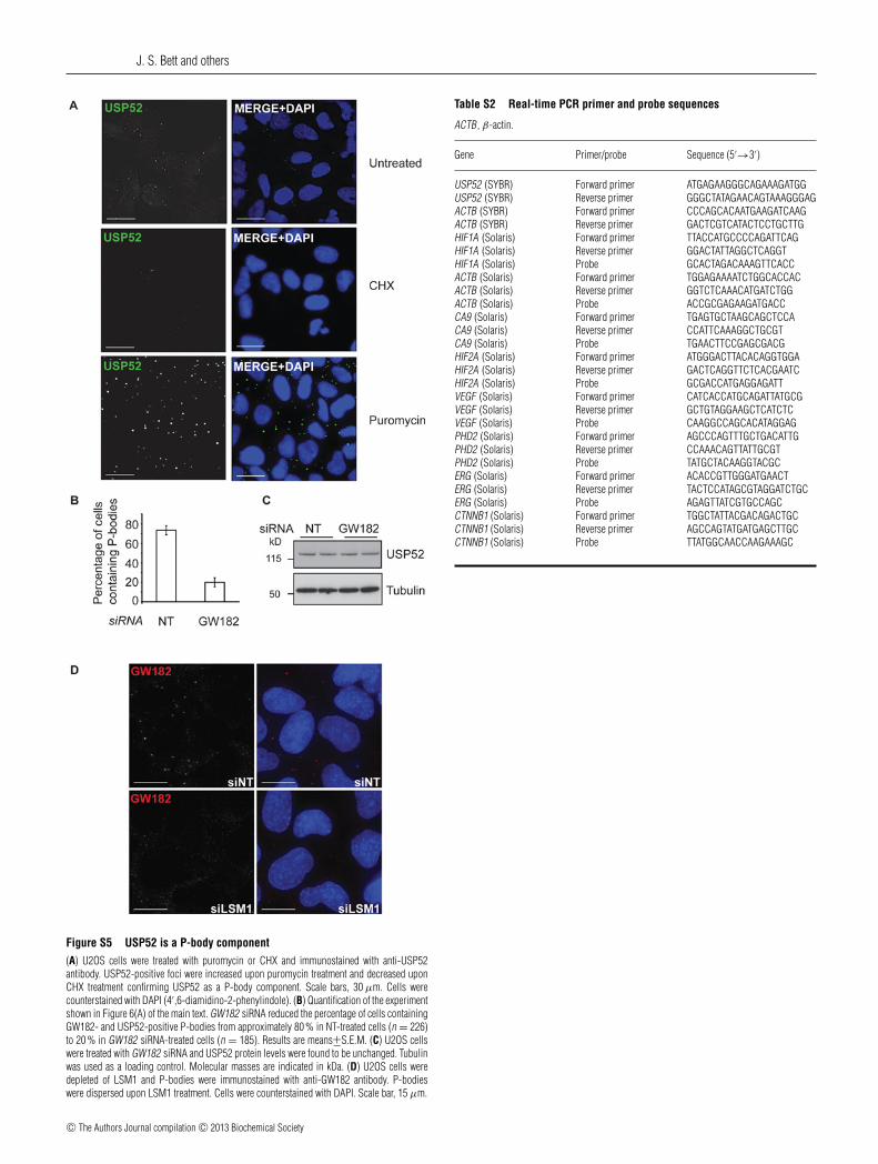
Figure S5 USP52 is a P-body component(A) U2OS cells were treated with puromycin or CHX and immunostained with anti-USP52antibody. USP52-positive foci were increased upon puromycin treatment and decreased uponCHX treatment confirming USP52 as a P-body component. Scale bars, 30 μm. Cells werecounterstained with DAPI (4′,6-diamidino-2-phenylindole). (B) Quantification of the experimentshown in Figure 6(A) of the main text. GW182 siRNA reduced the percentage of cells containingGW182- and USP52-positive P-bodies from approximately 80 % in NT-treated cells (n = 226)to 20 % in GW182 siRNA-treated cells (n = 185). Results are means+−S.E.M. (C) U2OS cellswere treated with GW182 siRNA and USP52 protein levels were found to be unchanged. Tubulinwas used as a loading control. Molecular masses are indicated in kDa. (D) U2OS cells weredepleted of LSM1 and P-bodies were immunostained with anti-GW182 antibody. P-bodieswere dispersed upon LSM1 treatment. Cells were counterstained with DAPI. Scale bar, 15 μm.
Table S2 Real-time PCR primer and probe sequences
ACTB, β-actin.
Gene Primer/probe Sequence (5′→3′)
USP52 (SYBR) Forward primer ATGAGAAGGGCAGAAAGATGGUSP52 (SYBR) Reverse primer GGGCTATAGAACAGTAAAGGGAGACTB (SYBR) Forward primer CCCAGCACAATGAAGATCAAGACTB (SYBR) Reverse primer GACTCGTCATACTCCTGCTTGHIF1A (Solaris) Forward primer TTACCATGCCCCAGATTCAGHIF1A (Solaris) Reverse primer GGACTATTAGGCTCAGGTHIF1A (Solaris) Probe GCACTAGACAAAGTTCACCACTB (Solaris) Forward primer TGGAGAAAATCTGGCACCACACTB (Solaris) Reverse primer GGTCTCAAACATGATCTGGACTB (Solaris) Probe ACCGCGAGAAGATGACCCA9 (Solaris) Forward primer TGAGTGCTAAGCAGCTCCACA9 (Solaris) Reverse primer CCATTCAAAGGCTGCGTCA9 (Solaris) Probe TGAACTTCCGAGCGACGHIF2A (Solaris) Forward primer ATGGGACTTACACAGGTGGAHIF2A (Solaris) Reverse primer GACTCAGGTTCTCACGAATCHIF2A (Solaris) Probe GCGACCATGAGGAGATTVEGF (Solaris) Forward primer CATCACCATGCAGATTATGCGVEGF (Solaris) Reverse primer GCTGTAGGAAGCTCATCTCVEGF (Solaris) Probe CAAGGCCAGCACATAGGAGPHD2 (Solaris) Forward primer AGCCCAGTTTGCTGACATTGPHD2 (Solaris) Reverse primer CCAAACAGTTATTGCGTPHD2 (Solaris) Probe TATGCTACAAGGTACGCERG (Solaris) Forward primer ACACCGTTGGGATGAACTERG (Solaris) Reverse primer TACTCCATAGCGTAGGATCTGCERG (Solaris) Probe AGAGTTATCGTGCCAGCCTNNB1 (Solaris) Forward primer TGGCTATTACGACAGACTGCCTNNB1 (Solaris) Reverse primer AGCCAGTATGATGAGCTTGCCTNNB1 (Solaris) Probe TTATGGCAACCAAGAAAGC
c© The Authors Journal compilation c© 2013 Biochemical Society

P-bodies regulate HIF1A mRNA stability
Table S3 Sequences of FISH probes
DNA probe Sequence (5′→3′)
HIF1A sense control 1 CTCACAGATGATGGTGACATGATTTACATTTCTGATAATGTGAACAAATACATGGGATTAHIF1A sense control 2 ATGGATGATGACTTCCAGTTACGTTCCTTCGATCAGTTGTCACCATTAGAAAGCAGTTCCHIF1A sense control 3 CTATGTAGTTGTGGAAGTTTATGCTAATATTGTGTAACTGATATTAAACCTAAATGTTCTHIF1A probe (antisense) 1 GAGTGTCTACTACCACTGTACTAAATGTAAAGACTATTACACTTGTTTATGTACCCTAATHIF1A probe (antisense) 2 TACCTACTACTGAAGGTCAATGCAAGGAAGCTAGTCAACAGTGGTAATCTTTCGTCAAGGHIF1A probe (antisense) 3 GATACATCAACACCTTCAAATACGATTATAACACATTGACTATAATTTGGATTTACAAGA
Table S4 List of USP52-interacting proteins identified from MS/MS analysis
Available as an Excel file at http://www.biochemj.org/bj/451/bj4510185add.htm
Received 4 January 2013/31 January 2013; accepted 11 February 2013Published as BJ Immediate Publication 11 February 2013, doi:10.1042/BJ20130026
c© The Authors Journal compilation c© 2013 Biochemical Society