the partition coefficient of gd-dtpa reflects...
TRANSCRIPT

JOURNAL OF CARDIOVASCULAR MAGNETIC RESONANCE1
Vol. 6, No. 1, pp. 33–42, 2004
VIABILITY
The Partition Coefficient of Gd-DTPA Reflects Maintained Tissue Viabilityin a Canine Model of Chronic Significant Coronary Stenosis
Katie S. Lekx,1,* Frank S. Prato,1 Jane Sykes,1 and Gerald Wisenberg2
1Department of Nuclear Medicine, Imaging Program, Lawson Health Research
Institute, and 2Division of Cardiology and The Department of Medical
Biophysics, University of Western Ontario, London, Ontario, Canada
ABSTRACT
Purpose. The underlying assumption in delayed enhancement or constant infusion
techniques to detect infarcted myocardium is that the partition coefficient (l) of Gd-
DTPA only increases in permanently damaged tissue. This assumption is supported in
canine models of stunned and infarcted myocardium but has not been adequately
tested in models of chronic, reversibly damaged tissue. Methods. A significant
coronary stenosis was maintained for 3 (n=9) or 10 (n=4) weeks in a canine model.
Myocardial perfusion was assessed using radioactively labeled microspheres, and
Doppler flow was used to monitor the effect on flow caused by the stenosis formation.
Function and in vivo l were assessed using magnetic resonance imaging (MRI) and
a constant infusion of Gd-DTPA. 201Tl and 111In-DTPA were used to assess ex vivo
myocardial viability and l, respectively. Results. Baseline Doppler-measured blood
flow through the left anterior descending coronary artery was reduced by 72.4±1.6%
(SEM) during the stenosis formation. However, shortly after creation of the stenosis
and at sacrifice, regional myocardial blood flow at rest was not decreased in the
Region at Risk (RAR) despite the persistence of the stenosis. Perfusion reserve in this
model, measured using adenosine stress, was significantly reduced. The in vivo lvalues in the RAR and remote tissue ranged between 0.32–0.45 mL/g and 0.31–0.42
mL/g, respectively. 201Tl uptake was maintained in all tissue, confirming the
maintenance of tissue viability. Global function was unchanged while regional
function was significantly depressed at 10 days but returned to baseline values by
*Correspondence: Katie S. Lekx, Department of Nuclear Medicine, St. Joseph’s Health Care, 268 Grosvenor St., London, Ontario
N6A 4V2, Canada; Fax: 519-646-6399; E-mail: [email protected].
33
DOI: 10.1081/JCMR-120027803 1097-6647 (Print); 1532-429X (Online)
Copyright D 2004 by Marcel Dekker, Inc. www.dekker.com

ORDER REPRINTS
day 21. Conclusions. This study is consistent with the hypothesis that l is not
increased in reversibly dysfunctional myocardium.
Key Words: MR imaging; Myocardial viability; Canine model; Coronary artery
disease; Significant coronary artery stenosis; Gd-DTPA; Partition Coefficient.
INTRODUCTION
Coronary artery disease (CAD) remains the largest
cause of morbidity and mortality in developed coun-
tries (Wijns et al., 1998), and the extent of left ven-
tricular dysfunction is one of the most important
prognostic determinants. The extent of functional im-
pairment varies, particularly in postmyocardial infarc-
tion patients and those with severe coronary artery
disease, and revascularization of the myocardium may
result in the recovery of function (Wijns et al., 1998).
This improvement in function does not occur, however,
if the majority of the functional impairment is secondary
to myocardial infarction (by definition irreversible) or
extensive fibrosis. Therefore, functional improvement
can only occur if there is residual viability in these
regions, with minimal fibrosis. Distinguishing different
types of reversible injury from irreversible damage is of
clinical importance in order to appropriately select pa-
tients for revascularization and to aid in determining an
individual patient’s prognosis.
Previous experiments in our laboratory have
investigated infarcted and stunned myocardium using
gadolinium diethylenetriaminepetaacetic acid (Gd-
DTPA) with magnetic resonance imaging (MRI) (Pe-
reira et al., 2000; Thornhill et al., 2001). In both cases,
a constant infusion of Gd-DTPA was used to assess the
specificity and sensitivity of differentiating injured (ei-
ther reversibly or irreversibly) myocardial tissue from
normal, healthy myocardial tissue. Pereira et al. (2000)
confirmed the sensitivity of the method in their models
of both acute and chronic myocardial infarction while
Thornhill et al. (2001) confirmed the specificity of this
method in a model of stunned myocardium. The deter-
mination of distribution volume in different forms of
reversibly injured tissue is of interest since some
authors (Kim et al., 2000; Pereira et al., 2000; Thorn-
hill et al., 2001) claim that the partition coefficient (l)
is only increased in infarcted tissue, while others
(Oshinski et al., 2001; Schwitter et al., 1997) claim that
reversibly injured tissue also produces increases in land, therefore, the specificity of MRI for determining
myocardial viability has been questioned. Our hypoth-
esis was that myocytes subtended by a significantly
stenosed coronary artery, but with impaired left ven-
tricular function, would still have the ability to exclude
Gd-DTPA and the tissue would not enhance on T1-
weighted images. Thus, the partition coefficient of Gd-
DTPA would be similar to that found in stunned and
normal myocardium. If true, this would further support
MRI, with a constant infusion of Gd-DTPA, as both a
sensitive and specific method for the determination of
tissue viability.
In this study we produced a canine model of
significant, perfusion reserve limiting stenosis and
confirmed, using measures of viability, function, and
regional myocardial blood flow, the presence of re-
versible myocardial injury and measured the distribu-
tion volume of Gd-DTPA in myocardial tissue served
by the stenosis.
METHODS
Canine Model
After several preliminary experiments, we deter-
mined that a reduction of blood flow through the left
anterior descending (LAD) coronary artery, measured
using Doppler ultrasound, to 20–30% of baseline was
the maximum degree of flow reduction possible before
permanent myocardial damage (infarction) occurred.
The change in blood flow was created using a cir-
cumferential stenosing device and measured using a
Doppler flow probe (Transonic Systems Inc., Ithaca,
NY) placed around the LAD artery immediately pro-
ximal to the stenosis. Two different approaches were
used to create a ‘‘permanent’’ coronary artery stenosis.
In four dogs, a balloon occluder was used that allowed
small incremental changes in blood flow until the de-
sired degree of stenosis was achieved. However, since
balloon occluders can deflate over time, we used a
second method in nine dogs once we had determined
the degree of stenosis required. The second stenosing
device was a self-locking, electrical tie (approximately
5mm in width) surrounded by a hollow plastic cylinder
or tubing to cushion the vessel. Selective coronary
angiography was performed either following surgery or
prior to sacrifice to validate the patency of the stenosed
artery and confirm methodology. Absolute regional
myocardial blood flow was assessed at various points
throughout the experiment using differently labeled
radioactive microspheres. The MR imaging was
34 Lekx et al.

ORDER REPRINTS
performed, using previously validated methods (Pereira
et al., 1996) (refer to Imaging Protocol section below)
to evaluate function and quantitative in vivo partition
coefficient values in the tissue at risk (Region at Risk;
RAR) and the remote tissue. The RAR tissue is defined
as the region served by the stenotic artery and with
blood flow <0.3 mL/min/g (measured by microspheres)
during a complete transient occlusion (45 sec). Ex vivo
tissue partition coefficient and viability were measured
using 111In-DTPA and 201Tl, respectively, and com-
pared to in vivo results, as previously reported (Pereira
et al., 1996; Pereira et al., 2000; Thornhill et al., 2001).
Animal Preparation and Surgery
All studies were in accordance with the University
of Western Ontario Council on Animal Care guide-
lines. The results from 13 female dogs (three beagles,
10 mongrels) are reported in this study. Nine animals
were studied for 3 weeks and four animals were
studied for 10 weeks. Anesthesia was induced with
Propofol 1% intravenously and then maintained using
2–2.5% isoflurane after endotracheal intubation. This
was performed in preparation for surgery and all
subsequent follow-up imaging sessions. During sur-
gery, a left thoracotomy was performed to expose the
heart and two small sections, each of approximately
0.8cm in length, of the LAD were dissected free of
the heart wall. The Doppler flow probe (proximal) and
stenosing device (distal) were placed in these two
regions, with the flow probe placed at least three vessel
diameters proximal to the stenosing device. A femoral
artery catheter was inserted for reference blood
withdrawal during microsphere injections. Regional
myocardial blood flow measurements were acquired at
baseline, during the transient occlusion to determine
the RAR, 15 minutes after stenosis formation, and just
prior to sacrifice using four differently labeled, ra-
dioactive microspheres. Reference blood was with-
drawn beginning one minute prior to the injection of
either 141Ce-, 85Sr-, 46Sc-, or 95Nb-labeled microspheres
(Perkin–Elmer, Boston, MA) directly into the left
atrium, and continued for another 4 minutes at a rate of
1.94 mL/min. Coronary angiography was performed in
eight dogs either following surgery or prior to sacrifice to
validate the patency of the stenosed artery and confirm
methodology (Fig. 1).
Two additional canines were acutely instrumented
in the same fashion to assess perfusion reserve in our
model of significant coronary artery stenosis. Adeno-
sine stress was induced before and after stenosis
formation using 0.14 mg/kg/min infusion maintained
for 8 minutes. Radioactively labeled microspheres were
injected at baseline, for RAR measurement, and 4 min
into each adenosine infusion. 201Tl and 111In-DTPA
were administered in the same manner as in the other
animals to determine myocardial viability and distri-
bution volume of Gd-DTPA. Animals were sacrificed
immediately following the termination of the Gd-
DTPA/111In-DTPA 1 hr constant infusion, approximate-
ly 4 hours from the start of the experiment.
Imaging Protocol
Imaging was performed prior to surgery [Baseline
(B), n=13], at 2–3 days (F1, n=13) and at 7 or 10 days
(F2, n=13) following surgery, and weekly thereafter
(F3 – F10, no experiments at week 6) until the
completion of the experiment at either 3 or 10 weeks.
All imaging was performed on a Siemens Vision 1.5 T
clinical system (Siemens, Erlangen, Germany), and the
same imaging protocol was used for all imaging
sessions. A schematic of this protocol is shown in
Fig. 2. Animals were placed prone in a head coil, and
the heart centered within the coil. Scout images were
acquired, followed by precontrast saturation recovery
turboFLASH (srTFL) images (TR/TE 2.4/1.2 ms, 8 mm
slice thickness, 13� flip, FOV 300 mm) throughout the
left ventricle. A 0.2 mmol/kg/min bolus of Gd-DTPA
was injected at a rate of 46 mL/min, followed by a
1 hr constant infusion of 0.004 mmol/kg/min. Short
axis, long axis, and four chamber cine images (TR/TE
Figure 1. X-ray angiogram in one animal after placement of
Transonic flow probe and stenosing device. Hatched arrow
indicates flow probe and white arrow indicates position of
stenosing device around the LAD coronary artery. Stenosis
degree in this animal was 76.2%; blood flow reduction in
artery measured by flow probe was 71.7%. X-ray angiography
was used to validate methodology and confirm creation of
the stenosis.
Gd-DTPA and Viability of Coronary Stenosis 35
ORDER REPRINTS
10/4.8 ms, 8 mm slice thickness, 30� flip, FOV 254–
300 mm) were obtained to visualize contractile func-
tion for qualitative and quantitative analysis. The srTFL
images were again acquired before the completion of
the Gd-DTPA constant infusion for determination of in
vivo partition coefficient (see Image and Data Analysis
section below).
Low- and mid-dose dobutamine (5 mmol/kg/min
and 10 mmol/kg/min, respectively) infusions were
performed for 5 minutes each. Cine images were
acquired at two slice positions in the last 2 minutes of
each infusion to assess the inotropic reserve of
myocardium subtended by the chronically stenosed
coronary artery.
On the day of sacrifice, 201Tl (DuPont Canada,
Markham, Ontario) was injected approximately 2.5 hr
prior to sacrifice to assess tissue viability. The imaging
protocol at sacrifice remained the same, but trace
amounts of 111In-DTPA (Frosst Radiopharmaceuticals,
Kirkland, Quebec, Canada) were added to the Gd-
DTPA constant infusion and maintained until sacrifice
to assess the distribution volume of DTPA chelates
(i.e., measure partition coefficient) ex vivo. Just prior
to sacrifice another measure of regional myocardial
blood flow was obtained using microspheres.
Potassium chloride was injected to sacrifice the
animal and the heart quickly excised and imaged using
a high-resolution T1-weighted 3D FLASH sequence
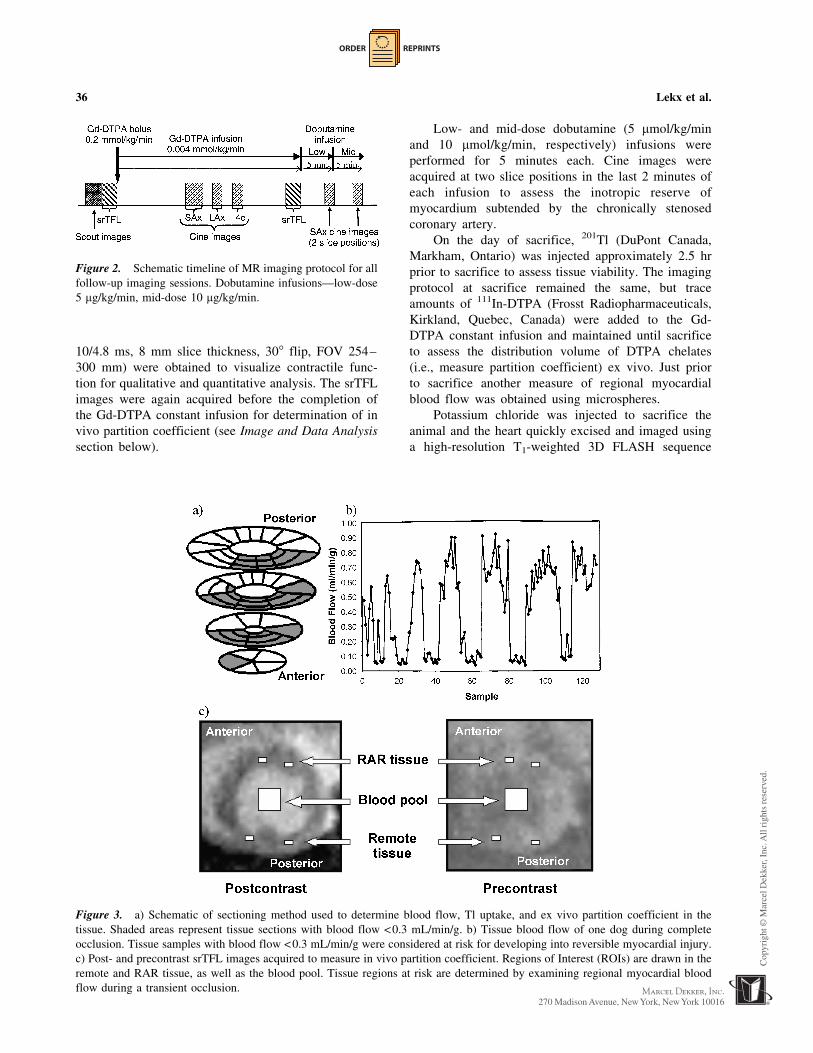
Figure 3. a) Schematic of sectioning method used to determine blood flow, Tl uptake, and ex vivo partition coefficient in the
tissue. Shaded areas represent tissue sections with blood flow <0.3 mL/min/g. b) Tissue blood flow of one dog during complete
occlusion. Tissue samples with blood flow <0.3 mL/min/g were considered at risk for developing into reversible myocardial injury.
c) Post- and precontrast srTFL images acquired to measure in vivo partition coefficient. Regions of Interest (ROIs) are drawn in the
remote and RAR tissue, as well as the blood pool. Tissue regions at risk are determined by examining regional myocardial blood
flow during a transient occlusion.
Figure 2. Schematic timeline of MR imaging protocol for all
follow-up imaging sessions. Dobutamine infusions—low-dose
5 mg/kg/min, mid-dose 10 mg/kg/min.
36 Lekx et al.

ORDER REPRINTS
(TR/TE 22/10 msec, 1 mm slice thickness, 40� flip, in-
plane resolution 0.5 mm) to map the distribution of
Gd-DTPA. The heart was then sliced into five to seven
slices and visually inspected for signs of necrosis. The
slices were then sectioned into 75–130 pieces weigh-
ing 0.2–1.2 g each. These tissue samples were counted
for radioactivity to determine myocardial blood flow
by microspheres, tissue viability by 201Tl uptake, and
ex vivo partition coefficient of Gd-DTPA by 111In-
DTPA (Pereira et al., 1996). Fig. 3a shows an example
of how the heart is sectioned and Fig. 3b is an example
of blood flow during the 45 sec occlusion that deter-
mines the RAR. This information is then used to assess
the tissue viability, blood flow, and partition coefficient
in the RAR and remote tissue.
Image and Data Analysis
All data were analyzed after sacrifice, and the
RAR tissue had been determined to ensure accurate
analysis of RAR and remote tissue parameters. In vivo
partition coefficient values were determined from the
pre- and postcontrast srTFL images. Regions of Interest
(ROIs) were drawn in the remote and RAR tissue (see
Fig. 3c). The change in signal intensity (DSI) from
post- to precontrast in both the tissue regions and the
blood pool was determined using AnalyzeAVW (Robb,
1990) and in vivo partition coefficient was thus
determined as (Pereira et al., 1999):
l ¼ DSItissue
DSIbloodð1Þ
Quantitative wall motion was assessed by measur-
ing left ventricular ejection fraction (LVEF) using
Siemens-based software, ARGUS, which is a semi-
quantitative segmentation program that draws contours
around the blood pool of the end-diastolic and end-
systolic images of each slice and calculates LVEF as:
LVEF ¼ EDV � ESV
EDV� 100 ð2Þ
Qualitative wall motion analysis was performed on
all cine MR images throughout the left ventricle at
every time-point in each animal, including after low-
and mid-dose dobutamine infusions. Each short axis
cine at each slice position was assessed for wall motion
in six different regions: septal, anteroseptal, anterolat-
eral, lateral, inferolateral, and inferoseptal. An experi-
enced cardiologist (GW), blind to the cardiac states of
each wall motion study, scored each of these regions
on a scale from 0–6. The scoring was as follows: 0—
hyperkinetic, 1—normal, 2—mildly hypokinetic, 3—
moderately hypokinetic, 4—severely hypokinetic, 5—
akinetic, and 6—dyskinetic.
Average blood flow, ex vivo partition coefficient,
and normalized Tl uptake values were calculated in
both the RAR tissue and remote tissue. In vivo par-
tition coefficient values were calculated by placing
ROIs (method described above) in the regions that
corresponded to tissue sections with reduced blood
flow during the 45 sec occlusion, and therefore at risk
of forming reversible myocardial injury.
Perfusion Reserve Determination
Perfusion reserve during adenosine stress was
determined in two separate animal experiments as
noted above in Animal Preparation and Surgery.
Perfusion reserve was calculated as average regional
myocardial blood flow in the RAR and remote tissue
during adenosine stress normalized by regional myo-
cardial blood flow at rest in the same tissue regions.
Statistical Analysis
All result values are expressed as mean value plus
or minus standard error of the mean (SEM). Analysis
of Variance (ANOVA) was performed on the blood
flow, in vivo and ex vivo partition coefficient, nor-
malized Tl uptake, and quantitative functional data to
determine the relationship between the RAR and
remote tissue regions of these parameters. If the
ANOVA revealed significance, further investigation
was performed using Tukey posthoc analysis. Levene’s
statistic was computed to determine whether homoge-
neity of variance was present or not. If not, inhomog-
eneous data sets were square-root transformed and
analyzed in the same manner as listed above. If homo-
geneity of variance was still not present, nonparametric
statistical analysis was performed and compared to
results obtained using parametric tests. Repeated mea-
sures analysis was performed on the qualitative wall
motion data to determine if a relationship existed
between the RAR and remote tissue regions at the
various time-points studied. Nonparametric analysis
was performed to confirm parametric results since the
wall motion scoring was a subjective analysis. An
alpha value of P<0.05 was considered significant.
RESULTS
The acute functional effects of the coronary
stenoses averaged 72.4±1.6% (n=9) below baseline
values, determined by the flow change through the
LAD using the flow probe. Luminal diameter reduction
using x-ray angiography revealed an approximate
stenosis degree of 75–80% (all dogs). Regional
Gd-DTPA and Viability of Coronary Stenosis 37

ORDER REPRINTS
myocardial blood flow results, determined by radioac-
tive microspheres, are shown in Fig. 4. All blood flow
measurements were taken at rest under general anes-
thetic. Baseline blood flow was 0.64 mL/min/g in the
RAR and 0.74 mL/min/g in the remote tissue. Blood
flow in both the RAR and remote tissue was in-
significantly increased (0.67 mL/min/g and 0.92 mL/
min/g, respectively; P=NS) 15 min following the
stenosis formation compared to baseline. At sacrifice,
blood flow was again insignificantly increased in both
the RAR (0.85 mL/min/g; P=NS) and remote tissue
(0.90 mL/min/g; P=NS), despite maintained stenosis
formation. Although blood flow tended to be higher in
the remote tissue compared to the RAR tissue at all
time-points measured, there was no significant differ-
ence in the flow to these regions at any time.
The normalized Tl uptake results from 11 animals
and ex vivo partition coefficient results from all 13
animals are shown in Table 1, as well as the mean and
SEM values. In two animals, 201Tl was not available
at the time of sacrifice. A minor 3% significant re-
duction was noted in the normalized Tl uptake values
in the RAR vs. remote tissue (P<0.05). However, the
Tl uptake values were within the range of normal tis-
sue in both the RAR and remote tissue. No significant
difference in ex vivo l was noted between the two
tissue regions.
All in vivo partition coefficient results for the
duration of the experiment in all dogs are shown in
Fig. 5. Average values ranged between 0.31–0.42 mL/g
in remote tissue and 0.32–0.45 mL/g in RAR tissue.
The partition coefficient of the remote tissue was sig-
nificantly lower than that of the RAR tissue at day 14
(0.34 mL/g vs. 0.39 mL/g; P<0.05). No other significant
differences were found either within or between the
remote and RAR tissue groups. All values were below
0.7 mL/g, the upper limit for normal l. Therefore, all lvalues were within the range of normal tissue.
Additionally, no signs of infarction were noted during
the visual inspection during sectioning of the heart.
Figure 4. Regional myocardial blood flow in the RAR tissue
(&) and in the remote tissue (.) at baseline, 15 min after
stenosis formation, and just prior to sacrifice. Error bars
represent SEM. No significant differences were noted
either between the tissue regions or over the three differ-
ent measurements.
Figure 5. In vivo partition coefficient results in remote
tissue and tissue at risk prior to surgery (B) and at the follow-
ups after surgery (F1–F10). (Numbers) indicate ‘‘n’’ at each
imaging session. Error bars indicate the standard error of the
mean. Significance (*) between the RAR and remote tissue
was noted at F3 (Day 14). No significant differences within the
tissue groups were found.
Table 1. Normalized Tl uptake and ex vivo l results in both
the remote and RAR tissue in all 13 dogs.
TI Uptake* Ex vivo l
DOG REM RAR REM RAR
1 0.99 0.95 0.21 0.18
2 0.97 0.89 0.45 0.31
3 0.98 0.90 0.33 0.33
4 0.96 0.95 0.34 0.42
5 0.96 0.94 0.31 0.32
6 0.94 0.94 0.33 0.49
7 0.94 0.92 0.34 0.32
8 0.94 0.95 0.49 0.38
9 0.95 0.92 0.28 0.28
10 N/A N/A 0.33 0.34
11 N/A N/A 0.29 0.29
12 0.95 0.96 0.39 0.39
13 0.97 0.91 0.34 0.34
AVG 0.96 0.93 0.34 0.34
SEM 0.005 0.007 0.020 0.021
Also shown are the average values and SEM. A significant (*)
difference was noted between the remote and RAR Tl uptake.
Tl uptake and l were within the range of normal tissue in both
the RAR and remote tissue, and all tissue regions were
considered viable based on the Tl uptake values.
38 Lekx et al.

ORDER REPRINTS
Left ventricular ejection fraction (LVEF) results
from all dogs are shown in Fig. 6. Average ejection
fraction values ranged from 30.8% to 41.7%. No sig-
nificant differences were noted at any time-point. Nor-
malization of global ejection fraction results to heart
rate revealed similar results as without normalization,
although there did appear to be a tendency for
normalized ejection fraction to be slightly reduced up
to one week following stenosis formation.
Qualitative regional wall motion results in all 13
animals at rest and during stress up to Follow-up 4 (F4)
(21 days) are shown in Figs. 7 and 8, respectively. Wall
motion in the RAR in the four animals studied up to 10
weeks was not different from the wall motion seen at
Follow-up 4. The RAR tissue results from two slice
positions are shown in Fig. 7a, and the remote tissue
results from the same two slice positions are shown in
Fig. 7b. Wall motion in the RAR tissue was signifi-
cantly decreased at F2 (10 days), with higher wall
motion score following stenosis formation (P<0.05),
but had returned to baseline values by either 3 (n=9) or
10 (n=4) weeks. In the remote tissue, significant
changes in regional function were not observed. Wall
motion in all remote regions was similar and had a wall
thickening score of approximately 1 (normal wall mo-
tion). Of the 13 animals studied, 10 (76%) demonstrated
an evident regional reduction in wall motion score at F2
that returned to baseline by F4.
Wall motion results during dobutamine stress are
shown in Fig. 8 from one slice position in the RAR (a),
and remote (b) tissue regions. Image quality from the
most apical slice was poor due to increased motion
artefact and hence could not be reliably analyzed.
Statistical analysis was performed only on those animals
Figure 6. Left ventricular ejection fraction (%) results in all
dogs at rest at Baseline (B) and Follow-ups 1–10 (F1–F10).
Error bars represent SEM. LVEF did not change over either the
3-week or 10-week protocols.
Figure 8. Wall motion scores during dobutamine stimulation
in one slice position in the (a) RAR, and (b) remote tissue
regions. Dashed line, At rest; Solid symbol, 5 min low-dose
dobutamine; Open symbol, 5 min mid-dose dobutamine.
Figure 7. Wall motion scores at rest throughout the
experiment in the (a) RAR and (b) remote tissue. a) RAR
tissue; significant decrease in wall thickening score at Day 10
compared to baseline. b) Remote tissue; no significant change
in wall thickening score. Key: RAR tissue: &, mid-apical slice,
5, mid-basal slice; Remote tissue: ., mid-apical slice 6, mid-
basal slice. * indicates significance at Day 10 (F2) in two
different slice positions (P<0.05).
Gd-DTPA and Viability of Coronary Stenosis 39

ORDER REPRINTS
with complete dobutamine image sets. No significant
differences between time and dobutamine dose were
noted (7/13 dogs, slice 1 and 4/13 dogs, slice 2). In both
the RAR and remote tissue regions, low-dose and mid-
dose dobutamine increased function at each time-point.
As expected, low-dose dobutamine increased function to
a lesser degree than mid-dose dobutamine.
Perfusion reserve results are shown in Fig. 9.
Perfusion reserve was attenuated in the RAR tissue
after stenosis formation compared to the same tissue
region prior to stenosis (1.20 mL/min/g vs. 1.89 mL/
min/g; P<0.001). Perfusion reserve was comparable in
the remote tissue after stenosis formation compared to
prior to stenosis (1.99 vs. 1.90 mL/min/g; P=NS).
DISCUSSION
Since 201Tl uptake and both the in vivo and ex
vivo partition coefficient values were within the normal
range, with no evidence of myocardial necrosis upon
visual inspection, increases in partition coefficient are
demonstrated, in this study, to be specific for the
detection of infarcted myocardium. Our group (Thorn-
hill et al., 2001) and others (Kim et al., 2000) have
previously shown this to be a sensitive method for
detecting myocardial necrosis.
Despite a significant stenosis maintained through-
out both the 3-week and 10-week experiments, regional
myocardial blood flow at rest, measured with micro-
spheres, was not reduced, in contrast to previous
reports using a similar model in swine (Liedtke et al.,
1995). However, when we attempted to increase the
stenosis degree to greater than 75–80%, in the quest to
reduce resting blood flow, infarction resulted. In fact,
resting blood flow in this study was actually slightly
increased (although insignificantly) in both the tissue at
risk and remote tissue following stenosis and at
sacrifice. Kudej et al. (1998) also noted a gradual
increase in coronary artery blood flow in pigs after
formation of stenosis with a hydraulic occluder and
readjusted the occluder to match the desired blood flow
reduction originally achieved, which resulted in my-
ocardial infarction. The increase in blood flow ob-
served in our current experiments could either be a
transient compensatory mechanism (acutely) or an in-
crease in collateral recruitment (acutely) or develop-
ment (chronically). Our experiments have shown that a
significant but noncritical (flow limiting) coronary ar-
tery stenosis in a single vessel does not lead to a
progressive reduction in resting blood flow or contrac-
tile function, and would not appear to produce the
clinical phenomenon of hibernating myocardium in this
canine model.
Some groups (Gerber et al., 1996; Vanoverschelde
et al., 1993) have reported that hibernating myocardi-
um is a by-product of repeated intermittent episodes of
stress or demand-provoked ischemia associated with
stunning, with normal resting flow initially, rather than
being caused by a sustained decrease in perfusion.
Each episode of stunning, with transient left ventricular
dysfunction, ultimately culminates in persistent chronic
dysfunction or hibernating myocardium. In this study,
however, regional dysfunction only occurred at 10 days
and did not persist throughout the experiment, despite
repeated sessions of pharmacological stress by dobuta-
mine and the fact that the animals’ daily activity,
including run exercise, was not restricted. The acute
studies demonstrated that perfusion reserve was re-
duced at the time of surgery, but we could not obtain
similar information during the course of the 3- or 10-
week studies. It may therefore be possible that per-
fusion reserve was only restricted at the time of surgery
and normalized over time, and may explain why hi-
bernating myocardium was not produced. Further
experiments will monitor perfusion reserve over the
entire 3-week protocol, using the same model. Of
importance to note, however, is that in the 10-week
experiments, x-ray angiography was performed both
early and late, and no significant change in stenosis
degree was noted over that time frame.
Canty and Fallavollita (1999, 2001) stress the
importance of studying models of noncritical but flow
Figure 9. Perfusion reserve in two additional dogs before
and 30 min after stenosis formation in both the RAR and
remote tissue is calculated as blood flow during stress divided
by blood flow at rest. Infusing 0.14 mg/kg/min adenosine for
8 minutes induced stress, and blood flow was measured by
injecting radioactively labeled microspheres 4 min into
infusion. Perfusion reserve was significantly (*; P<0.001)
attenuated in the tissue distal to the stenosis (RAR tissue)
compared to remote tissue following stenosis formation.
40 Lekx et al.

ORDER REPRINTS
reserve limiting stenosis, creating hibernating myocar-
dium, for longer periods of time, possibly up to 3
months, before tissue with reduced blood flow and
function will develop. In our experiments, with a
maintained significant coronary stenosis, similar in
degree to the percentage stenosis reported by these
investigators, neither resting myocardial blood flow nor
contractile function was reduced compared to baseline
after 10 weeks. We conclude that chronic hibernating
myocardium does not develop within up to ten weeks
of chronic significant coronary artery stenosis in this
model. At this point, we are unsure why dysfunction
developed only at 10 days and did not persist to 3 or
10 weeks. The 45sec, transient occlusion was not
sufficient to cause a reduction in wall motion. Ad-
ditionally, this transient occlusion was performed at the
conclusion of the experiment in the 10-week experi-
ments, rather than at the beginning of experiments as in
the 3-week protocol, and wall motion reduction was
still noted. Possibly the manipulation of the coronary
artery during surgery might explain why the reduction
in function occurs, although not on a delayed basis.
However, this is only speculative and does not explain
the reduction in wall motion at only one time-point.
The quest for a sensitive and specific measure of
myocardial viability is extremely important (Thornhill
et al., 2003), since restoration of blood flow could
restore function to dysfunctional regions. The sensitivity
of contrast-enhanced MRI is well-known; however, the
specificity has been challenged by delayed enhancement
experiments performed in rats that argue that infarct
size is overestimated due to a transient increase in
partition coefficient in reversibly damaged tissue
surrounding the infarct zone (Oshinski et al., 2001;
Saeed et al., 2001). Our use of a constant infusion
technique, as opposed to delayed enhancement, elim-
inates any altered washout effects, since this technique
eliminates the effect of flow on enhancement (Tong
et al., 1993). The results found in this study indicate
that partition coefficient is not increased in reversibly
damaged myocardium, as manifest by the transient
reduction in contractility, caused by a significant
coronary artery stenosis, which further implies that
MRI is a specific indicator of myocardial viability.
However, further studies of other types of reversible
damage, especially hibernating myocardium, will have
to be done to confirm the specificity of this method.
This study reports that in the case of chronic
coronary artery stenosis, partition coefficient values
remain within the range of normal tissue. Although
hibernating myocardium was not created, as per the
original goal of this project, the results obtained give
additional insight into the mechanisms leading/not
leading to hibernating myocardium and, in fact, do
not support the notion that it occurs secondary to
repetitive stunning. Also, given that 201Tl uptake was
normal in both the tissue at risk and the remote tissue,
the experiments provide further evidence that the
measurement of the partition coefficient of Gd-DTPA
using MRI is both a sensitive and specific indicator of
tissue viability.
ABBREVIATIONS
srTFL saturation recovery turboFLASH
SAx short axis
LAx long axis
4c 4 chamber
ACKNOWLEDGMENTS
The authors would like to thank Jenny Gibbons
and Lela Noonan for animal and experiment assistance;
Dick J. Drost for technical assistance using the
Siemens MR unit; Berlex Canada for providing Gd-
DTPA (Magnevistk formulation); Yves Bureau for
statistical analysis assistance; and the Canadian Insti-
tutes for Health Research, the Ontario government, and
the Natural Sciences and Engineering Research Council
of Canada for financial support.
REFERENCES
Canty, J. M. Jr., Fallavollita, J. A. (1999). Resting
myocardial flow in hibernating myocardium:
validating animal models of human pathophysiol-
ogy. Am J. Physiol. 277(1 Pt 2):H417–H422.
Review.
Canty, J. M., Fallavollita, J. A. (2001). Lessons from
experimental models of hibernating myocardium.
Coron. Artery Dis. 12:371–380.
Gerber, B. L., Vanoverschelde, J.-L., Bol, A., Michel,
C., Labar, D., Wijns, W., Melin, J. A. (1996).
Myocardial blood flow, glucose uptake, and
recruitment of inotropic reserve in chronic left
ventricular ischemic dysfunction. Implications for
the pathophysiology of chronic myocardial hiber-
nation. Circulation 94:651–659.
Kim, R. J., Wu, E., Rafael, A., Chen, E.-L., Parker, M.
A., Simonetti, O., Klocke, F. J., Bonow, R. O.,
Judd, R. M. (2000). The use of contrast-enhanced
magnetic resonance imaging to identify reversible
Gd-DTPA and Viability of Coronary Stenosis 41

ORDER REPRINTS
myocardial dysfunction. N. Engl. J. Med. 343:
144–153.
Kudej, R. K., Ghaleh, B., Sato, N., Shen, Y.-T.,
Bishop, S. P., Vatner, S. F. (1998). Ineffective
perfusion-contraction matching in conscious,
chronically instrumented pigs with an extended
period of coronary stenosis. Circ. Res. 82:1199–
1205.
Liedtke, A. J., Renstrom, B., Nellis, S., Hall, J. L.,
Stanley, W. C. (1995). Mechanical and metabolic
function in pig hearts after 4 days of chronic
coronary stenosis. JACC 26(3):815–825.
Oshinski, J. N., Yang, Z., Jones, J. R., Mata, J. F.,
French, B. A. (2001). Imaging time after Gd-
DTPA injection is significant in using delayed
enhancement to determine infarct size accurately
with magnetic resonance imaging. Circulation
104:2838–2842.
Pereira, R. S., Prato, F. S., Wisenberg, G., Sykes, J.
(1996). The determination of myocardial viability
using Gd-DTPA in a canine model of acute
myocardial ischemia and reperfusion. Magn.
Reson. Med. 36:684–693.
Pereira, R. S., Prato, F. S., Sykes, J., Wisenberg, G.
(1999). Assessment of myocardial viability using
MRI during constant infusion of Gd-DTPA: fur-
ther studies at early and later periods of reperfu-
sion. Magn. Reson. Med. 42:60–68.
Pereira, R. S., Prato, F. S., Lekx, K. S., Sykes, J.,
Wisenberg, G. (2000). Contrast-enhanced MRI
for the assessment of myocardial viability after
permanent coronary artery occlusion. Magn.
Reson. Med. 44:309–316.
Robb, R. A. (1990). A software system for interactive
and quantitative analysis of biomedical images. In:
Høhne, K. H., Fuchs, H., Pizer, S. M., eds. 3D
Imaging in Medicine, NATO ASI Series. Berlin:
Springer-Verlag, pp. 333–361.
Saeed, M., Lund, G., Wendland, M. F., Bremerich, J.,
Weinmann, H.-J., Higgins, C. B. (2001). Mag-
netic resonance characterization of the peri-
infarction zone of reperfused myocardial infarc-
tion with necrosis-specific and extracellular
nonspecific contrast media. Circulation 103:
871–876.
Schwitter, J., Saeed, M., Wendland, M. F., Derugin, N.,
Canet, E., Brasch, R. C., Higgins, C. B. (1997).
Influence of severity of myocardial injury on
distribution of macromolecules: extravascular
versus intravascular gadolinium-based magnetic
resonance contrast agents. JACC 30(4):1086–
1094.
Thornhill, R. E., Prato, F. S., Pereira, R. S., Wisenberg,
G., Sykes, J. (2001). Examining a canine model of
stunned myocardium using Gd-DTPA-enhanced
MRI. Magn. Reson. Med. 45(5):864–871.
Thornhill, R. E., Prato, F. S., Wisenberg, G. (2003). The
assessment of myocardial viability: a review of
current diagnostic imaging approaches. JCMR
4(3):381–410.
Tong, C. Y., Prato, F. S., Wisenberg, G., Lee, T. Y.,
Carroll, E., Sandler, D., Wills, J. (1993). Techni-
ques for the measurement of the local myocardial
extraction efficiency for inert diffusible contrast
agents such as gadopentate dimeglumine. Magn.
Reson. Med. 30:332–336.
Vanoverschelde, J.-L., Wijns, W., Depre, C., Essamri,
B., Heyndrickx, G. R., Borgers, M., Bol, A.,
Melin, J. A. (1993). Mechanisms of chronic re-
gional post-ischemic dysfunction in humans: new
insights from the study of noninfarcted collateral-
dependent myocardium. Circulation 87:1513–
1523.
Wijns, W., Vatner, S. F., Camici, P. G. (1998).
Mechanisms of disease: hibernating myocardium.
New Engl. J. Med. 339(3):173–181.
Received July 9, 2002
Accepted July 13, 2003
42 Lekx et al.

Request Permission/Order Reprints
Reprints of this article can also be ordered at
http://www.dekker.com/servlet/product/DOI/101081JCMR120027803
Request Permission or Order Reprints Instantly!
Interested in copying and sharing this article? In most cases, U.S. Copyright Law requires that you get permission from the article’s rightsholder before using copyrighted content.
All information and materials found in this article, including but not limited to text, trademarks, patents, logos, graphics and images (the "Materials"), are the copyrighted works and other forms of intellectual property of Marcel Dekker, Inc., or its licensors. All rights not expressly granted are reserved.
Get permission to lawfully reproduce and distribute the Materials or order reprints quickly and painlessly. Simply click on the "Request Permission/ Order Reprints" link below and follow the instructions. Visit the U.S. Copyright Office for information on Fair Use limitations of U.S. copyright law. Please refer to The Association of American Publishers’ (AAP) website for guidelines on Fair Use in the Classroom.
The Materials are for your personal use only and cannot be reformatted, reposted, resold or distributed by electronic means or otherwise without permission from Marcel Dekker, Inc. Marcel Dekker, Inc. grants you the limited right to display the Materials only on your personal computer or personal wireless device, and to copy and download single copies of such Materials provided that any copyright, trademark or other notice appearing on such Materials is also retained by, displayed, copied or downloaded as part of the Materials and is not removed or obscured, and provided you do not edit, modify, alter or enhance the Materials. Please refer to our Website User Agreement for more details.