the pathomechanisms of periodontal disease
TRANSCRIPT

Gingivitis and periodontitis are inflammatory diseases developing due to the protection and fight against plaque bacteria
The pathomechanisms of periodontal disease
The pathomechanisms of periodontal disease
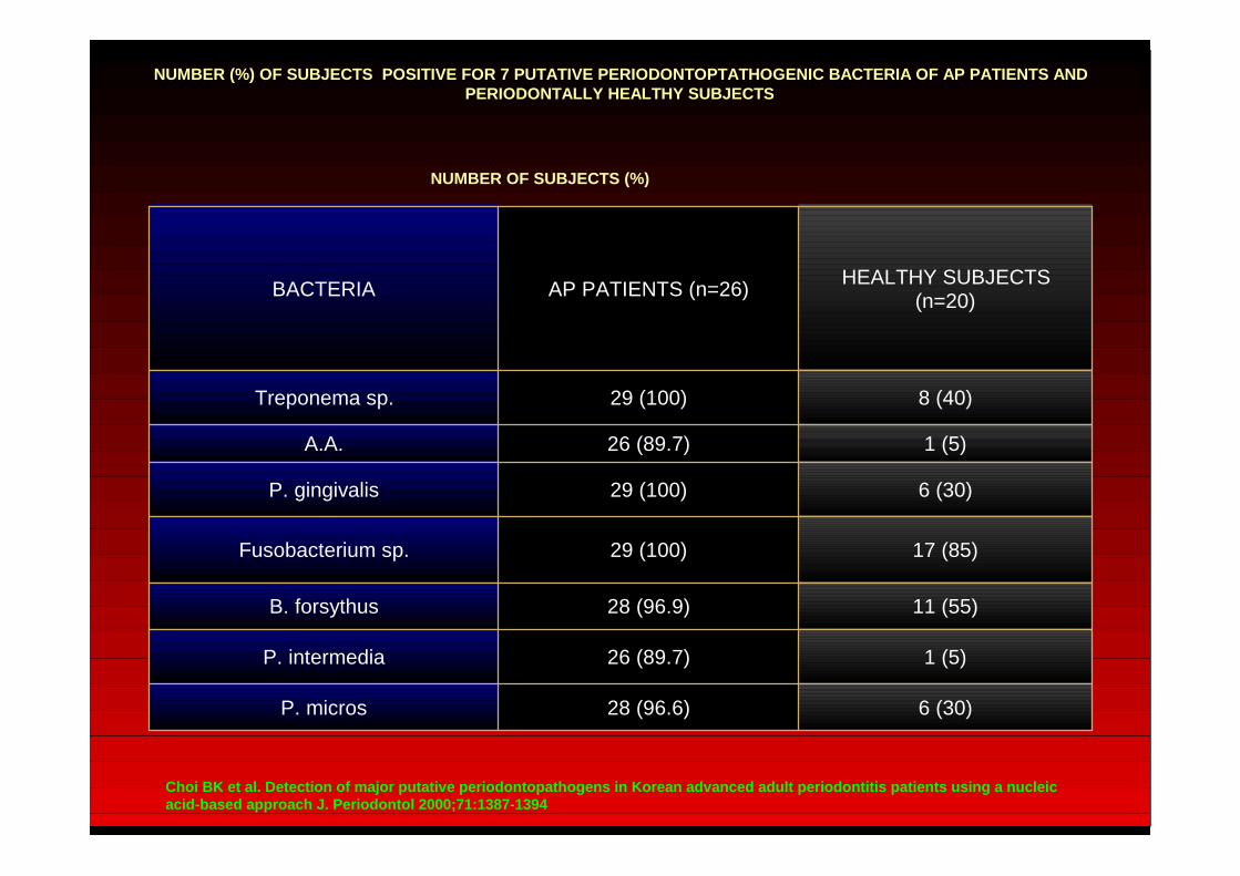
NUMBER (%) OF SUBJECTS POSITIVE FOR 7 PUTATIVE PER IODONTOPTATHOGENIC BACTERIA OF AP PATIENTS AND PERIODONTALLY HEALTHY SUBJECTS
Choi BK et al. Detection of major putative periodon topathogens in Korean advanced adult periodontitis patients using a nucleic acid-based approach J. Periodontol 2000;71:1387-139 4
BACTERIA AP PATIENTS (n=26)HEALTHY SUBJECTS
(n=20)
Treponema sp. 29 (100) 8 (40)
A.A. 26 (89.7) 1 (5)
P. gingivalis 29 (100) 6 (30)
Fusobacterium sp. 29 (100) 17 (85)
B. forsythus 28 (96.9) 11 (55)
P. intermedia 26 (89.7) 1 (5)
P. micros 28 (96.6) 6 (30)
NUMBER OF SUBJECTS (%)

Host defense processes responsible for tissue destructions
Host defense processes responsible for tissue destructions
Bacterial plaque is necessary but not sufficient for destructive periodontitis
Destructive periodontitis occurs in a small percentage of adult population
Week correlation between dental plaque and periodontal tissue destruction
Tween studies proved that genetic factors can be responsible for about half of the clinical manifestation of periodontitis
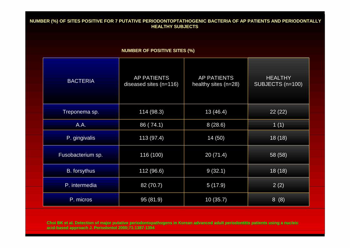
NUMBER (%) OF SITES POSITIVE FOR 7 PUTATIVE PERIODONTOPTATHOGENIC BACTERIA OF AP PATIENTS AND PERIODON TALLY HEALTHY SUBJECTS
Choi BK et al. Detection of major putative periodon topathogens in Korean advanced adult periodontitis patients using a nucleic acid-based approach J. Periodontol 2000;71:1387-139 4
BACTERIAAP PATIENTS
diseased sites (n=116)AP PATIENTS
healthy sites (n=28)HEALTHY
SUBJECTS (n=100)
Treponema sp. 114 (98.3) 13 (46.4) 22 (22)
A.A. 86 ( 74.1) 8 (28.6) 1 (1)
P. gingivalis 113 (97.4) 14 (50) 18 (18)
Fusobacterium sp. 116 (100) 20 (71.4) 58 (58)
B. forsythus 112 (96.6) 9 (32.1) 18 (18)
P. intermedia 82 (70.7) 5 (17.9) 2 (2)
P. micros 95 (81.9) 10 (35.7) 8 (8)
NUMBER OF POSITIVE SITES (%)

Theoretically the absolutely healhty gingivia histologically shows no inflammatory reaction at all This can only be achieved by experimentally clean and plaque free circumstancies
The histomorphometry of the biopsies from this "super healthy" pristine gingiva shows 40% epithelial cells and 60% connective tissue.
Healthy gingivia Healthy gingivia
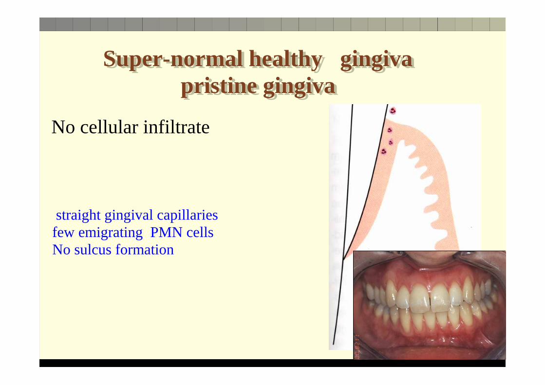
Super-normal healthy gingiva pristine gingiva
Super-normal healthy gingiva pristine gingiva
No cellular infiltrate
straight gingival capillaries few emigrating PMN cells No sulcus formation
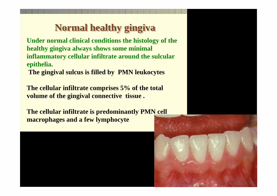
Normal healthy gingiva Normal healthy gingiva Under normal clinical conditions the histology of the healthy gingiva always shows some minimal inflammatory cellular infiltrate around the sulcula r epithelia. The gingival sulcus is filled by PMN leukocytes
The cellular infiltrate comprises 5% of the total volume of the gingival connective tissue .
The cellular infiltrate is predominantly PMN cell macrophages and a few lymphocyte

Normal healthy gingiva Normal healthy gingiva max. cellular infiltrate 5%predominantly PMN cells T -B lymphocyetesmonocytes/ macrophages
slight vascular proliferationcapillary loops slight proliferation of junctional epitheliumsulcus formation

Clinically healthy gingiva Clinically healthy gingiva
defensive mechanisms:
a. local antibody productionb. PMN leukocytes and monocytes - phagocytosis in the crevice c. sulcus complement system d. sulcus epithelium continuous desquamation e. intact epithelial barrier f. sulcus fluid diluting effect

Controlling the EC Matrixphysiological turnover
• MMPs, - balanced with a group of • Tissue Inhibitors of Metalloproteinases (TIMPs),
• to keep matrix remodeling highly regulated • (Hannas et al., 2007).
• MMPs and TIMPs are regularly expressed in healthy periodontal tissues and maintains a homeostasis
• (Gonçalves et al., 2008).

Controling alveolar boneturnover
• The major regulatory mechanism of osteoclast activity
receptor RANK (receptor activator of nuclear factor-κB), and its ligand RANKL,
• and its soluble counterpart OPG (osteoprotegerin)
• The integrity of bone tissues depends on themaintenance of a delicate equilibrium between boneresorption by osteoclasts and bone formation byosteoblasts.
• Alveolar bone loss is a key event in PD.


Generation of inflammatory stimuli:How bacteria set up inflammatory
responses in the gingiva?•The primary aetiologic factor of periodontal disease isthe bacterial biofilm.
•Gram-positive and Gram-negative bacteria possess virulence factors • That may cause direct destruction to periodontal tissues • Or rather stimulates host cells to activate a wide range of inflammatory responses
• These responses are intended to eliminate the microbial challenge but may often cause further tissue damage.


Due to bacterial irritation the gingival mast cells degranulate and liberate vasoactive substances : histamine, serotonine
The earliest sign of inflammatory reaction is manifested in the vasculature : the capillary network expands, the capillaries forms loops
Abundant number of PMN leukocyte, lymphocytes and monocytes gather around the sulcular epithelia
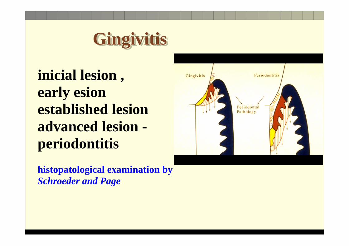
Gingivitis Gingivitis
Gingivitis Gingivitis
inicial lesion , early esionestablished lesionadvanced lesion - periodontitis
histopatological examination by Schroeder and Page
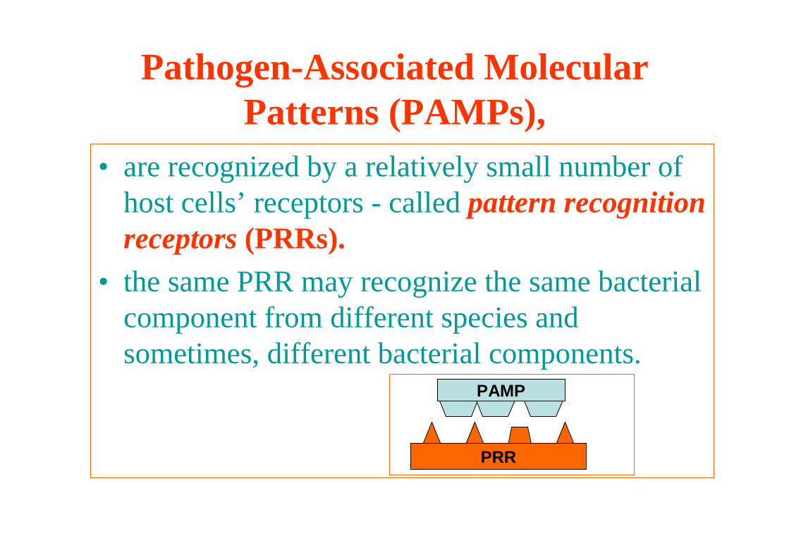
Pathogen-AssociatedMolecular Patterns (PAMPs),
• are recognized by a relatively small number of host cells’ receptors - called pattern recognition receptors(PRRs).
• the same PRR may recognize the same bacterial component from different species and sometimes, different bacterial components.
PAMP
PRR
The Toll-like receptors
• The innate host response • recognition of microbial components as
“danger signals” by host cells• subsequent production of inflammatory
mediators
The Toll-like receptors (TLRs) areexpressed
• resident cells –epithelial cells• leukocytes•• Activate the innate immune response by binding to
various bacterial components :
• lipopolysaccharide [LPS],• bacterial DNA, • diacyl lipopeptides, • peptidoglycan, • (Mahanonda and Pichyangkul, 2007).

After TLR activation, an intracellularsignaling cascade is stimulated,
• leading to• the activation of transcription factors• subsequent inflammatory cytokine
expression,• leukocyte migration, • osteoclastogenesis
• (Nakamura et al., 2008; Ukai et al., 2008; Gelani et al., 2009; Lima et al., 2010).

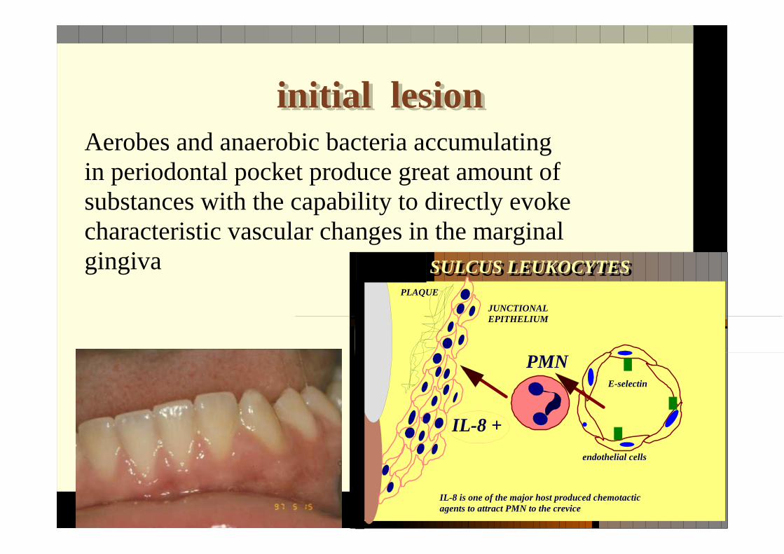
initial lesion initial lesionAerobes and anaerobic bacteria accumulating in periodontal pocket produce great amount of substances with the capability to directly evoke characteristic vascular changes in the marginal gingiva
SULCUS LEUKOCYTESSULCUS LEUKOCYTES
PMN
PLAQUE
JUNCTIONAL EPITHELIUM
E-selectin
endothelial cells
IL-8 +
IL-8 is one of the major host produced chemotactic agents to attract PMN to the crevice

mast cell
PLAQUE
JUNCTIONAL EPITHELIUM
IL-8 +
.....:::::
PGE LTN4
histamine, serotonin
bradykinin, plasmin
platelet
serotonin, thromboxan, platelet derived growth factor PGF
Initial laesion - vascular changes

PMN
PLAQUE
JUNCTIONAL EPITHELIUM
.....:::::
INCREASED CREVICULAR FLUID
extravasation
Initial laesion - vascular changes - initial reactions
IgG, IgM, IgA
complement

PMN
PLAQUE
JUNCTIONAL EPITHELIUM
.....::::: extravasatio
n
Initial laesion - cellular reactions
MONOCYTE
LYMPHOCYTE
ADHESION
cellular infiltrate 5%
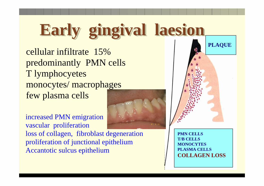
Early gingival laesionEarly gingival laesioncellular infiltrate 15%predominantly PMN cells T lymphocyetesmonocytes/ macrophages few plasma cells
increased PMN emigrationvascular proliferation loss of collagen, fibroblast degeneration proliferation of junctional epitheliumAccantotic sulcus epithelium
PLAQUE
PMN CELLST/B CELLSMONOCYTESPLASMA CELLS
COLLAGEN LOSS

PMN
PLAQUE
JUNCTIONAL EPITHELIUM
.....::::: extravasatio
MONOCYTA
Th 1 - Th2 -Th0 LYMPHOCYTA
ADHESION
cellular infiltrate 15%
plasma cells
aerly laesion - cellular reaction 5-7 days

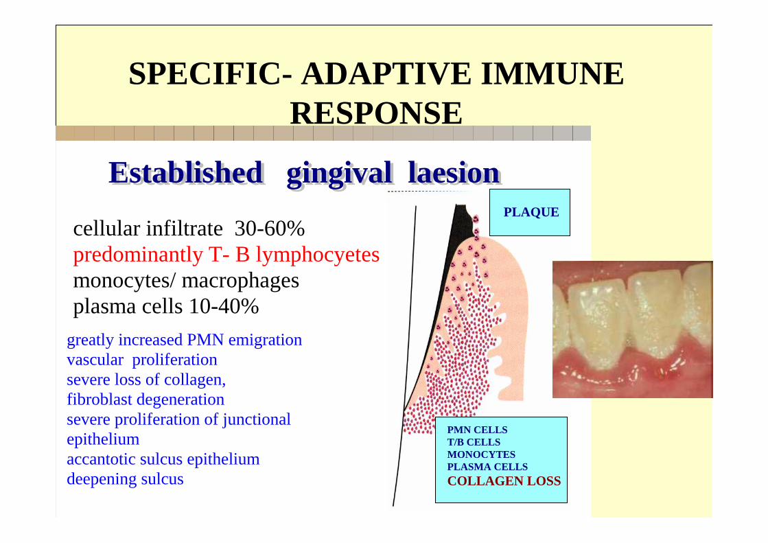
Established gingival laesionEstablished gingival laesion
cellular infiltrate 30-60%predominantly T- B lymphocyetesmonocytes/ macrophages plasma cells 10-40%
greatly increased PMN emigration vascular proliferation severe loss of collagen, fibroblast degeneration severe proliferation of junctional epitheliumaccantotic sulcus epithelium deepening sulcus
PMN CELLST/B CELLSMONOCYTESPLASMA CELLS
COLLAGEN LOSS
PLAQUE

PMN
PLAQUE
JUNCTIONAL EPITHELIUM
.....::::: extravasatio
MONOCYTES
Th 1 - Th2 -Th0 LYMPHOCYTES
ADHESION
cellular infiltrate 30-60% plasma cells 10-30%
plasma cells
established laesion - cellular reaction 7-21 days

COMPOSITION OF GINGIVA AT DAY 0 AND DAY 28 IN EXPERIMENTAL GINGIVITIS STUDY ON
DOGS
baseline Day 280%
20%
40%
60%
80%
100%
non infiltrated tissuejunctional epitheliumoral epitheliuminfiltrated conn tiss

COMPOSITION OF GINGIVA AT DAY 0 AND DAY 28 IN EXPERIMENTAL GINGIVITIS STUDY ON
DOGS
baseline Day 280%
20%
40%
60%
80%
100%
collagenfibroblastvasculaturelukocyteresidual tissue
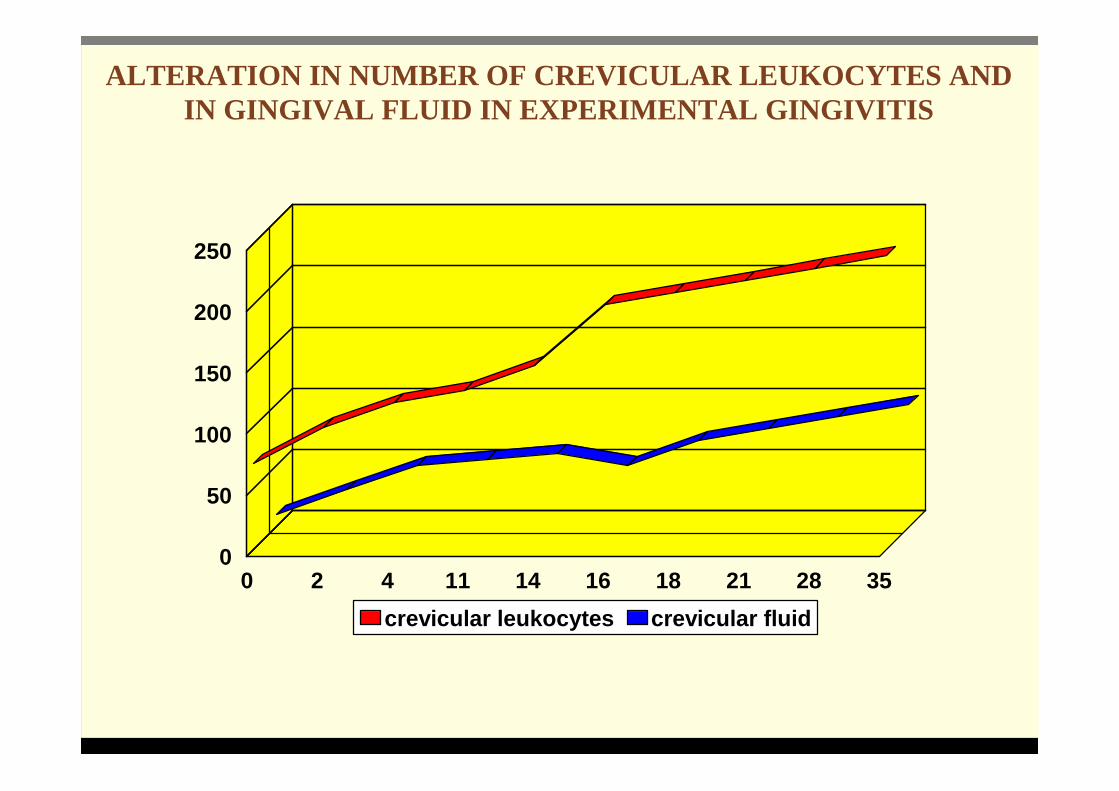
ALTERATION IN NUMBER OF CREVICULAR LEUKOCYTES AND IN GINGIVAL FLUID IN EXPERIMENTAL GINGIVITIS
0 2 4 11 14 16 18 21 28 350
50
100
150
200
250
crevicular leukocytes crevicular fluid
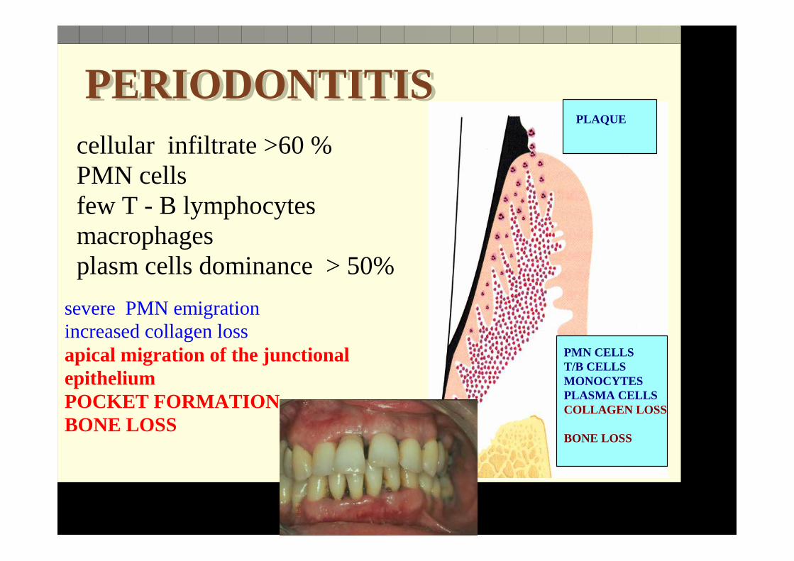
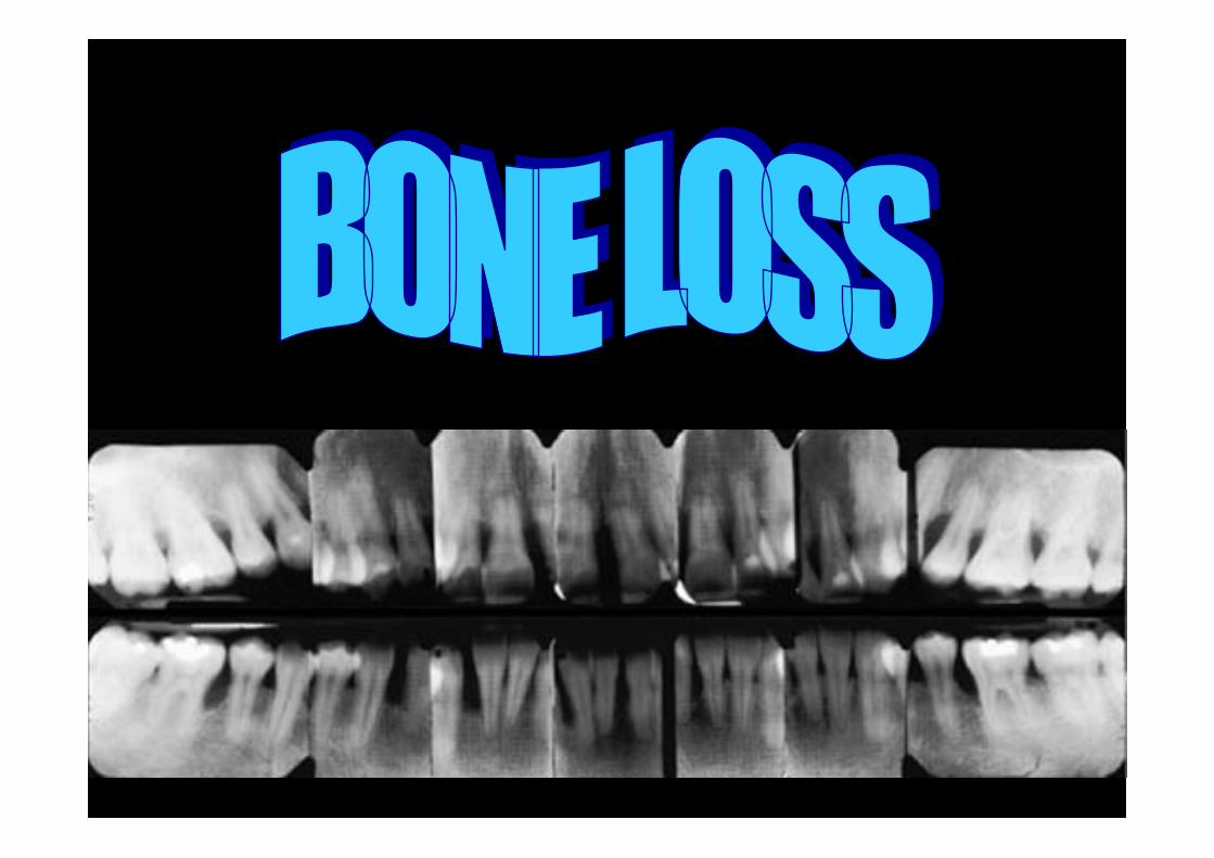
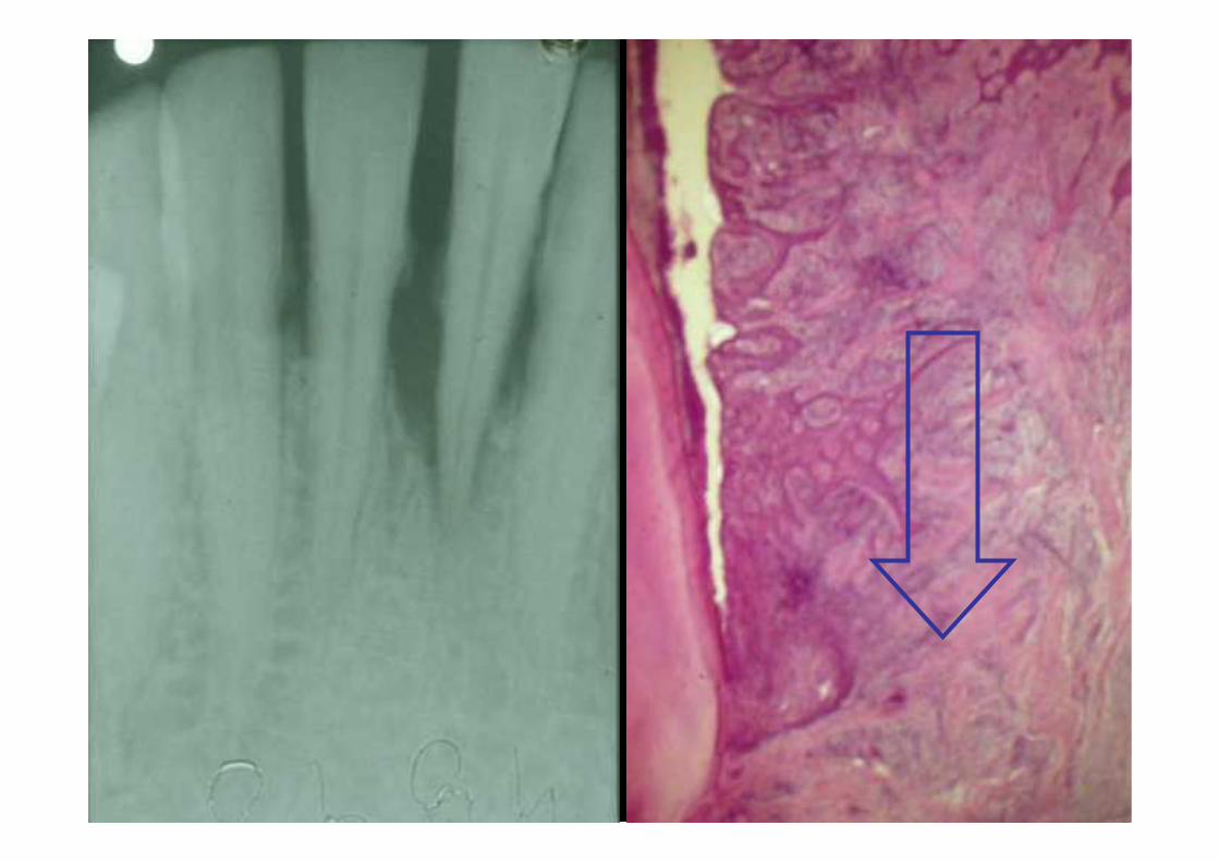
PERIODONTITIS PERIODONTITIS cellular infiltrate >60 %PMN cellsfew T - B lymphocytesmacrophages plasm cells dominance > 50%
severe PMN emigrationincreased collagen loss apical migration of the junctionalepitheliumPOCKET FORMATION BONE LOSS
PMN CELLST/B CELLSMONOCYTESPLASMA CELLSCOLLAGEN LOSS
BONE LOSS
PLAQUE

PMN
PLAQUE
JUNCTIONAL EPITHELIUM
.....::::: extravasatio
MONOCYTES
Th 1 - Th2 LYMPHOCYTES
ADHESION
cellular infiltration >60% plasma CELLS >50%
plasma cells
periodontitis - cellular reactions
IgIg
IgIg

NUMEROUS PLASMA CELLS
Host defense processes responsible for tissue destructions
Host defense processes responsible for tissue destructions
There are four distinct level of protection against oral bacteria
salivagingival crevicegingival tissue systemic immunity
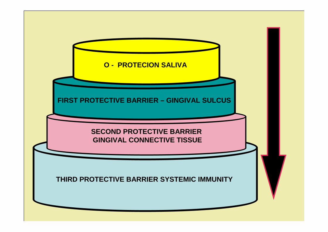
THIRD PROTECTIVE BARRIER SYSTEMIC IMMUNITY
SECOND PROTECTIVE BARRIERGINGIVAL CONNECTIVE TISSUE
FIRST PROTECTIVE BARRIER – GINGIVAL SULCUS
O - PROTECION SALIVA

Host defense processes responsible for tissue destructions
Host defense processes responsible for tissue destructions
mucine salivary lactoferin lysosyme secretory IgA Whole saliva - IgG and IgM molecules
0 barrier level
Saliva contains several antibacterial factors that can control bacterial growth and spreading

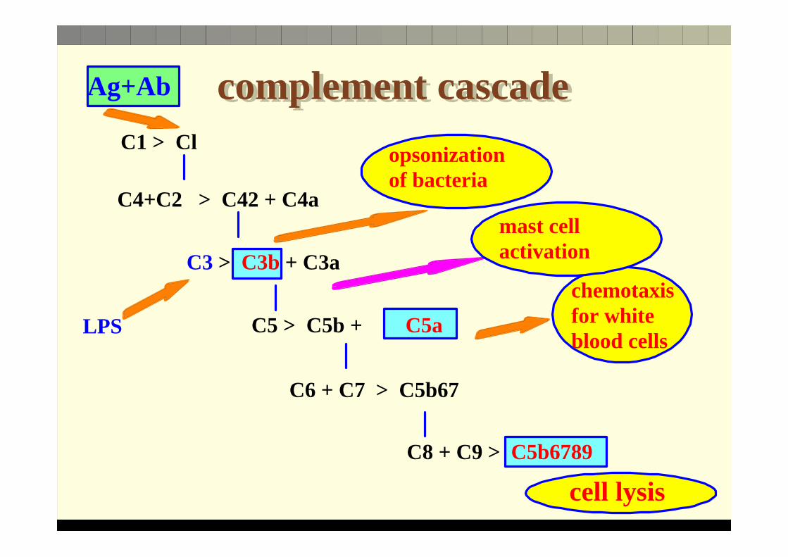
complement cascadecomplement cascadeC1 > Cl
C4+C2 > C42 + C4a
C6 + C7 > C5b67
LPS
C8 + C9 > C5b6789
C5 > C5b + C5a
C3 > C3b + C3a
Ag+Ab
opsonization of bacteria
chemotaxis for white blood cells
mast cell activation
cell lysis

Host defense processes responsible for tissue destructions
Host defense processes responsible for tissue destructions
Sulcus epithelium Secretes cytokines and chemokines (IL-8) Antibacterial peptides (αααα-defensin, ββββ- defensin)The Langerhans cells' membrane receptors play crucial role in innate protection
1st protective barrier gingival sulcus
Many sophisticated and effective antibacterial mechanisms to keep bacteria out of tissues

Host defense processes responsible for tissue destructions
Host defense processes responsible for tissue destructions
1st protective barrier gingival sulcus
A layer of PMN leukocytes separated bacterial plaque from gingival epithelium
Crevicular PMN cells phagocytose bacteria The majority of catabolic enzymes from PMN cells get into the crevicular fluid and will not cause tissue damage. Monocytes in the sulcus can phagocytose PMN cells and bacterial debris clearing the waste products The functional aberrations of sulcus leukocytes can lead to severe periodontal destructions

SULCUS
BLEEDING


Host defense processes responsible for tissue destructions
Host defense processes responsible for tissue destructions
1st protective barrier gingival sulcus
Humoral factors
The crevicular complement system is one of the earliest reactions Bacteria in the sulcus can activate complement by the classic and alternative pathways
C3b complement is an opsonine
Abundant crevicular IgG and IgA molecules . Bacteria can directly stimulate B lymphocytes as a mytogen.
complement cascadecomplement cascadeC1 > Cl
C4+C2 > C42 + C4a
C6 + C7 > C5b67
LPS
C8 + C9 > C5b6789
C5 > C5b + C5a
C3 > C3b + C3a
Ag+Ab
opsonization of bacteria
chemotaxis for white blood cells
mast cell activation
cell lysis

Classic and alternative complement cascadeC3 convertas
ALTERNATÍVEC3
C3+H2O
C3bFactor B
C3bBb
CLASSIC CRP AG+IgG,IgM
C1q
C4 + C2
C4bC2b C2A
C3b - C3a anaphylatoxinamplificatio
regulation - regulation +C5a C5b CR1-3 + C3bi
chemotaxis lysis C6-C9 opsonisation
mast cells
Host defense processes responsible for tissue destructions
Host defense processes responsible for tissue destructions
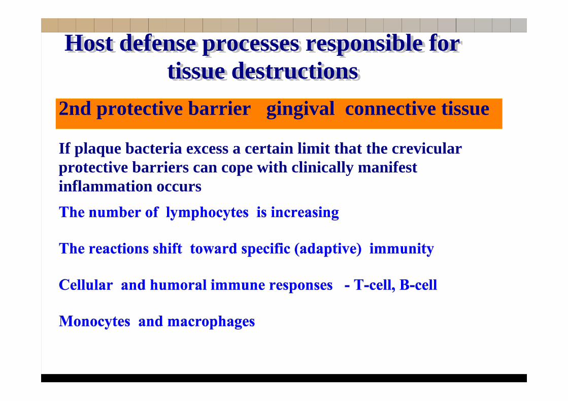
2nd protective barrier gingival connective tissue
If plaque bacteria excess a certain limit that the crevicular protective barriers can cope with clinically manifest inflammation occurs


Host defense processes responsible for tissue destructions
Host defense processes responsible for tissue destructions
In young healthy individuals the serum antibody titter is significantly lower than in healthy adults.
3rd protective barrier systemic immune response
Most healthy adults carries specific serum (IgM, IgG and IgA) against oral periodontopathogenic bacteria

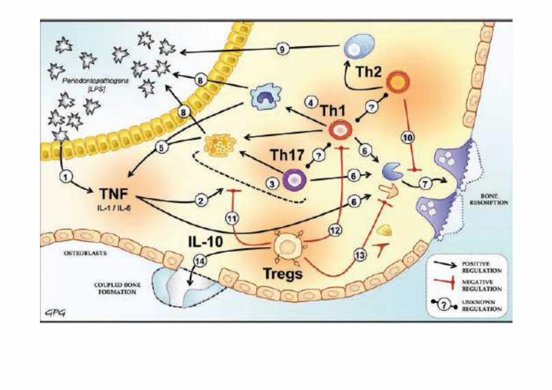
Mechanisms responsible for periodontal tissue destructions
Mechanisms responsible for periodontal tissue destructions
soluble proteolytic enzymes low molecular weight vaste products (urea, sulfides etc) endotoxin (lipopolysaccharide- LPS) exotoxin - i.e.- leukotoxin
Direct bacterial factors
The major cause is bacterial plaque. Bacteria can directly damage periodontal tissuesbut this is only a non significant factor in tissue destruction

Mechanisms responsible for periodontal tissue destructions
Mechanisms responsible for periodontal tissue destructions
innate immunity "adaptíve reactions" acquired immunity
The role of the host in the periodontal tissue destructions
Many different immune processes play role in the pathomechanisms of periodontatis . These are the decisive factors

innate immunity innate immunity
Humoral and cellular elements of innate immunity
Proteolytic enzymes proteinazestissue collagenaze - matrix metalloproteaz MMP - produced by PMN cells and monocytes
Collagenases from PMN leukocytes and fibroblasts can digest type I, II and III collagen tripla-helix and cause extracellular matrix degradation
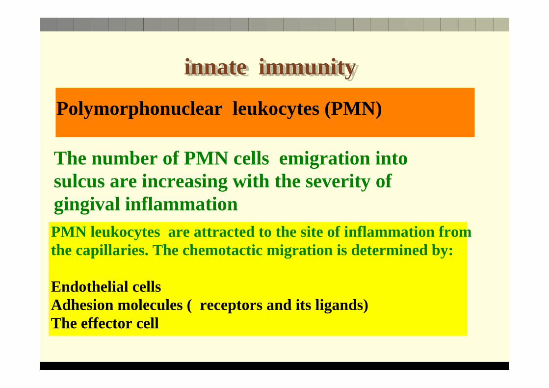
innate immunity innate immunity
Polymorphonuclear leukocytes (PMN)
The number of PMN cells emigration into sulcus are increasing with the severity of gingival inflammation PMN leukocytes are attracted to the site of inflammation from the capillaries. The chemotactic migration is determined by:
Endothelial cells Adhesion molecules ( receptors and its ligands)The effector cell
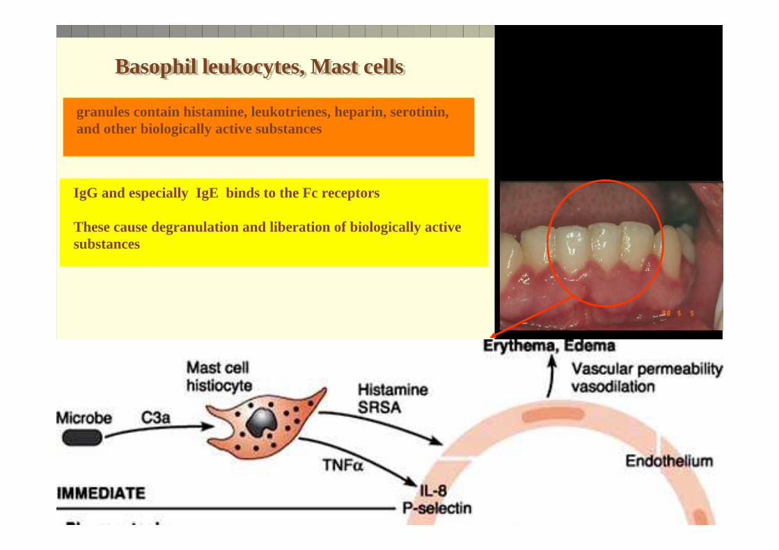
Basophil leukocytes, Mast cells Basophil leukocytes, Mast cells
granules contain histamine, leukotrienes, heparin, serotinin, and other biologically active substances
IgG and especially IgE binds to the Fc receptors
These cause degranulation and liberation of biologically active substances

Polymorphonuclear leukocytes (PMN) Polymorphonuclear leukocytes (PMN)
Several adhesion molecules assist the extravasation and traversing of PMN leukocytes across gingival connective tissue and also emigration though sulcus epithelium
E-selectinAdhesinEndothelial Adhesion Molecules (ELAM)Intercellular Adhesion Molecules (ICAM)

SCHEMATIC ILLUSTRATION OF THE PROSESS WHERBY NEUTROPHILS ARE ATTARCTED INTO THE JUNCTIONAL
EPITHELIUM
LPSPROTEASESfMet-Leu-Phe
release of proinflammatory cytokines IL-1, TNF
endothelial cell increase their adhesion molecule expression ICAM-1, ELAM-1
postcapillary venulejunctional
epithelium
chemotactic gradient of C5a, LTB4, IL-8, fMet-Leu-Phe
PMN lukocytes migrate through the junctional epithelium into the crevice
PMN binding and extravasation
PLAQUE

THE PROTECTIVE ROLE OF PMN CAN BE DEVIDED TO SIX STAGES.

The leukocyte contacts, rolls, sticks and extravasates out of the blood vessel prior to beginning its journey to the site of inflammation
activation of endothelial cells
random contact
rolling sticking extravasation
E-selectin integrins
PMN rolling
blood vessel wall
LPS
CAM-1selectin

Molecules, cells and processes influencing the increased adherence of leukocytes to blood vessels so that they can extravasate to chemotact towards the microbes
activation of endothelial cells
PMN rolling
LPS
CAM-1
selectin
tooth surface
dental plaque bacteria
LPS
LPS
protein
DIRECT
protein
INDIRECT
PGE2IL-8MMP
IL-1bTNF-a
LPSBACTERIA

Polymorphonuclear leukocytes (PMN) Polymorphonuclear leukocytes (PMN)
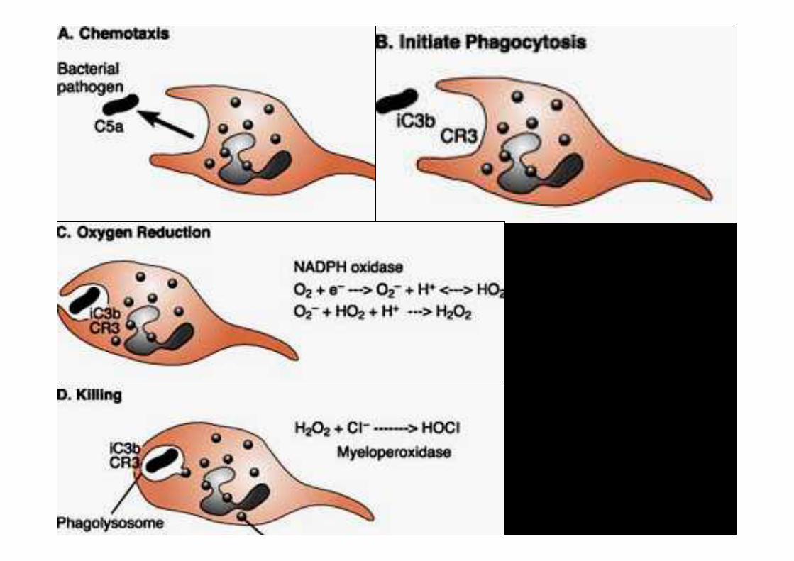
The main function of PMN leukocytes phagocytosis. The precondition for phagocytosis migration towards chemotactic stimulus
Chemotactic stimulicomplement C5a, leukortrien B4,interleukin-8 bacterial metabolites
Polymorphonuclear leukocytes (PMN) Polymorphonuclear leukocytes (PMN)
There are two chemotactic receptors with different affinities
High affinity receptor is responsible for chemotactic movements Low affinity receptors will ignite the oxidative burst and degranulation and prepare the cells for phagocytosis

Polymorphonuclear leukocytes (PMN) Polymorphonuclear leukocytes (PMN)
The activity of PMN cells can lead to severe tissue destruction - periodontal abscess
Chemotactic stimuli can determine the caracter of the PMN cellular response - protective or destructive .
The most importatnt chemotactic molecules are: C5a, LTB4 és az IL-8. IL-8 is less potent in activation of phagocytosis, but more ponent in enhancing MMP production

Polymorphonuclear leukocytes (PMN) Polymorphonuclear leukocytes (PMN)
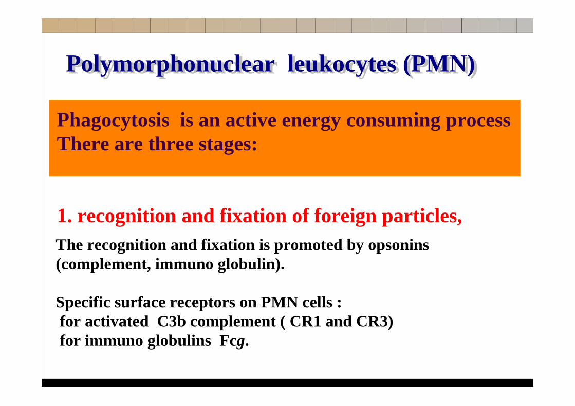
Phagocytosis is an active energy consuming processThere are three stages:
1. recognition and fixation of foreign particles, 2. engulfing foreign particles3. degradation and digestion of foreign particles

Polymorphonuclear leukocytes (PMN) Polymorphonuclear leukocytes (PMN)
Phagocytosis is an active energy consuming processThere are three stages:
1. recognition and fixation of foreign particles, 2. engulfing foreign particles3. degradation and digestion of foreign particles
Polymorphonuclear leukocytes (PMN) Polymorphonuclear leukocytes (PMN)
Phagocytosis is an active energy consuming processThere are three stages:
1. recognition and fixation of foreign particles, The recognition and fixation is promoted by opsonins(complement, immuno globulin).
Specific surface receptors on PMN cells : for activated C3b complement ( CR1 and CR3) for immuno globulins Fcg.


Polymorphonuclear leukocytes (PMN) phagocytosisPolymorphonuclear leukocytes (PMN) phagocytosis
bacteria
opsonins
developing phagosome
phagosome
FcI, FcII, FcIII receptors CD14
receptors
adherent bacteria
CR1 CR3 receptors
LPS
Polymorphonuclear leukocytes (PMN) Polymorphonuclear leukocytes (PMN)
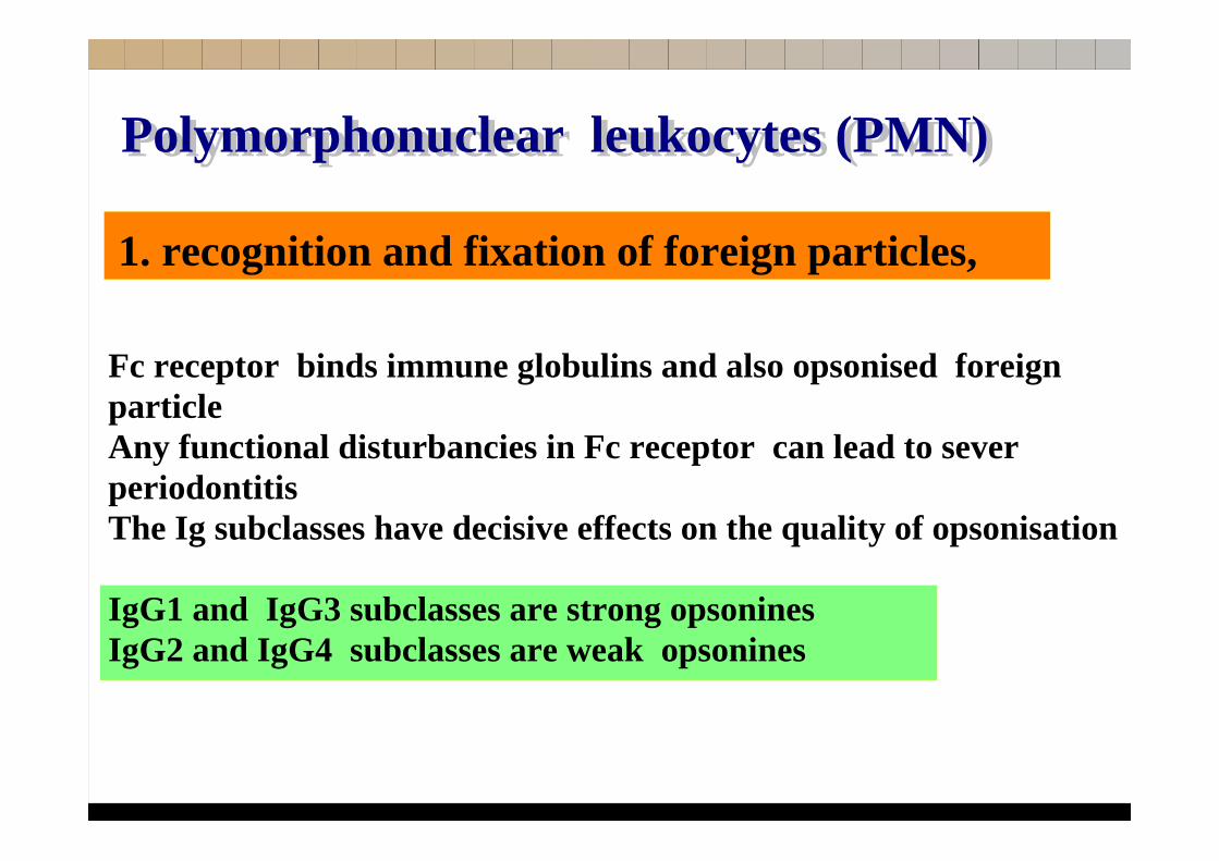
1. recognition and fixation of foreign particles,
Fc receptor binds immune globulins and also opsonised foreign particleAny functional disturbancies in Fc receptor can lead to sever periodontitis The Ig subclasses have decisive effects on the quality of opsonisation IgG1 and IgG3 subclasses are strong opsonines IgG2 and IgG4 subclasses are weak opsonines

Polymorphonuclear leukocytes (PMN)
Polymorphonuclear leukocytes (PMN)
Fc receptor
Fc receptor is a very critical coupling factor between non specific innate reactions and humoral immunity. On the cell membrane of Neutrophils there are many receptors, with different affinity
FcI high affinity receptorFcII and FcIII low affinity receptors
FcII receptor binds all Ig subclassesFcIII binds only IgG1 és IgG3 subclassesFcIII receptor cannot ignite oxidative burstFcII receptor can activate the whole process of phagocytosis

Polymorphonuclear leukocytes (PMN) Polymorphonuclear leukocytes (PMN)
Fc receptor
In localized juvenile periodontitis the expression of FcII and FcIII receptors is down regulated in crevicular PMN cells
The circulating PMN cells show normal values

Polymorphonuclear leukocytes (PMN)
Polymorphonuclear leukocytes (PMN)
CD14 receptor
A PMN leukocytes can opsonise bacteria without Ig and complements CD14 receptors on PMN cell membranes can bind LPS
PMN cells can phagocytose bacteria if complement or specific Ig molecules are destroyed by bacterial virulence factors

Polymorphonuclear leukocytes (PMN) phagocytosisPolymorphonuclear leukocytes (PMN) phagocytosis
bacteria
opsonins
developing phagosome
phagosome
FcI, FcII, FcIII receptors CD14
receptors
adherent bacteria
CR1 CR3 receptors
Polymorphonuclear leukocytes (PMN) Polymorphonuclear leukocytes (PMN)
Phagocytosis is an active energy consuming processThere are three stages:
1. recognition and fixation of foreign particles, 2. engulfing foreign particles3. degradation and digestion of foreign particles
Polymorphonuclear leukocytes (PMN) Polymorphonuclear leukocytes (PMN)
There are different cytoplasmatic granules
Three type of granules exist:primary or azurophil granules secondary or specific granules terciary of C granules
Primary granules is identified by its peroxidase content myeloperoxidase, lysozyme és proteinase enzymes. Secondary granules are peroxidase negative - Containing:lactoferin, B12 binding protein, fibronectin receptors, laminin receptors Secondary granules are released chiefly extracellularly while primarly granules serving the intracellulary digestion .

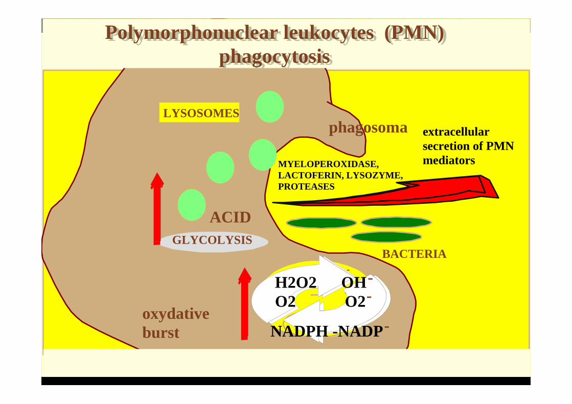
Polymorphonuclear leukocytes (PMN) phagocytosis
Polymorphonuclear leukocytes (PMN) phagocytosis
GLYCOLYSIS
LYSOSOMES
BACTERIA
MYELOPEROXIDASE, LACTOFERIN, LYSOZYME, PROTEASES
ACID
-
-
-
extracellular secretion of PMN mediators
NADPH -NADP
H2O2 OHO2 O2-
-
oxydative burst
phagosoma

Polymorphonuclear leukocytes (PMN) Polymorphonuclear leukocytes (PMN)
PMN leukocytes make up the first defensive line with the complement system and Ig molecules neutrofil/complement/antibody axis
Deficiencies in PMN functions - greater risk for destructive periodontal disease Leukocyte Adhesion DeficiencyPapillon-LeFevre syndromeChediak-Higashi syndromescyclic neutropenia leukemialocalized juvenile periodontitis

LEUKOCTE ADHESION DEFICIENCY, (LAD)
Selectin
CD-15
Integrin
CD-11 a,b,c
CD-18
SCHEMATIC ILLUSTRATION OF THE PROSESS WHERBY NEUTROPHILS ARE ATTARCTED INTO THE JUNCTIONAL
EPITHELIUM
LPSPROTEASESfMet-Leu-Phe
release of proinflammatory cytokines IL-1, TNF
endothelial cell increase their adhesion molecule expression ICAM-1, ELAM-1
postcapillary venulejunctional
epithelium
chemotactic gradient of C5a, LTB4, IL-8, fMet-Leu-Phe
PMN lukocytes migrate through the junctional epithelium into the crevice
PMN binding and extravasation
PLAQUE

LEUKOCTE ADHESION DEFICIENCY, (LAD)
As integrin molecules are alsoresponsible forbinding theopsinized antigens, in this disease notonly the diapedesisbut also thephagocytosis is hampered

Papillon LeFevre syndromeGene mutation on 11 chromosome (11q14-q21)
cathepsin C gene
.HART TC, HART PS, BOWDEN DW és mts. Mutation of the cathepsin C gene are responsible for Papillon-Lefevre syndrome J Med Genet 1999; 36:881-888
.HART TC, ZHANG Y, FIRATI E és mts. Identificaton of cathepsin C mutations in ethnically diverse Papillon-Lefevre syndrome patients J Med Genet 2000; 37: 927- 931

.
Papillon LeFevre syndromeGene mutation on 11 chromosome (11q14-q21) cathepsin C ge ne

Chronic granulomatozus disease
PMN leukocytes NADPH oxydaze enzyme disfunction
The disease is inherited by recessive inheritance
The PMN cellsare not able toproduce reactivefree oxygenradicals and is notcapable of killingphagocytosedbacteria
Polymorphonuclear leukocytes (PMN) phagocytosis
Polymorphonuclear leukocytes (PMN) phagocytosis
GLYCOLYSIS
LYSOSOMES
BACTERIA
MYELOPEROXIDASE, LACTOFERIN, LYSOZYME, PROTEASES
ACID
-
-
-
extracellular secretion of PMN mediators
NADPH -NADP
H2O2 OHO2 O2-
-
oxydative burst
phagosoma

Mononuclear cells monocytes and macrophages
Mononuclear cells monocytes and macrophages
Langerhans cells and gingival macrophages,
macrophages make up 2% of the cellular elements of the gingival sulcus fluid Plays important role in the production inflammatory and regenerative cytokines (IL-1, IL-6, TNF, TGF, PGE2 LTB4 )

Mononuclear cells monocytes and macrophages
Mononuclear cells monocytes and macrophages
The reactivity and cytokine production by monocytes determine the course of the inflammatory periodontal disease and the severity of tissue destruction
The life span of the monocytes is much longer than that of the PMN leukocytes After phagocytosis the cells survive .
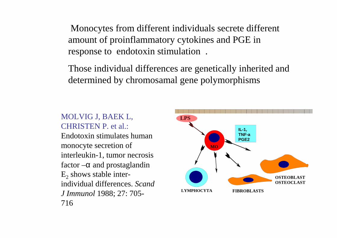
Monocytes fromdifferent individuals secrete differentamount of proinflammatory cytokines and PGE in response to endotoxin stimulation .
Those individual differences are genetically inherited anddetermined by chromosamal gene polymorphisms
MOLVIG J, BAEK L, CHRISTEN P. et al.:Endotoxin stimulates human monocyte secretion of interleukin-1, tumor necrosis factor –α and prostaglandin E2 shows stable inter-individual differences. Scand J Immunol 1988; 27: 705-716
MO
FIBROBLASTS
IL-1,TNF-aPGE2
OSTEOBLASTOSTEOCLAST
LYMPHOCYTA
LPS

A nocleotid base change on the locus of IL-1B +3953 of the homologechromosome will lead to a four fold increase in IL-1 production bymonocytes
KORNMAN KS, diGIOVINE FS. Genetic variations in cytokine expression: a risk factor for severity of adult periodontitis Ann Periodontol 1998; 3:327-338
C - T G - T-889 +4845
G - T-511 +3953
C - T
IL-1 alpha
IL-1 beta

The destructive and protective hostresponse
• A “maximum” intensity of host immune inflammatoryreaction leads to excessive tissue damage withoutenhancing the control of infection
• (Trombone et al., 2009).
• Patients presenting hyper-inflammatory genetic variantspresent the same frequency and load of red-complexperiodontopathogens and A. actinomycetemcomitans
• as patients genetically not prone to develop exacerbatedresponses
• (Ferreira et al., 2008);
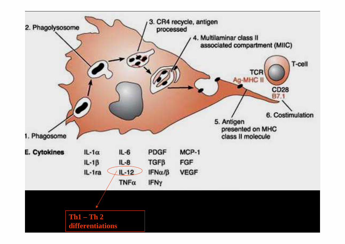
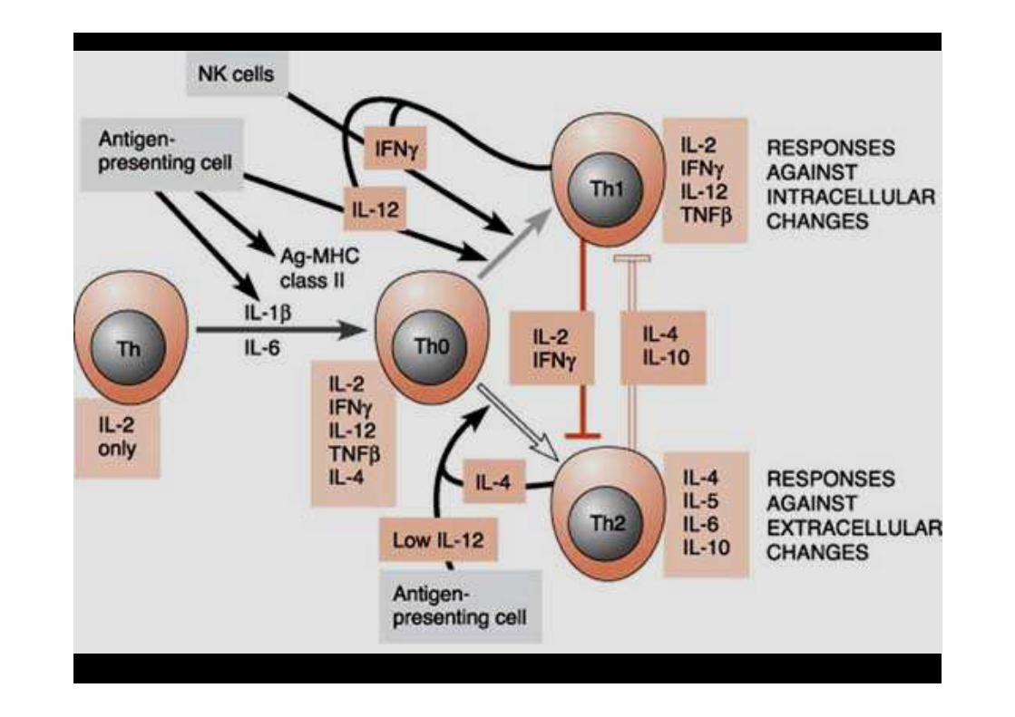
Th1 – Th 2 differentiations

Natural killer lymphocytes ( NK cells )
Natural killer lymphocytes ( NK cells )
Non committed lymphocytesThey simple bind to target cells and kill them without previous sensitization
Provide immediate non specific reaction
High affinity Fc receptors for IgG which enables them to play an important role in antibody dependent cell mediated cytolysis

inflammatory cytokinesinflammatory cytokines
secondary inflammatory mediator productionsecondary inflammatory mediator production
PAF, histamine, bradykinin, PGE2

Cytokines Cytokines
cytokiens are soluble proteins produced by cells and act on other cell from the same cell line or on different cells lines autocrine regulation
paracrine regulation

Common features
Cytokines Cytokines
1. locally produced and acting and degrading locally 2. several cytokines have similar biologic effects 3. Most inflammatory cytokine pre coded in mRNA - and a signal can trigger the translation and secretion 4. Very strong hormones, acting on high affinity membrane receptors in very low concentration 10 - 10 Mol 5. Several cells have different membrane cytokin receptors and can give opposing answers 6. Cells have membrane receptors with different affinity
-9 -8
Interleukin 1 (IL-1)Interleukin 1 (IL-1)IL-1 plays a crucial role in the pathogenesis of periodontal destruction
Disease activity in periodontal pockets leads immediately to increased ( 3-4 fold) IL-1ββββ concentration
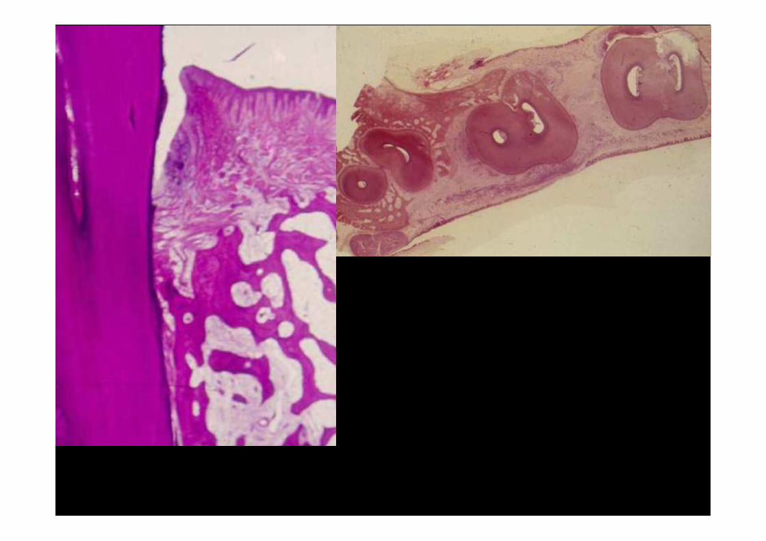
Interleukin 1 (IL-1)Interleukin 1 (IL-1)One of the earliest cytokinesOsteoclast stimulating factor LPS stimulated lymphocytes in cell cultures produced a substance which increased bone resorption in bone organ cultures
IL-1 exists in two forms IL-1 a and b.

Interleukin 1 (IL-1)Interleukin 1 (IL-1)Locally acts to up-regulate adhesion molecules on fibroblasts, endothelial cells, lymphocytes, PMN leukocytes and monocytes
Bacterial LPS stimulates sulcus Langerhans cells to produce IL-1αααα and ββββ-t. They play important role in periodontal inflammation
Directly increase all catabolic processes in connective tissue

Increases PGE2 secretion by fibroblasts and monocytes which in turn stimulates vasodilatation, oedema and bone resorption
Stimulates PMN leukocytes and monocytes MMP production
IL-1 is autostimulatory acts on other cells to produce more IL-1
Interleukin 1 (IL-1)Interleukin 1 (IL-1)

Interleukin 1 (IL-1)Interleukin 1 (IL-1)
There are IL-1 receptor antagonists (IL-1ra)
They can bind to IL-1 receptors and occupies IL-1 receptors IL-1ra synthesis is enhanced by steroids and certain anti inflammatory cytokines IL-4, IL-10.

Interleukin-6 (IL-6) Interleukin-6 (IL-6) Produced by lymphocytes, fibroblasts and monocytes.
IL-6 promotes the proliferation and differentiation of B lymphocytes, T lymphocytes and the differentiation of monocytes into multinucleated osteoclasts
IL-6 produced during inflammation plays essential role in periodontal bone resorption .

IL-6IL-6

Interleukin-6 (IL-6) Interleukin-6 (IL-6)
Estrogens and progesterons inhibit the secretion of IL-6 by mononuclear cells and osteoblasts
Supposedly this plays an important role in the postmenopausal osteoporosis and the increased severity of periodontal bone loss after menopause.

Tumor necrosis factor alpha (TNFαααα)
Tumor necrosis factor alpha (TNFαααα)
Promotes matrix degradation and bone resorption Less potent than IL-1. The mechanisms of action on bone metabolism is different from that of IL-1. It exerts a decoupling effect on bone, inhibiting bone formation and facilitating osteoclastic bone resorption
Chronic low levels of IL-1 and TNFαααα in the bloodstream can promote endothelial cell damage and atherosclerosis !!!!!

Tumor necrosis factor alfa (TNFαααα) Tumor necrosis factor alfa (TNFαααα) In low concentration protect against inflammation.In high concentration extremely toxic to the tissues Causing abortion in pregnant animals
Triggers the release of histamine, serotinin Promotes matrix degradation and bone resorption Less potent than IL-1.

INFLAMMATORY BONE RESORPTION -RANKL
• Inflammatory bone diseases enhance the local RANKL expression and the RANKL/OPG ratio is shifted
• ( Liu és mtsai., 2003, Taubnamés mtsai., 2001, Teng ésmtsai., 2000) .
• Interleukin-1, IL -6 and TNF-α are strong bone resobers and they increase the RANKL/OPG expression in osteoblasts and other stromal cells. These cells can locally control the extent of bone resorption
• (Lerner 2004, Liu 2003 Nafasawa és mtsai., 2007).

The role of osteoblasts in the osteoclastogenesisand modulationof bone resorption

The molecular communicationfactors
betweenosteoblasts andosteoclasts
• Macrophag Colony Stimulating Factor ( M-CSF ) • Receptor Aktivator of Nuclera Factor K Ligand
(RANKL). • The M-CSF binds to the membrane receptors of
osteoclast precursors igniting their proliferation and ensures their survival
• RANKL is a trigger factor, that facilitates the differentiation of osteoclast precursor cells and stimulates the resorptive capacity of the matured k osteoclasts
• (Yasuda és mtsai., 1998 , Kong és mtsai., 1999, Lacey és mtsai., 1998 ).
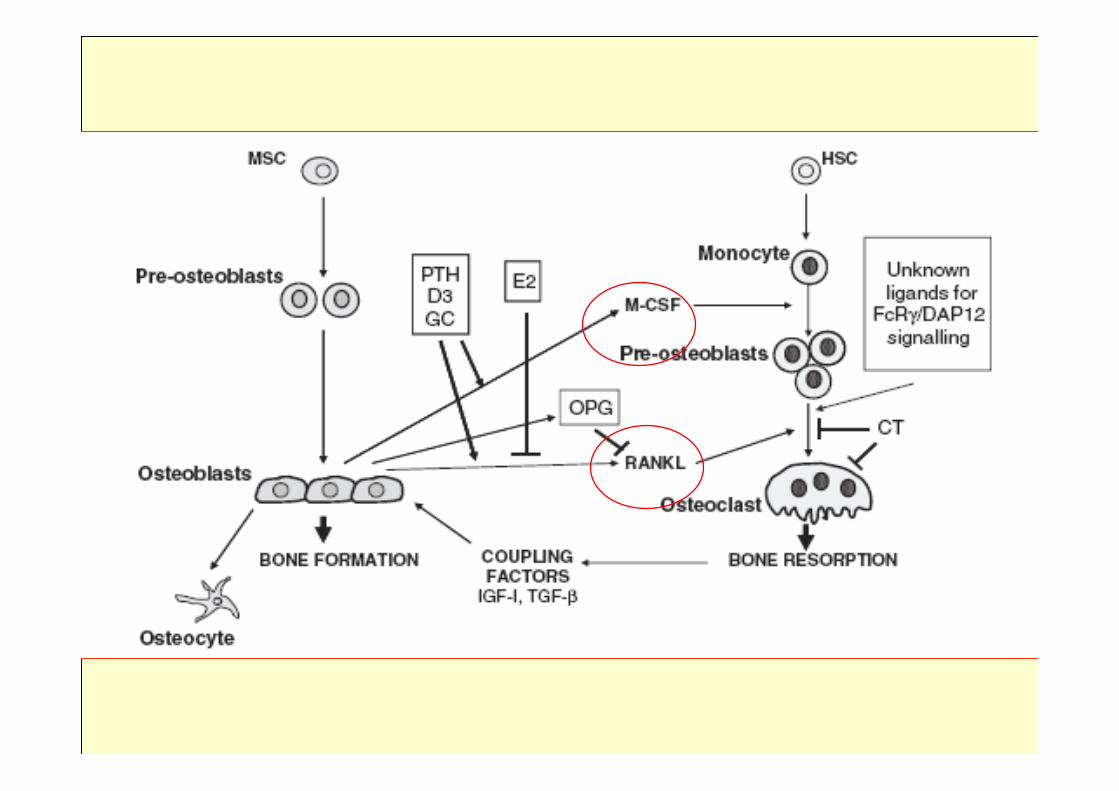

The role of osteoblasts in the osteoclastogenesis and modulation of bone
resorption
• The effect of RANKL can be antagonized by osteoprotegerinnel (OPG) (Simonet és mtsai., 1997).
• OPG synthesized by osteoblasts and other stromal cells.
• OPG can bind to RANKL- and can block the RANKL/RANK coupling and the triggering of osteoclasts .
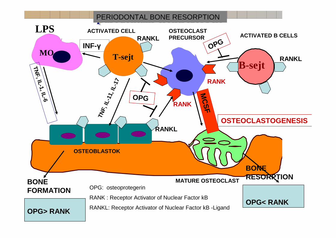
LPS
MO
B-sejt
parodontális csontrezorbció parodontális csontrezorbció
T-sejt
BONE FORMATION
BONE RESORPTION
OPG> RANKOPG< RANK
ACTIVATED CELL OSTEOCLAST PRECURSOR ACTIVATED B CELLS
OSTEOBLASTOK
MATURE OSTEOCLAST
TNF, IL-1, IL-6
TNF,
IL-1
1, IL
-17
INF-γ
MC
SF
OSTEOCLASTOGENESIS
OPG
OPG
RANKL
RANKL
RANKL
RANK
RANK
OPG: osteoprotegerin
RANK : Receptor Activator of Nuclear Factor kB
RANKL: Receptor Activator of Nuclear Factor kB -Ligand
PERIODONTAL BONE RESORPTION


Chemotactic cytokinesIL-8
Chemotactic cytokinesIL-8
Chemokines Produced by epithelial cells, monocytes and endothelial cells stimulates by IL-1, LPS or TNFαααα. IL-8 stimulates MMP secretion by PMN leukocytes
IL-8IL-8In gingivitis the LTB4 concentration in the gingival sulcus is high In destructive periodontitis , especially in periodontal abscess the IL-8 concentration is high
The IL-8 evoked PMN response is more destructive due to the elevated MMP production
PMN cell response attracted by LTB4 or C5a chemotactic factors is more protective due to the stimulated phagocytosis

ProstaglandinProstaglandinModulation of arachidonic acid metabolism
tissue demage
membrane phospholipids
arachidonic acid
Leukotrienes Lipoxins
PGEprostacyclinsthromboxane
2
lipoxygenase
COX-1COX-2
Cell-cell and enzyme interactions
ProstaglandinProstaglandinCOX-1 constitutive cyclooxigenase
COX-2 - inflammatory cyclooxigenase Two isoforms of cyclooxygenase enzymesCOX-1 COX-2
COX-1 PG-s for homeostasis
COX-2 PG-s for inflammation
local inflammatory PG production
local inflammatory PG production monocytes
macrophagesfibroblasts
PMN leukocytes other white blood cells
endothelial cells
monocytes macrophages
fibroblastsPMN leukocytes
other white blood cellsendothelial cells
ProstaglandinProstaglandin IL-1 ββββ or TNFαααα stimulus on monocytes immediately induce COX-2 gen transcription and de novo production of PGE, The same stimulus has no effect on the COX-1 enzyme system. In the periodontal tissues the main source of PGE2 is monocytes and fibroblasts

ProstaglandinProstaglandinCOX-1 constitutive cyclooxigenase
COX-2 - inflammatory cyclooxigenase Genetically determined variance in host immune response to bacterial
infections has been identified in individuals with agressive
periodontitis

ProstaglandinProstaglandinCOX-1 constitutive cyclooxigenase
COX-2 - inflammatory cyclooxigenase
Monocytes from agressive periodontitis patients showed
increased PGE production upon stimulation with LPS
Shapira L et al. The secretion of PGE, IL-1b and TNFa by adherent munonuclear cells from early onset periodontitis patients J. Periodontol 1994;65:139-146

ProstaglandinProstaglandin
PGE increases vascular permeability, vasodilatation and edema. Stimulate MMP production by monocytes and fibroblasts and consequently promotes connective tissue matrix catabolism
Directly and indirectly stimulates bone resorption Synergistically enhances biologic effect of TNFαααα and IL-1

ProstaglandinProstaglandin
In gingivitis the PGE2 concentration of the crevicular fluid is significantly increased.
In gingivitis there is a three fold increase
In periodontitis there is an additional 3-5 fold increase

PG is a potent bone resorber
periodontitisperiapical periodontitiscyst orthodontic tooth movement traumatic occlusionmalignant tumors
NONSTEROID ANTI-INFLAMMATORY DRUGS
NONSTEROID ANTI-INFLAMMATORY DRUGS
ASPIRINAPRANAX
CATAFLAMDICLOFENAC
DONALGINFLUGALINHOTEMIN
INDOMETACINUMNAPROSYNPROFÉNIDSURGAMTILCOTIL
VOLTAREN
ASPIRINAPRANAX
CATAFLAMDICLOFENAC
DONALGINFLUGALINHOTEMIN
INDOMETACINUMNAPROSYNPROFÉNIDSURGAMTILCOTIL
VOLTAREN
Reparative and anabolic cytokinesReparative and anabolic cytokines
During wound healing and tissue repair monocytes platelets, fibroblasts and other cells produce factors with anabolic biological effects
Platelet Derived Growth Factor (PDGF) Fibroblastic Growth Factor (FGF)Insuline-like Growth Factor (IGF)Transforming Growth Factor (TGF)Bone Morphogenic Proteins (BMPs).
Reparative and anabolic cytokinesReparative and anabolic cytokines
1. These are chemotactic for fibroblasts, periodontal ligament mesenchymal cells, and osteoprogenitor cells2. Many, as a growth factor induce cellular differentiation of connective tissue mesenchymal cells into matured matrix-secreting cells 3. Many of the growth factors are incorporated into the newly formed matrix I.e. Bone contains large amount of BMP, TGF and IGF.

Reparative and anabolic cytokinesReparative and anabolic cytokinesMatrix degradation immediately triggers the compensatory anabolic cytokine production
If bacterial stimuli persist the pro-inflammatory cytokines will suppress the regenerative processes,
After the bacterial factors having been eliminated the regenerative processes will dominate the tissue reactions and the connective tissue regenerates

In periodontal inflammation the majority of the pro-inflammatory cytokines are produced by monocytes. The monocyte reaction plays a decisive role in the determination of the severity of inflammatory reaction and tissue damage
The healing is also promoted by anabolic cytokines produced by monocytes
Reparative and anabolic cytokinesReparative and anabolic cytokines
Established gingival laesionEstablished gingival laesion
cellular infiltrate 30-60%predominantly T- B lymphocyetesmonocytes/ macrophages plasma cells 10-40%
greatly increased PMN emigration vascular proliferation severe loss of collagen, fibroblast degeneration severe proliferation of junctional epitheliumaccantotic sulcus epithelium deepening sulcus
PMN CELLST/B CELLSMONOCYTESPLASMA CELLS
COLLAGEN LOSS
PLAQUE
SPECIFIC- ADAPTIVE IMMUNE RESPONSE
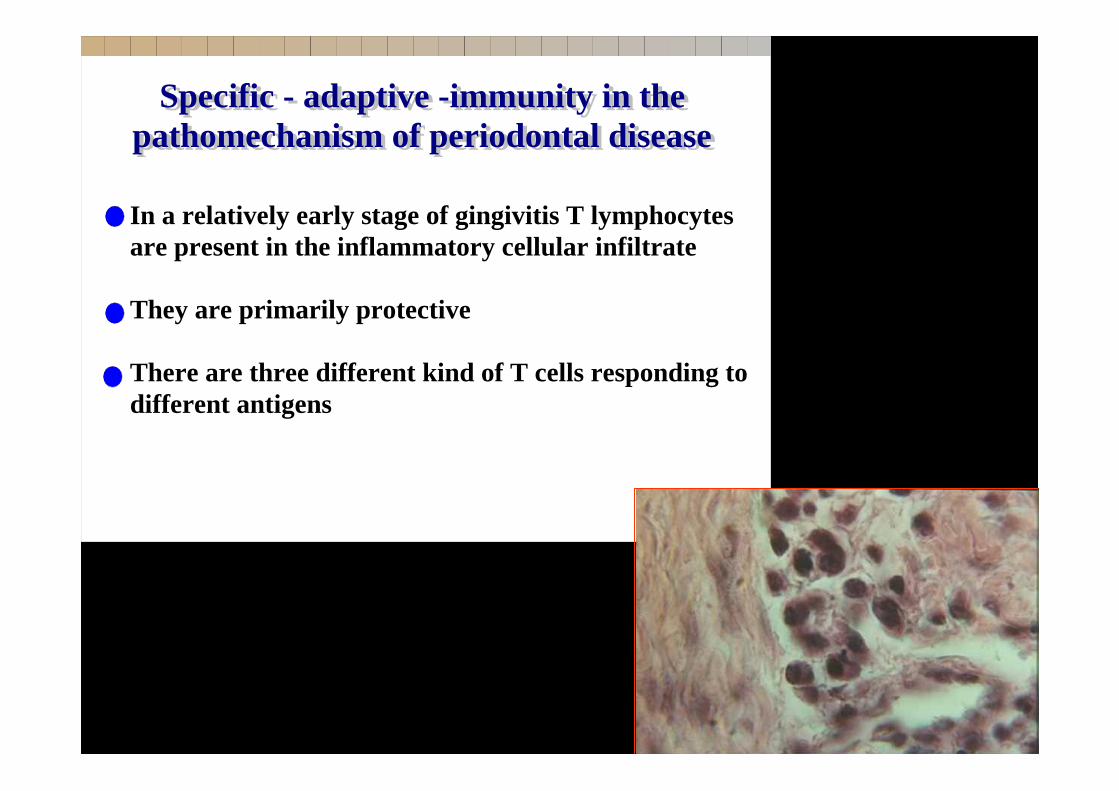
Specific - adaptive -immunity in the pathomechanism of periodontal disease
Specific - adaptive -immunity in the pathomechanism of periodontal disease
In a relatively early stage of gingivitis T lymphocytes are present in the inflammatory cellular infiltrate
They are primarily protective There are three different kind of T cells responding to different antigens
Specific immunity in the pathomechanism of periodontal disease
Specific immunity in the pathomechanism of periodontal disease
1- CD8+ cytotoxic lymphocytes, 2 - CD4+ lymphocytes3 - natural killer T lymphocytes
CD4+ cells are helper T cells They are divided into two sub groupsTh1 and Th2 helper cellsTh17 and Treg cells (regulatory)

CYTOKINES PRODUCED BY TYPE 1 (TH1) AND TYPE 2 (TH2) CD4+ HELPER CELLS
TH1TH2
IL-2 INFγγγγ TNF-ββββ
IL-5
IL-4
IL-6 IL-10IL-13
ENHANCE CELL MEDIATED RESPONSE
ENHANCE HUMORAL IMMUNE RESPONSE

Specific immunity in the pathomechanism of periodontal disease
Specific immunity in the pathomechanism of periodontal disease
Th1 and Th2 cells play different role in periodontitis
Th1 cells primarly associated with active inflammation and restricted to a relatively limited area
Th2 cells are widely spread all over the tissue and rather associated with chronic inflammation

• It has beenreported that T-cells are• involved in bone destructionvia IL -17
production, which in turn is describedas aninducer of RANKL production
• (Kotake et al., 1999; Satoet al., 2006)
BONE RESORPTION BY ADAPTIVE IMMUNE RESPONSE

The destructivehost responsefromthe tissuedamageperspectiveTh2
• Th2 cells’ commitmentand action areprimarily dependenton IL -4, the
• prototypical Th2 cytokine,
• which also acts as a B-cell stimulatory Factor
• (Murphy and Reiner,
CYTOKINES PRODUCED BY TYPE 1 (TH1) AND TYPE 2 (TH2) CD4+ HELPER CELLS
TH1TH2
IL-2 INF γγγγ TNF-ββββ
IL-5
IL-4
IL-6 IL-10IL-13
ENHANCE CELL MEDIATED RESPONSE
ENHANCE HUMORAL IMMUNE RESPONSE

The destructivehost responsefromthe tissuedamageperspective
• IL -6 contributes to B-cell differentiationand antibody production
• B-cells produce RANKL in response toperiodontal pathogenstimulation
• The majority of B-cells in periodontallesions are RANKL+
• (Kawai et al., Han et al., 2009),
The destructivehost responsefromthe tissuedamageperspective
• In a later stage B-cells outnumber T-cells inperiodontal lesions,
• the predominance of a Th2-type response inperiodontal lesions
• It potentially leads to the accumulationofRANKL -producing cells and, consequently, to tissue destructionand bone loss
• (Gemmellet al., 2002b)
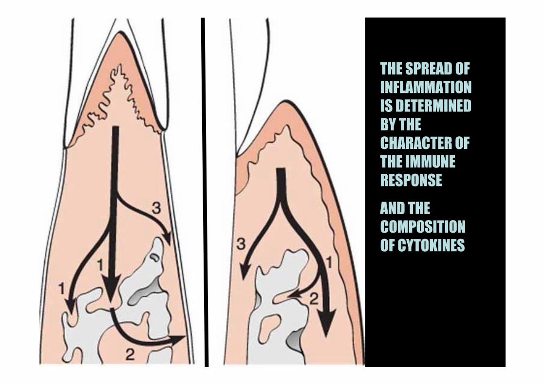
THE SPREAD OF
INFLAMMATION
IS DETERMINED
BY THE
CHARACTER OF
THE IMMUNE
RESPONSE
AND THE
COMPOSITION
OF CYTOKINES

lymphocyte signaling lymphokines lymphocyte signaling lymphokines
MO T CELL
Ag presentation
IL-1
CD-25 receptor expression
antigen specific clone
IL-4IL-5B cell
plasma cells
IgG
IgG
IgG
IgG IL-6
antigen specific clone
Th-1
IL-4
Th-2
IL-2
autocrin activation
CD-8+
T cell proliferation
IL-10IL-13
IL-2MHC II
IFNγγγγ

BONE RESORPTION

PERIODONTAL INFALMMATORY BONE RESORPTION
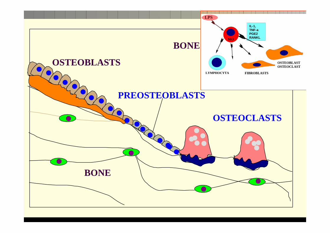
THE INFLAMMATORY CYTOKINES AND OTHER LOCAL FACTORS – LIKE PGE - WILL UPSET THE BALANCE OF THE NORMAL COUPLED BONE REMODELING AND SHIFTS THIS TOWARDS NET BONE LOSS
- PARTLY BY INHIBITING BONE FORMATION
- RARTLY BY PROMOTING OSTEOCLASTIC BONE RESORPTION ,

MO
FIBROBLASTS
IL-1,TNF-aPGE2RANKL
OSTEOBLASTOSTEOCLAST
LYMPHOCYTA
LPS
IL-1
IL-6
LYMPHOTOXIN
LOCAL REGULATION IN INFLAMMATION
..........
..... ....
... .
......... ..
BONE REMODELING
OSTEOBLASTS
BONE
PREOSTEOBLASTS
OSTEOCLASTS
MO
FIBROBLASTS
IL-1,TNF-aPGE2RANKL
OSTEOBLASTOSTEOCLAST
LYMPHOCYTA
LPS

LOCAL REGULATION BONE RESORPTION
• DIRECT STIMULATION ON OSTEOCLAST PRECURSORS
•INDIRECT STIMULATION ON MATURED OSTEOCLATS
• LOCALLY STIMULATES OSTEOBLASTIC PGE SYNTHESIS WHICH IN TURN STIMULATES BONE RESORPTION
•SEPCIFIC RECEPTOR ON OSTEOCLASTS FOR IL-1
• ITS PERMANENT PRESENCE INHIBITS BONE FORMATION
• AT EARLY STAGE STIMULATES OSTEBLAST PRECURSORS TO PROLIFERATE BUT THE MATURED OSTEOBLASTS ARE BLOCKED
IL-1POTENT BONE RESORBING CYTOKINE

LOCAL REGULATION BONE RESORPTION
• NO DIRECT EFFECT ON BONE RESORPTION
• DIRECT STIMULATION ON OSTECLAST PRECURSORS
• INDIRECT STIMULATION OF BONE RESORPTION
IL-6

LOCAL REGULATION BONE RESORPTION
• INDIRECTLY STIMULATES OSTEOCLASTIC BONE RESORPTION
• LOCALLY ENHANCES PGE PRODUCTION
• ITS PERMANENT PRESENCE INHIBITS MATURE OSTEOBLASTS BUT ALSO STIMULATES PRECURSOR CELLS REPLICATION AND DIFFERENTIATION
TNF -LYMPHOTOXIN

The key between T cells and osteoclasticactivatios is RANKL
•• Receptor Activator of Nuclear Factor kB
(RANK)• ITS RANKL LIGAND CAN BE FOUND IN
OSTEOBLAST, T AND B CELLS

LPS
MO
B-sejt
PERIODONTAL BONE RESORPTION PERIODONTAL BONE RESORPTION
T-sejt
Bone formationBone resorption
OPG> RANKOPG< RANK
ACTIVATED T-CELL OSTEOCLAST PRECURSOR ACTIVATED B-CELLS
OSTEOBLASTOK
MATURED OC
TNF, IL-1, IL-6
TNF,
IL-1
1, IL
-17
INF-γ
MC
SF
OSTEOCLASTOGENESIS
OPG
OPG
RANKL
RANKL
RANKL
RANK
RANK
OPG: osteoproteregin
RANK : Receptor Activator of Nuclear Factor kB
RANKL: Receptor Activator of Nuclear Factor kB -Ligand

The role of osteoblasts in the osteoclastogenesisand modulationof bone resorption

The molecular communicationfactors
betweenosteoblasts andosteoclasts
• Macrophag Colony Stimulating Factor ( M-CSF ) • Receptor Aktivator of Nuclera Factor K Ligand
(RANKL). • The M-CSF binds to the membrane receptors of
osteoclast precursors igniting their proliferation and ensures their survival
• RANKL is a trigger factor, that facilitates the differentiation of osteoclast precursor cells and stimulates the resorptive capacity of the matured k osteoclasts
• (Yasuda és mtsai., 1998 , Kong és mtsai., 1999, Lacey és mtsai., 1998 ).


The role of osteoblasts in theosteoclastogenesis and modulationof bone
resorption
• After RANKL having been bound to the membrane receptors (RANK) of osteoclast precursor cells previously activated by M-CSF significant changes are taking place in the cell and will be able to rezorb and digest bone matrix
• (Takayanagi és mtsai., 2002).

LOCAL REGULATION BONE RESORPTION
• STIMULATES OC PRECURSOR DIFFERENTIATION
• STIMULATES MATURED OSTEOCLAST ACTIVITY
• MEDIATES SEVERAL OTHER LOCAL FACTORS’ EFFECT ON BONE –EGF, IGF, TGF
• IT IS LOCALLY PRODUCED IN A LARGE QUANTITY BY OSTEO BLASTS AND THIS HAS MAJOR EFFECT ON COUPLED BONE REMODELIN G
• IN VITRO LOW DOSES STIMULATES BONE FORMATION WHIL E LARGE DOSES ENAHNCES BONE RESOPRTION
•IN VIVO ITS MAJOR EFFECT ON BONE IS TO STIMULATE PERIOSTEAL BONE FORMATION
PROSTAGLANDIN GROUP

The regulation of periodontal boneresoprtion and formation
• PDL and gingival fibroblasts play a key role in the local regulation of RANKL and osteoprotegerin (OPG) .
• PDL fibroblasts can synthesize both RANKL and OPG
• The decrease in OPG by PDL fibroblasts will enhance alveolar bone resorption
• (Hasegawa és mtsai., 2002)
SYSTEMIC REGULATION PARATHYROID HORMONE -PTH
CALCITONIN1,25 DIHYDROXY VITAMIN D
STEROID HORMONES GROWTH HORMONES THYROID HORMONES
3

.. .
L TFGβ
A TFGβ
CSONT
OSTEOID
+
+_
hormonePTH
plasminogenActivator
matrix képzés
a TFGβ ΜΙΝΤ β ΜΙΝΤ β ΜΙΝΤ β ΜΙΝΤ COUPLING FACTOR

bone matrix
nucleus
Golgi
H
lysosomes
+Ca sensorproteniase
Na
K
HCIO3
CI
Ruffled Border
Ca++
ACTIVE OSTEOCLAST
bone resorption


OSTEOCYTA OSTEOBLAST OSTEOCLAST
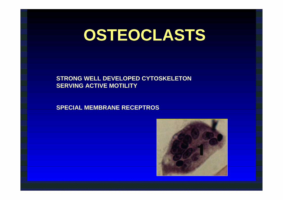
OSTEOCLASTS
STRONG WELL DEVELOPED CYTOSKELETON SERVING ACTIVE MOTILITY
SPECIAL MEMBRANE RECEPTROS

OSTEOCLASTS ENZYME SYSTEM
ACID PHOSPHATASE
ββββ-GLUCORONIDASE
CYSTEIN PROTEASE – CATHEPSIN B
TISSUE PLASMINOGEN ACTIVATOR
MATRIX METALLOPROTESES COLLAGENASE
LYSOZYME


LACUNALIS CSONTRESORPTIO
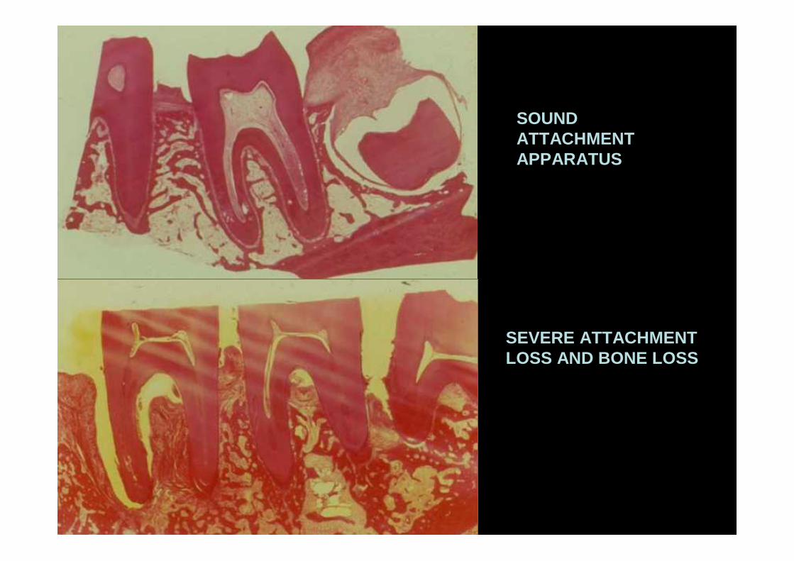
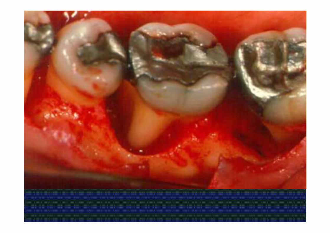
SOUND ATTACHMENT APPARATUS
SEVERE ATTACHMENT LOSS AND BONE LOSS


poor oral hygiene
normal flora exogenous infection
pathoge-nic flora
PMNclearance
Antibodyresponse
bacterialpenetration
gingivitis
monocytelymphocyteaxis
cytokinesPGE2mediators
inflammationtissue destruction
pocketingbone loss
Systemicexposure
Yes
Noparodontitis
pathogenesis

poor oral hygiene
normal flora exogenous infection
pathoge-nic flora
PMNclearance
Antibodyresponse
bacterialpenetration
gingivitis
monocytelymphocyteaxis
cytokinesPGE2mediators
inflammationtissue destruction
pocketingbone loss
Systemicexposure
Yes
No
host stress
_
+
disease cycle

poor oral hygiene
normal flora exogenous infection
pathoge-nic flora
PMNclearance
Antibodyresponse
bacterialpenetration
gingivitis
monocytelymphocyteaxis
cytokinesPGE2mediators
inflammationtissue destruction
pocketingbone loss
Systemicexposure
No
Risk factorsgenetic traitsdiabetessmoking
_
++
Yes

poor oral hygiene
normal flora exogenous infection
pathoge-nic flora
PMNclearance
Antibodyresponse
gingivitis
monocytelymphocyteaxis
cytokinesPGE2mediators
inflammationtissue destruction
pocketingbone loss
Systemicexposure
Yes
No
debridement
_
++ bacterialpenetration
growthfactors
healingrepairregeneration
treatment cycle