the pathophysiology of letter-by-letter reading - · pdf filel. cohen et al./neuropsychologia...
TRANSCRIPT

Neuropsychologia 42 (2004) 1768–1780
The pathophysiology of letter-by-letter reading
Laurent Cohena,b,∗, Carole Henrya,b, Stanislas Dehaeneb, Olivier Martinauda,b,Stéphane Lehéricyc, Cathy Lemerb, Sophie Ferrieuxa
a Institut de Neurologie, Hˆopital de la Salpˆetrière, 47/83 Bd de l’Hˆopital, 75651 Paris CEDEX 13, Franceb INSERM U562, Service Hospitalier Frédéric Joliot, CEA/DSV, Orsay, Francec Service de Neuroradiologie, Hˆopital de la Salpˆetrière, AP-HP, Paris, France
Received 24 March 2004; received in revised form 28 April 2004; accepted 30 April 2004
Abstract
Pure alexia is a frequent and incapacitating consequence of left occipitotemporal lesions. It is thought to result from the disruption or thedisconnection of the visual word form area (VWFA), a region reproducibly located within the left occipito-temporal sulcus, and encoding theabstract identity of strings of visual letters. Alexic patients often retain effective single letter recognition abilities, and develop an effortfulletter-by-letter reading strategy which is the basis of most rehabilitation techniques. We study a patient who developed letter-by-letterreading following the surgical removal of left occipito-temporal regions. Using anatomical and functional MRI in the patient and innormal controls, we show that alexia resulted from the deafferentation of left fusiform cortex, and we analyze the network of brain regionssubtending letter-by-letter reading. We propose that during letter-by-letter reading (1) letters are identified in the intact right-hemisphericvisual system, with a central role for the region symetrical to the VWFA; (2) letters are serially transferred to the left hemisphere throughthe intact segment of the corpus callosum; (3) word identity is eventually recovered in the left hemisphere through verbal working memoryprocesses involving inferior frontal and supramarginal cortex.© 2004 Elsevier Ltd. All rights reserved.
Keywords:Reading; Alexia; fMRI; Fusiform; Language
1. Introduction
Pure alexia is a frequent and incapacitating consequenceof left occipito-temporal lesions (Binder & Mohr, 1992;Damasio & Damasio, 1983). Affected patients suddenly loseexpert reading abilities that they have acquired through yearsof academic training. At the same time, speech comprehen-sion and production, as well as word spelling, are preserved.The essence of this lost perceptual skill is (1) letter identi-fication invariant for position, size, font, and case; and (2)the fast and parallel identification of arrays of several let-ters (Dehaene et al., 2001; McCandliss, Cohen, & Dehaene,2003). It is not before the age of 10 that children showthe adult reading pattern (Aghababian & Nazir, 2000), i.e.short word reading latencies that are independent of wordlength, at least within a range of about three to seven lettersand in optimal display conditions (Lavidor, Ellis, Shillcock,& Bland, 2001; Weekes, 1997). In severe cases, known asglobal alexia, patients cannot access abstract letter identity
∗ Corresponding author. Tel.:+33 1 42161849/42161802;fax: +1 44 24 52 47.
E-mail address:[email protected] (L. Cohen).
(Miozzo & Caramazza, 1998), and are unable to name evensingle letters (Dejerine, 1892). More often, alexic patientskeep effective letter recognition abilities and develop an ef-fortful letter-by-letter reading strategy, which is the basisof most rehabilitation techniques (Greenwald & GonzalezRothi, 1998). Eventually, this procedure may become quiteeffective, although it remains easily detectable in the exces-sive effect of word length on reading latencies (Behrmann,Black, & Bub, 1990).
A variety of questions and hypotheses have been putforward concerning letter-by-letter reading (for selectedreferences seeMontant & Behrmann, 2000). Does it re-flect the partial preservation of normal premorbid processes(Behrmann, Plaut, & Nelson, 1998), or is it based on novelstrategies (Speedie, Rothi, Heilman, 1982)? Is it basedon residual left-hemispheric (LH) or on compensatoryright-hemispheric (RH) mechanisms (Coslett & Saffran,1998)? Does reveal a general impairment in processing si-multaneous visual objects (Farah & Wallace, 1991) or com-plex displays (Montant & Behrmann, 2000), or is it specificto reading? Such issues may be clarified by refering to asimple model of the visual stages of reading (McCandlisset al., 2003). Letters displayed in one hemifield are first an-
0028-3932/$ – see front matter © 2004 Elsevier Ltd. All rights reserved.doi:10.1016/j.neuropsychologia.2004.04.018
L. Cohen et al. / Neuropsychologia 42 (2004) 1768–1780 1769
alyzed through a cascade of contralateral retinotopic areas,which compute increasingly abstract representations. Even-tually, a representation of letter identities is created in thevisual word form area (VWFA), reproducibly located withinthe left occipito-temporal sulcus, at about TC –42, –63, –15(Cohen et al., 2003). This representation is invariant forparameters such as size, position, case, font. The transfer ofvisual information from lower-order retinotopic cortices tothe VWFA takes place within the left hemisphere for stim-uli displayed in the right visual field (RVF). For left visualfield (LVF) stimuli, information is conveyed through inter-hemispheric fiber tracts that course in the splenium of thecorpus callosum and over the posterior horns of the lateralventricles (Binder & Mohr, 1992; Molko et al., 2002). TheVWFA then projects to structures involved in phonologicalor lexico-semantic processing.
In this framework, pure alexia is thought to result eitherfrom the disruption of the VWFA itself, or from impairedprojections to or from this system. We recently showedthat the critical lesion for pure alexia overlaps accuratelywith the VWFA, as identified by its activation duringnormal reading (Cohen et al., 2003). In this study, func-tional imaging data were obtained in a patient with typicalletter-by-letter reading (patient F.). We observed that theintact right-hemispheric cortex symmetrical to the VWFA,which we labeled the R-VWFA, showed a pattern of acti-vation normally specific to the VWFA itself, i.e. strongeractivation for alphabetic strings than for chequeboards.Moreover, there were abnormally strong activations in a leftfrontoparietal network related to verbal working memory.This pattern supported the idea that in letter-by-letter read-ing, (1) alphabetic symbols are identified in the R-VWFA,(2) they are serially transferred to the left hemisphere, and(3) word identity is recovered by the left hemisphere throughan effortful verbal working memory process. Note that thisconstrual is in line with a classical account of letter-by-letterreading (e.g.Binder & Mohr, 1992; Speedie et al., 1982).
Patient F.’s lesion spared the dorsal bank of the calcarinesulcus, and accordingly the lower right visual quadrant wasintact. As we found some residual activations at coordinatesclose to those of the VWFA, it could not be excluded thata partially spared left-hemispheric pathway leading fromV1 to the VWFA could contribute, or even be essential, toletter-by-letter reading. Here we study a young patient (C.Z.)who presented letter-by-letter reading following the surgicalresection of left occipito-temporal structures, sparing theVWFA but including the entire primary visual cortex andall fiber tracts leading to the VWFA from both the right andthe left visual fields.
Our aims were to assess the account of letter-by-letterreading summarized before, and to fully document a case ofpure alexia due to deafferentation of an intact VWFA. Fi-nally, the complete surgical removal of left posterior regions,as opposed to patchy lesions following infarcts, may helpto delimit the role of residual left hemispheric structures inletter-by-letter reading.
2. Medical history and lesion description
Patient C.Z. was a 19-year-old student, 100% right-handedaccording to the Edinburgh inventory (Oldfield, 1971). Shewas operated twice for the complete resection of a leftoccipital neuroectodermal tumor, revealed by intracranialhypertension and right hemianopia. Surgery was followedby radiotherapy and chemotherapy. The present study wascarried out between 6 and 12 months after the surgery.There was no indication of relapse on close clinical and ra-diological follow-up. Following the intervention, the patientcomplained of being unable to read, while she could writenormally. Her family also reported substantial memorydifficulties.
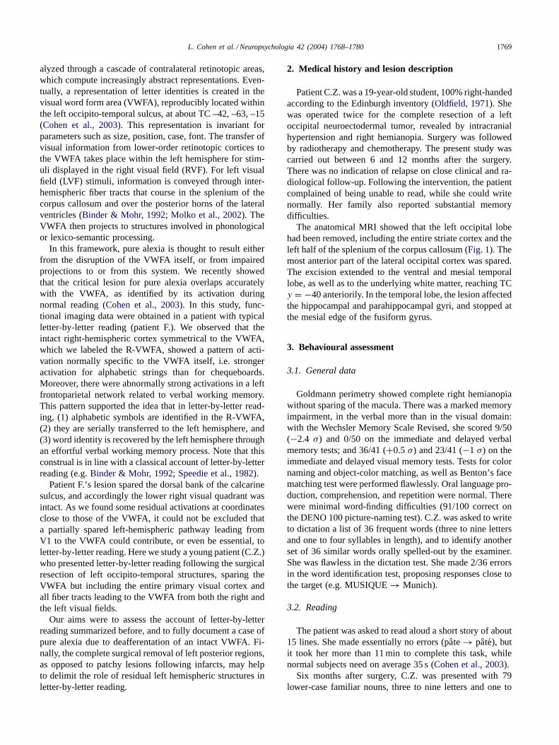
The anatomical MRI showed that the left occipital lobehad been removed, including the entire striate cortex and theleft half of the splenium of the corpus callosum (Fig. 1). Themost anterior part of the lateral occipital cortex was spared.The excision extended to the ventral and mesial temporallobe, as well as to the underlying white matter, reaching TCy = −40 anteriorily. In the temporal lobe, the lesion affectedthe hippocampal and parahippocampal gyri, and stopped atthe mesial edge of the fusiform gyrus.
3. Behavioural assessment
3.1. General data
Goldmann perimetry showed complete right hemianopiawithout sparing of the macula. There was a marked memoryimpairment, in the verbal more than in the visual domain:with the Wechsler Memory Scale Revised, she scored 9/50(−2.4 σ) and 0/50 on the immediate and delayed verbalmemory tests; and 36/41 (+0.5σ) and 23/41 (−1 σ) on theimmediate and delayed visual memory tests. Tests for colornaming and object-color matching, as well as Benton’s facematching test were performed flawlessly. Oral language pro-duction, comprehension, and repetition were normal. Therewere minimal word-finding difficulties (91/100 correct onthe DENO 100 picture-naming test). C.Z. was asked to writeto dictation a list of 36 frequent words (three to nine lettersand one to four syllables in length), and to identify anotherset of 36 similar words orally spelled-out by the examiner.She was flawless in the dictation test. She made 2/36 errorsin the word identification test, proposing responses close tothe target (e.g. MUSIQUE→ Munich).
3.2. Reading
The patient was asked to read aloud a short story of about15 lines. She made essentially no errors (pate→ pate), butit took her more than 11 min to complete this task, whilenormal subjects need on average 35 s (Cohen et al., 2003).
Six months after surgery, C.Z. was presented with 79lower-case familiar nouns, three to nine letters and one to
1770 L. Cohen et al. / Neuropsychologia 42 (2004) 1768–1780
Fig. 1. Normalized T1-weighted MRI of patient C.Z., showing the surgical resection of most of the left occipital lobe including the left half of the splenium,the posterior hemispheric white matter, the hippocampal and parahippocampal gyri. The white arrowhead points to the spared mid-fusiform cortex.
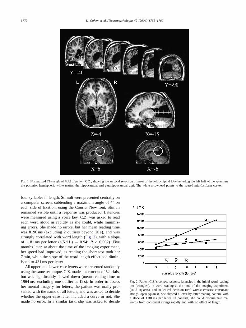
four syllables in length. Stimuli were presented centrally ona computer screen, subtending a maximum angle of 4◦ oneach side of fixation, using the Courier New font. Stimuliremained visible until a response was produced. Latencieswere measured using a voice key. C.Z. was asked to readeach word aloud as rapidly as she could, while minimiz-ing errors. She made no errors, but her mean reading timewas 8196 ms (excluding 2 outliers beyond 20 s), and wasstrongly correlated with word length (Fig. 2), with a slopeof 1181 ms per letter(r(5 d.f .) = 0.94; P < 0.002). Fivemonths later, at about the time of the imaging experiment,her speed had improved, as reading the short text took her7 min, while the slope of the word length effect had dimin-ished to 431 ms per letter.
All upper- and lower-case letters were presented randomlyusing the same technique. C.Z. made no error out of 52 trials,but was significantly slowed down (mean reading time=1964 ms, excluding one outlier at 12 s). In order to assessher mental imagery for letters, the patient was orally pre-sented with the name of all letters, and was asked to decidewhether the upper-case letter included a curve or not. Shemade no error. In a similar task, she was asked to decide
Fig. 2. Patient C.Z.’s correct response latencies in the initial word readingtest (triangles), in word reading at the time of the imaging experiment(solid squares), and in lexical decision (real words: crosses; consonantstrings: open squares). She showed a letter-by-letter reading pattern, witha slope of 1181 ms per letter. In contrast, she could discriminate realwords from consonant strings rapidly and with no effect of length.

L. Cohen et al. / Neuropsychologia 42 (2004) 1768–1780 1771
whether lower-case letters included a descender (j, g, p, q,y). She made 4/26 errors, all corresponding to an initial mis-understanding of instructions.
In order to assess the access to abstract letter identity, twofurther tests were proposed to C.Z., using only letters withwidely different upper-case and lower-case shapes. First, shewas presented with 10 upper-case and 10 lower-case lettersand was asked to write down each letter in the opposite case.She made a single error (q → G). Second, the patient waspresented with 80 pairs of letters horizontally displayed ona computer screen. The two letters in a pair were printedin different cases, and C.Z. had to decide whether the twoletters corresponded to the same grapheme (e.g. a A) or not(e.g. a B), and to respond using one of two keys. C.Z. made2/80 errors. In order to make it more sensitive to subtleimpairments of letter processing, this task was run again,with stimuli displayed for only 200 ms and followed by amasking pattern (######). The patient made 24/80 errors.However, as this high error rate could be due to hemianopia,the test was run once again, with the two letters displayedone above the other. C.Z. still made 12/80 errors, with a biasfor responding “different”.
Finally, the patient was submitted to lexical decision tasks.She was first asked to discriminate words versus stringsof consonants. She was presented with three lists of 30items each. Each list comprised 15 frequent words (log10frequency per million >2), 4–6 letters long, and 15 conso-nant strings matched in length to the words. Stimuli weredisplayed for 200 ms and followed by a masking pattern(######). C.Z. had to decide whether stimuli were realwords or not, and to respond using one of two keys. The pa-tient judged the task to be easy, explaining that she could in-stantly pick out large consonant clusters, and she performedway above chance (17/90 errors; 19%;P < 0.0001). Shehad a mean RT of 947 ms, and latencies did not increase withword length, suggesting that, in this task, the patient did notresort to letter-by-letter reading (Fig. 2). To further clarifywhether the patient could indeed detect clusters of conso-nants beyond the reach of letter-by-letter reading, two lists ofsix-letter stimuli were created, for which the decision couldbe based only on the last three letters. The first list, whichwas run three times, included 12 words with three initialconsonants, and 12 matched consonant strings with the samefirst three letters (e.g. SPHERE versus SPHCRQ). The sec-ond list, which was run twice, included 25 frequent words,and 25 matched non-words, with the same first three lettersbut three final consonants (e.g. BONDIR versus BONDFL).Rare letters such as K, X, Z were excluded. The patientresponded better than chance with both lists (21/72 errors;29%; P < 0.025; and 35/100 errors; 35%;P < 0.05, re-spectively), confirming that she could pick out consonantclusters better than expected on the basis of left-to-rightletter-by-letter reading.
In summary, the patient showed typical pure alexia, witha sparing of oral language and of spelling abilities. She re-sorted to letter-by-letter word reading, with a slope of read-
ing latencies that was initially over 1 s per letter. The identi-fication of single letters was accurate, but abnormally slow,with errors in a cross-case matching task under short displayconditions. Finally, C.Z. could apparently spot consonantstrings with relatively good accuracy and speed. She wasindeed faster at detecting consonant strings than at readingthe shortest words, or even single letters.
3.3. Brain imaging
The patient was submitted to the same imaging procedureand compared to the same group of nine normal controls asdescribed inCohen et al. (2003).
3.4. Procedure
The patient was presented with four types of stimuli: a fix-ation point, real words, consonant strings, and chequerboardscovering approximately the same field as letter strings. The128 words were a subset of the list used for the behaviouralassessment, including 38 three to four words, 60 five to sevenletter words, 30 eight to nine letter words. Consonant stringswere matched one-to-one to real words in number of letters.The frequency distribution of consonants was the same inconsonant strings as in words. Stimuli were presented cen-trally for 1700 ms, followed by a 800 ms fixation point. Thepatient was instructed to pay attention equally to all typesstimuli and to read real words covertly.
The patient received four fMRI sequences, each compris-ing 16 experimental blocks, i.e. four blocks of each condi-tion. Each block comprised eight trials. Blocks were pre-sented in pseudo-random order within sequences, so as tomaximize the variety of transitions between conditions whileavoiding any repetition of the same condition in successiveblocks. Trials were presented in random order within blocks.The order of blocks within sequences and the order of trialswithin blocks differed across the four sequences.
3.5. Imaging parameters
Each sequence consisted of 10 s of initial fixation, fol-lowed by 16 stimulation blocks of 20 s each, as describedbefore. In each sequence, 132 functional volumes sensi-tive to blood oxygen level dependent (BOLD) contrast wereacquired with a T2-weighted gradient echo, echo planarimaging sequence on a 1.5 tesla Signa Imager (TR (relax-ation time)= 2500 ms,α (flip angle)=90, TE (echo time)= 60 ms, field of view= 240 mm× 240 mm, in plane res-olution = 3.75 mm× 3.75 mm). Each volume comprised17 axial slices of 5 mm thickness covering most of thebrain. The first four volumes were discarded to reach sig-nal equilibrium. High-resolution T1-weighted images (3Dfast gradient-echo inversion recovery sequence, TI (inver-sion time)= 400 ms, TR= 11 ms, TE=2 ms,α = 20, fieldof view = 240 mm × 240 mm, slice thickness=1.5 mm,

1772 L. Cohen et al. / Neuropsychologia 42 (2004) 1768–1780
in-plane resolution= 0.94 mm× 0.94 mm) were also ac-quired for anatomical localization, as well as T2-weightedfast spin echo and FLAIR axial slices.
3.6. Analysis of MRI data
Functional images were analysed with the StatisticalParametric Mapping software (SPM99). To correct for mo-tion, functional scans were realigned using the first imageas a reference. As functional images were locally distortedby metallic surgical material, functional images were trans-formed to TC (Talairach & Tournoux, 1988) using thestandard EPI template, applying nonlinear normalizationparameters while masking the lesion and the magnetic ar-tifact (Brett, Leff, Rorden, & Ashburner, 2001). Functionalimages were then smoothed with a Gaussian spatial filter(5 mm FWHM). The resulting images had cubic voxels of3 mm× 3 mm× 3 mm. The anatomical image was linearlytransformed to TC using the standard T1 template.
Activation on each of the 4 types of trials was modeled bya combination of the standard SPM haemodynamic functionand its temporal derivative. Only the former function wasused for statistical contrasts. Four additional variables ofnon-interest modelled constant differences across the foursequences. Long-term signal variations were eliminatedwith a high-pass filter set at 240 s. Low-pass filtering wasachieved by convolution with the haemodynamic responsefunction. In individual analyses, we used an uncorrectedvoxelwise threshold ofP < 0.001, a clusterwise thresh-old of P < 0.05 corrected, and all peak voxels mentionedin the descriptions of activation networks had a statisticalsignificance ofP < 10−5, unless stated otherwise.
4. Results
We will first delineate the overall network of activatedareas, then concentrate on regions involved in processingalphabetic stimuli, and eventually focus on letter-by-letterreading by contrasting words versus consonant strings. Weput an emphasis on occipito-temporal regions, and partic-ularly on the distribution of activations across the lesionedleft-hemispheric and the intact right-hemispheric cortices.Whenever relevant, individual analyses of the patient’s dataare supplemented with comparisons to the group of normalcontrols.
4.1. Overall reading network
4.1.1. Alphabetic stimuli versus fixationIn order to identify the overall reading network, we con-
trasted alphabetic stimuli (i.e. words and consonant strings)versus fixation (Fig. 3). Ventral occipito-temporal activationswill be reported last.
There were bilateral activations in the horizontal (left: TC–30, −57, 51; Z > 15; right: TC 27, –48, 54;Z > 15)
and posterior (left: TC –21, –66, 30;Z > 15; right: TC 33,–63, 21;Z > 15) segments of the intraparietal sulcus; inthe SMA/cingulate (TC –3, 9, 57;Z > 15); in the dorsolat-eral (left: TC –27, 42, 15;Z = 6.68; right: TC 21, 69, 24;Z = 6.87 and TC 30 45 30;Z = 7.55) and orbital (left:TC −33 45 –24;Z > 10; right: TC 12, 63, –15;Z = 7.30)prefrontal cortex; in the precentral cortex (left: TC−48, 0,30; Z > 15; right: 51, 21, 33;Z > 10); in Broca’s areaand its right-sided counterpart (left: TC –42, 27, 0;Z > 9;right: TC 48, 24, –9;Z = 6.59). Although bilateral, mostof those activations showed left-hemispheric predominance.Additionally there were left-sided activations in the supra-marginal gyrus (SMG) (TC –57, –33, 33;Z = 7.84) and thethalamus (TC –12, –18, 6;Z > 8).
In the LH, there was a residual stripe of activation in theventral occipito-temporal cortex just lateral to the lesion.This activation followed the fusiform gyrus in its posterior(TC –36, –84, –12;Z = 4.60; TC −33, –75, –18;Z >
10) and middle segment (TC –33 –57 –15;Z = 6.07), andextended anteriorily to TCY = −39. Note that the peak atTC –33, –57, –15 was close to the normal coordinates ofthe VWFA.
The symmetrical activations in the RH were more exten-sive, and the intensity of activations as measured by the per-cent change of BOLD signal was about three times higherthan in the left-hemispheric ventral temporal cortex (Fig.3). Within the right parieto-occipito-temporal cluster, ventralactivations started from the occipital pole (TC 24, –96, –18;Z > 20), and followed the fusiform gyrus in its posterior (TC42, –87, –21;Z > 15) and middle segment (TC 51,−69,–21;Z > 9), again extending to TCY = −39. This stripe in-cluded the region symmetrical to the VWFA. Slightly moredorsally there were activation foci in the mesial (TC 21–81,0; Z > 9) and lateral (TC 39–81, –6;Z > 15) posterioroccipital cortex.
4.1.2. Alphabetic stimuli versus chequerboardsIn order to identify within this network which regions
were more specifically involved in processing alphabeticstimuli, we compared alphabetic stimuli versus chequer-boards, while masking by words vs. chequerboards and con-sonants vs. chequerboards (P < 0.01 each). This analysisrevealed a large network almost encompassing the entirenetwork identified relative to fixation.
In the left occipito-temporal region, there was again astripe of activation following the fusiform gyrus from itsposterior segment (TC−36, –84, –21;Z = 5.63; TC−33,–75, –18;Z = 7.73) to TCY = −57. In the RH, activationsdid not start from the occipital pole, but from the posteriorsegment of the fusiform gyrus (TC 45 –87 –21;Z > 15),extended to its middle portion (TC 54, –69, –21;Z = 6.45)including the region symmetrical to the VWFA (TC 39, –51,–27;Z = 6.65), and anteriorily to TCY = −45. Note thatthe occipital pole did not appear in this contrast because itwas equally activated by chequerboards (TC 24, –93, –18;Z > 15) and alphabetic stimuli relative to fixation (Fig. 3).

L. Cohen et al. / Neuropsychologia 42 (2004) 1768–1780 1773
Fig. 3. Top: General reading network as revealed by the contrast of alphabetic stimuli versus fixation, overlaid on a 3D render of the patient’s brain.Bottom: Percent change in BOLD signal relative to fixation in selected occipito-temporal voxels. Histogram bars correspond to chequerboards, alphabeticstimuli, consonant strings, and real words, respectively. Except for the right posterior occipital cluster, all regions showed a stronger activation foralphabetic stimuli than for chequerboards. Contrary to the normal pattern, activations were about 3 times stronger in the right than in the left hemisphere,and the R-VWFA showed stronger activations for alphabetic stimuli than for chequerboards.
4.1.3. Occipito-temporal activations in the patient and incontrols
Thus two anomalies were observed in the patient’soccipito-temporal activations. First, in normals,contrastingalphabetic stimuli versus fixationelicits roughly symmet-rical ventral occipito-temporal activations. In the patient,activations were three times stronger in the right than in theleft hemisphere, in the cortex just lateral to the lesion. Sec-ond, in normals,contrasting alphabetic stimuli versus che-
querboardsshowed exclusively left-sided occipito-temporalactivations. In the patient however, such activations pre-dominated in the right hemisphere.
In order to assess the abnormality of this pattern, wecompared the contrast of alphabetic stimuli versus che-querboards in the patient relative to the group of controls(voxelwise P < 0.02; P < 0.05 for cluster extent cor-rected within the patient’s overall reading network). Thisshowed right-hemispheric activation in the occipital cor-

1774 L. Cohen et al. / Neuropsychologia 42 (2004) 1768–1780
tex (TC 33, –63, 15;Z = 5.18), the posterior fusiformgyrus (TC 45, –78, –12;Z = 4.83), and extending in theparahippocampal region (TC 27, –48, –6;Z = 3.08). Therewas also a small cluster in the region symmetrical to theVWFA (TC 42, –54, –27;Z = 2.73) below the extentthreshold. There was no activation in the correspondingleft-hemispheric regions. This confirms that the patient’sright occipito-temporal cortex symmetrical to the VWFAwas strongly and abnormally involved in the processingof alphabetic stimuli, as opposed to simple geometricalpatterns.
Fig. 4. Top: Right occipital, predominantly left frontal, and left parietal regions activated by words vs. consonant strings, and associated with letter-by-letterreading. Middle: The same regions are found when comparing activations by words vs consonant in the patient relative to normal controls. Note thatright occipital activations are visible on the left lateral view due to the resection of the left occipital pole. Bottom: Percent change in BOLD signalrelative to fixation in selected voxels with stronger activation for words than for consonants.
4.2. Letter-by-letter reading: words versus consonantstrings
Beyond an increased reliance on right temporal cortexfor alphabetic processing, letter-by-letter reading should re-quire a more extensive network of areas in order to pro-gressively figure out word identity. A critical contrast todelimit this network is the difference between real wordsand consonant string (Fig. 4). Indeed, the patient could spotrapidly and accurately consonant strings, and she thereforeengaged fully in letter-by-letter reading with real words

L. Cohen et al. / Neuropsychologia 42 (2004) 1768–1780 1775
only. Note that according to her own report, the patientwas not always sure of having correctly identified targetwords, a limitation which could be expected consideringthe relatively rapid rate of stimulation. However, she al-ways attempted to identify words through letter-by-letterreading. The activation patterns reported now confirmedthat words and consonant strings were actually processeddifferently.
The subtraction of words minus consonant strings showedleft hemispheric activations in the left precentral cortex (TC–60, 9, 33;Z = 6.53), Broca’s area (TC –42, 27, 0;Z =6.04), and the SMG (TC –66, –27, 42;Z = 5.27; cluster-wiseP < 0.1 corrected). There were bilateral activations inthe SMA (left: TC –9, 21, 54;Z = 4.81; right: TC 3, 30,51; Z = 3.98; clusterwiseP < 0.06 corrected). In the RH,there were activations in the dorsolateral (TC 45, 24, 21;Z = 4.33) and mesial (TC 9, 45, 21;Z = 4.46; clusterwiseP < 0.06 corrected) prefrontal cortex; in the inferior frontalgyrus (TC 30, 27, –12;Z = 5.74); and in the mesial occip-ital cortex (TC 18, –84, 9;Z = 5.06; and TC 30, –63, 3;Z = 4.42).
In order to assess statistically the deviation of this patternfrom the norm, we compared the contrast of words versusconsonants in the patient relative to the group of controls(voxelwiseP < 0.02; P < 0.05 for cluster extent correctedwithin the patient’s overall reading network). This procedureshowed a network essentially identical to the previous anal-ysis (Fig. 4): left precentral (TC –57, 9, 30;Z = 3.51; andTC −39, –3, 30;Z = 3.34), Broca’s area (TC –33, 21, –9;Z = 3.80), right inferior frontal (TC 33, 30, –9;Z = 3.75),and right occipital activations (TC 18, –78, 6;Z = 4.40;andTC 30, –60, 0;Z = 3.85) (all Ps< 0.001). Activations inthe left SMG (TC –66, –24, 39;Z = 3.51; P < 0.001),bilateral mesial frontal cortex, and right dorsolateral pre-frontal were also present but too small to reach clusterwisesignificance.
5. Discussion
5.1. Pure alexia due to a disconnected VWFA?
We proposed that the VWFA, as identified in a numberof functional studies using PET or fMRI (Beauregard et al.,1997; Brunswick, McCrory, Price, Frith, & Frith, 1999; Fiez,Balota, Raichle, & Petersen, 1999; Cohen et al., 2000, 2002;Dehaene et al., 2001; Dehaene, Le Clec’H, Poline, LeBihan, & Cohen, 2002; Paulesu et al., 2000; Wagner et al.,1998), MEG or intracerebral recordings (Allison, McCarthy,Nobre, Puce, & Belger, 1994; Allison, Puce, Spencer, &McCarthy, 1999; Cohen et al., 2000; Nobre, Allison, &McCarthy, 1994; Salmelin, Service, Kiesilä, Uutela, &Salonen, 1996; Tarkiainen, Helenius, Hansen, Cornelissen,& Salmelin, 1999; Simos et al., 2002) plays a key role inreaching an abstract visual representation of letter strings,independent from position, font, or case, on the basis of
fast and parallel letter identification. In this framework,a selective disruption of the VWFA should result in anisolated reading deficit, in the absence of other languageimpairments, i.e. pure alexia. The current evidence fromanatomo-clinical correlations is compatible with this hy-pothesis, as it points to the mid-portion of the left fusiformgyrus as the critical lesion site for pure alexia (Beversdorf,Ratcliffe, Rhodes, & Reeves, 1997; Binder & Mohr, 1992;Cohen et al., 2003; Damasio & Damasio, 1983).
However, a close analysis of patient C.Z.’s lesion suggeststhat it actually spared the fusiform cortex and the VWFA(Fig. 1). In Talairach normalized space, the peak activationof the VWFA in control subjects falls at about TC –42,–63, –15 (Cohen et al., 2003). At this anteroposterior level(y = −63), patient C.Z.’s lesion extended laterally only tox = –28, that is more mesial than the VWFA of any nor-mal subject (Fig. 5). This point is confirmed by consideringanatomical landmarks rather than normalized coordinates.In individual analyses, the VWFA consistently falls in thedepth of the occipito-temporal sulcus, lateral to the fusiformgyrus (Cohen et al., 2000; Cohen et al., 2002; Dehaene et al.,2002; Gauthier et al., 2000; Puce, Allison, Asgari, Gore, &McCarthy, 1996), a region that was clearly spared in patientC.Z. Finally, the patient showed residual activations at thenormal coordinates of the VWFA.
Considering that the VWFA itself was spared, we pro-pose that the reading deficit resulted from its deafferentationfrom visual input. As visible onFig. 1, the left occipital lobeand the occipito-temporal white matter were extensively re-moved, including callosal connexions, depriving the remain-ing fusiform gyrus of input connections from both left- andright-hemispheric lower-level visual cortex (Catani, Jones,Donato, & Ffytche, 2003). Indeed, the patient’s white mat-ter lesion encompassed and far exceeded the typical cal-losal lesion responsible for alexia in the left visual hemifield(Suzuki et al., 1998). In such cases, deafferentation of theVWFA from right-hemispheric input was demonstrated by areduced activation by left-sided as compared to right-sidedwords (Cohen et al., 2000; Cohen et al., 2003). Moreover, theinterhemispheric pathway critically involved in hemialexia,as delineated using diffusion tensor imaging, falls within theregion that was resected in the present case (Molko et al.,2002). As a point of history, one may note that the originalalexic patient reported byDejerine (1892)also had an intactmid-fusiform cortex, while the lesion affected the underly-ing white matter (Dejerine, 1895).
5.2. Role of residual left fusiform activations
Although patient C.Z.’s VWFA was deprived of visualinput, it was significantly activated during the imaging ex-periment. However, those activations were much weakerthan their right-hemispheric counterpart, due to corticalexcision and to the disconnection of remaining cortex.Their functional meaning remains unclear. It should bestressed that C.Z.’s pattern of alexia was perfectly indistin-

1776 L. Cohen et al. / Neuropsychologia 42 (2004) 1768–1780
Fig. 5. Patient C.Z.’s lesion was just mesial to the left fusiform gyrus, thus sparing the VWFA, as identified by its activation in normal controls (left),by its residual activation in patient C.Z. (middle), and by the overlap of cortical lesions in patients with pure alexia (right;Cohen et al., 2003).
guishable from other more common patients with corticaldamage actually destroying the fusiform cortex, like forinstance patients reported byCohen et al. (2003). Thisdemonstrates that the preservation of an intact but deaf-ferented fusiform cortex makes no difference as regardsreading performance. It thus seems safe to conclude thatthe patient’s VWFA played no causal role in letter-by-letterreading.
The residual activation of the VWFA probably resultedfrom top-down influences reaching the VWFA throughpreserved long-distance and U-shaped association fibers.Indeed ventral areas devoted to object recognition can beactivated in the absence of visual input during mental im-agery (O’Craven & Kanwisher, 2000). Patient C.Z. wasexcellent in tasks of mental imagery involving letter shapes,an ability to which the VWFA possibly contributed (for anopposite case seeBartolomeo, Bachoud-Levi, Chokron, &Degos, 2002). There is also evidence that infero-temporalregions can receive top-down activation by auditory words,whenever orthographic processing is required. ThusBurton,Small, & Blumstein (2000)observed a left infero-temporalactivation (TC−58, −56, −8) when subjects performedsame-different judgments on spoken syllables, only whenthey had to extract the first phoneme of the syllable, but notwhen their decision was based on the syllable as a whole.The former task is not within the reach of illiterate subjects,and requires reading-dependent phonological awareness(Morais & Kolinsky, 1994). Similarly, Booth et al. (2002,2003) observed an activation of the left VWFA (TC−45,−57, −12) when subjects engaged in a spelling task onauditory words, but not when they performed a rhymingtask on the same stimuli. It is an open question whetherthe residual activations of the patient’s VWFA could bemodulated by such task manipulations.
In summary, patient C.Z.’s VWFA was anatomicallyspared, but deafferented of all visual input, explaining theoccurrence of pure alexia. Residual top-down activationswere observed in the VWFA, but they probably played noactual role in letter-by-letter reading.
5.3. The mechanisms of letter-by-letter reading
One maya priori assume that efficient letter-by-letterreading requires the cooperation of a letter identification de-vice, presumably located in the ventral visual stream, and ofa verbal working memory network in charge of combiningletters and of accessing word identity, presumably involvingleft-hemispheric perisylvian fronto-parietal structures. Thefunctional imaging study of patient C.Z. allowed us to fleshout these hypotheses, which will be discussed in turn.
5.4. Letter identification and the right occipito-temporalcortex
We argued that although the VWFA itself was sparedby the resection, it was deprived of any visual input andprobably played no causal role in letter-by-letter reading.The right-sided occipito-temporal cortex, including the R-VWFA, appears as a likely substitute for the purpose of iden-tifying visual letters. There is indeed convergent evidencethat the right hemisphere is able to identify letters, from stud-ies of split-brain patients (Baynes & Eliassen, 1998), patientswith left hemispherectomy (Patterson, Vargha-Khadem, &Polkey, 1989), or deep dyslexia from large left-sided lesions(Coltheart, 1980). Furthermore, the residual reading abili-ties of pure alexic patients may be lost following disrup-tion of right occipito-temporal systems, either following asubsequent right-sided lesion (Bartolomeo, Bachoud-Levi,Degos, & Boller, 1998), or by means of transcranial mag-netic stimulation (Coslett & Monsul, 1994).
Regarding the specific involvement of the R-VWFA, ourhypothesis is supported by the abnormal pattern of activa-tion of this region observed in patient C.Z. First, the R-VWFA was activated 3 times more strongly by alphabeticstimuli relative to rest than the VWFA proper, while activa-tions are roughly symmetrical in normal subjects. Second,the R-VWFA and more posterior right fusiform cortex weremore activated by alphabetic stimuli than by chequerboards,a key functional feature of the normal VWFA. This pattern

L. Cohen et al. / Neuropsychologia 42 (2004) 1768–1780 1777
was similar to what was observed in another letter-by-letterreader (Cohen et al., 2003). That the R-VWFA should takeup letter identification probabaly does not imply a thoroughmodification of its functional properties. Indeed, in the pro-tocol used with patient C.Z., normal controls, the R-VWFAis activated by alphabetic stimuli relative to fixation with al-most the same strength as the VWFA. This suggests that,in the right hemisphere also, letters are encoded in high-level ventral visual cortex devoted to object recognition. Aleft-hemispheric predominance emerges most clearly whencontrasting letter strings versus higher-level visual controlssuch as chequerboards (Cohen et al., 2002), textures (Puceet al., 1996), faces (Gauthier et al., 2000; Puce et al., 1996),buildings (Hasson, Levy, Behrmann, Hendler, & Malach,2002), or pseudoletters (Price, Wise, & Frackowiak, 1996).Some of the specificities of the VWFA as compared to theR-VWFA may be that the VWFA builds up a representationof letter strings more invariant for changes of case (Dehaeneet al., 2004; Dehaene et al., 2001; see alsoKoutstaal et al.,2001) and of spatial position (Cohen et al., 2000; Cohenet al., 2002; Hasson et al., 2002), and is more selectivelytuned to alphabetic stimuli than to other types of objects(Hasson, Harel, Levy, & Malach, 2003).
Hence one may expect that the substitution of the VWFAby the right-hemispheric structures should not be perfect.This may explain the patient’s abnormal performance in de-manding letter processing tasks, particularly in the cross-case matching task with fast stimulus presentation. If indeedthe VWFA is the key system for computing the equivalencebetween upper- and lower-case letters (Dehaene et al., 2001;Polk & Farah, 2002), its disruption is likely to make the taskdifficult, as in C.Z., or even impossible in other alexic pa-tients (Chanoine, Teixeira Ferreira, Demonet, Nespoulous,& Poncet, 1998; Miozzo and Caramazza, 1998).
In addition to the right fusiform, the right primary vi-sual cortex emerged as a likely component of letter-by-letterreading in patient C.Z. Contrary to what prevails in normalcontrols, early visual cortices were activated more stronglyby words, which require letter-by-letter reading, than byconsonant strings, which the patient could rapidly pick out.A number of imaging studies have shown that attentionalcontrol modulates the activation of the primary visual cor-tex (for a review seePessoa, Kastner, & Ungerleider, 2003).Thus attended stimuli elicit stronger activations in striateand extrastriate cortex than non-attended stimuli (Muller,Bartelt, Donner, Villringer, & Brandt, 2003; Somers, Dale,Seiffert, & Tootell, 1999). Attentional amplification was alsoevidenced in patients with visual extinction, in whom con-sciously perceived stimuli elicited stronger activations thanextinguished stimuli in the fusiform gyrus but also in the pri-mary visual cortex (TC 8, –94, 0) (Vuilleumier et al., 2001).It may be relevant to reading that when multiple object arepresented simultaneously, they exert mutual inhibition, re-sulting in decreased visual activations relative to sequentialpresentation (Kastner, De Weerd, Desimone, & Ungerleider,1998). However, directing attention towards one of the stim-
uli compensates this reduction of activity. Similarly, one mayspeculate that the mutual inhibition among simultaneouslydisplayed letters is compensated by successively attendingto the letters in the stimulus word, explaining increased acti-vations during letter-by-letter reading. Finally, the increasedoccipital activation may also reflect the contribution of earlyvisual cortex to the storage of visual information in workingmemory during letter-by-letter reading (Super, Spekreijse,& Lamme, 2001).
5.5. Verbal working memory and the left perisylvian cortex
A central component of letter-by-letter reading is thepiecemeal assembly and identification of the target word,based on the serial recognition of letters. This may requirea variety of operations, such as the storage in short termmemory of an increasing set of letters, their phonologicalrecoding, their combination into larger units, repeated ac-cess to the lexicon, etc. Accordingly, patient C.Z. showedstronger than normal activations in areas related to languageand verbal working memory: Broca’s area, the left SMG,mesial and dorsolateral prefrontal cortex (for reviews ofactivations seeCabeza & Nyberg, 2000; D’Esposito, Postle,& Rypma, 2000), in agreement withCohen et al. (2003).For instance, using tasks involving the maintenance andmanipulation of letter strings,Postle, Berger, & D’Esposito(1999) found ventral and dorsal prefrontal and left SMGactivations closely matching those observed in patient C.Z.during letter-by-letter reading. Those areas showed differ-ential sensitivity to memory load and to the complexity ofmanipulations in working memory, pointing to distinct andinteracting functional roles (see alsoGruber & von Cramon,2003).
5.6. Interhemispheric connections
If the network subtending letter-by-letter reading is dis-tributed across the two hemispheres, its operation requiresspared interhemispheric connections, to ensure the trans-fer of letter identities from the RH to the LH. It has beenproposed that in alexic patients with an additional callosallesion, this transfer is impeded, yielding global alexia ratherthan letter-by-letter reading (Binder & Mohr, 1992; Cohenet al., 2003). Therefore it seems surprising that patient C.Z.could read letter-by-letter despite the resection of her pos-terior callosal connections (Fig. 1). The case of patient C.Z.is indeed inconsistent withBinder and Mohr (1992)con-clusion. These authors isolated the white matter above theposterior ventricular horn as the critical interhemisphericpathway allowing letter-by-letter reading, a region whichhad been removed in patient C.Z.
To gain some clarification on this issue, one may dis-tinguish three types of interhemispheric communicationinvolved in normal reading and in alexia.First, normalreading of letter displayed in the LVF is based on projec-tions from right-sided visual cortex such as V4 toward the

1778 L. Cohen et al. / Neuropsychologia 42 (2004) 1768–1780
VWFA in the left fusiform gyrus. Lesion data and diffusiontensor imaging indicates that this pathway goes throughthe splenium of the corpus callosum, possibly its mostventral segment (Suzuki et al., 1998), and over the lateralventricles (Molko et al., 2002). Severing those connectionsyields alexia in the LVF.Second, we propose that duringletter-by-letter reading, the R-VWFA communicates theoutcome of letter identification to the LH language systemthrough a pathway whose anatomical substrate is not pre-cisely defined. Severing those connections should preventletter-by-letter reading in alexic patients. It is plausible thatsuch connexions are more anterior than those projectingto the VWFA, as they connect more anterior regions. Suchheterotopic callosal connections have been evidenced byDi Virgilio & Clarke (1997), who studied the brain of aprosopagnosic patient with a right occipito-temporal lesionoverlapping with the R-VWFA. They showed that monosy-naptic projection from this region reach language areas inthe LH, including Broca’s area, Wernicke’s area and the in-ferior parietal lobule. Such connections could be involved inthe transfer of letter identities across hemispheres. However,their exact trajectory in the callosum and the hemisphericwhite matter is not known.Third, there are indications thatsemantic information can be transferred from the right tothe left hemisphere through the anterior half of the cor-pus callosum. This pathway allows for the indirect namingof words displayed to the RH, at least in subjects whoseRH can access the meaning of printed words or numbers(Cohen & Dehaene, 1996; Gazzaniga, 2000; Sidtis, Volpe,Holtzman, Wilson, & Gazzaniga, 198). It is possible that thestrategic transfer of letter identites across hemispheres dur-ing letter-by-letter reading can use a variety of more or lessanterior anatomical pathways, and thus adapt to a variety ofcallosal lesions. Future research combining diffusion tensorand functional imaging may help delineate the anatomi-cal substrates of those different types of interhemisphericexchange (Catani et al., 2003; Molko et al., 2002).
5.7. Residual parallel letter perception?
Although the focus of the present study was on letter-by-letter reading, we conclude by briefly discussing the openissue of residual parallel letter processing in pure alexia. Inthe initial behavioural study, we observed that patient C.Z.performed quite well when asked to discriminate words fromconsonant strings, responding even faster than when readingaloud a single letter. There were some suggestions that thedetection of consonant strings was based on parallel letterprocessing: Response latencies were short, independent ofstimulus length, and the patient could detect consonant clus-ters located at the end of flashed words, i.e. beyond the scopeof letter-by-letter reading. Indeed a number of studies areindicative of parallel perception in implicit reading tasks inpure alexic patients. Implicit reading has been documentedusing lexical decision, semantic decision, or word superior-ity effect in letter detection (for reviews seeArguin, Bub, &
Bowers, 1998; Coslett & Saffran, 1998). The performanceof alexic patients in those tasks do not result from letter-by-letter reading, a strategy which must even be carefully pre-vented to allow implicit reading to emerge (Coslett, SaffranEM, Greenbaum S, & Schwartz, 1993). Accordingly, when-ever word length was studied, no influence of this parameteron lexical decision was found (Bub & Arguin, 1995; Coslett& Saffran, 1989). Similarly, briefly displayed masked words,although they escaped explicit report, primed subsequenttargets in an alexic patient. Importantly, the priming effectseemed to depend on the entire prime word, including itsfinal letters (Bowers, Arguin, & Bub, 1996). In brief, thereare limited but consistent indications that implicit readingrelies on parallel letter processing, as exemplified by patientC.Z.’s ability to detect consonant strings.
The cerebral mechanisms of such parallel perception inalexic patients is currently unknown. One hypothesis is thatthe intact RH visual system, presumably including the R-VWFA, can identify several letters in parallel. However,the subsequent processing of those letters would depend onthe current task demands (Cohen & Dehaene, 1995; Coslettet al., 1993). When the only requirement is to pick out un-familiar consonant strings, the RH could achieve the per-ceptual decision and trigger the response, with no need toconvey to the LH the identity of individual letters. In con-trast, when patients are engaged in explicit letter-by-letterreading, letters enter the internal spelling process only oneat a time, maybe due to the attentional scanning of the stim-ulus, to the code used for the inter-hemispheric transfer ofletter, to capacity limitations affecting this transfer, or to theincremental word recognition carried out by the LH. Thosespeculative hypotheses are compatible with the substantialnetwork of activations observed in the patient’s RH, butshould be assessed in the future with specifically tailoredexperimental designs.
Acknowledgements
We thank patient C.Z. for her participation. This studywas supported by the IFR 49 and by a “Cognitique” grantfrom the French Ministry of Research.
References
Aghababian, V., & Nazir, T. A. (2000). Developing normal reading skills:aspects of the visual processes underlying word recognition.Journalof Experimental Child Psychology, 76, 123–150.
Allison, T., McCarthy, G., Nobre, A., Puce, A., & Belger, A. (1994).Human extrastriate visual cortex and the perception of faces wordsnumbers and colors.Cerebral Cortex, 5, 544–554.
Allison, T., Puce, A., Spencer, D. D., & McCarthy, G. (1999).Electrophysiological studies of human face perception. I: Potentialsgenerated in occipitotemporal cortex by face and non-face stimuli.Cerebral Cortex, 9, 415–430.
Arguin, M., Bub, D. N., & Bowers, J. S. (1998). Extent and limitsof covert lexical activation in letter-by-letter reading.CognitiveNeuropsychology, 15, 53–92.

L. Cohen et al. / Neuropsychologia 42 (2004) 1768–1780 1779
Bartolomeo, P., Bachoud-Levi, A. C., Chokron, S., & Degos, J. D. (2002).Visually- and motor-based knowledge of letters: evidence from a purealexic patient.Neuropsychologia, 40, 1363–1371.
Bartolomeo, P., Bachoud-Levi, A. C., Degos, J. D., & Boller, F. (1998).Disruption of residual reading capacity in a pure alexic patient after amirror-image right-hemispheric lesion.Neurology, 50, 286–288.
Baynes, K., Eliassen, J. C. (1998). The visual lexicon: its access andorganization in commissurotomy patients. In: M. Beeman, C., Chiarello(Eds.), Right hemisphere language comprehension(pp. 79–104).Mahwah, NJ: Lawrence Erlbaum Associates.
Beauregard, M., Chertkow, H., Bub, D., Murtha, S., Dixon, R., & Evans, A.(1997). The neural substrate for concrete, abstract, and emotional wordlexica: a positron emission tomography study.Journal of CognitiveNeuroscience, 9, 441–461.
Behrmann, M., Black, S. E., & Bub, D. (1990). The evolution of purealexia: a longitudinal study of recovery.Brain and Language, 39, 405–427.
Behrmann, M., Plaut, D. C., & Nelson, J. (1998). A literature review andnew data supporting an interactive account of letter-by-letter reading.Cognitive Neuropsychology, 15, 7–51.
Beversdorf, D. Q., Ratcliffe, N. R., Rhodes, C. H., & Reeves, A. G.(1997). Pure alexia: clinical–pathologic evidence for a lateralized visuallanguage association cortex.Clinical Neuropathology, 16, 328–331.
Binder, J. R., & Mohr, J. P. (1992). The topography of callosal readingpathways. A case-control analysis.Brain, 115, 1807–1826.
Booth, J. R., Burman, D. D., Meyer, J. R., Gitelman, D. R., Parrish,T. B., & Mesulam, M. M. (2002). Functional anatomy of intra- andcross-modal lexical tasks.Neuroimage, 16, 7–22.
Booth, J. R., Burman, D. D., Meyer, J. R., Gitelman, D. R., Parrish, T.B., & Mesulam, M. M. (2003). Relation between brain activation andlexical performance.Human Brain Mapping, 19, 155–169.
Bowers, J. S., Arguin, M., & Bub, D. N. (1996). Fast and specific accessto orthographic knowledge in a case of letter-by-letter surface alexia.Cognitive Neuropsychology, 13, 525–567.
Brett, M., Leff, A. P., Rorden, C., & Ashburner, J. (2001). Spatialnormalization of brain images with focal lesions using cost functionmasking.Neuroimage, 14, 486–500.
Brunswick, N., McCrory, E., Price, C. J., Frith, C. D., & Frith, U. (1999).Explicit and implicit processing of words and pseudowords by adultdevelopmental dyslexics: A search for Wernicke’s Wortschatz.Brain,122, 1901–1917.
Bub, D. N., & Arguin, M. (1995). Visual word activation in pure alexia.Brain and Language, 49, 77–103.
Burton, M. W., Small, S. L., & Blumstein, S. E. (2000). The roleof segmentation in phonological processing: an fMRI investigation.Journal of Cognitive Neuroscience, 12, 679–690.
Cabeza, R., & Nyberg, L. (2000). Imaging cognition II: an empiricalreview of 275 PET and fMRI studies.Journal of CognitiveNeuroscience, 12, 1–47.
Catani, M., Jones, D. K., Donato, R., & Ffytche, D. H. (2003). Occipito-temporal connections in the human brain.Brain, 126, 2093–2107.
Chanoine, V., Teixeira Ferreira, C., Demonet, J. F., Nespoulous, J. L.,& Poncet, M. (1998). Optic aphasia with pure alexia: a mild form ofvisual associative agnosia? A case study.Cortex, 34, 437–448.
Cohen, L., & Dehaene, S. (1995). Number processing in pure alexia: theeffect of hemispheric asymmetries and task demands.NeuroCase, 1,121–137.
Cohen, L., & Dehaene, S. (1996). Cerebral networks for numberprocessing: evidence from a case of posterior callosal lesion.NeuroCase, 2, 155–174.
Cohen, L., Dehaene, S., Naccache, L., Lehéricy, S., Dehaene-Lambertz,G., & Hénaff, M. A. et al., (2000). The visual word form area: spatialand temporal characterization of an initial stage of reading in normalsubjects and posterior split-brain patients.Brain, 123, 291–307.
Cohen, L., Lehericy, S., Chochon, F., Lemer, C., Rivaud, S., & Dehaene,S. (2002). Language-specific tuning of visual cortex? Functionalproperties of the Visual Word Form Area.Brain, 125, 1054–1069.
Cohen, L., Martinaud, O., Lemer, C., Lehericy, S., Samson, Y., &Obadia, M. et al., (2003). Visual word recognition in the left andright hemispheres: anatomical and functional correlates of peripheralalexias.Cerebral Cortex, 13, 1313–1333.
Coltheart, M. (1980). Deep dyslexia: a right-hemisphere hypothesis. In:M. Coltheart, K. Patterson, J. C. Marshall (Eds.),Deep dyslexia(pp. 326–380). London: Routledge.
Coslett, H. B., & Monsul, N. (1994). Reading with the right hemisphere:evidence from transcranial magnetic stimulation.Brain and Language,46, 198–211.
Coslett, H. B., & Saffran, E. M. (1989). Evidence for preserved readingin pure alexia.Brain, 112, 327–359.
Coslett, H., Saffran, E. (1998). Reading and the right hemisphere: eidencefrom acquired dyslexia. In: M., Beeman, C., Chiarello (Eds.),Righthemisphere language comprehension( pp. 105–132). Mahwah, NJ:Lawrence Erlbaum Associates.
Coslett, H. B., Saffran, E. M., Greenbaum, S., & Schwartz, H. (1993).Reading in pure alexia: the effect of strategy.Brain, 116, 21–37.
Damasio, A. R., & Damasio, H. (1983). The anatomic basis of purealexia.Neurology, 33, 1573–1583.
Dehaene, S., Jobert, A., Naccache, L., Ciuciu, P., Poline, J. B., Le Bihan,D., & Cohen, L. (2004). Letter binding and invariant recognition ofmasked words.Psychological Science, 15, 307–313.
Dehaene, S., Le Clec’H, G., Poline, J. B., Le Bihan, D., & Cohen, L.(2002). The visual word form area: a prelexical representation of visualwords in the fusiform gyrus.Neuroreport, 13, 321–325.
Dehaene, S., Naccache, L., Cohen, L., Bihan, D. L., Mangin, J. F., &Poline, J. B. et al., (2001). Cerebral mechanisms of word masking andunconscious repetition priming.Nature Neuroscience, 4, 752–758.
Dejerine, J. (1892). Contribution à l’étude anatomo-pathologique etclinique des différentes variétés de cécité verbale.Mémoires de laSociété de Biologie, 4, 61–90.
Dejerine, J. (1895). Anatomie des centres nerveux (vol 1). Paris: Rueffet Cie.
D’Esposito, M., Postle, B. R., & Rypma, B. (2000). Prefrontal corticalcontributions to working memory: evidence from event-related fMRIstudies.Experimental Brain Research, 133, 3–11.
Di Virgilio, G., & Clarke, S. (1997). Direct interhemispheric visual inputto human speech areas.Human Brain Mapping, 5, 347–354.
Farah, M. J., & Wallace, M. A. (1991). Pure alexia as a visual impairment:a reconsideration.Cognitive Neuropsychology, 8, 313–334.
Fiez, J. A., Balota, D. A., Raichle, M. E., & Petersen, S. E. (1999).Effects of lexicality, frequency, and spelling-to-sound consistency onthe functional anatomy of reading.Neuron, 24, 205–218.
Gauthier, I., Tarr, M. J., Moylan, J., Skudlarski, P., Gore, J. C., &Anderson, A. W. (2000). The fusiform face area is part of a networkthat processes faces at the individual level.Journal of CognitiveNeuroscience, 12, 495–504.
Gazzaniga, M. S. (2000). Cerebral specialization and interhemisphericcommunication. Does the corpus callosum enable the human condition.Brain, 123, 1293–1326.
Greenwald, M. L., & Gonzalez Rothi, L. J. (1998). Lexical access via letternaming in a profoundly alexic and anomic patient: a treatment study.Journal of the International Neuropsychological Society, 4, 595–607.
Gruber, O., & von Cramon, D. Y. (2003). The functional neuroanatomyof human working memory revisited. Evidence from 3-T fMRI studiesusing classical domain-specific interference tasks.Neuroimage, 19,797–809.
Hasson, U., Harel, M., Levy, I., & Malach, R. (2003). Large-scalemirror-symmetry organization of human occipito-temporal object areas.Neuron, 37, 1027–1041.
Hasson, U., Levy, I., Behrmann, M., Hendler, T., & Malach, R. (2002).Eccentricity bias as an organizing principle for human high-order objectareas.Neuron, 34, 479–490.
Kastner, S., De Weerd, P., Desimone, R., & Ungerleider, L. G. (1998).Mechanisms of directed attention in the human extrastriate cortex asrevealed by functional MRI.Science, 282, 108–111.

1780 L. Cohen et al. / Neuropsychologia 42 (2004) 1768–1780
Koutstaal, W., Wagner, A. D., Rotte, M., Maril, A., Buckner, R. L., &Schacter, D. L. (2001). Perceptual specificity in visual object priming:functional magnetic resonance imaging evidence for a lateralitydifference in fusiform cortex.Neuropsychologia, 39, 184–199.
Lavidor, M., Ellis, A. W., Shillcock, R., & Bland, T. (2001). Evaluatinga split processing model of visual word recognition: effects of wordlength.Brain Research Cognitive Brain Research, 12, 265–272.
McCandliss, B. D., Cohen, L., & Dehaene, S. (2003). The visual wordform area: expertise for reading in the fusiform gyrus.Trends inCognitive Science, 7, 293–299.
Miozzo, M., & Caramazza, A. (1998). Varieties of pure alexia:The case of failure to access graphemic representations.CognitiveNeuropsychology, 15, 203–238.
Molko, N., Cohen, L., Mangin, J. F., Chochon, F., Lehéricy, S., &Le Bihan, D. et al., (2002). Visualizing the neural bases of adisconnection syndrome with diffusion tensor imaging.Journal ofCognitive Neuroscience, 14, 629–636.
Montant, M., & Behrmann, M. (2000). Pure alexia.Neurocase, 6, 265–294.
Morais, J., & Kolinsky, R. (1994). Perception and awareness inphonological processing: the case of the phoneme.Cognition, 50, 287–297.
Muller, N. G., Bartelt, O. A., Donner, T. H., Villringer, A., & Brandt,S. A. (2003). A physiological correlate of the Zoom Lens of visualattention.Journal of Neuroscience, 23, 3561–3565.
Nobre, A. C., Allison, T., & McCarthy, G. (1994). Word recognition inthe human inferior temporal lobe.Nature, 372, 260–263.
O’Craven, K. M., & Kanwisher, N. (2000). Mental imagery of faces andplaces activates corresponding stimulus-specific brain regions.Journalof Neuroscience, 12, 1013–1023.
Oldfield, R. C. (1971). The assessment and analysis of handedness: theEdinburgh inventory.Neuropsychologia, 9, 97–113.
Patterson, K., Vargha-Khadem, F., & Polkey, C. E. (1989). Reading withone hemisphere.Brain, 112, 39–63.
Paulesu, E., McCrory, E., Fazio, F., Menoncello, L., Brunswick, N., &Cappa, S. F. et al., (2000). A cultural effect on brain function.NatureNeuroscience, 3, 91–96.
Pessoa, L., Kastner, S., & Ungerleider, L. G. (2003). Neuroimagingstudies of attention: from modulation of sensory processing to top-down control.Journal of Neuroscience, 23, 3990–3998.
Polk, T. A., & Farah, M. J. (2002). Functional MRI evidence for anabstract, not perceptual, word-form area.Journal of ExperimentalPsychology General, 131, 65–72.
Postle, B. R., Berger, J. S., & D’Esposito, M. (1999). Functionalneuroanatomical double dissociation of mnemonic and executivecontrol processes contributing to working memory performance.Proceedings of the National Academy of Sciences of United States ofAmerica, 96, 12959–12964.
Price, C. J., Wise, R. J. S., & Frackowiak, R. S. J. (1996). Demonstratingthe implicit processing of visually presented words and pseudowords.Cerebral Cortex, 6, 62–70.
Puce, A., Allison, T., Asgari, M., Gore, J. C., & McCarthy, G. (1996).Differential sensitivity of human visual cortex to faces letterstrings,and textures: a functional magnetic resonance imaging study.Journalof Neuroscience, 16, 5205–5215.
Salmelin, R., Service, E., Kiesilä, P., Uutela, K., & Salonen, O.(1996). Impaired visual word processing in dyslexia revealed withmagnetoencephalography.Annals of Neurology, 40, 157–162.
Sidtis, J. J., Volpe, B. T., Holtzman, J. D., Wilson, D. H., & Gazzaniga, M.S. (1981). Cognitive interaction after staged callosal section: evidencefor transfer of semantic activation.Science, 212, 344–346.
Simos, P. G., Breier, J. I., Fletcher, J. M., Foorman, B. R., Castillo,E. M., & Papanicolaou, A. C. (2002). Brain mechanisms for readingwords and pseudowords: an integrated approach.Cerebral Cortex, 12,297–305.
Somers, D. C., Dale, A. M., Seiffert, A. E., & Tootell, R. B. H. (1999).Functional MRI reveals spatially specific attentional modulation inhuman primary visual cortex.Proceedings of the National Academyof Sciences of United States of America, 96, 1663–1668.
Speedie, L. J., Rothi, L. J., & Heilman, K. M. (1982). Spelling dyslexia:a form of cross-cuing.Brain Lang, 15, 340–352.
Super, H., Spekreijse, H., & Lamme, V. A. (2001). A neural correlate ofworking memory in the monkey primary visual cortex.Science, 293,120–124.
Suzuki, K., Yamadori, A., Endo, K., Fujii, T., Ezura, M., & Takahashi,A. (1998). Dissociation of letter and picture naming resulting fromcallosal disconnection.Neurology, 51, 1390–1394.
Talairach, J., Tournoux, P. (1988).Co-planar stereotaxic-atlas of thehuman brain. 3-Dimensional proportional system: approach to cerebralimaging. Translated by Mark Rayport. New-York: Thieme MedicalPublishers Inc.
Tarkiainen, A., Helenius, P., Hansen, P. C., Cornelissen, P. L., &Salmelin, R. (1999). Dynamics of letter string perception in the humanoccipitotemporal cortex.Brain, 122, 2119–2132.
Vuilleumier, P., Sagiv, N., Hazeltine, E., Poldrack, R. A., Swick, D.,& Rafal, R. D. et al., (2001). Neural fate of seen and unseen facesin visuospatial neglect: a combined event-related functional MRI andevent-related potential study.Proceedings of the National Academy ofSciences of United States of America, 98, 3495–3500.
Wagner, A. D., Schacter, D. L., Rotte, M., Koutstaal, W., Maril, A.,& Dale, A. M. et al., (1998). Building memories: remembering andforgetting of verbal experiences as predicted by brain activity.Science,281, 1188–1191.
Weekes, B. S. (1997). Differential effects of number of letters on wordand nonword naming latency.Quarterly Journal of ExperimentalPsychology, 50A, 439–456.