the pelvic growth in extant and extinct i hominids...
TRANSCRIPT
ÅNTKkOP00ClE •
CHRISTINE BERGE
pp. 47-56 • 1995
THE PELVIC GROWTH IN EXTANT AND EXTINCTI HOMINIDS: IMPLICATIONS FOR THE EVOLUTION
OF BODY PROPORTIONS AND BODY SIZEIN HUMANS
ABSTRACT: A sample of 52 juvenile and adult hipbones of modern humans of various origins and one adult hipboneand a juvenile ilium of Australopithecus africanus (Sts14, MLD 7) are biometrically compared with a sample of 107juvenile and adult hipbones ofAfrican great apes. The results indicate that the pelvic growth pattern fully differentiatesmodern humans not onlyfrom the great apes, but also from thefirst hominids, who retained an ape-like pelvic growth.Heterochronic processes on the hominid post-cranium lead to a modification of body proportions (longer hindlimbsand narrower trunk) and height, which increases with delayed maturity and the growth spurt at the end of the adolescentperiod.
KEY WORDS: Os coxae — Ontogenetic allometry — Heterochrony —Australopithecus africanus Homo sapiens — Pantroglodytes — Gorilla gorilla
INTRODUCTION
In recent decades, numerous studies have focused on the
pelvic morphology of the first hominids to reconstruct their
locomotor behavior (Stern and Susman, 1983; Susman et
al., 1984; McHenry, 1986; Berge, 1991a, 1991b, 1992,
1994; Jungers, 1991; Oxnard and Hoyland-Wilks, 1994;
for example). From a functional viewpoint, the pelvic
morphology of Australopithecus indicates some similar-
ity to that of modern humans, which may be interpreted in
terms of bipedal adaptation. For example, a very broad
sacrum and a very short ilium segment linking up the sacro-
iliac joint to the hip joint (segment CAI, on Figure 1) im-
ply that the australopithecine pelvis was adapted topressure from body weight; that is to say, it was adapted
I fers
to bipedalism.
from that of
However,
modern humans
the australopithecine
by sharing greater
pelvis
breadth
dif-
at two points: firstly, at the level of the iliac blades, whichare laterally extended and almost flat, and secondly at thelevel of the pelvic cavity, which is particularly enlarged-between the two hip joints. The reconstruction of the load-ing constraints and movements of bipedalism from thebony structure suggests that the australopithecines walked
with large rotatory movements of the pelvis and shoul-ders, using a sort of waddling gait (Zihlman and Hunter,1972; Berge, 1994). The extended lower limb would havelacked stabilization during walking, probably because theyalso retained the capacity for partly arboreal behavior(Susman, 1983; Susman et al., 1984; Deloison, 1991;Tardieu, 1991; Berge, 1994; for example).
From an evolutionary viewpoint, the pelvic morphol-ogy of Australopithecus must be understood in the lightof ontogenetic changes in the hominid lineage. Previousresults demonstrated that the pelvic proportions of an adultAustralopithecus are very close to that of a human fetus(Berge, 1993; Berge, 1996). The shape similarity between
the adult ancestor and the fetal descendant suggests thatsome heterochronic changes, such as changes in growthduration, rhythm and velocity, may occur in hominid evo-
lution to modify the pelvic shape. Human evolution is clas-
sically cited as an example of neotenic process on grounds
of the cranium morphology, which seems to have retained
juvenile traits in adulthood (Bolk, 1926; Gould, 1977,
Shea, 1989; Dean and Wood, 1984). However, as noted
by Shea (1989), human growth and morphology cannot
be considered to be the result of a single neotenie process,
47
Christine
but rather of a mosaic evolution, i. e. a great variety of
accelerations, retardations, extensions and other develop-
mental changes.The present study analyses the growth pattern of the
hipbone in modern humans and australopithecines in com-
parison with the growth pattern ofAfrican great apes. The
results lead us to analyse change in body proportions in
hominid evolution in terms of heterochronic processes.
Different approaches are defined for studying hetero-
chronic changes (Gould, 1977; Alberch et al., 1979; Shea,
1985a, 1989; McKinney and McNamara, 1991). Here we
compared the growth patterns of modern humans and Af-
rican great apes by calculating ontogenetic allometries of
hipbone dimensions. The ilium growth pattern ofAustralopithecus is estimated from the observation of ju-
venile and adult ilia belonging to the same species: A.africanus.
MATERIAL AND METHODS
The skeletal sample of modern specimens comprises 52juvenile and adult hipbones of humans of specified ages(coll. Orfila of the Institut d' Anatomie, centre Universitaire
des Saints-Péres, Paris; and Laboratoire d' Anthropologie,MNHN), and 107 adult and juvenile hipbones of African
great apes (unspecified ages). The ape sample comprises:
63 Pan troglodytes and 44 Gorilla gorilla (Musée Royalde l' Afrique Centrale, Tervuren, and Laboratoired' Anatomie Comparée, MNHN Paris).
The fossil sample studied from casts comprises: theadult hipbone of Australopithecus africanus Sts 14(Robinson, 1972) and the juvenile ilium A. africanus
MLD 7 (Dart,1949a, 1949b, 1958).
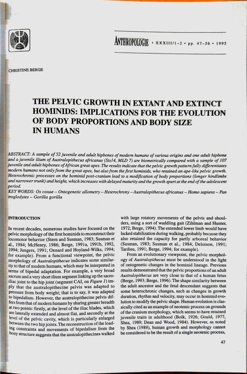
11 dimensions were measured on the hipbone of mod-
ern humans and African great apes (Table 1, Figure 1).
Ten ontogenetic allometric coefficients (slopes of the re-
gression lines) were calculated in humans andAfrican great
FIGURE 1. Measurements of the human hipbone. A; external view'B: internal view. O: centre of the acetabular ring (external andinternal points). S: Scalenion; T: Tuberion. (for key to measure-ments, see Table I), b: acetabulo-cristal buttress; t: cristal tubercle,
48
apes by least-square regression of the hipbone dimenagainst the hipbone length (log-data) (Table 2). Aallows us to compare the growth patterns in apes andmans. Also, growth curves of some ilium dimensions Qdrawn in modern humans to serve as a referencecomparison of the juvenile and adult australopithecinq
TABLE 1. Key and definition for hipbone measurements. Forexplanation, see Berge (1991m 1993).
Key
PELILLPULISLILBCAICRI
SAPACEIMB
Pelvic measurements
hipbone lengthilium lengthpubis lengthischium lengthischium breadthcaudal ilium lengthcranial ilium lengthiliac plane breadthsacral plane breadthacetabular diameterminimal ilium breadth
RESULTS AND DISCUSSION
An overview of the pelvic growth
Pelvic growth is relatively complex: the hipbone grows
a group of long bones, and the sacrum as a group of v
brae. Considering the single hipbone, there are eight
tres for development (Gray, 1901): three are primary
(corresponding to the three bones: ilium, ischium,pubis), and five secondary (one for the iliac crest, one
the anterior inferior iliac spine, one for the ischial tubero
osity, one for the pubic symphysis, one for the
cartilage). The increase in size and shape of the hipboßle
is due to the differential remodelling and reshaping
ilium, ischium and pubis. The three bones grow by dep
sition of new bone on the lateral borders, and resorption
of osseous matter on the medial borders. The shape changet
of the hipbone results from differences in growth direct
tion, time and velocity of the various bone segments
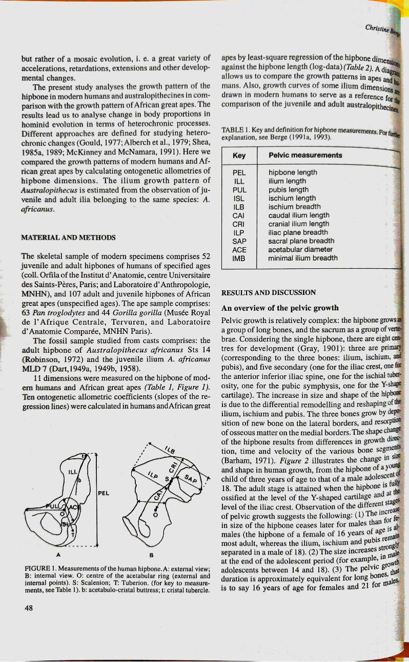
(Barham, 1971). Figure 2 illustrates the change in size
and shape in human growth, from the hipbone of a young
child of three years of age to that of a male adolescentd
18. The adult stage is attained when the hipbone is
ossified at the level of the Y-shaped cartilage and at the
level of the iliac crest. Observation of the different stages
of pelvic growth suggests the following: (I) The increaß
in size of the hipbone ceases later for males than for fee
males (the hipbone of a female of 16 years of age is
most adult, whereas the ilium, ischium and pubis remaJP
separated in a male of 18). (2) The size increases strongly
inat the end of the adolescent period (for example,
adolescents between 14 and 18). (3) The pelvic grothat
duration is approximately equivalent for long bones,
is to say 16 years of age for females and 21 for males'

The pelvic Growth in Extant and Extinct Hominids: Implications for the Evolution of Body Proportions and Body Size in Humans
. TABLE 2. Ontogenetic allometric coefficients calculated by least-square regression in common chimpanzees, gorillas, and modernhumans. Each hipbone dimension (log-data) is regressed against the hipbone length (log-data). See definition of hip bone measurements,Table l, Figure l. N: number of specimens; b: allotnetric coefficients (regression slopes); SE(b): standard error of b. R2: coefficient ofdetermination.
Pan troglodytes Gorilla gorilla Homo sapienskey
ILL 63 1.01
1.1
ISL 0.99ILB 1.16CAI 0.99CRI 1.03
1.17
SAP 0.93ACE 0.84IMB 1
cl(b)
0.02
0.04
0.03
0.04
0.03
0.04
0.03
0.06
0.06
0.05
0.98
0.93
0.96
0.94
0.95
0.93
0.95
0.82
0.85
0.86
N
44
b
0.99
1.05
0.96
1.06
0.99
0.89
1.19
0.79
0.65
1.01
cl(b)
0.01
0.02
0.02
0.02
0.03
0.04
0.03
0.04
0.05
0.04
R2
1
0.98
0.98
0.98
0.97
0.9
0.98
0.9
0.93
0.95
N
52
D
b
0.97
0.97
1.05
0.98
0.88
0.99
0.97
1.05
1.09
0.9
0.01
0.02
0.02
0.01
0.02
0.02
0.02
0.02
0.03
0.01
0.97
0.97
0.99
0.99
0.97
0.97
0.99
0.98
0.97
0.99
FIGURE 2. The growth of the hurnan hipbone illustrated by five hipbones from the coll. Ortila. •rhe hipbones are represented on the salliescale (internal views). A: male aged 2 years, 9 months and 14 days; B: female aged 1() years, 10 months and 18 days; C: male aged13 years, 7 months and 20 days; D: female aged 16 years, 6 months; E: male aged 18 years.
respectively. The hipbone of the chimpanzee, by compari-son, ceases to grow at the age of 12 (Barham, 1971). Theprolonged growth at the end of the adolescent period is auniquely human feature, which corresponds to a growthspurt in body height (Bogin, 1988). Chimpanzees andgorillas attain their adult size at the time of their sexualmaturity, which is at 8—12 years of age (Shea, 1985b;Leigh, 1992).
The growth pattern of the hipbone in modern humansas compared to that of the African great apesColeman (1971), who compared the growth of the hip-bone in humans and chimpanzees, concluded that theirpelvic growth patterns do not differ in terms of directionOf growth. In fact, as suggested by Barham (1971), differ-ential velocities and times of growth modify the hipboneshape. Figure 3 illustrates the shape differences in thegrowth patterns of modern humans and African great apesin terms of allometric changes. Ontogenetic allometriccoefficients are calculated in reference to the maximal
length of the hipbone (Table 2). We may analyse the shapechange of the hipbone in the following manner: Each pat-tern is represented by ten ontogenetic allometric coeffi-cients radiating from a point. A perfect circle of radius Iwould correspond to an isometric pattern, i. e. homotheticchanges relative to the length of the hipbone. A deforma-tion of the circle corresponds either to a positive allometry(a radius above I indicates that the dimension grows morein proportion to the hipbone length), or to a negativeallometry (a radius below I indicates that the dimensiongrows less than the hipbone length). Thus, we observethat the common chimpanzees and gorillas share for themost part the same growth pattern. The most remarkablechange in shape during ape growth concerns the ilium,which becomes proportionally broader at the level of theiliac plane (ILP, ILB), and narrower at the level of thesacral plane (SAP), with a relatively smaller acetabulum(ACE), and a narrower minimal ilium breadth (IMB).
A previous study has demonstrated that the pelves ofgreat apes (Pongo, Pan, and Gorilla) have retained nu-
49

ILL
12IMB PUL
06
ACE ISL
SAP
CAIpan
CRI
FIGURE 3. The hipbone growth patterns in African great apes andmodern humans. In each species, the growth pattern is representedby 10 ontogenetic coefficients (see Tables 1 and 2) radiating froma point. A deformation of the circle corresponds to positive ornegative allometries (see text).
Christine Berge
merous morphological traits inherited from a commonadaptation to arboreal behavior (Berge, 199 la). For ex.ample, a very long and thin ilium segment linking up thesacro-iliac joint to the hip joint, a small acetabulum, and avery narrow sacral region, indicate that gravity inducestensile forces on the interarticular pelvic segments duringsuspensory and climbing behavior. The present study dem.onstrates that most of these morphological traits are notmodified during growth, even though the heavy adults (andmore specifically the male gorillas) are unable to move intrees and must adopt a terrestrial locomotion. For exam.ple, the hip of the male gorillas remains thin and elon-gated, and consequently badly adapted for compressiveforces induced by body weight in quadrupedalism. Froma functional viewpoint, the very powerful hip and trunkmusculature attached to very broad iliac blades is essen-tial for the adult chimps and gorillas to balance body massin terrestrial quadrupedalism. From an evolutionary view-point, positive allometries of the ilium breadth and of theiliac plane breadth seem to be a general trend in mam-mals, which leads the larger animals to have a broaderpelvis than smaller ones. This allometric phenomenon maybe observed either within a single species (ontogeneticand static allometries) or within different species (evolu-tionary allometry), for example, among catarrhines (Berge,
FIGURE 4. Pelvic proportions and ilium morphology in modern humans and australopithecines. The pelves are represented with thesame hipbone length, corresponding, respectively, to a length of 2 cm (A), 4 cm (B), 22 cm (C), and 16 cm (D). A: human fetus aged 5 anda half months; B: human neonate; C: adult human; D: adult australopithecine (AL 288 reconstructed by Schmid, 1983). S: anteriorsuperior iliac spine; T: cristal tubercle (see text).
50

The pelvic Growth in Extant and Extinct Hominids: Implications for the Evolution of Body Proportions and Body Size in Humans
1993, 1995), and other non-human mammals, such as
canids, and felids (Berge, unpublished data). Only humans
have a reversed allometric phenomenon, as explained be-
low,The human displays a fully different growth pattern of
the hipbone. The ilium growth is characterized by a pro-
portional increase of the sacral plane breadth (SAP), and
of the acetabulum diameter (ACE), contrasting with a rela-
tive decrease of the iliac blade breadth at the level of the
iliac plane (ILB, ILP) in proportion to the hipbone length.
Other pelvic dimensions are modified with growth, such
as the segment CAI which becomes proportionally shorter,
whereas the ischium segment (ISL) becomes proportion-
ally longer. From a functional viewpoint, most of themorphological traits described in the human pelvis as re-
lated to bipedalism are accentuated with pelvic growth.
For example, a broad sacral region, a large acetabulum,contrasting to a relatively narrow pelvis at the level of the
iliac crests, and a short hipbone (short and robust ilium
segment linking up the sacro-iliac joint to the hip joint).Ontogenetic changes lead to the following modifications:
First, the whole pelvic structure, which supports body
weight, becomes progressively more robust with pelvic
growth. Second, the iliac blade becomes proportionally
narrower with a proportional narrower ilium, at the level
of the iliac plane. In fact, we may observe, in figure 4, that
the iliac plane, which is fully flat in the fetus (Figure 44),
becomes incurved in the adult with the formation of the
internal fossa iliaca. Thus, the maximal breadth of the adult
pelvis is not situated between the two anterior superior
iliac spines, as is the case in the fetal pelvis, but between
the two cristal tubercles. The attainment of a proportion-
FIGURE 5. Cranial view of the hominid iliac crest (the dorsal pelvic
part is found above, and the ventral one below): A: modern human;
B: australopithecine (AL 288 reconstructed by Schmid, 1983). T:
Cristal tubercle; S: anterior superior iliac spine; dotted lines: iliac
crest projected onto sagittal and frontal planes.
ally narrower pelvis at the level of the anterior superioriliac spines, with more sagittally positioned iliac blades,facilitates walking with adducted lower limbs in humans(Berge, 1994). From an evolutionary viewpoint, the changein ilium shape is the consequence of a new morphologicaltrait, the acetabulo-cristal buttress (and the cristal buttresson the iliac crest), which appears at the period of birth inmodern humans (Figure 4B). The acetabulo-cristal but-
tress, which thickens the external face of the human ilium,
is the key element of this study. The acetabulo-cristal but-
tress and the cristal buttress, which appear at the time of
birth, are progressively displaced during growth a more
backward position relative to the anterior superior iliac
spine (Figure 4C). This morphological trait allows the adult
ilium to be strongly incurved and above all sagittally po-
sitioned (Figure 5A). The acetabulo-cristal buttress and
the cristal tubercle exist in no other mammals, even though
some heavy animals, such as male gorillas, may have
slightly incurved but transversally positioned ilia (pers.
observations, contra Reynolds, 1931, who described «a
beginning tuberosity in many male gorillas»).
The growth pattern of the ilium in Australopithecus
africanus
The adult pelvis in Australopithecines resembles to some
extent that of a fetal human. The australopithecine pelvis
is very broad with laterally extended iliac blades, as is the
fetal pelvis in modern humans (Figures 4D, 5B). As on
the neonate ilium, the australopithecine ilium is almost
flat, with a cristal tubercle very close to the anterior supe-
rior iliac spine, and the fossa iliaca is almost unformed.
Such morphological traits are common to the whole group
of gracile and robust australopithecines, and correspond
to the same evolutionary stage in hominid evolution
(Mednick, 1955; Zihlman, 1971; Day, 1973; McHenry,
1975, 1986; Berge, 1984; Sigmon, 1986; for example).
The fossil MLD 7 allows us to determine some pelvic
change at the end of the adolescent period, which may
partly explain why adult Australopithecus are so different
from adult Homo in terms of body proportions.
A left juvenile ilium MLD 7 was found by Dart (1949a,
1949b, 1958) with a right ischium and a mandibula be-
longing to the same specimen. Dart, who compared the
dentition to those of a human juvenile, suggests that the
skeleton is that of a child of 12 years of age (Dart, 1948).
The ilium development corroborates that the skeleton was
of a pre-pubertal age, though it was probably aged be-
tween 8 and 10.
Comparison of the juvenile and adult ilia from the same
species (Australopithecus africanus) suggests a pelvic
growth pattern closer to that of African great apes than to
that of modern humans, for the following reasons:
(l) The juvenile ilium attained almost the same size as
the adult ilium of Sts 14 (Figure 6). In Figure 7, the growth
curve of the ilium length (ILL) clearly indicates that the
growth spurt after puberty, which characterizes modern
humans, was absent in the first hominids. In humans, the
51

ilium increases in length after growth reaches a plateaubetween the ages of 8 and 14. MLD 7 has the same iliumlength as a human child of around 8, and Sts 14 the sameilium length as a child of 10. It seems evident that theaustralopithecines ceased to grow a short time after pu-berty, probably at 10—12 years of age, as is the case withAfrican great apes.
(2) On the external face of the ilium (Figure 6A—B),the acetabulo-cristal buttress is clearly present in the adultSts14, but barely formed with no cristal tubercle in thejuvenile MLD 7. Dart (1958) described an acetabulo-cristalbuttress on MLD 7 as a «generalized thickening of theanterior cristal region, with no localized cristal tubercle».Such an observation suggests that the australopithecinebuttress was probably formed during the juvenile period,under the effects of gravitational forces in bipedalism.
(3) On the internal face of the ilium (Figure 6C—D),the iliac plane (ILP) is more enlarged on the adult iliumSts14 than on that of the juvenile MLD 7, whereas thesacral plane (SAP) is of a similar dimension in the twofossils. The growth curves in modern humans indicate thatthe ilium breadth scales with a growth spurt after the pe-riod of puberty, maintaining the same proportions as theilium length (Figures 7 and 8; see also the ontogeneticallometric coefficients of ILL and ILB, on Table 2). InFigure 8, the posterior part of the human ilium, that is tosay the sacral plane (SAP), increases relatively slowly in
b
52
Christine Berge
breadth after puberty, as does the ventral-lateral part, thatis to say the iliac plane (ILP), We observe that the propor-tions between ILP and SAP remain almost unchanged froma 10—12 year old's ilium to that of an adult (Figure 8). Thecomparison between MLD 7 and Sts14 suggests that theaustralopithecine ilium increases in breadth but not inlength during the period of puberty (Figures 7 and 8). Infigure 8, the australopithecine ilium scales in breadth withthe same proportions as the human ilium during the peeriod of puberty. However, the proportions between ILPand SAP change. The sacral plane ceases to grow beforethe period of puberty, whereas the iliac plane continues toincrease more markedly than in human growth during thesame period.
As is clearly from Figure 8, the pelvic morphology ofthe first hominids and modern humans is very much closerin the juvenile period than in adulthood. The reasons arerelated to their pelvic growth patterns and accentuate taxo-nomic differences. The pelvic growth pattern, inaustralopithecines is very similar to that of the Africangreat apes, both in growth duration and in pelvic changes.For example, the chimp and gorilla ilia increase in breadthwith the same change in proportions of ILP and SAP asobserved above (see, Table 2, Figure 3). The human growthpattern implies different pelvic changes in terms ofontogenetic allometries, and a growth spurt of the pelvicsize at the end of the adolescent period.
FIGURE 6. The pelvic growth in AustralopithecuSafricanus. The fossils are represented on the same scale•
AC: Adult hipbone Sts 14; BD: juvenile ilium MLD 7;
AB: external view; CD: internal view; b: acetabu10-cristal buttress; ILL ilium length (reconstructed for
MLD 7); ILP: iliac plane breadth; t: cristal tubercle ;
SAP: sacral plane breadth (for interpretation, see text)e

The PelviC Growth in Extant and Extinct Hominids: Implications for the Evolution of Body Proportions and Body Size in Humans
ILLHomo
MLD7• Sts14
Age
5 10 15 25FIGURE 7. Growth curve of the ilium length in modern humans and australopithecines. Axis-X: age in years; axis-Y: ilium length (ILL)in 1/10 mm. MLD7 and Sts14: juvenile and adult australopithecines. Plain line: human growth curve; dotted line: reconstructed growthcurve in australopithecines.
ILB
MLD7 0—
Homo
-—•Sts14
Homo
— OSts14
Age
5 10 15 20 25
FIGURE 8. Growth curve of the ilium breadth in modern humans and australopithecines. Axis-X: age in years; axis-Y: ilium breadth(LB), iliac plane breadth (ILP), and sacral plane breadth (SAP) in 1/10 mm. MLD7 and Sts14: juvenile and adult australopithecines.Plain lines: human growth curve; dotted line: reconstructed growth curves in australopithecines.
Evolutionary change in body proportions and bodysize in hominids
The ilium bone, which links the trunk to the hindlimbsegments, is particularly relevant in the reconstruction ofthe post-cranium growth in hominid fossils. There are tworeasons for this. First of all, the ilium grows in length inthe same way as long bone, that is to say as a hindlimbsegment. This signifies that the ilium growth inAustralopithecus may give some information as to the rateof growth rhythm and velocity for the whole hindlimband for the stature. In modern humans, the growth spurtOf the ilium length comes with a similar growth spurt of
the hindlimb length and of the stature at the end of theadolescent period. In australopithecines, the ilium lengthceases to grow earlier, at probably 10—12 years of age,and this is the reason why the australopithecines had veryshort hindlimbs in proportion to the trunk, and conse-quently a small stature (for body proportions and bodyheight in australopithecines, see, for example, McHenry,1978; 1992a, 1992b; McHenry and Temerin, 1979; Jungersand stern, 1983; Schmid, 1983, 1991; Wolpoff, 1983;Jungers, 1988, 1991; Preuchoft and Witte, 1991). Sec-ondly, the maximal pelvic breadth measured at the levelof the iliac blades is also a trunk breadth which is rela-tively similar in size to the thorax breadth (see, Schultz,
53

20 cm
FIGURE 9. Body proportions in australopithecines:The reconstructed skeleton of AL 288 (Schmid, 1983).
1961). To illustrate this purpose, we may observe, on the
reconstructed AL 288 (Schmid, 1983), that the broad and
laterally positioned iliac blades of Lucy correspond to a
very broad and funnel-shaped thorax as in pongids,whereas the short hipbone corresponds to a short hindlimb
and a small stature (Figure 9). Thus, heterochronic changes
of the ilium morphology in hominids may reflect similar
heterochronic changes for the whole post-cranium.
One of the most important results of the present study
is that the australopithecine morphology appears to repre-
sent an ancestral-like morphology for the human lineage.
The main argument is that we observe similar change in
the ilium morphology in human ontogeny and hominid
phylogeny (human neonates retain some «australo-
pithecine-like» traits). The reconstruction of the ilium
growth in Australopithecus africanus suggests that: (1)
the delayed maturity and growth spurt in body size ap-
peared after Australopithecus; and (2) the change in trunk
morphology, that is to say a narrower and barrel-shaped
thorax, also appeared at the same period in the humanlineage. Moreover, other fossils suggest that the changein post-cranium growth arose probably with the first Homo
erectus. For example, the NariokotomeHomo erectus skel-
eton seems to be an adolescent of 11 years of age whencompared to human development. His body size, and body
proportions, are very similar to those of modern humans,suggesting a similar post-cranium growth pattern (see, Ruff
and Walker, 1993). However, it would seem that Homo
54
Christine Berge
erectus retained some primitive traits in terms of pelvicgrowth pattern. For example, the acetabulo-cristal buttress
has a more ventral position in adult Homo erectus than inadult humans (Sigmon, 1986, Berge, 1993).
Conclusion as to heterochronic processes
Three heterochronic processes corresponding to an accel-erated change in ilium morphology, may be described inhominid evolution. ( 1) Pre-displacement: we suppose thata new morphological trait (acetabulo-cristal buttress andcristal tubercle) appeared during the juvenile period inaustralopithecines, most probably with the practice ofbipedalism. This morphological trait was displaced dur-ing evolution, until it appeared at birth in human descend-
ants. (2)Acceleration: an accelerated change in ilium shape
and pelvic proportions allows human descendants to have
a proportionally narrower pelvis, with more incurved and
sagittally positioned iliac blades, and consequently a nar-
rower and funnel-shaped thorax. This phenomenon is il-
lustrated by differences in ilium and pelvic growth patterns
in terms of ontogenetic allometry. (3) Hyper- morphosis:
the delayed maturity and growth spurt at the end of the
adolescent period allows a change in body propåtions
(i. e. longer hindlimb segments) and an increase in height.
Pre-displacement, acceleration, and hypermorphosis are
three phylogenetic phenomena producing recapitulation,
that is to say leading to a peramorphic descendant in terms
of post-cranium morphology. The opposite result wasfound in the skull morphology, which tends to induce a
paedomorphic descendant (Gould, 1977, Shea, 1989, Dean
and Wood, 1984).As previously suggested by Shea (1989),
it is now evident that the hominids evolve with a greatdiversity of evolutionary processes, implyingpaedomorphic changes for the skull and peramorphicchanges for the post-cranium.
AKNOWLEDGMENTS
I am very grateful to Dr. V. Vanöata , to the Primatological
Group in the Czech Republic, and to the CzechAnthrop0*
logical Society, for inviting me to participate at the Sym-
posium «Primate Ontogeny» in Treöt (10—14 September
1995). My thanks go to Pr. J.-P. Lassau, Pr. Thys Van den
Audenaer, Pr. J. Langaney, Pr. J. Repérant, for allowing
me to study the pelvic material, and to J. W. Underhill for
stylistic corrections. This research is supported by the
CNRS (URA 11 37).
REFERENCES
ALBERCH R, GOULD S. J., OSTER G. F. and WAKE D. B., 1979:
Size and shape in ontogeny and phylogeny. Paleobiol.
BARHAM W. W., 1971: A longitudinal study of the growth of the
chimpanzee bony pelvis. Proc. 3rd Congr. Primat. Zürich, 1970
BERGE C., 1984: Multivariate analysis of the pelvis for hominids
and other extant primates: Implications for locomotion and sys-

pelvic Growth in Extant and j'ombddt: Implicat/&lifm
temøticg of the different q»ecie of *ugt'ikvitbecioe•. j.
BERGE C.. 1991 a: Size and
6: 365-376.bÄGE C . 1991 b' Quelle est la signification fonctionne"e du
large de Australopithecug afarenrig (AL 288)' In: Yand B. SentJ1 (eds.): Cahiers de Paléoanthropologie, Peig: CNRS,
BERGE C., 1992: Analyse morphométrique fonctjcmnelle du pelvisdes A ustralopithéques (Australopithecus afarenrig, A. africnag):Interprétations locomotrices et obstétricaJes. In: M, T'Nss•iM(ed): Cinq millions, I 'Aventure Humaine, Bruxelles:E.R.A.ULS6:49-62.
BERGE C., 1993: L'Ev01ution de la Hancbe et du Pelvis degHominidés. Bipédie, Parturition, Croissance,AJJomfiie. Cahierjde Paléoanthropologie, CNRS, Paris.
BERGE C. , 1994: How did the australopithecines walk? Abiomechanical study of the hip and thigh of Australopi&cugafarensis. J. Hum. Evol. 26:259—273.
BERGE C., 1995: La croissance du pelvis de l'homme compare åcelle des pongidés africains: Implication dans l'évolution de labipédie. Anthropologie et Préhistorie, 106: 1—12.
BERGE C., 1996: Evolution and growth of the hominid pelvis: A pre-liminary study of the ilium shape by the thin-plate spline. M.Corti, A. Loy, L Marcus, G, Naylor, D. Slice (eds.): Advancesin Morphometrics. Plenum Press, pp. 441—448.
OGIN B., 1988: Patterns of Human Growth. Cambridge: CambridgeUniv. Press.
OLK L., 1926: On the problem of anthropogenesis. Proc SectionSciences Kon. Akad. Wetens. Amsterdam 29: 465-475.
LEMAN H., 1971: Comparison of the pelvic growth patterns ofChimpanzee and Man. Proc 3rd Congr. Primat. Zürich 1970 (I):176-182.
ART R. A., 1948: The adolescent mandibule of Australopithecusprometheus. Am. J. phys. Anthropol. 6:391-411.
ART R. A.. 1949a: The first pelvic bones of Australopithecusprometheus: Preliminary note. Am. J. Phys. Anthropol. 7:255-258.
ART R. A. , 1949b: Innominate fragments of Australopithecusprometheus. Am. J. Phys. Anthropol. 7:301—334.
ART R. A., 1958: A further adolescent australopithecine ilium from
Makapansgat. Am. J. phys. Anthropol. 16.473-479.
AY M. H., 1973: Locomotor features of the lower limb in hominids.
In: F. Zuckerman (ed.): The Concept of Human Evolution, Sym.
Zool. soc. Lond. 33: 29-51.EAN M. C., WOOD B. A., 1984: Phylogeny, neoteny and growth of
the cranial base in hominoids. Folia primatol. 43: 157—180.
ELOISON, Y., 1991: Les australopithéques marchaient-ils comme
nous? In: Y. Coppens and B. Senut (eds.): Cahiers de Paléo-
anthropologie, Paris: CNRS, pp. 177—186.
ULD S. J., 1977: Ontogeny and Phylogeny. Cambridge, Mass.:
Harvard Univ. Press.GRAY H., 1901 : Anatomy, Descriptive and Surgical. Running Press,
Philadelphia, Pennsylvania (new ed. 1974).
'UNGERS W. L., 1988: Lucy's length: stature reconstruction in
Australopithecus afarensis (AL 288—1) with implications for
other small-bodied hominids. Am. J. Phys. Anthropol. 76:
227-231.UNGERS W. L., 1991 : A pygmy perspective on body size and shape
in Australopithecus ufarensis (AL 288—1, «Lucy»). In: Y.
Coppens and B. Senut (eds.): Cahiers de Paléoanthropologie,
Paris: CNRS, pp. 215-224.JUNGERS W. L.. STERN J. T. Jr., 1983: Body proportions, skeletal
allometry and locomotion in Hadar fossils: a reply to Wolpoff.
J. Evol. 12: 673-684.LEIGH S, R., 1992: Patterns of variation in the ontogeny of primate
body size dimorphism. J. Human Evol. 23:27—50.
McHENRY H. M.. 1975: A new pelvic fragment from Swartkrans and
the relationship between the robust and the gracile australo-
pithecines. Am. J. Phys. anthropol. 43; 245—262.
McHENRY H. M.. 1978: Fore- and bindJjmb proportions in Plio-
Pleistocene hominid pelvic bones. Am, J. Phys. 43:
263-270.McHENRY H. M., 1986: The first bipeds: A comparison or the
afarensis and A. africanus postcranjum and implications (or the
evolution of bipedalism. J. Evol. 15; 177—191,
McHEVRY H. •ipe and brqfy ptopnttiong in erry
V . TF-MÉRN L Abipedalvqm- Evi&nce front the reconfAnthrop 22
L. MCNAMARA R J.of Chogeny New York. LoÜn: Plenum
VEDNKJK L W, The evolution of the humon i)ititn An1 203-2t6
OXNARD HOYLAND-WTLRS C. Hominidevidence. In: L Freedman. N
PRELOKFT H. wr1TE H. Biomechanical theof body shape In: Y, Coppens and B, Sesut
(edg P:ris: CNRS. pp.REYNOLDS E. 1931 evolution or the human pelvis in relaoon
the mechantgm of erect posture. Pam Peabody Mgs,
ROBINSON J. T . Chi.ago. Loo&m: Unm of Chicago Press.
RUFF C. B. WALKER A.. size body shapeWaJker and R. Leakey (eds.): The Nuri"kotome HomoSkelettn Harvard Umv. Press. Cambridge. Mass. pp. 234—26$.
SCHMID R. 1983: Eine rekonstruction des Skelettes von A.L. 288—1(H*iar) Konsequenzen. Folia primetol. 40: 283-306.
SCHMID P.. 1991 : The trunk or the australopithecines. In: Y. Coppengand B. Senut (eds. Cahiers de Puleoanthropotogie. CN RSVpp. 225-234.
SCHULTZ A, H.. 1961: Prinuatologja. Handbook of Primgtology.Lieferung 5: Vertebral Column and Thorax. S. Karger,New York.
SHEA B. T.. 1985a: Ontogenetic anometry and scaling, A discussioøbased on the growth and form ot the skull in African apes. In W.L. Jungers (ed.): Size and Scaling in Primate Biology NewPlenum Press, pp. 17S-205.
SHEA B, T.. 1985b: The ontogeny of sexual dimorphism in the Afri-can apes. Am. J. Prtnutol. 8: 183—188.
SHEA B. T., 1989: Heterochrony jn human evolution: the caw fcmneoteny reconsi&red. Am. J. Phys. Anthropol. 32'.69—jOJ.
SIGMON B. A.. 1986: Evolution in the homjmd peivjs. PaleonL Afr26:25-32.
STERN J. T. Jr.. SUSMAN L, 1983: The locomotor anatomy ofAustralopithecus afarensiJ. Am. J. Phys. Anthropol.
SUSMAN, R. L, 1983: Evolution of the human foot: Evidence from
the plio-pleisuxene hominids in foot and ankle. Am.
dicfoot soc. 3: 365-376.SUSMAN R. L, STERN J. T. Jr., JUNGERS W. L. 1984: Arboreality
and bipedality in the Hadar hominids. Folia primacol. 43;113-156
TARDIEU C., 1991: Etu& comparative déplacemeots du centre
de gravité du corps pendant la marche par une nouvelle methode
d'analyse Mise l'épreuve d'une hypochése
évolutive. In: Y. Coppens and B. Senut (eds.); Cahiers de
Paléoanthropologie, Pans: CNRS, pp. 49—58.
WOLPOFFM. H., 1983: Lucy's legs. J. 12:443-453
ZIHLMAN, AL (1971) The question of locomotor differences io
Australopithecus. Proc. 3rd Int. Congr. Primatology, ZUogb,
1970, vol. 1:54-66
ZIHLMAN A. L, HUNTER W. S., 1972: A biomechanical
Christine Berge
CNRS, URA Il 37
Lab d'Anatomie Compare, MNHN
55 rue Button75(Åi5 Paris, France
55