the phou protein from escherichia coli interacts with phor...
TRANSCRIPT

The PhoU Protein from Escherichia coli Interacts with PhoR, PstB,and Metals To Form a Phosphate-Signaling Complex at theMembrane
Stewart G. Gardner, Kristine D. Johns, Rebecca Tanner, William R. McCleary
Department of Microbiology and Molecular Biology, Brigham Young University, Provo, Utah, USA
Robust growth in many bacteria is dependent upon proper regulation of the adaptive response to phosphate (Pi) limitation. Thisresponse enables cells to acquire Pi with high affinity and utilize alternate phosphorous sources. The molecular mechanisms of Pi
signal transduction are not completely understood. PhoU, along with the high-affinity, Pi-specific ATP-binding cassette trans-porter PstSCAB and the two-component proteins PhoR and PhoB, is absolutely required for Pi signaling in Escherichia coli. Lit-tle is known about the role of PhoU and its function in regulation. We have demonstrated using bacterial two-hybrid analysisand confirmatory coelution experiments that PhoU interacts with PhoR through its PAS (Per-ARNT-Sim) domain and that italso interacts with PstB, the cytoplasmic component of the transporter. We have also shown that the soluble form of PhoU is adimer that binds manganese and magnesium. Alteration of highly conserved residues in PhoU by site-directed mutagenesisshows that these sites play a role in binding metals. Analysis of these phoU mutants suggests that metal binding may be impor-tant for PhoU membrane interactions. Taken together, these results support the hypothesis that PhoU is involved in the forma-tion of a signaling complex at the cytoplasmic membrane that responds to environmental Pi levels.
Escherichia coli employs seven genes whose products sense en-vironmental phosphate (Pi) and control the expression of the
Pho regulon (1, 2). These genes include phoB, phoR, pstS, pstC,pstA, pstB, and phoU. Together, these genes are necessary andsufficient for Pi signal transduction. While the identities of thecorresponding signaling proteins have been known for some time(3–5), the mechanisms by which they function to transduce the Pi
signal have not yet been fully elucidated.The hub of the signaling pathway consists of the two-component
signaling proteins PhoB and PhoR (6). PhoB is a typical winged-helixresponse regulator that upon aspartyl phosphorylation forms adimer, which binds to DNA sequences upstream of Pho regulongenes to recruit RNA polymerase and initiate transcription (7–10).PhoR is the bifunctional histidine autokinase/phospho-PhoB phos-phatase that donates a phosphoryl group to PhoB when environmen-tal Pi is limiting and removes the phosphoryl group from phosphor-ylated PhoB (PhoB�P) when environmental Pi is abundant (11, 12).PhoR is an integral membrane protein that does not contain a signif-icant periplasmic domain but does contain a membrane-spanningregion (Mem), a cytoplasmic charged region (CR), a Per-ARNT-Sim(PAS) domain (11, 13), with prototypical Dimerization/Histidinephosphorylation (DHp) and Catalytic ATP binding (CA) domains atits C terminus (14). Since PhoR does not contain a significantperiplasmic sensory domain, it is assumed that its PAS domain sensesa cytoplasmic signal, but the nature of the signal is not known.Changes in intracellular Pi concentration are not the signal, becausethese levels remain relatively constant in the presence of various ex-tracellular Pi concentrations and different Pi-signaling states (15, 16).
It has been suggested that the PhoR/PhoB proteins assess Pi
availability by monitoring the activity of the Pst transporter (17).The Pst proteins form a type I ATP-binding cassette (ABC) im-porter that exhibits high-affinity, Pi-specific transport (18–20).PstS is the periplasmic Pi-binding protein, PstC and PstA are in-tegral membrane proteins that form the pore through which Pi
passes, and PstB is the dimeric, cytoplasmic ATPase. Based upon
data from other well-described ABC transporters (21), Pi-boundPstS likely stimulates the ATPase activity of PstB to facilitate thesignificant conformational changes that are involved in Pi trans-port (20). When external Pi levels are above �4 �M, the trans-porter exists in a high-activity state that stimulates the phospho-PhoB phosphatase activity of PhoR (5). The low-activity-statetransporter signals for the high autokinase activity of PhoR, whichleads to elevated levels of phospho-PhoB.
In addition to the four transporter proteins, PhoU is also re-quired for Pi signal transduction but not for transport through thePstSCAB complex (22). When phoU is mutated or deleted, PhoR isa constitutive PhoB kinase leading to high expression of the Pho regu-lon genes. This is accompanied by poor growth and frequently leadsto the accumulation of compensatory mutations in phoR, phoB, orthe pstSCAB genes. It has been reported that PhoU is a peripheralmembrane protein that is a negative regulator of the signaling path-way and that it modulates Pi transport through the PstSCAB proteins(22–24). Multiple crystal structures have been reported for PhoUproteins in various organisms (25–27). Each of the structures showsthat PhoU consists of two fairly symmetric, three-alpha-helix bundles(Fig. 1). These structures show several different quaternary struc-tures: monomer, dimer, trimer, and even hexamer. Metal ions arefound associated with two of these structures and are coordinated byhighly conserved amino acid residues that are found in each three-helix bundle (25, 26). PhoU from Thermotoga maritima coordi-
Received 14 January 2014 Accepted 15 February 2014
Published ahead of print 21 February 2014
Address correspondence to William R. McCleary, [email protected].
Supplemental material for this article may be found at http://dx.doi.org/10.1128/JB.00029-14.
Copyright © 2014, American Society for Microbiology. All Rights Reserved.
doi:10.1128/JB.00029-14
May 2014 Volume 196 Number 9 Journal of Bacteriology p. 1741–1752 jb.asm.org 1741
on June 24, 2018 by guesthttp://jb.asm
.org/D
ownloaded from

nates trinuclear and tetranuclear iron clusters (25), while PhoUfrom Streptococcus pneumoniae shows zinc ions bound (26). Onemetal group is bound by residues that correspond to E. coli PhoUD58, N62, E100, and D104, and another is bound by D161, D165,E200, and D204 (Fig. 1). It is not known whether these metals areimportant for physiological function or if they are artifacts of crys-tallization conditions.
It is not known how PhoU functions in the signaling pathway.However, two general classes of models have been suggested:PhoU may mediate the formation of a signaling complex betweenthe PstSCAB transporter and PhoR (2, 27), or it may produce asoluble messenger that is recognized by the cytoplasmic domainsof PhoR (consistent with observations reported by Hoffer andTommassen [28] and by Rao et al. [29]). No experimental evi-dence has yet been presented supporting physical interactions be-tween signaling proteins.
We employed bacterial adenylate cyclase two-hybrid (BACTH)analysis as well as coelution experiments to show that PhoU inter-acts with both PhoR and PstB. Our results show that the PhoU/PhoR interaction involves the PAS domain of PhoR. This is con-sistent with the presence of a Pi-signaling complex consisting ofthe PstSCAB transporter, PhoU, and PhoR. We have also shownthat the soluble form of E. coli PhoU is a dimer that binds manga-nese and magnesium at conserved sites and that metal bindingmay be important for localizing PhoU to the membrane.
MATERIALS AND METHODSBacterial stains, growth media, and growth conditions. The bacterialstrains and plasmids used in this study are shown in Table 1. P1clr trans-ductions were carried out as previously described (31). Strains were grownin LB medium (36) at 37°C or in morpholinepropanesulfonic acid(MOPS) defined medium containing either 0.06% glucose and 2.0 mM Pi
(MOPS HiPi) or 0.4% glucose and 0.1 mM Pi (MOPS LoPi) (37). Whenindicated, kanamycin, ampicillin, or chloramphenicol was used at 50 �g/ml, 50 �g/ml, or 40 �g/ml, respectively. BACTH strains were grown onMOPS LoPi plates with 0.2% maltose as a carbon source with ampicillin(50 �g/ml), kanamycin (30 �g/ml), and isopropyl-�-D-thiogalactopyra-noside (IPTG) at 0.5 mM and 5-bromo-4-chloro-3-indolyl-�-D-galacto-pyranoside (X-Gal) at 40 �g/ml. The His-tagged versions of PhoR, PhoU,and PstB and the other site-directed mutants were created using theQuikChange site-directed mutagenesis kit from Agilent Technologies andverified by DNA sequence analysis (see Table S1 in the supplemental ma-terial for primer sequences).
E. coli PhoU structure prediction. The three-dimensional structureof the PhoU protein from E. coli was predicted by using the Phyre2 website (http://www.sbg.bio.ic.ac.uk/phyre2) (38). The PhoU sequence wassubmitted as a query and was analyzed in intensive mode. The serverindicated that 216 residues were modeled at �90% accuracy. The pre-dicted structures were displayed using the software program MacPyMol(Schrödinger, LLC).
Bacterial adenylate cyclase two-hybrid analysis. Two-hybrid screenswere carried out using the plasmids pKT25 and pUT18C from theBACTH kit and grown in the BTH101 indicator strain (EuroMedex).Control plasmids, coding for T18 and T25 fragments that were fused to aleucine zipper (GCN4), were also provided by the BACTH system kit.Plasmid constructs carrying phoR, phoR truncations, pstB, and phoU fu-sions to the T18 or T25 fragments were generated by cloning PCR frag-ments that incorporated XbaI and KpnI restriction sites by using theprimers listed in Table S1 in the supplemental material. About 1 �l ofovernight cultures were spotted on MOPS–maltose–ampicillin– kanamy-cin–IPTG–X-Gal plates and grown at 30°C until color developed.
�-Galactosidase activity assays. �-Galactosidase has long been uti-lized as an easily assayable enzyme to measure gene expression, and in theBACTH system, gene expression is linked to protein-protein interactions(31). Assays of �-galactosidase activity were based on methods publishedpreviously (39). Specifically, quadruplicate cultures in LB with ampicillin,kanamycin, and IPTG were grown at 30°C with shaking overnight. Then,50 �l of overnight culture was added to 150 �l LB in a 96-well flat-bottomplate (Greiner Bio-One), and the optical density at 600 nm (OD600) valueswere read. In addition, in a 1.7-ml centrifuge tube, 200 �l of overnightculture was added to 800 �l of Z buffer (16 g Na2HPO4 · 12H2O, 6.25 gNaH2PO4 · H2O, 0.75 g KCl, 0.246 g MgSO4 · 7H2O, and 2.7 ml of �-mer-captoethanol added to 1 liter of water and pH adjusted to 7.0 [39]). Onedrop of 0.1% SDS and 2 drops of chloroform were added to the cells, andsamples were vortexed vigorously for 15 s. The tubes were spun for 1 minat 16,000 � g in a benchtop centrifuge to pellet cell debris and chloroform.Two hundred microliters of the cell lysates were then loaded into wells ofa 96-well flat-bottom plate. Forty microliters of 0.4% ONPG (o-nitrophe-nyl-�-D-galactopyranoside) in Z buffer was added to each sample, and theOD420 values were read once a minute for 30 min in a plate reader that wasmaintained at 28°C (BioTek Synergy HT). Different units of activity for�-galactosidase activity assays in 96-well plates have been reported forvarious studies (40, 41). Units of activity were calculated as follows:units � (1,000 � slope of a line fit to OD420 in mOD/min)/(4 � OD600 of1:4-diluted overnight sample), based on previously described tests for theBACTH system (39).
PstB/PhoU coelution experiments. Cell cultures were grown over-night in 250 ml MOPS LoPi medium (except the mixed culture, whichcombined the �pstB strain containing pRR48 in 125 ml MOPS HiPi withthe �pstB strain containing p48pstB-His in 125 ml MOPS LoPi). Sampleswere collected by centrifugation at 5,000 � g and stored at 20°C. Cell
FIG 1 Modeled structure of E. coli PhoU. The structure of the PhoU proteinfrom Escherichia coli was modeled based upon known structures from theProtein Data Bank using the Phyre2 server. The protein is colored blue to red,going from the amino terminus to the carboxyl terminus. The six helices arelabeled from to 1 to 6. The side chains of several highly conserved aminoacids are shown. In particular, D58, N62, E100, and D104 form a putativemetal binding site between helices 2 and 3, and D161, D165, E200, and D204form another metal binding site between helices 5 and 6. Also shown is theF194 residue used in the fluorescence assays.
Gardner et al.
1742 jb.asm.org Journal of Bacteriology
on June 24, 2018 by guesthttp://jb.asm
.org/D
ownloaded from

pellets were thawed on ice and resuspended in 5 ml PstB lysis buffer (50mM Tris-HCl [pH 7.2], 300 mM NaCl, and 20 mM imidazole) with pro-tease inhibitor cocktail for use in purification of histidine-tagged proteins(Sigma). Cells were lysed by one passage at 4,000 lb/in2 and three addi-tional passages at 18,000 lb/in2 using a Microfluidics LV1 cell disruptor.The crude lysate was then cleared by centrifugation in a Sorval SLA-600TC rotor at 10,000 � g at 4°C for 10 min. An aliquot of the cleared, crudelysate (0.5 ml) was collected and stored at 20°C for later analysis. Theremaining cleared crude lysate was loaded onto a fresh 1-ml HisTrap FF
nickel column (GE Healthcare). The HisTrap column was then loadedonto a GE Healthcare Akta Prime Plus liquid chromatography system.The column was washed with 20 ml of PstB lysis buffer followed by elutionwith 5 ml of PstB elution buffer (Tris-HCl [pH 7.2], 300 mM NaCl, and250 mM imidazole). Wash and elution samples were collected in 1-mlfractions and stored at 20°C before analysis.
PhoR/PhoU coelution experiments. SG1 cells (a �phoB �phoR�pstSCAB-phoU strain) were transformed with p48phoRNHis or pRR48(as a negative control) and p116U2 (Table 1). Fifty-milliliter cultures were
TABLE 1 Strains and plasmids
Strain or plasmid Description Source or reference
E. coli strainsBW25113 Wild type 30CSH126 recA Tn10, Tetr 31BM261 PpstS::Ptac �pitA::FRT �pitB::FRT pRR48 23BM263 PpstS::Ptac �pitA::FRT �pitB::FRT �phoU pRR48 23BM265 BM263 pstB::pBU3 recA1, encodes wild-type phoU This studyBM266 BM263 pstB::pBQ3 recA1, encodes phoUQuad This studyBTH101 F cya-99 araD139 galE15 galK16 rpsL1 (Strr) hsdR2 mcrA1 mcrB1 EuroMedexBW26337 BW25113 �pstSCAB-phoU::FRT 30BW26390 BW25113 �pstB::FRT Yale CGSCJW0390-2 BW25113 �phoR::kan Yale CGSCANCH1 �phoBR::kan 32SG1 BW26337, �phoBR::kan from ANCH1 by P1clr transduction This study
PlasmidspKG116 pACYC184-based replicon, Camr, nahR 33p116U2 Camr phoU expression plasmid, salicylate inducible 23p116U2His p116U2 with a C-terminal 6�His tag This studyp116U2D58A p116U2 with D58A mutation This studyp116U2E100A p116U2 with E100A mutation This studyp116U2R101A p116U2 with R101A mutation This studyp116U2E200A p116U2 with E200A mutation This studyp116U2R201A p116U2 with R201A mutation This studyp116U2D204A p116U2 with D204A mutation This studyp116U2D58A,N62A p116U2 with D58A and N62A mutations This studyp116U2E100A,R101A p116U2 with E100A and R101A mutations This studyp116U2E200A,R201A p116U2 with E200A and R201A mutations This studyp116U2Quad p116U2 with E100A, R101A, E200A, and R201A mutations This studypBU3 Camr temp-sensitive, carries phoU This studypBQ3 Camr temp-sensitive, carries phoUQuad This studypRR48 pBR322-based replicon, Ampr, lacIq 34pIB307 pMAK705-based vector with a temp-sensitive replicon 35p48phoR pRR48-based phoR expression plasmid, lactose inducible This studyp48phoRNHis p48phoR with an amino-terminal 6�His tag This studyp48pstB pRR48 with pstB cloned into the NdeI and KpnI sites, lactose inducible This studyp48pstB-His p48pstB with a carboxyl-terminal 6�His tag Ths studypKT25 Kanr, BACTH plasmid for T25 fragment of AC EuroMedexpUT18C Ampr, BACTH plasmid for T18 fragment of AC EuroMedexpUT18zip Ampr, BACTH plasmid for T18 fragment fused to leucine zipper EuroMedexpKT25zip Kanr, BACTH plasmid for T25 fragment fused to leucine zipper EuroMedexpKT25phoU Kanr, BACTH plasmid for phoU-T25 fusion This studypKT25quad Kanr, BACTH plasmid for phoUQuad-T25 fusion This studypKT25pstB Kanr, BACTH plasmid for pstB-T25 fusion This studypUT18phoU Ampr, BACTH plasmid for phoU-T18 fusion This studypUT18Cquad Ampr, BACTH plasmid for phoUQuad-T18 fusion This studypUT18CD85A Ampr, BACTH plasmid for phoUD85A-T18 fusion This studypUT18CE100A Ampr, BACTH plasmid for phoUE100A-T18 fusion This studypUT18CA147E Ampr, BACTH plasmid for phoUA147E-T18 fusion This studypUT18CE200A Ampr, BACTH plasmid for phoUE200A-T18 fusion This studypUT18CE200A, R201A Ampr, BACTH plasmid for phoUE200A, R201A-T18 fusion This studypUT18CphoRN-C Ampr, BACTH plasmid for phoR-T18 fusion This studypUT18CRN-DHp Ampr, BACTH plasmid for N-DHp portion of PhoR-T18 fusion This studypUT18CRN-PAS Ampr, BACTH plasmid for N-PAS portion of PhoR-T18 fusion This studypUT18CRN-CR Ampr, BACTH plasmid for N-CR portion of PhoR-T18 fusion This studypUT18CRPAS Ampr, BACTH plasmid for PhoR PAS-T18 fusion This studypUT18CRCR-C Ampr, BACTH plasmid for CR-C portion of PhoR-T18 fusion This studypUT18CRPAS-C Ampr, BACTH plasmid for PAS-C portion of PhoR-T18 fusion This studypUT18CRDHp-C Ampr, BACTH plasmid for DHp-C portion of PhoR-T18 fusion This study
PhoU Interacts with PstB and PhoR
May 2014 Volume 196 Number 9 jb.asm.org 1743
on June 24, 2018 by guesthttp://jb.asm
.org/D
ownloaded from

grown overnight in LB with ampicillin, kanamycin, and 100 �M IPTG.These cells were collected by centrifugation, resuspended in 5 ml PhoUlysis buffer (50 mM Tris, 0.5 M NaCl, 2 mM imidazole, and 14 mM�-mercaptoethanol, pH 7.2) and 25 �l of protease inhibitor cocktail foruse in purification of histidine-tagged proteins (Sigma). Then, cells werelysed by one passage at 5,000 lb/in2 and three passages at 18,000 lb/in2
through a Microfluidics LV1 microfluidizer. Lysates were cleared by cen-trifugation, and a lysate sample was taken. Cleared lysates were mixed with1 ml of nickel-nitrilotriacetic acid (Ni-NTA) agarose slurry (Qiagen) andshaken for 1 h on ice. Samples were then loaded onto a 1-ml disposablepolypropylene column (Qiagen); flowthrough was collected and reap-plied to the column. Ten milliliters of PhoU lysis buffer was applied forthe first wash, followed by 6 ml PhoU wash buffer (like PhoU lysis bufferbut with 35 mM imidazole) for the second wash. Finally, 2 ml of PhoUelution buffer (PhoU lysis buffer with 0.5 M imidazole) was applied toelute the bound proteins. We added equal volumes of Laemmli samplebuffer (Bio-Rad) to each sample and boiled for 5 min, and 5 �l of each wasused for Western blot analysis (the lysate sample was diluted 1:20 to runon the gel).
AP and Western immunoblot assays. The alkaline phosphatase (AP)assays were carried out as described previously (42). The immunoblotassays were performed as described previously (43, 44) using a polyclonalrabbit anti-PhoU antibody and a mouse anti-penta-His antibody (Qia-gen). Immunoblots were visualized using the WesternBreeze chemilumi-nescent Western blot immunodetection kit (Invitrogen).
PhoU purification and gel filtration. The DH5 E. coli strain harbor-ing p116U2His was grown overnight in LB medium containing 40 �g/mlchloramphenicol and 100 �M sodium salicylate. Cells were harvested bycentrifugation and resuspended in lysis buffer (20 mM NaPO4, 0.5 MNaCl, 20 mM imidazole, 0.5 mM dithiothreitol [DTT], 10% glycerol, and0.1 mM phenylmethylsulfonyl fluoride [PMSF], pH 7.4), after which theywere lysed by one passage at 5,000 lb/in2 and three passages at 18,000lb/in2 through a Microfluidics LV1 microfluidizer. The crude lysate wascentrifuged at 12,000 � g for 15 min, and the supernatant fraction wasused for purification on an Akta Prime-plus chromatography system (GEHealthcare) using a 1-ml HisTrap column as directed by the manufac-turer. Specifically, we washed with 20 ml of a mixture of 80% lysis bufferand 10% elution buffer (20 mM NaPO4, 0.5 M NaCl, 0.5 M imidazole, 0.5mM DTT, and 10% glycerol). Then, we eluted with 100% elution buffercollecting 1-ml fractions. All steps were carried out at a 1-ml/min flowrate. It should be noted that the PhoU-His protein remained soluble onlyat concentrations below �0.7 mg/ml, whereas mutant derivatives fell outof solution at concentrations above �0.1 mg/ml. Gel filtration chroma-tography was also performed with this system using a HiPrep 16/60 Sep-hacryl S-200 HR column (GE Healthcare) and a 0.5-ml sample loop run at0.7 ml/min at 4°C. Standards of 450, 158, 45, 25, and 12.5 kDa were rununder the same conditions to create a calibration curve used to predict thesize of soluble PhoU.
ICP-MS. Samples of purified PhoU-His were collected from the His-Trap column in 1-ml fractions. For a buffer-only control, the same puri-fication protocol was followed using these same buffers without loadingany cell lysate onto the HisTrap column. PhoU-His and the correspond-ing buffer-only 1-ml fractions were collected during elution and thendiluted to 12 ml of 5% nitric acid. These samples were incubated for 3 h at55°C, centrifuged for 10 min at 2,250 � g, filtered through a 0.2-�mNalgene syringe filter (Thermo Scientific), and analyzed for metal contentusing inductively coupled plasma mass spectrometry (ICP-MS) on anElan6000 instrument using the TotalQuant method. We scanned over thewhole range using distilled deionized water as the blank and an optimiza-tion solution containing 10 ppb of various metals as the standard. Fivereplicates (each) were performed, and the metal content of the PhoU-Hisfraction was compared to that of the buffer-only control.
Fluorescence assays for metal binding. To confirm metal binding, weused the PhoU-His F194W protein purified by metal affinity chromatog-raphy, dialyzed extensively in dialysis buffer (20 mM sodium phosphate,
0.5 M NaCl, 0.1 mM DTT, and 10% glycerol, pH 7.4), and assayed in aFlouroMax-3 spectrofluorometer with excitation at 280 nm (the excita-tion wavelength for tryptophan) at 21°C in a temperature-controlled con-tinuously stirred chamber and scanned for fluorescence from 300 to 400nm. For manganese interactions, we assayed purified protein (70 �gF194W PhoU and 26.4 �g E100A R201A F194W E200A R201A PhoU[QuadW]) in dialysis buffer. To the protein samples, we added 10 mMMnCl2 stepwise to get 5 �M, 25 �M, 75 �M, and 150 �M final concen-trations of manganese and scanned the sample for fluorescence. Thesescans were performed in triplicate. We averaged the fluorescence valuesfor the 345- to 355-nm wavelengths, subtracted the corresponding bufferplus MnCl2 control values, corrected for dilution, and then fit curves tothe data to determine the Kd (dissociation constant) and derived the peakfluorescence of each scan. For MgCl2, we followed the same protocol as forMnCl2 but added stocks of MgCl2 stepwise to get 25 �M, 75 �M, 150 �M,250 �M, 500 �M, 1.5 mM, and 2.75 mM final concentrations.
Integrated strain construction. Temperature-sensitive plasmids car-rying a 300-bp fragment of pstB upstream of phoU were constructed inorder to introduce phoU (or its mutant derivative) back into the chromo-some at its native location. To create the plasmid insert, the 3= end ofthe pstB gene extending 67 bp into the phoU gene was first amplifiedfrom chromosomal DNA obtained from BW25113 using the primersPstBfor and PstBrev (see Table S1 in the supplemental material forprimer sequences). The phoU gene was then amplified from p116U2 orp116U2Quad using the PhoUfor and PhoUrev primers. These two PCRfragments were purified, diluted 1:50, combined, and amplified again us-ing the PstBfor and PhoUrev primers to generate a PCR fragment encod-ing the 3= end of the pstB gene upstream of phoU, exactly as found in thechromosome. This PCR product was subsequently digested with XbaI andHindIII (sites that were engineered into the primers) and ligated into thetemperature-sensitive plasmid pIB307, which had been similarly digested.The resulting plasmids were called pBU3 and pBQ3 and carried phoU andphoU encoding the E100A, R101A, E200A, and R201A substitutions(phoUQuad), respectively. The plasmids were then transformed intoBM263, which contains a chromosomal deletion of phoU, and subse-quently grown at 30°C. To select for cells in which the whole plasmidintegrated into the chromosome via RecA-mediated recombination in thepstB region, several colonies from each transformation were streaked ontofresh LB chloramphenicol plates and incubated at 42°C. To prevent exci-sion of the integrated plasmid, isolated colonies were grown in LB me-dium at 37°C and then transduced with P1 phage carrying a marked recAallele from E. coli strain CSH126. The resulting strain with an integratedphoU gene was called BM265, and the strain with the integrated quadruplemutant was called BM266. Strains were confirmed by PCR analysis.
Cell fractionation. Membranes were prepared from the indicatedstrains by harvesting cells from 300 ml of liquid cultures grown to station-ary phase in LB. Cells were harvested by centrifugation and resuspendedin 14 ml of cold buffer A (phosphate-buffered saline [PBS], 1 mM DTT,and 0.1 mM PMSF, pH 7.2). The cells were then lysed by three to fourpassages through a Microfluidics M-110L microfluidizer at 4°C. Thecrude lysates were centrifuged for 10 min at 13,000 � g to remove unbro-ken cells and aggregated proteins, and 5 ml of the supernatant fractionswas then subjected to ultracentrifugation at 100,000 � g for 1 h at 4°C inan L100XP Beckman Coulter ultracentrifuge using a type 90 Ti rotor. Thesoluble fractions were saved, and the pelleted fractions were resuspendedin 5 ml of 0.1% SDS. Fifty microliters of each fraction was mixed with 50�l Laemmli sample buffer (Bio-Rad) prepared with �-mercaptoethanoland boiled for 10 min.
RESULTSBacterial two-hybrid analysis shows interactions between Pi-signaling proteins. Since preliminary experiments to isolate a sta-ble signaling complex from membranes using affinity chromatog-raphy with nonionic detergents were unsuccessful, we employedalternate approaches to address this important question. Since
Gardner et al.
1744 jb.asm.org Journal of Bacteriology
on June 24, 2018 by guesthttp://jb.asm
.org/D
ownloaded from
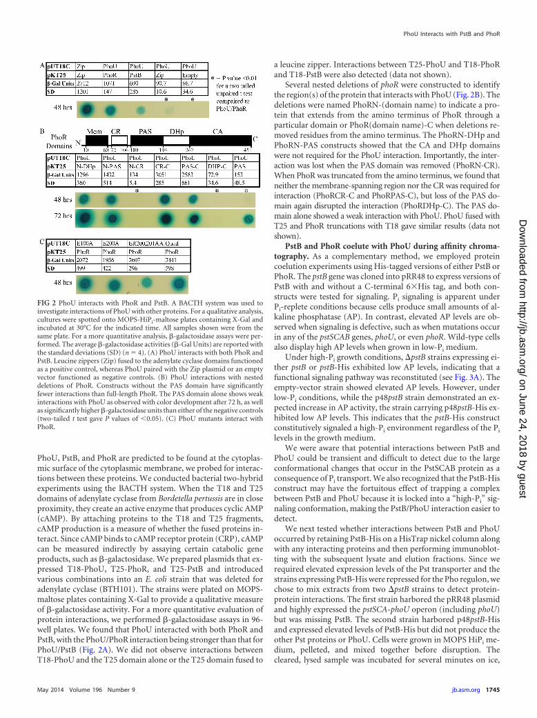
PhoU, PstB, and PhoR are predicted to be found at the cytoplas-mic surface of the cytoplasmic membrane, we probed for interac-tions between these proteins. We conducted bacterial two-hybridexperiments using the BACTH system. When the T18 and T25domains of adenylate cyclase from Bordetella pertussis are in closeproximity, they create an active enzyme that produces cyclic AMP(cAMP). By attaching proteins to the T18 and T25 fragments,cAMP production is a measure of whether the fused proteins in-teract. Since cAMP binds to cAMP receptor protein (CRP), cAMPcan be measured indirectly by assaying certain catabolic geneproducts, such as �-galactosidase. We prepared plasmids that ex-pressed T18-PhoU, T25-PhoR, and T25-PstB and introducedvarious combinations into an E. coli strain that was deleted foradenylate cyclase (BTH101). The strains were plated on MOPS-maltose plates containing X-Gal to provide a qualitative measureof �-galactosidase activity. For a more quantitative evaluation ofprotein interactions, we performed �-galactosidase assays in 96-well plates. We found that PhoU interacted with both PhoR andPstB, with the PhoU/PhoR interaction being stronger than that forPhoU/PstB (Fig. 2A). We did not observe interactions betweenT18-PhoU and the T25 domain alone or the T25 domain fused to
a leucine zipper. Interactions between T25-PhoU and T18-PhoRand T18-PstB were also detected (data not shown).
Several nested deletions of phoR were constructed to identifythe region(s) of the protein that interacts with PhoU (Fig. 2B). Thedeletions were named PhoRN-(domain name) to indicate a pro-tein that extends from the amino terminus of PhoR through aparticular domain or PhoR(domain name)-C when deletions re-moved residues from the amino terminus. The PhoRN-DHp andPhoRN-PAS constructs showed that the CA and DHp domainswere not required for the PhoU interaction. Importantly, the inter-action was lost when the PAS domain was removed (PhoRN-CR).When PhoR was truncated from the amino terminus, we found thatneither the membrane-spanning region nor the CR was required forinteraction (PhoRCR-C and PhoRPAS-C), but loss of the PAS do-main again disrupted the interaction (PhoRDHp-C). The PAS do-main alone showed a weak interaction with PhoU. PhoU fused withT25 and PhoR truncations with T18 gave similar results (data notshown).
PstB and PhoR coelute with PhoU during affinity chroma-tography. As a complementary method, we employed proteincoelution experiments using His-tagged versions of either PstB orPhoR. The pstB gene was cloned into pRR48 to express versions ofPstB with and without a C-terminal 6�His tag, and both con-structs were tested for signaling. Pi signaling is apparent underPi-replete conditions because cells produce small amounts of al-kaline phosphatase (AP). In contrast, elevated AP levels are ob-served when signaling is defective, such as when mutations occurin any of the pstSCAB genes, phoU, or even phoR. Wild-type cellsalso display high AP levels when grown in low-Pi medium.
Under high-Pi growth conditions, �pstB strains expressing ei-ther pstB or pstB-His exhibited low AP levels, indicating that afunctional signaling pathway was reconstituted (see Fig. 3A). Theempty-vector strain showed elevated AP levels. However, underlow-Pi conditions, while the p48pstB strain demonstrated an ex-pected increase in AP activity, the strain carrying p48pstB-His ex-hibited low AP levels. This indicates that the pstB-His constructconstitutively signaled a high-Pi environment regardless of the Pi
levels in the growth medium.We were aware that potential interactions between PstB and
PhoU could be transient and difficult to detect due to the largeconformational changes that occur in the PstSCAB protein as aconsequence of Pi transport. We also recognized that the PstB-Hisconstruct may have the fortuitous effect of trapping a complexbetween PstB and PhoU because it is locked into a “high-Pi” sig-naling conformation, making the PstB/PhoU interaction easier todetect.
We next tested whether interactions between PstB and PhoUoccurred by retaining PstB-His on a HisTrap nickel column alongwith any interacting proteins and then performing immunoblot-ting with the subsequent lysate and elution fractions. Since werequired elevated expression levels of the Pst transporter and thestrains expressing PstB-His were repressed for the Pho regulon, wechose to mix extracts from two �pstB strains to detect protein-protein interactions. The first strain harbored the pRR48 plasmidand highly expressed the pstSCA-phoU operon (including phoU)but was missing PstB. The second strain harbored p48pstB-Hisand expressed elevated levels of PstB-His but did not produce theother Pst proteins or PhoU. Cells were grown in MOPS HiPi me-dium, pelleted, and mixed together before disruption. Thecleared, lysed sample was incubated for several minutes on ice,
FIG 2 PhoU interacts with PhoR and PstB. A BACTH system was used toinvestigate interactions of PhoU with other proteins. For a qualitative analysis,cultures were spotted onto MOPS-HiPi-maltose plates containing X-Gal andincubated at 30°C for the indicated time. All samples shown were from thesame plate. For a more quantitative analysis, �-galactosidase assays were per-formed. The average �-galactosidase activities (�-Gal Units) are reported withthe standard deviations (SD) (n � 4). (A) PhoU interacts with both PhoR andPstB. Leucine zippers (Zip) fused to the adenylate cyclase domains functionedas a positive control, whereas PhoU paired with the Zip plasmid or an emptyvector functioned as negative controls. (B) PhoU interactions with nesteddeletions of PhoR. Constructs without the PAS domain have significantlyfewer interactions than full-length PhoR. The PAS domain alone shows weakinteractions with PhoU as observed with color development after 72 h, as wellas significantly higher �-galactosidase units than either of the negative controls(two-tailed t test gave P values of �0.05). (C) PhoU mutants interact withPhoR.
PhoU Interacts with PstB and PhoR
May 2014 Volume 196 Number 9 jb.asm.org 1745
on June 24, 2018 by guesthttp://jb.asm
.org/D
ownloaded from

subjected to affinity chromatography, and then processed forWestern immunoblotting with anti-His antibody to detect thePstB-His protein and with anti-PhoU antibody to detect PhoU.The mixed sample showed bands for PstB-His and PhoU in thelysate and elution fractions (Fig. 3B). These results demonstratedthat PstB-His retained PhoU on the column through the washingprocedure. A �pstB strain harboring the empty vector pRR48 wasgrown in MOPS LoPi medium and subjected to the same protocol.
As shown in Fig. 3A, this strain constitutively expressed high levelsof AP and also produced elevated amounts of PhoU (Fig. 3B,central panel). No PstB-His was observed in any fraction, but astrong PhoU band was observed only in the crude fraction, indi-cating that PhoU does not bind to the HisTrap nickel column inthe absence of PstB-His. When a �pstSCAB-phoU strain contain-ing p48pstB-His was analyzed, it showed low-level expression ofPstB-His in the crude fraction but accumulation in the elutionfraction. This strain also showed the absence of PhoU in everyfraction, indicating that the antibody was specific for PhoU.
We also confirmed the PhoU/PhoR interaction with coelutionexperiments. We constructed a plasmid that expressed PhoR withan amino-terminal 6�His tag (p48PhoRNHis) and confirmed itssignaling activity by introducing it into the �phoR deletion strainJW0390-2 (45) and performing AP assays (data not shown). Be-cause of difficulties with protein expression levels in this geneticbackground, we assayed interactions between PhoR and PhoU inSG1, a strain in which genes for all seven of the Pi-signaling pro-teins were deleted. Either pRR48 or p48PhoRNHis was intro-duced into the SG1 strain containing p116U2. The new strainswere grown in LB medium, pelleted, lysed, and subjected to affin-ity chromatography. Figure 3C shows that PhoU was observed inthe lysate fractions of both strains but was found in the elutionfraction only when PhoR-His was also present. The anti-His anti-bodies showed that PhoR-His was produced only when SG1 har-bored p48PhoRNHis. These results show that PhoU interacts withboth PhoR and PstB, and they are consistent with the presence ofa Pi-signaling complex. To further understand the role of PhoU inPi signaling, we characterized its native structure, explored thefunctions of the its most highly conserved residues by mutationalanalysis, and determined whether PhoU binds metal ions.
E. coli PhoU forms a dimer. We cloned phoU, to encode anative protein or a version with a C-terminal 6�His tag (PhoU-His), into pKG116 and confirmed that the constructs were func-tional by complementation analysis (data not shown). We thenpurified PhoU-His using immobilized metal ion affinity chroma-tography and analyzed its native structure using gel filtrationchromatography. Under nonreducing conditions, PhoU-His(with a predicted molecular mass of 28,240 Da) eluted in severalpeaks corresponding to high-molecular-mass oligomers (data notshown). The oligomers were resolved when �-mercaptoethanolwas included in the sample buffer. We think that these oligomersrepresent nonfunctional forms of PhoU because the side chains ofeach of its five Cys residues are predicted to extend toward theinterior of the protein. Any covalent bonds involving these Cysresidues would require PhoU to be partially or completely un-folded. When PhoU-His was analyzed with �-mercaptoethanol inthe column buffer, it eluted as a single peak with an apparentmolecular mass of 52.1 kDa, which is comparable to the predictedmolecular mass of a PhoU dimer (Fig. 4A).
We used the BACTH system to confirm our gel filtration ex-periments. Using compatible plasmids that expressed T18-PhoUand T25-PhoU, we observed that PhoU interacts with itself (Fig.4B). We did not observe interactions when T18-PhoU was coex-pressed with the T25 fragment alone or when T25-PhoU was co-expressed with the T18 fragment.
E. coli PhoU binds manganese and magnesium. To screen formetals bound to PhoU, the PhoU-His protein was purified andanalyzed by inductively coupled plasma mass spectrometry (ICP-MS). We identified manganese as a potential bound metal because
FIG 3 PstB and PhoR coelute with PhoU. (A) Genetic complementation of a�pstB mutation with p48pstB and p48pstB-His. The �pstB E. coli strainBW26390 was transformed with the indicated plasmids, and AP assays wereperformed. Cells were grown in MOPS HiPi or MOPS LoPi medium, whereindicated. (B) Binding of PhoU to PstB-His. Cells were lysed, and the solubleextracts were subjected to nickel affinity purification by passage over a HisTrapcolumn. Identical samples from the lysate and elution fraction were separatedby SDS-PAGE, transferred to nitrocellulose membranes, and analyzed by im-munodetection. The mixed lysate sample contained extracts from a strainexpressing high levels of PstS, PstC, PstA, and PhoU with a sample expressinghigh levels of PstB-His. The use of the mixed lysate was necessary because theexpression of PstB-His decreased the expression of the Pho regulon in the�pstB genetic background. (C) Plasmids expressing PhoR-His (PhoR with anamino-terminal 6�His tag) and PhoU were both transformed into a �phoBR�pstSCAB-phoU strain (SG1). Nickel affinity chromatography was used toretain the PhoR-His on the affinity column. Cell lysate and elusion fractionswere analyzed by Western blotting to see that PhoU coelutes with PhoR-His.
Gardner et al.
1746 jb.asm.org Journal of Bacteriology
on June 24, 2018 by guesthttp://jb.asm
.org/D
ownloaded from

significantly more manganese was found with the PhoU-His sam-ple than with a buffer only control (t test with a P value of 2.4 �107). Nickel and zinc were also identified as potentially bindingto PhoU; however these metals may be artifacts from the 6�Histag. We did not detect a significant increase in iron in the samplecontaining PhoU compared with the control.
We then used a fluorescence assay to further investigate metalbinding by PhoU. Tryptophan fluorescence intensity changes andthe maximum wavelength shifts when the polarity of its local en-vironment changes. Assays that follow changes in intrinsic tryp-tophan fluorescence are frequently used to detect changes in localprotein environments (46). We engineered a PhoU-His F194Wmutant and confirmed that the construct is still functioned in Pi
signaling (data not shown). This engineered tryptophan is located6 residues upstream of the conserved putative metal binding res-idue, E200, and is on the same face of the -helix as one of thepredicted metal binding sites (Fig. 1). PhoU-His F194W was ex-cited at 280 nm, and emission was followed from 300 to 400 nm.When manganese was added to the sample, fluorescence was en-hanced in a pattern that fit a predicted binding curve (Fig. 5A).Fitting a curve to the peak fluorescence values with various con-centrations of manganese, we found that PhoU-His F194Wbound manganese with an apparent Kd (dissociation constant) of18.3 �M, with a standard error of 6.2, based on three replicates(Fig. 5B). This value is similar to intracellular levels of manganesein E. coli reported between 15 �M (47) and 21.1 �M (48), al-though it is proposed that the free manganese levels in E. coli aremuch lower than this (49). Magnesium and manganese oftenoverlap in function for cellular processes, and magnesium is atmuch higher concentrations in the cell than manganese (50). So,we also tested whether the PhoU-His F194W protein bound mag-nesium and found that it bound this metal with a Kd of �1.5 �0.68 mM (see Fig. S3 in the supplemental material). The high Kd
for magnesium may explain why we did not observe this metalbound to PhoU in the ICP-MS experiment. Since the amount offree magnesium in E. coli cell is between 1 and 2 mM (51), bindingthis metal may be important for its physiological function. We saw
a shift in the wavelength of peak fluorescence with metal binding.We did not see any significant fluorescence change with zinc ornickel (data not shown). Zinc and nickel are known to bind to6�His tags (52). Apparently, binding at the C-terminal 6�His tagis far enough away that it does not alter F194W construct fluores-cence.
Mutational analysis of conserved residues. To examine therole of metal binding in Pi signaling, we mutated the phoU gene toencode proteins in which several of the highly conserved chargedresidues were converted to alanine residues. These plasmids wereintroduced into a �phoU strain in which the pstSCAB-phoU pro-moter was replaced with a lactose-inducible promoter (BM263) toprevent the poor-growth phenotype seen in phoU mutants thatoverexpress a functional PstSCAB transporter (23). Strains weregrown overnight in a high-Pi medium, and AP assays were per-formed to assess signaling (Fig. 6A). Each of the mutants displayednearly wild-type signaling activity, repressing the expression ofalkaline phosphatase. The strongest phenotype was associatedwith the R201A mutation, which still left PhoU with nearly 80% ofits wild-type activity. Immunoblot analysis showed that each ofthe mutant proteins was expressed at a level similar to those of theothers (data not shown).
We created three mutants that carried two mutations in a sin-gle site, D58A/N62A, E100A/R101A, and E200A/R201A, to test ifit was necessary to alter more than a single residue within a puta-tive metal binding pocket to block function. Each of these doublemutants also retained significant PhoU signaling activity. We thencreated a quadruple mutant which had mutations in each three-
FIG 4 PhoU forms a dimer. (A) E. coli PhoU-His was purified and analyzed ona gel filtration column. The PhoU-His protein eluted as a single peak at anelution volume between previously run 45-kDa and 68-kDa standards. PhoU-His has a predicted molecular mass of 28.2 kDa, so this peak corresponds withthe expected size of a PhoU dimer. (B) BACTH analysis of PhoU interactionswith itself. Strains grown in LB medium with different combinations of plas-mids were spotted onto MOPS-HiPi-maltose plates containing X-Gal and in-cubated at 30°C.
FIG 5 PhoU binds manganese. (A) A representative emission scan of tryp-tophan fluorescence when purified E. coli PhoU-His F194W (F194) wasmixed with increasing levels of manganese and excited at 280 nm. The additionof manganese caused an enhancement of the fluorescence and a shift in thepeak fluorescence. (B) Manganese binding curve. With the addition of man-ganese, the mean change in fluorescence between 345 nm and 355 nm wasplotted, and a binding curve was fit to the data (error bars represent � stan-dard errors; n � 3).
PhoU Interacts with PstB and PhoR
May 2014 Volume 196 Number 9 jb.asm.org 1747
on June 24, 2018 by guesthttp://jb.asm
.org/D
ownloaded from

helix bundle to alter residues predicted to bind metal (E100A andE200A) as well as mutations of two other highly conserved resi-dues (R101A and R201A). This version of PhoU also retained�80% of its signaling activity.
Analysis of phoU integrants. Since the mutations were carriedon a medium-copy-number plasmid, we postulated that the lack of aprominent phenotype was due to elevated expression levels ratherthan to those residues not being important for function. To addressthis possibility, we engineered two new strains, BM265 and BM266,which inserted the wild-type phoU gene or the phoUQuad variantinto the chromosome of the �phoU strain BM263 at its normal loca-tion. AP assays were performed with strains that were grown over-night in MOPS HiPi medium with or without 50 �M IPTG. Thisamount of IPTG was found to be the minimal amount needed toachieve full repression of the Pho regulon in BM261 (a strain forwhich the native Ppst promoter was replaced by the Ptac promoter).Analysis of the parent strain BM261 showed that repression of thePho regulon required expression of the pstSCAB-phoU operon as APwas derepressed in the absence of IPTG (Fig. 6B). The Pho regulonwas induced both in the presence and absence of IPTG in the �phoUstrain BM263. The strain containing the integrated phoU geneshowed a response that was indistinguishable from that of the BM261
parent strain, indicating that the reconstruction of the pstSCAB-phoUoperon was functional and was regulated in a normal pattern. How-ever, the phoUQuad mutant showed a pattern that was like that of the�phoU strain BM263, indicating that when expressed at lower levelsfrom the chromosome, the PhoUQuad mutant was not functional.We performed Western blotting to ensure that the signaling differ-ences between the phoU construct and the phoUQuad mutant werenot due to differences in protein expression or protein stability (Fig.6C). These results support the conclusion that elevated expression ofphoU from a multicopy-number plasmid suppresses the phenotypeof the site-directed phoU mutations.
We hypothesized that these conserved sites may be involved intargeting PhoU to its proper cellular location. When it is expressedfrom a plasmid, there may be sufficient PhoU throughout the cellso that targeted membrane localization is not required for signaltransmission. However, proper cellular targeting would be essen-tial for signaling when the phoU gene is present in single copy andexpression levels are low. To test this membrane affinity hypoth-esis for these conserved residues, plasmids expressing severalphoU mutants were introduced into the �phoU strain BM263, andmembrane localization experiments were performed. Theamount of PhoU in the membrane fraction progressively de-
FIG 6 Signaling phenotypes of PhoU mutants. (A) Alkaline phosphatase expression was used as a reporter of Pi signaling. Plasmids with the mutant phoUconstructs were introduced into a strain for which the native Ppst promoter was replaced by the Ptac promoter. For ease in referring to this strain, it is called STAC,for PstS operon, Ptac promoter. A �phoU STAC strain (BM263) was used to test phoU mutants for signaling. Assays were performed in triplicate, and the errorbars represent � standard deviations of the measurements. (B) Signaling phenotypes of the integrated phoU constructs. Cells were grown overnight on a rollerdrum at 37°C in MOPS HiPi medium in the presence or absence of 50 �M IPTG, and AP assays were performed in triplicate. The error bars represent � standarddeviations. (C) BM265 (phoU) and BM266 (phoUQuad) were grown in the presence or absence of 50 �M IPTG in MOPS HiPi and then harvested and processedfor visualizing protein expression by immunoblot analysis.
Gardner et al.
1748 jb.asm.org Journal of Bacteriology
on June 24, 2018 by guesthttp://jb.asm
.org/D
ownloaded from

creased as the number of mutations in PhoU increased from zeroto four (Fig. 7).
Biochemical analysis of the PhoUQuad protein. We analyzedthe PhoUQuad mutant protein for dimer formation and metalbinding. When examined by gel filtration chromatography, thePhoUQuad protein eluted from the column at the same volume asthe PhoU-His protein (see Fig. S1 in the supplemental material).Moreover, the PhoUQuad mutant interacted with itself in theBACTH assay (Fig. 4D). Gel filtration experiments analyzingPhoU-His with buffers that contained either EDTA to chelatemetal or excess manganese to saturate metal binding did not pro-duce any changes in the elution profiles (see Fig. S2). From theseobservations, we conclude that PhoU forms a dimer and that theE100, R101, E200, and R201 residues are not essential for dimerformation.
We also tested the purified PhoU-His Quad F194W proteinusing the fluorescence assay to measure metal binding and foundthat it did not bind manganese as well as PhoU-His F194W (Kd of85.2 �M � 26.3 versus a Kd of 18.3 �M � 6.2 with no overlap of94% confidence intervals of the calculated Kds based on triplicatetests). The fluorescence peak shift observed with PhoU-His QuadF194W was significantly less than the shift caused by PhoU-HisF194W (4.2 nm for the F194W mutant versus 1.7 nm for the“Quad” F194W mutant; P value of 0.020 from a two-tailed ttest). We did not observe fluorescence enhancement when thePhoUQuadF194W protein was reacted with magnesium. Sincethe wild-type and mutant proteins both form dimers and the mu-tant protein does not appear to bind metals as well, we concludethat PhoU dimerization is independent of metal binding. In addi-tion, mutations in predicted metal binding residues did not dis-rupt interactions with PhoR when they were assayed using theBACTH system.
DISCUSSION
This work provides evidence that the PhoU, PhoR, and PstB pro-teins physically interact. The demonstration of this interaction is
important because it provides a framework for understanding Pi
signal transduction. Our current model of this signaling pathwayis that PhoU is required for the formation of a signaling complexthat is comprised of PstSCAB and PhoR (Fig. 8). We propose thatPhoU contains multiple nonoverlapping binding sites where itinteracts with the membrane, PhoR, and PstB.
We found that PhoU’s interaction with PhoR requires the PASdomain of PhoR (Fig. 2B). PAS domains often function in signal-ing through sensing physical or chemical stimuli (53). Structuralstudies of histidine kinases have shown that the correct spatialpositioning of the DHp and CA domains is essential for theirkinase and phosphatase activities (54–57). In some cases, it isthought that physical interactions between the CA and DHp do-mains lead to phosphatase activity and that altering these interac-tions upon receiving a signal allows for autokinase activity (57).There have also been several studies of PAS domain structures inhistidine kinases (53, 56, 58, 59). In one study, the authors pro-posed that the PAS domain interacts with the CA domain to in-hibit the CA/DHp domain interaction that is required for kinaseactivity and that signal binding to the PAS domain frees up the CAdomain and allows for kinase activity (59). Disruption of the in-teractions between the PAS domain and other domains may lead
FIG 7 Membrane localization of PhoU and its mutant derivatives. BM263cells harboring p116U2 or several derived plasmids carrying mutations thatalter conserved residues were grown at 37°C in LB medium. Cells were dis-rupted, and the cell components were separated into soluble and membranefractions by ultracentrifugation. A total of 1.5 �g of proteins from the solublefractions and 5 �g from the membrane fractions were separated by SDS-PAGE, transferred to nitrocellulose, and probed with polyclonal rabbit antise-rum to PhoU.
FIG 8 Model of Pho regulon expression control. PhoR contains two trans-membrane segments (TM1 and TM2), a charged region (CR), and PAS, DHp,and CA domains. PhoR either phosphorylates or dephosphorylates the re-sponse regulator PhoB on a conserved aspartic acid residue (D) of its receiverdomain (REC). Upon phosphorylation, PhoB binds DNA and activates ex-pression of Pho regulon genes. The PstSCAB transporter may signal Pi levelsthrough the alternating conformations that are inherent to its transport pro-cess. Thus, PhoU may respond to mechanical forces in its interaction with thePstSCAB transporter and transmit that information to PhoR through its PASdomain. Proper signaling complex formation requires PhoU to localize to thecytoplasmic face of the inner membrane. Metal binding (M2 ) by PhoU isimportant for its interaction with the membrane, especially when expressed atlow levels, as is the case when cells are grown under phosphate-replete condi-tions.
PhoU Interacts with PstB and PhoR
May 2014 Volume 196 Number 9 jb.asm.org 1749
on June 24, 2018 by guesthttp://jb.asm
.org/D
ownloaded from

to changes in activity (55). One example reported that the PASdomain is essential for kinase function (60), while another foundthat the PAS domain is essential for phosphatase function (61).Clearly, PAS domains play a role in regulating the activity of manyhistidine kinases. Our data support a model in which the PASdomain of PhoR receives its input through direct interactions withPhoU. PAS domains frequently bind small molecules; our data donot address whether the PAS domain of PhoR binds a small mol-ecule that allows it to interact with PhoU.
According to our signaling complex model, PhoR is able tosense through PhoU the conformational states of PstSCAB as aconsequence of Pi transport and then modulate its kinase/phos-phatase equilibrium toward the appropriate response. This modelcould accommodate variations in which the signaling complex isstable regardless of the activity of the Pst transporter or the possi-bility that the complex is transient and is formed only under asubset of conditions. For example, under high-Pi conditions, PstBmay interact with PhoU and favor the binding of PhoR at the PASdomain. This may allow the PAS domain to interact with the CAdomain and shift the activity toward phospho-PhoB phosphataseactivity. Conversely, under low-Pi conditions, PhoU may not havea stable interaction with the PAS domain of PhoR, which stabilizesthe autokinase activity. This hypothesis is consistent with the ob-served unregulated kinase activity of PhoR in the absence of PhoUor a functional PstSCAB transporter (22).
In support of a signaling complex involving PhoU, we havepreviously shown that PhoU modulates Pi transport through thePst transporter (23). It seems likely that such modulation wouldinvolve direct protein interactions between PhoU and PstSCAB.Moreover, Oganesyan et al. reported that PhoU contains foldssimilar to Bag domains (a class of cofactors of the eukaryotic chap-erone Hsp70 family) and proposed that PhoU may associate withthe ATPase domain of PhoR (CA domain) and cause it to releasePhoB, thus turning off the signaling cascade in a manner similar toBag domains’ association with Hsp70 (27). While supporting aninteraction between PhoU and PhoR, our results show that unlikethe Bag/Hsp70 paradigm, PhoU interacts with the PAS domain ofPhoR and not its ATP-binding CA domain. The dimeric nature ofPhoU may also be important as it interacts with the dimeric PstBand PhoR proteins.
Our data suggest that the roles of the most highly conservedresidues within the PhoU protein family are to bind metal ions,which function to target PhoU to the membrane. Membrane tar-geting may be especially important when PhoU is present at lowconcentrations, such as when Pi is abundant. There are severalexamples where metal binding by proteins allows them to localizeto the membrane. For example, annexins play a crucial role inCa2 signaling by binding Ca2 that forms a bridge between theprotein and the phospholipid head groups (62). Metal is bound bythe protein through interactions with carbonyl and carboxylgroups of the protein and bound to the membrane through inter-action with the phosphoryl moieties of the phospholipids (63). Asimilar mechanism is employed by the alpha-toxin protein fromClostridium perfringens, where binding of Ca2 is linked to mem-brane binding and triggers the opening of the active site (64).
The highly conserved metal-binding residues of PhoU are im-portant for signaling only when the phoU gene is expressed insingle copy from the chromosome. They are not essential for sig-naling when expressed from a plasmid. It therefore seems unlikely
that these residues are part of an enzymatic active site that pro-duces a soluble message that is part of the signaling process.
By assaying for coelution of PhoU following the retention ofPstB-His on a HisTrap nickel column, we were able to detect pro-tein-protein interactions between these two proteins. We assumethat this method was effective because the PstB-His protein wasincorporated into a complete transporter consisting of PstC andPstA, where the proper protein conformations required for phys-ical interactions were maintained. Coomassie stain-treated gels ofthe elution fractions showed many bands (not shown), includingthose of the predicted sizes for PstC and PstB, indicating that theaffinity chromatography step enriched for the PstB-His proteinbut did not produce a purified complex. It seems likely that thecomplexity of the eluate was due to the nature of the sample ap-plied to the column, which consisted of soluble proteins as well asproteins imbedded in membrane vesicles.
A few studies have investigated PhoU’s interaction with otherproteins. One group used fluorescence resonance energy transfer(FRET) analysis to determine protein-protein interactions of var-ious proteins involved in the Pi starvation response and failed tofind an interaction between PhoU and either PhoR or PhoB (65).Another study used a yeast two-hybrid assay to show that in Ed-wardsiella tarda PhoU interacts with PhoB and Fur, but they didnot see any interaction with PhoR (66). However, the conflictingresults between these studies imply that more evidence is neces-sary to fully understand all of the proteins that interact with PhoU.It is possible that fusing PhoU to enhanced cyan fluorescent pro-tein in the FRET analysis and using a truncated PhoR proteinexpressed in yeast cells prevented the PhoU/PhoR interaction orthe detection methods were not sensitive enough to identify thePhoU/PhoR interaction.
We used a BACTH system to identify and characterize PhoU/PhoR interactions. Others have had success using these systems toidentify domains from bacterial proteins that interact and havefound the BACTH system is especially useful for testing mem-brane bound proteins (39, 67). Our results confirm that PhoUdoes interact with PhoR. Given the multiple PhoR and PhoU con-structs that show interaction (Fig. 2), it is unlikely that all of thedifferent constructs are false positives. These results are also con-firmed with PhoU coeluting with PhoR-His (Fig. 3).
ACKNOWLEDGMENTS
We thank John Bell, Jen Nelson, and Liz Gibbons for their assistance withthe fluorescence assays and Lance Moses for help with ICP-MS analysis.We thank several undergraduate students, Geoffrey Johnston, RachelWinn, Austin Callison, and Annaliese Gabrielle, for help in the initialcharacterization of the mutant strains and Casey Callison, Kirk Richard-son, Michael Barrus, and Gregory Bowden for excellent technical assis-tance.
This work was supported by Public Health Service grant R15GM96222from the National Institute of General Medical Sciences. R.T. was partiallysupported by an Undergraduate Mentoring Environment Grant fromBrigham Young University.
REFERENCES1. Hsieh YJ, Wanner BL. 2010. Global regulation by the seven-component
Pi signaling system. Curr. Opin. Microbiol. 13:198 –203. http://dx.doi.org/10.1016/j.mib.2010.01.014.
2. Wanner BL. 1996. Phosphorous assimilation and control of the phos-phate regulon, p 1357–1381. In Neidhardt FC, Curtiss R, III, Ingraham JL,Lin ECC, Low KB, Magasanik B, Reznikoff WS, Riley M, Schaechter M,
Gardner et al.
1750 jb.asm.org Journal of Bacteriology
on June 24, 2018 by guesthttp://jb.asm
.org/D
ownloaded from

Umbarger HE (ed), Escherichia coli and Salmonella: cellular and molecularbiology, 2nd ed. ASM Press, Washington, DC.
3. Rao NN, Torriani A. 1990. Molecular aspects of phosphate transport inEscherichia coli. Mol. Microbiol. 4:1083–1090. http://dx.doi.org/10.1111/j.1365-2958.1990.tb00682.x.
4. Torriani-Gorini A. 1987. The birth of the Pho regulon, p 3–11. In Torri-ani-Gorini A, Rothman FG, Silver S, Wright A, Yagil E (ed), Phosphatemetabolism and cellular regulation in microorganisms. American Societyfor Microbiology, Washington, DC.
5. Wanner BL. 1993. Gene regulation by phosphate in enteric bacteria. J.Cell. Biochem. 51:47–54. http://dx.doi.org/10.1002/jcb.240510110.
6. Makino K, Shinagawa H, Amemura M, Kawamoto T, Yamada M, NakataA. 1989. Signal transduction in the phosphate regulon of Escherichia coliinvolves phosphotransfer between PhoR and PhoB proteins. J. Mol. Biol.210:551–559. http://dx.doi.org/10.1016/0022-2836(89)90131-9.
7. Bachhawat P, Swapna GV, Montelione GT, Stock AM. 2005. Mecha-nism of activation for transcription factor PhoB suggested by differentmodes of dimerization in the inactive and active states. Structure (Camb.)13:1353–1363. http://dx.doi.org/10.1016/j.str.2005.06.006.
8. Makino K, Amemura M, Kim S, Nakata A, Shinagawa H. 1993. Role ofthe �70 subunit of RNA polymerase in transcriptional activation by acti-vator protein PhoB in Escherichia coli. Genes Dev. 7:149 –160. http://dx.doi.org/10.1101/gad.7.1.149.
9. Makino K, Shinagawa H, Amemura M, Nakata A. 1986. Nucleotidesequence of the phoB gene, the positive regulatory gene for the phosphateregulon of Escherichia coli K-12. J. Mol. Biol. 190:37– 44. http://dx.doi.org/10.1016/0022-2836(86)90073-2.
10. McClearyWR.1996.TheactivationofPhoBbyacetylphosphate.Mol.Microbiol.20:1155–1163. http://dx.doi.org/10.1111/j.1365-2958.1996.tb02636.x.
11. Carmany DO, Hollingsworth K, McCleary WR. 2003. Genetic and bio-chemical studies of phosphatase activity of PhoR. J. Bacteriol. 185:1112–1115. http://dx.doi.org/10.1128/JB.185.3.1112-1115.2003.
12. Makino K, Shinagawa H, Amemura M, Nakata A. 1986. Nucleotidesequence of the phoR gene, a regulatory gene for the phosphate regulon ofEscherichia coli. J. Mol. Biol. 192:549 –556. http://dx.doi.org/10.1016/0022-2836(86)90275-5.
13. Etzkorn M, Kneuper H, Dunnwald P, Vijayan V, Kramer J, GriesingerC, Becker S, Unden G, Baldus M. 2008. Plasticity of the PAS domain anda potential role for signal transduction in the histidine kinase DcuS. Nat.Struct. Mol. Biol. 15:1031–1039. http://dx.doi.org/10.1038/nsmb.1493.
14. Dutta R, Qin L, Inouye M. 1999. Histidine kinases: diversity of domainorganization. Mol. Microbiol. 34:633– 640. http://dx.doi.org/10.1046/j.1365-2958.1999.01646.x.
15. Rao NN, Roberts MF, Torriani A, Yashphe J. 1993. Effect of glpT andglpD mutations on expression of the phoA gene in Escherichia coli. J. Bac-teriol. 175:74 –79.
16. Shulman RG, Brown TR, Ugurbil K, Ogawa S, Cohen SM, den HollanderJA. 1979. Cellular applications of 31P and 13C nuclear magnetic resonance.Science 205:160–166. http://dx.doi.org/10.1126/science.36664.
17. Peterson CN, Mandel MJ, Silhavy TJ. 2005. Escherichia coli starvationdiets: essential nutrients weigh in distinctly. J. Bacteriol. 187:7549 –7553.http://dx.doi.org/10.1128/JB.187.22.7549-7553.2005.
18. Amemura M, Makino K, Shinagawa H, Kobayashi A, Nakata A. 1985.Nucleotide sequence of the genes involved in phosphate transport andregulation of the phosphate regulon in E. coli. J. Mol. Biol. 184:241–250.http://dx.doi.org/10.1016/0022-2836(85)90377-8.
19. Rees DC, Johnson E, Lewinson O. 2009. ABC transporters: the power tochange. Nat. Rev. Mol. Cell Biol. 10:218 –227. http://dx.doi.org/10.1038/nrm2646.
20. Webb DC, Rosenberg H, Cox GB. 1992. Mutational analysis of theEscherichia coli phosphate-specific transport system, a member of the traf-fic ATPase (or ABC) family of membrane transporters. J. Biol. Chem.267:24661–24668.
21. Davidson AL, Dassa E, Orelle C, Chen J. 2008. Structure, function, andevolution of bacterial ATP-binding cassette systems. Microbiol. Mol. Biol.Rev. 72:317–364. http://dx.doi.org/10.1128/MMBR.00031-07.
22. Steed PM, Wanner BL. 1993. Use of the rep technique for allele replace-ment to construct mutants with deletions of the pstSCAB-phoU operon:evidence of a new role for the PhoU protein in the phosphate regulon. J.Bacteriol. 175:6797– 6809.
23. Rice CD, Pollard JE, Lewis ZT, McCleary WR. 2009. Employment of apromoter-swapping technique shows that PhoU modulates the activity of
the PstSCAB2 ABC transporter in Escherichia coli. Appl. Environ. Micro-biol. 75:573–582. http://dx.doi.org/10.1128/AEM.01046-08.
24. Surin BP, Dixon NE, Rosenberg H. 1986. Purification of the PhoUprotein, a negative regulator of the pho regulon of Escherichia coli K-12. J.Bacteriol. 168:631– 635.
25. Liu J, Lou Y, Yokota H, Adams PD, Kim R, Kim SH. 2005. Crystalstructure of a PhoU protein homologue: a new class of metalloproteincontaining multinuclear iron clusters. J. Biol. Chem. 280:15960 –15966.http://dx.doi.org/10.1074/jbc.M414117200.
26. Madej T, Addess KJ, Fong JH, Geer LY, Geer RC, Lanczycki CJ, Liu C,Lu S, Marchler-Bauer A, Panchenko AR, Chen J, Thiessen PA, Wang Y,Zhang D, Bryant SH. 2012. MMDB: 3D structures and macromolecularinteractions. Nucleic Acids Res. 40:D461–D464. http://dx.doi.org/10.1093/nar/gkr1162.
27. Oganesyan V, Oganesyan N, Adams PD, Jancarik J, Yokota HA, Kim R,Kim SH. 2005. Crystal structure of the “PhoU-like” phosphate uptakeregulator from Aquifex aeolicus. J. Bacteriol. 187:4238 – 4244. http://dx.doi.org/10.1128/JB.187.12.4238-4244.2005.
28. Hoffer SM, Tommassen J. 2001. The phosphate-binding protein of Esche-richia coli is not essential for Pi-regulated expression of the pho regulon. J.Bacteriol. 183:5768 –5771. http://dx.doi.org/10.1128/JB.183.19.5768-5771.2001.
29. Rao NN, Wang E, Yashphe J, Torriani A. 1986. Nucleotide pool in phoregulon mutants and alkaline phosphatase synthesis in E. coli. J. Bacteriol.166:205–211.
30. Datsenko KA, Wanner BL. 2000. One-step inactivation of chromosomalgenes in Escherichia coli K-12 using PCR products. Proc. Natl. Acad. Sci.U. S. A. 97:6640 – 6645. http://dx.doi.org/10.1073/pnas.120163297.
31. Miller J. 1992. A short course in bacterial genetics: a laboratory manualand handbook for Escherichia coli and related bacteria. Cold Spring Har-bor Laboratory Press, Cold Spring Harbor, NY.
32. Yamada M, Makino K, Amemura M, Shinagawa H, Nakata A. 1989.Regulation of the phosphate regulon of Escherichia coli: analysis of mutantphoB and phoR genes causing different phenotypes. J. Bacteriol. 171:5601–5606.
33. Buron-Barral MC, Gosink KK, Parkinson JS. 2006. Loss- and gain-of-function mutations in the F1-HAMP region of the Escherichia coli aero-taxis transducer Aer. J. Bacteriol. 188:3477–3486. http://dx.doi.org/10.1128/JB.188.10.3477-3486.2006.
34. Studdert CA, Parkinson JS. 2005. Insights into the organization anddynamics of bacterial chemoreceptor clusters through in vivo crosslinkingstudies. Proc. Natl. Acad. Sci. U. S. A. 102:15623–15628. http://dx.doi.org/10.1073/pnas.0506040102.
35. Blomfield IC, Vaughn V, Rest RF, Eisenstein BI. 1991. Allelic exchangein Escherichia coli using the Bacillus subtilis sacB gene and a temperature-sensitive pSC101 replicon. Mol. Microbiol. 5:1447–1457. http://dx.doi.org/10.1111/j.1365-2958.1991.tb00791.x.
36. Sambrook J, Fritsch RF, Maniatis T. 1989. Molecular cloning: a labora-tory manual, 2nd ed. Cold Spring Harbor Laboratory Press, Cold SpringHarbor, NY.
37. Neidhardt FC, Bloch PL, Smith DF. 1974. Culture medium for entero-bacteria. J. Bacteriol. 119:736 –747.
38. Kelley LA, Sternberg MJ. 2009. Protein structure prediction on the Web:a case study using the Phyre server. Nat. Protoc. 4:363–371. http://dx.doi.org/10.1038/nprot.2009.2.
39. Battesti A, Bouveret E. 2012. The bacterial two-hybrid system based onadenylate cyclase reconstitution in Escherichia coli. Methods 58:325–334.http://dx.doi.org/10.1016/j.ymeth.2012.07.018.
40. Griffith KL, Wolf RE, Jr. 2002. Measuring beta-galactosidase activity inbacteria: cell growth, permeabilization, and enzyme assays in 96-well ar-rays. Biochem. Biophys. Res. Commun. 290:397– 402. http://dx.doi.org/10.1006/bbrc.2001.6152.
41. Thibodeau SA, Fang R, Joung JK. 2004. High-throughput beta-galactosidase assay for bacterial cell-based reporter systems. Biotech-niques 36:410 – 415.
42. Zundel CJ, Capener DC, McCleary WR. 1998. Analysis of the conservedacidic residues in the regulatory domain of PhoB. FEBS Lett. 441:242–246.http://dx.doi.org/10.1016/S0014-5793(98)01556-7.
43. Ruiz N, Silhavy TJ. 2003. Constitutive activation of the Escherichia coli Phoregulon upregulates rpoS translation in an Hfq-dependent fashion. J. Bac-teriol. 185:5984 –5992. http://dx.doi.org/10.1128/JB.185.20.5984-5992.2003.
44. Schurdell MS, Woodbury GM, McCleary WR. 2007. Genetic evidence
PhoU Interacts with PstB and PhoR
May 2014 Volume 196 Number 9 jb.asm.org 1751
on June 24, 2018 by guesthttp://jb.asm
.org/D
ownloaded from

suggests that the intergenic region between pstA and pstB plays a role in theregulation of rpoS translation during phosphate limitation. J. Bacteriol.189:1150 –1153. http://dx.doi.org/10.1128/JB.01482-06.
45. Baba T, Ara T, Hasegawa M, Takai Y, Okumura Y, Baba M, DatsenkoKA, Tomita M, Wanner BL, Mori H. 2006. Construction of Escherichiacoli K-12 in-frame, single-gene knockout mutants: the Keio collection.Mol. Syst. Biol. 2:1–11. http://dx.doi.org/10.1038/msb4100050.
46. Vivian JT, Callis PR. 2001. Mechanisms of tryptophan fluorescence shiftsin proteins. Biophys. J. 80:2093–2109. http://dx.doi.org/10.1016/S0006-3495(01)76183-8.
47. Anjem A, Varghese S, Imlay JA. 2009. Manganese import is a key element ofthe OxyR response to hydrogen peroxide in Escherichia coli. Mol. Microbiol.72:844 – 858. http://dx.doi.org/10.1111/j.1365-2958.2009.06699.x.
48. Waters LS, Sandoval M, Storz G. 2011. The Escherichia coli MntRminiregulon includes genes encoding a small protein and an efflux pumprequired for manganese homeostasis. J. Bacteriol. 193:5887–5897. http://dx.doi.org/10.1128/JB.05872-11.
49. Golynskiy MV, Gunderson WA, Hendrich MP, Cohen SM. 2006. Metalbinding studies and EPR spectroscopy of the manganese transport regu-lator MntR. Biochemistry 45:15359 –15372. http://dx.doi.org/10.1021/bi0607406.
50. Silver S, Johnseine P, King K. 1970. Manganese active transport inEscherichia coli. J. Bacteriol. 104:1299 –1306.
51. Alatossava T, Jutte H, Kuhn A, Kellenberger E. 1985. Manipulation ofintracellular magnesium content in polymyxin B nonapeptide-sensitizedEscherichia coli by ionophore A23187. J. Bacteriol. 162:413– 419.
52. Lopez C, Sanchez J, Hermida L, Zulueta A, Marquez G. 2004. Cysteinemediated multimerization of a recombinant dengue E fragment fused to theP64k protein following immobilized metal ion affinity chromatography. Pro-tein Expr. Purif. 34:176–182. http://dx.doi.org/10.1016/j.pep.2003.11.018.
53. Moglich A, Ayers RA, Moffat K. 2009. Structure and signaling mecha-nism of Per-ARNT-Sim domains. Structure 17:1282–1294. http://dx.doi.org/10.1016/j.str.2009.08.011.
54. Sousa EH, Tuckerman JR, Gondim AC, Gonzalez G, Gilles-GonzalezMA. 2013. Signal transduction and phosphoryl transfer by a FixL hybridkinase with low oxygen affinity: importance of the vicinal PAS domain andreceiver aspartate. Biochemistry 52:456 –565. http://dx.doi.org/10.1021/bi300991r.
55. Wang C, Sang J, Wang J, Su M, Downey JS, Wu Q, Wang S, Cai Y, XuX, Wu J, Senadheera DB, Cvitkovitch DG, Chen L, Goodman SD, HanA. 2013. Mechanistic insights revealed by the crystal structure of a histi-dine kinase with signal transducer and sensor domains. PLoS Biol. 11:e1001493. http://dx.doi.org/10.1371/journal.pbio.1001493.
56. Winnen B, Anderson E, Cole JL, King GF, Rowland SL. 2013. Role of the
PAS sensor domains in the Bacillus subtilis sporulation kinase KinA. J.Bacteriol. 195:2349 –2358. http://dx.doi.org/10.1128/JB.00096-13.
57. Gao R, Stock AM. 2009. Biological insights from structures of two-component proteins. Annu. Rev. Microbiol. 63:133–154. http://dx.doi.org/10.1146/annurev.micro.091208.073214.
58. Szurmant H, White RA, Hoch JA. 2007. Sensor complexes regulatingtwo-component signal transduction. Curr. Opin. Struct. Biol. 17:706 –715. http://dx.doi.org/10.1016/j.sbi.2007.08.019.
59. Yamada S, Sugimoto H, Kobayashi M, Ohno A, Nakamura H, Shiro Y.2009. Structure of PAS-linked histidine kinase and the response regulatorcomplex. Structure 17:1333–1344. http://dx.doi.org/10.1016/j.str.2009.07.016.
60. Echenique JR, Trombe MC. 2001. Competence modulation by the NADHoxidase of Streptococcus pneumoniae involves signal transduction. J. Bacte-riol. 183:768 –772. http://dx.doi.org/10.1128/JB.183.2.768-772.2001.
61. Gutu AD, Wayne KJ, Sham LT, Winkler ME. 2010. Kinetic character-ization of the WalRKSpn (VicRK) two-component system of Streptococcuspneumoniae: dependence of WalKSpn (VicK) phosphatase activity on itsPAS domain. J. Bacteriol. 192:2346 –2358. http://dx.doi.org/10.1128/JB.01690-09.
62. Gerke V, Creutz CE, Moss SE. 2005. Annexins: linking Ca2 signalling tomembrane dynamics. Nat. Rev. Mol. Cell Biol. 6:449 – 461. http://dx.doi.org/10.1038/nrm1661.
63. Swairjo MA, Concha NO, Kaetzel MA, Dedman JR, Seaton BA. 1995.Ca(2 )-bridging mechanism and phospholipid head group recognitionin the membrane-binding protein annexin V. Nat. Struct. Biol. 2:968 –974. http://dx.doi.org/10.1038/nsb1195-968.
64. Titball RW, Naylor CE, Miller J, Moss DS, Basak AK. 2000. Opening ofthe active site of Clostridium perfringens alpha-toxin may be triggered bymembrane binding. Int. J. Med. Microbiol. 290:357–361. http://dx.doi.org/10.1016/S1438-4221(00)80040-5.
65. Baek JH, Kang YJ, Lee SY. 2007. Transcript and protein level analyses ofthe interactions among PhoB, PhoR, PhoU and CreC in response to phos-phate starvation in Escherichia coli. FEMS Microbiol. Lett. 277:254 –259.http://dx.doi.org/10.1111/j.1574-6968.2007.00965.x.
66. Chakraborty S, Sivaraman J, Leung KY, Mok YK. 2011. Two-component PhoB-PhoR regulatory system and ferric uptake regulatorsense phosphate and iron to control virulence genes in type III and VIsecretion systems of Edwardsiella tarda. J. Biol. Chem. 286:39417–39430.http://dx.doi.org/10.1074/jbc.M111.295188.
67. Mikkelsen H, Hui K, Barraud N, Filloux A. 2013. The pathogenicityisland encoded PvrSR/RcsCB regulatory network controls biofilm forma-tion and dispersal in Pseudomonas aeruginosa PA14. Mol. Microbiol. 89:450 – 463. http://dx.doi.org/10.1111/mmi.12287.
Gardner et al.
1752 jb.asm.org Journal of Bacteriology
on June 24, 2018 by guesthttp://jb.asm
.org/D
ownloaded from