the physics of protons for patient treatment andrew j ... j. wroe, jerry d. slater, james m. slater...
TRANSCRIPT

The Physics of Protons for Patient Treatment
Andrew J. Wroe, Jerry D. Slater, James M. Slater
Loma Linda University
Introduction
Presently one of the major health risks mankind faces is cancer. More than one in three people
will suffer from this disease or side effects of its treatment at some stage in their lives (1). Because of
the deleterious effects that cancer and, oftentimes, treatment modalities have on the human population,
better treatment techniques are constantly being sought. Besides surgery, external-beam radiation
therapy is a mainstay of cancer treatment and cure. High-energy protons are an important innovation in
external-beam radiation therapy, providing highly conformal dose distributions and thus sparing normal
tissues through benefits afforded by the Bragg peak. This paper will focus on the physics of protons
and facility design in enabling this particle to be used for the clinical benefit of patients.
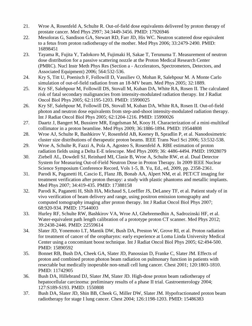
The Power of the Bragg Peak
In radiation therapy it is desirable to maximize dose delivered to the tumor volume whilst
minimizing dose to surrounding normal structures. In the ideal case one would want to deliver the
entire radiation dose to the target volume, with no dose delivered to surrounding structures. Protons and
other heavy charged particles come close to fulfilling this objective: they deposit most of their energy
in a high-dose peak (known as the Bragg peak) at the end of their track (Error! Reference source not
found.). This peak is created through an exponential increase in stopping power towards the end of the
protons’ track. Hence, as a heavy charged particle (such as a proton) slows down, the amount of energy
it deposits per unit length covered increases exponentially, creating a high-dose peak (2). The depth of
this peak in a given material (such as a patient) depends on its initial energy; varying this energy allows
the high-dose region to be placed at any depth.
The depth dose profile of protons is in stark contrast to X-rays (considered as a standard
external beam radiation modality), which achieve maximum dose either at or just below the surface of
the patient. It is only through the superposition of multiple treatment fields from multiple directions
that an elevated radiation dose to the target volume is achievable with X-rays. A further disadvantage
of X-rays is that they deposit dose beyond the target. Protons stop at the Bragg peak, ensuring that no
dose is delivered from the primary radiation field beyond the target. This has great benefit in reducing
the dose to structures that lie beyond the target and minimizing the integral dose experienced by the
patient (Error! Reference source not found.).
Proton Beam Delivery
Proton delivery techniques can be categorized as passive or active in the delivery of a uniform
dose to the treatment volume. Passive techniques, which have been most commonly used in the clinical
setting (3, 4), spread the beam laterally using a combination of gold (or lead) and Lexan foils (5). The
combination of two materials, one of low and the other of high atomic number, produces a flat beam of
constant flux and a constant range. Typically a dual scattering foil arrangement (6) is utilized that is
optimized to deliver a flat field of the cross sectional area required for treatment. The beam is then
modulated in depth using a rotating plastic wheel (7, 8) that effectively allows for the superposition of
multiple Bragg peaks of varying energy and intensity to create a region of uniform high dose called the
spread-out Bragg peak (SOBP) (6). The beam is then collimated by brass or Cerrobend® apertures and
its penetration depth is varied by means of a wax bolus. Such an arrangement creates a uniform dose
across the treatment volume, as displayed in Error! Reference source not found..
Active techniques (9-12) employ a magnetically guided proton pencil beam in combination with
dynamic changes of beam energy and beam intensity during treatment. One advantage of the active
system is that it minimizes interaction between the primary beam and beam modifying devices, in turn
minimizing the production of secondary particles. Further, it has the potential to treat complex tumor
volumes with greater precision and improved normal tissue sparing. However, the dosimetry and beam

delivery is also more complex and problematic; errors in this regard can lead to high and low dose
regions and an incomplete treatment of the tumor volume. Organ motion during treatment is another
complicating factor that also needs to be considered for effective and accurate treatment of the tumor
volume.
Facility Design
Proton therapy facilities are incredibly complex systems needing to accelerate, transport, and
deliver charged protons to a given target volume as specified by the clinician. Unlike X-ray facilities
that contain the accelerator, gantry, and beam delivery systems within a single room or bunker, proton
therapy departments typically use a single accelerator to provide high-energy protons for a number of
treatment rooms (6). Protons are accelerated in the accelerator (cyclotron or synchrotron) and then
transported along an evacuated beam pipe to the treatment room. During acceleration and transport,
magnets control proton direction and also focus the proton beam overcoming Coulombic repulsion of
the like charged particles. Beam is delivered to one treatment room at a time as requested by the
treating therapist while the other treatment rooms are setting up and aligning the patient (13).
When the beam arrives at the proton room it must be delivered to the target site. This is
typically achieved using a gantry that allows for beam delivery 360 degrees around the patient. Patients
meanwhile are aligned using lasers and X-rays images to ensuring accurate alignment relative to the
beam delivery system (isocenter). The combination of gantry proton delivery with six-degree-of-
freedom robotic patient positioners (Error! Reference source not found.) allows clinicians the greatest
flexibility in proton beam delivery in treating the target volume while sparing critical structures.
Treatment times per beam is of the order of 1-2 minutes, during which time the patient feels nothing
and is under constant audio and visual monitoring by the therapy team.
Clinical Advantages of Protons
Protons provide a clinical advantage over other external-beam radiation therapy techniques
through the Bragg peak. The Bragg peak (or SOBP, which is used clinically) allows for the delivery of
fewer beams to achieve the same target coverage (Error! Reference source not found.), in turn limiting
the integral dose delivered to other organs. The ability of protons to stop and produce no primary
proton exit dose is a distinct advantage over X-rays in limiting dose to the rest of the body and sparing
critical structures (Figs. 6 and 7). This is especially critical for pediatric cases (Error! Reference source
not found.), where additional dose to non-target structures can lead to increased instances of secondary
malignancies due to longer life expectancy.
The ability to treat with fewer beams in proton therapy not only limits unwanted dose to the
patient but also speeds up treatment time. Faster treatment times limit intra-fraction motion (motion
during treatment), allowing for a more accurate delivery to the target, which is essential as treatment
become more conformal with tighter margins.
The Out-of-Field Dose Question
It has been demonstrated that protons provide a significant advantage over other external-beam
radiation modalities owing to their depth-dose distribution, which allows for maximum dose to be
delivered to the tumor volume with no primary particle dose beyond the distal edge. This allows for
significantly fewer beams to be utilized, resulting in a much lower integral dose to surrounding critical
structures (14). Recently the whole-body dose delivered by protons and other ions was called into
question by Eric Hall (15). This dose, which is of particular importance in pediatric patients with long
life expectancy and greater susceptibility to radiation-induced cancers, is delivered by secondary
neutrons that are produced through primary beam interactions with both the patient and beam-
modifying devices. The dose-equivalent values per unit of prescribed proton dose presented in the
report of Hall (15) have been questioned (16-18), but the concern remains because of the large
uncertainties involved in neutron dose measurements and the RBE of neutrons (19).
Measurements have been completed by a number of groups (20-23) to answer these questions
and provide guidelines for physicians in specifying treatment. These are typically of the order of

several mSv/Gy close to the edge of the treatment field where the measured result is influenced by
scattered high-energy protons, to sub mSv/Gy at lateral displacements typically larger than 20 cm.
Error! Reference source not found. demonstrates the potential of active proton beam delivery systems in
reducing the out-of-field dose component to the patient over passive delivery techniques such as those
employed at the Paul Scherrer Institute (PSI), Harvard Cyclotron Laboratory (HCL), LLUMC, and the
Midwest Proton Research Institute (MPRI).
The characteristics of the proton beam (field size, proton energy, modulation etc.) also impact
the dose equivalent delivered out of the field. Error! Reference source not found. demonstrates how the
external field dose equivalent can vary by more than an order of magnitude based on field
characteristics. For a comprehensive summary of the data on this issue, the reader is referred to (20).
A comparison of the studies has shown that the out-of-field doses experienced by passively
delivered proton therapy is comparable or less than those delivered in X-ray therapy (24-26), especially
close to the field edge. Additionally, the use of fewer treatment beams by passively delivered proton
therapy allows for reduced integral doses to surrounding organs, which may be of clinical benefit.
Active beam scanning of protons can further reduce the out-of-field dose delivered by proton therapy;
however, the method for tumor coverage is more complex and needs careful consideration to ensure
that uniform dose to the tumor volume is not compromised. Choice of therapy is an important clinical
decision that the physician must make in consultation with the patient, weighing in many factors
including the inherent radiation protection issue of out-of-field doses; these may present an important
factor, especially for younger patients.
Research Opportunities
Proton therapy provides a unique opportunity to complete research for a number of applications.
Firstly, as proton therapy is a growing radiation treatment modality, there is much interest in
developing technologies and treatment techniques for clinical applications. Active beam scanning (10,
13), radiosurgery, multi-leaf collimators (27), radiation dosimetry (28-30), immobilization, robotic
patient positioners (13) and treatment verification (31, 32) are all being investigated in order to provide
tools to the clinicians to improve the efficacy of proton radiation therapy. Protons are also being
investigated as a possible imaging modality (33), with the hope that performing proton CT scans will
reduce the range uncertainties and patient imaging dose. Meanwhile, clinicians are further developing
treatment protocols for a wide range of treatment sites (3, 4, 34-38) in turn improving the efficacy of
proton treatment while expanding its application to more patients.
As protons are also the predominant radiation species in space, proton therapy institutions
provide an area for Earth-based testing prior to spaceflight. This can include electronics testing to
ascertain failure rates and improve design, detector development for radiation protection and
spaceflight (39-41) and radiobiology studies in order to achieve a greater understanding of the effects
that low-dose radiation will have on astronauts (42-44). LLUMC has a dedicated research room that
allows for protons between 20 and 250MeV to be delivered to the experiment along one of three
dedicated beam lines. Field sizes of 0.1-50cm diameter can be delivered with passive delivery
techniques, while beam scanning allows this to be increased to over 100cm (44). The dedicated
research room allows for experiments to be completed separate from patient treatment, ensuring that
experiments can remain set up without disturbance for extended periods. Experiments may also be
completed in one of the four treatment rooms where clinically commissioned proton fields of 100-
250MeV can be delivered to the experimental setup up to 18cm in diameter. The presence of
laboratory, engineering and physics support at LLUMC ensures that even the most complex
experiments can be supported by a multi-discipline team of proton delivery experts with 20+ years of
experience.

References
1. American Cancer Society. Cancer Facts & Figures 2012; accessed 15 August 2012 at
http://www.cancer.org/Research/CancerFactsFigures/CancerFactsFigures/cancer-facts-figures-
2012.
2. Australian National Proton Facility Discussion Paper, Australian National Proton Facility
Steering Committee, 2001.
3. Slater JD Clinical applications of proton radiation treatment at Loma Linda University: review
of a fifteen-year experience. Technol Cancer Res Treat 2006; 5:81-89. PMID: 16551128
4. Slater JD, Rossi CJ, Yonemoto LT, Bush DA, Jabola BR, Levy RP, et al. Proton therapy for
prostate cancer: the initial Loma Linda University experience. Int J Radiat Oncol Biol Phys
2004; 59:348-352. PMID: 15145147
5. Koehler AM, Schneider RJ, Sisterson JM. Flattening of proton dose distributions for large-field
radiotherapy. Med Phys 1977; 4:297-301. PMID: 407436
6. Coutrakon G, Bauman M, Lesyna D, Miller D, Nusbaum J, Slater J, et al. A prototype beam
delivery system for the proton medical accelerator at Loma Linda. Med Phys 1991; 18:1093-
1099. PMID: 1661367
7. Wilson RR. Radiological use of fast protons. Radiology 1946; 47:487-491. PMID: 20274616
8. Koehler AM, Schneider RJ, Sisterson JM. Range modulators for protons and heavy ions. Nucl
Inst Meth 1975; 131:437-440.
9. Lomax A, Albertini F, Bolsi A, Steneker M, Boehringer T, Coray A, et al. Intensity modulated
proton therapy at PSI: things we have learnt (and are still learning). Radiother Oncol 2005; 76:
S54-S55.
10. Lomax AJ, Albertini F, Boehringer T, Bols A, Bosshardt M, CorayA. et al. Spot scanning
proton therapy: treatment planning and treatment verification. Radiother Oncol 2006; 78:S21.
11. Pedroni E, Boehringer T, Coray A, Egger E, Grossmann M, Lin SX, et al. Initial experience of
using an active beam delivery technique at PSI. Strahlenther Onkol 1999; 175:18-20.PMID:
10394388
12. Pedroni E. The new proton scanning gantry of PSI: a system designed for IMPT delivery in the
whole body including moving targets. RadiotherOncol 2006; 78:S71.
13. Lesyna D. Facility overview for a proton beam treatment center. Technol Cancer Res Treat
2007; 6: 41-48. PMID: 17668950
14. Slater JM. Selecting the optimum particle for radiation therapy. Technol Cancer Res Treat
2007; 6:35-39. PMID: 17558950
15. Hall EJ. Intensity-modulated radiation therapy, protons, and the risk of second cancers. Int J
Radiat Oncol Biol Phys 2006; 65:1-7. PMID: 16618572
16. Macklis R. In regards to Hall: intensity-modulated radiation therapy, protons, and the risk of
second cancers (Int J Radiat Oncol Biol Phys 2006;65 : 1-7). Int J Radiat Oncol Biol Phys
2006; 66:1593-1594.
17. Gottschalk B. Neutron dose in scattered and scanned proton beams: in regard to Eric J Hall (Int
J Radiat Oncol Biol Phys 2006;65 : 1-7). Int J Radiat Oncol Biol Phys 2006; 66:1594.
18. Paganetti H, Bortfeld T, Delaney TF. Neutron dose in proton radiation therapy: in regard to Eric
J. Hall (Int J Radiat Oncol Biol Phys 2006;65 : 1-7). Int J Radiat Oncol Biol Phys 2006;
66:1594-1595.
19. Brenner DJ, Hall EJ. Secondary neutrons in clinical proton radiotherapy: a charged issue.
Radiother Oncol 2008; 86:165-170. PMID: 18192046
20. Wroe A, Clasie B, Kooy H, Flanz J, Schulte R, Rosenfeld A. Out-of-field dose equivalents
delivered by passively scattered therapeutic proton beams for clinically relevant field
configurations. Int J Radiat Oncol Biol Phys 2009; 73:306-313. PMID: 19100924
21. Wroe A, Rosenfeld A, Schulte R. Out-of-field dose equivalents delivered by proton therapy of
prostate cancer. Med Phys 2997; 34:3449-3456. PMID: 17926946
22. Mesoloras G, Sandison GA, Stewart RD, Farr JD, His WC. Neutron scattered dose equivalent
to a fetus from proton radiotherapy of the mother. Med Phys 2006; 33:2479-2490. PMID:
16898451
23. Tayama R, Fujita Y, Tadokoro M, Fujimaki H, Sakae T, Terunuma T. Measurement of neutron
dose distribution for a passive scattering nozzle at the Proton Medical Research Center
(PMRC). Nucl Instr Meth Phys Res (Section a - Accelerators, Spectrometers, Detectors, and
Associated Equipment) 2006; 564:532-536.
24. Kry S, Titt U, Poenisch F, Followill D, Vassiliev O, Mohan R, Salehpour M. A Monte Carlo
simulation of out-of-field radiation from an 18-MV beam. Med Phys 2005; 32:1889.
25. Kry SF, Salehpour M, Followill DS, Stovall M, Kuban DA, White RA, Rosen II. The calculated
risk of fatal secondary malignancies from intensity-modulated radiation therapy. Int J Radiat
Oncol Biol Phys 2005; 62:1195-1203. PMID: 15990025
26. Kry SF, Salehpour M, Followill DS, Stovall M, Kuban DA, White RA, Rosen II. Out-of-field
photon and neutron dose equivalents from step-and-shoot intensity-modulated radiation therapy.
Int J Radiat Oncol Biol Phys 2005; 62:1204-1216. PMID: 15990026
27. Daartz J, Bangert M, Bussiere MR, Engelsman M, Kooy H. Characterization of a mini-multileaf
collimator in a proton beamline. Med Phys 2009; 36:1886-1894. PMID: 19544808
28. Wroe AJ, Schulte R, Bashkirov V, Rosenfeld AB, Keeney B, Spradlin P, et al. Nanodosimetric
cluster size distributions of therapeutic proton beams. IEEE Trans Nucl Sci 2006; 53:532-538.
29. Wroe A, Schulte R, Fazzi A, Pola A, Agosteo S, Rosenfeld A. RBE estimation of proton
radiation fields using a Delta E-E telescope. Med Phys 2009; 36: 4486-4494. PMID: 19928079
30. Ziebell AL, Dowdell SJ, Reinhard MI, Clasie B, Wroe A, Schulte RW, et al. Dual Detector
System for Measuring Out-of-Field Neutron Dose in Proton Therapy. In 2009 IEEE Nuclear
Science Symposium Conference Record, Vols 1-5, B. Yu, Ed., ed, 2009, pp. 2358-2361.
31. Parodi K, Paganetti H, Cascio E, Flanz JB, Bonab AA, Alpert NM, et al. PET/CT imaging for
treatment verification after proton therapy: a study with plastic phantoms and metallic implants.
Med Phys 2007; 34:419-435. PMID: 17388158
32. Parodi K, Paganetti H, Shih HA, Michaud S, Loeffler JS, DeLaney TF, et al. Patient study of in
vivo verification of beam delivery and range, using positron emission tomography and
computed tomography imaging after proton therapy. Int J Radiat Oncol Biol Phys 2007;
68:920-934. PMID: 17544003
33. Hurley RF, Schulte RW, Bashkirov VA, Wroe AJ, Ghebremedhin A, Sadrozinski HF, et al.
Water-equivalent path length calibration of a prototype proton CT scanner. Med Phys 2012;
39:2438-2446. PMID: 22559614
34. Slater JD, Yonemoto LT, Mantik DW, Bush DA, Preston W, Grove RI, et al. Proton radiation
for treatment of cancer of the oropharynx: early experience at Loma Linda University Medical
Center using a concomitant boost technique. Int J Radiat Oncol Biol Phys 2005; 62:494-500.
PMID: 15890592
35. Bonnet RB, Bush DA, Cheek GA, Slater JD, Panossian D, Franke C, Slater JM. Effects of
proton and combined proton photon beam radiation on pulmonary function in patients with
resectable but medically inoperable non-small cell lung cancer. Chest 2001; 120:1803-1810.
PMID: 11742905
36. Bush DA, Hillebrand DJ, Slater JM, Slater JD. High-dose proton beam radiotherapy of
hepatocellular carcinoma: preliminary results of a phase II trial. Gastroenterology 2004;
127:S189-S193. PMID: 1550808
37. Bush DA, Slater JD, Shin BB, Cheek G, Miller DW, Slater JM. Hypofractionated proton beam
radiotherapy for stage I lung cancer. Chest 2004; 126:1198-1203. PMID: 15486383

38. Gridley DS, Grover RS, Loredo LN, Wroe AJ, Slater JD. Proton-beam therapy for tumors of the
CNS. Exp Rev Neurother 2010; 10:319-330. PMID: 20136386
39. Pisacane VL, Dolecek QE, Maas F, Nelson ME, Taddei PJ, Zhao Z, et al. MIcroDosimeter
iNstrument (MIDN) on MidSTAR-I. 2006; SAE Trans J Aerospace 2006-01-2146.
40. Schulte RW, Wroe AJ, Bashkirov VA, Garty GY, Breskin A, Chechik R, et al. Nanodosimetry-
based quality factors for radiation protection in space. Z Med Phys 2008; 18:286-296. PMID:
19205218
41. Wroe AJ, Cornelius IM, Rosenfeld AB, Pisacane VL, Ziegler JF, Nelson E, et al.
Microdosimetry simulations of solar protons within a spacecraft. IEEE Trans Nucl Sci 2005;
52:2591-2596.
42. Gridley DS, Freeman TL, Makinde AY, Wroe AJ, Luo-Owen X, Tian J, et al. Comparison of
proton and electron radiation effects on biological responses in liver, spleen and blood. Int J
Radiat Biol 2011; 87:1173-1181. PMID: 22035456
43. Maks CJ, Wan XS, Ware JH, Romero-Weaver AL, Sanzari AK, Wilson JM, et al. Analysis of
white blood cell counts in mice after gamma- or proton-radiation exposure. Radiat Res 2011;
176:170-176. PMID: 21476859
44. Coutrakon GB, Benton ER, Gridley DS, Hickey T, Hubbard J, Koss P, et al. Simulation of a 36
h solar particle event at LLUMC using a proton beam scanning system. Nucl Instr Meth Phys
Res (Section B-Beam Interactions with Materials and Atoms) 2007; 261:791-794.
45. Paul Scherrer Institute Website: http://radmed.web.psi.ch/asm/gantry/gantry_master.html 2007.
46. Yan X, Titt U, Koehler AM, Newhauser WD. Measurement of neutron dose equivalent to
proton therapy patients outside of the proton radiation field. Nucl Instr Meth Phys Res (Section
a - Accelerators Spectrometers Detectors and Associated Equipment) 2002; 476:429-434.
47. Schneider U, Agosteo S, Pedroni E, Besserer J. Secondary neutron dose during proton therapy
using spot scanning. Int J Radiat Oncol Biol Phys 2002; 53:244-251. PMID: 12007965
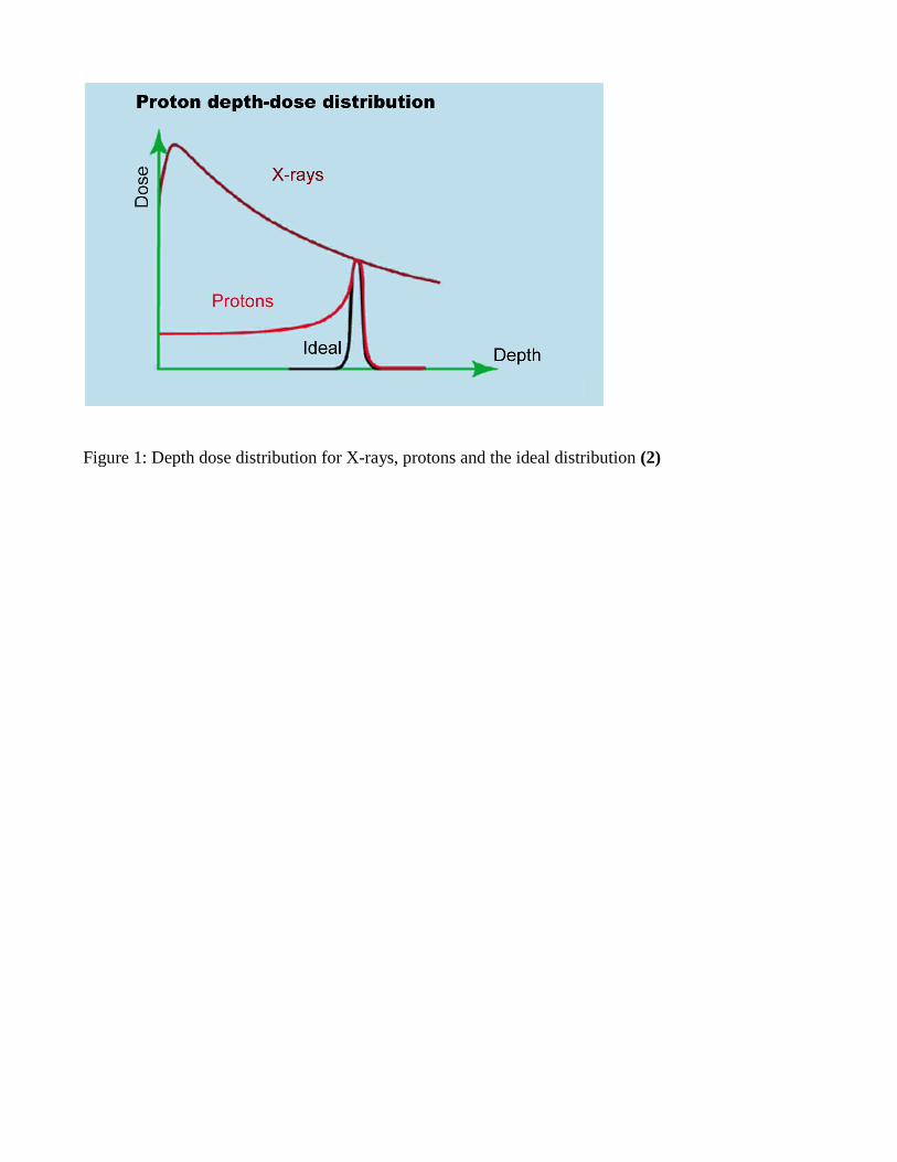
Figure 1: Depth dose distribution for X-rays, protons and the ideal distribution (2)
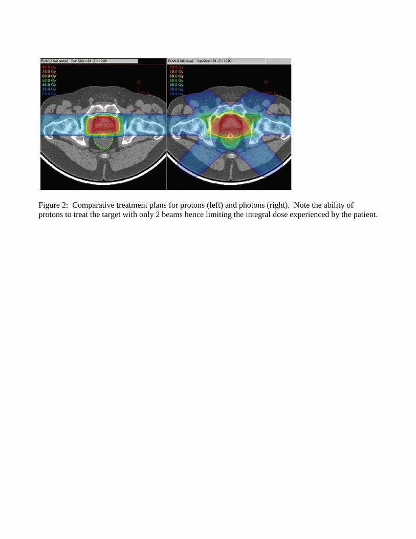
Figure 2: Comparative treatment plans for protons (left) and photons (right). Note the ability of
protons to treat the target with only 2 beams hence limiting the integral dose experienced by the patient.

Figure 3: Comparison of the X-ray depth dose curve with the SOBP used in passive proton beam
delivery for clinical treatment (2)

Figure 4: A representation of active scanning, illustrating how the superposition of multiple Bragg
peaks or hot spots can be used to create a uniform dose across a desired volume of irregular shape (45)

Figure 5: Schematic of the robotic patient positioner and gantry beam delivery system at LLUMC (13)
Figure 6: Comparative treatment plans for protons (left) and X-rays (right) in a liver treatment.
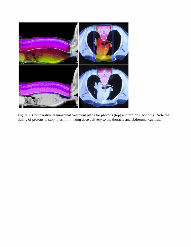
Figure 7: Comparative craniospinal treatment plans for photons (top) and protons (bottom). Note the
ability of protons to stop, thus minimizing dose delivery to the thoracic and abdominal cavities.

Figure 8: Measured data on out-of-field dose equivalent in proton therapy from a number of centers
using a range of measurement techniques and devices. Results are presented from HCL (46), PSI (47),
LLUMC (21) and MPRI (22)

Figure 9: Measured data on out-of-field dose equivalent in proton therapy for a number of different
treatment sites and beam configurations (20)