the pocket guide to neurocritical care
TRANSCRIPT
THE POCKET GUIDE TO
NEUROCRITICAL CARE:
A concise reference for the evaluation and management of neurologic emergencies
by the
Neurocritical Care Society
Marin E. Darsie, MD Asma M. Moheet, MD
ii
THE POCKET GUIDE TO
NEUROCRITICAL CARE:
A concise reference for the evaluation and management of neurologic emergencies
by the
Neurocritical Care Society
Marin E. Darsie, MD Assistant Professor of Emergency Medicine Division of Neurocritical Care University of Wisconsin School of Medicine and Public Health Madison, WI, USA Asma M. Moheet, MD Assistant Professor of Neurology and Neurosurgery Associate Director, Neuro Critical Care Unit Cedars-Sinai Medical Center Los Angeles, CA, USA
iii
For our families, our patients, and their families. Thank you for teaching us.
Copyright © 2017 by Neurocritical Care Society
All rights reserved. No part of this publication may be reproduced, stored in a retrieval system, or transmitted in any form or by any means, electronic,
mechanical, photocopying, recording, or otherwise, without the prior written permission of the author.
Disclaimer: This content was developed by the Neurocritical Care Society (NCS) and is intended to be used as an educational tool and should not be construed as medical advice. The editors and authors have made every effort to present accurate and complete information at the time of publication, however new information on any of the topics contained in this book may emerge. The authors, editors, publisher, or any party involved in the production of this work cannot guarantee that all data presented herein is complete or accurate, and these parties are not responsible for any omissions or errors in this text or liable for outcomes due to use of this text. Note that this includes drug information. The information in this text cannot replace independent evaluation by a qualified medical professional. Except when otherwise cited, the views expressed by the authors and editors are their own and are not necessarily the views of any institution with which the authors or editors are affiliated. Practitioners should refer to institutional policies and practices where appropriate. References have been provided by chapter authors at the end of each chapter. NCS respects the intellectual property rights of others and prohibits users from reproducing content that violates another party’s intellectual property rights. If you believe that any material infringes upon any copyright which you own or control, you may send a written notification of such infringement to NCS.
ISBN-13: 978-1-943909-00-1
Library of Congress Control Number: 2017951923
Cover Design and Original Graphics by Daniel Walsh
Printed in the United States of America
iv
PREFACE
While staffing the Neurocritical Care Society booth at the 2016 SCCM Conference, members of the NCS Resident/Fellow Committee realized there was demand for a quick reference guide to neurocritical care when person after person inquired if we had any for sale. Over the next 15 months, the Resident/Fellow Committee developed a vision which has become The Pocket Guide to Neurocritical Care: A concise reference for the evaluation and management of neurologic emergencies.
Over 40 authors have contributed to this project. Residents, fellows, and APPs were recruited as junior authors to provide authorship opportunities for trainees at the beginning of their careers. They were paired with established leaders in the field of neurocritical care who served as the senior authors for each chapter.
The aim of this book was to create a resource for trainees and other members of the NCCU multidisciplinary team of varying backgrounds on bedside management and pearls for a variety of neurocritical care conditions. With guidance from the NCS Educational Products Committee, seventeen high-yield topics were identified which include the most commonly encountered neurologic emergencies and basics of critical care medicine explained through the neurocritical care perspective. This book is not intended to be a definitive reference text, rather it aims to arm the reader with a grasp of a neurocritical care topic in less than 15 minutes.
v
ACKNOWLEDGEMENTS This project would not have been possible without the time, efforts and vision of all the members of the NCS Resident/Fellow Committee, specifically Sherri Braksick, Tobias Kulik, Deepa Malaiyandi, and Anand Venkatraman for their assistance with editing.
We would like to recognize the members of the NCS Educational Products Committee for their oversight and support in developing this project, as well as their time spent editing and reviewing its content for accuracy.
We would like to thank Becca Stickney and Sara Memmen for all their help in launching the first edition of The Pocket Guide to Neurocritical Care.
Finally, we would like to thank our families. We are the best versions of ourselves because of your love and support.
Marin Darsie and Asma Moheet August 2017
vi
ABBREVIATIONS + positive
¯̄ decreased
increased
AAN American Academy of Neurology
Ab antibody
ABCs airway, breathing, circulation
ABG arterial blood gas
AC assist control
ACA anterior cerebral artery
ACh acetylcholine
AChEI acetylcholinesterase inhibitor(s)
AChR acetylcholinesterase receptor(s)
ACEI angiotensin-converting enzyme inhibitor(s)
ACLS Advanced Cardiac Life Support
AComm anterior communicating artery
AED anti-epileptic drug
AF atrial fibrillation
AG anion gap
AHA American Heart Association
AICA anterior inferior cerebellar artery
AIS acute ischemic stroke
ALS amyotrophic lateral sclerosis
AKA also known as
ARDS acute respiratory distress syndrome
aSAH aneurysmal subarachnoid hemorrhage
ATLS Advanced Trauma Life Support
AVDO2 arterio-venous difference of oxygen consumption
AVM arteriovenous malformation
BCx blood culture
BBB blood-brain barrier
BID twice daily
BMP basic metabolic panel
BP blood pressure
BTF Brain Trauma Foundation
BSAS Bedside Shivering Assessment Scale
C Celsius
Ca2+ calcium
CABG coronary artery bypass graft
CAS carotid artery stenting
CBC complete blood count
CBF cerebral blood flow
CCM cerebral cavernous malformation
CEA carotid endarterectomy
CHF congestive heart failure
CIDP chronic inflammatory demyelinating polyneuropathy
COPD chronic obstructive pulmonary disease
CIM critical illness myopathy
CKD chronic kidney disease
CMP comprehensive metabolic panel
CMRO2 cerebral metabolic rate of oxygen
CMV cytomegalovirus
CN cranial nerve
CNS central nervous system
CO cardiac output
CPP cerebral perfusion pressure
CPR cardiopulmonary resuscitation
CrCl creatinine clearance
CSE convulsive status epilepticus
CSF cerebrospinal fluid
CSWS cerebral salt wasting syndrome
vii
CT computerized tomography
CTA CT angiography
CTV CT venogram
CVAD central venous access device
CVR cerebral vascular resistance
CXR chest x-ray
D day
DAI diffuse axonal injury
dAVF dural arteriovenous fistula
DBP diastolic blood pressure
DCD donation after circulatory death
DCI delayed cerebral ischemia
DH decompressive hemicraniectomy
DI diabetes insipidus
DKA diabetic ketoacidosis
DNI do not intubate
DNR do not resuscitate
DSA digital subtraction angiography
DTR deep tendon reflexes
DVT deep vein thrombosis
EBV Epstein-Barr virus
ECMO extracorporeal membrane oxygenation
ED Emergency Department
EDH epidural hematoma
EEG electroencephalogram
EKG electrocardiogram
EMG electromyography
EN enteral nutrition
ENLS Emergency Neurologic Life Support
ENT ear/nose/throat or otolaryngology
EOM extraocular muscles
ETT endotracheal tube
EVD external ventricular drain
FFP fresh frozen plasma
FOUR Full Outline of Unresponsiveness
FVC forced vital capacity
GBS Guillain-Barré syndrome
GCS Glasgow Coma Scale
GI gastrointestinal
GOSE Glasgow Outcome Scale- Extended
GTC generalized tonic-clonic
H hour
HCG human chorionic gonadotropin HD hemodialysis
Hgb hemoglobin
HIT heparin-induced thrombocytopenia
HOB head of bed
HSV Herpes simplex virus
HTLV-1 Human T-lymphotropic virus type 1
HTN hypertension
HTS hypertonic saline
IBW ideal body weight
ICA internal carotid artery
ICH intracerebral hemorrhage
ICP intracranial pressure
ICU intensive care unit
IgA immunoglobulin A
IM intramuscular
IV intravenous
IVC inferior vena cava
IVF intravenous fluids
IVH intraventricular hemorrhage
IVIg intravenous immunoglobulin
IV tPA intravenous tissue plasminogen activator
KCL potassium chloride
viii
LE lower extremity
LFTs liver function tests
LKWT last known well time
LOS length of stay
LP lumbar puncture
MAP mean arterial pressure
MC myasthenic crisis
MCA middle cerebral artery
MCS minimally conscious state
MEP maximal expiratory pressure
MG myasthenia gravis
MH malignant hyperthermia
MHS malignant hemispheric stroke
MI myocardial infarction
Min minute
mL milliliter
MOA mechanism of action
MRI magnetic resonance imaging
mRS modified Rankin Scale
MV mechanical ventilation
NCCU Neuro Critical Care Unit
NCS nerve conduction study OR Neurocritical Care Society
NCSE non-convulsive status epilepticus
NG nasogastric
NIF negative inspiratory force
NIPPV noninvasive positive pressure ventilation
NM neuromuscular
NMBA neuromuscular blockade agent
NMJ neuromuscular junction
NOAC novel oral anticoagulant
NS normal saline
NSAID nonsteroidal anti-inflammatory drugs
O2 oxygen
OG orogastric
OOB out of bed
OSA obstructive sleep apnea
OSM osmolar
OT occupational therapy or therapist
PbtO2 brain tissue oxygen tension
PCA posterior cerebral artery
PComm posterior communicating artery
PE pulmonary embolus
PEEP positive end expiratory pressure
PEG percutaneous endoscopic gastrostomy
PFO patent foramen ovale
PICA posterior inferior cerebellar artery
PIV peripheral intravenous line
PLEX plasmapheresis
Pplat plateau pressure
PRN pro re nata, as needed
PRVC pressure regulated volume control
PT physical therapy or therapist
PVS persistent vegetative state
QID four times daily
QOD every other day
RA rheumatoid arthritis
RASS Richmond Agitation and Sedation Scale
R/O rule out
RCT randomized control trial
RN registered nurse
ROM range of motion
ROSC return of spontaneous circulation
RR respiratory rate
RSI rapid sequence intubation
ix
RT respiratory therapy or therapist
RTA renal tubular acidosis
RVR rapid ventricular response
SAH subarachnoid hemorrhage
SBP systolic blood pressure
SC subcutaneous
SCA superior cerebellar artery
SCD sequential compression device
SDH subdural hematoma
Se sensitivity
SE status epilepticus
Sec second
SIADH syndrome of inappropriate antidiuretic hormone secretion
SLE systemic lupus erythematosus
s/p status post
Sp specificity
SpO2 peripheral capillary oxygen saturation
SSEPs somatosensory evoked potentials
SSRI selective serotonin reuptake inhibitor
SSS sick sinus syndrome
ST speech therapy or therapist
TB tuberculosis
TBI traumatic brain injury
TCD transcranial doppler ultrasound
TH therapeutic hypothermia
TIA transient ischemic attach
TMJ temporomandibular joint TMP-SMX trimethoprim- sulfamethoxazole
tPA tissue plasminogen activator
TTM targeted temperature management
U units
UA urinalysis
UCx urine culture
UE upper extremity
UH unfractionated heparin
UMN upper motor neuron
UTI urinary tract infection
VF ventricular fibrillation
VT tidal volume
VT ventricular tachycardia
VTE venous thromboembolism
VZV Varicella zoster virus
w/ with
WFNS World Federation of Neurological Surgeons
WNV West Nile virus
w/o without
yo years old
x
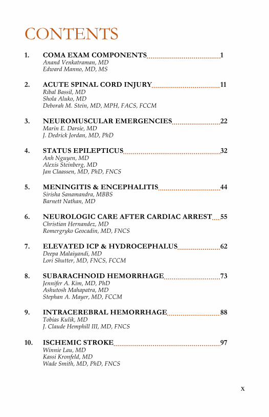
CONTENTS 1. COMA EXAM COMPONENTS 1 Anand Venkatraman, MD Edward Manno, MD, MS 2. ACUTE SPINAL CORD INJURY 11 Ribal Bassil, MD Shola Aluko, MD Deborah M. Stein, MD, MPH, FACS, FCCM 3. NEUROMUSCULAR EMERGENCIES 22 Marin E. Darsie, MD J. Dedrick Jordan, MD, PhD 4. STATUS EPILEPTICUS 32 Anh Nguyen, MD Alexis Steinberg, MD Jan Claassen, MD, PhD, FNCS 5. MENINGITIS & ENCEPHALITIS 44 Sirisha Sanamandra, MBBS Barnett Nathan, MD 6. NEUROLOGIC CARE AFTER CARDIAC ARREST 55 Christian Hernandez, MD Romergryko Geocadin, MD, FNCS 7. ELEVATED ICP & HYDROCEPHALUS 62 Deepa Malaiyandi, MD Lori Shutter, MD, FNCS, FCCM 8. SUBARACHNOID HEMORRHAGE 73 Jennifer A. Kim, MD, PhD Ashutosh Mahapatra, MD Stephan A. Mayer, MD, FCCM 9. INTRACEREBRAL HEMORRHAGE 88 Tobias Kulik, MD J. Claude Hemphill III, MD, FNCS 10. ISCHEMIC STROKE 97 Winnie Lau, MD Kassi Kronfeld, MD Wade Smith, MD, PhD, FNCS
xi
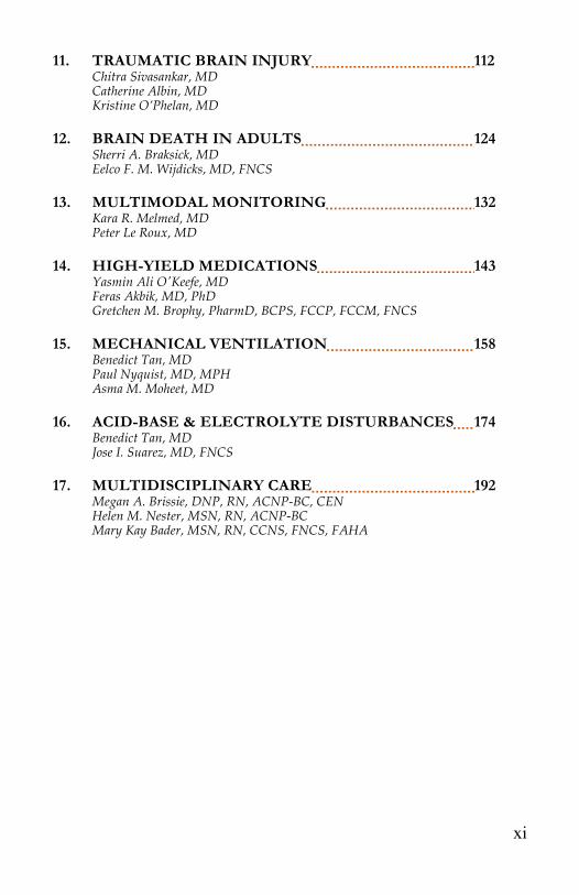
11. TRAUMATIC BRAIN INJURY 112 Chitra Sivasankar, MD Catherine Albin, MD Kristine O’Phelan, MD 12. BRAIN DEATH IN ADULTS 124 Sherri A. Braksick, MD Eelco F. M. Wijdicks, MD, FNCS 13. MULTIMODAL MONITORING 132 Kara R. Melmed, MD Peter Le Roux, MD 14. HIGH-YIELD MEDICATIONS 143 Yasmin Ali O'Keefe, MD Feras Akbik, MD, PhD Gretchen M. Brophy, PharmD, BCPS, FCCP, FCCM, FNCS 15. MECHANICAL VENTILATION 158 Benedict Tan, MD Paul Nyquist, MD, MPH Asma M. Moheet, MD 16. ACID-BASE & ELECTROLYTE DISTURBANCES 174 Benedict Tan, MD Jose I. Suarez, MD, FNCS 17. MULTIDISCIPLINARY CARE 192 Megan A. Brissie, DNP, RN, ACNP-BC, CEN Helen M. Nester, MSN, RN, ACNP-BC Mary Kay Bader, MSN, RN, CCNS, FNCS, FAHA
1
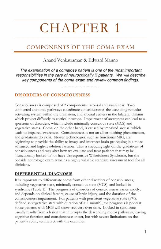
CHAPTER 1 COMPONENTS OF THE COMA EXAM
Anand Venkatraman & Edward Manno
The examination of a comatose patient is one of the most important responsibilities in the care of neurocritically ill patients. We will describe
key components of the coma exam and review common findings. ………………………
DISORDERS OF CONSCIOUSNESS Consciousness is comprised of 2 components: arousal and awareness. Two connected anatomic pathways coordinate consciousness: the ascending reticular activating system within the brainstem, and arousal centers in the bilateral thalami which project diffusely to cortical neurons. Impairment of awareness can lead to a spectrum of disorders, which include minimally conscious state (MCS) and vegetative states. Coma, on the other hand, is caused by impaired arousal which leads to impaired awareness. Consciousness is not an all-or-nothing phenomenon, and gradations do exist. Newer technologies, such as functional MRI, are beginning to provide the ability to image and interpret brain processing in a more advanced and high-resolution fashion. This is shedding light on the gradations of consciousness and may alter how we evaluate and treat patients that may be “functionally locked-in” or have Unresponsive Wakefulness Syndrome, but the bedside neurologic exam remains a highly valuable standard assessment tool for all clinicians. DIFFERENTIAL DIAGNOSIS It is important to differentiate coma from other disorders of consciousness, including vegetative state, minimally conscious state (MCS), and locked-in syndrome (Table 1). The prognosis of disorders of consciousness varies widely, and depends on clinical factors, cause of brain injury, and the duration of the consciousness impairment. For patients with persistent vegetative state (PVS, defined as vegetative state with duration of > 1 month), the prognosis is poorest. Some patients with MCS will show recovery over time. Locked-in syndrome usually results from a lesion that interrupts the descending motor pathways, leaving cognitive function and consciousness intact, but with severe limitations on the patient’s ability to interact with the examiner.
2
POSSIBLE CAUSES OF COMA Bihemispheric phenomena, such as medication or drug toxicities, generalized status epilepticus, metabolic disorders and meningoencephalitides can all cause poor responsiveness or coma, with or without focal neurologic findings. Coma may also be caused by brain lesions affecting the thalamus and brainstem, since these contain crucial arousal-supporting neurons. The latter may be associated with focal neurologic findings. It is essential to rule out reversible causes of coma in cases when the etiology is not known (Table 2). NEUROLOGIC EXAM IN COMA The initial exam is important for localization and identifying the cause of coma. Serial exams to assess interval change are equally important. Acute neurologic deterioration can signal AIS, ICH, seizure, worsening edema, hydrocephalus, or elevated ICP. Hourly vital sign assessments and neurologic checks are the norm in newly-admitted NCCU patients. In some, such as those admitted after surgical or endovascular procedures, the frequency of assessments may need to be higher. We recommend the use of standardized scales to assess disorders of consciousness. The best known is the Glasgow Coma Scale (GCS), of which the arbitrary definition of coma is GCS 8 or less (E2V2M4). See Table 3 for reference. Limitations of GCS: § Can miss locked-in states and subtle changes in consciousness § Does not assess pupillary and other brainstem reflexes § Patients with similar scores may go on to have different outcomes § Assigns greater weight to motor response than eye opening and verbal responses. § Intubated patients default to a T score on verbal The Full Outline of UnResponsiveness (FOUR) score can also be used, and addresses some shortcomings of the GCS: § Incorporates brainstem function and respiratory pattern, allowing for better
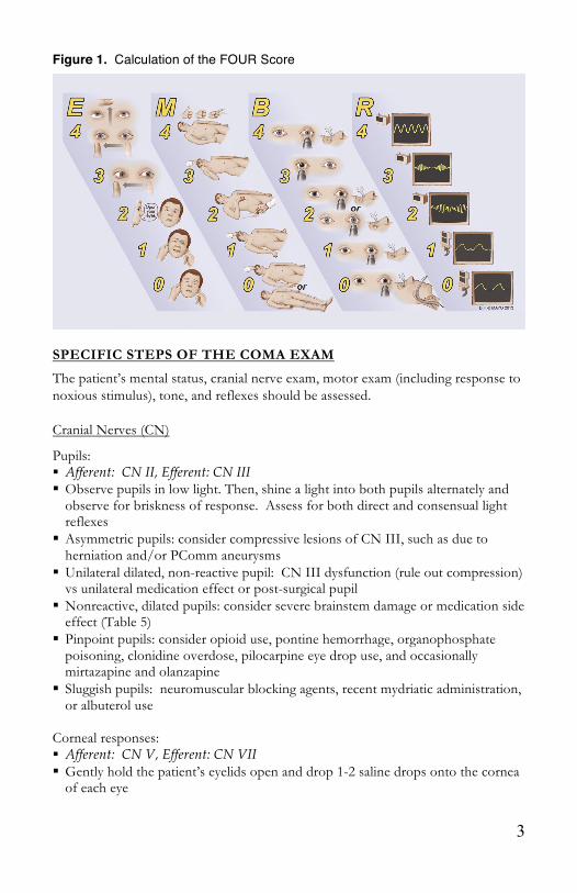
localization § Can help recognize a locked-in state § Can recognize various stages of herniation The calculation of the FOUR Score is illustrated in Figure 1, and is additionally described in Table 4.
3
Figure 1. Calculation of the FOUR Score
SPECIFIC STEPS OF THE COMA EXAM
The patient’s mental status, cranial nerve exam, motor exam (including response to noxious stimulus), tone, and reflexes should be assessed. Cranial Nerves (CN)
Pupils: § Afferent: CN II, Efferent: CN III § Observe pupils in low light. Then, shine a light into both pupils alternately and
observe for briskness of response. Assess for both direct and consensual light reflexes
§ Asymmetric pupils: consider compressive lesions of CN III, such as due to herniation and/or PComm aneurysms
§ Unilateral dilated, non-reactive pupil: CN III dysfunction (rule out compression) vs unilateral medication effect or post-surgical pupil
§ Nonreactive, dilated pupils: consider severe brainstem damage or medication side effect (Table 5)
§ Pinpoint pupils: consider opioid use, pontine hemorrhage, organophosphate poisoning, clonidine overdose, pilocarpine eye drop use, and occasionally mirtazapine and olanzapine
§ Sluggish pupils: neuromuscular blocking agents, recent mydriatic administration, or albuterol use
Corneal responses: § Afferent: CN V, Efferent: CN VII § Gently hold the patient’s eyelids open and drop 1-2 saline drops onto the cornea
of each eye
4
§ Cotton swab can also be used but use caution as with repeated testing this can lead to corneal ulceration
§ There is a blinking response if this pathway is intact Blink to threat: § Afferent: CN II, Efferent: CN VII § Briskly move your hand into the patient’s visual field while holding his/her eyelid
open. The patient should blink. Gaze: § Hold eyes open and observe direction of gaze in neutral head position § Eye movements involve coordinated functioning of multiple CN, frontal lobe
and brainstem centers § Gaze deviation also occurs due to involvement of frontal eye fields in each
hemisphere: destructive lesions cause ipsilateral gaze deviation, stimulation causes contralateral deviation
§ Cortical ischemic stroke patients demonstrate gaze directed towards hemisphere of the stroke.
§ Seizure patients demonstrate gaze directed away from seizing hemisphere, and may have gaze towards the hemisphere post-ictally
§ Brainstem strokes can cause impaired gaze towards the side of the stroke § Forced downgaze may be seen in thalamic hemorrhages, pineal mass lesions, and
severe hydrocephalus § Bilateral CN VI palsy seen in ↑ ICP
EOMs: § Innervation of extraocular muscles: Lateral Rectus CN VI, Superior Oblique CN
IV, All others CN III § Fixation and tracking are normal findings § Fixation: eyes looking at an object and not moving from that position § Tracking: eyes moving as the object or the examiner moves, to follow them § Roving eye movements: slow and conjugate to-and-fro movements § Can be seen in toxic and metabolic conditions where brainstem is intact. Light
stages of sleep and lighter coma also cause this § Nystagmus: fast, beating movements to one side (may indicate ongoing seizures)
¨ Other causes: phenytoin toxicity, brain lesions like those seen in stroke or multiple sclerosis, inner ear disorders, and metabolic disorders like thiamine deficiency
¨ Down-beating nystagmus may be seen in disorders of the craniocervical junction or cerebellar flocculus
¨ Up-beating nystagmus may be seen in cerebellar vermis involvement, and sometimes in lesions of the medulla
¨ Acute lesions in the pons can cause rapid downward jerking of the eyes with slow return to normal position, called ocular bobbing
Fundoscopy: § Evaluate optic disc and nerve
5
§ Blurring of optic disc margins is indicative of ↑ ICP, but absence of blurring does not automatically indicate normal ICP. Subhyaloid hemorrhages can also be seen with ↑ ICP ¨ Terson’s syndrome: subhyaloid hemorrhage in SAH
Oculocephalic or “doll’s eyes” reflex (OCR): § Afferent: CN VIII and proprioceptive pathways from the cervical level, Efferent:
CN III and VI § Confirm stability of cervical spine, then move head briskly in one direction and
then the other with the eyelids held open § Interpretation of OCR responses in a comatose patient:
¨ In a normal OCR, eyes move conjugately in the direction opposite to head movement
¨ In abnormal OCR, eyes stay in fixed position in the head, implying brainstem disease
Cold calorics (oculovestibular response or OVR): § Afferent: CN VIII, Efferent: CN III and VI § Do this if OCRs are absent § Ensure patency of ear canal and ability of water to reach tympanic membrane § Instill 50-60 mL of ice cold water into each ear over 1 minute using a syringe § Test each side individually with several minutes between testing of each § Normal: slow conjugate deviation towards the irrigated side and fast horizontal
nystagmus to the contralateral ear § Abnormal: no fast nystagmus in patients with cerebral damage but intact
brainstem reflexes. No slow deviation and no fast nystagmus implies cerebral and brainstem damage
Gag reflex: § Afferent: CN IX, Efferent: CN X § Tested by stimulating the back of the patient’s throat with a tongue depressor or
suction catheter Cough reflex: § Afferent: CN X, Efferent: CN X § In an intubated patient can be tested by inserting a suction catheter into the
patient’s ETT or tracheostomy tube Motor A normal patient should follow commands. In a comatose patient it is often necessary to administer noxious stimuli centrally, which may include sternal rub or supraorbital ridge pressure. Do not perform supraorbital ridge pressure in the presence of facial fractures. If there is no response to central noxious stimulus, peripheral stimulus (such as application of nailbed pressure) should be performed.
6
§ Patients may localize to the stimulus, withdraw away from the stimulus, flex, extend, or have no response at all. Grimacing may also be observed.
§ Spinal reflexes may lead to lower extremity movements even in patients with severe brain damage or brain death (e.g. triple flexion response of hip, knee, and ankle flexion)
§ Decorticate posturing: upper extremity flexion and lower extremity extension, typically from a lesion above the red nucleus of the midbrain.
§ Decerebrate posturing: upper and lower extremity extension is typically from a lesion below the red nucleus.
§ Unilateral or bilateral posturing may be seen based on location of lesion causing it.
§ Postanoxic myoclonus is common in patients following cardiac arrest. Occasionally it may indicate ongoing seizure activity, EEG is recommended.
Tone and Reflexes ↑ tone, brisk reflexes and upgoing toes are indicative of a lesion in the spinal cord or brain. § If unilateral, usually indicates a lesion on the opposite side. § Symmetric hyperreflexia can be normal, especially in young patients, but may also
indicate bilateral lesions, especially in the brainstem and spinal cord. In rare instances, symmetric hyperreflexia might indicate conditions like serotonin syndrome.
§ People with brisk reflexes usually do not have upgoing toes, so this can be a good way to differentiate pathological cases from physiologic hyperreflexia.
§ Brisk reflexes and ↑ tone in lower extremities but not upper extremities are indicative of lesion below the level of the cervical spinal cord.
RESPIRATORY PATTERNS IN COMATOSE PATIENTS Medication side effects should be ruled out first. Sedating medications tend to cause slow regular breathing, whereas salicylate overdose can cause rapid breathing. In intubated patients, assess synchrony with the ventilator and degree of effort, including actual vs set respiratory rate. Abnormal breathing may manifest more prominently on spontaneous ventilator modes. Types of abnormal breathing: § Cheyne-Stokes: oscillation between fast and slow breathing (multiple causes
including bilateral hemispheric lesions, heart failure, etc.) § Apneustic: rapid breathing with inspiratory pauses (pontine lesions) § Biot’s: quick shallow breaths followed by pause after four to five cycles
(medullary damage) § Kussmaul: rapid, deep and labored breaths (metabolic acidosis)