the polyphosphate kinase gene ppk2 is required for - mbio
TRANSCRIPT

The Polyphosphate Kinase Gene ppk2 Is Required for Mycobacteriumtuberculosis Inorganic Polyphosphate Regulation and Virulence
Yu-Min Chuang,a Deborah A. Belchis,b Petros C. Karakousisa,c
Department of Medicine, Johns Hopkins University, School of Medicine, Baltimore, Maryland, USAa; Department of Pathology, Johns Hopkins University, School ofMedicine, Baltimore, Maryland, USAb; Department of International Health, Johns Hopkins Bloomberg School of Public Health, Baltimore, Maryland, USAc
ABSTRACT The Mycobacterium tuberculosis gene Rv3232c/MT3329 (ppk2) encodes a class II polyphosphate kinase, which hydro-lyzes inorganic polyphosphate (poly P) to synthesize GTP. We assessed the role of ppk2 in M. tuberculosis poly P regulation, anti-biotic tolerance, and virulence. A ppk2-deficient mutant (ppk2::Tn) and its isogenic wild-type (WT) and complemented (Comp)strains were studied. For each strain, the intrabacillary poly P content, MIC of isoniazid, and growth kinetics during infection ofJ774 macrophages were determined. Multiplex immunobead assays were used to evaluate cytokines elaborated during macro-phage infection. The requirement of ppk2 for M. tuberculosis virulence was assessed in the murine model. The ppk2::Tn mutantwas found to have significantly increased poly P content and a 4-fold increase in the MIC of isoniazid relative to the WT andComp strains. The ppk2::Tn mutant showed reduced survival at day 7 in activated and naive J774 macrophages relative to theWT. Naive ppk2::Tn mutant-infected macrophages showed increased expression of interleukin 2 (IL-2), IL-9, IL-10, IL-12p70,and gamma interferon (IFN-�) relative to WT-infected macrophages. The ppk2::Tn mutant exhibited significantly lower lungCFU during acute murine infection compared to the control groups. ppk2 is required for control of intrabacillary poly P levelsand optimal M. tuberculosis growth and survival in macrophages and mouse lungs.
IMPORTANCE Mycobacterium tuberculosis, the causative agent of tuberculosis (TB), is a highly successful human pathogen be-cause it has developed mechanisms to multiply and survive in the lungs by circumventing the immune system. Identification ofvirulence factors responsible for M. tuberculosis growth and persistence in host tissues may assist in the development of novelstrategies to treat TB. In this study, we found that the mycobacterial enzyme polyphosphate kinase 2 (PPK2) is required for con-trolling intracellular levels of important regulatory molecules and for maintaining susceptibility to the first-line anti-TB drugisoniazid. In addition, PPK2 was found to be required for M. tuberculosis growth in the lungs of mice, at least in part by sup-pressing the expression of certain key cytokines and chemokines by inactivated lung macrophages.
Received 25 January 2013 Accepted 26 April 2013 Published 21 May 2013
Citation Chuang YM, Belchis DA, Karakousis PC. 2013. The polyphosphate kinase gene ppk2 is required for Mycobacterium tuberculosis inorganic polyphosphate regulation andvirulence. mBio 4(3):e00039-13. doi:10.1128/mBio.00039-13.
Invited Editor Roberto Docampo, University of Georgia Editor L. David Sibley, Washington University School of Medicine
Copyright © 2013 Chuang et al. This is an open-access article distributed under the terms of the Creative Commons Attribution-Noncommercial-ShareAlike 3.0 Unportedlicense, which permits unrestricted noncommercial use, distribution, and reproduction in any medium, provided the original author and source are credited.
Address correspondence to Petros C. Karakousis, [email protected].
Tuberculosis (TB) remains a major global health problem (1, 2).One of the major obstacles to global TB eradication efforts is
the prolonged treatment course required to cure active TB, whichis believed to reflect the organism’s ability to remain in a nonrep-licating persistent state with reduced susceptibility to conven-tional antituberculous drugs.
Inorganic polyphosphate (poly P), a linear polymer of manytens or hundreds of inorganic phosphate residues linked by high-energy phosphoanhydride bonds, appears to play an importantregulatory role in the transition to bacterial persistence (3, 4).Bacteria accumulate poly P intracellularly when they encountergrowth-limiting conditions, such as phosphate depletion, aminoacid starvation, or osmotic stress (3, 5). Poly P accumulation hasbeen shown to modulate many different bacterial processes, in-cluding protein synthesis, nucleotide balance, lipid metabolism,energy utility, and susceptibility to antibiotics (3, 4). Bacterialpolyphosphate kinases are of two classes. Polyphosphate kinase 1(PPK1) is responsible for poly P synthesis through hydrolysis ofATP (3). Although PPK2 enzymes synthesize poly P, they also
function as GTP synthases, using poly P as a substrate (3, 6–9).Deletion of the ppk2B gene in Corynebacterium glutamicum re-duced PPK activity and intrabacterial poly P content, while over-expression of ppk2B increased PPK activity and poly P content(10). Conversely, ppk2 deficiency in Campylobacter jejuni led todecreased poly P-dependent GTP synthesis and increased intra-cellular ATP/GTP ratio (9). Mutation of ppk2 attenuates bacterialsurvival under different growth-limiting conditions (7, 9, 10). Inaddition, PPK2 plays an important role in invasion and intracel-lular survival of C. jejuni in human intestinal epithelial cells (9).
The Mycobacterium tuberculosis genome contains genes thatencode both PPK1 (Rv2984) and PPK2 (Rv3232c), which play arole in poly P synthesis (7, 11). PPK1 is involved in the stress-induced mprAB-sigE-rel signaling pathway in mycobacteria (11)and appears to be encoded by an essential gene in M. tuberculosis(12). M. tuberculosis PPK2 is an octamer that catalyzes polyP-dependent phosphorylation of ADP to ATP at a rate �800-foldhigher than that of poly P synthesis (13). PPK2 also regulates theintracellular nucleotide pool and is required for survival of
RESEARCH ARTICLE
May/June 2013 Volume 4 Issue 3 e00039-13 ® mbio.asm.org 1
Dow
nloa
ded
from
http
s://j
ourn
als.
asm
.org
/jour
nal/m
bio
on 3
1 D
ecem
ber
2021
by
61.8
1.69
.191
.

FIG 1 Complementation of ppk2::Tn. (A) RT-PCR analysis of wild-type M. tuberculosis CDC 1551 expression of intergenic regions from MT3331 todesA3/MT3326 during late log phase. Chromosomal DNA was used as a control. Primer sets 1 to 5 (Table 1) target specific intergenic areas, as indicated in thefigure; primer set 6 targets ppk2. (B) PCR analysis of genomic DNA from the wild-type, ppk2::Tn, and ppk2::Tn Comp strains. Primer set A targets the ppk2 gene(bp �50 to 430), yielding a 480-bp product in the wild type, a 2,547-bp product in the ppk2::Tn mutant, and both products in the ppk2::Tn Comp strain. Primerset B targets bp 108 to 661 of ppk2, yielding a 553-bp product in all strains. (C) Southern hybridization demonstrating binding of the ppk2-specific probe toKpnI-digested fragments of the expected size in the wild type (1,669 bp), ppk2::Tn mutant (2,266 bp), and ppk2::Tn Comp strain (2,266 and 6,095 bp). (D)RT-PCR analysis of ppk2 expression in the wild-type, ppk2::Tn, and ppk2::Tn Comp strains during early stationary phase of growth.
Chuang et al.
2 ® mbio.asm.org May/June 2013 Volume 4 Issue 3 e00039-13
Dow
nloa
ded
from
http
s://j
ourn
als.
asm
.org
/jour
nal/m
bio
on 3
1 D
ecem
ber
2021
by
61.8
1.69
.191
.

M. smegmatis under acidic, heat, and hypoxic conditions (7). Inaddition, deficiency of ppk2 or ppk1 is associated with decreasedmycobacterial survival during macrophage infection (7, 11).However, the role of PPK2 in M. tuberculosis virulence is un-known.
In this study, we investigated the role of ppk2 on M. tuberculosispoly P homeostasis and susceptibility to the cell wall-acting agentisoniazid. Next, we determined the impact of ppk2 deficiency onM. tuberculosis pathogenesis in the animal host by studying thegrowth, survival, and pathology of a ppk2-deficient mutant in themurine model of TB infection. To investigate host factors respon-sible for growth restriction of the ppk2-deficient mutant duringacute infection in mouse lungs, we used multiplex immunobeadassays to evaluate cytokines elaborated by macrophages infectedwith the ppk2-deficient mutant and control strains.
RESULTSppk2 is coexpressed as part of a three-gene operon. In order totest the role of ppk2 (Rv3232c/MT3329) in M. tuberculosis viru-lence, a ppk2-deficient mutant (ppk2::Tn) was generated (14). Toguide our complementation strategy, we studied potential cotran-scription of genes flanking ppk2 using reverse transcription-PCR(RT-PCR). ppk2 is located within a putative operon comprising
Rv3234c/MT3331, Rv3233c/MT3330, Rv3232c/MT3329, Rv3231c/MT3328, Rv3230c/MT3327, and Rv3229c/MT3326. As shown inFig. 1A, the two genes upstream of ppk2 (Rv3234c/MT3331 andRv3233c/MT3330) are cotranscribed with ppk2, but the threedownstream genes are expressed independently in a single tran-scriptional unit.
A DNA fragment containing the Rv3234c/MT3331, Rv3233c/MT3330, and ppk2 genes, as well as 269 bp of the 5= flankingsequence of MT3331 was cloned into the pMH94H plasmid andthen recombined into ppk2::Tn to generate a ppk2 complementedstrain (ppk2::Tn Comp). PCR amplification with primers target-ing ppk2 (Table 1) yielded products of 487 bp and 2,554 bp in thewild-type and ppk2::Tn strains, respectively, and both productswere present, as expected, in the ppk2::Tn Comp strain (Fig. 1B).Southern hybridization, using a specific probe targeting ppk2 (Ta-ble 1) after restriction digestion of genomic DNA with KpnI con-firmed the generation of a 1,669-bp fragment in the wild-typestrain and a 2,266-bp fragment in the ppk2::Tn strain (Fig. 1C).The two expected products (2,266 bp and 6,095 bp) were observedin the ppk2::Tn Comp strain. As confirmation of functional ppk2complementation in the ppk2::Tn Comp strain, partial restorationof ppk2 expression was observed during early stationary phase(Fig. 1D). RT-PCR demonstrated that expression of the two up-
TABLE 1 Primers used in this study
Primer Directiona Sequence
Primers used for coexpression studiesb
Primer set 1 F GTAACGAATTCCGGCCAACTR TACATCTGCGACTGGGTTCC
Primer set 2 F CCAAGCTGCTGGAGACCTACR CGGGAAGATGGGCAATATAA
Primer set 3 F GTGGAAAAGCCAAAGGTCAAR ACCTGATCGATCGTGATTGG
Primer set 4 F ACCAAGAGCTGCCATTTCTGR CACAATGGTCCCTGGCTTTA
Primer set 5 F CGAAGTGGCAAGAGCGTAGR CCGATCTCCATGTTTTCGAT
Primer set 6 (ppk2) F GTGGATATACCATCCGTTGATGTGR GCGTACAGAAACCCATGACC
sigA primer set F ACCTCAAACAGATCGGCAAGR TGGATTTCCAGCACCTTCTC
Primers used for complemented strain generation/confirmationc
1st cloning F GCGGAGCTC GTACTCGACGACGGGTGTGR GCGTCTAGACAACGAGTCGCTGCAACA
2nd cloning F CGTCGCTCATGAGATGGTGCCCTTGGTGGR GCATCGAACGTTATGTTAAAACGACGGCCAGTGAAT
Primer set A F GGTACTCGGTGATCCGTTTGR AAGCACTGGACGAGCTTCTG
Primer set B Same as primer set 6ppk2 primer set (probe in Southern blot) F CGTCTACCAAGCCGAATTGT
R GCACCATCATCTCGTCCTTTGene-specific primers used for qRT-PCR studies
Rv0496 F AGAGGACCCTAACGGCAAATR TTTCCACCGCTTCTATCGAC
Rv1026 F AACTCGATTCGCTTGCTGATR TGGTGAAACGTCAGCAGTTC
ppk1 F CTCAAGACGCACTGCAAGACR TGAACAAGTCGGTCAAGTCG
sigA F TCGAGGTGATCAACAAGCTGR TGGATTTCCAGCACCTTCTC
a F, forward; R, reverse.b Primer sets 1 to 6 target regions depicted in Fig. 1A.c Primer sets A and B target regions depicted in Fig. 1B, and primer set 6 and sigA primers were used for RT-PCR in Fig. 1D.
PPK2 Is Required for M. tuberculosis Virulence
May/June 2013 Volume 4 Issue 3 e00039-13 ® mbio.asm.org 3
Dow
nloa
ded
from
http
s://j
ourn
als.
asm
.org
/jour
nal/m
bio
on 3
1 D
ecem
ber
2021
by
61.8
1.69
.191
.

stream genes (Rv3234c/MT3331 and Rv3233c/MT3330) and twodownstream genes (Rv3231c/MT3328 and Rv3230c/MT3327) wasnot disrupted in the mutant or complemented strains (see Fig. S1in the supplemental material). The ppk2::Tn mutant exhibitedwild-type growth during the logarithmic phase in supplementedMiddlebrook 7H9 broth but achieved a lower density relative tothe wild type during stationary phase. The in vitro growth of theppk2::Tn Comp strain was similar to that of the mutant (Fig. S2).
ppk2 is required for control of intrabacillary poly P levels andsusceptibility to isoniazid. Previous studies have shown thatPPK2 hydrolyzes inorganic polyphosphate (poly P) to generateGTP from GDP, contributing to a decreased ATP/GTP ratio inMycobacterium smegmatis (7). Consistent with a role for PPK2 inpoly P hydrolysis during exponential growth of M. tuberculosis, wefound that the ppk2::Tn mutant exhibited significantly higher polyP content relative to the isogenic wild-type (P � 0.0038) andppk2::Tn Comp (P � 0.0028) strains (Fig. 2A). Quantitative RT-PCR (qRT-PCR) revealed that ppk1 expression was significantlyreduced (P � 0.001) and expression of the putative exopolyphos-phatase Rv1026 was increased (P � 0.004) in the ppk2::Tn mutantrelative to the wild type (Fig. 2B), consistent with negative- andpositive-feedback mechanisms, respectively. However, expressionof the exopolyphosphatase gene Rv0496 was slightly decreased inthe ppk2::Tn mutant than in the wild type (P � 0.028). Consistentwith the hypothesis that poly P accumulation is associated with M.tuberculosis tolerance to the cell wall-active agent isoniazid (15),the MIC of isoniazid increased from 0.06 �g/ml for the wild-typeand ppk2::Tn Comp strains to 0.24 �g/ml for the ppk2::Tn mutantstrain. As in the case of M. smegmatis (7), M. tuberculosis ppk2deficiency was associated with a higher ATP/GTP ratio relative tothe wild-type (P � 0.07) and ppk2::Tn Comp (P � 0.02; Fig. 2C)strains. Each experiment was performed using three biologicalreplicates.
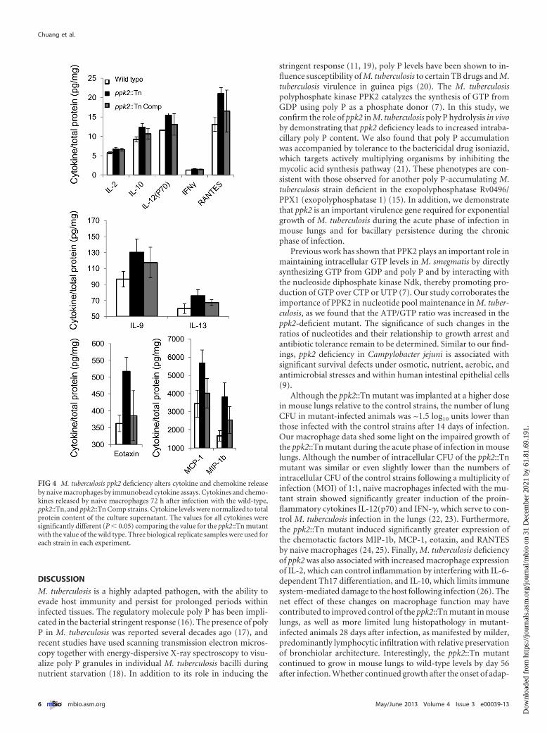
ppk2 is required for M. tuberculosis survival during infectionof naive macrophages and suppression of proinflammatory cy-tokine release. On the basis of previous reports, ppk2 appears toplay an important role in M. tuberculosis intracellular survival (7).To further investigate the mechanism of defective growth of theppk2::Tn mutant, we infected naive and activated murine J774macrophages with each of the three strains. The numbers of intra-cellular CFU were lower in gamma interferon (IFN-�)-activatedmacrophages 7 days after infection with the ppk2::Tn mutant rel-ative to the corresponding values for the wild-type (P � 0.004)and ppk2::Tn Comp strains (Fig. 3A). On day 1 after infection,activated macrophages infected with the ppk2::Tn mutant pro-duced significantly higher levels of granulocyte colony-stimulating factor (G-CSF) relative to macrophages infected withthe wild-type strain (data not shown; P � 0.03). The levels of allother cytokines and chemokines elaborated by activated macro-phages on day 1 or day 3 were either below the limit of detection ofthe assay or showed no differences between groups. The ppk2::Tnmutant showed defective growth relative to the wild-type andppk2::Tn Comp strains on day 5 (P � 0.016) and day 7 (P � 0.049)after infection of naive macrophages (Fig. 3B). At day 1 after in-fection of naive macrophages, cytokine and chemokine levels wereeither below the limit of detection of the assay or showed no dif-ferences between groups. However, on day 3 after infection, thelevels of interleukin 2 (IL-2) (P � 0.044), IL-9 (P � 0.04), IL-10(P � 0.012), IL-12(p70) (P � 0.001), IL-13 (P � 0.046), eotaxin(P � 0.005), IFN-� (P � 0.035), monocyte chemotactic peptide 1
FIG 2 ppk2 is required for M. tuberculosis control of intracellular poly P levelsand ATP/GTP ratio. (A) Intrabacillary poly P levels were measured in thewild-type, ppk2::Tn, and ppk2::Tn Comp strains during mid-log phase using aDAPI-based method and normalized to total protein content of extract lysate.Values that were significantly different (P � 0.05) are indicated by a bar andasterisk. Values are means � standard deviations (error bars) for three biolog-ical replicate samples. (B) qRT-PCR analysis of gene expression of ppk2::Tnmutant and ppk2::Tn Comp strain compared to that of the wild type duringlate log phase of growth in supplemented Middlebrook 7H9 broth, expressedas a change in the normalized cycle threshold (��CT). (C) Intrabacillary ATP/GTP ratio of the wild-type, ppk2::Tn, and ppk2::Tn Comp strains during latelog phase, as measured by HPLC. The values for ppk2::Tn and ppk2::Tn Compstrains were significantly different (P � 0.05) as indicated by the bar andasterisk. The values for the ppk2::Tn mutant and the wild-type strain were notsignificantly different (P � 0.07). Three biological replicate samples were usedfor each strain in each experiment.
Chuang et al.
4 ® mbio.asm.org May/June 2013 Volume 4 Issue 3 e00039-13
Dow
nloa
ded
from
http
s://j
ourn
als.
asm
.org
/jour
nal/m
bio
on 3
1 D
ecem
ber
2021
by
61.8
1.69
.191
.

(MCP-1) (P � 0.02), macrophage inflammatory protein 1b (MIP-1b) (P � 0.011), and RANTES (regulated upon activation, normalT cell expressed and secreted) (P � 0.004) were more highly in-duced by the ppk2::Tn mutant than by the wild-type and ppk2::TnComp strains, despite similar intracellular CFU values (Fig. 4).Each macrophage CFU and cytokine experiment was performedusing three biological replicates.
ppk2 enhanced M. tuberculosis growth and survival in mouselungs. In order to test the role of ppk2 in M. tuberculosis virulencein the mammalian host, separate groups of BALB/c mice wereaerosol infected with the wild type (1.82 � 0.1), ppk2::Tn mutant(2.24 � 0.06), and ppk2::Tn Comp strain (1.9 � 0.1) (all valueslog10 number of CFU/lung). During the first 14 days after infec-tion, wild-type and ppk2::Tn Comp strains showed typical expo-nential growth, increasing by 4.34 log10 units and 4.15 log10 units,
respectively. On the other hand, ppk2::Tnshowed a severe growth defect during theacute phase of the infection relative towild type, increasing by only 2.29 log10
units (P � 0.001) (Fig. 5A). After the on-set of adaptive immunity (day 28), thewild-type bacillary burden declined to5.85 � 0.15 1og10 units, after which a rel-atively stable lung census was main-tained. Interestingly, the lung bacillaryburden of the ppk2::Tn mutant continuedto increase until day 56, when it wasequivalent to that of animals infectedwith the wild type but then declined dur-ing the chronic phase of infection, suchthat by day 119, it was 0.63 log10 unitlower than that of wild type (P � 0.098).The ppk2::Tn mutant also showed defec-tive growth in mouse spleens, as the num-bers of CFU in ppk2::Tn-infected mousespleens were significantly lower than thenumbers in spleens in mice infected withthe wild-type and ppk2::Tn Comp strainsat days 28, 84, and 119 (P � 0.001 for alltime points) (Fig. 5B).
Mouse body weights were not signifi-cantly different between groups at anytime point (see Fig. S3A in the supple-mental material). However, by day 28 af-ter infection, the mean normalized lung(see Fig. S3B in the supplemental mate-rial) and spleen (see Fig. S3C) weights ofppk2::Tn-infected mice were significantlylower than the corresponding values ofmice infected with the wild type (P �0.001 and P � 0.023, respectively). Grosspathology revealed no major differencesin the number or size of lung tubercle le-sions between groups at any time point(Fig. S4).
After 28 days of infection, histologicalevaluation revealed moderate inflamma-tion with a predominance of macro-phages and bronchiolar obliteration inthe lungs of mice infected with the wild-
type and ppk2::Tn Comp strains (Fig. 5C). In lung samples frommice infected with the ppk2::Tn mutant, mild inflammation pre-dominantly comprising lymphocytes was noted. Lymphocytic in-volvement was observed in 15/41 (36.6%) bronchioles in miceinfected with the wild type, 8/50 (16%) in mice infected with theppk2::Tn mutant, and 12/21 (57.1%) in mice infected with theppk2::Tn Comp strain. Acid-fast staining revealed more bacilli inlungs in mice infected with the wild-type and ppk2::Tn Compstrains than in mice infected with the ppk2::Tn mutant. After56 days of infection, the lungs of each group exhibited similardegrees of bronchiolitis, bronchiolar obliteration, intra-alveolarinflammation, and perivascular lymphocytic inflammation, withequal numbers of histiocytes and lymphocytes. Each data pointfrom mouse experiments represents data obtained from 5 ani-mals.
FIG 3 ppk2 deficiency impairs M. tuberculosis growth and survival in J774 macrophages. Growth andsurvival of the wild type, ppk2::Tn mutant, and ppk2::Tn Comp strain after infection of activated (A) andnaive (B) J774 murine macrophages. Values that were significantly different (P � 0.05) are indicated bya bar and asterisk. Three biological replicate samples were used for each strain in each experiment.
PPK2 Is Required for M. tuberculosis Virulence
May/June 2013 Volume 4 Issue 3 e00039-13 ® mbio.asm.org 5
Dow
nloa
ded
from
http
s://j
ourn
als.
asm
.org
/jour
nal/m
bio
on 3
1 D
ecem
ber
2021
by
61.8
1.69
.191
.
DISCUSSION
M. tuberculosis is a highly adapted pathogen, with the ability toevade host immunity and persist for prolonged periods withininfected tissues. The regulatory molecule poly P has been impli-cated in the bacterial stringent response (16). The presence of polyP in M. tuberculosis was reported several decades ago (17), andrecent studies have used scanning transmission electron micros-copy together with energy-dispersive X-ray spectroscopy to visu-alize poly P granules in individual M. tuberculosis bacilli duringnutrient starvation (18). In addition to its role in inducing the
stringent response (11, 19), poly P levels have been shown to in-fluence susceptibility of M. tuberculosis to certain TB drugs and M.tuberculosis virulence in guinea pigs (20). The M. tuberculosispolyphosphate kinase PPK2 catalyzes the synthesis of GTP fromGDP using poly P as a phosphate donor (7). In this study, weconfirm the role of ppk2 in M. tuberculosis poly P hydrolysis in vivoby demonstrating that ppk2 deficiency leads to increased intraba-cillary poly P content. We also found that poly P accumulationwas accompanied by tolerance to the bactericidal drug isoniazid,which targets actively multiplying organisms by inhibiting themycolic acid synthesis pathway (21). These phenotypes are con-sistent with those observed for another poly P-accumulating M.tuberculosis strain deficient in the exopolyphosphatase Rv0496/PPX1 (exopolyphosphatase 1) (15). In addition, we demonstratethat ppk2 is an important virulence gene required for exponentialgrowth of M. tuberculosis during the acute phase of infection inmouse lungs and for bacillary persistence during the chronicphase of infection.
Previous work has shown that PPK2 plays an important role inmaintaining intracellular GTP levels in M. smegmatis by directlysynthesizing GTP from GDP and poly P and by interacting withthe nucleoside diphosphate kinase Ndk, thereby promoting pro-duction of GTP over CTP or UTP (7). Our study corroborates theimportance of PPK2 in nucleotide pool maintenance in M. tuber-culosis, as we found that the ATP/GTP ratio was increased in theppk2-deficient mutant. The significance of such changes in theratios of nucleotides and their relationship to growth arrest andantibiotic tolerance remain to be determined. Similar to our find-ings, ppk2 deficiency in Campylobacter jejuni is associated withsignificant survival defects under osmotic, nutrient, aerobic, andantimicrobial stresses and within human intestinal epithelial cells(9).
Although the ppk2::Tn mutant was implanted at a higher dosein mouse lungs relative to the control strains, the number of lungCFU in mutant-infected animals was ~1.5 log10 units lower thanthose infected with the control strains after 14 days of infection.Our macrophage data shed some light on the impaired growth ofthe ppk2::Tn mutant during the acute phase of infection in mouselungs. Although the number of intracellular CFU of the ppk2::Tnmutant was similar or even slightly lower than the numbers ofintracellular CFU of the control strains following a multiplicity ofinfection (MOI) of 1:1, naive macrophages infected with the mu-tant strain showed significantly greater induction of the proin-flammatory cytokines IL-12(p70) and IFN-�, which serve to con-trol M. tuberculosis infection in the lungs (22, 23). Furthermore,the ppk2::Tn mutant induced significantly greater expression ofthe chemotactic factors MIP-1b, MCP-1, eotaxin, and RANTESby naive macrophages (24, 25). Finally, M. tuberculosis deficiencyof ppk2 was also associated with increased macrophage expressionof IL-2, which can control inflammation by interfering with IL-6-dependent Th17 differentiation, and IL-10, which limits immunesystem-mediated damage to the host following infection (26). Thenet effect of these changes on macrophage function may havecontributed to improved control of the ppk2::Tn mutant in mouselungs, as well as more limited lung histopathology in mutant-infected animals 28 days after infection, as manifested by milder,predominantly lymphocytic infiltration with relative preservationof bronchiolar architecture. Interestingly, the ppk2::Tn mutantcontinued to grow in mouse lungs to wild-type levels by day 56after infection. Whether continued growth after the onset of adap-
FIG 4 M. tuberculosis ppk2 deficiency alters cytokine and chemokine releaseby naive macrophages by immunobead cytokine assays. Cytokines and chemo-kines released by naive macrophages 72 h after infection with the wild-type,ppk2::Tn, and ppk2::Tn Comp strains. Cytokine levels were normalized to totalprotein content of the culture supernatant. The values for all cytokines weresignificantly different (P � 0.05) comparing the value for the ppk2::Tn mutantwith the value of the wild type. Three biological replicate samples were used foreach strain in each experiment.
Chuang et al.
6 ® mbio.asm.org May/June 2013 Volume 4 Issue 3 e00039-13
Dow
nloa
ded
from
http
s://j
ourn
als.
asm
.org
/jour
nal/m
bio
on 3
1 D
ecem
ber
2021
by
61.8
1.69
.191
.

tive immunity is due to compensatorymetabolic changes in the mutant (e.g.,poly P hydrolysis through increased ex-pression of exopolyphosphatase [PPX])and/or defective host immunity remainsto be determined.
We observed incomplete restorationof all wild-type phenotypes followingcomplementation of ppk2. Despite ourefforts to preserve the native promoter ofppk2 in the ppk2::Tn Comp strain, it ispossible that regulation of the gene maybe altered at the recombination site,which is supported by the RT-PCR datashowing mildly decreased expression ofppk2 in the ppk2::Tn Comp strain relativeto the wild type during early stationaryphase (Fig. 1D). Another difference be-tween the wild-type and complementedstrains is the presence of two copies of theupstream genes Rv3233c/MT3330 andRv3234c/MT3331 in the latter. However,the expression levels of these genes didnot differ significantly between these twostrains by RT-PCR (see Fig. S1 in the sup-plemental material). Incomplete comple-mentation may be attributed to polar ef-fects of the transposon insertion in ppk2;however, expression of the two upstreamand two downstream genes was not dis-rupted, as revealed by RT-PCR (Fig. S1).
In summary, our data show that ppk2(Rv3232c/MT3329) is required to main-tain M. tuberculosis homeostasis of poly Pand nucleotide pools. In particular, dys-regulation of poly P balance in theppk2::Tn mutant may have contributedto phenotypic tolerance to isoniazid.Finally, we show for the first time thatPPK2 plays an important role in M.tuberculosis-host interactions, perhapsby suppressing macrophage productionof proinflammatory cytokines and chemo-kines, thereby promoting bacillary growthduring the acute phase of infection inmouse lungs.
MATERIALS AND METHODSBacterial strains. An M. tuberculosis strain de-ficient in ppk2/Rv3232c/MT3329 (ppk2::Tn)was generated by transposon mutagenesis ofthe wild-type M. tuberculosis strain CDC 1551(14). The point of insertion of the himar1transposon, which contains a kanamycin re-sistance cassette, was determined to be at nu-cleotide position 8 in the ppk2 gene (27). ADNA fragment containing MT3329 and thecoregulated genes MT3330 and MT3331,along with 269 bp of upstream and down-stream flanking sequences, was cloned into themycobacterial integrating vector pMH94H
FIG 5 ppk2 is required for M. tuberculosis virulence in the murine model. (A) Separate groups of micewere aerosol infected with the wild-type, ppk2::Tn, and ppk2::Tn Comp strains, and the number of CFUin the lungs of mice were determined at various time points postinfection. Values that were significantlydifferent are indicated as follows: *, P � 0.001 comparing the value for the ppk2::Tn mutant with thevalue for the wild type or ppk2::Tn Comp strain; **, P � 0.001 comparing the value for the ppk2::Tnmutant with the value for the wild type or ppk2::Tn Comp strain. The values for the ppk2::Tn Compstrain and the wild type were significantly different (P � 0.004). Five biological replicate samples wereused for each strain in each experiment. (B) Spleen CFU of the wild-type, ppk2::Tn, and ppk2::Tn Compstrains at various time points postinfection. Values that were significantly different (P � 0.05) areindicated by a bar and asterisk. Five biological replicate samples were used for each strain in eachexperiment. (C) Histological analysis of mouse lungs infected with wild-type, ppk2::Tn, and ppk2::TnComp strains at day 28 postinfection. Magnifications, �40 (a, d, and g), �200 (b, e, and h), and �400(c, f, and i).
PPK2 Is Required for M. tuberculosis Virulence
May/June 2013 Volume 4 Issue 3 e00039-13 ® mbio.asm.org 7
Dow
nloa
ded
from
http
s://j
ourn
als.
asm
.org
/jour
nal/m
bio
on 3
1 D
ecem
ber
2021
by
61.8
1.69
.191
.

using the BspHI and AclI sites to yield a single-copy plasmid conferringhygromycin resistance (28, 29). The complementation plasmid was intro-duced into the ppk2::Tn mutant by electroporation, and transformantswere selected on hygromycin-containing 7H10 plates. The complementedstrain (ppk2::Tn Comp) was confirmed by PCR and Southern blotting.Briefly, genomic DNA from the wild-type, ppk2::Tn, and ppk2::Tn Compstrains was isolated, digested with KpnI (NEB), separated by 1% agarosegel electrophoresis, and blotted onto positive-charge nylon membranesby standard capillary transfer. The DNA templates were hybridized withPCR-generated, digoxigenin-11-dUTP-labeled gene probes recognizingthe region of ppk2 from 108 to 601 bp. After cross-linking, probe bindingwas detected with an antidigoxigenin alkaline phosphatase conjugateantibody and developed with disodium 3-(4-methoxyspiro {1,2-dioixetane-3,2=-(5=-chloro)tricycle[3.3.1.13,7]decan}-4-yl) phenyl phos-phate (CSPD) substrate (Roche Diagnostics).
Determination of intrabacillary inorganic polyphosphate and nu-cleotide content. A 4=,6-diamidino-2-phenylindole (DAPI)-basedmethod was used to determine intracellular polyphosphate (poly P) con-tent in each strain (15, 30). Briefly, the bacteria were lysed by bead beatingin 5 M guanidinium thiocyanate (GITC; Sigma)—50 mM Tris-HCl(pH 7.0) buffer. The total protein levels of the lysates were determined bycolorimetric protein assay (Bio-Rad) and used to normalize poly P con-tent. Poly P was harvested by glass milk from Geneclean III kit (MP Bio-medicals LLC) and then treated with DNase (Ambion) and RNase (NEB)before elution with distilled water (95°C, pH 8.0). Each elution was fur-ther diluted 1:10 in distilled water and mixed with DAPI (Roche Diagnos-tics) at a final concentration of 100 �g/ml. The poly P concentration wasdetermined by fluorescence of the DAPI-poly P complex following exci-tation at 415 nm and emission at 525 nm by FLUOstar OPTIMA (BMGLabtech). The fluorescence intensity of DAPI-containing solution wasused as a background signal for blanks. Increasing concentrations of polyP (type 65; Sigma-Aldrich) ranging from 2.5 �g/ml to 312.5 ng/ml wereused to generate a standard curve. For nucleotide measurements, bacilliwere pelleted by centrifugation at 8,000 rpm at 4°C for 10 min. The pelletswere resuspended in ice-cold buffer (acetonitrile-methanol-water [40:40:20]) and incubated on ice for 15 min. The cell suspension was heated at95°C for 10 min and cooled on ice for 5 min. The cell debris was removedby centrifugation, and the supernatant was concentrated by evaporationand reconstituted in distilled water. ATP and GTP (Sigma) were used togenerate the reference peak. Cell lysate (50 �l) was injected into a reverse-phase high-performance liquid chromatograph (HPLC) (Waters 2690)fitted with a Sunfire C18 column and UV detector (Waters 2487) to studyATP and GTP contents at the same time. The resulting peaks were ana-lyzed with Waters Empower software, and the area of ATP and GTP peakswas used to determine the ratio for further comparison.
MIC determination. Logarithmically growing wild-type, ppk2::Tn,and Comp cultures (5 � 104/ml) were inoculated in 15-ml conical tubescontaining 2 ml of Middlebrook 7H9 broth without Tween 80 and withincreasing concentrations of isoniazid, as follows: 0, 0.03, 0.06, 0.12, 0.24,0.48, and 0.96 �g/ml. Bacterial growth was determined by the presence ofvisible pellets after 10 days of standing culture at 37°C. The MIC wasrecorded as the lowest concentration of antibiotic for which there was novisible pellet.
Quantitative polymerase chain reaction and RT-PCR. Total RNAwas extracted from 50-ml M. tuberculosis cultures and treated with DNase,and cDNA was generated using Superscript III (Invitrogen) (31). cDNAcorresponding to each transcript was subjected to 40 cycles of PCR forquantification using gene-specific primers (Table 1) and an iCycler 5.0(Bio-Rad). The cycle threshold (CT) value obtained for each gene wasnormalized with that of the housekeeping gene sigA (32), and the normal-ized change in CT (�CT) was calculated (15). Reverse transcription-PCR(RT-PCR) was used to determine genes coexpressed with ppk2/Rv3232c/MT3329 using the intergenic primers listed in Table 1. RT-PCR was alsoused to determine the expression of genes upstream and downstream ofthe ppk2 transposon insertion in the mutant and complemented strains
(see Fig. S1 in the supplemental material). Primers used for the expressionof upstream and downstream genes include the following: for Rv3234c/MT3331, forward (F) primer CAATGTATGTCGGGTTGCTG and re-verse (R) primer TGCAGTTGCTCGTCACTACC; for Rv3233c/MT3330,F primer CTGGTCGATGCCAGGACTAT and R primer AGGTCTCCAGCAGCTTGGTA; for Rv3231c/MT3328, F primer TCACGATCGATCAG-GTTGTC and R primer CAGAAATGGCAGCTCTTGGT; for Rv3230c/MT3327, F primer GCTTCCTGTCCACCCACTT and R primer CGTTCGAAGCATCGACATTA; and for sigA, F primer TCGAGGTGATCAACAAGCTG and R primer TGGATTTCCAGCACCTTCTC.
Murine infections and virulence endpoints. BALB/c mice (4 to 6weeks old, female; Charles River Laboratories, Wilmington, MA) werehoused in a biosafety level 3, specific-pathogen-free facility and fed waterand chow ad libitum. All procedures followed protocols approved by theInstitutional Animal Care and Use Committee at Johns Hopkins Univer-sity. Separate groups of mice were infected with wild-type, ppk2::Tn, orppk2::Tn Comp strains (~100 bacilli implanted per animal lung) via theaerosol route using a Glas-Col inhalation exposure system. Five mice ineach group were sacrificed at days 1, 14, 28, 56, 84, and 119. Organs werehomogenized and plated for CFU determination, and randomly selectedsections were fixed with formalin for histological analysis, which was per-formed by a pulmonary pathologist blind or unaware of the specimenidentity (D. A. Belchis). Lung and spleen samples harvested from ppk2::Tn-infected mice at the last time point were plated on Middlebrook 7H11agar plates with or without kanamycin. No difference in CFU was ob-served, indicating stability of the transposon insertion throughout theentire experiment.
Macrophage infections and cytokine assays. The mousemacrophage-like cell line J774.1 was maintained and infected with eachM. tuberculosis strain (33). Macrophages were activated by treatment withmouse gamma interferon (IFN-�) (Invitrogen) overnight and lipopoly-saccharide from Escherichia coli O26:B6 (Sigma) for 3 h before infection.Naive macrophages were divided 1 day before infection without any treat-ment. At day 0, 104 macrophages were infected with an equal number oflogarithmically growing bacilli of the wild-type, ppk2::Tn, and ppk2::TnComp strains. Intracellular M. tuberculosis was recovered and plated ondays 0, 1, 3, 5, and 7. At days 1 and 3, the supernatant of each culture wascollected and frozen at �80°C until analysis. Macrophage-secreted cyto-kines were analyzed by immunobead cytokine assays (mouse cytokine23-plex assay; Bio-Rad).
Statistical analysis. Data from at least three biological replicates wereused to calculate the means and standard deviations (SDs) for graphingpurposes. Statistical analysis employed the unpaired Student’s t test, and aP value of �0.05 was considered significant.
SUPPLEMENTAL MATERIALSupplemental material for this article may be found at http://mbio.asm.org/lookup/suppl/doi:10.1128/mBio.00039-13/-/DCSupplemental.
Figure S1, TIF file, 0.1 MB.Figure S2, TIF file, 0.1 MB.Figure S3, TIF file, 0.3 MB.Figure S4, TIF file, 1.4 MB.
ACKNOWLEDGMENTS
This work was supported by NIH grants AI083125 and HL106786.
REFERENCES1. Lawn SD, Zumla AI. 2011. Tuberculosis. Lancet 378:57–72.2. Fauci AS, NIAID Tuberculosis Working Group. 2008. Multidrug-
resistant and extensively drug-resistant tuberculosis: the National Insti-tute of Allergy and Infectious Diseases Research agenda and recommen-dations for priority research. J. Infect. Dis. 197:1493–1498.
3. Rao NN, Gómez-García MR, Kornberg A. 2009. Inorganicpolyphosphate: essential for growth and survival. Annu. Rev. Biochem.78:605– 647.
Chuang et al.
8 ® mbio.asm.org May/June 2013 Volume 4 Issue 3 e00039-13
Dow
nloa
ded
from
http
s://j
ourn
als.
asm
.org
/jour
nal/m
bio
on 3
1 D
ecem
ber
2021
by
61.8
1.69
.191
.

4. Kulaev I, Kulakovskaya T. 2000. Polyphosphate and phosphate pump.Annu. Rev. Microbiol. 54:709 –734.
5. Kornberg A, Rao NN, Ault-Riché D. 1999. Inorganic polyphosphate: amolecule of many functions. Annu. Rev. Biochem. 68:89 –125.
6. Zhang H, Ishige K, Kornberg A. 2002. A polyphosphate kinase (PPK2)widely conserved in bacteria. Proc. Natl. Acad. Sci. U. S. A. 99:16678 –16683.
7. Sureka K, Sanyal S, Basu J, Kundu M. 2009. Polyphosphate kinase 2: amodulator of nucleoside diphosphate kinase activity in mycobacteria.Mol. Microbiol. 74:1187–1197.
8. Ishige K, Zhang H, Kornberg A. 2002. Polyphosphate kinase (PPK2), apotent, polyphosphate-driven generator of GTP. Proc. Natl. Acad. Sci.U. S. A. 99:16684 –16688.
9. Gangaiah D, Liu Z, Arcos J, Kassem II, Sanad Y, Torrelles JB, Ra-jashekara G. 2010. Polyphosphate kinase 2: a novel determinant of stressresponses and pathogenesis in Campylobacter jejuni. PLoS One 5:e12142.
10. Lindner SN, Vidaurre D, Willbold S, Schoberth SM, Wendisch VF.2007. NCgl2620 encodes a class II polyphosphate kinase in Corynebacte-rium glutamicum. Appl. Environ. Microbiol. 73:5026 –5033.
11. Sureka K, Dey S, Datta P, Singh AK, Dasgupta A, Rodrigue S, Basu J,Kundu M. 2007. Polyphosphate kinase is involved in stress-inducedmprAB-sigE-rel signalling in mycobacteria. Mol. Microbiol. 65:261–276.
12. Jagannathan V, Kaur P, Datta S. 2010. Polyphosphate kinase from M.tuberculosis: an interconnect between the genetic and biochemical role.PLoS One 5:e14336. http://dx.doi.org/10.1371/journal.pone.0014336.
13. Shum KT, Lui EL, Wong SC, Yeung P, Sam L, Wang Y, Watt RM,Tanner JA. 2011. Aptamer-mediated inhibition of Mycobacterium tuber-culosis polyphosphate kinase 2. Biochemistry 50:3261–3271.
14. Lamichhane G, Zignol M, Blades NJ, Geiman DE, Dougherty A, Gros-set J, Broman KW, Bishai WR. 2003. A postgenomic method for pre-dicting essential genes at subsaturation levels of mutagenesis: applicationto Mycobacterium tuberculosis. Proc. Natl. Acad. Sci. U. S. A. 100:7213–7218.
15. Thayil SM, Morrison N, Schechter N, Rubin H, Karakousis PC. 2011.The role of the novel exopolyphosphatase MT0516 in Mycobacterium tu-berculosis drug tolerance and persistence. PLoS One 6:e28076. http://dx.doi.org/10.1371/journal.pone.0028076.
16. Hengge-Aronis R. 2002. Signal transduction and regulatory mechanismsinvolved in control of the sigma(s) (RpoS) subunit of RNA polymerase.Microbiol. Mol. Biol. Rev. 66:373–395.
17. Winder FG, Denneny JM. 1957. The metabolism of inorganic polyphos-phate in mycobacteria. J. Gen. Microbiol. 17:573–585.
18. Ward SK, Heintz JA, Albrecht RM, Talaat AM. 2012. Single-cell ele-mental analysis of bacteria: quantitative analysis of polyphosphates in My-cobacterium tuberculosis. Front. Cell. Infect. Microbiol. 2:63.
19. Sureka K, Ghosh B, Dasgupta A, Basu J, Kundu M, Bose I. 2008.Positive feedback and noise activate the stringent response regulator rel inmycobacteria. PLoS One 3:e1771. http://dx.doi.org/10.1371/journal.pone.0001771.
20. Singh R, Singh M, Arora G, Kumar S, Tiwari P, Kidwai S. 12 April 2013.Polyphosphate deficiency in Mycobacterium tuberculosis is associated withenhanced drug susceptibility and impaired growth in guinea pigs. J. Bac-teriol. http://dx.doi.org/10.1128/JB.00038-13.
21. Vilchèze C, Jacobs WR, Jr. 2007. The mechanism of isoniazid killing:clarity through the scope of genetics. Annu. Rev. Microbiol. 61:35–50.
22. Tomioka H, Tatano Y, Sano C, Shimizu T. 2011. Development of newantituberculous drugs based on bacterial virulence factors interfering withhost cytokine networks. J. Infect. Chemother. 17:302–317.
23. Sundareshan V, Modi J, Khardori NM. 2011. Mycobacteria and biolog-ical response modifiers: two sides of the relationship. Infect. Dis. Clin.North Am. 25:865– 893.
24. Lee SJ, Song OR, Lee YC, Choi YL. 2003. Molecular characterization ofpolyphosphate kinase (ppk) gene from Serratia marcescens. Biotechnol.Lett. 25:191–197.
25. Jose PJ, Griffiths-Johnson DA, Collins PD, Walsh DT, Moqbel R, TottyNF, Truong O, Hsuan JJ, Williams TJ. 1994. Eotaxin: a potent eosinophilchemoattractant cytokine detected in a guinea pig model of allergic air-ways inflammation. J. Exp. Med. 179:881– 887.
26. Banchereau J, Pascual V, O’Garra A. 2012. From IL-2 to IL-37: theexpanding spectrum of anti-inflammatory cytokines. Nat. Immunol. 13:925–931.
27. Lamichhane G, Tyagi S, Bishai WR. 2005. Designer arrays for definedmutant analysis to detect genes essential for survival of Mycobacteriumtuberculosis in mouse lungs. Infect. Immun. 73:2533–2540.
28. Lee MH, Pascopella L, Jacobs WR, Jr, Hatfull GF. 1991. Site-specificintegration of mycobacteriophage L5: integration-proficient vectors forMycobacterium smegmatis, Mycobacterium tuberculosis, and BacilleCalmette-Guerin. Proc. Natl. Acad. Sci. U. S. A. 88:3111–3115.
29. Chen P, Ruiz RE, Li Q, Silver RF, Bishai WR. 2000. Construction andcharacterization of a Mycobacterium tuberculosis mutant lacking the alter-nate sigma factor gene, sigF. Infect. Immun. 68:5575–5580.
30. Aschar-Sobbi R, Abramov AY, Diao C, Kargacin ME, Kargacin GJ,French RJ, Pavlov E. 2008. High sensitivity, quantitative measurementsof polyphosphate using a new DAPI-based approach. J. Fluoresc. 18:859 – 866.
31. Karakousis PC, Yoshimatsu T, Lamichhane G, Woolwine SC, Nuerm-berger EL, Grosset J, Bishai WR. 2004. Dormancy phenotype displayedby extracellular Mycobacterium tuberculosis within artificial granulomas inmice. J. Exp. Med. 200:647– 657.
32. Manganelli R, Dubnau E, Tyagi S, Kramer FR, Smith I. 1999. Differ-ential expression of 10 sigma factor genes in Mycobacterium tuberculosis.Mol. Microbiol. 31:715–724.
33. Bisson GP, Mehaffy C, Broeckling C, Prenni J, Rifat D, Lun DS, BurgosM, Weissman D, Karakousis PC, Dobos K. 2012. Upregulation of thephthiocerol dimycocerosate biosynthetic pathway by rifampin-resistant,rpoB mutant Mycobacterium tuberculosis. J. Bacteriol. 194:6441– 6452.
PPK2 Is Required for M. tuberculosis Virulence
May/June 2013 Volume 4 Issue 3 e00039-13 ® mbio.asm.org 9
Dow
nloa
ded
from
http
s://j
ourn
als.
asm
.org
/jour
nal/m
bio
on 3
1 D
ecem
ber
2021
by
61.8
1.69
.191
.