the proteome of cytosolic lipid droplets isolated from ... · lipid droplet proteome of caco-2/tc7...
TRANSCRIPT
Biol. Cell (2011) 103, 499–517 (Printed in Great Britain) doi:10.1042/BC20110024 Research article
The proteome of cytosolic lipiddroplets isolated from differentiatedCaco-2/TC7 enterocytes revealscell-specific characteristicsJulien Bouchoux*†‡§, Frauke Beilstein*†‡, Thomas Pauquai*†‡, I. Chiara Guerrera‖, Danielle Chateau*†‡,Nathalie Ly*†‡, Malik Alqub*†‡, Christophe Klein*†‡, Jean Chambaz*†‡§, Monique Rousset*†‡, Jean-MarcLacorte*†‡, Etienne Morel*†‡ and Sylvie Demignot*†‡§1
*Universite Pierre et Marie Curie-Paris 6, UMR S 872, Les Cordeliers, Paris 75006, France, †Inserm, U 872, Paris 75006, France,
‡Universite Paris Descartes, UMR S 872, Paris 75006, France, §Ecole Pratique des Hautes Etudes, Laboratoire de Pharmacologie Cellulaire
et Moleculaire, Paris 75006, France, and ‖Plateau Proteomes Necker, PPN, IFR94, Paris 75006, France
Background information. Intestinal absorption of alimentary lipids is a complex process ensured by enterocytesand leading to TRL [TAG (triacylglycerol)-rich lipoprotein] assembly and secretion. The accumulation of circulatingintestine-derived TRL is associated with atherosclerosis, stressing the importance of the control of postprandialhypertriglyceridaemia. During the postprandial period, TAGs are also transiently stored as CLDs (cytosolic lipiddroplets) in enterocytes. As a first step for determining whether CLDs could play a role in the control of enterocyteTRL secretion, we analysed the protein endowment of CLDs isolated by sucrose-gradient centrifugation fromdifferentiated Caco-2/TC7 enterocytes, the only human model able to secrete TRL in culture and to store transientlyTAGs as CLDs when supplied with lipids. Cells were analysed after a 24 h incubation with lipid micelles and thusin a state of CLD-associated TAG mobilization.
Results. Among the 105 proteins identified in the CLD fraction by LC-MS/MS (liquid chromatography coupled withtandem MS), 27 were directly involved in lipid metabolism pathways potentially relevant to enterocyte-specificfunctions. The transient feature of CLDs was consistent with the presence of proteins necessary for fatty acidactivation (acyl-CoA synthetases) and for TAG hydrolysis. In differentiated Caco-2/TC7 enterocytes, we identified forthe first time LPCAT2 (lysophosphatidylcholine acyltransferase 2), involved in PC (phosphatidylcholine) synthesis,and 3BHS1 (3-β-hydroxysteroid dehydrogenase 1), involved in steroid metabolism, and confirmed their partial CLDlocalization by immunofluorescence. In enterocytes, LPCAT2 may provide an economical source of PC, necessaryfor membrane synthesis and lipoprotein assembly, from the lysoPC present in the intestinal lumen. We also identifiedproteins involved in lipoprotein metabolism, such as ApoA-IV (apolipoprotein A-IV), which is specifically expressedby enterocytes and has been proposed to play many functions in vivo, including the formation of lipoproteins andthe control of their size. The association of ApoA-IV with CLD was confirmed by confocal and immunoelectronmicroscopy and validated in vivo in the jejunum of mice fed with a high-fat diet.
Conclusions. We report for the first time the protein endowment of Caco-2/TC7 enterocyte CLDs. Our resultssuggest that their formation and mobilization may participate in the control of enterocyte TRL secretion in acell-specific manner.
1 To whom correspondence should be addressed, at Equipe 4 ‘Differenciationintestinale et metabolisme lipidique’, UMR S 872, Centre de Recherche desCordeliers, 15 rue de l’ecole de medecine, Paris 75006, France ([email protected]).Key words: 3-β-hydroxysteroid dehydrogenase, apolipoprotein A-IV,Caco-2/TC7 cell, cytosolic lipid droplet, enterocyte, lysophosphatidylcholineacyltransferase 2 (LPCAT2), proteome.Abbreviations used: 3BHS1, 3-β-hydroxysteroid dehydrogenase 1; ABHD5,α/β-hydrolase-domain-containing protein 5; ACSL3, acyl-CoA synthetaselong-chain 3; ApoA-IV, apolipoprotein A-IV; CCT-α, choline-phosphatecytidylyltransferase A; CE, cholesterol ester; CLD, cytosolic lipid droplet; DAPI,4′,6-diamidino-2-phenylindole; DGAT, diacylglycerol acyltransferase; DGE,
diacylglyceryl ether; emPAI, exponentially modified protein abundance index;ER, endoplasmic reticulum; GM130, Golgi matrix 130; HSD17B11,17-β-hydroxysteroid dehydrogenase type 11; HSP60, heat-shock protein60 kDa; LC-MS/MS, liquid chromatography coupled with tandem MS; LDH,lactate dehydrogenase; LPCAT2, lysophosphatidylcholine acyltransferase 2;MALDI, matrix-assisted laser-desorption ionization; MGAT, monoacylglycerolacyltransferase; MGLL, monoacylglycerol lipase; TAG, triacylglycerol; MTTP,microsomal TAG transfer protein; NSDHL, NAD(P)-dependent steroiddehydrogenase-like; OA, oleic acid; PC, phosphatidylcholine; PDI, proteindisulfide-isomerase; PFA, paraformaldehyde; PL, phospholipid; PLIN, perilipin;TOF, time-of-flight; TRL, TAG-rich lipoprotein; VLDL, very-low-densitylipoprotein.
www.biolcell.org | Volume 103 (11) | Pages 499–517 499
Bio
log
y o
f th
e C
ell
w
ww
.bio
lcel
l.org
© 2011 The Author(s)
The author(s) has paid for this article to be freely available under the terms of the Creative Commons Attribution Non-Commercial Licence (http://creativecommons.org/licenses/by-nc/2.5/)which permits unrestricted non-commercial use, distribution and reproduction in any medium, provided the original work is properly cited.
J. Bouchoux and others
IntroductionThe intestinal absorption of dietary lipids is a highlyspecialized and complex process. In the lumen of theupper part of the small intestine, TAGs (triacylgly-cerols), which are the main dietary lipids, are hy-drolysed by the pancreatic carboxyl ester hydrolaseinto fatty acids and 2-monoglycerides that are ab-sorbed by enterocytes, which resynthesize TAG attheir ER (endoplasmic reticulum) membrane. Newlysynthesized TAGs are in part used for the assemblyof the intestine-specific TRLs (TAG-rich lipopro-teins), i.e. chylomicrons, in the secretory compart-ment. This occurs through the fusion of an ApoB(apolipoprotein B) molecule stabilized by lipidationvia MTTP (microsomal TAG transfer protein), witha lipid droplet formed independently in the ER lu-men. Other small exchangeable apolipoproteins, suchas ApoA-IV, associate with chylomicrons (Iqbal andHussain, 2009). Part of the newly synthesized TAG isstored as CLDs (cytosolic lipid droplets; Buschmannand Manke, 1981; Robertson et al., 2003). In entero-cytes, it has been shown that a dynamic accumula-tion and depletion of TAG in CLDs occur duringthe process of fat absorption (Zhu et al., 2009). Inhuman differentiated Caco-2 enterocytes, which pro-duce TRL when supplied with lipid micelles (Chat-eau et al., 2005; Luchoomun and Hussain, 1999), wepreviously demonstrated that TAG stored as CLDscan be mobilized to contribute to TRL production(Chateau et al., 2005) and that this TAG partitioncan be modulated by nutrients, e.g. glucose (Pauquaiet al., 2006) or polyphenols (Vidal et al., 2005). Thecellular mechanisms responsible for the formation ofCLDs and their mobilization to produce TRL, andthus the connection between storage and secretion ofTAG, are still poorly understood.
On their surface, CLDs have a protein endowmentthat has been characterized in various mammalian celltypes, including CHO K2 (Chinese-hamster ovaryK2) cells, human squamous epithelial carcinoma cells(A431), 3T3-L1 adipocytes, mammary epithelialcells and hepatic cells (for a review, see Hodges andWu, 2010). It includes members of the PLIN (peri-lipin) family (previously known as PAT family pro-teins), i.e. the structural proteins of CLDs, as well asproteins involved in many cellular functions, such aslipid metabolism, intracellular traffic or signalling.The proteome of CLDs emerges as variable depend-ing on the cell type. For example, PLIN-1 is found
specifically on the adipocyte lipid droplet, PLIN-5/OXPAT is expressed in cells that have a high capa-city for fatty acid oxidation, such as cardiac musclecells and PLIN-2/ADRP and PLIN-3/TIP47 are ubi-quitous (for a review, see Wolins et al., 2006). There-fore it can be hypothesized that the repertoire of pro-teins associated with CLDs has a functional impacton the metabolism of a given differentiated cell type.
To date, PLIN-2/ADRP and PLIN-3/TIP47 are theonly known proteins associated with CLDs in entero-cytes (Lee et al., 2009). The objective of the presentstudy was to characterize the proteome of enterocyteCLDs in order to determine whether intestinal spe-cificities exist in these organelles and to gain insightsinto their possible role in the balance between TAGstorage and lipoprotein production.
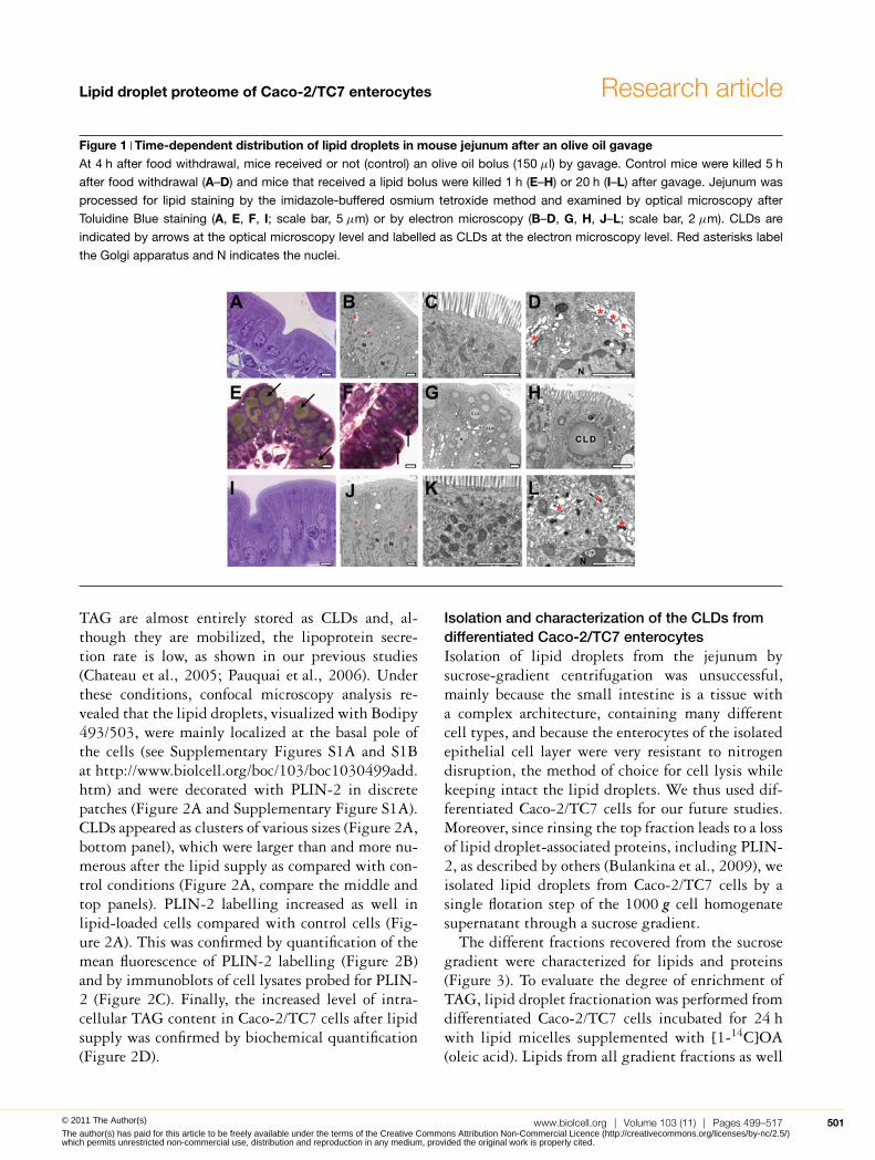
ResultsAccumulation of CLDs in mouse jejunum and inCaco-2/TC7 enterocytes after lipid supplyAs a first approach, we characterized the localizationof neutral lipids in enterocytes of mouse intestinalvilli as a function of time after olive oil gavage.
Morphological analysis showed that, in controlmice, the jejunum enterocytes were mostly devoidof CLDs (Figure 1A–1C) and no lipids could be visu-alized within the Golgi apparatus (Figure 1D). Bycontrast, 1 h after olive oil gavage, the cytoplasm ofjejunum enterocytes was full of lipid droplets (Fig-ures 1E–1H), which were localized at the apical poleof enterocytes above the nucleus and were larger atthe tip (Figure 1E) than in the lower part of the villi(Figure 1F). Electron microscopic analysis showedthat the large lipid droplets were not surrounded bya membrane and were thus cytosolic (Figure 1H).Despite the huge amount of cytosolic TAG observed1 h after gavage, none could be visualized 20 h later(Figure 1I–1K), highlighting the transient natureof this accumulation. At the same time, the Golgiapparatus clearly contained lipids, indicating activelipoprotein secretion (Figure 1L).
We have previously shown that human Caco-2/TC7 enterocytes, a clone that derives from theparental Caco-2 cell line, is able to produce TRLand store TAG as CLDs when supplied with lipidmicelles (Chateau et al., 2005; Vidal et al., 2005).After a 24 h incubation with lipid micelles, fattyacid uptake and TAG synthesis are completed,
500 C© The Authors Journal compilation C© 2011 Portland Press Limited© 2011 The Author(s)
The author(s) has paid for this article to be freely available under the terms of the Creative Commons Attribution Non-Commercial Licence (http://creativecommons.org/licenses/by-nc/2.5/)which permits unrestricted non-commercial use, distribution and reproduction in any medium, provided the original work is properly cited.
Lipid droplet proteome of Caco-2/TC7 enterocytes Research article
Figure 1 Time-dependent distribution of lipid droplets in mouse jejunum after an olive oil gavageAt 4 h after food withdrawal, mice received or not (control) an olive oil bolus (150 μl) by gavage. Control mice were killed 5 h
after food withdrawal (A–D) and mice that received a lipid bolus were killed 1 h (E–H) or 20 h (I–L) after gavage. Jejunum was
processed for lipid staining by the imidazole-buffered osmium tetroxide method and examined by optical microscopy after
Toluidine Blue staining (A, E, F, I; scale bar, 5 μm) or by electron microscopy (B–D, G, H, J–L; scale bar, 2 μm). CLDs are
indicated by arrows at the optical microscopy level and labelled as CLDs at the electron microscopy level. Red asterisks label
the Golgi apparatus and N indicates the nuclei.
TAG are almost entirely stored as CLDs and, al-though they are mobilized, the lipoprotein secre-tion rate is low, as shown in our previous studies(Chateau et al., 2005; Pauquai et al., 2006). Underthese conditions, confocal microscopy analysis re-vealed that the lipid droplets, visualized with Bodipy493/503, were mainly localized at the basal pole ofthe cells (see Supplementary Figures S1A and S1Bat http://www.biolcell.org/boc/103/boc1030499add.htm) and were decorated with PLIN-2 in discretepatches (Figure 2A and Supplementary Figure S1A).CLDs appeared as clusters of various sizes (Figure 2A,bottom panel), which were larger than and more nu-merous after the lipid supply as compared with con-trol conditions (Figure 2A, compare the middle andtop panels). PLIN-2 labelling increased as well inlipid-loaded cells compared with control cells (Fig-ure 2A). This was confirmed by quantification of themean fluorescence of PLIN-2 labelling (Figure 2B)and by immunoblots of cell lysates probed for PLIN-2 (Figure 2C). Finally, the increased level of intra-cellular TAG content in Caco-2/TC7 cells after lipidsupply was confirmed by biochemical quantification(Figure 2D).
Isolation and characterization of the CLDs fromdifferentiated Caco-2/TC7 enterocytesIsolation of lipid droplets from the jejunum bysucrose-gradient centrifugation was unsuccessful,mainly because the small intestine is a tissue witha complex architecture, containing many differentcell types, and because the enterocytes of the isolatedepithelial cell layer were very resistant to nitrogendisruption, the method of choice for cell lysis whilekeeping intact the lipid droplets. We thus used dif-ferentiated Caco-2/TC7 cells for our future studies.Moreover, since rinsing the top fraction leads to a lossof lipid droplet-associated proteins, including PLIN-2, as described by others (Bulankina et al., 2009), weisolated lipid droplets from Caco-2/TC7 cells by asingle flotation step of the 1000 g cell homogenatesupernatant through a sucrose gradient.
The different fractions recovered from the sucrosegradient were characterized for lipids and proteins(Figure 3). To evaluate the degree of enrichment ofTAG, lipid droplet fractionation was performed fromdifferentiated Caco-2/TC7 cells incubated for 24 hwith lipid micelles supplemented with [1-14C]OA(oleic acid). Lipids from all gradient fractions as well
www.biolcell.org | Volume 103 (11) | Pages 499–517 501© 2011 The Author(s)
The author(s) has paid for this article to be freely available under the terms of the Creative Commons Attribution Non-Commercial Licence (http://creativecommons.org/licenses/by-nc/2.5/)which permits unrestricted non-commercial use, distribution and reproduction in any medium, provided the original work is properly cited.

J. Bouchoux and others
Figure 2 TAG and PLIN-2 content in Caco-2/TC7 cells after incubation with lipid micelles for 24 hCaco-2/TC7 cells were cultured for 3 weeks on semi-permeable filters for differentiation and then supplied ( + ) or not ( − ;
control conditions) with lipid micelles for 24 h. (A) Cells were stained with Bodipy 493/503 (green) and labelled for PLIN-2 (red;
scale bars, 10 μm). Lower panels show higher magnifications and the insets display magnified areas. Acquisitions were made
from the basal pole of the cells, where most of the lipid droplets are localized (see also Supplementary Figures S1A and S1B).
(B) PLIN-2-associated fluorescence was quantified from (A) and expressed in arbitrary units.*P < 0.05 compared with control
cells. (C) Immunoblotting of PLIN-2 in cell lysates (40 μg of protein) and quantification. Results are expressed as percentage of
control. (D) TAG content in cell lysates. Results shown are the means +− S.E.M. for five independent experiments performed at
least in duplicate.*P < 0.05 when compared with control cells.
as from the homogenate and the supernatant wereextracted and lipid classes were separated by TLC.As expected, TAG were mainly recovered in frac-tion 1, the lowest-density fraction, and PL (phos-pholipids) in fraction 13, which contains cell mem-branes (Figure 3A). Along with TAG, fraction 1was also enriched in DGE (diacylglyceryl ether) andCE (cholesterol ester), both neutral and highly hy-drophobic lipids, which are thus probably bathedin the TAG core of the lipid droplets. DGE is aTAG analogue formed from batyl alcohol, a stableether analogue of 2-monoacylglycerol that we use to
evaluate the contribution of the MGAT (monoacyl-glycerol acyltransferase) pathway to TAG synthesis(Pauquai et al., 2006). The percentage of [1-14C]OAincorporated into TAG increased from 53.4 +− 3% inthe cell homogenate to 80.2 +− 0.6% in the super-natant loaded on the gradient and to 89.6 +− 0.6% infraction 1 (Figure 3B; see Supplementary Table S1at http://www.biolcell.org/boc/103/boc1030499add.htm). Interestingly, compared with the PL/TAG ra-tio, the DGE/TAG and the CE/TAG ratios remainedremarkably constant in these samples, indicating a co-enrichment of these hydrophobic lipids with TAG,
502 C© The Authors Journal compilation C© 2011 Portland Press Limited© 2011 The Author(s)
The author(s) has paid for this article to be freely available under the terms of the Creative Commons Attribution Non-Commercial Licence (http://creativecommons.org/licenses/by-nc/2.5/)which permits unrestricted non-commercial use, distribution and reproduction in any medium, provided the original work is properly cited.

Lipid droplet proteome of Caco-2/TC7 enterocytes Research article
Figure 3 Lipid and protein analysis of sucrose gradient fractions prepared from Caco-2/TC7 cellsCaco-2/TC7 cells were cultured on semi-permeable filters for 3 weeks for differentiation and then supplied with lipid micelles
for 24 h. For lipid analysis, lipid micelles were supplemented with [1-14C]OA. Cell homogenates were centrifuged for 10 min at
1000 g and the supernatant was fractionated on a sucrose gradient. The top to bottom fractions (1–13) were analysed for lipids
and proteins. (A) Autoradiography of a representative TLC of radiolabelled lipids extracted from cell homogenates, supernatants
and each fraction of the sucrose gradient (FA, fatty acids; DG, diacylglycerol). (B) The radioactive bands from TLC were excised
and the radioactivity was quantified by scintillation counting to evaluate the incorporation of [1-14C]OA into lipids. Results were
expressed for each fraction as percentage of total [1-14C]OA incorporated into each lipid class. (C) Distribution of PL (�) and TAG
(�) in the sucrose gradient fractions. Results are expressed as percentage of total [1-14C]OA incorporated. (D) Immunoblotting
of PLIN-2 (lipid droplet marker), LDH (cytosolic marker), PDI and calnexin (ER markers), GM130 (Golgi matrix protein marker),
HSP60 and prohibitin (mitochondrial markers) and catalase (peroxisome marker). Immunoblotting of ApoB48 was performed to
detect any contamination of the lipid droplet fraction with TRLs. Equal volumes of homogenate and supernatant were loaded on
gels to evaluate the recovery/loss of material after the 1000 g centrifugation step. The same percentage of each fraction of the
sucrose gradient was loaded on gels, except for fractions 1 and 2, which were 2-fold loaded in order to evaluate the presence
of organelle markers with a greater sensitivity.
the major component of the lipid droplet core (Sup-plementary Table S1). Conversely, the percentageof [1-14C]OA incorporated into PL decreased from41.1 +− 3.2% in the cell homogenate to 11.4 +− 0.8%in the supernatant and to 1.3 +− 0.1% in fraction 1.Overall, in fraction 1, the [1-14C]OA incorporatedinto neutral lipids (TAG + DGE + CE) accounted formore than 98% of the total radioactivity incorpor-ated and only for less than 2% in PL, as describedearlier (Bartz et al., 2007). Finally, 65.5 +− 11.5% of
the TAG and 3.6 +− 1% of the PL loaded on thesucrose gradient were recovered in the top fraction(Figure 3C). As expected, PLIN-2 was mainly re-covered in fraction 1 (Figure 3D). Overall, the lowestdensity fraction 1 was highly enriched in TAG andPLIN-2, two markers of lipid droplets.
A set of organelle markers was analysed by West-ern blotting to evaluate the relative purity of the isol-ated CLDs (Figure 3D). The lipid-droplet-containingfraction was slightly positive for LDH (lactate
www.biolcell.org | Volume 103 (11) | Pages 499–517 503© 2011 The Author(s)
The author(s) has paid for this article to be freely available under the terms of the Creative Commons Attribution Non-Commercial Licence (http://creativecommons.org/licenses/by-nc/2.5/)which permits unrestricted non-commercial use, distribution and reproduction in any medium, provided the original work is properly cited.
J. Bouchoux and others
dehydrogenase), an abundant cytosolic enzyme, andfor PDI (protein disulfide-isomerase), a luminal ERprotein that has been frequently identified in CLDfraction (Hodges and Wu, 2010). Markers of othercompartments, such as calnexin (ER), GM130 (Golgimatrix 130; Golgi apparatus), HSP60 (heat-shockprotein of 60 kDa) and prohibitin (mitochondria)and catalase (peroxisomes), were not detected infraction 1, although they were easily visualized inthe bottom fractions. Finally, we analysed the frac-tions for the presence of ApoB48, which is thenon-exchangeable TRL-associated intestinal form ofhuman ApoB. ApoB48 was not detected in the topfraction, while it was indeed detected in themembrane-containing bottom fraction (fraction 13),indicating that CLDs were not contaminated withTRL. Overall, although we cannot rule out a slightcontamination with cytosolic proteins, our results in-dicate that PLIN-2- and TAG-enriched fraction 1 wasdevoid of other organelles. We thus undertook theidentification of the CLD-associated proteins througha proteomic approach.
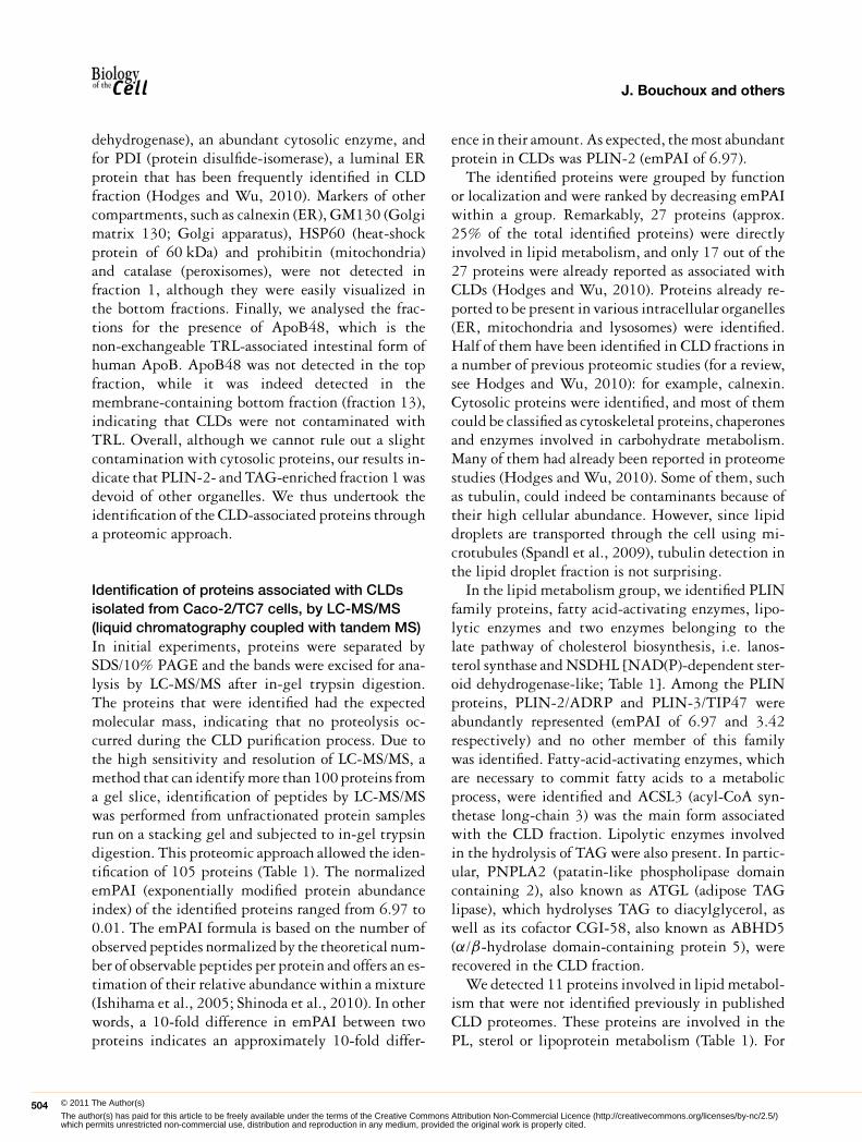
Identification of proteins associated with CLDsisolated from Caco-2/TC7 cells, by LC-MS/MS(liquid chromatography coupled with tandem MS)In initial experiments, proteins were separated bySDS/10% PAGE and the bands were excised for ana-lysis by LC-MS/MS after in-gel trypsin digestion.The proteins that were identified had the expectedmolecular mass, indicating that no proteolysis oc-curred during the CLD purification process. Due tothe high sensitivity and resolution of LC-MS/MS, amethod that can identify more than 100 proteins froma gel slice, identification of peptides by LC-MS/MSwas performed from unfractionated protein samplesrun on a stacking gel and subjected to in-gel trypsindigestion. This proteomic approach allowed the iden-tification of 105 proteins (Table 1). The normalizedemPAI (exponentially modified protein abundanceindex) of the identified proteins ranged from 6.97 to0.01. The emPAI formula is based on the number ofobserved peptides normalized by the theoretical num-ber of observable peptides per protein and offers an es-timation of their relative abundance within a mixture(Ishihama et al., 2005; Shinoda et al., 2010). In otherwords, a 10-fold difference in emPAI between twoproteins indicates an approximately 10-fold differ-
ence in their amount. As expected, the most abundantprotein in CLDs was PLIN-2 (emPAI of 6.97).
The identified proteins were grouped by functionor localization and were ranked by decreasing emPAIwithin a group. Remarkably, 27 proteins (approx.25% of the total identified proteins) were directlyinvolved in lipid metabolism, and only 17 out of the27 proteins were already reported as associated withCLDs (Hodges and Wu, 2010). Proteins already re-ported to be present in various intracellular organelles(ER, mitochondria and lysosomes) were identified.Half of them have been identified in CLD fractions ina number of previous proteomic studies (for a review,see Hodges and Wu, 2010): for example, calnexin.Cytosolic proteins were identified, and most of themcould be classified as cytoskeletal proteins, chaperonesand enzymes involved in carbohydrate metabolism.Many of them had already been reported in proteomestudies (Hodges and Wu, 2010). Some of them, suchas tubulin, could indeed be contaminants because oftheir high cellular abundance. However, since lipiddroplets are transported through the cell using mi-crotubules (Spandl et al., 2009), tubulin detection inthe lipid droplet fraction is not surprising.
In the lipid metabolism group, we identified PLINfamily proteins, fatty acid-activating enzymes, lipo-lytic enzymes and two enzymes belonging to thelate pathway of cholesterol biosynthesis, i.e. lanos-terol synthase and NSDHL [NAD(P)-dependent ster-oid dehydrogenase-like; Table 1]. Among the PLINproteins, PLIN-2/ADRP and PLIN-3/TIP47 wereabundantly represented (emPAI of 6.97 and 3.42respectively) and no other member of this familywas identified. Fatty-acid-activating enzymes, whichare necessary to commit fatty acids to a metabolicprocess, were identified and ACSL3 (acyl-CoA syn-thetase long-chain 3) was the main form associatedwith the CLD fraction. Lipolytic enzymes involvedin the hydrolysis of TAG were also present. In partic-ular, PNPLA2 (patatin-like phospholipase domaincontaining 2), also known as ATGL (adipose TAGlipase), which hydrolyses TAG to diacylglycerol, aswell as its cofactor CGI-58, also known as ABHD5(α/β-hydrolase domain-containing protein 5), wererecovered in the CLD fraction.
We detected 11 proteins involved in lipid metabol-ism that were not identified previously in publishedCLD proteomes. These proteins are involved in thePL, sterol or lipoprotein metabolism (Table 1). For
504 C© The Authors Journal compilation C© 2011 Portland Press Limited© 2011 The Author(s)
The author(s) has paid for this article to be freely available under the terms of the Creative Commons Attribution Non-Commercial Licence (http://creativecommons.org/licenses/by-nc/2.5/)which permits unrestricted non-commercial use, distribution and reproduction in any medium, provided the original work is properly cited.

Lipid droplet proteome of Caco-2/TC7 enterocytes Research article
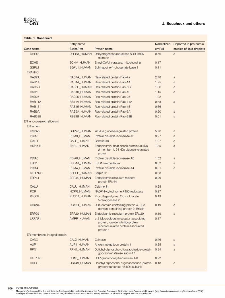
Table 1 List of identified (by LC-MS/MS) proteins associated with cytosolic lipid droplets isolated from Caco-2/TC7 cellsCaco-2/TC7 cells were cultured on semi-permeable filters for 3 weeks for differentiation and then supplied with lipid micelles for 24 h. Cellhomogenates were centrifuged for 10 min at 1000 g and the supernatant was fractionated on a sucrose gradient. The top fraction, containingthe cytosolic lipid droplets, was analysed by LC-MS/MS for protein identification. Proteins were identified with at least two unique peptides. AMascot search was performed allowing 1 ‘max. missed cleavage’, 20 p.p.m. ‘peptides mass tolerance’, 0.3 ‘fragment mass tolerance’ andagainst Homo sapiens taxonomy (20401 sequences). Mascot search results were filtered by P = 0.01 and false discovery rate below 1.6%.The proteins were identified in at least two out of three independent experiments. The proteins already reported in mammalian CLD proteomeare indicated (a, reported in the recent review of Hodges and Wu, 2010; b, Moessinger et al., 2011). Proteins are grouped by function orlocalization and, inside a group, proteins are ranked by decreasing emPAI.
Entry name Normalized Reported in proteomic
Gene name SwissProt Protein name emPAI studies of lipid droplets
Lipid metabolism
PLIN proteins
PLIN2 PLIN2_HUMAN Perilipin-2, adipose differentiation-relatedprotein, adipophilin
6.97 a
PLIN3 PLIN3_HUMAN Perilipin-3, mannose-6-phosphatereceptor-binding protein, TIP47
3.42 a
Fatty acid metabolism
ACSL3 ACSL3_HUMAN Long-chain-fatty-acid-CoA ligase 3 2.65 a
ACSL4 ACSL4_HUMAN Long-chain-fatty-acid-CoA ligase 4 0.15 a
Acylglycerol metabolism
MGLL MGLL_HUMAN Monoglyceride lipase 0.67 a
ABHD5 ABHD5_HUMAN 1-Acylglycerol-3-phosphateO-acyltransferase, abhydrolasedomain-containing protein 5, CGI-58
0.31 a
PNPLA2 PLPL2_HUMAN Patatin-like phospholipasedomain-containing protein 2, adiposetriacylglycerol lipase
0.15 a
Phospholipid metabolism
PCYT1A PCY1A_HUMAN Choline-phosphate cytidylyltransferase A 1.58
LPCAT2 PCAT2_HUMAN Lysophosphatidylcholine acyltransferase 2 0.30 b
Sterol metabolism
HSD17B11 DHB11_HUMAN Oestradiol 17-β-dehydrogenase 11 2.78 a
LSS ERG7_HUMAN Lanosterol synthase 2.46 a
CYB5R3 NB5R3_HUMAN NADH–cytochrome b5 reductase 3 1.63 a
NSDHL NSDHL_HUMAN Sterol-4-α-carboxylate 3-dehydrogenase,decarboxylating
1.55 a
EPHX1 HYEP_HUMAN Epoxide hydrolase 1 0.31
HSD3B1 3BHS1_HUMAN 3-β-Hydroxysteroid dehydrogenase 0.24
DHRS3 DHRS3_HUMAN Short-chain dehydrogenase/reductase 3 0.13 a
DHCR7 DHCR7_HUMAN 7-Dehydrocholesterol reductase 0.06
HSD17B7 DHB7_HUMAN 3-Oxo-steroid reductase 0.04 a
Lipoprotein metabolism
APOA4 APOA4_HUMAN Apolipoprotein A-IV 6.24
P4HB PDIA1_HUMAN Protein disulfide-isomerase 4.71 a
MTTP MTP_HUMAN Microsomal triacylglycerol transfer proteinlarge subunit
0.53
APOE APOE_HUMAN Apolipoprotein E 0.48
Other lipidic metabolism
FAF2 FAF2_HUMAN FAS-associated factor 2, UBXD8 1.77 a
RDH10 RDH10_HUMAN Retinol dehydrogenase 10 1.24
www.biolcell.org | Volume 103 (11) | Pages 499–517 505© 2011 The Author(s)
The author(s) has paid for this article to be freely available under the terms of the Creative Commons Attribution Non-Commercial Licence (http://creativecommons.org/licenses/by-nc/2.5/)which permits unrestricted non-commercial use, distribution and reproduction in any medium, provided the original work is properly cited.
J. Bouchoux and others
Table 1 Continued
Entry name Normalized Reported in proteomic
Gene name SwissProt Protein name emPAI studies of lipid droplets
DHRS1 DHRS1_HUMAN Dehydrogenase/reductase SDR familymember 1
0.56 a
ECHS1 ECHM_HUMAN Enoyl-CoA hydratase, mitochondrial 0.17
SGPL1 SGPL1_HUMAN Sphingosine-1-phosphate lyase 1 0.11
TRAFFIC
RAB7A RAB7A_HUMAN Ras-related protein Rab-7a 2.78 a
RAB1A RAB1A_HUMAN Ras-related protein Rab-1A 1.75 a
RAB5C RAB5C_HUMAN Ras-related protein Rab-5C 1.66 a
RAB10 RAB10_HUMAN Ras-related protein Rab-10 1.15 a
RAB25 RAB25_HUMAN Ras-related protein Rab-25 1.02
RAB11A RB11A_HUMAN Ras-related protein Rab-11A 0.68 a
RAB15 RAB15_HUMAN Ras-related protein Rab-15 0.66
RAB6A RAB6A_HUMAN Ras-related protein Rab-6A 0.20 a
RAB33B RB33B_HUMAN Ras-related protein Rab-33B 0.01 a
ER (endoplasmic reticulum)
ER lumen
HSPA5 GRP78_HUMAN 78 kDa glucose-regulated protein 5.76 a
PDIA3 PDIA3_HUMAN Protein disulfide-isomerase A3 3.27 a
CALR CALR_HUMAN Calreticulin 1.97 a
HSP90B ENPL_HUMAN Endoplasmin, heat-shock protein 90 kDaβ member 1, 94 kDa glucose-regulatedprotein
1.85 a
PDIA6 PDIA6_HUMAN Protein disulfide-isomerase A6 1.52 a
ERO1L ERO1A_HUMAN ERO1-like protein α 0.82 a
PDIA4 PDIA4_HUMAN Protein disulfide-isomerase A4 0.81 a
SERPINH SERPH_HUMAN Serpin H1 0.38
ERP44 ERP44_HUMAN Endoplasmic reticulum residentprotein ERp44
0.29
CALU CALU_HUMAN Calumenin 0.28
POR NCPR_HUMAN NADPH–cytochrome P450 reductase 0.27
PLOD2 PLOD2_HUMAN Procollagen-lysine, 2-oxoglutarate5-dioxygenase 2
0.19
UBXN4 UBXN4_HUMAN UBX domain-containing protein 4, UBXdomain-containing protein 2, Erasin
0.19 a
ERP29 ERP29_HUMAN Endoplasmic reticulum protein ERp29 0.19 a
LRPAP1 AMRP_HUMAN α-2-Macroglobulin receptor-associatedprotein, low-density lipoproteinreceptor-related protein-associatedprotein 1
0.17
ER membrane, integral protein
CANX CALX_HUMAN Calnexin 0.66 a
AUP1 AUP1_HUMAN Ancient ubiquitous protein 1 0.35 a
RPN1 RPN1_HUMAN Dolichyl-diphospho-oligosaccharide–proteinglycosyltransferase subunit 1
0.34 a
UGT1A6 UD16_HUMAN UDP-glucuronosyltransferase 1-6 0.22
DDOST OST48_HUMAN Dolichyl-diphospho-oligosaccharide–proteinglycosyltranferase 48 kDa subunit
0.18 a
506 C© The Authors Journal compilation C© 2011 Portland Press Limited© 2011 The Author(s)
The author(s) has paid for this article to be freely available under the terms of the Creative Commons Attribution Non-Commercial Licence (http://creativecommons.org/licenses/by-nc/2.5/)which permits unrestricted non-commercial use, distribution and reproduction in any medium, provided the original work is properly cited.
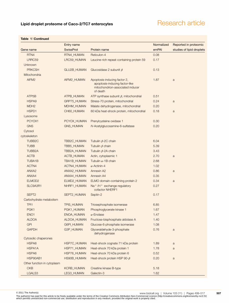
Lipid droplet proteome of Caco-2/TC7 enterocytes Research article
Table 1 Continued
Entry name Normalized Reported in proteomic
Gene name SwissProt Protein name emPAI studies of lipid droplets
RTN4 RTN4_HUMAN Reticulon-4 0.08
LRRC59 LRC59_HUMAN Leucine-rich repeat-containing protein 59 0.17
Unknown
PRKCSH GLU2B_HUMAN Glucosidase 2 subunit β 0.13
Mitochondria
AIFM2 AIFM2_HUMAN Apoptosis-inducing factor 2,apoptosis-inducing factor-likemitochondrion-associated inducerof death
1.87 a
ATP5B ATPB_HUMAN ATP synthase subunit β, mitochondrial 0.51
HSPA9 GRP75_HUMAN Stress-70 protein, mitochondrial 0.24 a
MDH2 MDHM_HUMAN Malate dehydrogenase, mitochondrial 0.20
HSPD1 CH60_HUMAN 60 kDa heat-shock protein, mitochondrial 0.18 a
Lysosome
PCYOX1 PCYOX_HUMAN Prenylcysteine oxidase 1 0.30
GNS GNS_HUMAN N-Acetylglucosamine-6-sulfatase 0.20
Cytosol
cytoskeleton
TUBB2C TBB2C_HUMAN Tubulin β-2C chain 6.04
TUBB TBB5_HUMAN Tubulin β chain 5.39
TUBB2A TBB2A_HUMAN Tubulin β-2A chain 3.43
ACTB ACTB_HUMAN Actin, cytoplasmic 1 2.70 a
TUBA1B TBA1B_HUMAN Tubulin α-1B chain 2.68
ACTN4 ACTN4_HUMAN α-Actinin-4 1.02
ANXA2 ANXA2_HUMAN Annexin A2 0.86 a
ANXA4 ANXA4_HUMAN Annexin A4 0.35
ELMOD2 ELMD2_HUMAN ELMO domain-containing protein 2 0.34 a
SLC9A3R1 NHRF1_HUMAN Na+ /H+ exchange regulatorycofactor NHERF1
0.27
SEPT2 SEPT2_HUMAN Septin-2 0.17
Carbohydrate metabolism
TPI1 TPIS_HUMAN Triosephosphate isomerase 6.85
PGK1 PGK1_HUMAN Phosphoglycerate kinase 1 1.67
ENO1 ENOA_HUMAN α-Enolase 1.47
ALDOA ALDOA_HUMAN Fructose-bisphosphate aldolase A 1.40
GPI G6PI_HUMAN Glucose-6-phosphate isomerase 1.08
GAPDH G3P_HUMAN Glyceraldehyde-3-phosphatedehydrogenase
0.76 a
Cytosolic chaperones
HSPA8 HSP7C_HUMAN Heat-shock cognate 71 kDa protein 1.89 a
HSPA1A HSP71_HUMAN Heat-shock 70 kDa protein 1 1.78 a
HSPA6 HSP76_HUMAN Heat-shock 70 kDa protein 6 0.52
HSP90AB1 HS90B_HUMAN Heat-shock protein HSP 90-β 0.20 a
Other function in cytoplasm
CKB KCRB_HUMAN Creatine kinase B-type 5.18
LGALS3 LEG3_HUMAN Galectin-3 1.62
www.biolcell.org | Volume 103 (11) | Pages 499–517 507© 2011 The Author(s)
The author(s) has paid for this article to be freely available under the terms of the Creative Commons Attribution Non-Commercial Licence (http://creativecommons.org/licenses/by-nc/2.5/)which permits unrestricted non-commercial use, distribution and reproduction in any medium, provided the original work is properly cited.

J. Bouchoux and others
Table 1 Continued
Entry name Normalized Reported in proteomic
Gene name SwissProt Protein name emPAI studies of lipid droplets
GSTP1 GSTP1_HUMAN Glutathione transferase P 0.57
GSTA1 GSTA1_HUMAN Glutathione transferase A1 0.44
NDRG1 NDRG1_HUMAN Protein NDRG1 0.26
TRIO TRIO_HUMAN Triple functional domain protein 0.02
Miscellaneous
METTL7B MET7B_HUMAN Methyltransferase-like protein 7B 2.97 a
METTL7A MET7A_HUMAN Methyltransferase-like protein 7A 1.59 a
SCCPDH SCPDH_HUMAN Probable saccharopine dehydrogenase 1.56 a
ATP1B1 P AT1B1_HUMAN Sodium/potassium-transporting ATPasesubunit β-1
0.79
CDH17 CAD17_HUMAN Cadherin-17 0.68
GPA33 GPA33_HUMAN Cell-surface A33 antigen 0.55
C2orf43 CB043_HUMAN UPF0554 protein C2 or f43 0.48 a
GANAB GANAB_HUMAN Neutral α-glucosidase AB 0.26
ATP1A1 AT1A1_HUMAN Sodium/potassium-transporting ATPasesubunit α1
0.11
EEF1A1 EF1A1_HUMAN Elongation factor 1-α1 0.09 a
KPNB1 IMB1_HUMAN Importin subunit β-1 0.07
CLIC1 CLIC1_HUMAN Chloride intracellular channel protein 1 0.01
example, PCYT1A [also known as CCT-α (choline-phosphate cytidylyltransferase A)], the enzyme thatcatalyses the rate-limiting step in the choline path-way for PC (phosphatidylcholine) synthesis (Vance,2008), and LPCAT2 (lysophosphatidylcholine acyl-transferase 2), which re-acylates lysophospholipidsby using acyl-CoA, were present in the lipid dropletfraction.
Proteins involved in the metabolism of lipopro-tein were also identified in the CLD fraction ofCaco-2/TC7 enterocytes. MTTP and its subunit,PDI A1, were detected as well as the exchangeableApoA-IV. By contrast, ApoB, the constitutive non-exchangeable apolipoprotein of TRL, was not iden-tified in the proteome. In Caco-2/TC7 cells, after a24 h incubation with lipid micelles, lipid dropletsare mainly localized at the basal pole of cells, butsome of them are localized in the supranuclear region(Supplementary Figures S1A and S1B and Chateauet al., 2005). ApoB is restricted to the apical re-gion of the cell (Supplementary Figures S1A andS1B and Morel et al., 2004) and, in this area, as weshow here, lipid droplets were surrounded by PLIN-2but devoid of ApoB (Supplementary Figure S1C). As
expected, ApoB colocalized with the ER marker cal-nexin, and, in the perinuclear region, Bodipy-labelledlipid droplets could also be found in close vicinityof calnexin (Supplementary Figure S1D). However,ApoB was never detected in these ER-neighbouringlipid droplets.
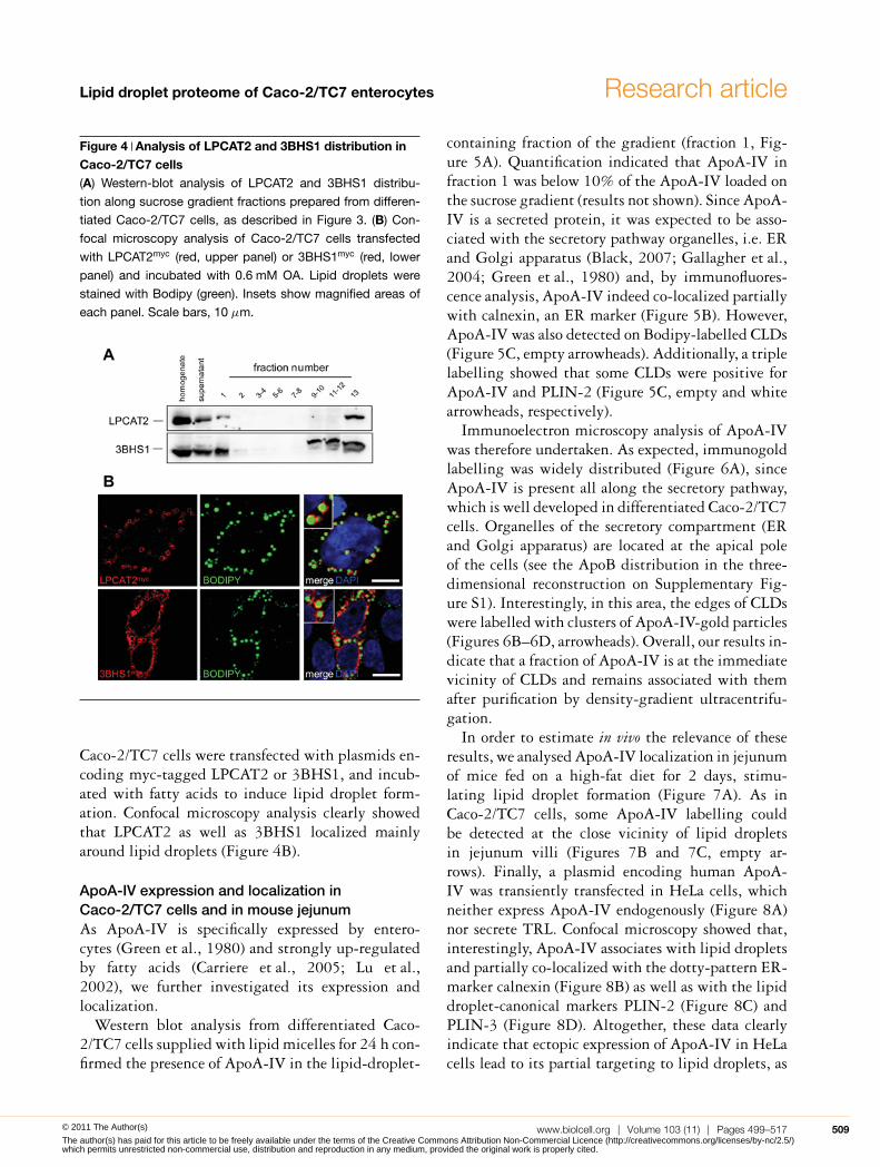
LPCAT2 and 3BHS1 (3-β-hydroxysteroiddehydrogenase 1) expression and localizationin Caco-2/TC7 cellsAs mentioned above, some proteins involved in lipidmetabolism were identified for the first time in aCLD proteome. We analysed the expression and thelocalization of LPCAT2, an enzyme involved in thePC metabolism, and of 3BHS1, an enzyme involvedin the metabolism of steroids, because they were sofar known to localize to the ER membrane (Agarwaland Garg, 2010; Morimoto et al., 2010; Wang et al.,2007) and have not been described yet in enterocytes.Western blot analysis of the sucrose gradient frac-tions showed that LPCAT2 and 3BHS1 were indeedmainly associated with the membrane fraction (frac-tion 13). However, both proteins were also detected inthe lipid-droplet-containing fraction 1 (Figure 4A).
508 C© The Authors Journal compilation C© 2011 Portland Press Limited© 2011 The Author(s)
The author(s) has paid for this article to be freely available under the terms of the Creative Commons Attribution Non-Commercial Licence (http://creativecommons.org/licenses/by-nc/2.5/)which permits unrestricted non-commercial use, distribution and reproduction in any medium, provided the original work is properly cited.
Lipid droplet proteome of Caco-2/TC7 enterocytes Research article
Figure 4 Analysis of LPCAT2 and 3BHS1 distribution inCaco-2/TC7 cells(A) Western-blot analysis of LPCAT2 and 3BHS1 distribu-
tion along sucrose gradient fractions prepared from differen-
tiated Caco-2/TC7 cells, as described in Figure 3. (B) Con-
focal microscopy analysis of Caco-2/TC7 cells transfected
with LPCAT2myc (red, upper panel) or 3BHS1myc (red, lower
panel) and incubated with 0.6 mM OA. Lipid droplets were
stained with Bodipy (green). Insets show magnified areas of
each panel. Scale bars, 10 μm.
Caco-2/TC7 cells were transfected with plasmids en-coding myc-tagged LPCAT2 or 3BHS1, and incub-ated with fatty acids to induce lipid droplet form-ation. Confocal microscopy analysis clearly showedthat LPCAT2 as well as 3BHS1 localized mainlyaround lipid droplets (Figure 4B).
ApoA-IV expression and localization inCaco-2/TC7 cells and in mouse jejunumAs ApoA-IV is specifically expressed by entero-cytes (Green et al., 1980) and strongly up-regulatedby fatty acids (Carriere et al., 2005; Lu et al.,2002), we further investigated its expression andlocalization.
Western blot analysis from differentiated Caco-2/TC7 cells supplied with lipid micelles for 24 h con-firmed the presence of ApoA-IV in the lipid-droplet-
containing fraction of the gradient (fraction 1, Fig-ure 5A). Quantification indicated that ApoA-IV infraction 1 was below 10% of the ApoA-IV loaded onthe sucrose gradient (results not shown). Since ApoA-IV is a secreted protein, it was expected to be asso-ciated with the secretory pathway organelles, i.e. ERand Golgi apparatus (Black, 2007; Gallagher et al.,2004; Green et al., 1980) and, by immunofluores-cence analysis, ApoA-IV indeed co-localized partiallywith calnexin, an ER marker (Figure 5B). However,ApoA-IV was also detected on Bodipy-labelled CLDs(Figure 5C, empty arrowheads). Additionally, a triplelabelling showed that some CLDs were positive forApoA-IV and PLIN-2 (Figure 5C, empty and whitearrowheads, respectively).
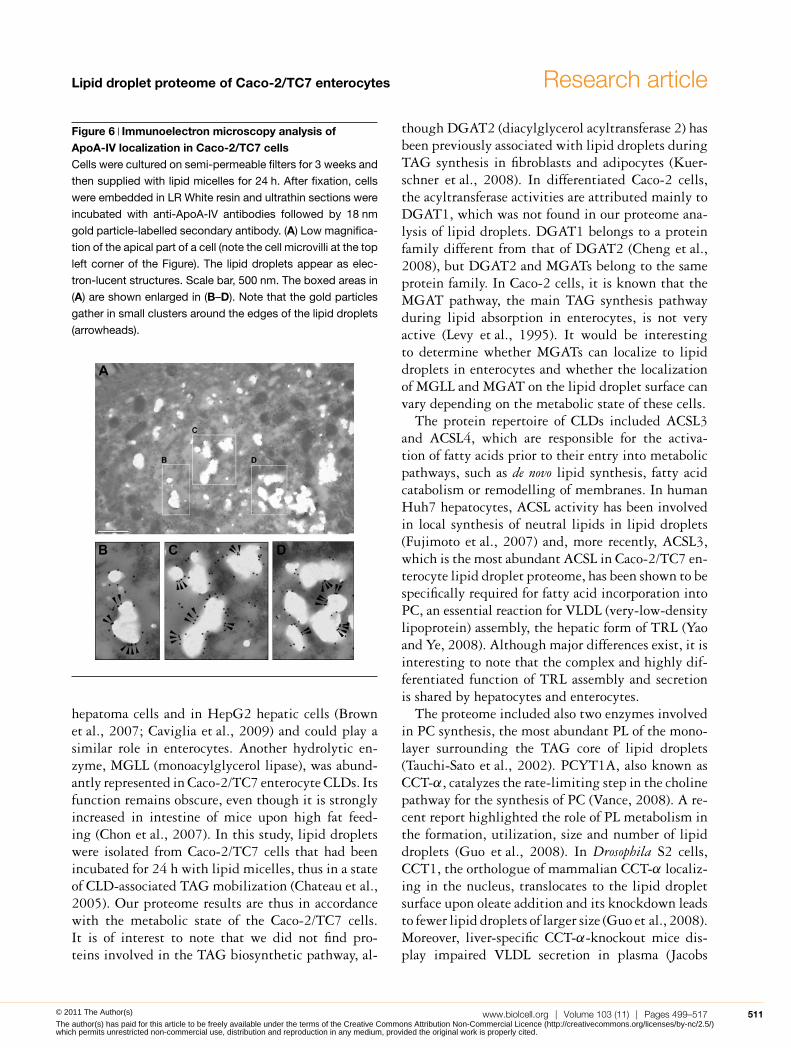
Immunoelectron microscopy analysis of ApoA-IVwas therefore undertaken. As expected, immunogoldlabelling was widely distributed (Figure 6A), sinceApoA-IV is present all along the secretory pathway,which is well developed in differentiated Caco-2/TC7cells. Organelles of the secretory compartment (ERand Golgi apparatus) are located at the apical poleof the cells (see the ApoB distribution in the three-dimensional reconstruction on Supplementary Fig-ure S1). Interestingly, in this area, the edges of CLDswere labelled with clusters of ApoA-IV-gold particles(Figures 6B–6D, arrowheads). Overall, our results in-dicate that a fraction of ApoA-IV is at the immediatevicinity of CLDs and remains associated with themafter purification by density-gradient ultracentrifu-gation.
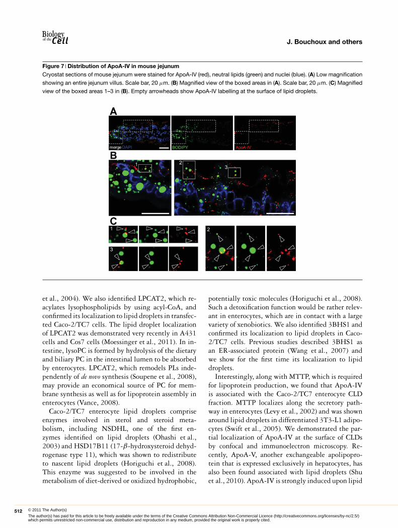
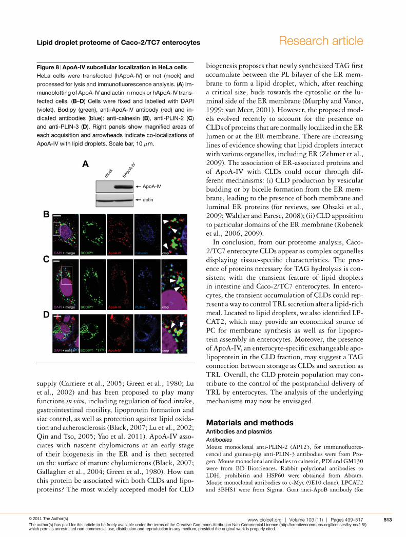
In order to estimate in vivo the relevance of theseresults, we analysed ApoA-IV localization in jejunumof mice fed on a high-fat diet for 2 days, stimu-lating lipid droplet formation (Figure 7A). As inCaco-2/TC7 cells, some ApoA-IV labelling couldbe detected at the close vicinity of lipid dropletsin jejunum villi (Figures 7B and 7C, empty ar-rows). Finally, a plasmid encoding human ApoA-IV was transiently transfected in HeLa cells, whichneither express ApoA-IV endogenously (Figure 8A)nor secrete TRL. Confocal microscopy showed that,interestingly, ApoA-IV associates with lipid dropletsand partially co-localized with the dotty-pattern ER-marker calnexin (Figure 8B) as well as with the lipiddroplet-canonical markers PLIN-2 (Figure 8C) andPLIN-3 (Figure 8D). Altogether, these data clearlyindicate that ectopic expression of ApoA-IV in HeLacells lead to its partial targeting to lipid droplets, as
www.biolcell.org | Volume 103 (11) | Pages 499–517 509© 2011 The Author(s)
The author(s) has paid for this article to be freely available under the terms of the Creative Commons Attribution Non-Commercial Licence (http://creativecommons.org/licenses/by-nc/2.5/)which permits unrestricted non-commercial use, distribution and reproduction in any medium, provided the original work is properly cited.

J. Bouchoux and others
Figure 5 Intracellular distribution of ApoA-IV in Caco-2/TC7 cellsCells were cultured on semi-permeable filters for 3 weeks and then supplied with lipid micelles for 24 h. (A) Western-blot analysis
of ApoA-IV distribution in sucrose gradient fractions prepared from differentiated Caco-2/TC7 cells as described in Figure 3.
(B) Cells were labelled for ApoA-IV (red) and calnexin (green). Empty arrowheads indicate co-localizations. Nuclei were stained
with DAPI (blue). Scale bar, 10 μm. (C) Cells were labelled for ApoA-IV (red), PLIN-2 (fuchsia), neutral lipids (green) and nuclei
(blue). Scale bar, 5 μm. The boxed area in the merged Figure is shown magnified on the right. White and empty arrowheads
indicate PLIN-2 and ApoA-IV labelling respectively at the surface of lipid droplets.
observed in Caco-2/TC7 enterocytes (Figures 5 and6) and mouse intestinal cells (Figure 7).
DiscussionWe report here the first proteome analysis of CLDsisolated from differentiated Caco-2/TC7 enterocytes.Observed in enterocytes in vivo (Buschmann andManke, 1981) and in cell culture models (Chateauet al., 2005), lipid droplets have only been recentlysuggested as potential players in the physiology oflipid absorption in the gastrointestinal tract (Pauquaiet al., 2006; Zhu et al., 2009). The proteome of dif-ferentiated Caco-2/TC7 enterocyte CLDs, which wereformed after a supply of dietary lipids, revealed severalcharacteristics. For example, among the PLIN pro-tein family, we identified PLIN-2/ADRP and PLIN-3/TIP47, but no PLIN-1, expressed mainly by adipo-
cytes, or PLIN-5/OXPAT, expressed in cells that havea high capacity of fatty acid oxidation. This findingis in good agreement with recent data from mouseintestine (Lee et al., 2009). Approximately 25% ofthe proteins identified in the Caco-2/TC7 enterocyteCLD fraction were involved in lipid metabolism, andwere mainly linked to the metabolism of TAGs, PLs,sterols and to the assembly of TRLs (Table 1). In-terestingly, among these 27 proteins, 11 were newlyidentified proteins associated with CLDs.
The proteome included proteins necessary for TAGhydrolysis, supporting a potential dynamic role ofCLDs in the control of TRL production in entero-cytes. PNPLA2, which hydrolyse TAGs to diacylgly-cerol, and its co-activator ABHD5 were present inthe proteome. ABHD5/CGI-58 was shown to facil-itate the mobilization of cytoplasmic TAG and theassembly and secretion of TRL in McA RH7777 rat
510 C© The Authors Journal compilation C© 2011 Portland Press Limited© 2011 The Author(s)
The author(s) has paid for this article to be freely available under the terms of the Creative Commons Attribution Non-Commercial Licence (http://creativecommons.org/licenses/by-nc/2.5/)which permits unrestricted non-commercial use, distribution and reproduction in any medium, provided the original work is properly cited.
Lipid droplet proteome of Caco-2/TC7 enterocytes Research article
Figure 6 Immunoelectron microscopy analysis ofApoA-IV localization in Caco-2/TC7 cellsCells were cultured on semi-permeable filters for 3 weeks and
then supplied with lipid micelles for 24 h. After fixation, cells
were embedded in LR White resin and ultrathin sections were
incubated with anti-ApoA-IV antibodies followed by 18 nm
gold particle-labelled secondary antibody. (A) Low magnifica-
tion of the apical part of a cell (note the cell microvilli at the top
left corner of the Figure). The lipid droplets appear as elec-
tron-lucent structures. Scale bar, 500 nm. The boxed areas in
(A) are shown enlarged in (B–D). Note that the gold particles
gather in small clusters around the edges of the lipid droplets
(arrowheads).
hepatoma cells and in HepG2 hepatic cells (Brownet al., 2007; Caviglia et al., 2009) and could play asimilar role in enterocytes. Another hydrolytic en-zyme, MGLL (monoacylglycerol lipase), was abund-antly represented in Caco-2/TC7 enterocyte CLDs. Itsfunction remains obscure, even though it is stronglyincreased in intestine of mice upon high fat feed-ing (Chon et al., 2007). In this study, lipid dropletswere isolated from Caco-2/TC7 cells that had beenincubated for 24 h with lipid micelles, thus in a stateof CLD-associated TAG mobilization (Chateau et al.,2005). Our proteome results are thus in accordancewith the metabolic state of the Caco-2/TC7 cells.It is of interest to note that we did not find pro-teins involved in the TAG biosynthetic pathway, al-
though DGAT2 (diacylglycerol acyltransferase 2) hasbeen previously associated with lipid droplets duringTAG synthesis in fibroblasts and adipocytes (Kuer-schner et al., 2008). In differentiated Caco-2 cells,the acyltransferase activities are attributed mainly toDGAT1, which was not found in our proteome ana-lysis of lipid droplets. DGAT1 belongs to a proteinfamily different from that of DGAT2 (Cheng et al.,2008), but DGAT2 and MGATs belong to the sameprotein family. In Caco-2 cells, it is known that theMGAT pathway, the main TAG synthesis pathwayduring lipid absorption in enterocytes, is not veryactive (Levy et al., 1995). It would be interestingto determine whether MGATs can localize to lipiddroplets in enterocytes and whether the localizationof MGLL and MGAT on the lipid droplet surface canvary depending on the metabolic state of these cells.
The protein repertoire of CLDs included ACSL3and ACSL4, which are responsible for the activa-tion of fatty acids prior to their entry into metabolicpathways, such as de novo lipid synthesis, fatty acidcatabolism or remodelling of membranes. In humanHuh7 hepatocytes, ACSL activity has been involvedin local synthesis of neutral lipids in lipid droplets(Fujimoto et al., 2007) and, more recently, ACSL3,which is the most abundant ACSL in Caco-2/TC7 en-terocyte lipid droplet proteome, has been shown to bespecifically required for fatty acid incorporation intoPC, an essential reaction for VLDL (very-low-densitylipoprotein) assembly, the hepatic form of TRL (Yaoand Ye, 2008). Although major differences exist, it isinteresting to note that the complex and highly dif-ferentiated function of TRL assembly and secretionis shared by hepatocytes and enterocytes.
The proteome included also two enzymes involvedin PC synthesis, the most abundant PL of the mono-layer surrounding the TAG core of lipid droplets(Tauchi-Sato et al., 2002). PCYT1A, also known asCCT-α, catalyzes the rate-limiting step in the cholinepathway for the synthesis of PC (Vance, 2008). A re-cent report highlighted the role of PL metabolism inthe formation, utilization, size and number of lipiddroplets (Guo et al., 2008). In Drosophila S2 cells,CCT1, the orthologue of mammalian CCT-α localiz-ing in the nucleus, translocates to the lipid dropletsurface upon oleate addition and its knockdown leadsto fewer lipid droplets of larger size (Guo et al., 2008).Moreover, liver-specific CCT-α-knockout mice dis-play impaired VLDL secretion in plasma (Jacobs
www.biolcell.org | Volume 103 (11) | Pages 499–517 511© 2011 The Author(s)
The author(s) has paid for this article to be freely available under the terms of the Creative Commons Attribution Non-Commercial Licence (http://creativecommons.org/licenses/by-nc/2.5/)which permits unrestricted non-commercial use, distribution and reproduction in any medium, provided the original work is properly cited.
J. Bouchoux and others
Figure 7 Distribution of ApoA-IV in mouse jejunumCryostat sections of mouse jejunum were stained for ApoA-IV (red), neutral lipids (green) and nuclei (blue). (A) Low magnification
showing an entire jejunum villus. Scale bar, 20 μm. (B) Magnified view of the boxed areas in (A). Scale bar, 20 μm. (C) Magnified
view of the boxed areas 1–3 in (B). Empty arrowheads show ApoA-IV labelling at the surface of lipid droplets.
et al., 2004). We also identified LPCAT2, which re-acylates lysophospholipids by using acyl-CoA, andconfirmed its localization to lipid droplets in transfec-ted Caco-2/TC7 cells. The lipid droplet localizationof LPCAT2 was demonstrated very recently in A431cells and Cos7 cells (Moessinger et al., 2011). In in-testine, lysoPC is formed by hydrolysis of the dietaryand biliary PC in the intestinal lumen to be absorbedby enterocytes. LPCAT2, which remodels PLs inde-pendently of de novo synthesis (Soupene et al., 2008),may provide an economical source of PC for mem-brane synthesis as well as for lipoprotein assembly inenterocytes (Vance, 2008).
Caco-2/TC7 enterocyte lipid droplets compriseenzymes involved in sterol and steroid meta-bolism, including NSDHL, one of the first en-zymes identified on lipid droplets (Ohashi et al.,2003) and HSD17B11 (17-β-hydroxysteroid dehyd-rogenase type 11), which was shown to redistributeto nascent lipid droplets (Horiguchi et al., 2008).This enzyme was suggested to be involved in themetabolism of diet-derived or oxidized hydrophobic,
potentially toxic molecules (Horiguchi et al., 2008).Such a detoxification function would be rather relev-ant in enterocytes, which are in contact with a largevariety of xenobiotics. We also identified 3BHS1 andconfirmed its localization to lipid droplets in Caco-2/TC7 cells. Previous studies described 3BHS1 asan ER-associated protein (Wang et al., 2007) andwe show for the first time its localization to lipiddroplets.
Interestingly, along with MTTP, which is requiredfor lipoprotein production, we found that ApoA-IVis associated with the Caco-2/TC7 enterocyte CLDfraction. MTTP localizes along the secretory path-way in enterocytes (Levy et al., 2002) and was shownaround lipid droplets in differentiated 3T3-L1 adipo-cytes (Swift et al., 2005). We demonstrated the par-tial localization of ApoA-IV at the surface of CLDsby confocal and immunoelectron microscopy. Re-cently, ApoA-V, another exchangeable apolipopro-tein that is expressed exclusively in hepatocytes, hasalso been found associated with lipid droplets (Shuet al., 2010). ApoA-IV is strongly induced upon lipid
512 C© The Authors Journal compilation C© 2011 Portland Press Limited© 2011 The Author(s)
The author(s) has paid for this article to be freely available under the terms of the Creative Commons Attribution Non-Commercial Licence (http://creativecommons.org/licenses/by-nc/2.5/)which permits unrestricted non-commercial use, distribution and reproduction in any medium, provided the original work is properly cited.
Lipid droplet proteome of Caco-2/TC7 enterocytes Research article
Figure 8 ApoA-IV subcellular localization in HeLa cellsHeLa cells were transfected (hApoA-IV) or not (mock) and
processed for lysis and immunofluorescence analysis. (A) Im-
munoblotting of ApoA-IV and actin in mock or hApoA-IV trans-
fected cells. (B–D) Cells were fixed and labelled with DAPI
(violet), Bodipy (green), anti-ApoA-IV antibody (red) and in-
dicated antibodies (blue): anti-calnexin (B), anti-PLIN-2 (C)
and anti-PLIN-3 (D). Right panels show magnified areas of
each acquisition and arrowheads indicate co-localizations of
ApoA-IV with lipid droplets. Scale bar, 10 μm.
supply (Carriere et al., 2005; Green et al., 1980; Luet al., 2002) and has been proposed to play manyfunctions in vivo, including regulation of food intake,gastrointestinal motility, lipoprotein formation andsize control, as well as protection against lipid oxida-tion and atherosclerosis (Black, 2007; Lu et al., 2002;Qin and Tso, 2005; Yao et al. 2011). ApoA-IV asso-ciates with nascent chylomicrons at an early stageof their biogenesis in the ER and is then secretedon the surface of mature chylomicrons (Black, 2007;Gallagher et al., 2004; Green et al., 1980). How canthis protein be associated with both CLDs and lipo-proteins? The most widely accepted model for CLD
biogenesis proposes that newly synthesized TAG firstaccumulate between the PL bilayer of the ER mem-brane to form a lipid droplet, which, after reachinga critical size, buds towards the cytosolic or the lu-minal side of the ER membrane (Murphy and Vance,1999; van Meer, 2001). However, the proposed mod-els evolved recently to account for the presence onCLDs of proteins that are normally localized in the ERlumen or at the ER membrane. There are increasinglines of evidence showing that lipid droplets interactwith various organelles, including ER (Zehmer et al.,2009). The association of ER-associated proteins andof ApoA-IV with CLDs could occur through dif-ferent mechanisms: (i) CLD production by vesicularbudding or by bicelle formation from the ER mem-brane, leading to the presence of both membrane andluminal ER proteins (for reviews, see Ohsaki et al.,2009; Walther and Farese, 2008); (ii) CLD appositionto particular domains of the ER membrane (Robeneket al., 2006, 2009).
In conclusion, from our proteome analysis, Caco-2/TC7 enterocyte CLDs appear as complex organellesdisplaying tissue-specific characteristics. The pres-ence of proteins necessary for TAG hydrolysis is con-sistent with the transient feature of lipid dropletsin intestine and Caco-2/TC7 enterocytes. In entero-cytes, the transient accumulation of CLDs could rep-resent a way to control TRL secretion after a lipid-richmeal. Located to lipid droplets, we also identified LP-CAT2, which may provide an economical source ofPC for membrane synthesis as well as for lipopro-tein assembly in enterocytes. Moreover, the presenceof ApoA-IV, an enterocyte-specific exchangeable apo-lipoprotein in the CLD fraction, may suggest a TAGconnection between storage as CLDs and secretion asTRL. Overall, the CLD protein population may con-tribute to the control of the postprandial delivery ofTRL by enterocytes. The analysis of the underlyingmechanisms may now be envisaged.
Materials and methodsAntibodies and plasmidsAntibodiesMouse monoclonal anti-PLIN-2 (AP125, for immunofluores-cence) and guinea-pig anti-PLIN-3 antibodies were from Pro-gen. Mouse monoclonal antibodies to calnexin, PDI and GM130were from BD Biosciences. Rabbit polyclonal antibodies toLDH, prohibitin and HSP60 were obtained from Abcam.Mouse monoclonal antibodies to c-Myc (9E10 clone), LPCAT2and 3BHS1 were from Sigma. Goat anti-ApoB antibody (for
www.biolcell.org | Volume 103 (11) | Pages 499–517 513© 2011 The Author(s)
The author(s) has paid for this article to be freely available under the terms of the Creative Commons Attribution Non-Commercial Licence (http://creativecommons.org/licenses/by-nc/2.5/)which permits unrestricted non-commercial use, distribution and reproduction in any medium, provided the original work is properly cited.

J. Bouchoux and others
immunoflurescence) was from Chemicon International andmouse monoclonal anti-ApoB antibody (1D1, for Western blots)was obtained from the Heart Institute of the University of Ott-awa (Canada). Rabbit anti-ApoA-IV antibody was provided byM. Zakin (Institut Pasteur, Paris, France) and sheep polyclonalanti-PLIN-2 (for Western blots) antibody was provided by J.McLauchlan (Targett-Adams et al., 2003).
PlasmidsFor Myc-tagged human LPCAT2 plasmid, residues 141–1775of the LPCAT2 gene were amplified by PCR from the IMAGEclone 3347690 and inserted between the EcoRV and XbaI sitesof the mammalian expression vector pCDNA3.1-myc (Pasde-loup et al., 2009). GGCGATATCATGAGCCGGTGCGCCC/ATATCTAGATCAGTCATCTTTTTTGTCTGAGGACTCTC-TTCATG were used as primers. For myc-HSD3B1 plasmid,residues 88–1209 of the HSD3B1 gene were amplified fromIMAGE clone 4755300 and inserted between the EcoRIand NotI restriction sites of pCDNA3.1-myc. GCTG-AATTCGATGGCCATGACGGGCTGG/TCAGCGGCCGCT-CACTGAGTCTTGGACTTCAGGTTCTC were used asprimers. The human ApoA-IV cDNA (hApoA-IV) was kindlyprovided by G.S. Shelness (Wake Forest University).
Animals and treatmentsMale C57BL/6 mice (6–8 weeks old) were purchased fromCharles River. Mice were maintained in a 12 h light/12 h darkcycle and fed with a chow diet (AO3, SAFE). All experimentalprocedures were in accordance with institutional regulations forthe care and use of laboratory animals. To analyse the distri-bution of lipid droplets in jejunum as a function of time aftergavage, groups of mice (three mice per group) were fasted for4 h; then two groups were given a bolus of olive oil (150 μl)while the third one was kept as a control. At 1 h after the lipidbolus, which is the time when TAG reaches a peak in plasma(Hernandez Vallejo et al., 2009), control mice and one groupthat received a lipid bolus were killed for jejunum analysis. Thelast group was maintained without food and killed 20 h after thelipid bolus. In other experiments, mice were fed with a high-fatdiet (24% lard and 3% sunflower oil) ad libitum for 2 days. Micehad free access to water during all procedures.
Cell culture, lipid supply and transfectionsCaco-2/TC7 cells were plated at a density of 0.25×10⁶ cells persmall insert (23.1 mm diameter; Becton Dickinson) or 2.6×10⁶
cells per large insert (75 mm diameter; Corning) and were grownas described previously (Chateau et al., 2005) for differenti-ation. After 18 days of culture, lipid micelles (2 mM sodiumtaurocholate, 0.6 mM OA, 0.2 mM lysoPC, 0.05 mM choles-terol and 0.2 mM 1-O-octadecyl-rac-glycerol, a stable analogueof monoacylglycerol) were prepared in a serum-free medium, aspreviously described (Pauquai et al., 2006), and added to the up-per compartment for 24 h, a time that allows the determinationof both TRL secretion and CLD accumulation (Chateau et al.,2005). When appropriate, lipid micelles were supplementedwith 0.1 μCi of [1-14C]OA per ml of final medium as describedpreviously (Chateau et al., 2005).
HeLa cells were grown at 37◦C and 5% CO2 in MEM (min-imal essential medium) supplemented with antibiotics, glutam-
ine (Invitrogen) and 10% foetal calf serum (AbCys). For plasmidtransfection, Caco-2/TC7 or HeLa cells were seeded on cover-slips and transfected with 0.5 μg of the indicated plasmid bylipofection (LipofectamineTM 2000; Invitrogen) according to themanufacturer’s instructions. To induce lipid droplet production,0.6 mM OA/BSA was added to the culture medium.
Cells were then processed for microscopy or cell fractiona-tion, as indicated in the next sections, or rinsed twice withice-cold PBS, scraped into lysis buffer (1% Triton X-100 and5 mM EDTA in PBS) supplemented with a 2% protease inhib-itor cocktail (P8340; Sigma) and kept frozen until analysis.
Jejunum processing for optical and electron microscopyJejunum was excised, rinsed in 0.1 M phosphate buffer (pH 7.4),cut longitudinally and laid flat in 2.5% glutaraldehyde/0.1 Mcacodylate buffer (pH 7.4). Jejunum were cut into small blocksand kept for 2 h in this solution for fixation. Blocks were in-cubated for 1 h in 0.1 M imidazole buffer (pH 7.4) containing2% osmium tetroxide for neutral lipid staining (Angermullerand Fahimi, 1982). All material was dehydrated with ethanoland embedded in Epon 812. Semi-thin sections were stainedfor 1 min with Toluidine Blue (1% in 1% sodium tetraborate)and examined by optical microscopy. Ultrathin sections werecontrast-stained with 3% lead citrate for 1 min and examinedwith a Jeol 100 CX-II electron microscope.
Confocal fluorescence microscopyJejunum pieces were rinsed, cut longitudinally and laid flat in4% (w/v) PFA (paraformaldehyde) for 30 min before embeddingin Tissue-Tek. Jejunum cryosections or cells were fixed with 4%PFA, permeabilized by 0.05% saponin in PBS and incubatedwith primary antibodies and then rinsed in PBS. After incuba-tion with appropriate cyanin dyes or Alexa Fluor®-conjuguatedfluorescent secondary antibodies (Jackson Immunoresearch), tis-sue sections or cells were stained for neutral lipids by incubationwith Bodipy 493/503 (Invitrogen). After nuclear staining byDAPI (4′,6-diamidino-2-phenylindole), samples were examinedby laser scanning confocal microscopy (LSM 510 or LSM 710microscope; Carl Zeiss). Fluorescence intensity was quantifiedusing ImageJ software.
Immunoelectron microscopyCells were fixed with 4% PFA in cacodylate buffer for 2 h.After alcohol-graded dehydration, cells were embedded in LRWhite (Electron Microscopy Sciences), and ultrathin sectionswere incubated with rabbit anti-human ApoA-IV antibody andthen with 18 nm gold particle-labelled donkey anti-rabbit IgGs(Jackson Immunoresearch). Sections were analysed in a Jeol100CX II electron microscope. Images were captured with anErlangshen 1000 camera and software (Gatan; Roper Scientific).
Subcellular fractionationLipid droplets were isolated by density-gradient centrifugationfrom Caco-2/TC7 cells incubated for 24 h with lipid micelles,using a protocol adapted from Yu et al. (2000) and Brasaemleand Wolins (2006). Cell layers (two 75 mm diameter inserts percentrifuge tube) were rinsed briefly (three times with cold PBS)and scraped and the volume of the cell homogenate was adjustedto 2 ml with buffer A (25 mM Tris/HCl, pH 7.4, 100 mM KCl,1 mM EDTA and 5 mM EGTA) containing a protease inhibitor
514 C© The Authors Journal compilation C© 2011 Portland Press Limited© 2011 The Author(s)
The author(s) has paid for this article to be freely available under the terms of the Creative Commons Attribution Non-Commercial Licence (http://creativecommons.org/licenses/by-nc/2.5/)which permits unrestricted non-commercial use, distribution and reproduction in any medium, provided the original work is properly cited.
Lipid droplet proteome of Caco-2/TC7 enterocytes Research article
cocktail (Complete® protease inhibitor cocktail from Roche).Cells were lysed twice using a cell disruption bomb (Parr In-strument Company; 1200 lbf/in2, 10 min; 1 lbf/in2 = 6.9 kPa).Cell homogenates were centrifuged at 1000 g for 10 min at 15◦C.The lipid droplet-containing supernatant was adjusted to 0.33 Msucrose in a final volume of 3 ml, using a 1 M sucrose solution inbuffer A, and transferred into a 12 ml ultracentrifugation tube.The supernatant was then overlaid sequentially with 3 ml of0.25 M sucrose-containing buffer A, 3 ml of 0.125 M sucrose-containing buffer A and 2 ml of 25 mM Tris/HCl (pH 7.4) con-taining 1 mM EDTA, 1 mM EGTA and the protease inhibitorcocktail to form a discontinuous sucrose gradient ranging from0.33 to 0 M. Tubes were centrifuged for 2 h (150000 g, 15◦C) ina Beckman SW41 rotor and 1 ml fractions were recovered fromthe top to the bottom (12 fractions). The pellet (fraction 13)was rinsed in PBS and suspended in 2 ml of 10 mM Tris/HCl(pH 7.4) buffer containing 0.25 M sucrose and protease inhibit-ors. Fractions were stored at − 80◦C until use.
Protein concentration and lipid analysisProtein concentration was determined by the Bio-Rad DC pro-tein assay with BSA as the standard. Quantification of the TAGcontent in cell lysates was performed using the PAP150TG kit(Biomerieux) as described previously (Pauquai et al., 2006). Forthe analysis of the lipid classes present in homogenates, su-pernatants and sucrose gradient fractions, lipids were extractedwith chloroform/methanol (2:1, v/v) and separated by TLC as de-scribed previously (Chateau et al., 2005). The radioactive bandswere excised and the radioactivity was quantified by scintillationcounting to evaluate the incorporation of [1-14C]OA into lipids.
Western blottingTotal cell lysates and fractions isolated from sucrose gradientwere fractionated by SDS/10% PAGE (5% for ApoB48) andproteins were transferred on to a nitrocellulose membrane. Afterincubation in TBS-T (20 mM Tris/HCl, pH 7.6, 137 mM NaCland 0.1% Tween 20) supplemented with 10% non-fat driedskimmed milk powder, blots were probed with primary anti-bodies in TBS-T containing 5% non-fat dried skimmed milkpowder and then with the appropriate peroxidase-conjugatedsecondary antibodies (Vector Laboratories). Blots were developedwith ECL® reagent (Amersham Pharmacia Biotech). Bands werevisualized with the Image Reader LAS-4000 (Fujifilm) and quan-tified using ImageJ software.
In-gel trypsin digestion, nanochromatography and MSanalysisThe 1 ml top fractions recovered from the sucrose-density-gradient ultracentrifugation were freeze-dried, dissolved inLaemmli buffer and subjected to SDS/PAGE under reducingconditions. Migration was stopped before the migration frontentered the resolving gel, so that all the proteins of one samplewere still contained in a single band. Gels were silver stained us-ing a MS-compatible silver stain and protein bands were digestedwith trypsin as described earlier (Blouin et al., 2010).
Peptides were separated with an Ultimate3000 (Dionex) seriesHPLC, using a C18 trap column Acclaim pepmap1000 C18[5 mm particles, 100 A (1 A = 0.1 nm) pore, 300 mm i.d.(inner diameter) and 5 mm length] and an analytical column
C18pepmap100 (3 mm, 15 cm length, 75 mm i.d., 100 A). Pep-tides were separated on a gradient of 40 min ranging from 93%solution A (0.1% trifluoroacetic acid)/7% solution B (80% acet-onitrile and 20% solution A) to 50% solution A/50% solutionB. Eluted fractions were spotted on-line on a MALDI (matrix-assisted laser-desorption ionization) target using a Probot (Di-onex) fraction collector. Spotted fractions were mixed 1:4 with2 mg/ml α-CHC (α-cyano-4-hydroxycinnamate; Laser Biolabs)in 70% acetonitrile containing 0.1% trifluoroacetic acid andGlu-fibrinopeptide at 3 fmol per spot. Fractions were collectedand analysed using a 4800 MALDI-TOF (time-of-flight) analyser(ABI).
Spectra acquisition and processing were performed using the4000 series Explorer software (ABI) version 3.5.28193 build1011 in positive reflection mode. External calibration was per-formed using four calibration points spotted throughout theplate, additional internal calibration being performed using theGlu-fibrinopeptide (m/z = 1570.677). For each fraction, 500 MSspectra were acquired in the range of 700–4000 Da. For each MSspectrum, the eight most abundant peaks were selected for frag-mentation (1000 MS/MS spectra per precursor). Global MS/MSpeak lists were subjected to an in-house mascot (Matrix Science)version 2.2 search engine for protein identification (Perkins et al.,1999). The Swiss Prot 56.8 (410518 sequences; 148080998residues) release database was used with human as species se-lection. Parent and fragment mass tolerances were, respectively,set to 20 p.p.m. and 0.3 Da, partial modification (oxidation) ofmethionine residues being allowed. A filter was applied to thesearch in order to reduce false positives and matching redund-ancies of the same peptide in several hits. Peptide score above20 and FDR (false discovery rate) below 1.6% (P < 0.01) wererequired. Only proteins identified with at least two unique pep-tides were retained. Whenever the result was ambiguous, spectrawere manually checked.
The emPAI offers a label-free, approximate estimation of therelative abundance of proteins within a mixture (Ishihama et al.,2005; Shinoda et al., 2010). To compare the emPAI factor acrossindependent biological experiments, we needed to make it in-dependent from the amount of sample injected. The normalizedemPAI factor was obtained by calculating first the average ratioof the Mascot emPAI factor of each protein divided by the totalnumber of proteins identified in the experiment. This was thenmultiplied by the average number of proteins identified in thethree experiments.
Statistical analysisResults are presented as means +− S.E.M. Statistical significancewas evaluated using the Student’s t test for unpaired data.
Author contributionNathalie Ly and Malik Alqub contributed toWestern-blot and immunofluorescence analyses andChristophe Klein assisted with confocal microscopyanalysis. Chiara Guerrera performed the MS analysesand Danielle Chateau performed the electron micro-scopy analyses. All other authors were involved in theconception, design and realization of the experiments,
www.biolcell.org | Volume 103 (11) | Pages 499–517 515© 2011 The Author(s)
The author(s) has paid for this article to be freely available under the terms of the Creative Commons Attribution Non-Commercial Licence (http://creativecommons.org/licenses/by-nc/2.5/)which permits unrestricted non-commercial use, distribution and reproduction in any medium, provided the original work is properly cited.
J. Bouchoux and others
the collection, analysis and interpretation of data andthe writing of the manuscript or revising it criticallyfor important intellectual content. Sylvie Demignotwas also the co-ordinator of the project.
AcknowledgementsWe acknowledge Francois Guillonneau (PlateformeProteomique Universite Paris Descartes, Paris,France) for MS/MS analysis, Julien Bricambert fortechnical assistance during his training, Philippe Car-dot for helpful suggestions and Zeina Chamoun for acritical reading of the paper prior to submission. Wethank J. McLauchlan, M. Zakin, D. Pasdeloup andG.S. Shelness for kindly providing antibodies andplasmids. Confocal microscopy was performed usingthe facilities of the Centre d’Imagerie Cellulaire et deCytometrie of UMR S 872.
FundingJuien Bouchoux and Frauke Beilstein were the recip-ients of an ANRS (Agence Nationale de Recherchessur le Sida et les hepatites; Paris, France) fellowshipand Thomas Pauquai was the recipient of a MENRT(Ministere de l’Education Nationale, de la Recher-che, et de la Technologie) fellowship. This work waspartially supported by the ANRS.
ReferencesAgarwal, A.K. and Garg, A. (2010) Enzymatic activity of the human
1-acylglycerol-3-phosphate-O-acyltransferase isoform 11:upregulated in breast and cervical cancers. J. Lipid Res. 51,2143–2152
Angermuller, S. and Fahimi, H.D. (1982) Imidazole-buffered osmiumtetroxide: an excellent stain for visualization of lipids intransmission electron microscopy. Histochem. J. 14, 823–835
Bartz, R., Li, W.H., Venables, B., Zehmer, J.K., Roth, M.R., Welti, R.,Anderson, R.G., Liu, P. and Chapman, K.D. (2007) Lipidomicsreveals that adiposomes store ether lipids and mediatephospholipid traffic. J. Lipid Res. 48, 837–847
Black, D.D. (2007) Development and physiological regulation ofintestinal lipid absorption. I. Development of intestinal lipidabsorption: cellular events in chylomicron assembly and secretion.Am. J. Physiol. Gastrointest. Liver Physiol. 293, G519–G524
Blouin, C.M., Le Lay, S., Eberl, A., Kofeler, H.C., Guerrera, I.C., Klein,C., Le Liepvre, X., Lasnier, F., Bourron, O., Gautier, J.F. et al. (2010)Lipid droplet analysis in caveolin-deficient adipocytes: alterationsin surface phospholipid composition and maturation defects.J. Lipid Res. 51, 945–956
Brasaemle, D.L. and Wolins, N.E. (2006) Isolation of lipid dropletsfrom cells by density gradient centrifugation. Curr. Protoc. CellBiol. Chapter 3, Unit 3.15
Brown, J.M., Chung, S., Das, A., Shelness, G.S., Rudel, L.L. and Yu,L. (2007) CGI-58 facilitates the mobilization of cytoplasmictriglyceride for lipoprotein secretion in hepatoma cells. J. LipidRes. 48, 2295–2305
Bulankina, A.V., Deggerich, A., Wenzel, D., Mutenda, K., Wittmann,J.G., Rudolph, M.G., Burger, K.N. and Honing, S. (2009) TIP47functions in the biogenesis of lipid droplets. J. Cell Biol. 185,641–655
Buschmann, R.J. and Manke, D.J. (1981) Morphometric analysis ofthe membranes and organelles of small intestinal enterocytes. II.lipid-fed hamster. J. Ultrastruct. Res. 76, 15–26
Carriere, V., Vidal, R., Lazou, K., Lacasa, M., Delers, F., Ribeiro, A.,Rousset, M., Chambaz, J. and Lacorte, J.M. (2005) HNF-4-dependent induction of apolipoprotein A-IV gene transcription byan apical supply of lipid micelles in intestinal cells. J. Biol. Chem.280, 5406–5413
Caviglia, J.M., Sparks, J.D., Toraskar, N., Brinker, A.M., Yin, T.C.,Dixon, J.L. and Brasaemle, D.L. (2009) ABHD5/CGI-58 facilitatesthe assembly and secretion of apolipoprotein B lipoproteins byMcA RH7777 rat hepatoma cells. Biochim. Biophys. Acta 1791,198–205
Chateau, D., Pauquai, T., Delers, F., Rousset, M., Chambaz, J. andDemignot, S. (2005) Lipid micelles stimulate the secretion oftriglyceride-enriched apolipoprotein B48-containing lipoproteinsby Caco-2 cells. J. Cell Physiol. 202, 767–76
Cheng, D., Iqbal, J., Devenny, J., Chu, C.H., Chen, L., Dong, J.,Seethala, R., Keim, W.J., Azzara, A.V., Lawrence, R.M. et al. (2008)Acylation of acylglycerols by acyl coenzyme A:diacylglycerolacyltransferase 1 (DGAT1). Functional importance of DGAT1 in theintestinal fat absorption. J. Biol. Chem. 283, 29802–29811
Chon, S.H., Zhou, Y.X., Dixon, J.L. and Storch, J. (2007) Intestinalmonoacylglycerol metabolism: developmental and nutritionalregulation of monoacylglycerol lipase and monoacylglycerolacyltransferase. J. Biol. Chem. 282, 33346–33357
Fujimoto, Y., Itabe, H., Kinoshita, T., Homma, K.J., Onoduka, J., Mori,M., Yamaguchi, S., Makita, M., Higashi, Y., Yamashita, A. andTakano, T. (2007) Involvement of ACSL in local synthesis of neutrallipids in cytoplasmic lipid droplets in human hepatocyte HuH7.J. Lipid Res. 48, 1280–1292
Gallagher, J.W., Weinberg, R.B. and Shelness, G.S. (2004) apoA-IVtagged with the ER retention signal KDEL perturbs the intracellulartrafficking and secretion of apoB. J. Lipid Res. 45, 1826–1834
Green, P.H., Glickman, R.M., Riley, J.W. and Quinet, E. (1980) Humanapolipoprotein A-IV. Intestinal origin and distribution in plasma.J. Clin. Invest. 65, 911–919
Guo, Y., Walther, T.C., Rao, M., Stuurman, N., Goshima, G.,Terayama, K., Wong, J.S., Vale, R.D., Walter, P. and Farese, R.V.(2008) Functional genomic screen reveals genes involved inlipid-droplet formation and utilization. Nature 453, 657–661
Hernandez Vallejo, S.J., Alqub, M., Luquet, S.,Cruciani-Guglielmacci, C., Delerive, P., Lobaccaro, J.M.,Kalopissis, A.D., Chambaz, J., Rousset, M. and Lacorte, J.M.(2009) Short-term adaptation of postprandial lipoprotein secretionand intestinal gene expression to a high-fat diet. Am. J. Physiol.Gastrointest. Liver Physiol. 296, G782–G792
Hodges, B.D. and Wu, C.C. (2010) Proteomic insights into anexpanded cellular role for cytoplasmic lipid droplets. J. Lipid Res.51, 262–273
Horiguchi, Y., Araki, M. and Motojima, K. (2008) Identification andcharacterization of the ER/lipid droplet-targeting sequence in17beta-hydroxysteroid dehydrogenase type 11. Arch. Biochem.Biophys. 479, 121–130
Iqbal, J. and Hussain, M. (2009) Intestinal lipid absorption. Am. J.Physiol. Endocrinol. Metab. 296, E1183–E1194
Ishihama, Y., Oda, Y., Tabata, T., Sato, T., Nagasu, T., Rappsilber, J.and Mann, M. (2005) Exponentially modified protein abundanceindex (emPAI) for estimation of absolute protein amount inproteomics by the number of sequenced peptides per protein.Mol. Cell Proteomics 4, 1265–1272
Jacobs, R.L., Devlin, C., Tabas, I. and Vance, D.E. (2004) Targeteddeletion of hepatic CTP:phosphocholine cytidylyltransferase alphain mice decreases plasma high density and very low densitylipoproteins. J. Biol. Chem. 279, 47402–47410
516 C© The Authors Journal compilation C© 2011 Portland Press Limited© 2011 The Author(s)
The author(s) has paid for this article to be freely available under the terms of the Creative Commons Attribution Non-Commercial Licence (http://creativecommons.org/licenses/by-nc/2.5/)which permits unrestricted non-commercial use, distribution and reproduction in any medium, provided the original work is properly cited.
Lipid droplet proteome of Caco-2/TC7 enterocytes Research article
Kuerschner, L., Moessinger, C. and Thiele, C. (2008) Imaging of lipidbiosynthesis: how a neutral lipid enters lipid droplets. Traffic 9,338–352
Lee, B., Zhu, J., Wolins, N.E., Cheng, J.X. and Buhman, K.K. (2009)Differential association of adipophilin and TIP47 proteins withcytoplasmic lipid droplets in mouse enterocytes during dietary fatabsorption. Biochim. Biophys. Acta 1791, 1173–1180
Levy, E., Mehran, M. and Seidman, E. (1995) Caco-2 cells as a modelfor intestinal lipoprotein synthesis and secretion. FASEB J. 9,626–635
Levy, E., Stan, S., Delvin, E., Menard, D., Shoulders, C., Garofalo, C.,Slight, I., Seidman, E., Mayer, G. and Bendayan, M. (2002)Localization of microsomal triglyceride transfer protein in the Golgi:possible role in the assembly of chylomicrons. J. Biol. Chem. 277,16470–16477
Lu, S., Yao, Y., Meng, S., Cheng, X. and Black, D.D. (2002)Overexpression of apolipoprotein A-IV enhances lipid transport innewborn swine intestinal epithelial cells. J. Biol. Chem. 277,31929–31937
Luchoomun, J. and Hussain, M.M. (1999) Assembly and secretion ofchylomicrons by differentiated Caco-2 cells. Nascent triglyceridesand preformed phospholipids are preferentially used for lipoproteinassembly. J. Biol. Chem. 274, 19565–19572
Moessinger, C., Kuerschner, L., Spandl, J., Shevchenko, A. andThiele, C. (2011) Human lysophosphatidylcholine acyltransferase 1and 2 are located to lipid droplets, where they catalyze theformation of phosphatidylcholine. J. Biol. Chem. 286, 5599–5606
Morel, E., Demignot, S., Chateau, D., Chambaz, J., Rousset, M. andDelers, F. (2004) Lipid-dependent bidirectional traffic of apolipo-protein B in polarized enterocytes. Mol. Biol. Cell 15, 132–141
Morimoto, R., Shindou, H., Oda, Y. and Shimizu, T. (2010)Phosphorylation of lysophosphatidylcholine acyltransferase 2 atSer34 enhances platelet-activating factor production in endotoxin-stimulated macrophages. J. Biol. Chem. 285, 29857–29862
Murphy, D.J. and Vance, J. (1999) Mechanisms of lipid-bodyformation. Trends Biochem. Sci. 24, 109–115
Ohashi, M., Mizushima, N., Kabeya, Y. and Yoshimori, T. (2003)Localization of mammalian NAD(P)H steroid dehydrogenase-likeprotein on lipid droplets. J. Biol. Chem. 278, 36819–36829
Ohsaki, Y., Cheng, J., Suzuki, M., Shinohara, Y., Fujita, A. andFujimoto, T. (2009) Biogenesis of cytoplasmic lipid droplets: fromthe lipid ester globule in the membrane to the visible structure.Biochim. Biophys. Acta 1791, 399–407
Pasdeloup, D., Blondel, D., Isidro, A.L. and Rixon, F.J. (2009)Herpesvirus capsid association with the nuclear pore complex andviral DNA release involve the nucleoporin CAN/Nup214 and thecapsid protein pUL25. J. Virol. 83, 6610–6623
Pauquai, T., Bouchoux, J., Chateau, D., Vidal, R., Rousset, M.,Chambaz, J. and Demignot, S. (2006) Adaptation of enterocyticCaco-2 cells to glucose modulates triacylglycerol-rich lipoproteinsecretion through triacylglycerol targeting into the endoplasmicreticulum lumen. Biochem. J. 395, 393–403
Perkins, D.N., Pappin, D.J., Creasy, D.M. and Cottrell, J.S. (1999)Probability-based protein identification by searching sequencedatabases using mass spectrometry data. Electrophoresis 20,3551–3567
Qin, X. and Tso, P. (2005) The role of apolipoprotein AIV on thecontrol of food intake. Curr. Drug Targets 6, 145–151
Robenek, H., Buers, I., Hofnagel, O., Robenek, M.J., Troyer, D. andSevers, N.J. (2009) Compartmentalization of proteins in lipiddroplet biogenesis. Biochim. Biophys. Acta 1791, 408–418
Robenek, H., Hofnagel, O., Buers, I., Robenek, M.J., Troyer, D. andSevers, N.J. (2006) Adipophilin-enriched domains in the ERmembrane are sites of lipid droplet biogenesis. J. Cell Sci. 119,4215–4224
Robertson, M.D., Parkes, M., Warren, B.F., Ferguson, D.J., Jackson,K.G., Jewell, D.P. and Frayn, K.N. (2003) Mobilisation of enterocytefat stores by oral glucose in humans. Gut 52, 834–839
Shinoda, K., Tomita, M. and Ishihama, Y. (2010) emPAI Calc – for theestimation of protein abundance from large-scale identificationdata by liquid chromatography-tandem mass spectrometry.Bioinformatics 26, 576–577
Shu, X., Nelbach, L., Ryan, R.O. and Forte, T.M. (2010) ApolipoproteinA-V associates with intrahepatic lipid droplets and influencestriglyceride accumulation. Biochim. Biophys. Acta 1801, 605–608
Soupene, E., Fyrst, H. and Kuypers, F.A. (2008) Mammalianacyl-CoA:lysophosphatidylcholine acyltransferase enzymes. Proc.Natl. Acad. Sci. U.S.A. 105, 88–93
Spandl, J., White, D.J., Peychl, J. and Thiele, C. (2009) Live cellmulticolor imaging of lipid droplets with a new dye, LD540. Traffic10, 1579–1584
Swift, L.L., Kakkad, B., Boone, C., Jovanovska, A., Jerome, W.G.,Mohler, P.J. and Ong, D.E. (2005) Microsomal triglyceride transferprotein expression in adipocytes: a new component in fatmetabolism. FEBS Lett. 579, 3183–3189
Targett-Adams, P., Chambers, D., Gledhill, S., Hope, R.G., Coy, J.F.,Girod, A. and McLauchlan, J. (2003) Live cell analysis and targetingof the lipid droplet-binding adipocyte differentiation-relatedprotein. J. Biol. Chem. 278, 15998–16007
Tauchi-Sato, K., Ozeki, S., Houjou, T., Taguchi, R. and Fujimoto, T.(2002) The surface of lipid droplets is a phospholipid monolayerwith a unique fatty acid composition. J. Biol. Chem. 277,44507–44512
van Meer, G (2001) Caveolin, cholesterol, and lipid droplets? J. CellBiol. 152, F29–F34
Vance, D.E. (2008) Role of phosphatidylcholine biosynthesis in theregulation of lipoprotein homeostasis. Curr. Opin. Lipidol. 19,229–234
Vidal, R., Hernandez-Vallejo, S., Pauquai, T., Texier, O., Rousset, M.,Chambaz, J., Demignot, S. and Lacorte, J.M. (2005) Appleprocyanidins decrease cholesterol esterification and lipoproteinsecretion in Caco-2/TC7 enterocytes. J. Lipid Res. 46, 258–268
Walther, T.C. and Farese, Jr, R.V. (2008) The life of lipid droplets.Biochim. Biophys. Acta 1791, 459–466
Wang, L., Salavaggione, E., Pelleymounter, L., Eckloff, B., Wieben, E.and Weinshilboum, R. (2007) Human 3beta-hydroxysteroiddehydrogenase types 1 and 2: Gene sequence variation andfunctional genomics. J. Steroid Biochem. Mol. Biol. 107, 88–99
Wolins, N.E., Brasaemle, D.L. and Bickel, P.E. (2006) A proposedmodel of fat packaging by exchangeable lipid droplet proteins.FEBS Lett. 580, 5484–5491
Yao, H. and Ye, J. (2008) Long chain acyl-CoA synthetase3-mediated phosphatidylcholine synthesis is required for assemblyof very low density lipoproteins in human hepatoma Huh7 cells.J. Biol. Chem. 283, 849–854
Yao, Y., lLu, S., Huang, Y., Beeman-Black, C.C., Lu, R., Pan, X.,Hussain, M.M. and Black, D.D. (2011) Regulation of microsomaltriglyceride transfer protein (MTTP) by apolipoprotein A-IV innewborm swine intestinal epithelial cells. Am. J. Physiol.Gastrointest. Liver Physiol. 300, G357–G363
Yu, W., Cassara, J. and Weller, P.F. (2000) Phosphatidylinositide3-kinase localizes to cytoplasmic lipid bodies in humanpolymorphonuclear leukocytes and other myeloid-derived cells.Blood 95, 1078–1085
Zehmer, J.K., Huang, Y., Peng, G., Pu, J., Anderson, R.G. and Liu, P.(2009) A role for lipid droplets in inter-membrane lipid traffic.Proteomics 9, 914–921
Zhu, J., Lee, B., Buhman, K.K. and Cheng, J.X. (2009) A dynamic,cytoplasmic triacylglycerol pool in enterocytes revealed by ex vivoand in vivo coherent anti-Stokes Raman scattering imaging.J. Lipid Res. 50, 1080–1089
Received 15 February 2011/25 July 2011; accepted 26 July 2011
Published as Immediate Publication 26 July 2011, doi:10.1042/BC20110024
www.biolcell.org | Volume 103 (11) | Pages 499–517 517© 2011 The Author(s)
The author(s) has paid for this article to be freely available under the terms of the Creative Commons Attribution Non-Commercial Licence (http://creativecommons.org/licenses/by-nc/2.5/)which permits unrestricted non-commercial use, distribution and reproduction in any medium, provided the original work is properly cited.

Biol. Cell (2011) 103, 499–517 (Printed in Great Britain) doi:10.1042/BC20110024
Supplementary online data
The proteome of cytosolic lipid droplets isolated fromdifferentiated Caco-2/TC7 enterocytes reveals cell-specificcharacteristics
Julien Bouchoux*†‡§, Frauke Beilstein*†‡, Thomas Pauquai*†‡, I. Chiara Guerrera‖, Danielle Chateau*†‡,Nathalie Ly*†‡, Malik Alqub*†‡, Christophe Klein*†‡, Jean Chambaz*†‡§, Monique Rousset*†‡, Jean-MarcLacorte*†‡, Etienne Morel*†‡ and Sylvie Demignot*†‡§1
*Universite Pierre et Marie Curie-Paris 6, UMR S 872, Les Cordeliers, Paris 75006, France, †Inserm, U 872, Paris 75006, France,
‡Universite Paris Descartes, UMR S 872, Paris 75006, France, §Ecole Pratique des Hautes Etudes, Laboratoire de Pharmacologie Cellulaire
et Moleculaire, Paris 75006, France, and ‖Plateau Proteomes Necker, PPN, IFR94, Paris 75006, France
Figure S1 ApoB does not localize on lipid dropletsCaco-2/TC7 cells were cultured for 3 weeks on semi-permeable filters for differentiation and then supplied with lipid micelles
for 24 h. (A) Cells were labelled with DAPI (violet) and Bodipy (green), and for perilipin-2 (PLIN-2, blue) and apoB (red). XY plans
from XZ confocal acquisition are shown from top of the cell (apical) every 2 μm. Scale bar = 5 μm. (B) Cells were labelled with
DAPI (violet), Bodipy (green) and with anti-apoB antibody (red). XZ confocal acquisition was made and the three-dimensional
image was created using Zen Zeiss software and is shown from apical to basal pole. (C) Cells were labelled with DAPI (violet)
and Bodipy (green), and for apoB (red) and perilipin-2 (PLIN-2, blue). Confocal acquisition was made at the cell perinuclear plan.
Scale bar = 5 μm. (D) Cells were labelled with Bodipy (green), and anti-calnexin (blue) and anti-ApoB (red) antibodies. Confocal
acquisition was made at the subapical (left panel) or perinuclear (right panel) plan. Scale bar = 10 μm.
1 To whom correspondence should be addressed, at Equipe 4 ‘Differenciationintestinale et metabolisme lipidique’, UMR S 872, Centre de Recherche desCordeliers, 15 rue de l’ecole de medecine, Paris 75006, France ([email protected]).
www.biolcell.org | Volume 103 (11) | Pages 499–517© 2011 The Author(s)
The author(s) has paid for this article to be freely available under the terms of the Creative Commons Attribution Non-Commercial Licence (http://creativecommons.org/licenses/by-nc/2.5/)which permits unrestricted non-commercial use, distribution and reproduction in any medium, provided the original work is properly cited.

J. Bouchoux and others
Table S1 Quantification of [1-14C]oleic acid incorporated into phospholipids (PL), triacylglycerols (TAG), diacylglyceryl ether(DGE) and cholesterol esters (CE) of homogenate, supernatant and sucrose gradient fractions 1 and 13Differentiated Caco-2/TC7 cells were supplied with lipid micelles supplemented with [1-14C]oleic acid for 24 h. Cell homogenates werecentrifuged for 10 min at 1000 g and the supernatant was fractionated on a sucrose gradient to isolate lipid droplets. Lipids were extractedand fractionated by TLC. Radioactive bands were excised and the radioactivity was counted. Results are expressed, for each fraction, asa percentage of [1-14C]oleic acid incorporated into each lipid class. Lipid ratios are also indicated. Results shown are means +− S.E.M.;n.d., not detectable.
Fraction number
Lipid or ratio Homogenate Supernatant 1 13
PL (%) 41.1 +− 3.2 11.4 +− 0.8 1.3 +− 0.1 85.5 +− 3.1
TAG (%) 53.4 +− 3.0 80.2 +− 0.6 89.6 +− 0.6 14.5 +− 3.1
DGE (%) 2.6 +− 0.1 4.0 +− 0.2 4.3 +− 0.1 n.d.
CE (%) 2.9 +− 0.2 4.4 +− 0.3 4.8 +− 0.5 n.d.
Total (%) 100 100 100 100
PL/TAG ratio 0.772 +− 0.107 0.141 +− 0.011 0.014 +− 0.001 6.166 +− 1.753
DGE/TAG ratio 0.048 +− 0.003 0.050 +− 0.002 0.047 +− 0.001
CE/TAG ratio 0.054 +− 0.001 0.056 +− 0.004 0.054 +− 0.006
Received 15 February 2011/25 July 2011; accepted 26 July 2011
Published as Immediate Publication 26 July 2011, doi:10.1042/BC20110024
C© The Authors Journal compilation C© 2011 Portland Press Limited© 2011 The Author(s)
The author(s) has paid for this article to be freely available under the terms of the Creative Commons Attribution Non-Commercial Licence (http://creativecommons.org/licenses/by-nc/2.5/)which permits unrestricted non-commercial use, distribution and reproduction in any medium, provided the original work is properly cited.