the pruritus- and th2-associated cytokine il-31 promotes ... · the pruritus- and th2-associated...
TRANSCRIPT

The pruritus- and TH2-associated cytokine IL-31promotes growth of sensory nerves
Micha Feld, PhD,a Richard Garcia, PhD,b J€org Buddenkotte, PhD,a Shintaro Katayama, PhD,c Katherine Lewis, PhD,b
Gareth Muirhead, MSc,d Peter Hevezi, PhD,e Kristin Plesser, MSc,a Holger Schrumpf, MSc,a Kaarel Krjutskov, PhD,c
Olga Sergeeva, PhD,f Hans Werner M€uller, PhD,g Sophia Tsoka, PhD,d Juha Kere, MD, PhD,c Stacey R. Dillon, PhD,b
Martin Steinhoff, MD, PhD,a,h* and Bernhard Homey, MDa* D€usseldorf, Germany, Seattle, Wash, Huddinge, Sweden, London,
United Kingdom, Irvine, Calif, and Dublin, Ireland
Background: Pruritus is a cardinal symptom of atopicdermatitis, and an increased cutaneous sensory network isthought to contribute to pruritus. Although the immune cell–IL-31–neuron axis has been implicated in severe pruritus duringatopic skin inflammation, IL-31’s neuropoietic potentialremains elusive.Objective: We sought to analyze the IL-31–relatedtranscriptome in sensory neurons and to investigate whether IL-31 promotes sensory nerve fiber outgrowth.Methods: In vitro primary sensory neuron culture systems weresubjected to whole-transcriptome sequencing, ingenuitypathway analysis, immunofluorescence, and nerve elongation, aswell as branching assays after IL-31 stimulation. In vivo weinvestigated the cutaneous sensory neuronal network in wild-type, Il31-transgenic, and IL-31 pump–equipped mice.Results: Transgenic Il31 overexpression and subcutaneouslydelivered IL-31 induced an increase in the cutaneous nerve fiberdensity in lesional skin in vivo. Transcriptional profiling of IL-31–activated dorsal root ganglia neurons revealed enrichmentfor genes promoting nervous system development and neuronaloutgrowth and negatively regulating cell death. Moreover, thegrowth cones of primary small-diameter dorsal root ganglianeurons showed abundant IL-31 receptor a expression. Indeed,
From athe Department of Dermatology, University Hospital D€usseldorf; bZymoGenetics
(a Bristol-Myers Squibb Company), Seattle; cthe Department of Biosciences and
Nutrition, Karolinska Institutet, Huddinge; dthe Department of Informatics, King’s
College London; ePhysiology and Biophysics, University of California, Irvine; fthe
Department of Neurophysiology and gthe Molecular Neurobiology Laboratory,
Neurology, Heinrich Heine University, D€usseldorf; and hthe Department of Derma-
tology and UCD Charles Institute for Translational Dermatology, Dublin.
*These authors contributed equally to this work.
Supported by the European Union’s Seventh Framework Programme FP7/2007-2013
under grant agreement no. 261366 (to B.H.), the SFI IvP award, National Institutes of
Health (AR059402-01A1)/National Institute of Arthritis and Musculoskeletal and
Skin Diseases grant R01 (AR059402), Toray Japan, and the Skin and Cancer Hospital
Charity Dublin (to M.S.).
Disclosure of potential conflict of interest: R. Garcia declares he is an employee of and
received travel funding from Bristol-Myers Squibb. K. Lewis declares she is an
employee and stockholder of receives travel funding from Bristol-Myers Squibb. S. R.
Dillon is an employee of and stockholder in and receives travel funding from Bristol-
Myers Squibb. M. Steinhoff declares that he has received grants from SFI IVP, the
Debra Foundation, and the City of Dublin Skin Cancer Hospital Charity. The rest of the
authors declare that they have no relevant conflicts of interest.
Received for publication July 14, 2015; revised January 25, 2016; accepted for publica-
tion February 4, 2016.
Corresponding author: Bernhard Homey, MD, Department of Dermatology, University
Hospital D€usseldorf, Moorenstrasse 5, 40225 D€usseldorf, Germany. E-mail:
0091-6749/$36.00
� 2016 American Academy of Allergy, Asthma & Immunology
http://dx.doi.org/10.1016/j.jaci.2016.02.020
IL-31 selectively promoted nerve fiber extension only in small-diameter neurons. Signal transducer and activator oftranscription 3 phosphorylation mediated IL-31–inducedneuronal outgrowth, and pharmacologic inhibition of signaltransducer and activator of transcription 3 completelyabolished this effect. In contrast, transient receptor potentialcation channel vanilloid subtype 1 channels were dispensable forIL-31–induced neuronal sprouting.Conclusions: The pruritus- and TH2-associated novel cytokineIL-31 induces a distinct transcriptional program in sensoryneurons, leading to nerve elongation and branching both in vitroand in vivo. This finding might help us understand the clinicalobservation that patients with atopic dermatitis experienceincreased sensitivity to minimal stimuli inducing sustained itch.(J Allergy Clin Immunol 2016;nnn:nnn-nnn.)
Key words: IL-31, IL-31 receptor a, dorsal root ganglia, atopicdermatitis, nerve growth, cutaneous hyperinnervation
In patients with atopic dermatitis (AD), a chronic TH2-dominated inflammatory skin disease, pruritus is the cardinalsymptom with the most significant adverse effect on patients’quality of life and high socioeconomic costs.1,2 Physical andpsychological stress responses in patients with AD3 and TH2cytokine–related skin barrier defects4 might promote the pruritussensation. Development of chronic pruritus relies not only onincreased availability of itch mediators but also most likely takesadvantage of increased density of cutaneous neuronal networkswith prolonged sensory nerve fibers extending into the epidermalcompartment.5-7 Moreover, the diameter of these fibers appears tobe thicker in skin of patients with AD because of an increasednumber of axons on single nerve fibers.8 Phenotypic characteriza-tion of cutaneous nerve fibers reveals an increased number of sub-stance P–positive and/or calcitonin gene-related protein–positivenerve fibers in the skin of atopic subjects.9-11 Several reports haveproposed a role for neurotrophins, cytokines, or both in AD-associated cutaneous nerve growth.11,12 However, the mechanismthat controls sensory nerve fiber growth in patients with atopicskin inflammation and that might contribute to the typical pruritichypersensitivity of patients with AD still remains elusive.
The novel atopy-associated cytokine IL-31 plays a crucial rolein AD, asthma, allergic rhinitis, and mastocytosis.13-15 IL-31 be-longs to the IL-6 family of cytokines16 and is mainly, but notexclusively, produced by activated TH2 cells.17,18 Transcriptionof the Il31 gene in TH2 and mast cells requires IL-4 signaling.19
The IL-31 receptor subunits IL-31 receptor a (IL-31RA) and on-costatin M receptor b are coexpressed on sensory neurons,20,21
and recent evidence indicates that IL-31 from skin-infiltrating
1

J ALLERGY CLIN IMMUNOL
nnn 2016
2 FELD ET AL
Abbreviations used
AD: A
topic dermatitisDRG: D
orsal root gangliaERK: E
xtracellular signal-regulated kinaseIL-31RA: IL
-31 receptor aIPA: In
genuity pathway analysisNGF: N
erve growth factorPGP9.5: P
rotein gene product 9.5PI3K: P
hosphoinositide 3-kinasePrph: P
eripherin geneRNA-Seq: R
NA sequencingSTAT3: S
ignal transducer and activator of transcription 3STRT: S
ingle cell–tagged reverse transcriptionTg: T
ransgenicTrkA: T
ropomyosin receptor kinase ATRPV1: T
ransient receptor potential cation channel vanilloidsubtype 1
TH2 lymphocytes can communicate with sensory neurons,thereby triggering the development of pruritus and skin lesionsin mice.17,18,22,23 Skin areas devoid of T-lymphocyte infiltrationare not affected by IL-31 signaling. Indeed, in patients withAD, IL-31 provides a novel link connecting Staphylococcus-related T-cell activation and pruritus.20
We reported recently that IL-31–induced pruritus in micerequires functional ion channels, namely transient receptor poten-tial cation channel vanilloid subtype 1 (TRPV1) and transientreceptor potential A1, on cutaneous sensory neurons and that thisprocess is uncoupled from mast cells.18 Moreover, pharmacologicinhibition of extracellular signal-regulated kinase (ERK1/2)signaling hampers IL-31–mediated pruritus.18 However, neitherimmunosuppressants nor m-opioid receptor or a histamine H1
antagonist alleviate pruritus elicited by exogenous IL-31.24 Incontrast, in mice with chronic atopy-like skin inflammation, itch-scratch cycles are significantly reduced by administration ofneutralizing anti–IL-31 or anti–IL-31RA antibodies.22,24 Intrigu-ingly, a recent clinical phase I trial in patients with AD using a hu-manized mAb targeting IL-31RA demonstrated significantimprovement of pruritus,25 providing further evidence that IL-31links TH2-related inflammation to pruritus.
Although the TH2 cell–IL-31–sensory neuron axis and its rolein pruritus are now well established, the question of whether IL-31 is also involved in the increased density of sensory networkswithin the skin remains elusive.
METHODS
Mice and sample collectionSix to 8-week-old wild-type C57BL/6, Il31ra, and Trpv1 knockout mice
were kept under specific pathogen-free conditions. Il31 transgenic (Tg)
mice specifically overexpressing Il31 under the Em-Lck promoter in lympho-
cytes17 and control littermates were housed for up to 9 months under specific
pathogen-free conditions until characteristic lesions developed spontaneously.
For further details, see the Methods section in this article’s Online Repository
at www.jacionline.org.
Preparation and treatment of dorsal root ganglia
neuronsAdult C57BL/6mice and Il31ra knockoutmicewere killed andDRGs from
the lumbar, thoracic, and cervical regions were removed to prepare sensory
neurons from dissociated dorsal root ganglia (DRG) neurons. For further de-
tails, see the Methods section in this article’s Online Repository.
Immunofluorescence and image analysisOCT-embedded skin samples were cut in 20-mm-thick sections to analyze
cutaneous innervation. For further details, see the Methods section in this
article’s Online Repository.
RNA sequencing and data analysisThe single cell–tagged reverse transcription (STRT) method was used26
with minor modifications to measure transcription initiation at the 59 end of
polyA1 transcripts starting from 10 ng of total RNA as template. For further
details, see the Methods section in this article’s Online Repository.
Western blottingProteins from IL-31–activated DRG neurons were harvested with Roti-
Load buffer (Carl Roth, Karlsruhe, Germany) supplemented with
2-mercaptoethanol at the indicated time points and boiled for 10 minutes.
For further details, see the Methods section in this article’s Online Repository.
Quantitative real-time PCRRNAwas prepared from IL-31– and nerve growth factor (NGF)–activated
DRG neurons with the RNeasy kit (Qiagen, Hilden, Germany) and reverse
transcribed with SuperScript II (Invitrogen, Carlsbad, Calif), according to the
manufacturer’s instructions. For further details, see theMethods section in this
article’s Online Repository.
Statistical analysisResults are expressed asmeans6 SEMs. At least 3 independent experiments
were conducted (n>_ 3). Statistical analysiswas performedwithGraphPadPrism
5 software (GraphPad software, La Jolla, Calif). Significance was evaluated by
using the paired t test,Mann-Whitney test,Wilcoxonmatched-pairs signed-rank
test, and 1-way ANOVAwith post hoc Tukey, Newman-Keuls, or Dunnett tests.
Significance was set at a P value of less than .05.
RESULTS
Transgenic overexpression of Il31 results in
increased cutaneous innervationIL-31 is associated with AD and directly activates peripheral
sensory neurons to induce pruritus.18 Patients with AD withchronic pruritus show increased cutaneous innervation.5 To un-ravel an additional role for IL-31 in cutaneous innervation, wetook advantage of Il31Tg mice, which have an AD-like skinphenotype with severe pruritus spontaneously affecting the napeof the neck and ears (Fig 1, A, and see Fig E1 in this article’s On-line Repository at www.jacionline.org). First, we characterizedthe nerve fiber density in lesional and nonlesional skin fromIl31Tg mice17 and healthy skin from wild-type littermates (Fig1, B) by using immunofluorescence to visualize protein geneproduct 9.5 (PGP9.5)1 nerve fibers. Our results demonstratedthat Il31Tg mice show a marked and significant increase in thecutaneous nerve fiber density in lesional skin (8.5 6 2.7PGP9.51 fibers, P < .01) compared with that in uninvolved orhealthy skin (Il31Tg nonlesional: 0.75 6 0.4 PGP9.51 fibers orhealthy C57BL/6 wild-type: 0.42 6 0.2 PGP9.51 fibers; Fig 1,B and C). Expression of the DRG neuron-specific transcriptperipherin gene (Prph) is increased in the skin of Il31Tgmice (see Fig E2, G, in this article’s Online Repository at

FIG 1. Skin innveration is dysregulated in Il31 transgenics. A, Development of pruritic skin lesions in the
nape of the neck during conventional housing. B, Immunofluorescence of PGP9.51 nerve fibers in Il31Tgskin and wild-type littermates (epidermis oriented upward). C, Quantification of cutaneous PGP9.51 nerve
fibers. D, Measurement of epidermal thickening. **P < .01 and ***P < .001, ANOVA with Tukey or Dunnett
post hoc testing.
J ALLERGY CLIN IMMUNOL
VOLUME nnn, NUMBER nn
FELD ET AL 3
www.jacionline.org). Moreover, Il31Tg lesional skin showssignificantly increased epidermal thickening (67.2 6 31.2 mm,P < .001) compared with uninvolved skin (Fig 1, D). To furthersubstantiate our findings, we investigated changes in the cuta-neous nerve fiber density in BALB/c mice supplemented withan iso-osmotic pump dispensing mIL-31 for 14 days subcutane-ously and in naive BALB/c mice.We found that continuous deliv-ery of exogenous mIL-31 promotes development of AD-like skinlesions, pruritus/scratching, and hair loss starting from day 6 on-ward (see Fig E3, D, in this article’s Online Repository at www.jacionline.org). Lesional skin from IL-31–treated BALB/c miceis hyperinnervated compared with nonlesional skin and skinfrom naive, healthy BALB/c mice (see Fig E3, A and B), andthe abundance of the DRG neuron-specific transcript Prph isenhanced in lesional skin from mIL-31–treated BALB/c mice(see Fig E3, E). Moreover, IL-31 increases epidermal thickeningin BALB/c mice (see Fig E3, C).
IL-31 induces distinct genes related to neuronal
growth in primary DRG neuronsTo further study IL-31’s neuropoietic potential and to compare
its function with the well-characterized neurotrophin NGF, we
analyzed their transcriptional profiles using RNA sequencing(RNA-Seq; see Fig E4 in this article’s Online Repository at www.jacionline.org). First, we found that IL-31 and NGF significantlyupregulate 259 and 216 genes in dissociated DRG neurons,respectively. Notably, only 31 genes were shared between thesetreatment conditions (Fig 2, A). Surprisingly, very few geneswere downregulated (6 by IL-31 and 12 by NGF), suggestingthat both factors activate DRG neurons (Fig 2, A). To validatethe IL-31 data set, 6 randomly selected upregulated genes(Il31ra, Shroom3, Nts, Rcan1, Sema6D, and Cyth2) wereanalyzed in biological replicates (n 5 4-5), and 5 of 6 geneswere significantly induced by IL-31 (see Fig E2), supportingthe reliability of the RNA-Seq data set.
Next, differentially expressed genes were annotated andassigned to functional biological classes. Although upregulatingdifferent gene sets, both IL-31– and NGF-related genes belong tothe same functional classes, such as cell morphology/adhesion,gene expression, (protein) metabolism, neuronal, and trans-port(er) (Fig 2, B). Moreover, ingenuity pathway analysis (IPA)revealed that 7 of the 10 most significantly enriched pathwaysare shared between IL-31– and NGF-activated neurons (eukary-otic translation initiation factor 2 signaling, oxidative phosphory-lation, gluconeogenesis I, mitochrondrial dysfunction, unfolded

FIG 2. mIL-31– and NGF-induced genes relate to similar biological processes but upregulate different sets of
genes. A, Differential gene expression profiles in stimulated and dissociated DRG neurons. B, Functional
annotations of IL-31– and NGF-induced genes. C, Chart showing signaling pathways related to IL-31 and
NGF activation of DRG neurons. D-F, Charts showing enrichment of functional annotations in the categories
of nervous system development (Fig 2,D), cell death and survival (Fig 2, E), and cellular movement (Fig 2, F).The activation z score indicates the direction of regulation: positive (score > 1) and negative (score < 21).
EIF2, Eukaryotic translation initiation factor 2; ILK, integrin-linked kinase; MAPK, mitogen-activated protein
kinase. #Number of enriched molecules.
J ALLERGY CLIN IMMUNOL
nnn 2016
4 FELD ET AL
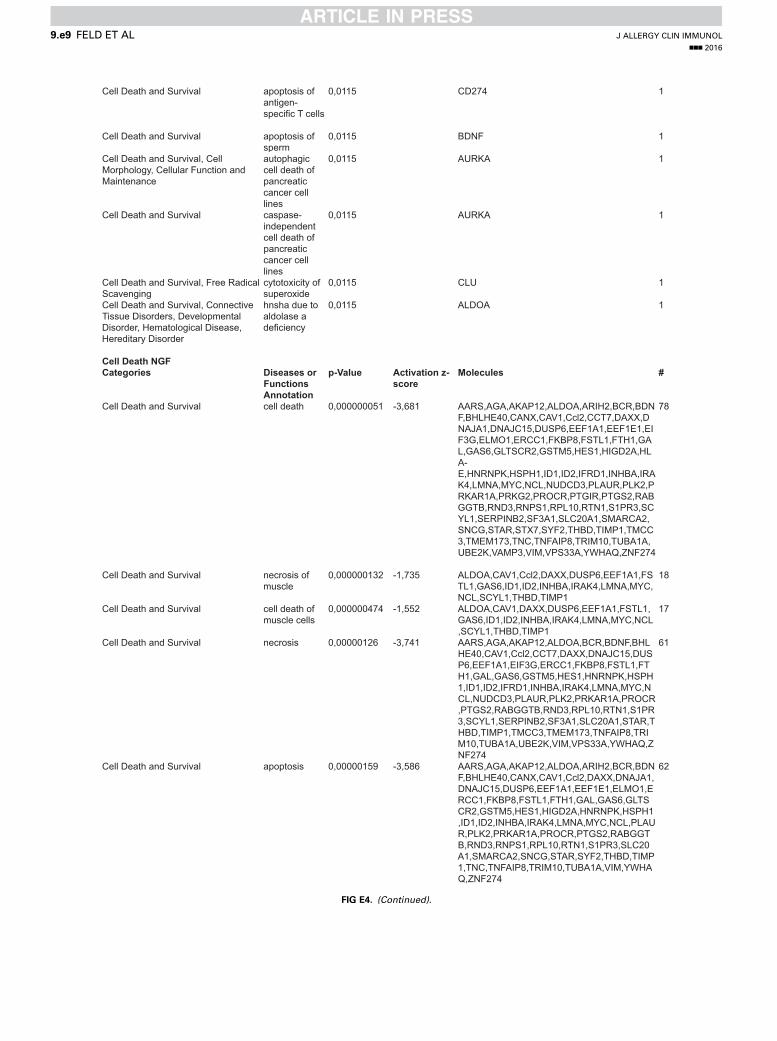
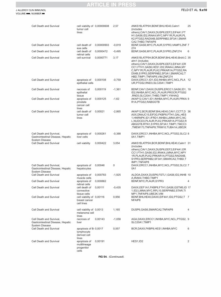
protein response, glycolysis I, and ERK/mitogen-activatedprotein kinase signaling; Fig 2, C). However, IPA also identifiedenrichment of signaling pathways specific for IL-31 (eg, 14-3-3signaling, mechanistic target of rapamycin signaling, phosphoi-nositide 3-kinase [PI3K]/AKT signaling, and integrin-linkedkinase signaling) or NGF (eg, Gi signaling, cyclic AMP–mediated signaling, and cholesterol biosynthesis I; see Fig E4in this article’s Online Repository at www.jacionline.org).Interestingly, IPA indicated that differentially expressed genes
associated with either IL-31 or NGF stimulation are related to avariety of functional annotations within 3 central categories:nervous system development (Fig 2, D), cell death and survival(Fig 2, E), and cellular movement (Fig 2, F). In these categoriesIL-31– and NGF-induced gene sets not only promote overlappingbiological responses but also relate to different functionalannotations. However, if linked to different functionalannotations, the IL-31– and NGF-activated genes can be assignedto similar biological functions promoting neuronal growth

FIG 3. IL-31RA is expressed on small-diameter neurons, and mIL-31 promotes axonal growth in DRG
explants. A, Colocalization of IL-31RA and TrkA (NGF receptor) in DRG sections. *Colocalization of IL-31RA
with TrkA. B, The minority of IL-31RA1 small-diameter DRG neurons coexpress TrkA. C, Identification of
IL-31RA in growth cones of DRG neurons. D, mIL-31–induced neuronal sprouting in DRG explants.
***P < .001, Mann-Whitney test.
J ALLERGY CLIN IMMUNOL
VOLUME nnn, NUMBER nn
FELD ET AL 5
(regeneration of peripheral nerve, neuronal outgrowth, and cellsurvival; see Fig E4) and cell movement (Fig 2, F). Indeed, withinthe category of nervous system development, IL-31 positivelyregulates the quantity of neurites and the extension of neurites,whereas NGF induces genes positively regulating the quantityof neurons and growth of axons (activation z score > 1; Fig 2,D). In contrast, activation of functions like necrosis, apoptosis,and cell death is negatively regulated in response to both IL-31and NGF (activation z score < 21; Fig 2, E).
IL-31 promotes neuronal growth in small-diameter
neuronsTo analyze the distribution pattern and coexpression of IL-
31RA and tropomyosin receptor kinase A (TrkA; the specificNGF receptor) in neuron subsets, we performed immunofluores-cence staining on primary DRG neurons isolated from wild-typemice. We found that IL-31RA is expressed only on small-diameter neurons (diameter, <20 mm), whereas TrkA is foundon small- and large-diameter (diameter, >20 mm) neurons (Fig 3,A and B, upper panel). Only 21% 6 9% of all IL-31RA1 DRGneurons also stain positive for TrkA (Fig 3, B, lower panel).Moreover, in IL-31–activated DRG neurons, IL-31RA is locatedin the growth cones and cell bodies (Fig 3, C). To furtherinvestigate the role of IL-31 in neuronal outgrowth, whole DRGexplants from wild-type mice (n 5 6) were stimulated withIL-31 or control (n 5 19 DRGs per condition), and the length
of the 10 longest neurites per DRG was measured. Interestingly,stimulation of whole DRG explants with IL-31 for 3 days leadsto (1) increased neuronal growth and (2) neurite elongation(Fig 3, D). Quantitation of the length of the 10 longest neuritesper DRG explant revealed that IL-31 significantly promotes nervefiber elongation (1993 6 33.6 mm, P < .0001) compared withcontrol values (1458 6 28.6 mm; Fig 3, D). To further validatethe finding that only small-diameter neurons are responsive toIL-31, we used cultured dissociated DRG neurons (n 5 43-58neurons per condition in 5 independent experiments) to measurethe diameter of each neuron and to treat these neurons with IL-31,NGF, or vehicle control for 24 hours. In dissociated DRG neuronswith a small diameter (<20 mm), we found that both IL-31(318.1 6 17.9 mm, P < .001) and NGF (325 6 27.8 mm,P < .01) induce nerve fiber elongation compared with controlneurons (220.6 6 14.2 mm; Fig 4, A and B). Interestingly, incontrast to NGF, IL-31 also promoted increased branching ofthe longest neurites extending from small-diameter neuronsprepared from wild-type mice (<20 mm, P 5 .01; Fig 4, C). Inlarge-diameter neurons (>20 mm) only NGF, but not IL-31,affected nerve fiber elongation (Fig 4, D). To confirm that IL-31biology is mediated by its specific receptor IL-31RA, weprepared dissociated DRG neurons derived from Il31ra-deficientmice. In contrast to wild-type mice, IL-31 did not augmentneuronal elongation and branching of small-diameter neuronsof Il31ra2/2 mice (207 6 14.1 mm) when compared withuntreated control mice (178.5 6 21.5 mm; Fig 4, E and F).
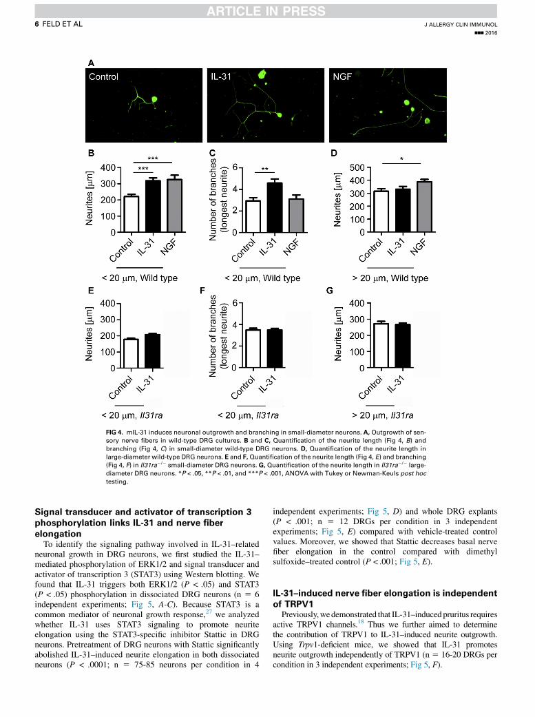
FIG 4. mIL-31 induces neuronal outgrowth and branching in small-diameter neurons. A, Outgrowth of sen-
sory nerve fibers in wild-type DRG cultures. B and C, Quantification of the neurite length (Fig 4, B) andbranching (Fig 4, C) in small-diameter wild-type DRG neurons. D, Quantification of the neurite length in
large-diameter wild-type DRG neurons. E and F,Quantification of the neurite length (Fig 4, E) and branching
(Fig 4, F) in Il31ra2/2 small-diameter DRG neurons. G, Quantification of the neurite length in Il31ra2/2 large-
diameter DRG neurons. *P < .05, **P < .01, and ***P < .001, ANOVA with Tukey or Newman-Keuls post hoctesting.
J ALLERGY CLIN IMMUNOL
nnn 2016
6 FELD ET AL
Signal transducer and activator of transcription 3
phosphorylation links IL-31 and nerve fiber
elongationTo identify the signaling pathway involved in IL-31–related
neuronal growth in DRG neurons, we first studied the IL-31–mediated phosphorylation of ERK1/2 and signal transducer andactivator of transcription 3 (STAT3) using Western blotting. Wefound that IL-31 triggers both ERK1/2 (P < .05) and STAT3(P < .05) phosphorylation in dissociated DRG neurons (n 5 6independent experiments; Fig 5, A-C). Because STAT3 is acommon mediator of neuronal growth response,27 we analyzedwhether IL-31 uses STAT3 signaling to promote neuriteelongation using the STAT3-specific inhibitor Stattic in DRGneurons. Pretreatment of DRG neurons with Stattic significantlyabolished IL-31–induced neurite elongation in both dissociatedneurons (P < .0001; n 5 75-85 neurons per condition in 4
independent experiments; Fig 5, D) and whole DRG explants(P < .001; n 5 12 DRGs per condition in 3 independentexperiments; Fig 5, E) compared with vehicle-treated controlvalues. Moreover, we showed that Stattic decreases basal nervefiber elongation in the control compared with dimethylsulfoxide–treated control (P < .001; Fig 5, E).
IL-31–induced nerve fiber elongation is independent
of TRPV1Previously,wedemonstrated that IL-31–inducedpruritus requires
active TRPV1 channels.18 Thus we further aimed to determinethe contribution of TRPV1 to IL-31–induced neurite outgrowth.Using Trpv1-deficient mice, we showed that IL-31 promotesneurite outgrowth independently of TRPV1 (n 5 16-20 DRGs percondition in 3 independent experiments; Fig 5, F).

FIG 5. mIL-31–activated STAT3 signaling regulates neurite elongation in DRG neurons. A-C, Quantitation of
mIL-31–induced ERK1/2 and STAT3 phosphorylation in DRG neurons. D and E, Stattic-related outgrowth
inhibition in dissociated DRG neurons (Fig 5, D) and DRG explants (Fig 5, E). F, Quantitation of outgrowth
in Trpv12/2 neurons. *P < .05 and ***P < .001, Wilcoxonmatched-pairs signed-rank test, ANOVAwith Tukey
post hoc testing. DMSO, Dimethyl sulfoxide.
J ALLERGY CLIN IMMUNOL
VOLUME nnn, NUMBER nn
FELD ET AL 7
DISCUSSIONIn patients with AD, agonizing itch sensation involves an
increased density of cutaneous sensory nerve fibers.5 Currentconcepts implicate an imbalance of neurotrophins and repulsivefactors as participating in pruritus-associated pathology.6
Because AD represents a chronic inflammatory disease, a rolefor TH2 cytokines in promoting the elongation of sensory nervesduring atopic skin inflammation has also been proposed.12 Here,for the first time in a murine model, we report that the TH2-relatedand atopy-associated cytokine IL-31 directly promotes nerve fiberelongation in vitro and in vivo (Figs 1, 3, and 4). Moreover, wedemonstrate that IL-31 increases neurite branching in activatedDRG neurons (Fig 4, C). Intriguingly, AD-related factors, suchas IL-4 and staphylococcal enterotoxin B, augment IL-31production in human subjects,20,28 suggesting that endogenousand/or environmental triggers might engage the IL-31 pathwayto promote cutaneous innervation in atopic subjects. Indeed,transcriptional profiling of IL-31–activated DRG neurons frommice further supports a neuropoietic function of IL-31. Pathwayanalysis revealed enrichment for pathways related to neurogene-sis in general and to neurite branching and elongation of neuritesin particular (Fig 2,D, and see theMethods section in this article’sOnline Repository). Interestingly, IPA indicated an IL-31–induced innervation of the skin response (see Fig E4). Moreover,
our findings are also supported by initial correlative findings byMurota et al.29 IL-31 has been assigned to the IL-6 family ofcytokines,16 which emerged as a cytokine family with dualfunctions: inflammation and neuropoiesis.30 The IL-6 familymembers leukemia inhibitory factor, ciliary neurotrophic factor,and oncostatin M regulate nervous system development, neuronalsurvival, or repair30-34 through a selective receptor heterodimerspecific for each IL-6 family member. However, unlike all otherIL-6 family–related receptor heterodimers, the IL-31 receptorcomplex uses a unique gp130-like receptor chain IL-31RA.16
Our recent studies revealed that IL-31RA is expressed exclusivelyon a subset of small-diameter neurons (<20 mm; Fig 3, A) ofTRPV11 peptidergic murine DRG neurons, which evokesprofound scratching in mice through an ERK1/2-dependentmechanism.18 The clinical relevance of these findings issupported by the positive results of a recent clinical study usinga neutralizing anti–IL-31RA mAb to control pruritus in patientswith AD.25 Here we demonstrate that IL-31RA is targeted tothe growth cones of primary sensory neurons (Fig 3, C),further suggesting an essential role for IL-31 in neuron–growthcone guidance, as demonstrated previously for otherneurotrophins.35,36
Previously, the dysregulation of NGF and semaphorin 3Awasproposed to explain how increased cutaneous sensory nerve

J ALLERGY CLIN IMMUNOL
nnn 2016
8 FELD ET AL
density is regulated in patients with AD.6 In this modelkeratinocyte-derived NGF is one of the mediators determiningthe innervation density in skin of patients with AD, which ischaracterized by decreased levels of the repulsive axon guidancefactor semaphorin 3A.37,38 Nonetheless, current evidencesuggests that additional TH2 inflammation–related mechanismsare triggering skin innervation and scratching behavior in micewith chronic AD.12 Although intradermal injection of mIL-31induces intense immediate pruritus in mice,18 Hawro et al23
reported that skin prick testing with human IL-31 only induceddelayed pruritus (9/20 subjects) 2 hours after injection. Hencefurther research will be necessary to investigate how IL-31directly or indirectly communicates with neurons to causepruritus in human subjects.
In the present study we extend our understanding of theneuronal function of IL-31 promoting the elongation ofIL-31RA1 neurons (Figs 3, D, and 4). Immunofluorescenceanalysis of IL-31RA1 neurons further revealed that only theminority of IL-31–responsive neurons coexpresses TrkA (Fig 3,A and B), proposing that IL-31 elongates a unique subset ofitch-conducting sensory nerve fibers that are unresponsive toNGF. To further characterize the differences/similarities betweenIL-31– and NGF-induced nerve fiber out growth, we comparedthe transcriptional profiles of murine DRG neurons culturedwith IL-31 or NGF bymeans of whole-transcriptome sequencing.Intriguingly, although IL-31 and NGF enrich genes that relate tosimilar biological processes, the transcriptional profile is quitedifferent (Fig 2), suggesting that both agonists might activatedifferent neurons. IL-31 stimulation leads to enrichment ofsignaling pathways linked to nervous system development andsurvival: PI3K/AKT, mitogen-activated protein kinase, mamma-lian target of rapamycin, and 14-3-3 signaling.39-42 Interestingly,pathways enriched and positively regulated in response to IL-31,like PI3K/AKT and integrin-linked kinase signaling, triggerperipheral axon regeneration,43 axonal elongation,41 andneuronal polarity.44 STAT3 was identified as (1) an importantpositive regulator of peripheral nerve regeneration and neuronaloutgrowth27,45 and (2) a protective factor rescuing from axonaldegeneration,46 thus strongly suggesting a role for STAT3 inIL-31–related nerve fiber growth responses. These findings aresupported by IL-31–induced STAT3 phosphorylation in murineneurons (Fig 5, A and B) and the fact that pharmacologic inhibi-tion of the STAT3 pathway completely abolishes IL-31–mediatedneuron outgrowth (Fig 5, D and E). Interestingly, it has beenreported recently that NGF requires basal activity of the IL-6family receptor–related gp130 to induce STAT3 phosphorylationand to promote neuronal outgrowth inDRG neurons,45 suggestingthat both IL-31 and NGF promote nerve fiber elongation throughSTAT3 phosphorylation but target different neuron subsets.Recently, we could demonstrate that IL-31–induced itchsensation in mice critically depends on ERK1/2 and functionalTRPV1 channels.18 Although previous studies associated Trpv1with neuritogenesis,47 our findings in TRPV1-deficient miceshow that IL-31–induced sensory neuron elongation and branch-ing were independent of TRPV1 engagement (Fig 5, F). Hencethese results suggest that one stimulus, IL-31, uses differentsignaling traits to evoke distinct downstream phenotypes.
In patients with AD, intractable pruritus causes severe clinicalchallenges and relies, on the activation of the IL-31 axis, amongother factors. Based on previous findings and the results of thepresent study, we suggest the following model for the role of the
IL-31 axis in the skin. First, during atopic skin inflammation,immune cells, such as TH2 cells, mast cells, or both, communicatewith cutaneous IL-31RA1 sensory neurons through IL-31secretion and induce itch sensations. Second, subsequently,chronic IL-31–driven ‘‘atopic’’ stimulation of cutaneous sensoryneurons can result in cutaneous sensory nerve fiber elongationand branching, leading to increased sensory nerve fiber densityin patients with AD. Third, given the enhanced sensory networkwithin the skin, it is conceivable that itch thresholds aredecreasing and that minimal doses of pruritogens can result intriggering symptoms in atopic subjects.
Taken together, because of its role in inflammation, itch, andnerve elongation, therapeutic targeting of the IL-31/IL-31RApathway might be an attractive approach to break viciousitch-scratch-eczema cycles and to improve the patient’s qualityof life.
We thank Michaela Fastrich for expert technical assistance. The compu-
tations were performed with resources provided by SNIC through Uppsala
Multidisciplinary Center for Advanced Computational Science (UPPMAX)
under Project b2014069.
Key messages
d The pruritus- and TH2-associated novel cytokine IL-31 in-duces a distinct transcriptional program in sensoryneurons.
d IL-31 induces the outgrowth of sensory neurons in aSTAT3-dependent manner.
d Translational effect: IL-31–associated nerve elongationmight be involved in skin hypersensitivity of patientswith AD to pruritogenic trigger factors.
REFERENCES
1. Bieber T. Atopic dermatitis. N Engl J Med 2008;358:1483-94.
2. Eyerich K, Novak N. Immunology of atopic eczema: overcoming the Th1/Th2
paradigm. Allergy 2013;68:974-82.
3. Hamilton JD, Su�arez-Fari~nas M, Dhingra N, Cardinale I, Li X, Kostic A, et al.
Dupilumab improves the molecular signature in skin of patients with moderate-
to-severe atopic dermatitis. J Allergy Clin Immunol 2014;134:1293-300.
4. Kabashima K. New concept of the pathogenesis of atopic dermatitis: interplay
among the barrier, allergy, and pruritus as a trinity. J Dermatol Sci 2013;70:3-11.
5. Cevikbas F, Steinhoff A, Homey B, Steinhoff M. Neuroimmune interactions in
allergic skin diseases. Curr Opin Allergy Clin Immunol 2007;7:365-73.
6. Tominaga M, Takamori K. Itch and nerve fibers with special reference to atopic
dermatitis: therapeutic implications. J Dermatol 2014;41:205-12.
7. Emtestam L, Hagstr€omer L, Dou YC, Sartorius K, Johansson O. PGP 9.5 distribu-
tion patterns in biopsies from early lesions of atopic dermatitis. Arch Dermatol Res
2012;304:781-5.
8. Urashima R, Mihara M. Cutaneous nerves in atopic dermatitis. A histological,
immunohistochemical and electron microscopic study. Virchows Arch 1998;432:
363-70.
9. Pincelli C, Fantini F, Massimi P, Girolomoni G, Seidenari S, Giannetti A. Neuro-
peptides in skin from patients with atopic dermatitis: an immunohistochemical
study. Br J Dermatol 1990;122:745-50.
10. J€arvikallio A, Harvima IT, Naukkarinen A. Mast cells, nerves and neuropeptides in
atopic dermatitis and nummular eczema. Arch Dermatol Res 2003;295:2-7.
11. Ikoma A, Steinhoff M, St€ander S, Yosipovitch G, Schmelz M. The neurobiology of
itch. Nat Rev Neurosci 2006;7:535-47.
12. Oh MH, Oh SY, Lu J, Lou H, Myers AC, Zhu Z, et al. TRPA1-dependent pruritus
in IL-13-induced chronic atopic dermatitis. J Immunol 2013;191:5371-82.
13. Liu W, Luo R, Chen Y, Sun C, Wang J, Zhou L, et al. Interleukin-31 promotes help-
er T cell type-2 inflammation in children with allergic rhinitis. Pediatr Res 2015;
77:20-8.

J ALLERGY CLIN IMMUNOL
VOLUME nnn, NUMBER nn
FELD ET AL 9
14. Lei Z, Liu G, Huang Q, Lv M, Zu R, Zhang GM, et al. SCF and IL-31 rather than
IL-17 and BAFF are potential indicators in patients with allergic asthma. Allergy
2008;63:327-32.
15. Hartmann K, Wagner N, Rabenhorst A, Pflanz L, Leja S, F€orster A, et al.
Serum IL-31 levels are increased in a subset of patients with mastocytosis
and correlate with disease severity in adult patients. J Allergy Clin Immunol
2013;132:232-5.
16. Cornelissen C, L€uscher-Firzlaff J, Baron JM, L€uscher B. Signaling by IL-31 and
functional consequences. Eur J Cell Biol 2012;91:552-66.
17. Dillon SR, Sprecher C, Hammond A, Bilsborough J, Rosenfeld-Franklin M, Pre-
snell SR, et al. Interleukin 31, a cytokine produced by activated T cells, induces
dermatitis in mice. Nat Immunol 2004;5:752-60.
18. Cevikbas F, Wang X, Akiyama T, Kempkes C, Savinko T, Antal A, et al. A sensory
neuron-expressed IL-31 receptor mediates T helper cell-dependent itch: involve-
ment of TRPV1 and TRPA1. J Allergy Clin Immunol 2014;133:448-60.
19. Park K, Park JH, Yang WJ, Lee JJ, Song MJ, Kim HP. Transcriptional activation of
the IL31 gene by NFAT and STAT6. J Leukoc Biol 2012;91:245-57.
20. Sonkoly E, Muller A, Lauerma AI, Pivarcsi A, Soto H, Kemeny L, et al. IL-31: a
new link between T cells and pruritus in atopic skin inflammation. J Allergy Clin
Immunol 2006;117:411-7.
21. Kato A, Fujii E, Watanabe T, Takashima Y, Matsushita H, Furuhashi T, et al. Dis-
tribution of IL-31 and its receptor expressing cells in skin of atopic dermatitis.
J Dermatol Sci 2014;74:229-35.
22. Grimstad O, Sawanobori Y, Vestergaard C, Bilsborough J, Olsen UB, Grønhøj-
Larsen C, et al. Anti-interleukin-31-antibodies ameliorate scratching behaviour
in NC/Nga mice: a model of atopic dermatitis. Exp Dermatol 2009;18:35-43.
23. Hawro T, Saluja R, Weller K, Altrichter S, Metz M, Maurer M. Interleukin-31 does
not induce immediate itch in atopic dermatitis patients and healthy controls after
skin challenge. Allergy 2014;69:113-7.
24. Kasutani K, Fujii E, Ohyama S, Adachi H, Hasegawa M, Kitamura H, et al. Anti-
IL-31 receptor antibody is shown to be a potential therapeutic option for treating
itch and dermatitis in mice. Br J Pharmacol 2014;171:5049-58.
25. Nemoto O, Shiramoto M, Hanada R, Matsuki S, Imayama S, Kato M, et al. Safety
and tolerability of a humanized monoclonal antibody to the Interleukin-31
receptor; results of a phase I, single ascending dose study, in healthy volunteers
and patients with atopic dermatitis. Abstract presented at: European Academy of
Dermatology and Venereology Meeting, Istanbul, Turkey; October 2-6, 2015.
26. Islam S, Kj€allquist U, Moliner A, Zajac P, Fan JB, L€onnerberg P, et al. Highly
multiplexed and strand-specific single-cell RNA 5’ end sequencing. Nat Protoc
2012;7:813-28.
27. Bareyre FM, Garzorz N, Lang C, Misgeld T, B€uning H, Kerschensteiner M. In vivo
imaging reveals a phase-specific role of STAT3 during central and peripheral
nervous system axon regeneration. Proc Natl Acad Sci U S A 2011;108:6282-7.
28. Stott B, Lavender P, Lehmann S, Pennino D, Durham S, Schmidt-Weber CB.
Human IL-31 is induced by IL-4 and promotes TH2-driven inflammation.
J Allergy Clin Immunol 2013;132:446-54.
29. Murota H, El-latif MA, Tamura T, Katayama I. Olopatadine hydrochloride
decreases tissue interleukin-31 levels in an atopic dermatitis mouse model. Acta
Derm Venereol 2014;94:78-9.
30. Slaets H, Nelissen S, Janssens K, Vidal PM, Lemmens E, Stinissen P, et al. Oncos-
tatin M reduces lesion size and promotes functional recovery and neurite outgrowth
after spinal cord injury. Mol Neurobiol 2014;50:1142-51.
31. Murphy M, Reid K, Hilton DJ, Bartlett PF. Generation of sensory neurons is
stimulated by leukemia inhibitory factor. Proc Natl Acad Sci U S A 1991;88:
3498-501.
32. Moidunny S, Vinet J, Wesseling E, Bijzet J, Shieh CH, van Ijzendoorn SC, et al.
Adenosine A2B receptor-mediated leukemia inhibitory factor release from astrocytes
protects cortical neurons against excitotoxicity. J Neuroinflammation 2012;9:198.
33. Gallagher D, Gutierrez H, Gavalda N, O’Keeffe G, Hay R, Davies AM. Nuclear
factor-kappaB activation via tyrosine phosphorylation of inhibitor kappaB-alpha
is crucial for ciliary neurotrophic factor-promoted neurite growth from developing
neurons. J Neurosci 2007;27:9664-9.
34. Liu H, Liu G, Bi Y. CNTF regulates neurite outgrowth and neuronal migration
through JAK2/STAT3 and PI3K/Akt signaling pathways of DRG explants with
gp120-induced neurotoxicity in vitro. Neurosci Lett 2014;569:110-5.
35. Gallo G, Letourneau PC. Regulation of growth cone actin filaments by guidance
cues. J Neurobiol 2004;58:92-102.
36. Li Y, Jia YC, Cui K, Li N, Zheng ZY, Wang YZ, et al. Essential role of TRPC chan-
nels in the guidance of nerve growth cones by brain-derived neurotrophic factor.
Nature 2005;434:894-8.
37. Dou YC, Hagstr€omer L, Emtestam L, Johansson O. Increased nerve growth factor
and its receptors in atopic dermatitis: an immunohistochemical study. Arch Derma-
tol Res 2006;298:31-7.
38. Tominaga M, Ogawa H, Takamori K. Decreased production of semaphorin 3A in
the lesional skin of atopic dermatitis. Br J Dermatol 2008;158:842-4.
39. Cosker KE, Segal RA. Neuronal signaling through endocytosis. Cold Spring Harb
Perspect Biol 2014;6(2).
40. Patapoutian A, Reichardt LF. Trk receptors: mediators of neurotrophin action. Curr
Opin Neurobiol 2001;11:272-80.
41. Christie KJ, Zochodne D. Peripheral axon regrowth: new molecular approaches.
Neuroscience 2013;240:310-24.
42. Shimada T, Fournier AE, Yamagata K. Neuroprotective function of 14-3-3 proteins
in neurodegeneration. Biomed Res Int 2013;2013:564534.
43. Saijilafu, Hur EM, Liu CM, Jiao Z, Xu WL, Zhou FQ. PI3K-GSK3 signalling reg-
ulates mammalian axon regeneration by inducing the expression of Smad1. Nat
Commun 2013;4:2690.
44. Guo W, Jiang H, Gray V, Dedhar S, Rao Y. Role of the integrin-linked kinase (ILK)
in determining neuronal polarity. Dev Biol 2007;306:457-68.
45. Quarta S, Baeumer BE, Scherbakov N, Andratsch M, Rose-John S, Dechant G,
et al. Peripheral nerve regeneration and NGF-dependent neurite outgrowth of adult
sensory neurons converge on STAT3 phosphorylation downstream of neuropoietic
cytokine receptor gp130. J Neurosci 2014;34:13222-33.
46. Selvaraj BT, Frank N, Bender FL, Asan E, Sendtner M. Local axonal function of
STAT3 rescues axon degeneration in the pmn model of motoneuron disease. J Cell
Biol 2012;199:437-51.
47. Goswami C, Rademacher N, Smalla KH, Kalscheuer V, Ropers HH, Gundelfinger
ED, et al. TRPV1 acts as a synaptic protein and regulates vesicle recycling. J Cell
Sci 2010;123:2045-57.

J ALLERGY CLIN IMMUNOL
nnn 2016
9.e1 FELD ET AL
METHODS
Mice and sample collectionSix to 8-week-old wild-type C57BL/6 and Trpv1 knockout mice were
kept under specific pathogen-free conditions. Il31Tg mice specifically
overexpressing Il31 under the Em-Lck promoter in lymphocytesE1 and
control littermates were housed for up to 9 months under specific
pathogen-free conditions until characteristic lesions developed spontane-
ously. Subsequently, punch biopsy specimens from lesional and nonlesional
skin were collected. An IL-31 pump study in BALB/c mice was performed.
Briefly, BALB/c mice received 20 mg/d recombinant mouse IL-31
(mIL-31; ZymoGenetics, Seattle, Wash) for 14 days delivered subcutane-
ously through a miniosmotic pump (Alzet Osmotic Pump; Durect, Cupertino,
Calif). The skin phenotype was monitored daily and scored based on pruritus,
hair loss, and lesion development: 0, no scratching or hair loss; 1, minimal
scratching and thinning of the coat in small areas; 2, scratching with minor
hair loss; 3, scratching with moderate hair loss and lesion development;
and 4, excessive scratching with severe hair loss and lesion development.
Skin biopsy specimens were taken from the area in which the pump
was dispensing mIL-31. All animal procedures were approved by the
ZymoGenetics Institutional Animal and Care and Use Committee or by the
local government committee, the ‘‘Landesamt fuer Natur, Umwelt und
Verbraucherschutz Nordrhein-Westfalen.’’
Preparation and treatment of DRG neuronsTo prepare sensory neurons from dissociated DRG neurons, adult C57BL/6
mice were killed, and DRGs from the lumbar, thoracic, and cervical regions
were removed. DRGs were trimmed of connective tissue and nerve roots and
then treated with 3 mg/mL collagenase (Sigma-Aldrich, St Louis, Mo) and
0.25mg/mL trypsin (PAALaboratories/GEHealthcare) for 30minutes. DRGs
were triturated for dissociation to prepare a single-cell suspension. To generate
whole DRG explants, DRGs derived from C57BL/6 mice or Trpv1 knockout
mice were isolated, as described above, except that connective tissue was
digested with 0.3% collagenase for 30 minutes only. Both dissociated DRG
neurons and whole DRG explants were plated onto cell-culture dishes (for
RNA and protein extraction) or glass slides (for immunofluorescence) coated
with poly-L-lysine (0.1-1 mg/mL, Sigma-Aldrich) and laminin (5 mg/mL,
Sigma-Aldrich). Cells were cultured in minimal essential medium
supplemented with 10% horse serum, 1% penicillin/streptomycin, 1%
vitamins, 1% N2-supplement, and 2%B27-supplement for recovery. Cultured
DRGs were stimulated with 100 ng/mLmIL-31 (ZymoGenetics) or 10 ng/mL
NGF (Sigma-Aldrich) for the indicated time points. In selected experiments
2 mmol/L (dissociated DRG neurons) or 20 mmol/L (whole DRG explants)
of the STAT3 inhibitor V/Stattic (Calbiochem, San Diego, Calif) was applied
1 hour before IL-31 stimulation.
Immunofluorescence and image analysisOCT-embedded skin samples were cut in 20-mm-thick sections to
analyze cutaneous innervation. Sections were either fixed with Zambonis
fixative (MORPHISTO, Frankfurt, Germany) or 4% paraformaldehyde,
blocked with 2% BSA and 10% donkey serum in 0.5% Triton X-100
containing PBS, and stained with 2.5 mg/mL rabbit anti-PGP9.5 pAb (EMD
Millipore, Billerica, Mass), anti-rabbit Alexa Fluor 555 (Invitrogen), and 49-6-diamidino-2-phenylindole dihydrochloride (DAPI; Molecular Probes,
Eugene, Ore). Confocal image stacks at 320 magnification were taken
with the Zeiss LSM 510 (Zeiss, Oberkochen, Germany) supplemented with
Zen software and ‘‘flattened’’ into a single image before nerve fiber
quantification. Cutaneous PGP9.51 fibers were quantified in preprocessed
images by 3 independent blind researchers. DRG neurons were first fixed
with 1% paraformaldehyde/7.5% sucrose for 30 minutes and then for
another 30 minutes in 2% paraformaldehyde/15% sucrose to quantify axonal
growth and to identify the cellular IL-31RA localization. DRG neurons were
stained with 10 mg/mL anti–bIII-tubulin (Sigma-Aldrich), 14.8 mg/mL rat
anti-mouse IL-31RA mAb (ZymoGenetics), and anti-rabbit Alexa Fluor
488 or anti-rat Alexa Fluor 594 (both Invitrogen), according to standard
methods. Images were taken with a Zeiss Cell Observer, Axiovision and
Mosaix software, or a Zeiss LSM 510 supplemented with Zen software
(all from Zeiss). The length of each neurite was measured as the distance
from the end of the neurite straight back to the DRG body radially.E2 Cry-
opreserved, OCT-embedded whole DRGs plucked from the spinal columns
of C57BL/6 mice were cut in 10-mm-thick sections. Sections were stained
with 14.8 mg/mL rat anti-mouse IL-31RA (ZymoGenetics), 2.9 mg/mL rab-
bit anti-mouse TrkA (Abcam, Cambridge, United Kingdom), anti-rat Alexa
Fluor 555, and anti-rabbit Alexa Fluor 488 (both from Invitrogen), accord-
ing to standard methods, to visualize the distribution of IL-31– and NGF-
responsive neurons. Photographs were taken with the Zeiss Cell Observer.
For all immunofluorescence staining, at least 3 independent experiments
were performed.
RNA-Seq and data analysisThe STRT method was used,E3 with minor modifications, to measure tran-
scription initiation at the 59 end of polyA1 transcripts starting from 10 ng of
total RNA as template. RNA samples fromDRGneurons (from n5 3 indepen-
dent experiments) were arranged in the plate, and each one was sequenced
with an individual barcode. The capture buffer contained 10 mmol/L Tris-
HCl (pH 8.0), 0.1% Triton X-100, 800 nmol/L T30-VN-oligo, 2 mmol/L
dNTP mixture, and 2 mmol/L template switching oligos barcoding the
sample. The cDNAs were pooled into one tube by using 10% PEG-6000
and 0.9mol/LNaCl and amplified by using 14 cycles of PCR and 10 additional
cycles to introduce the complete sets of adapters for Illumina sequencing (Il-
lumina, San Diego, Calif). The libraries were size selected (200-400 bp) by
using sequential AMPure XP bead selection protocol and 0.73 and 0.223ratios.
The sequences of the STRT libraries were preprocessed to (1) demultiplex
by sample barcodes; (2) exclude redundant reads to reduce PCR bias by unique
molecular identifiersE4; (3) align the reads to the mouse reference genome
mm9, the mouse ribosomal DNA repeating unit (GenBank:BK000964), and
spike-in sequences by using TopHatE5; (4) quantify the expression levels in
50-bp strand-specific windows sliding in 25-bp steps; and (5) perform the
basal quality check of the library and the sequencing. Then the differential
expression was tested by using SAMstrt.E6
For analysis, only sequences that aligned to coding DNA sequence, coding
upstream, and coding 59 untranslated region were selected, and duplicates
were removed. Annotation was done manually with information in GenBank,
PubMed, and pfam alignments to assign individual genes to functional classes
to generate a detailed (level 1: 44 classes for IL-31–cultured DRG neurons and
48 classes for NGF cultured DRG neurons) annotation or an overview (level 2:
10 classes for both culture conditions) with broader functional classes. Genes
that were not characterized and did not have any informative pfammotifs were
annotated as ‘‘Unknown.’’ Summary (level 2) data sets were then plotted in a
bar chart format to compare functional classes after induction of gene
expression by IL-31 or NGF.
Biological functions associated with differentially expressed genes were
identified by using IPA.We performed a global canonical pathway enrichment
analysis and functional enrichment analysis of diseases and functions. Three
functional categories of interest were considered: cell death, cellular
movement, and nervous system development. Differentially expressed genes
associated with NGF and IL-31 were analyzed independently to identify
biological functions and were ranked were based on the summed -log(P value)
across both NGF and IL-31.
Western blottingProteins from IL-31–activated DRG neurons were harvested with
Roti-Load buffer (Carl Roth) supplemented with 2-mercaptoethanol at the
indicated time points and boiled for 10 minutes. Whole DRG lysates were
separated by means of SDS-PAGE and transferred onto nitrocellulose
membranes, according to standard methods. The primary antibodies anti-
pERK1/2 (Thr202/Tyr204 [1:1000]) and anti-pSTAT3 (Tyr705 [1:2000]; both
fromCell Signaling, Danvers,Mass) and 0.3mg/mL anti–bIII-tubulin (Sigma-
Aldrich) and appropriate peroxidase-conjugated secondary antibodies (GE
Healthcare, Fairfield, Conn) were used, according to the manufacturer’s

J ALLERGY CLIN IMMUNOL
VOLUME nnn, NUMBER nn
FELD ET AL 9.e2
instructions. Changes in phosphorylation status were evaluated by using
ImageJ software (National Institutes of Health, Bethesda, Md).
Quantitative real-time PCRRNAwas prepared from IL-31– and NGF-activated DRG neurons with the
RNeasy kit (Qiagen) and reverse transcribed with SuperScript II (Invitrogen),
according to the manufacturer’s instructions. Quantitation of mRNA levels was
performedbyusing real-timefluorescencedetectionwithAbsoluteSYBRGreen
ROX mix (Applied Biosystems, Foster City, Calif). The primers used were as
follows: Il31ra forward, 59-ttcaagacattgtcaatcagtgtg-39; Il31ra reverse, 59-gtcactgttttgatgctaagtagaaga-39; Shroom3 forward, 59-ggggcttcaccctgaaag-39;Shroom3 reverse, 59-tttgcccccttcttcaatct-39; Nts forward, 59-agctggtgtgcct-gactctc-39; Nts reverse, 59-ccagggctctcacatcttct-39; Rcan1 forward, 59-ggctgca-caagaccgagt-39; Rcan1 reverse, 59-tgtgaacttcctatgtgtaaagtctga-39; Sema6d
forward, 59-tttgctttccataaccacagc-39; Sema6d reverse, 59- ctggacttcccaccgta-
cac-39; Cyth2 forward, 59-tcttcaacccagaccgagag-39; Cyth2 reverse, 59-gccgcttccaagtcttcac-39; Prph forward, 59-tctgatcaggacaattgagacc-39; and Prph
reverse, 59-cactgtgctgttccttctgg-39. Primers and probes specific for 18S RNA
were obtained from Life Technologies (Darmstadt, Germany). Target gene
expression was analyzed on an Applied Biosystems Prism 7000 supplemented
with SDS 1.2.3 software, and the expression profile was normalized by using
18S expression.
REFERENCES
E1. Dillon SR, Sprecher C, Hammond A, Bilsborough J, Rosenfeld-Franklin M, Pre-
snell SR, et al. Interleukin 31, a cytokine produced by activated T cells, induces
dermatitis in mice. Nat Immunol 2004;5:752-60.
E2. Deister C, Schmidt CE. Optimizing neurotrophic factor combinations for neurite
outgrowth. J Neural Eng 2006;3:172-9.
E3. Islam S, Kj€allquist U, Moliner A, Zajac P, Fan JB, L€onnerberg P, et al. Highly mul-
tiplexed and strand-specific single-cell RNA 59 end sequencing. Nat Protoc 2012;7:813-28.
E4. Kivioja T, V€ah€arautio A, Karlsson K, Bonke M, Enge M, Linnarsson S, et al.
Counting absolute numbers of molecules using unique molecular identifiers. Nat
Methods 2011;9:72-4.
E5. Trapnell C, Pachter L, Salzberg SL. TopHat: discovering splice junctions with
RNA-Seq. Bioinformatics 2009;25:1105-11.
E6. Bareyre FM, Garzorz N, Lang C, Misgeld T, B€uning H, Kerschensteiner M. In vivo
imaging reveals a phase-specific role of STAT3 during central and peripheral ner-
vous system axon regeneration. Proc Natl Acad Sci U S A 2011;108:6282-7.

FIG E1. Skin phenotype in Il31Tgmice. Spontaneous development of skin lesions in Il31Tgmice in the nape
of the neck (white arrow; A) and ears (B), as well as development of signs of hair loss on the back (whitearrowhead).
J ALLERGY CLIN IMMUNOL
nnn 2016
9.e3 FELD ET AL
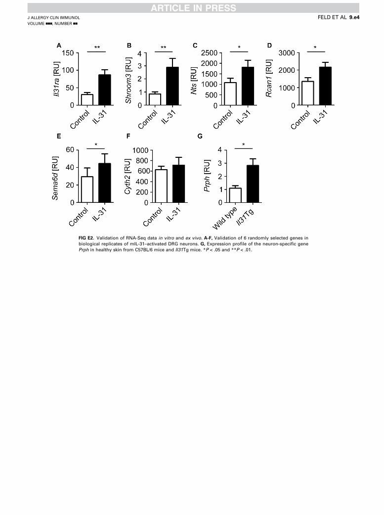
FIG E2. Validation of RNA-Seq data in vitro and ex vivo. A-F, Validation of 6 randomly selected genes in
biological replicates of mIL-31–activated DRG neurons. G, Expression profile of the neuron-specific gene
Prph in healthy skin from C57BL/6 mice and Il31Tg mice. *P < .05 and **P < .01.
J ALLERGY CLIN IMMUNOL
VOLUME nnn, NUMBER nn
FELD ET AL 9.e4
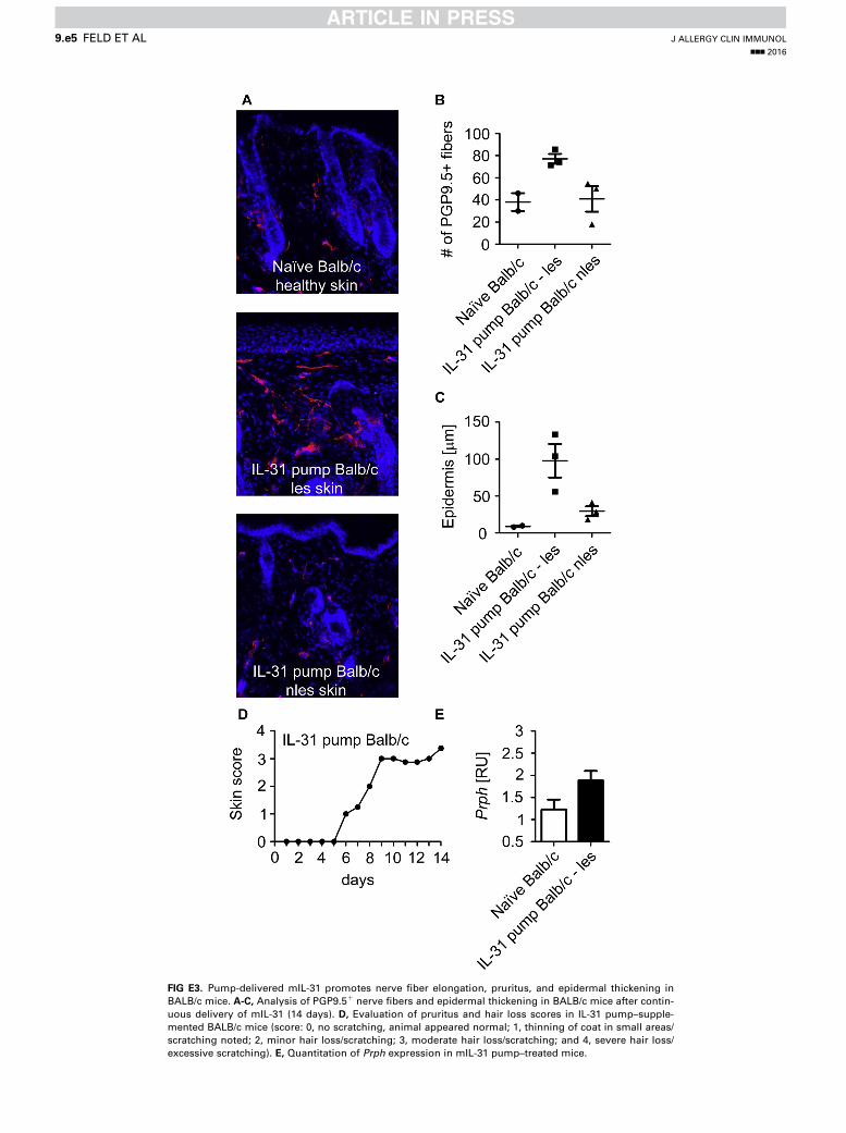
FIG E3. Pump-delivered mIL-31 promotes nerve fiber elongation, pruritus, and epidermal thickening in
BALB/c mice. A-C, Analysis of PGP9.51 nerve fibers and epidermal thickening in BALB/c mice after contin-
uous delivery of mIL-31 (14 days). D, Evaluation of pruritus and hair loss scores in IL-31 pump–supple-
mented BALB/c mice (score: 0, no scratching, animal appeared normal; 1, thinning of coat in small areas/
scratching noted; 2, minor hair loss/scratching; 3, moderate hair loss/scratching; and 4, severe hair loss/
excessive scratching). E, Quantitation of Prph expression in mIL-31 pump–treated mice.
J ALLERGY CLIN IMMUNOL
nnn 2016
9.e5 FELD ET AL

Pathway enrichment
Canonical Pathway NGF (-log(pval))
IL31 (-log(pval))
EIF2 Signaling 4,076162032 5,770226787Oxidative Phosphorylation 2,373652826 3,561817896Gluconeogenesis I 2,75247761 2,538198233Mitochondrial Dysfunction 2,18297669 3,06674445714-3-3-mediated Signaling 0,977161324 4,18874801Regulation of eIF4 and p70S6K Signaling
0,771230656 4,290506996
mTOR Signaling 0,559983575 4,161762165Unfolded protein response 1,808776853 2,457844517Glycolysis I 2,75247761 1,475661409ERK5 Signaling 0,903754489 3,096407492Cell Cycle: G2/M DNA Damage Checkpoint Regulation
1,086662778 2,612326854
Huntingtons Disease Signaling 1,131338349 2,268249295ERK/MAPK Signaling 1,444333791 1,663914012p70S6K Signaling 0,49296076 2,584065887Androgen Signaling 1,028634218 2,028639509Corticotropin Releasing Hormone Signaling
1,637297387 1,399660473
Lipid Antigen Presentation by CD1 1,582793893 1,444153866Cholesterol Biosynthesis I 2,166794329 0,853384957Cholesterol Biosynthesis II (via 24,25-dihydrolanosterol)
2,166794329 0,853384957
NAD Phosphorylation and Dephosphorylation
2,166794329 0,853384957
Cholesterol Biosynthesis III (via Desmosterol)
2,166794329 0,853384957
PI3K/AKT Signaling 0,474183421 2,512556553p38 MAPK Signaling 1,563830177 1,329732885IGF-1 Signaling 0,615321847 2,271162156ILK Signaling 0,967964816 1,674021323Glioma Invasiveness Signaling 1,746168958 0,852280425Glutamate Receptor Signaling 1,746168958 0,852280425cAMP-mediated signaling 2,248397132 0,33258727NRF2-mediated Oxidative Stress Response
2,078200976 0,462608915
Protein Kinase A Signaling 1,081369893 1,445574343Myc Mediated Apoptosis Signaling 0,962883602 1,530998754Germ Cell-Sertoli Cell Junction Signaling
0,336079235 1,966388416
Ephrin B Signaling 0,292521918 1,994703268Remodeling of Epithelial Adherens Junctions
0,850144967 1,356374596
tRNA Charging 2,19647265Breast Cancer Regulation by Stathmin1
0,547707608 1,624231874
Superpathway of Cholesterol Biosynthesis
1,522658161 0,556367364
i Signaling 1,528871761 0,393308768Axonal Guidance Signaling 0 1,913094255Coagulation System 1,344717051 0,47598352Nitric Oxide Signaling in the Cardiovascular System
1,800409145 0
Antigen Presentation Pathway 1,301192155 0,456528928Epoxysqualene Biosynthesis 1,714124955Neurotrophin/TRK Signaling 0,318527905 1,372379138Asparagine Degradation I 1,63907355Adenine and Adenosine Salvage I 1,63907355Endoplasmic Reticulum Stress Pathway
1,617513148
PEDF Signaling 0,300852237 1,310056029Diphthamide Biosynthesis 1,54013191Small Cell Lung Cancer Signaling 1,497881299NGF Signaling 1,449110891PCP pathway 1,439462254Creatine-phosphate Biosynthesis 1,343036327eNOS Signaling 1,31226812 0
FIG E4. Data set 2: IPA output. Data are related to pathway enrichment and
functional annotations. Data set 1: accession to RNA-Seq raw data; data are
uploaded with the European Nucleotide Archive at http://www.ebi.ac.uk/
ena/data/view/PRJEB9128 and do not appear here.
J ALLERGY CLIN IMMUNOL
VOLUME nnn, NUMBER nn
FELD ET AL 9.e6

Cell Death IL-31Categories Diseases or
Functions Annotation
p-Value Activation z-score
Molecules #
Cell Death and Survival necrosis 0,000117 -1,455 ADRM1,ALDOA,APRT,ATF3,ATF4,ATP1A1,AURKA,BDNF,CCT5,CD274,CLU,CST3,CTBP1,DDIT3,DTYMK,FKBP4,GNAO1,GNB2,GSTM5,HSPA4,ILK,ITGA7,LDHA,LRPAP1,MAD2L1,MAPK3,NDEL1,NEFH,Nppb,NSMCE1,NTS,PAFAH2,PGRMC1,PIK3IP1,PLA2G7,PROCR,PSAP,PTTG1,Pvr,RABGGTA,RAN,RHOA,RPS3,RPS6KB1,RTN4,SCYL1,SDC4,SERPINB2,SIN3B,SIVA1,SNCA,SPARC,SPIN1,THBD,TNIP2,TUBA1A,TUBB,TUBB3,VTI1B,XAF1,YWHAB,YWHAQ,YWHAZ
63
Cell Death and Survival apoptosis of neurons
0,000182 -0,559 ATF3,ATF4,BDNF,DDIT3,GNAO1,GSTM5,ILK,LRPAP1,NDEL1,NTS,PLA2G7,RHOA,RPS3,SIVA1,SNCA,VTI1B,YWHAB
17
Cell Death and Survival apoptosis of male germ cells
0,000307 BDNF,CLU,EHD1,HSPA4,HSPBP1,INSL6,PAFAH1B2
7
Cell Death and Survival cell death of fibroblast cell lines
0,000418 0,928 ATF3,ATF4,CLU,DDIT3,FKBP4,GNB2,GSTM5,ILK,MAPK3,NSMCE1,Pvr,RHOA,SDC4,SERPINB2,SIVA1,YWHAZ
16
Cell Death and Survival cell death 0,000522 -1,796 ADRM1,ALDOA,APRT,ARL6IP5,ATF3,ATF4,ATP1A1,AURKA,BDNF,CANX,CCT5,CD274,CLU,CST3,CTBP1,CTSH,CYSLTR2,DDIT3,DTYMK,EHD1,FKBP4,GABARAP,GNAO1,GNB2,GSTM5,HSPA4,HSPBP1,IL4R,ILK,INSL6,ITGA7,LDHA,LRPAP1,MAD2L1,MAL,MAPK3,NDEL1,NEFH,Nppb,NSMCE1,NTS,PAFAH1B2,PAFAH2,PGRMC1,PIK3IP1,PLA2G7,PROCR,PSAP,PTTG1,Pvr,RABGGTA,RAN,RHOA,RPS3,RPS6KB1,RTN4,SCYL1,SDC4,SERPINB2,SH3BGRL3,SIN3B,SIVA1,SNCA,SPARC,SPIN1,THBD,TNIP2,TUBA1A,TUBB,TUBB3,VTI1B,XAF1,YWHAB,YWHAQ,YWHAZ
75
Cell Death and Survival neuronal cell death
0,000612 -0,086 ATF3,ATF4,BDNF,CLU,DDIT3,GNAO1,GSTM5,ILK,LDHA,LRPAP1,NDEL1,NEFH,NTS,PIK3IP1,PLA2G7,RHOA,RPS3,SIVA1,SNCA,VTI1B,YWHAB,YWHAZ
22
Cardiovascular System Development and Function, Cell Death and Survival
cell viability of endothelial cells
0,00064 1,951 BDNF,DDIT3,ILK,ITGB1BP1,TNIP2 5
Cell Death and Survival cell death of connective tissue cells
0,00123 0,197 ATF3,ATF4,CLU,CTBP1,DDIT3,FKBP4,GNB2,GSTM5,ILK,LRPAP1,MAPK3,NSMCE1,Pvr,RHOA,RPS6KB1,SDC4,SERPINB2,SIVA1,YWHAZ
19
Cell Death and Survival cell death of endothelial cells
0,00138 -1,706 ATF3,BDNF,DDIT3,MAPK3,Nppb,PROCR,RHOA,THBD,TNIP2
9
Cell Death and Survival apoptosis 0,00142 -1,616 ALDOA,ARL6IP5,ATF3,ATF4,ATP1A1,AURKA,BDNF,CANX,CD274,CLU,CST3,CTBP1,CTSH,DDIT3,DTYMK,EHD1,FKBP4,GABARAP,GNAO1,GNB2,GSTM5,HSPA4,HSPBP1,IL4R,ILK,INSL6,ITGA7,LDHA,LRPAP1,MAD2L1,MAPK3,NDEL1,Nppb,NTS,PAFAH1B2,PAFAH2,PIK3IP1,PLA2G7,PROCR,PSAP,PTTG1,RABGGTA,RHOA,RPS3,RPS6KB1,RTN4,SDC4,SH3BGRL3,SIN3B,SIVA1,SNCA,SPARC,THBD,TNIP2,TUBA1A,VTI1B,XAF1,YWHAB,YWHAQ,YWHAZ
60
Cell Death and Survival, Cellular Compromise, Neurological Disease, Tissue Morphology
neurodegeneration of cholinergic neurons
0,00193 BDNF,SNCA 2
Cell Death and Survival necrosis of epithelial tissue
0,00256 -0,476 ATF3,AURKA,BDNF,CLU,DDIT3,FKBP4,LDHA,MAPK3,NDEL1,Nppb,PROCR,RHOA,SIVA1,SNCA,SPARC,THBD,TNIP2,YWHAQ
18
FIG E4. (Continued).
J ALLERGY CLIN IMMUNOL
nnn 2016
9.e7 FELD ET AL

Cell Death and Survival, Cellular Compromise, Neurological Disease, Tissue Morphology
neurodegeneration of motor neurons
0,00257 BDNF,NEFH,SCYL1 3
Cell Death and Survival, Cellular Assembly and Organization, Cellular Development, Nervous System Development and Function, Tissue Development, Tissue Morphology
regeneration of axons
0,00263 0,068 BDNF,NDEL1,RHOA,RTN4 4
Cell Death and Survival apoptosis of endothelial cells
0,00278 -1,446 ATF3,BDNF,DDIT3,MAPK3,Nppb,PROCR,RHOA,TNIP2
8
Cell Death and Survival cell viability of breast cancer cell lines
0,00311 1,912 BDNF,CLU,EIF4A1,ILK,MAD2L1,PGRMC1,RHOA
7
Cell Death and Survival cell death of hippocampal neurons
0,0032 0,44 BDNF,DDIT3,LRPAP1,RPS3,SNCA 5
Cell Death and Survival, Cellular Compromise, Neurological Disease, Tissue Morphology
neurodegeneration of neurites
0,0032 -0,747 LRPAP1,NEFH,PSAP,SCYL1,SNCA 5
Cell Death and Survival apoptosis of kidney cells
0,00354 1,992 ATF3,ATP1A1,CLU,DDIT3,ILK 5
Cell Death and Survival apoptosis of central nervous system cells
0,00416 -1,391 BDNF,GNAO1,LRPAP1,RHOA,RPS3,RPS6KB1,YWHAB
7
Cell Death and Survival necrosis of kidney
0,00458 -0,184 APRT,ATF3,ATP1A1,CLU,DDIT3,GNB2,GSTM5,ILK,LDHA,NDEL1,SNCA,YWHAQ
12
Cell Death and Survival cell death of tumor cell lines
0,00508 -0,763 ADRM1,ATF3,ATF4,ATP1A1,AURKA,BDNF,CCT5,CD274,CLU,CTBP1,DDIT3,DTYMK,GNAO1,HSPA4,ILK,MAD2L1,MAPK3,PAFAH2,PGRMC1,PLA2G7,PSAP,PTTG1,RABGGTA,RHOA,RPS3,RPS6KB1,RTN4,SIN3B,SIVA1,SNCA,SPARC,SPIN1,TUBA1A,TUBB3,XAF1,YWHAZ
36
Cell Death and Survival cell death of central nervous system cells
0,0061 -0,26 BDNF,CLU,DDIT3,GNAO1,LRPAP1,RHOA,RPS3,RPS6KB1,SNCA,YWHAB
10
Cell Death and Survival cell death of retinal ganglion cells
0,00642 BDNF,LRPAP1,RHOA 3
Cell Death and Survival apoptosis of tumor cell lines
0,00744 -1,535 ATF3,ATF4,AURKA,BDNF,CD274,CLU,CTBP1,DDIT3,DTYMK,HSPA4,ILK,MAD2L1,MAPK3,PAFAH2,PLA2G7,PSAP,PTTG1,RABGGTA,RHOA,RPS3,RPS6KB1,RTN4,SIN3B,SIVA1,SNCA,SPARC,TUBA1A,XAF1,YWHAZ
29
Cell Death and Survival cell death of kidney cells
0,00865 0,252 ATF3,ATP1A1,CLU,DDIT3,GNB2,GSTM5,ILK,LDHA,NDEL1,SNCA,YWHAQ
11
Cell Death and Survival apoptosis of cerebral cortex cells
0,00869 -1,479 BDNF,GNAO1,LRPAP1,RPS3,YWHAB 5
Cell Death and Survival cell death of cytotoxic T cells
0,00952 CD274,CST3 2
Cell Death and Survival cell death of neuroblastoma cell lines
0,00968 -0,546 BDNF,CCT5,CLU,DDIT3,GNAO1,SNCA,TUBA1A
7
Cell Death and Survival regeneration of cells
0,00971 0,468 BDNF,DDIT3,NDEL1,RHOA,RTN4 5
Cell Death and Survival cell death of cerebral cortex cells
0,00978 -0,118 BDNF,CLU,DDIT3,GNAO1,LRPAP1,RPS3,SNCA,YWHAB
8
Cell Death and Survival apoptosis of myeloma cell lines
0,0111 -1 ATF4,AURKA,DDIT3,RPS6KB1 4
Cancer, Cell Death and Survival, Tumor Morphology
apoptosis of TM4t cells
0,0115 DDIT3 1
FIG E4. (Continued).
J ALLERGY CLIN IMMUNOL
VOLUME nnn, NUMBER nn
FELD ET AL 9.e8
Cell Death and Survival apoptosis of antigen-specific T cells
0,0115 CD274 1
Cell Death and Survival apoptosis of sperm
0,0115 BDNF 1
Cell Death and Survival, Cell Morphology, Cellular Function and Maintenance
autophagic cell death of pancreatic cancer cell lines
0,0115 AURKA 1
Cell Death and Survival caspase-independent cell death of pancreatic cancer cell lines
0,0115 AURKA 1
Cell Death and Survival, Free Radical Scavenging
cytotoxicity of superoxide
0,0115 CLU 1
Cell Death and Survival, Connective Tissue Disorders, Developmental Disorder, Hematological Disease, Hereditary Disorder
hnsha due to aldolase a deficiency
0,0115 ALDOA 1
Cell Death NGFCategories Diseases or
Functions Annotation
p-Value Activation z-score
Molecules #
Cell Death and Survival cell death 0,000000051 -3,681 AARS,AGA,AKAP12,ALDOA,ARIH2,BCR,BDNF,BHLHE40,CANX,CAV1,Ccl2,CCT7,DAXX,DNAJA1,DNAJC15,DUSP6,EEF1A1,EEF1E1,EIF3G,ELMO1,ERCC1,FKBP8,FSTL1,FTH1,GAL,GAS6,GLTSCR2,GSTM5,HES1,HIGD2A,HLA-E,HNRNPK,HSPH1,ID1,ID2,IFRD1,INHBA,IRAK4,LMNA,MYC,NCL,NUDCD3,PLAUR,PLK2,PRKAR1A,PRKG2,PROCR,PTGIR,PTGS2,RABGGTB,RND3,RNPS1,RPL10,RTN1,S1PR3,SCYL1,SERPINB2,SF3A1,SLC20A1,SMARCA2,SNCG,STAR,STX7,SYF2,THBD,TIMP1,TMCC3,TMEM173,TNC,TNFAIP8,TRIM10,TUBA1A,UBE2K,VAMP3,VIM,VPS33A,YWHAQ,ZNF274
78
Cell Death and Survival necrosis of muscle
0,000000132 -1,735 ALDOA,CAV1,Ccl2,DAXX,DUSP6,EEF1A1,FSTL1,GAS6,ID1,ID2,INHBA,IRAK4,LMNA,MYC,NCL,SCYL1,THBD,TIMP1
18
Cell Death and Survival cell death of muscle cells
0,000000474 -1,552 ALDOA,CAV1,DAXX,DUSP6,EEF1A1,FSTL1,GAS6,ID1,ID2,INHBA,IRAK4,LMNA,MYC,NCL,SCYL1,THBD,TIMP1
17
Cell Death and Survival necrosis 0,00000126 -3,741 AARS,AGA,AKAP12,ALDOA,BCR,BDNF,BHLHE40,CAV1,Ccl2,CCT7,DAXX,DNAJC15,DUSP6,EEF1A1,EIF3G,ERCC1,FKBP8,FSTL1,FTH1,GAL,GAS6,GSTM5,HES1,HNRNPK,HSPH1,ID1,ID2,IFRD1,INHBA,IRAK4,LMNA,MYC,NCL,NUDCD3,PLAUR,PLK2,PRKAR1A,PROCR,PTGS2,RABGGTB,RND3,RPL10,RTN1,S1PR3,SCYL1,SERPINB2,SF3A1,SLC20A1,STAR,THBD,TIMP1,TMCC3,TMEM173,TNFAIP8,TRIM10,TUBA1A,UBE2K,VIM,VPS33A,YWHAQ,ZNF274
61
Cell Death and Survival apoptosis 0,00000159 -3,586 AARS,AGA,AKAP12,ALDOA,ARIH2,BCR,BDNF,BHLHE40,CANX,CAV1,Ccl2,DAXX,DNAJA1,DNAJC15,DUSP6,EEF1A1,EEF1E1,ELMO1,ERCC1,FKBP8,FSTL1,FTH1,GAL,GAS6,GLTSCR2,GSTM5,HES1,HIGD2A,HNRNPK,HSPH1,ID1,ID2,INHBA,IRAK4,LMNA,MYC,NCL,PLAUR,PLK2,PRKAR1A,PROCR,PTGS2,RABGGTB,RND3,RNPS1,RPL10,RTN1,S1PR3,SLC20A1,SMARCA2,SNCG,STAR,SYF2,THBD,TIMP1,TNC,TNFAIP8,TRIM10,TUBA1A,VIM,YWHAQ,ZNF274
62
FIG E4. (Continued).
J ALLERGY CLIN IMMUNOL
nnn 2016
9.e9 FELD ET AL
Cell Death and Survival cell viability of tumor cell lines
0,00000608 2,57 ANKS1B,ATP5H,BDNF,BHLHE40,Calm1 (includes others),CAV1,DAXX,DUSP6,EEF2,EIF4A1,FTH1,GAS6,ID2,IRAK4,MYC,NPY1R,PLAUR,PLK2,PTGS2,RAD54B,SERPINB2,SF3A1,SMARCA2,THBD,TNFAIP8
25
Cell Death and Survival cell death of eye cells
0,00000903 -0,819 BDNF,GAS6,MYC,PLAUR,S1PR3,VAMP3,ZNF274
7
Cell Death and Survival cell death of retinal cells
0,0000472 -0,495 BDNF,GAS6,MYC,PLAUR,S1PR3,ZNF274 6
Cell Death and Survival cell survival 0,0000771 3,17 ANKS1B,ATP5H,BCR,BDNF,BHLHE40,Brd4,Calm1 (includes others),CAV1,DAXX,DUSP6,EEF2,EIF4A1,ERCC1,FTH1,GAS6,HES1,ID2,IRAK4,LMNA,MYC,NPY1R,PLAUR,PLK2,PRKAR1A,PTGS2,RAD54B,S1PR3,SERPINB2,SF3A1,SMARCA2,THBD,TIMP1,TNFAIP8,VIM,ZNF274
35
Cell Death and Survival apoptosis of epithelial cells
0,000108 -0,759 DAXX,ERCC1,ID1,ID2,INHBA,MYC,NCL,PLAUR,PTGS2,RND3,SLC20A1,TIMP1
12
Cell Death and Survival necrosis of epithelial tissue
0,000119 -1,361 BDNF,CAV1,DAXX,DUSP6,ERCC1,GAS6,ID1,ID2,INHBA,MYC,NCL,PLAUR,PROCR,PTGS2,RND3,SLC20A1,THBD,TIMP1,YWHAQ
19
Cell Death and Survival apoptosis of prostate cancer cell lines
0,000125 -1,62 AKAP12,CAV1,ID1,INHBA,MYC,PLAUR,PRKAR1A,PTGS2,RABGGTB
9
Cell Death and Survival cell death of tumor cell lines
0,00021 -2,865 AKAP12,BCR,BDNF,BHLHE40,CAV1,CCT7,DAXX,DNAJC15,EIF3G,FKBP8,FTH1,GAL,HES1,HNRNPK,ID1,IFRD1,INHBA,LMNA,MYC,NCL,NUDCD3,PLAUR,PLK2,PRKAR1A,PTGS2,RABGGTB,RTN1,S1PR3,SF3A1,TIMP1,TMCC3,TMEM173,TNFAIP8,TRIM10,TUBA1A,UBE2K
36
Cell Death and Survival, Gastrointestinal Disease, Hepatic System Disease
apoptosis of liver cells
0,000261 -0,388 DAXX,ERCC1,INHBA,MYC,NCL,PTGS2,SLC20A1,TIMP1
8
Cell Death and Survival cell viability 0,000422 3,054 ANKS1B,ATP5H,BCR,BDNF,BHLHE40,Calm1 (includes others),CAV1,DAXX,DUSP6,EEF2,EIF4A1,ERCC1,FTH1,GAS6,ID2,IRAK4,LMNA,MYC,NPY1R,PLAUR,PLK2,PRKAR1A,PTGS2,RAD54B,S1PR3,SERPINB2,SF3A1,SMARCA2,THBD,TIMP1,TNFAIP8
31
Cell Death and Survival, Gastrointestinal Disease, Hepatic System Disease
apoptosis of hepatocytes
0,00046 0 DAXX,ERCC1,INHBA,MYC,NCL,PTGS2,SLC20A1
7
Cell Death and Survival apoptosis of muscle cells
0,000783 -1,925 ALDOA,DAXX,DUSP6,FSTL1,GAS6,ID2,INHBA,IRAK4,THBD,TIMP1
10
Cell Death and Survival apoptosis of retinal cells
0,000862 BDNF,MYC,PLAUR,S1PR3 4
Cell Death and Survival cell death of connective tissue cells
0,00111 -0,435 DAXX,EEF1A1,FKBP8,FTH1,GAS6,GSTM5,ID1,ID2,LMNA,MYC,RPL10,SERPINB2,STAR,TIMP1,TNFAIP8,UBE2K,VIM
17
Cell Death and Survival cell viability of breast cancer cell lines
0,00116 0,956 BDNF,BHLHE40,DAXX,EIF4A1,ID2,PTGS2,TNFAIP8
7
Cell Death and Survival cell viability of melanoma cell lines
0,0013 1,165 DUSP6,GAS6,SMARCA2,TNFAIP8 4
Cell Death and Survival, Gastrointestinal Disease, Hepatic System Disease
necrosis of liver
0,00143 -1,058 AGA,DAXX,ERCC1,INHBA,MYC,NCL,PTGS2,SLC20A1,TIMP1
9
Cell Death and Survival apoptosis of B-lymphocyte derived cell lines
0,0017 0,557 BCR,DAXX,FKBP8,HES1,INHBA,MYC 6
Cell Death and Survival apoptosis of multilineage progenitor cells
0,00191 HES1,ID2 2
FIG E4. (Continued).
J ALLERGY CLIN IMMUNOL
VOLUME nnn, NUMBER nn
FELD ET AL 9.e10
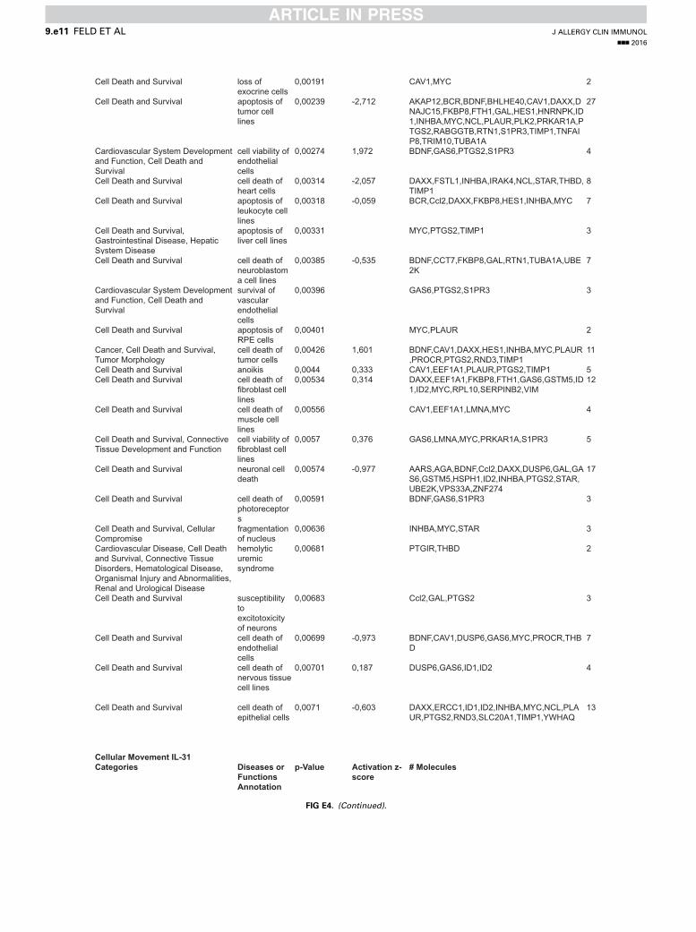
Cell Death and Survival loss of exocrine cells
0,00191 CAV1,MYC 2
Cell Death and Survival apoptosis of tumor cell lines
0,00239 -2,712 AKAP12,BCR,BDNF,BHLHE40,CAV1,DAXX,DNAJC15,FKBP8,FTH1,GAL,HES1,HNRNPK,ID1,INHBA,MYC,NCL,PLAUR,PLK2,PRKAR1A,PTGS2,RABGGTB,RTN1,S1PR3,TIMP1,TNFAIP8,TRIM10,TUBA1A
27
Cardiovascular System Development and Function, Cell Death and Survival
cell viability of endothelial cells
0,00274 1,972 BDNF,GAS6,PTGS2,S1PR3 4
Cell Death and Survival cell death of heart cells
0,00314 -2,057 DAXX,FSTL1,INHBA,IRAK4,NCL,STAR,THBD,TIMP1
8
Cell Death and Survival apoptosis of leukocyte cell lines
0,00318 -0,059 BCR,Ccl2,DAXX,FKBP8,HES1,INHBA,MYC 7
Cell Death and Survival, Gastrointestinal Disease, Hepatic System Disease
apoptosis of liver cell lines
0,00331 MYC,PTGS2,TIMP1 3
Cell Death and Survival cell death of neuroblastoma cell lines
0,00385 -0,535 BDNF,CCT7,FKBP8,GAL,RTN1,TUBA1A,UBE2K
7
Cardiovascular System Development and Function, Cell Death and Survival
survival of vascular endothelial cells
0,00396 GAS6,PTGS2,S1PR3 3
Cell Death and Survival apoptosis of RPE cells
0,00401 MYC,PLAUR 2
Cancer, Cell Death and Survival, Tumor Morphology
cell death of tumor cells
0,00426 1,601 BDNF,CAV1,DAXX,HES1,INHBA,MYC,PLAUR,PROCR,PTGS2,RND3,TIMP1
11
Cell Death and Survival anoikis 0,0044 0,333 CAV1,EEF1A1,PLAUR,PTGS2,TIMP1 5Cell Death and Survival cell death of
fibroblast cell lines
0,00534 0,314 DAXX,EEF1A1,FKBP8,FTH1,GAS6,GSTM5,ID1,ID2,MYC,RPL10,SERPINB2,VIM
12
Cell Death and Survival cell death of muscle cell lines
0,00556 CAV1,EEF1A1,LMNA,MYC 4
Cell Death and Survival, Connective Tissue Development and Function
cell viability of fibroblast cell lines
0,0057 0,376 GAS6,LMNA,MYC,PRKAR1A,S1PR3 5
Cell Death and Survival neuronal cell death
0,00574 -0,977 AARS,AGA,BDNF,Ccl2,DAXX,DUSP6,GAL,GAS6,GSTM5,HSPH1,ID2,INHBA,PTGS2,STAR,UBE2K,VPS33A,ZNF274
17
Cell Death and Survival cell death of photoreceptors
0,00591 BDNF,GAS6,S1PR3 3
Cell Death and Survival, Cellular Compromise
fragmentation of nucleus
0,00636 INHBA,MYC,STAR 3
Cardiovascular Disease, Cell Death and Survival, Connective Tissue Disorders, Hematological Disease, Organismal Injury and Abnormalities, Renal and Urological Disease
hemolytic uremic syndrome
0,00681 PTGIR,THBD 2
Cell Death and Survival susceptibility to excitotoxicity of neurons
0,00683 Ccl2,GAL,PTGS2 3
Cell Death and Survival cell death of endothelial cells
0,00699 -0,973 BDNF,CAV1,DUSP6,GAS6,MYC,PROCR,THBD
7
Cell Death and Survival cell death of nervous tissue cell lines
0,00701 0,187 DUSP6,GAS6,ID1,ID2 4
Cell Death and Survival cell death of epithelial cells
0,0071 -0,603 DAXX,ERCC1,ID1,ID2,INHBA,MYC,NCL,PLAUR,PTGS2,RND3,SLC20A1,TIMP1,YWHAQ
13
Cellular Movement IL-31Categories Diseases or
Functions Annotation
p-Value Activation z-score
# Molecules
FIG E4. (Continued).
J ALLERGY CLIN IMMUNOL
nnn 2016
9.e11 FELD ET AL
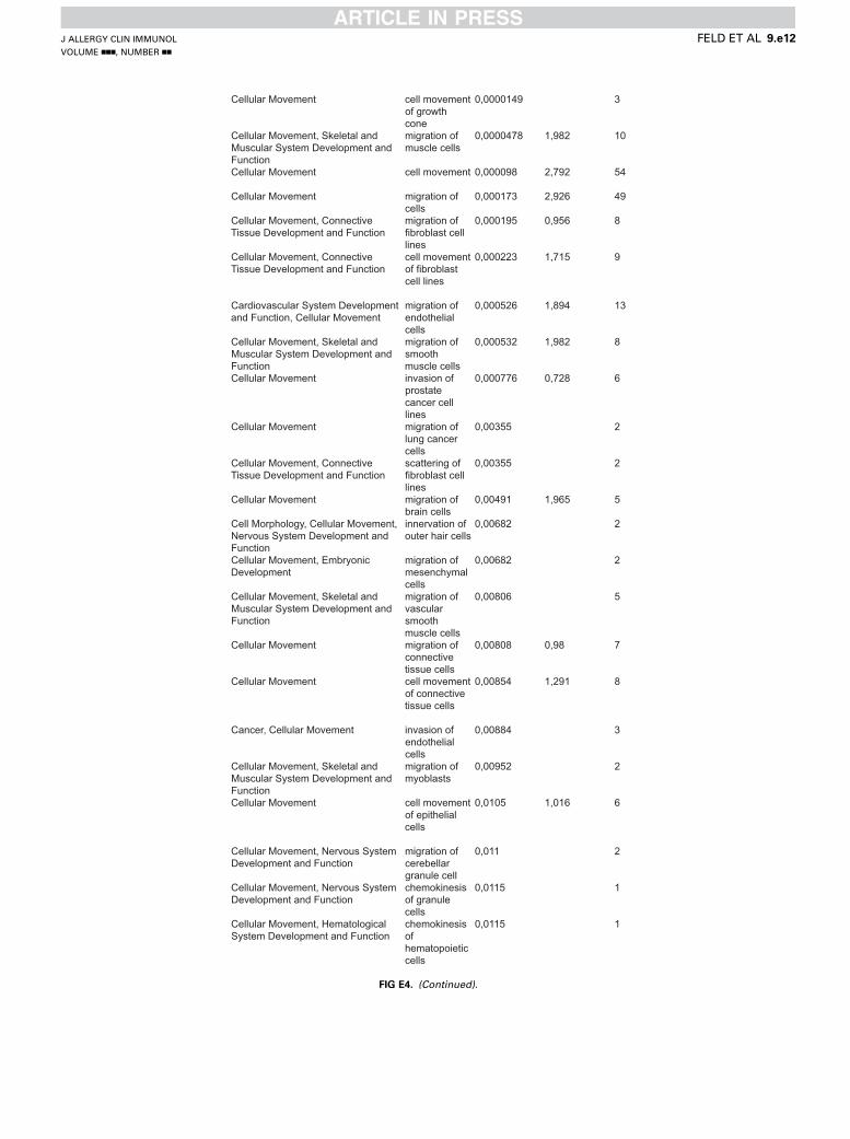
Cellular Movement cell movement of growth cone
0,0000149 3
Cellular Movement, Skeletal and Muscular System Development and Function
migration of muscle cells
0,0000478 1,982 10
Cellular Movement cell movement 0,000098 2,792 54
Cellular Movement migration of cells
0,000173 2,926 49
Cellular Movement, Connective Tissue Development and Function
migration of fibroblast cell lines
0,000195 0,956 8
Cellular Movement, Connective Tissue Development and Function
cell movement of fibroblast cell lines
0,000223 1,715 9
Cardiovascular System Development and Function, Cellular Movement
migration of endothelial cells
0,000526 1,894 13
Cellular Movement, Skeletal and Muscular System Development and Function
migration of smooth muscle cells
0,000532 1,982 8
Cellular Movement invasion of prostate cancer cell lines
0,000776 0,728 6
Cellular Movement migration of lung cancer cells
0,00355 2
Cellular Movement, Connective Tissue Development and Function
scattering of fibroblast cell lines
0,00355 2
Cellular Movement migration of brain cells
0,00491 1,965 5
Cell Morphology, Cellular Movement, Nervous System Development and Function
innervation of outer hair cells
0,00682 2
Cellular Movement, Embryonic Development
migration of mesenchymal cells
0,00682 2
Cellular Movement, Skeletal and Muscular System Development and Function
migration of vascular smooth muscle cells
0,00806 5
Cellular Movement migration of connective tissue cells
0,00808 0,98 7
Cellular Movement cell movement of connective tissue cells
0,00854 1,291 8
Cancer, Cellular Movement invasion of endothelial cells
0,00884 3
Cellular Movement, Skeletal and Muscular System Development and Function
migration of myoblasts
0,00952 2
Cellular Movement cell movement of epithelial cells
0,0105 1,016 6
Cellular Movement, Nervous System Development and Function
migration of cerebellar granule cell
0,011 2
Cellular Movement, Nervous System Development and Function
chemokinesis of granule cells
0,0115 1
Cellular Movement, Hematological System Development and Function
chemokinesis of hematopoietic cells
0,0115 1
FIG E4. (Continued).
J ALLERGY CLIN IMMUNOL
VOLUME nnn, NUMBER nn
FELD ET AL 9.e12

Cellular Assembly and Organization, Cellular Function and Maintenance, Cellular Movement
dispersion of Golgi apparatus
0,0115 1
Cellular Assembly and Organization, Cellular Function and Maintenance, Cellular Movement
dispersion of lysosome
0,0115 1
Cellular Movement NGFCategories Diseases or
Functions Annotation
p-Value Activation z-score
Molecules #
Cardiovascular System Development and Function, Cellular Movement
migration of endothelial cells
0,00000516 1,95 BCAS3,BDNF,CAV1,GAS6,ID1,MYC,NCL,PLAUR,PTGS2,S1PR3,SCG2,SPRY4,THBD,TIMP1,VIM
15
Cellular Movement cell movement 0,0000259 3,147 AKAP12,ALDOA,AP2M1,BCAS3,BCR,BDNF,BHLHE40,CAV1,Ccl2,DAXX,DCLK1,DNAJA1,DUSP6,ELMO1,FSTL1,FTH1,GAS6,HARS,HNRNPK,ID1,ID2,INADL,INHBA,IRAK4,LMNA,MYC,NCL,PLAUR,PRKAR1A,PROCR,PTGIR,PTGS2,RND3,RNH1,RPL13A,S1PR3,SCG2,SERPINB2,SNCG,SPRY4,THBD,TIMP1,TNC,TNFAIP6,TNFAIP8,Tpm1,TUBA1A,VDAC3,VIM
49
Cellular Movement, Reproductive System Development and Function
migration of gonadal cell lines
0,0000729 2,183 ELMO1,NCL,PLAUR,S1PR3,THBD 5
Cellular Movement migration of cells
0,0000748 3,444 ALDOA,AP2M1,BCAS3,BCR,BDNF,BHLHE40,CAV1,Ccl2,DCLK1,ELMO1,FSTL1,FTH1,GAS6,HARS,HNRNPK,ID1,ID2,INADL,INHBA,IRAK4,LMNA,MYC,NCL,PLAUR,PRKAR1A,PROCR,PTGIR,PTGS2,RND3,RNH1,RPL13A,S1PR3,SCG2,SERPINB2,SNCG,SPRY4,THBD,TIMP1,TNC,TNFAIP6,TNFAIP8,Tpm1,TUBA1A,VIM
44
Cardiovascular System Development and Function, Cellular Movement
migration of vascular endothelial cells
0,000294 1,224 GAS6,ID1,NCL,PTGS2,S1PR3,SPRY4,THBD,TIMP1
8
Cellular Movement cell movement of neuroglia
0,000456 -0,927 BCR,BDNF,CAV1,Ccl2,GAS6,TNC 6
Cellular Movement homing 0,000698 0,913 AKAP12,BCR,BDNF,CAV1,Ccl2,DUSP6,GAS6,HARS,INHBA,IRAK4,MYC,PLAUR,PTGS2,RPL13A,S1PR3,SCG2,THBD
17
Cardiovascular System Development and Function, Cellular Movement
chemotaxis of vascular endothelial cells
0,00105 GAS6,PTGS2,THBD 3
Cellular Movement homing of cells
0,00134 1,05 AKAP12,BCR,BDNF,CAV1,Ccl2,DUSP6,GAS6,HARS,INHBA,MYC,PLAUR,PTGS2,RPL13A,S1PR3,SCG2,THBD
16
Cellular Movement cell movement of RPE cells
0,00137 MYC,PLAUR 2
Cellular Growth and Proliferation, Cellular Movement, Hematological System Development and Function
cytostasis of B-lymphocyte derived cell lines
0,00191 BCR,DAXX 2
Cellular Movement cell movement of tumor cell lines
0,0025 2,085 AKAP12,AP2M1,BCR,CAV1,Ccl2,ELMO1,GAS6,HNRNPK,ID1,MYC,NCL,PLAUR,PTGS2,S1PR3,SNCG,THBD,TIMP1,TNC,TNFAIP8,VIM
20
Cellular Movement chemotaxis 0,00262 1,227 BCR,BDNF,CAV1,Ccl2,DUSP6,GAS6,HARS,INHBA,IRAK4,PLAUR,PTGS2,RPL13A,S1PR3,SCG2,THBD
15
Cellular Assembly and Organization, Cellular Function and Maintenance, Cellular Movement, Nervous System Development and Function
endocytosis of synaptic vesicles
0,00301 CANX,PACSIN1,SNCG 3
Cellular Movement invasion of cells
0,00321 2,091 BCR,BDNF,BHLHE40,CAV1,ELMO1,FSTL1,ID1,ID2,INHBA,MYC,PLAUR,PTGS2,RND3,RNH1,S1PR3,SNCG,THBD,TIMP1,VIM,YWHAQ
20
FIG E4. (Continued).
J ALLERGY CLIN IMMUNOL
nnn 2016
9.e13 FELD ET AL

Cellular Movement invasion of fibroblast cell lines
0,00328 CAV1,ELMO1,FSTL1,RND3 4
Cellular Movement migration of tumor cells
0,00388 1,633 BDNF,CAV1,PLAUR,PTGS2,SERPINB2,TIMP1,TNC,VIM
8
Cellular Movement, Hematological System Development and Function, Immune Cell Trafficking
mobilization of antigen presenting cells
0,00401 Ccl2,IRAK4 2
Cell-To-Cell Signaling and Interaction, Cellular Movement, Hematological System Development and Function
recruitment of hematopoietic cells
0,00401 BDNF,Ccl2 2
Cellular Assembly and Organization, Cellular Movement, Nervous System Development and Function
transport of synaptic vesicles
0,00432 CANX,PACSIN1,SNCG,STX7 4
Cellular Movement invasion of breast cancer cell lines
0,00445 1,238 CAV1,ID1,ID2,MYC,PLAUR,PTGS2,SNCG,TIMP1
8
Cellular Movement chemotaxis of cells
0,00475 1,392 BCR,BDNF,CAV1,Ccl2,DUSP6,GAS6,HARS,INHBA,PLAUR,PTGS2,RPL13A,S1PR3,SCG2,THBD
14
Cellular Movement migration of brain cancer cell lines
0,00511 1,973 ELMO1,GAS6,NCL,PLAUR,TNC 5
Cellular Movement, Nervous System Development and Function
migration of neuroglia
0,00583 -1,131 BCR,BDNF,Ccl2,TNC 4
Cellular Movement migration of fibrosarcoma cell lines
0,00591 AP2M1,HNRNPK,PLAUR 3
Cellular Movement, Hematological System Development and Function, Immune Cell Trafficking, Inflammatory Response
cell movement of dendritic cells
0,00675 -0,064 Ccl2,HARS,IRAK4,S1PR3,SCG2,TIMP1 6
Cellular Movement, Nervous System Development and Function
migration of interneurons
0,00681 BDNF,PLAUR 2
Cellular Movement migration of tumor cell lines
0,00713 2,806 AP2M1,CAV1,ELMO1,GAS6,HNRNPK,ID1,MYC,NCL,PLAUR,PTGS2,S1PR3,SNCG,TIMP1,TNC,TNFAIP8,VIM
16
Nervous Development IL-31Categories Diseases or
Functions Annotation
p-Value Activation z-score
Molecules #
Nervous System Development and Function
regeneration of peripheral nerve
0,00000603 ATF3,BDNF,CLU 3
Nervous System Development and Function
abnormal morphology of sensory ganglion
0,000395 BDNF,VTI1B 2
Nervous System Development and Function
abnormal morphology of sensory nervous system
0,000784 BDNF,ETV1 2
Embryonic Development, Nervous System Development and Function, Organ Development, Organismal Development, Tissue Development
formation of molecular layer of cerebellum
0,000784 BDNF,SLC17A6 2
Cellular Assembly and Organization, Nervous System Development and Function
quantity of neurite branches
0,0013 BDNF,RHOA 2
Cell Morphology, Nervous System Development and Function
length of neurites
0,00147 -0,036 BDNF,GDI1,LRPAP1,NDEL1,RHOA 5
Nervous System Development and Function, Neurological Disease
degradation of myelin sheath
0,00193 PSAP,SERPING1 2
Cell Morphology, Cellular Assembly and Organization, Nervous System Development and Function
diameter of axons
0,00193 BDNF,NEFH 2
FIG E4. (Continued).
J ALLERGY CLIN IMMUNOL
VOLUME nnn, NUMBER nn
FELD ET AL 9.e14

Nervous System Development and Function, Tissue Morphology
morphology of nervous tissue
0,00208 BDNF,CKB,ETV1,LRPAP1,MAL,NDEL1,NEFH,PSAP,RHOA,RPS6KB1,RTN4,SCYL1,SLC17A6,SNCA,TUBA1A
15
Cellular Growth and Proliferation, Nervous System Development and Function
inhibition of neurons
0,00227 BDNF,KIF5B,RTN4 3
Cellular Assembly and Organization, Cellular Function and Maintenance, Nervous System Development and Function
quantity of neurites
0,00247 1,117 BDNF,CANX,LRPAP1,NEFH,SNCA 5
Cellular Development, Cellular Growth and Proliferation, Nervous System Development and Function, Tissue Development
outgrowth of neurons
0,00252 0,08 BDNF,CLU,FKBP4,GNAO1,ILK,MAPK3,NDEL1,NEFH,PSAP,RHOA,RTN4,TUBA1A,VTI1B,YWHAZ
14
Cell Death and Survival, Cellular Assembly and Organization, Cellular Development, Nervous System Development and Function, Tissue Development, Tissue Morphology
regeneration of axons
0,00263 0,068 BDNF,NDEL1,RHOA,RTN4 4
Cell Morphology, Hair and Skin Development and Function, Nervous System Development and Function
innervation of skin
0,00268 BDNF,ITGA7 2
Cellular Development, Cellular Growth and Proliferation, Nervous System Development and Function, Tissue Development
proliferation of sensory neurons
0,00268 BDNF,CLU 2
Cell Morphology, Nervous System Development and Function, Tissue Morphology
morphology of neurons
0,00406 BDNF,CKB,LRPAP1,MAL,NDEL1,NEFH,PSAP,RHOA,RPS6KB1,RTN4,SCYL1,SLC17A6,SNCA,TUBA1A
14
Nervous System Development and Function
myelination of nerves
0,00445 BDNF,MAL,RTN4 3
Cellular Development, Nervous System Development and Function, Tissue Development
myelination of neurons
0,0049 BDNF,ILK,RTN4 3
Cell Morphology, Cellular Assembly and Organization, Nervous System Development and Function
elongation of neurites
0,00509 0,594 ATF3,ITGA7,NDEL1,RHOA 4
Cell Morphology, Cellular Assembly and Organization, Cellular Function and Maintenance, Nervous System Development and Function
extension of neurites
0,00531 1,128 ATF3,BDNF,CYTH2,ILK,LRPAP1,NDEL1,RTN4
7
Cellular Assembly and Organization, Cellular Function and Maintenance, Nervous System Development and Function, Tissue Development
organization of neurofilaments
0,00562 NDEL1,NEFH 2
Cellular Assembly and Organization, Nervous System Development and Function
size of neurites
0,00562 BDNF,NEFH 2
Cell Morphology, Cellular Assembly and Organization, Nervous System Development and Function
elongation of axons
0,00589 ITGA7,NDEL1,RHOA 3
Cell Morphology, Cellular Assembly and Organization, Cellular Development, Cellular Growth and Proliferation, Nervous System Development and Function, Tissue Development
outgrowth of neurites
0,00605 -0,117 BDNF,FKBP4,GNAO1,ILK,MAPK3,NDEL1,NEFH,PSAP,RHOA,RTN4,TUBA1A,VTI1B,YWHAZ
13
Nervous System Development and Function
abnormal morphology of inferior ganglion of vagus nerve
0,00682 BDNF,VTI1B 2
Nervous System Development and Function
abnormal morphology of ventral root
0,00682 ETV1,NEFH 2
Cell Morphology, Cellular Movement, Nervous System Development and Function
innervation of outer hair cells
0,00682 BDNF,PSAP 2
Cell Morphology, Nervous System Development and Function
length of axons
0,00812 BDNF,RHOA 2
FIG E4. (Continued).
J ALLERGY CLIN IMMUNOL
nnn 2016
9.e15 FELD ET AL

Embryonic Development, Nervous System Development and Function, Organ Development, Organismal Development, Tissue Development
formation of hippocampus
0,00837 BDNF,CST3,NDEL1,RHOA,SNCA 5
Cell Morphology, Cellular Assembly and Organization, Cellular Development, Cellular Function and Maintenance, Nervous System Development and Function, Tissue Development
axonogenesis 0,00914 0,751 ATF3,BDNF,GDI1,ILK,NDEL1,NEFH,RHOA,RTN4
8
Nervous System Development and Function
abnormal morphology of petrosal ganglion
0,011 BDNF,VTI1B 2
Nervous System Development and Function
abnormal morphology of small trigeminal ganglion
0,011 BDNF,VTI1B 2
Cellular Development, Cellular Growth and Proliferation, Embryonic Development, Nervous System Development and Function, Organ Development, Organismal Development, Tissue Development
development of hippocampal neurons
0,011 BDNF,RHOA 2
Cellular Movement, Nervous System Development and Function
migration of cerebellar granule cell
0,011 BDNF,NDEL1 2
Nervous System Development and Function
myelination of sciatic nerve
0,011 BDNF,MAL 2
Cell-To-Cell Signaling and Interaction, Nervous System Development and Function
synaptic transmission of hippocampal neurons
0,011 BDNF,SNCA 2
Cell Morphology, Cellular Assembly and Organization, Cellular Development, Cellular Function and Maintenance, Nervous System Development and Function, Tissue Development
morphogenesis of neurites
0,0113 0,665 ATF3,BDNF,CLU,DTNBP1,EHD1,ILK,NDEL1,NEFH,PFN1,RHOA,RPS6KB1,RTN4
12
Nervous System Development and Function, Organ Morphology
abnormal morphology of Golgi tendon organ
0,0115 ETV1 1
Cell Morphology, Nervous System Development and Function, Tissue Morphology
abnormal morphology of adrenergic neurons
0,0115 BDNF 1
Nervous System Development and Function
abnormal morphology of pacinian corpuscle
0,0115 ETV1 1
Auditory and Vestibular System Development and Function, Cell Morphology, Nervous System Development and Function, Organ Morphology, Tissue Morphology
abnormal morphology of type I vestibular hair cells
0,0115 BDNF 1
Cell-To-Cell Signaling and Interaction, Nervous System Development and Function
action potential of dopaminergic neurons
0,0115 BDNF 1
Cell-To-Cell Signaling and Interaction, Nervous System Development and Function
activation of striatal neurons
0,0115 BDNF 1
Cell-To-Cell Signaling and Interaction, Nervous System Development and Function, Tissue Development
aggregation of Cajal-Retzius neurons
0,0115 BDNF 1
FIG E4. (Continued).
J ALLERGY CLIN IMMUNOL
VOLUME nnn, NUMBER nn
FELD ET AL 9.e16

Cell-To-Cell Signaling and Interaction, Cellular Assembly and Organization, Nervous System Development and Function
binding of synaptic vesicles
0,0115 SNCA 1
Cellular Movement, Nervous System Development and Function
chemokinesis of granule cells
0,0115 BDNF 1
Behavior, Nervous System Development and Function
declarative memory
0,0115 BDNF 1
Cellular Assembly and Organization, Nervous System Development and Function
density of neurofilaments
0,0115 NEFH 1
Cellular Development, Nervous System Development and Function, Tissue Development
differentiation of noradrenergic neurons
0,0115 BDNF 1
Cellular Development, Nervous System Development and Function, Tissue Development
differentiation of striatonigral neurons
0,0115 BDNF 1
Cell-To-Cell Signaling and Interaction, Cellular Function and Maintenance, Nervous System Development and Function
discharge of granule cells
0,0115 BDNF 1
Cell Morphology, Cellular Assembly and Organization, Cellular Development, Cellular Function and Maintenance, Nervous System Development and Function, Tissue Development
formation of Lewy neurites
0,0115 SNCA 1
Embryonic Development, Nervous System Development and Function, Organ Development, Organismal Development, Tissue Development
formation of internal granular layer of cerebellum
0,0115 BDNF 1
Cellular Growth and Proliferation, Nervous System Development and Function
formation of nervous tissue cell lines
0,0115 BDNF 1
Nervous System Development and Function
function of sensory nerve
0,0115 GLA 1
Cellular Function and Maintenance, Nervous System Development and Function
function of serotonergic neurons
0,0115 BDNF 1
Cellular Function and Maintenance, Nervous System Development and Function
hyperfunction of dopaminergic neurons
0,0115 BDNF 1
Nervous Development NGFCategories Diseases or
Functions Annotation
p-Value Activation z-score
Molecules #
Nervous System Development and Function
morphology of central nervous system
0,0000191 AGA,AKAP12,BCR,BDNF,CAV1,DCLK1,FKBP8,GAL,GM2A,HES1,ID1,ID2,MYC,NPY1R,PLAUR,SLC17A6,TUBA1A,UPP1,VIM
19
Nervous System Development and Function, Tissue Morphology
quantity of striatal neurons
0,0000486 BDNF,FTH1,INHBA 3
Nervous System Development and Function, Organ Morphology, Organismal Development
morphology of brain
0,0000621 AGA,AKAP12,BCR,BDNF,CAV1,DCLK1,FKBP8,GAL,GM2A,HES1,ID1,ID2,NPY1R,PLAUR,SLC17A6,TUBA1A,UPP1
17
Nervous System Development and Function, Organ Morphology, Organismal Development
abnormal morphology of brain
0,000117 AGA,AKAP12,BCR,BDNF,CAV1,DCLK1,FKBP8,GAL,GM2A,HES1,ID1,ID2,NPY1R,PLAUR,SLC17A6,UPP1
16
Cellular Development, Embryonic Development, Nervous System Development and Function, Organismal Development, Tissue Development
differentiation of neural precursor cells
0,000169 BDNF,HES1,ID1,ID2 4
FIG E4. (Continued).
J ALLERGY CLIN IMMUNOL
nnn 2016
9.e17 FELD ET AL
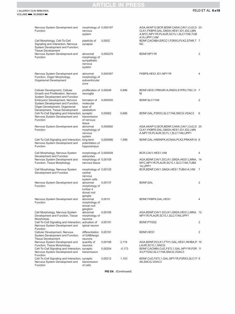
Nervous System Development and Function
morphology of nervous system
0,000187 AGA,AKAP12,BCR,BDNF,CANX,CAV1,Ccl2,DCLK1,FKBP8,GAL,GM2A,HES1,ID1,ID2,LMNA,MYC,NPY1R,PLAUR,SCYL1,SLC17A6,TUBA1A,UPP1,VIM
23
Cell Morphology, Cell-To-Cell Signaling and Interaction, Nervous System Development and Function, Tissue Development
plasticity of synapse
0,0002 BDNF,CACNB4,ERCC1,P2RX3,PLK2,STAR,TNC
7
Nervous System Development and Function
abnormal morphology of sympathetic nervous system
0,000279 BDNF,NPY1R 2
Nervous System Development and Function, Organ Morphology, Organismal Development
abnormal morphology of subventricular zone
0,000397 FKBP8,HES1,ID1,NPY1R 4
Cellular Development, Cellular Growth and Proliferation, Nervous System Development and Function
proliferation of neuroglia
0,00048 0,896 BDNF,HES1,PRKAR1A,RND3,S1PR3,TNC,VIM
7
Embryonic Development, Nervous System Development and Function, Organ Development, Organismal Development, Tissue Development
formation of molecular layer of cerebellum
0,000555 BDNF,SLC17A6 2
Cell-To-Cell Signaling and Interaction, Nervous System Development and Function
synaptic transmission of nervous tissue
0,00062 0,686 BDNF,GAL,P2RX3,SLC17A6,SNCG,VDAC3 6
Nervous System Development and Function
abnormal morphology of nervous system
0,000892 AGA,AKAP12,BCR,BDNF,CANX,CAV1,Ccl2,DCLK1,FKBP8,GAL,GM2A,HES1,ID1,ID2,LMNA,NPY1R,PLAUR,SCYL1,SLC17A6,UPP1
20
Cell-To-Cell Signaling and Interaction, Nervous System Development and Function
long-term potentiation of hippocampus
0,000906 1,599 BDNF,GAL,HNRNPK,KCNA4,PLK2,PRKAR1A 6
Cell Morphology, Nervous System Development and Function
morphology of astrocytes
0,000939 BCR,CAV1,HES1,VIM 4
Nervous System Development and Function, Tissue Morphology
morphology of nervous tissue
0,00108 AGA,BDNF,CAV1,DCLK1,GM2A,HES1,LMNA,MYC,NPY1R,PLAUR,SCYL1,SLC17A6,TUBA1A,UPP1
14
Cell Morphology, Nervous System Development and Function
morphology of central nervous system cells
0,00125 BCR,BDNF,CAV1,GM2A,HES1,TUBA1A,VIM 7
Nervous System Development and Function
abnormal morphology of lumbar 4 dorsal root ganglia
0,00137 BDNF,GAL 2
Nervous System Development and Function
abnormal morphology of dorsal root ganglion
0,0015 BDNF,FKBP8,GAL,HES1 4
Cell Morphology, Nervous System Development and Function, Tissue Morphology
abnormal morphology of neurons
0,00185 AGA,BDNF,CAV1,DCLK1,GM2A,HES1,LMNA,NPY1R,PLAUR,SCYL1,SLC17A6,UPP1
12
Cell-To-Cell Signaling and Interaction, Nervous System Development and Function
activation of spinal neuron
0,00191 BDNF,PTGS2 2
Cellular Development, Nervous System Development and Function, Tissue Development
differentiation of GABAergic neurons
0,00191 BDNF,HES1 2
Nervous System Development and Function, Tissue Morphology
quantity of neurons
0,00198 2,119 AGA,BDNF,DCLK1,FTH1,GAL,HES1,INHBA,PLAUR,SCYL1,SNCG
10
Cell-To-Cell Signaling and Interaction, Nervous System Development and Function
synaptic transmission
0,00204 -0,173 BDNF,CACNB4,Ccl2,FSTL1,GAL,NPY1R,P2RX3,PTGS2,SLC17A6,SNCG,VDAC3
11
Cell-To-Cell Signaling and Interaction, Nervous System Development and Function
synaptic transmission of cells
0,00212 1,103 BDNF,Ccl2,FSTL1,GAL,NPY1R,P2RX3,SLC17A6,SNCG,VDAC3
9
FIG E4. (Continued).
J ALLERGY CLIN IMMUNOL
VOLUME nnn, NUMBER nn
FELD ET AL 9.e18

Cell Morphology, Nervous System Development and Function, Tissue Morphology
morphology of neurons
0,00237 AGA,BDNF,CAV1,DCLK1,GM2A,HES1,LMNA,NPY1R,PLAUR,SCYL1,SLC17A6,TUBA1A,UPP1
13
Nervous System Development and Function, Organ Morphology, Organismal Development
abnormal morphology of forebrain
0,0027 CAV1,DCLK1,FKBP8,GM2A,HES1,ID2,SLC17A6,UPP1
8
Cellular Assembly and Organization, Cellular Function and Maintenance, Cellular Movement, Nervous System Development and Function
endocytosis of synaptic vesicles
0,00301 CANX,PACSIN1,SNCG 3
Nervous System Development and Function, Tissue Morphology
quantity of interneurons
0,00301 BDNF,DCLK1,PLAUR 3
Nervous System Development and Function, Organ Morphology, Organismal Development
abnormal morphology of cerebellum
0,00316 AGA,AKAP12,BCR,FKBP8,GM2A,UPP1 6
Cell Morphology, Nervous System Development and Function
abnormal morphology of astrocytes
0,00396 BCR,CAV1,HES1 3
Cell Morphology, Cellular Assembly and Organization, Cellular Development, Cellular Function and Maintenance, Nervous System Development and Function, Organ Morphology, Tissue Development
branching of hippocampal neurons
0,00401 BDNF,PACSIN1 2
Nervous System Development and Function, Organ Morphology, Organismal Development
abnormal morphology of cerebrum
0,00406 AGA,BDNF,CAV1,FKBP8,GAL,GM2A,PLAUR 7
Cellular Assembly and Organization, Cellular Movement, Nervous System Development and Function
transport of synaptic vesicles
0,00432 CANX,PACSIN1,SNCG,STX7 4
Nervous System Development and Function
sensory system development
0,00493 -0,404 BDNF,GAL,HES1,ID1,S1PR3 5
Cell-To-Cell Signaling and Interaction, Nervous System Development and Function
long-term potentiation of hippocampal cells
0,00508 BDNF,HNRNPK,KCNA4 3
Cellular Development, Embryonic Development, Nervous System Development and Function, Organismal Development, Tissue Development
differentiation of neurosphere cells
0,0058 BDNF,ID2 2
Nervous System Development and Function, Organ Morphology
thickness of cerebral cortex
0,0058 TUBA1A,VIM 2
Cellular Movement, Nervous System Development and Function
migration of neuroglia
0,00583 -1,131 BCR,BDNF,Ccl2,TNC 4
Cellular Assembly and Organization, Cellular Development, Cellular Growth and Proliferation, Nervous System Development and Function, Tissue Development
growth of axons
0,00586 1,498 BDNF,Ccl2,DCLK1,EIF2B2,KIF3C,VIM 6
Cell-To-Cell Signaling and Interaction, Nervous System Development and Function
long-term potentiation
0,00603 1,956 BDNF,GAL,HNRNPK,KCNA4,PLK2,PRKAR1A,TNC,VDAC3
8
Behavior, Nervous System Development and Function
circadian rhythm
0,00634 BHLHE40,ID2,PRKG2,SLC17A6,TNC 5
Cellular Movement, Nervous System Development and Function
migration of interneurons
0,00681 BDNF,PLAUR 2
Nervous System Development and Function
neuroprotection of hippocampus
0,00681 BDNF,PTGS2 2
Cellular Development, Nervous System Development and Function, Tissue Development
differentiation of neuronal progenitor cells
0,00683 BDNF,CAV1,S1PR3 3
Cellular Development, Nervous System Development and Function
development of neuroglia
0,00733 BDNF,EIF2B2,ID2,VIM 4
FIG E4. (Continued).
J ALLERGY CLIN IMMUNOL
nnn 2016
9.e19 FELD ET AL

FIG E4. (Continued).
J ALLERGY CLIN IMMUNOL
VOLUME nnn, NUMBER nn
FELD ET AL 9.e20
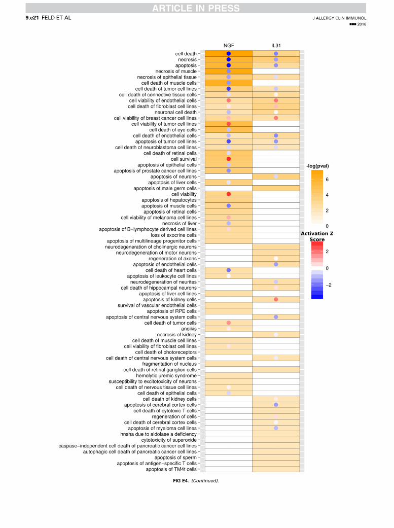
FIG E4. (Continued).
J ALLERGY CLIN IMMUNOL
nnn 2016
9.e21 FELD ET AL

FIG E4. (Continued).
J ALLERGY CLIN IMMUNOL
VOLUME nnn, NUMBER nn
FELD ET AL 9.e22
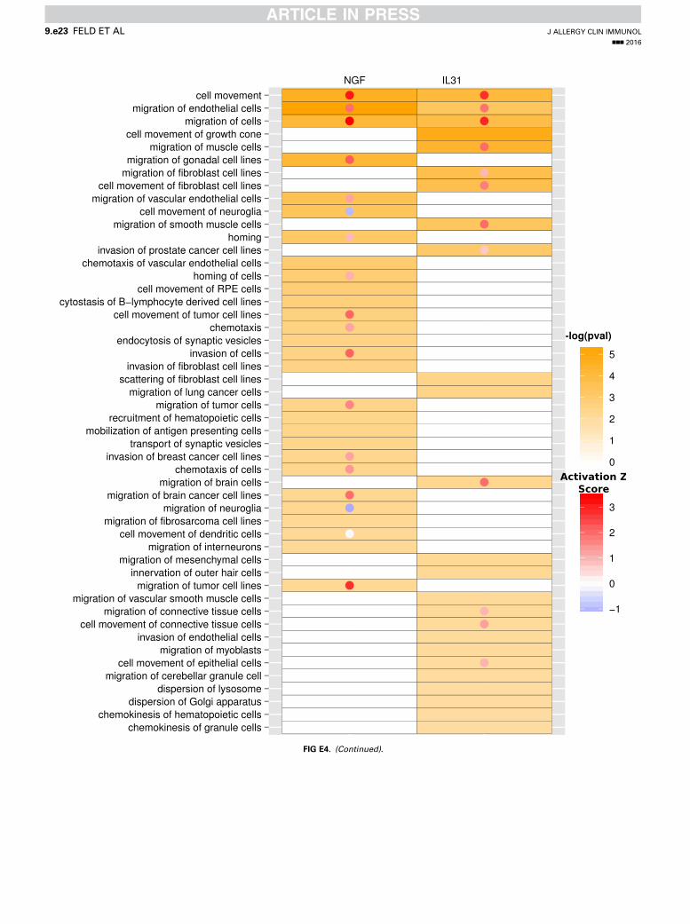
FIG E4. (Continued).
J ALLERGY CLIN IMMUNOL
nnn 2016
9.e23 FELD ET AL

FIG E4. (Continued).
J ALLERGY CLIN IMMUNOL
VOLUME nnn, NUMBER nn
FELD ET AL 9.e24