the pursuit of perfect vision: essential elements for achieving
TRANSCRIPT

Distributed with
The Pursuit of PERFECT VISIONEssential Elements for Achieving Optimal Cataract and Refractive Surgery Outcomes
CME MONOGRAPH
Visit www.mededicus.com for online testing and instant CME certificate.
LEARNING METHOD AND MEDIUMThis educational activity consists of a supplement and ten (10)study questions. The participant should, in order, read thelearning objectives contained at the beginning of thissupplement, read the supplement, answer all questions in thepost test, and complete the Activity Evaluation/Credit Requestform. To receive credit for this activity, please go towww.mededicus.com, Educational Activities tab, click the Post-Test & CME Certificate button and follow the instructionsprovided on the Post-Test and Activity Evaluation/CreditRequest form. This educational activity should take a maximumof 1.5 hours to complete.
CONTENT SOURCEThis continuing medical education (CME) activity capturescontent from a roundtable discussion held on January 22, 2013,in Waikoloa, Hawaii.
ACTIVITY DESCRIPTIONEven with many advances in cataract and refractive surgery,success is not guaranteed for every patient. New technologiesand new strategies for inflammation management can helpimprove results. This educational activity offers experts’insights and practical approaches for managing aspects ofcataract and refractive surgery that can improve theconsistency of good visual outcomes for patients.
TARGET AUDIENCEThis activity is intended for ophthalmologists.
LEARNING OBJECTIVESUpon completion of this activity, participants will be better able to:• Identify vision-limiting ocular comorbidities with preoperative
examinations in patients undergoing cataract surgery• Recognize, evaluate, and treat ocular surface disorders prior
to cataract and refractive surgery • Consistently select the appropriate intraocular lens (IOL) and
IOL power in patients undergoing cataract surgery • Apply anti-inflammatory regimens to prevent inflammation in
patients undergoing cataract and refractive surgery
ACCREDITATION STATEMENTThis activity has been planned and implemented in accordancewith the Essential Areas and Policies of the AccreditationCouncil for Continuing Medical Education through the jointsponsorship of The New York Eye and Ear Infirmary andMedEdicus LLC. The New York Eye and Ear Infirmary isaccredited by the ACCME to provide continuing medicaleducation for physicians.
AMA CREDIT DESIGNATION STATEMENTThe New York Eye and Ear Infirmary designates this enduringmaterial for a maximum of 1.5 AMA PRA Category 1 Credits™.Physicians should claim only the credit commensurate with theextent of their participation in the activity.
GRANTOR STATEMENTThis continuing medical education activity is supportedthrough an unrestricted educational grant from Bausch + LombIncorporated.
DISCLOSURE POLICY STATEMENTIt is the policy of The New York Eye and Ear Infirmary that thefaculty and anyone in a position to control activity contentdisclose any real or apparent conflicts of interest relating to thetopics of this educational activity, and also disclose discussionsof unlabeled/unapproved uses of drugs or devices during theirpresentation(s). The New York Eye and Ear Infirmary hasestablished policies in place that will identify and resolve allconflicts of interest prior to this educational activity. Fulldisclosure of faculty/planners and their commercialrelationships, if any, follows.
Highlights From a Roundtable Discussion
FACULTY
Terrence P. O’Brien, MD (CHAIR/MODERATOR)Professor of OphthalmologyCharlotte Breyer Rodgers Distinguished Chair in OphthalmologyDirector of the Refractive Surgery ServiceBascom Palmer Eye Institute Miami, Florida
Eric D. Donnenfeld, MDOphthalmic Consultants of Long IslandRockville Centre, New YorkClinical Professor of OphthalmologyNew York UniversityNew York, New YorkTrusteeGeisel School of Medicine at DartmouthHanover, New Hampshire
Edward J. Holland, MDDirector, Cornea Services Cincinnati Eye Institute Professor of Ophthalmology The University of CincinnatiCincinnati, Ohio
John D. Sheppard, MD, MMScPresidentVirginia Eye ConsultantsProfessor of OphthalmologyEastern Virginia Medical SchoolNorfolk, Virginia
ORIGINAL RELEASE: June 1, 2013LAST REVIEW: May 15, 2013EXPIRATION: June 30, 2014
Jointly sponsored by The New York Eye and Ear Infirmaryand MedEdicus LLC.
This continuing medical education activity is supported through anunrestricted educational grant from Bausch + Lomb Incorporated.

2
DISCLOSURESEric D. Donnenfeld, MD, had a financial agreement or affiliation during the pastyear with the following commercial interests in the form of Consultant/AdvisoryBoard: Abbott Medical Optics; Alcon, Inc; Allergan, Inc; Bausch + LombIncorporated; SARcode Bioscience, Inc; and TearScience, Inc.
Edward J. Holland, MD, had a financial agreement or affiliation during the pastyear with the following commercial interests in the form of Consultant/AdvisoryBoard: Abbott Medical Optics; Alcon Laboratories, Inc; Bausch + LombIncorporated; SARcode Bioscience, Inc; Senju Pharmaceutical Co, Ltd;TearScience, Inc; and WaveTec Vision Systems, Inc; Fees for promotional,advertising or non-CME services received directly from commercial interest or their Agents (eg, Speakers Bureaus): Alcon, Inc; and Bausch + LombIncorporated; Contracted Research: Abbott Medical Optics; Alcon Laboratories,Inc; and WaveTec Vision Systems, Inc.
Terrence P. O’Brien, MD, had a financial agreement or affiliation during the pastyear with the following commercial interests in the form of Honoraria: AbbottMedical Optics; Alcon, Inc; Allergan, Inc; Bausch + Lomb Incorporated; Bio-Tissue, Inc; and Merck & Co, Inc.
John D. Sheppard, MD, MMSc, had a financial agreement or affiliation during the past year with the following commercial interests in the form of Honoraria:AbbVie Inc; Alcon, Inc; Allergan, Inc; Bausch + Lomb Incorporated, ElevenBiosciences; EyeGate Pharmaceuticals, Inc; Lacrisciences LLC; Lux Biosciences,Inc; Merck & Co, Inc; Santen Pharmaceutical Co, Ltd; SARcode Bioscience, Inc;Vistakon; and 1-800-Doctors, Inc.
PEER REVIEW DISCLOSURETed Gerszberg, MD, has no relevant commercial relationships to disclose.
EDITORIAL SUPPORT DISCLOSURESCheryl Guttman Krader; Cynthia Tornallyay, RD, MBA, CCMEP; Kimberly Corbin,CCMEP; Barbara Aubel; and Barbara Lyon have no relevant commercialrelationships to disclose.
DISCLOSURE ATTESTATIONThe contributing physicians listed above have attested to the following:1) that the relationships/affiliations noted will not bias or otherwise influencetheir involvement in this activity;2) that practice recommendations given relevant to the companies with whomthey have relationships/affiliations will be supported by the best availableevidence or, absent evidence, will be consistent with generally accepted medicalpractice; and 3) that all reasonable clinical alternatives will be discussed when making practicerecommendations.
OFF-LABEL DISCUSSIONThis activity includes off-label discussion of the following agents: topicalazithromycin for meibomian gland dysfunction (MGD), loteprednol etabonate geland ointment for inflammation other than that related to cataract surgery,tetracyclines for MGD, topical cyclosporine for preventing dry eye after LASIK (laser-assisted in situ keratomileusis). Please refer to the official prescribing information fordiscussion of approved indications, contraindications, and warnings.
FOR DIGITAL EDITIONSSystem Requirements:If you are viewing this activity online, please ensure the computer you plan touse meets the following requirements:
• Operating System: Windows or Macintosh• Media Viewing Requirements: Flash Player or Adobe Reader• Supported Browsers: Microsoft Internet Explorer, Firefox, Google Chrome,
Safari, and Opera• A good Internet connection
The New York Eye and Ear Infirmary Privacy & Confidentiality PoliciesCME policies: http://www/nyee.edu/cme-enduring.html Hospital policies: http://www.nyee.edu/website-privacy.html
CME Provider Contact InformationFor questions about this activity, call 212-979-4383.
TO OBTAIN AMA PRA CATEGORY 1 CREDIT™ Online and Instant CertificateTo obtain AMA PRA Category 1 Credit™ for this activity, read the material in itsentirety and consult referenced sources as necessary. We offer instant certificateprocessing and support Green CME. Please take this post test and evaluationonline by going to www.MedEdicus.com, Educational Activities tab, and clickingthe Post-Test & CME Certificate button. Upon passing, you will receive yourcertificate immediately. You must score 70% or higher to receive credit for thisactivity, and may take the test up to 2 times.
DISCLAIMERThe views and opinions expressed in this educational activity are those of the faculty and do not necessarily represent the views of The New York Eye and Ear Infirmary, MedEdicus LLC, Bausch + Lomb Incorporated, orOphthalmology Times.
PURSUING PERFECT VISION INCATARACT SURGERY WITH ADVANCEDTECHNOLOGY IOLsPatients choosing advanced technology intraocular lenses(IOLs) for pseudophakia and those undergoing laserrefractive surgery demand excellent outcomes. Recently, apanel of leading cataract and refractive surgeons convenedto talk about the challenges ophthalmic surgeons face inpursuing the provision of perfect vision for these individuals as well as ways to surmount those challenges, includingstrategies for optimizing our diagnostic evaluation and theuse of newer anti-inflammatory medications to controlperioperative inflammation.
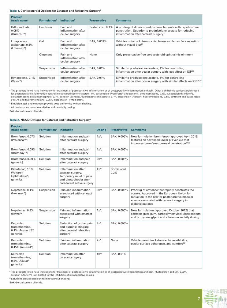
This continuing medical education activity presents thehighlights of our discussion and a summary of currentpharmacologic options for inflammation control (Tables 1 and 2, page 7). We sincerely hope that readers will find theinformation it contains useful for providing optimal care inbehalf of patients in their clinical practice.
—Terrence P. O’Brien, MD
CASE 1A 73-year-old woman has been using monovision contactlenses successfully for many years to correct her presbyopichyperopia. Six months after being diagnosed with bilateralcataracts by her optometrist, she presents reportingincreasing contact lens intolerance along with deteriorationof vision, dulling of color perception, and increasing glare.Both eyes are affected, but the left eye more than the right.She is eager to proceed with cataract surgery and hopes tohave the same IOL that allows a friend of hers freedom fromspectacles or contact lenses.
Consistent with her cataracts, her examination shows bestcorrected visual acuity of 20/30– OD, decreasing to 20/100with glare, and 20/50– OS, decreasing to 20/200 with glare.Schirmer I test results at 5 minutes are 8 mm OD and 7 mmOS, and tear film break-up time is less than 4 seconds.Clinical examination shows inspissation of the meibomianglands with surrounding telangiectasia, staining of theconjunctiva and inferior cornea with Rose Bengal, and afoamy tear film (Figure 1A-C).
CHALLENGE 1—Ocular Surface DiseaseThis is a patient who will have high expectations forsuccessful surgery with a presbyopia-correcting IOL, but the presence of ocular surface disease (OSD) raises someconcerns. How common is OSD in the cataract surgerypopulation, and what is the most common etiology?
This CME activity is copyrighted to MedEdicus LLC ©2013. All rights reserved.
A B
CFIGURE 1. Case 1: Slit-lampimages show signs of meibomiangland dysfunction and ocularsurface disease. Photos Courtesy of Terrence P. O’Brien, MD

3
DR SHEPPARD: It has been reported that approximatelythree-fourths of patients anticipating cataract surgery havesignificant OSD,1 and in my practice, more than half ofpatients aged older than 50 years have significant OSD.Evaporative dry eye associated with meibomian glanddysfunction (MGD) is the most common etiology, followedby a mixed pattern of MGD and aqueous tear deficiency,2which appears to be present in this patient.
DR DONNENFELD: Because OSD is so common amongpatients needing cataract surgery, surgeons must recognizethat managing the ocular surface is as important asperforming quality surgery for achieving quality visual outcomes.
DR O’BRIEN: We all strive for efficiencies in clinical practice,but have to be careful that patients receive a thoroughpreoperative evaluation and appropriate management suchthat they are not brought to surgery too quickly. How do youhandle screening for OSD?
DR SHEPPARD: We assume every patient who presents forelective surgery has OSD until proved otherwise. Thetechnicians who do the initial screening know to look for therelevant signs and symptoms, and patients with positivefindings are seen by the surgeon prior to placement of anydrops that might confound the diagnosis. Tear filmosmolarity and the Ocular Surface Disease Index (OSDI) areexcellent technician-driven screening tools.
DR HOLLAND: Dr Sheppard has brought up an importantpoint about having a system in place so that the technicianswho first see patients identify clues of OSD. Otherwise, whenthe surgeon sees the patient, it might be assumed that thepatient’s vision problems are cataract-related and that anyconjunctival redness and staining was caused by theanesthetic and dilating drops.
MGD is a straightforward diagnosis for cornea specialists,but it may be the most underdiagnosed, undertreated, and underappreciated disease in eye care worldwide.3Expression of the meibomian glands and examination of the secretions should be a routine part of the diagnosticevaluation to identify MGD. I perform the externalexamination using cotton-tip applicators, which provide atool for gland expression, are cleaner than the fingers, andenable lifting of the lid to examine the palpebral conjunctiva.
DR DONNENFELD: All our patients complete a questionnairethat is a modification of the OSDI, and the answers arereviewed by a technician. If there is concern about OSD, thepatient undergoes tear osmolarity testing, which is the mostsensitive diagnostic test for dry eye disease.4
We also obtain topography in all cataract patients prior toinstilling any drops. Loss of data in the visual axis on thetopographic image is a sign that the patient’s ocular surface is not ready for cataract surgery because it will beimpossible to get an accurate keratometry measurement.
I also examine patients prior to instillation of any drops, and based on the findings, might perform tear filminterferometry, which is helpful for diagnosing MGD.
DR O’BRIEN: I think we all agree that there has to be aheightened awareness to screen patients efficiently andthoroughly for OSD while being cognizant of the potential foradverse consequences if the condition is not controlledpreoperatively. What approach can be used to rapidly preparethe ocular surface to withstand the challenge of surgery?
DR DONNENFELD: Anti-inflammatory treatment is needed for rapid resolution of OSD, and a short course of a topicalcorticosteroid can be useful for improving MGD andaqueous-deficient dry eye disease.5,6
Lid margin and dry eye disease, however, are chronicconditions, so patients also require a long-term solution. For MGD, we use hot compresses, lid hygiene, and oralnutritional therapy for maintenance therapy, but we start allthese measures preoperatively.
DR HOLLAND: It is important to look at the severity of theOSD and to consider the patient’s expectations. For me,clinical signs of corneal disease, either marginal keratitisand/or significant corneal staining, represent sufficientseverity to postpone surgery and aggressively manage thepatient using a corticosteroid for acute treatment of lidinflammation and secondary keratoconjunctivitis.
DR O’BRIEN: Detecting the signs of inflammationpreoperatively is an essential step toward launchingappropriate measures to stabilize OSD in advance of surgery.We all agree that corticosteroids provide us with the bestmechanism, potency, and rapid onset of action to achievethis control in a short period of time before surgery. Havethere been recent advances in ophthalmic corticosteroidsthat help to achieve our goals preoperatively?
DR HOLLAND: There are a number of topical corticosteroidsto use. I consider the new gel preparation of loteprednol agood option for corticosteroid treatment in these patients.The gel is designed to have increased ocular surfaceretention and has a pH, demulcents, and a low concentrationof benzalkonium chloride that make it comfortable and ocularsurface-friendly.7 For patients who are very symptomatic, Iadd loteprednol ointment for bedtime use. The ointment ispreservative-free and provides even longer contact time onthe lids and ocular surface than does the gel. There are somefixed-combination corticosteroid-antibiotic ointments, mostof which contain an aminoglycoside. However, theantimicrobial effect of such ointments is usually not neededfor these patients, and the aminoglycoside antibiotics in theproducts can be toxic to the ocular surface.
I think thermal pulsation therapy is much more effective than warm compresses and lid hygiene for deliveringhyperthermia therapy and relieving meibomian glandobstruction. Nutritional supplementation with omega-3 fattyacids also is very important, but it serves as maintenancetherapy because the onset of efficacy takes several weeks.Patients must choose products that are the triglyceride formof omega-3s to assure good absorption.8 I recommend adaily dose of 4 to 6 g.
DR DONNENFELD: Thermal pulsation therapy usedpreoperatively in conjunction with pharmaceuticals canexpedite recovery of a normal tear film, and we use itcommonly in patients with MGD and cataracts. The onlytime I prescribe a corticosteroid-antibiotic combination forpatients with OSD is for the small minority with anteriorblepharitis in which Staphylococcus is often involved.9
DR SHEPPARD: And preferably, patients should be treatedwell in advance of surgery, probably 4 to 6 weeks.
For patients with MGD, I prescribe an oral tetracycline inaddition to the steroid and a nutritional supplement, andsometimes add thermal pulsation therapy or warmcompresses. Tetracyclines have an anti-inflammatory effectand restore the lipid properties of the meibum.10 For thesupplement, I prefer a product that contains gamma-linolenic acid as well as omega-3s. In a randomized,controlled, prospective, multicenter, masked trial, weaugmented and confirmed the work of other investigators,showing that patients with keratoconjunctivitis sicca andconcomitant MGD taking this combination benefit, withsignificant improvements in clinical signs, symptoms, andstabilization of ocular surface inflammatory biomarkers.11,12
I wait at least 3 weeks to reevaluate the keratometry and
4
staining. Once inflammation is controlled, I place a collagenpunctal plug to last at least through the period of the cataract surgery.
DR DONNENFELD: I usually wait to add oral doxycycline or topical azithromycin, but if patients are eager for rapidresolution, I start oral doxycycline 50 mg twice daily andthen reduce the dose to 50 mg once daily after 2 weeks inorder to minimize side effects.
DR HOLLAND: I also like topical azithromycin and mightprescribe doxycycline 50 mg/d, although I use doxycyclineless often now that I have been using dietary supplementsand a good topical corticosteroid.
CHALLENGE 2—Vision-Limiting ComorbiditiesDR O’BRIEN: Comorbid conditions limiting visual potentialrepresent another challenge to the pursuit of perfect visionafter cataract surgery. Certainly, a cataract can obscurevisualization of posterior ocular structures, including theoptic nerve and retina.
Suppose the preoperative optical coherence tomography(OCT) in the patient I discussed revealed an epiretinalmembrane. How would you manage her ocularcomorbidities?
DR SHEPPARD: Optical coherence tomography is a keycomponent of our pre-cataract evaluation, particularly forpatients using multifocal IOLs. In our practice, we consideran epiretinal membrane an absolute contraindication for amultifocal lens. However, accommodating IOLs, toric lenses,or monovision lenses are still viable options because they donot introduce significant aberration to the visual axis.
In today’s environment and in this patient’s best interest, I think referral for further evaluation by a retina specialist ismandatory. The retina specialist decides on the need foradditional testing and treatment.
DR HOLLAND: I agree that OCT should be standardpreoperative screening for patients seeking a multifocal IOLbecause any existing retinal pathology may amplify thedecreased contrast sensitivity inherent with multifocal IOLsand could increase the likelihood of patient dissatisfaction.However, we have had very good outcomes with toric lensesand monovision in patients with retinal pathology.
In our multispecialty practice, patients with an epiretinalmembrane are referred to one of our retina specialists. If retinal surgery is necessary, it is performed by the retina specialist during a combined procedure with thecataract surgery.
DR O’BRIEN: How do you manage a patient with a mild,nonvisually significant epiretinal membrane?
DR DONNENFELD: We make the patient aware that his or her visual rehabilitation may take longer, that there isincreased risk for retinal sequelae after cataract surgery, andthat the visual outcome may not be as optimal as he or shewould like.
In addition, we are much more aggressive with our medicalmanagement in patients who are at increased risk forpostsurgical cystoid macular edema (CME). While they maynot require retinal surgery, they still require expert cataractsurgery done with appropriate anti-inflammatory therapy toprevent inflammation and its adverse sequelae.
In a patient who is at increased risk for CME, I start anonsteroidal anti-inflammatory drug (NSAID) 1 week prior tosurgery and begin a pulsed-dose regimen of a corticosteroid 2hours before surgery. Then I continue both medications withan aggressive postoperative regimen. For the NSAID in theseat-risk patients, I have been prescribing bromfenac, 0.09%,
twice daily rather than at the recommended dose of oncedaily, and I use difluprednate for the corticosteroid because ofits potency. In a prospective, randomized trial, we founddifluprednate was more effective than prednisolone acetatefor controlling inflammation after cataract surgery.13
DR SHEPPARD: Topical steroids can have benefit forpreventing inflammation in the posterior pole,13 and I alsoprescribe an NSAID. I have been prescribing bromfenac once a day in routine cases and bromfenac, 0.09%, twice daily ornepafenac, 0.1%, 3 times a day in patients at high risk for CME(eg, those with diabetes or uveitis). Now we have newformulations of both of these NSAIDs, with the approval ofnepafenac, 0.3%, and bromfenac, 0.07%. Keeping in mind thatthe risk for CME peaks at 4 to 6 weeks postoperatively,14 I agreewith the recommendation to extend the duration of NSAIDtherapy into and beyond that period for high-risk patients.15
DR O’BRIEN: We all consider routine addition of an NSAIDimportant in our postoperative regimens in patients withdiabetes or uveitis because of their high risk for developingpostoperative CME. Singh and colleagues recentlycorroborated this practice in a study of patients withnonproliferative diabetic retinopathy undergoing cataractsurgery. The group randomized to receive a corticosteroid for2 weeks plus nepafenac for 3 months postoperatively hadsignificantly less macular edema on OCT than the controlstreated with the same corticosteroid regimen and vehicle.16
Per product labeling, all NSAIDs are recommended to be usedfor only 2 weeks after cataract surgery. Today, with optimizeddrug formulations compared with those of earlier NSAIDs, wesee fewer complications and we consider these medicationsquite safe. There have been occasional reports, however, ofadverse corneal effects, and extended use of an NSAID insusceptible patients may result in such complications asepithelial breakdown, corneal thinning, corneal erosion,corneal ulceration, or corneal perforation.
Now, consider a patient with a family history of glaucomaand cup-to-disc ratios of 0.4 OD and 0.6 OS. The intraocularpressure (IOP) is 14 mm Hg in both eyes. How would youapproach the preoperative evaluation?
DR SHEPPARD: The inter-eye difference in cup-to-disc ratiomay just be a physiologic difference, but the family historyindicates glaucoma risk, and the patient’s chart should becarefully reviewed. Additionally, OCT may be done to lookfor asymmetry in retinal nerve fiber layer (RNFL) thickness,and the patient should have a visual field, gonioscopy, andpachymetry.
DR DONNENFELD: My surgical decision would be predicatedby the RNFL thickness and visual field. If the RNFL thicknessis close to normal or if the visual field is not significantlyconstricted, I am very comfortable implanting a multifocalIOL. If RNFL thickness is significantly decreased or if there is significant visual field loss, then the loss of contrastsensitivity inherent with a multifocal IOL will be amplified.
Lindstrom and colleagues reported that patients with mildglaucoma may do very well with a multifocal IOL,17 but aninformed consent discussion will guide the decision for each patient.
For patients with glaucoma, I would reinforce the importance ofconsidering the potential for a steroid-induced IOP responsewhen prescribing anti-inflammatory treatment. According toChang and colleagues, an agent with less potential to raise IOP also should be considered in younger patients and in thosewith high myopia because these patients are at increased riskfor a steroid-induced IOP response.18 The corticosteroidssuggested by the researchers that minimize the potential forIOP elevation were loteprednol and fluorometholone. Foralternatives, they suggested using a topical NSAID alone or ashortened course of corticosteroid treatment.

5
CHALLENGE 3—Achieving Refractive AccuracyDR O’BRIEN: With monofocal IOLs, it was consideredreasonable or even desirable to have a low residualametropia, particularly low myopic astigmatism. Can wetolerate significant astigmatism with multifocal IOLs?
DR DONNENFELD: Patients with multifocal IOLs cannot dowell with residual refractive error. The general rule is thathalf a diopter of astigmatism is visually significant, althoughI have seen cases in which even that is too much. Surgeonsimplanting multifocal IOLs have to be willing and able to useall the tools available to correct astigmatism.
DR O’BRIEN: Because of the challenges of IOL powercalculation, achieving refractive accuracy after cataractsurgery is a particular problem in patients who had priorkeratorefractive surgery. Are you finding that this subgroupof patients has an increased interest in presbyopia-correctingIOLs?
DR DONNENFELD: Overwhelmingly, these patients areasking for an accommodating or multifocal IOL. After beingspectacle-free for years, they are strongly opposed toneeding eyeglasses again.
DR SHEPPARD: Patients who had radial keratotomy were thefirst postrefractive surgery patients to come to us for cataractsurgery, and they presented challenges far and above thosein patients who had photorefractive keratectomy (PRK) orLASIK (laser-assisted in situ keratomileusis). Post-radialkeratotomy patients need to know there is a very high riskfor needing an IOL exchange because of high residualrefractive error. If you can manage that expectation andconvincingly dissuade these patients from having amultifocal IOL, you have a chance of giving them gooduncorrected vision with monofocal monovision or managingtheir astigmatism to some extent.
DR HOLLAND: The first challenge in the postrefractivesurgery patient is to determine the appropriate implant. Thesecond challenge is obtaining accurate biometry to use inthe IOL power calculation. There are multiple IOL powerformulas available, but intraoperative aberrometry reallyhelps us select the right IOL power, and performing it addsless than 1 minute to the procedure. I recommend it for allmy postrefractive surgery patients.
CHALLENGE 4—Suppressing Surgically InducedInflammationDR O’BRIEN: Even with major technological advances thathave made surgery less traumatic, and even in the hands ofthe most skilled surgeons, ophthalmic surgery still causesinflammation. What can we do in the operating room tominimize inflammation?
DR HOLLAND: The obvious consideration is to be as good asurgeon as possible in terms of minimizing tissue traumaand complications. Protecting against ocular surfacedessication is important for avoiding an epithelial defect andsubsequent inflammation. With the intent to minimize therisk for epithelial trauma during surgery, we instruct patientsto keep their eyes closed while awaiting the procedure in the preoperative area, and reinforce this directive with anyaccompanying family members and the attending staff.Then, I place a dispersive viscoelastic on the cornea at the start of the case; it both protects the epithelium andimproves my visibility if the patient has some subtleepithelial changes.
DR O’BRIEN: I would add that when applying viscoelastic forocular surface protection, we have to be careful to apply itafter the antiseptic preparation in order not to interfere withthe action of the antiseptic.
DR SHEPPARD: Other measures for minimizing epithelialtrauma and inflammation include avoiding excessive dropsin the eyes, limiting preservative exposure, not placing thespeculum too early, not opening the eye too wide, and nothaving the microscope at full power during the entire case.Limiting the number of incisions is helpful, too. Placing themain incision on the steep axis may eliminate the need for at least 1 limbal relaxing incision. Additionally, use of thefemtosecond laser for lens fragmentation offers anopportunity to limit the amount of surgically inducedinflammation by reducing phacoemulsification time andoperative time.19
Finally, I apply a bandage contact lens in all my premium IOLpatients, patients with basement membrane dystrophies,and in anyone who sustains an epithelial defect duringsurgery: a bandage contact lens accelerates healing andlimits pain.
DR HOLLAND: Use of a femtosecond laser to make arcuateincisions instead of limbal relaxing incisions with a diamondor steel blade is advantageous for reducing ocular surfacetrauma, and it can also limit neuropathic changes becausethe incisions can be made shorter and a little more centrally located.
DR DONNENFELD: When performing femto-cataract surgery,it is important that the cataract surgery begin quickly afterthe femtosecond laser treatment, optimally within 20minutes, in order to minimize prostaglandin release, pupil constriction, declining anesthetic effect, and ocularsurface drying.
We have discovered that, surprisingly, patients who havefemto-cataract surgery overwhelmingly have morepostoperative discomfort than those having conventionalcataract surgery. The simple explanation is that femto-cataract surgery patients almost always have arcuateincisions, and they experience inflammation and irritationfrom those incisions. To suppress inflammation and tocontrol pain in these patients, it is helpful to have a strongcorticosteroid and the ability to extend contact time on theocular surface and to support the cornea. I think loteprednolgel may be useful for these procedures.
DR O’BRIEN: What are your considerations for controllinginflammation postoperatively?
DR SHEPPARD: Controlling inflammation postoperativelybegins with preoperative treatment. One thing to realize isthat, according to labeling, many corticosteroids used incataract surgery carry a recommendation for the agent to bestarted on the day after surgery. However, based on resultsof a preclinical study we conducted,20 I believe it is muchmore advantageous to treat preoperatively, beginning atleast the day before surgery, to optimally downregulate the inflammatory cascade that is activated by the surgical procedure.
DR HOLLAND: We have to be aggressive with medications tosuppress postoperative inflammation. Treatment efficacy,however, depends on the patient using the medication asdirected, including following instructions to shake the bottlewhen using a suspension. A study involving a corticosteroidsuspension found that even when patients were specificallyinstructed to read the label that stated “shake well,”compliance was poor.21 Furthermore, even when a brandedcorticosteroid suspension bottle is shaken, dose uniformityis poor.22 Initially, the dose dispensed tends to be less than100% of the labeled concentration; and the problems withdose uniformity are worse with a generic agent.22 Today,with difluprednate emulsion and loteprednol gel, we havecorticosteroid formulations providing good dose uniformitywithout any need for shaking the container.7,21

6
PURSUING PERFECT VISION AFTER LASIKCASE 2A 49-year-old perimenopausal woman with hyperopia andastigmatism presents with contact lens intolerance. Sheundergoes bilateral femtosecond laser-assisted LASIK withan excellent refractive outcome and unaided visual acuity of20/20. She returns several months later, very dissatisfied,however. As observed with Rose Bengal, she has 4+ stainingof the inferior cornea extending centrally (Figure 2). HerSchirmer 1 test result is 2 mm and tear film break-up time is2 seconds. What are the most common causes of patientdissatisfaction after laser vision correction? What mightexplain this patient’s outcome?
DR DONNENFELD: Dry eye remains the most significantproblem after LASIK, but we are getting much better aboutidentifying and managing it. After reviewing the world’sliterature on LASIK to determine patient satisfaction andquality of life following LASIK, the Joint LASIK Study TaskForce made the important recommendation that all patientswho present for LASIK should undergo a thorough screeningfor dry eye and be treated aggressively preoperatively if it isfound to be present.23
This patient had a “perfect storm” of conditions fordeveloping dry eye-related problems after LASIK. She wasperimenopausal and likely had preexisting dry eye, and shewas undergoing a hyperopic ablation, which causes morenerve damage with its larger flap and ablation area.
Because hyperopic patients seeking laser vision correctiontend to be older, I am extremely cognizant of the concern fordry eye, and so am aggressive with my management both interms of therapy and in terms of patient expectations. Clearlens extraction is often a better surgical option thankeratorefractive surgery for these patients.
DR HOLLAND: This case patient also presented with contactlens intolerance prior to LASIK, which was another clue toserious OSD.
Contact lens intolerance is one of the most common reasonspatients want refractive surgery, and dry eye is the mostcommon cause of contact lens intolerance. While myopicLASIK causes a little less dryness than does hyperopictreatment, this patient still would have the samepostoperative problems because of her preexisting dry eye.
I agree that for a hyperopic patient in the presbyopic agegroup, clear lens exchange is a preferred procedure. However,if the patient still wants laser vision correction, I wouldstrongly advocate delaying surgery and aggressively treatingthe OSD first. This patient probably has mixed aqueous teardeficiency and evaporative dry eye. Topical cyclosporine plus acorticosteroid is an excellent treatment for acute management.Cyclosporine addresses the aqueous tear deficiency, but thecorticosteroid acts more rapidly than cyclosporine to controlinflammation and will improve the tolerability of thecyclosporine.24 Still, it will probably take 6 to 8 weeks beforethe ocular surface is in the right condition for surgery.
DR O’BRIEN: Educating these patients about the reasons forpostponing surgery is important. They are usually eager toproceed, and so it is incumbent upon the staff to support thedecision to delay with proper counseling.
DR HOLLAND: Patients with no corneal staining can scheduletheir surgery without waiting to optimize the ocular surface,but I also start cyclosporine with loteprednol in them rightaway, with the goal of protecting against postoperativeaqueous deficiency. Since there is usually a 3- to 4-weekdelay until they undergo the procedure, the patients achievethe benefit of cyclosporine early postoperatively.
DR O’BRIEN: We hope to see minimal inflammation afterLASIK, but occasionally patients have more than theanticipated level. What are the consequences of uncontrolled inflammation?
DR SHEPPARD: Uncontrolled inflammation can lead toregression, particularly in PRK patients, setting the stage forhaze, irregular astigmatism, glare, and haloes, thus requiringan enhancement procedure.
DR DONNENFELD: Patients with atopic disease are at thehighest risk for inflammation following LASIK and they havea much greater risk for suboptimal refractive results and for diffuse lamellar keratitis. They need to be treated veryaggressively. The preoperative use of a corticosteroid plusan antihistamine can make a big difference in refractiveoutcomes for these patients.
DR O’BRIEN: We published a severity-based preoperativetreatment algorithm for optimizing the ocular surface prior toLASIK in patients with allergy, and it recommends use of adual-acting antihistamine/mast cell stabilizer for anyone withmild or more severe disease.25 Regarding the issue of dryeye and LASIK, however, it is important to choose a productthat has selective activity for the histamine-1 receptor andlow affinity for the receptors that mediate dry eye, a productsuch as bepotastine or epinastine.
NSAIDs were first demonstrated to be effective in reducingpain after excimer laser phototherapeutic keratectomy. DrSheppard, do you use an ophthalmic NSAID in LASIK patients?
DR SHEPPARD: I think the combination of a topical NSAIDand a topical steroid is relevant to LASIK surgery and has aparticular role in PRK patients. It is well known to PRKsurgeons who have used systemic steroids for postoperativepain management—as well as to any physician caring fortrauma patients who require aggressive systemic steroids—that steroids also have potent analgesic activity.
DR DONNENFELD: Because NSAIDs act by inhibitingprostaglandin production, I start treatment in surface ablationcases with 1 drop before surgery and 1 drop on top of thebandage contact lens at the end of surgery, and I instruct thepatient to instill 1 drop daily for 2 more days. I start acorticosteroid immediately following placement of thebandage contact lens and continue it for 4 to 6 weeks aftersurface ablation, a much longer period than that prescribedfor following lamellar surgery. Keeping in mind the risk for asteroid-induced IOP response during this prolonged course of treatment, I prescribe loteprednol gel because of its lowpotential to elevate IOP.26 Reducing exposure to benzalkoniumchloride also is desirable in surface ablation cases.
DR O’BRIEN: In summary, I think we concur that there aremultiple issues to consider as we aim to provide the bestoutcomes for our refractive cataract surgery and laser visioncorrection patients. It is important to be comprehensive withour preoperative screening; to be careful to delay surgery, as needed, while managing preexisting conditions that cancompromise outcomes; to be cognizant of the challenges ofIOL power calculations in patients with a history of refractivesurgery; and to be aggressive in controlling surgicallyinduced inflammation by using an appropriate medicationregimen incorporating optimal agents.
FIGURE 2: Case 2: Slit-lamp photograph afterRose Bengal instillationdiscloses marked diffusepunctate erosions acrossthe LASIK flap surface andfilament formation on theinferior conjunctiva. Photo Courtesy ofTerrence P. O’Brien, MD
7
a The products listed have indications for treatment of postoperative inflammation or of postoperative inflammation and pain. Other ophthalmic corticosteroids usedfor postoperative inflammation control include prednisolone acetate, 1%, suspension (Pred Forte® and generic), dexamethasone, 0.1%, suspension (Maxidex®),dexamethasone sodium phosphate, 0.1%, solution (generic), fluorometholone acetate, 0.1%, suspension (Flarex®), fluorometholone, 0.1%, ointment and suspension(FML®), and fluorometholone, 0.25%, suspension (FML Forte®).
b Emulsion, gel, and ointment provide dose uniformity without shaking.c All products are recommended for 4-times-daily dosing.
BAK=benzalkonium chloride.
Product (trade name) Formulationb Indication Dosing Preservative Comments
Bromfenac, 0.07%(Prolensa™)
Solution Inflammation and painafter cataract surgery
1x/d BAK, 0.005% New formulation bromfenac (approved April 2013)features an advanced lower pH vehicle that improves bromfenac corneal penetration31,32
Bromfenac, 0.09%(Bromday™)
Solution Inflammation and painafter cataract surgery
1x/d BAK, 0.005%
Bromfenac, 0.09%(generic)
Solution Inflammation and painafter cataract surgery
2x/d BAK, 0.005%
Diclofenac, 0.1%(VoltarenOphthalmic®,generics)
Solution Inflammation aftercataract surgery.Temporary relief of painand photophobia aftercorneal refractive surgery
4x/d Sorbic acid,0.2%
Nepafenac, 0.1%(Nevanac®)
Suspension Pain and inflammationassociated with cataractsurgery
3x/d BAK, 0.005% Prodrug of amfenac that rapidly penetrates thecornea. Approved in the European Union forreduction in the risk for postoperative macularedema associated with cataract surgery in diabetic patients
Nepafenac, 0.3%(Ilevro™)
Suspension Pain and inflammationassociated with cataractsurgery
1x/d BAK, 0.005% New formulation (approved October 2012) thatcontains guar gum, carboxymethylcellulose sodium,and propylene glycol and allows once-daily dosing
Ketorolactromethamine,0.4% (Acular LS®,generics)
Solution Reduction of ocular painand burning/ stingingafter corneal refractivesurgery
4x/d BAK, 0.006%
Ketorolactromethamine,0.45% (Acuvail®)
Solution Pain and inflammationafter cataract surgery
2x/d None Vehicle promotes ketorolac bioavailability, ocular surface adherence, and comfort33
Ketorolactromethamine,0.5% (Acular®,generics)
Solution Inflammation aftercataract surgery
4x/d BAK, 0.01%
a The products listed have indications for treatment of postoperative inflammation or of postoperative inflammation and pain. Flurbiprofen sodium, 0.03%, solution (Ocufen®) is indicated for the inhibition of intraoperative miosis.
b Solutions provide dose uniformity without shaking.
BAK=benzalkonium chloride.
Table 2. NSAID Options for Cataract and Refractive Surgerya
Product (trade name) Formulationb Indicationc Preservative Comments
Difluprednate, 0.05%(Durezol™)
Emulsion Pain andinflammation afterocular surgery
Sorbic acid, 0.1% A prodrug of difluoroprednisolone butyrate with rapid cornealpenetration. Superior to prednisolone acetate for reducinginflammation after cataract surgery13
Loteprednol etabonate, 0.5%(Lotemax®)
Gel Pain andinflammation afterocular surgery
BAK, 0.003% Vehicle contains 2 demulcents, favors ocular surface retentionwithout visual blur27
Ointment Pain andinflammation afterocular surgery
None Only preservative-free corticosteroid ophthalmic ointment
Suspension Inflammation afterocular surgery
BAK, 0.01% Similar to prednisolone acetate, 1%, for controllinginflammation after ocular surgery with less effect on IOP28
Rimexolone, 0.1%(Vexol®)
Suspension Inflammation afterocular surgery
BAK, 0.01% Similar to prednisolone acetate, 1%, for controllinginflammation after ocular surgery with similar effects on IOP29,30
Table 1. Corticosteroid Options for Cataract and Refractive Surgerya

1. Trattler WB, Reilly CD, Goldberg DF, et al. Cataract and dry eye:Prospective health assessment of cataract patients ocular surface study.Presented at: American Society of Cataract and Refractive SurgeryAnnual Meeting; March 26-30, 2011; San Diego, CA.
2. Lemp MA, Crews LA, Bron AJ, Foulks GN, Sullivan BD. Distribution ofaqueous-deficient and evaporative dry eye in a clinic-based patientcohort: a retrospective study. Cornea. 2012;31(5):472-478.
3. Geerling G, Tauber J, Baudouin C, et al. The International Workshop onMeibomian Gland Dysfunction: Report of the Subcommittee onManagement and Treatment of Meibomian Gland Dysfunction. InvestOphthalmol Vis Sci. 2011;52(4):2050-2064.
4. Lemp MA, Bron AJ, Baudoin C, et al. Tear osmolarity in the diagnosisand management of dry eye disease. Am J Ophthalmol. 2011;151(5):792-798.e1.
5. Pflugfelder SC, Maskin SL, Anderson B, et al. A randomized, double-masked, placebo-controlled, multicenter comparison of loteprednoletabonate ophthalmic suspension, 0.5% , and placebo for treatment ofkeratoconjunctivitis sicca in patients with delayed tear clearance. Am J Ophthalmol. 2004;138(3):444-457.
6. American Academy of Ophthalmology Cornea/External Disease Panel.Preferred Practice Pattern® Guidelines. Blepharitis – Limited Revision.San Francisco, CA: American Academy of Ophthalmology; 2011.www.aao.org/ppp. Accessed March 15, 2013.
7. Fong R, Leitritz M, Siou-Mermet R, Erb T. Loteprednol etabonate gel0.5% for postoperative pain and inflammation after cataract surgery:results of a multicenter trial. Clin Ophthalmol. 2012;6:1113-1124.
8. Dyerberg J, Madsen P, Møller JM, Aardestrup I, Schmidt EB.Bioavailability of marine n-3 fatty acid formulations. ProstaglandinsLeukot Essent Fatty Acids. 2010;83(3):137-141.
9. Lemp MA, Nichols KK. Blepharitis in the United States 2009: a survey-based perspective on prevalence and treatment. Ocul Surf. 2009;7(2 suppl):S1-S14.
10. Foulks GN, Borchman D, Yappert M, Kakar S. Topical azithromycin andoral doxycycline therapy of meibomian gland dysfunction: acomparative clinical and spectroscopic pilot study. Cornea. 2013;32(1):44-53.
11. Pinna A, Piccinini P, Carta F. Effect of oral linoleic and gamma-linolenicacid on meibomian gland dysfunction. Cornea. 2007;26(3):260-264.
12. Sheppard JD Jr, Pflugfelder SC, Singh R, et al. Long-term treatmentwith nutritional supplements containing gamma linolenic acid andomega 3 fatty acids improves moderate to severe keratoconjunctivitissicca. Cornea. In press.
13. Donnenfeld ED, Holland EJ, Solomon KD, et al. A multicenterrandomized controlled fellow eye trial of pulse-dosed difluprednate0.05% versus prednisolone acetate 1% in cataract surgery. Am JOphthalmol. 2011;152(4):609-617.
14. Pseudophakic Cystoid Macular Edema (Irvine-Gass Syndrome).EyeWiki. http://eyewiki.aao.org/Pseudophakic_Cystoid_Macular_Edema_(Irvine-Gass_Syndrome). Modified April 12, 2012. Accessed March 15, 2013.
15. O’Brien TP. Emerging guidelines for use of NSAID therapy to optimizecataract surgery patient care. Curr Med Res Opin. 2005;21(7):1131-1137.
16. Singh R, Alpern L, Jaffe GJ, et al. Evaluation of nepafenac in preventionof macular edema following cataract surgery in patients with diabeticretinopathy. Clin Ophthalmol. 2012;6:1259-1269.
17. Lindstrom RL. Ten thoughts on cataract surgery in the glaucoma patientand the use of accommodating IOLs. Presented at: 2008 AmericanSociety of Cataract and Refractive Surgery Glaucoma Day; April 4,2008; Chicago, IL.
18. Chang DF, Tan JJ, Tripodis Y. Risk factors for steroid response amongcataract patients. J Cataract Refract Surg. 2011;37(4):675-681.
19. Large series outcomes show increased safety, efficiency, efficacy withlaser-assisted cataract surgery. Ophthalmology Times Conference Brief.November 12, 2012. http://images2.advanstar.com/ophthalmologytimes/eNews/2012-AAO/2012_AAO_Day3.html. Accessed March 15, 2013.
20. Samudre SS, Lattanzio FA Jr, Williams PB, Sheppard JD Jr. Comparisonof topical steroids for acute anterior uveitis. J Ocul Pharmacol Ther.2004;20(6):533-547.
21. Apt L, Henrick A, Silverman LM. Patient compliance with use of topicalophthalmic corticosteroid suspensions. Am J Ophthalmol. 1979;87(2):210–214.
22. Stringer W, Bryant R. Dose uniformity of topical corticosteroidpreparations: difluprednate ophthalmic emulsion 0.05% versus brandedand generic prednisolone acetate ophthalmic suspension 1%. ClinOphthalmol. 2010;4:1119-1124.
23. Solomon KD, Fernández de Castro LE, Sandoval HP, et al; Joint LASIKStudy Task Force. LASIK world literature review: quality of life andpatient satisfaction. Ophthalmology. 2009;116(4):691-701.
24. Donnenfeld E, Sheppard JD, Holland EJ. Prospective, multicenter,randomized controlled study on the effect of loteprednol etabonate oninitiating therapy with cyclosporine A. Presented at: AmericanAcademy of Ophthalmology Annual Meeting; November 10-13, 2007;New Orleans, LA.
25. Bielory BP, O’Brien TP. Allergic complications with laser-assisted in-situkeratomileusis. Curr Opin Allergy Clin Immunol. 2011;11(5):483-491.
26. Comstock TL, Decory HH. Advances in corticosteroid therapy for ocularinflammation: loteprednol etabonate. Int J Inflam. 2012;2012:789623.[Epub 2012 Mar 28]
27. Coffey MJ, DeCory HH, Lane SS. Development of a non-settling gelformulation of 0.5% loteprednol etabonate for anti-inflammatory use asan ophthalmic drop. Clin Ophthalmol. 2013;7:299-312.
28. Lane SS, Holland EJ. Loteprednol etabonate 0.5% versus prednisoloneacetate 1.0% for the treatment of inflammation after cataract surgery. J Cataract Refract Surg. 2013;39(2):168-173.
29. Kavuncu S, Horoz H, Ardagil A, Erbil HH. Rimexolone 1% versusprednisolone acetate in preventing early postoperative inflammationafter cataract surgery. Int Ophthalmol. 2008;28(4):281-285.
30. Yaylali V, Ozbay D, Tatlipinar S, Yildirim C, Ozden S. Efficacy and safetyof rimexolone 1% versus prednisolone acetate 1% in the control ofpostoperative inflammation following phacoemulsification cataractsurgery. Int Ophthalmol. 2004;25(1):65-68.
31. Bausch + Lomb Incorporated. Bausch + Lomb receives FDA approvalfor PROLENSA™ (bromfenac ophthalmic solution) 0.07%. April 8, 2013.http://www.bausch.com/en/Our-Company/Recent-News/2013-Archive/FDA-Approval-for-PROLENSA. Accessed April 23, 2013.
32. Prolensa [package insert]. Tampa, FL: Bausch + Lomb Incorporated; 2013.
33. Allergan, Inc. Allergan receives FDA approval for ACUVAIL(TM)ophthalmic solution for the treatment of pain and inflammation followingcataract surgery. July 23, 2009. http://agn.client.shareholder.com/releasedetail.cfm?ReleaseID=398556. Accessed April 24, 2013.
REFERENCES
TO OBTAIN AMA PRA CATEGORY 1 CREDIT™ Online and Instant CertificateTo obtain AMA PRA Category 1 Credit™ for this activity, read the material in its entirety and consult referenced sourcesas necessary. We offer instant certificate processing and support Green CME. Please take this post test and evaluationonline by going to www.MedEdicus.com, Educational Activities tab, and clicking the Post-Test & CME Certificate button.Upon passing, you will receive your certificate immediately. You must score 70% or higher to receive credit for thisactivity, and may take the test up to 2 times.