the raccoon polyomavirus genome and tumor antigen
TRANSCRIPT

The Raccoon Polyomavirus Genome and Tumor AntigenTranscription Are Stable and Abundant in Neuroglial Tumors
Terza Brostoff, Florante N. Dela Cruz, Jr., Molly E. Church, Kevin D. Woolard, Patricia A. Pesavento
Department of Pathology, Microbiology, and Immunology, School of Veterinary Medicine, University of California, Davis, California, USA
ABSTRACT
Raccoon polyomavirus (RacPyV) is associated with 100% of neuroglial tumors in free-ranging raccoons. Other tumor-associatedpolyomaviruses (PyVs), including simian virus 40 (SV40), murine PyV, and Merkel cell PyV, are found integrated in the hostgenome in neoplastic cells, where they constitutively express splice variants of the tumor antigen (TAg) gene. We have previouslyreported that RacPyV exists only as an episome (nonintegrated) in neuroglial tumors. Here, we have investigated TAg transcrip-tion in primary tumor tissue by transcriptome analysis, and we identified the alternatively spliced TAg transcripts for RacPyV.We also determined that TAg was highly transcribed relative to host cellular genes. We further colocalized TAg DNA and mRNAby in situ hybridization and found that the majority of tumor cells showed positive staining. Lastly, we examined the stability ofthe viral genome and TAg transcription by quantitative reverse transcriptase PCR in cultured tumor cells in vitro and in a mousexenograft model. When tumor cells were cultured in vitro, TAg transcription increased nearly 2 log-fold over that of parentaltumor tissue by passage 17. Both episomal viral genome and TAg transcription were faithfully maintained in culture and in tu-mors arising from xenotransplantation of cultured cells in mice. This study represents a minimal criterion for RacPyV’s associa-tion with neuroglial tumors and a novel mechanism of stability for a polyomavirus in cancer.
IMPORTANCE
The natural cycle of polyomaviruses in mammals is to persist in the host without causing disease, but they can cause cancer inhumans or in other animals. Because this is an unpredictable and rare event, the oncogenic potential of polyomavirus is primar-ily evaluated in laboratory animal models. Recently, raccoon polyomavirus (RacPyV) was identified in neuroglial tumors of free-ranging raccoons. Viral copy number was consistently high in these tumors but was low or undetectable in nontumor tissue orin unaffected raccoons. Unlike other oncogenic polyomaviruses, RacPyV was episomal, not integrated, in these tumors. To de-termine the stability of the viral genome and sustained transcription of the oncogenic tumor antigen genes, we cultured primaryraccoon tumor cells and passaged them in mice, confirming the nonintegrated state of the virus and the maintenance of viralgene transcription throughout. RacPyV provides a naturally occurring and tractable model for a novel mechanism of polyomavi-rus-mediated oncogenesis.
Polyomaviruses (PyVs) infect a wide range of species, includinghumans. In well-studied PyVs, such as simian virus 40 (SV40),
the oncogenic large tumor antigen (LT) protein is necessary andsufficient to transform cells in culture and to create tumors inlaboratory animals (1–3). Despite this alarming potential, the typ-ical result of most PyV infections is a détente of long-term, asymp-tomatic persistence in their host species (4, 5). In 2008, Merkel cellpolyomavirus (MCPyV) was discovered as a clonally integratedvirus in human Merkel cell carcinoma (MCC), a rare but aggres-sive skin cancer (6). This is now considered the first example ofcancer attributed to naturally occurring PyV infection in humans(7, 8). In 2010, raccoon polyomavirus (RacPyV) was identified inneuroglial olfactory tract tumors of free-ranging raccoons (9). Todate, 17 raccoon tumors, all of which harbor abundant viral ge-nome, have been collected from the western United States.RacPyV DNA is comparatively low or undetectable in nontumorbrain tissue of affected animals and in unaffected (non-tumor-bearing) animals.
Like other PyVs, RacPyV has a circular double-stranded DNAgenome of 5 kb with a noncoding regulatory region (NCRR) di-viding the genome into the alternatively spliced early tumor anti-gen (TAg; nonstructural) region and the late capsid (VP; struc-tural) region (9). RacPyV is closely related to MCPyV at both thegenome and protein levels, and they both fall within a monophy-
letic clade of viruses that some have called almipolyomaviruses,which encode either an overprinting “ALTO” protein or a similarlyoverprinted middle tumor antigen (MT) protein (10). Almipolyo-maviruses include PyVs that are causative for naturally occurring tu-mors. The RacPyV TAg gene encodes the same predicted immuno-modulatory and cellular growth control domains as do other PyVs.However, proof that these gene products contribute to or cause neu-roglial tumors in raccoons has so far been limited to epidemiologicdata and comparative sequence analyses.
The RacPyV TAg gene encodes predicted large tumor antigen(LT) and small tumor antigen (sT) proteins. Several known onco-genic domains of PyV LT (a DnaJ domain and a retinoblastoma[pRB]-binding motif) and sT (protein phosphatase 2A [PP2A]binding motifs) are present as shown by sequence analysis (9).SV40 LT and sT, along with constitutive overexpression of human
Received 9 July 2014 Accepted 19 August 2014
Published ahead of print 27 August 2014
Editor: M. J. Imperiale
Address correspondence to Patricia A. Pesavento, [email protected].
Copyright © 2014, American Society for Microbiology. All Rights Reserved.
doi:10.1128/JVI.01912-14
12816 jvi.asm.org Journal of Virology p. 12816 –12824 November 2014 Volume 88 Number 21
on March 31, 2018 by guest
http://jvi.asm.org/
Dow
nloaded from

telomerase and the H-ras oncogene, are sufficient for completeprimary cell transformation in vitro (11). However, all knownPyVs, including those that have never been associated with natu-rally occurring tumors, encode these predicted oncogenic do-mains. Therefore, a robust collection of scientific evidence isneeded to support the classification of RacPyV as a causative agentin raccoon neuroglial tumors. This is especially critical becauseRacPyV exists as an episome in raccoon brain tumors, whereasMCPyV is clonally integrated in MCC (9, 12). As a minimal crite-rion for causality, regardless of its episomal state, we predict thatthe RacPyV genome and TAg transcription are consistently pres-ent in the tumor cell population. Additionally, TAg transcriptionand its function in tumors should be independent of productive/lytic infection. In the case of MCPyV, this is a result of truncationof LT, either by sequence mutation or by site of integration (7, 13,14). This occurs at the expense of the structural proteins, whichare not required for tumor formation or maintenance and arelargely repressed in MCC (15).
In this paper, we demonstrate that episomal RacPyV genomeand TAg transcription are abundant in naturally occurring tu-mors and in a case with metastasis. Furthermore, cultured pri-mary tumor cells inoculated intracranially into immunodeficientNod-scid gamma mice form and recapitulate naturally occurringtumors seen in wild raccoons. In the xenotransplant and in furthercell lines cultured from this tissue, episomal RacPyV genome isstable and unaltered and TAg transcript is abundant.
MATERIALS AND METHODSTranscriptome sequencing. Next-generation sequencing (TruSeq) wasutilized for whole-transcriptome analysis of two raccoon brain tumors(Rac 10 and Rac 12) and anatomical location-matched olfactory bulbtissues from two unaffected raccoons. Samples were submitted to the UCDavis Comprehensive Cancer Center’s Genomics Shared Resource forRNA isolation, RNA sequencing (RNA-Seq) library preparation and val-idation, and next-generation sequencing. Rac 10 presented here is thesame Rac 10 as that analyzed in the work of Dela Cruz et al., 2013 (9).Detailed procedures are provided below.
RNA isolation. Total cellular RNA was isolated from raccoon braintumors and normal olfactory bulb tissues (50 mg per sample) using theTRIzol reagent (Life Technologies, Carlsbad, CA) and a modified proto-col that incorporates an additional extraction with phenol-chloroform–isoamyl alcohol (25:24:1, pH 4.3). RNA quantity and quality were assessedon a NanoDrop spectrophotometer (Thermo Scientific, Waltham, MA)and an Agilent 2100 Bioanalyzer (Agilent Technologies, Westlake Village,CA), respectively.
Library preparation for RNA-Seq. RNA-Seq libraries were preparedfrom 1 �g total RNA using the TruSeq RNA sample preparation kit (Illu-mina, San Diego, CA) according to the manufacturer’s protocol. Briefly,polyadenylated mRNA was purified from total RNA and rRNA removedby two rounds of binding to magnetic poly(T) beads. This was followed byRNA fragmentation by incubation in the presence of divalent cations at94°C for 5 min. Double-stranded cDNA was generated by random-primed first-strand synthesis with SuperScript II reverse transcriptase(Life Technologies) and subsequent second-strand synthesis with RNaseH and DNA polymerase I. The cDNA was then blunt ended with T4 andKlenow DNA polymerases to remove 3= overhangs and fill in 5= over-hangs, phosphorylated with T4 polynucleotide kinase, and then 3=-Atailed by incubation with Klenow fragment (3=¡5= exo–) and dATP. Il-lumina paired-end adapters were ligated, followed by purification withAMPure XP beads. The library was enriched by high-fidelity PCR ampli-fication (15 cycles) with Phusion DNA polymerase (Thermo Scientific)and adapter-specific primers. The molar concentration of the libraries wasdetermined by measuring concentration with a Qubit fluorometer (Life
Technologies), determining the insert length with an Agilent 2100 Bio-analyzer, and then using quantitative PCR (qPCR)-based quantification(KAPA library quantification kit).
Next-generation sequencing and data analysis. Indexed librarieswere pooled and loaded on the cBot for cluster generation on TruSeqpaired-end flow cells, and paired-end sequencing (2 � 100 bp, paired-end; 4 libraries/lane) was performed with an Illumina HiSeq 2000 se-quencing system (BGI@UC Davis Joint Genome Center) using standardIllumina kit reagents (TruSeq SBS kit v3-HS). Image processing, basecalling, quality scoring (Phred), FASTQ file generation, and sample de-multiplexing were executed by HiSeq control software with real-timeanalysis (HCS 1.5/RTA 1.13) and CASAVA 1.8 software (Illumina).
Transcriptome analysis. (i) De novo transcriptome assembly andanalysis. Raw sequencing reads in FASTQ format were subjected to qual-ity trimming and adapter removal with Trimmomatic 0.32 using thefollowing options: ILLUMINACLIP:adapters.fa:2:30:10 TRAILING:3HEADCROP:15 SLIDINGWINDOW:4:15 MINLEN:70. Quality readswere pooled and used for de novo transcriptome assembly using the Trin-ity platform (r20131110). Assembled transcripts were quantified usingRSEM 1.2.7. To reduce the number of artifactually assembled transcripts,transcripts with poor read support (�1 fragment per kilobase of tran-script per million fragments mapped [FPKM]) were filtered from theinitial assembly. To segregate artifactually assembled false-fusion RacPyVtranscripts spanning the NCRR, RacPyV transcripts were separated intoearly (TAg) and late (VP) regions. The TAg gene was delimited by the startand stop codons of the LT open reading frame (ORF). The structural genewas delimited by the start codon of the VP2 ORF and the stop codon of theVP1 ORF. All retained transcripts were requantified using RSEM and usedas the final transcriptome assembly for subsequent analyses. Sequenceswere annotated via BLASTX (E value, �1e�9) against the UniProtKB/Swiss-Prot database (January 2014 build).
(ii) Read alignment to RacPyV genome. TopHat v2.0.10 and Bowtie 2v2.1.0 were used to align RNA-Seq reads to the RacPyV genomes andverify splicing events identified in the transcriptome assembly. Qualityreads from the two tumor samples were aligned to their respective RacPyVgenomes. Quality reads from the two normal olfactory bulb tissues werealigned to RacPyV strain R45 (GenBank accession no. JQ178241.1) (9).
Histology and in situ hybridization. Tissues from nine raccoons (Rac7, Rac 9, Rac 10, Rac 11, Rac 12, Rac 13, Rac 14, Rac 15, and Rac 16) werefixed in 10% buffered formalin, pH 7.2, for at least 48 h and then paraffinembedded. Mouse brains containing xenograft tumors (mouse A1 andmouse A3) were perfused with 4% paraformaldehyde (PFA), followed byovernight fixation and then paraffin embedding. For each tissue, 4-�msections were processed by standard histologic techniques. Colorimetricin situ hybridization (ISH) was performed on Superfrost Plus slides(Fisher Scientific, Pittsburgh, PA) by hand using the RNAscope kit (Ad-vanced Cell Diagnostics, Inc., Hayward, CA) according to the manufac-turer’s instructions. Briefly, tissue sections were pretreated with heat andprotease prior to hybridization with a set of probes complementary tonucleotides (nt) 966 to 2084 of RacPyV R45. A horseradish peroxidase-based signal amplification system was then hybridized to the target probesfollowed by color development with diaminobenzidine. Slides were coun-terstained with hematoxylin for 20 s and, following counterstaining, weredipped in xylene and covered with xylene-based SHUR/Mount coverslip-ping mounting medium (Triangle Biomedical Sciences, Durham, NC),and a coverslip was applied. Positive staining was identified as brown,punctate dots present in the nucleus and/or cytoplasm. Control probes forthe bacterial gene DapB (negative control) and for the housekeeping geneubiquitin C (positive control— evidence of adequate RNA) were in-cluded. The raccoon and mouse tissue (and control) slides were read bytwo veterinary pathologists (P.A.P. and M.E.C.) and classified as eitherpositive or negative. There was no observed hybridization in replicatetissue sections incubated with scrambled (unrelated) probes with similarguanine-cytosine content (n � 3), in normal brain tissue within the samesection (n � 6), or in brain tissues from RacPyV-negative raccoons (n �
RacPyV TAg Transcription in Neuroglial Tumors
November 2014 Volume 88 Number 21 jvi.asm.org 12817
on March 31, 2018 by guest
http://jvi.asm.org/
Dow
nloaded from
2). Positive cases had granular cell staining that was above the signal ob-served on the negative-control slides.
Raccoon cells and primary tumor tissue. Raccoon tumor tissue wascollected within 3 h of euthanasia (Rac 14), mechanically homogenized,and cultured as previously described (16). Briefly, tissue was enzymati-cally dissociated into individual cells and plated into NBE medium con-sisting of neurobasal medium (Life Technologies), N2 and B27 supple-ments (0.5� each; Life Technologies), and recombinant human basicfibroblast growth factor (bFGF) and epidermal growth factor (EGF) (25ng/ml each; R&D Systems, Minneapolis, MN).
Southern blot hybridization and RCA. Southern blot hybridizationand rolling circle amplification (RCA) were performed as previously de-scribed (9) for Rac 10. Briefly, DNA was extracted using a DNeasy kitaccording to the manufacturer’s instructions (Qiagen, Valencia, CA). ForSouthern blot hybridization, 1 �g of DNA, undigested or digested withKpnI at 37°C for 16 h, was electrophoresed on a 0.8% agarose gel. Sepa-rated DNA was transferred to positively charged nylon membranes(Roche, Indianapolis, IN) overnight by capillary action in 20� SSC (1�SSC is 0.15 M NaCl plus 0.015 M sodium citrate). DNA was UV cross-linked and subsequently hybridized with the RacPyV LT probe previouslydescribed (9) at 48°C for 16 h. Membranes were washed with washingbuffer (0.1 mol/liter maleic acid, 0.15 mol/liter NaCl; pH 7.5; 0.3% [vol/vol] Tween 20); developed with CDP-Star, ready-to-use (Roche), for upto 15 min; and visualized using a FluorChem E imaging system (Protein-Simple, Santa Clara, CA). RCA was performed on extracted DNA usingTempliPhi (GE Healthcare Life Sciences, Piscataway, NJ) according to themanufacturer’s instructions for plasmid DNA (1 ng extracted DNA, reac-tion mixture incubated at 30°C for 14 h).
Mice, xenotransplants, and xenotransplant culture. An intracranialorthotopic xenograft model was used to assess tumorigenicity of culturedcells. Six adult female NSG (NOD.Cg-Prkdcscid Il2rgtm1Wjl/SzJ) mice wereobtained from the UC Davis (UCD) Institute for Regenerative Cures andhoused at UCD in accordance with AAALAC regulations. All procedureswere reviewed and approved by the UCD Institute for Animal Care andUse Committee. Cells from the raccoon tumor-derived primary cell line(Rac 14) at passage 2 were resuspended in phosphate-buffered saline(PBS), and 5 �l containing 150,000 cells was injected per mouse stereot-actically into the striatum (coordinates: 2 mm anterior, 2 mm lateral tobregma, 2.5 mm deep from dura). There was no injection-associated mor-bidity or mortality. Mice were monitored for weight loss and euthanizedat 20% decrease in body weight in accordance with the UCD humaneendpoints. Two mice (mouse A2 and mouse A5) were immediately flash-frozen upon euthanasia, and tumors were used for virus DNA sequenceverification and homogenized for culture, as described above. Two addi-tional mice (mouse A1 and mouse A3) were perfused, and tissues wereimmersed in 4% PFA, followed by paraffin embedding, as described abovefor histology.
PCR and qRT-PCR. DNA from tumors Rac 10 and Rac 12 and fromxenotransplant mouse tumors mouse A2 and mouse A5 was extracted
using the DNeasy kit as described above (Qiagen). All DNA was amplifiedby PCR using RacPyV-specific primers, and amplicons were sequenced onan ABI 3730 sequencer (Applied Biosystems, South San Francisco, CA).RCA was performed on both mouse tumors as described above. TotalRNA was extracted from Rac 10 and Rac 12; cultured tumor cells from Rac14 at passage 5 (p5), p10, and p17; and cultured xenotransplant cells frommouse A2 and mouse A5 using the RNeasy kit according to the manufac-turer’s instructions (Qiagen). RNA was extracted from RacPyV-negativecultured raccoon cortical astrocytes as a negative control. Quantitativereverse transcriptase PCR (qRT-PCR) was performed on an ABI 7300real-time PCR system (Applied Biosystems) according to the manufactur-er’s instructions using a probe spanning the exon1/exon2 junction of LT.All samples were assayed in triplicate. The relative quantity of target tran-scripts was normalized to the number of mammalian 18S rRNA tran-scripts found in the same samples. The relative quantitation of target geneexpression was performed by calculating the comparative cycle threshold(CT). To verify the mouse brain tumors as true xenotransplants and notprimary virus-induced mouse tumors, DNA from two mouse tumors andfrom the same animals’ unaffected brains were extracted as describedabove and analyzed by PCR using mouse mitochondrial DNA-specificprimers.
RESULTS
All raccoons and mice presented in this study are summarized inTable 1.
RacPyV T antigen is highly transcribed in raccoon neuroglialtumors. To investigate RacPyV gene transcription, the tumor tis-sues of two affected RacPyV-positive raccoons (Rac 10 and Rac12) were selected for RNA-Seq analysis. Anatomically matched,RacPyV-negative olfactory bulb tissues of two raccoons were in-cluded as negative controls. Transcripts were quantified usingRNA-Seq by expectation-maximization (RSEM) and annotatedusing BLASTx homology to the UniProtKB database, and 13,473protein-coding genes were identified. RacPyV transcripts were de-tected in both tumor samples, and none were detected in the neg-ative controls.
The two tumor transcriptomes share many common features.With respect to the total polyadenylated RNA population, RacPyVTAg is highly expressed (Fig. 1). In Rac 10 and Rac 12, TAg tran-scription is estimated to be 61.25 and 532.50 transcripts per mil-lion (TPM), respectively. RacPyV structural (VP) gene transcrip-tion is reduced relative to that of TAg, estimated at 7.42 and 10.56TPM in Rac 10 and Rac 12, respectively. Of the 13,473 total genesidentified, TAg transcription ranked as the 155th and 2,166thmost abundantly transcribed genes and VP ranked 8,784th and9,339th, respectively.
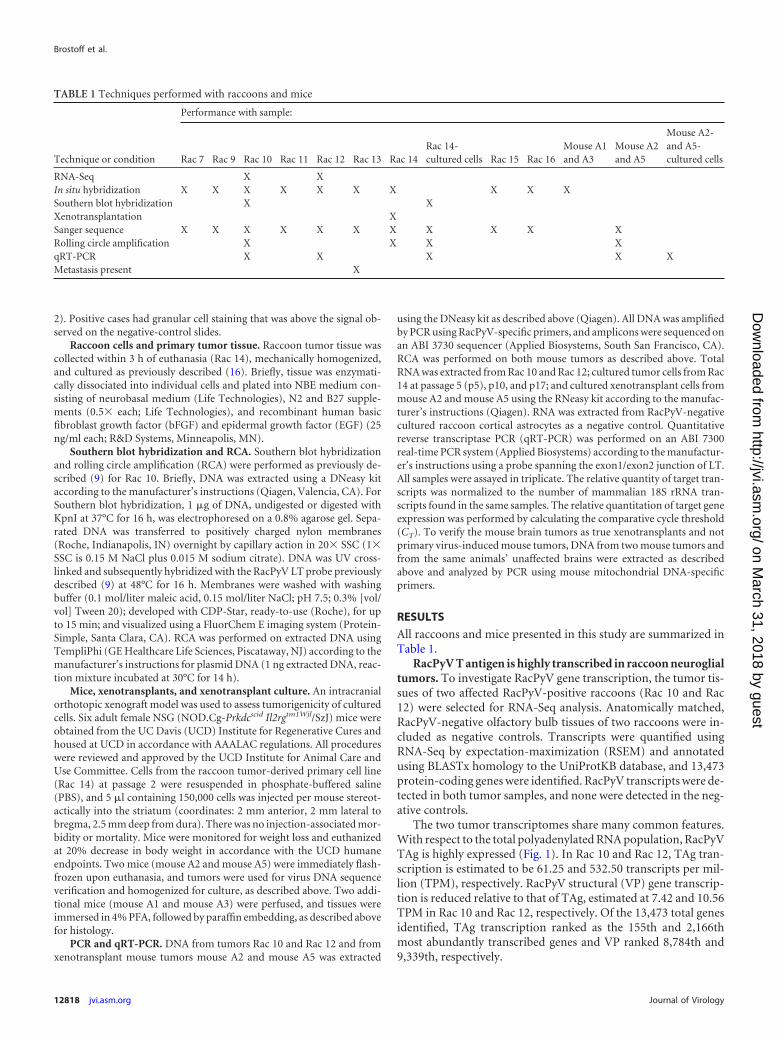
TABLE 1 Techniques performed with raccoons and mice
Technique or condition
Performance with sample:
Rac 7 Rac 9 Rac 10 Rac 11 Rac 12 Rac 13 Rac 14Rac 14-cultured cells Rac 15 Rac 16
Mouse A1and A3
Mouse A2and A5
Mouse A2-and A5-cultured cells
RNA-Seq X XIn situ hybridization X X X X X X X X X XSouthern blot hybridization X XXenotransplantation XSanger sequence X X X X X X X X X X XRolling circle amplification X X X XqRT-PCR X X X X XMetastasis present X
Brostoff et al.
12818 jvi.asm.org Journal of Virology
on March 31, 2018 by guest
http://jvi.asm.org/
Dow
nloaded from

The TAg gene of PyVs encodes a single transcript that is differ-entially spliced to form numerous proteins necessary for cellularmodulation, viral replication, and late gene transcription. Whileall PyVs encode LT and sT, they also encode a host of other tran-scripts that are virus specific. In order to determine the range ofTAg isoforms generated by tumor-associated RacPyV, we frac-tionally quantified the TAg transcripts constructed in the de novotranscriptome assembly and identified their respective ORFs. Rac10 and Rac 12 contain different genomic deletions within the LTintron region; consequent transcripts that retain elements of thisintron produce unique, tumor-specific TAg ORFs. In general, sev-eral isoforms were identified, including LT, sT, MT, and a novel35kT.
The full-length LT isoform comprises the majority (64% to66%) of TAg species and encodes the LT-associated motifs classi-cally observed in other polyomaviruses, including the DnaJ do-main, the pRB-binding domain, the origin binding domain(OBD), and the helicase domain (Fig. 2). In MCC-associatedMCPyV, signature mutations in the viral genome prematurelytruncate the LT ORF (7). Similar mutations have not been ob-served in the genomes of tumor-associated RacPyVs (9). How-ever, a truncated LT ORF may be produced by a subpopulation ofmutated RacPyV genomes. To determine if LT ORFs are prema-
FIG 1 TAg mRNA abundance is high relative to RacPyV VP gene and to totalgene transcription in tumors. Total RNA was extracted and filtered for poly-adenylated RNA from two RacPyV PCR-positive tumor samples (Rac 10 andRac 12) and two RacPyV PCR-negative-control olfactory bulbs. RNA was se-quenced on an Illumina HiSeq 2000, and pooled quality reads were de novoassembled to produce a transcriptome that identified 13,473 protein-codinggenes. Transcripts were quantified using RSEM, and genes from the tumorsamples were plotted in order of decreasing TPM. Asterisks and inverted tri-angles indicate RacPyV TAg and VP genes, respectively.
FIG 2 RacPyV TAg isoforms in two tumor samples shown by transcriptome analysis. RacPyV TAg transcripts were detected in two raccoon tumors, Rac 10 (A)and Rac 12 (B). Transcripts were identified by sequence homology to the RacPyV genome R45. Transcripts were quantified by RSEM, and percent abundance oftotal TAg transcripts was calculated for each isoform (right). Coverage pileups are plotted above a corresponding TAg schematic displaying the associated bindingmotifs and domains (DJ, DnaJ; PP2A, protein phosphatase 2A binding motif; pRB, retinoblastoma binding domain; OBD, origin binding domain). ORFs arerepresented by rectangular boxes with stop codons indicated by asterisks, diagonal lines indicate intron splice junctions, and flat lines indicate untranslatedregions.
RacPyV TAg Transcription in Neuroglial Tumors
November 2014 Volume 88 Number 21 jvi.asm.org 12819
on March 31, 2018 by guest
http://jvi.asm.org/
Dow
nloaded from

turely truncated, quality reads from tumors were aligned to theconstructed LT isoforms and examined for single nucleotide poly-morphisms (SNPs) that would prematurely truncate LT. No poly-morphisms were detected, suggesting that LT is intact in thesetumors. Rac 12 LT contains an intron that splices 7 amino acidsout of the helicase domain. This 21-nt intron is estimated to bepresent in all Rac 12 TAg transcripts but would alter only LT at thelevel of translation.
The sT isoform is unique in each tumor as a result of differen-tial splicing and genomic composition. In Rac 12, the sequenceimmediately prior to the sT splice donor site, which is observed inRac 10, is absent due to the genomic deletion of 522 to 544 bp.Furthermore, no stop codon exists in the Rac 12 LT intron prior tothe homologous Rac 10 sT intron sequence, and so a conventionalsT ORF does not exist in Rac 12. However, transcripts retainingthe LT intron encode an sT ORF defined by a downstream stopcodon in the remainder of the LT intron. In Rac 10, the deletion ofnucleotide 385 produces a truncated sT ORF that includes onlythe first of two protein phosphatase 2A (PP2A) binding motifs; theRac 12 sT ORF includes both of these motifs.
An MT isoform is identified uniquely in Rac 12. The intronobserved in Rac 10 sT is also present in Rac 12. However, due tothe deletion of 522 to 544 bp in Rac 12, an MT ORF is observed.Similar to the MTs observed in murine PyV, RacPyV MT encodesthe DnaJ binding domain, PP2A binding motifs, and the ALTOORF. No reads spanning the MT intron were observed in Rac 10.
A 35kT isoform is identified uniquely in Rac 12. The 35kT ORFencodes a predicted 35-kDa protein and shares common domainswith the similarly designated MCPyV 57kT and SV40 17kT. Allthree of these proteins contain the DnaJ binding domain andpRB-binding domain. Additionally, their ORFs span three exonsby utilizing the LT splice junction and a second downstream splicejunction (7, 17). Similarly to MCPyV 57kT, 35kT encodes its exon2/exon 3 junction within sequences encoding the LT OBD andhelicase domains, respectively. Similar to 17kT, the third exon ofthe 35kT ORF is out of frame with respect to the LT ORF andterminates shortly after the splice junction. No reads spanning thesecond 35kT intron were observed in Rac 10.
RacPyV TAg gene and transcript localization in neuroglialtumors by in situ hybridization. In order to examine spatial,semiquantitative, and cell-specific distribution of RacPyV DNAand mRNA, we performed in situ hybridization (ISH) on nineRacPyV tumors using a set of probes complementary to nt 966 to2084 (exon 2 of LT). The first 320 nt of this region are sharedamong all predicted splice variants, so this assay can recognize allTAg isoforms. Recognition of transcript alone by ISH is not pos-sible, because RacPyV is a double-stranded DNA virus. Therefore,signal is representative of both viral DNA and mRNA. In one case(Rac 13) where the brain tumor had metastasized to multipleother viscera (lymph node, adrenal gland, and liver), both braintumor and liver metastases were tested.
TAg ISH was positive in all nine cases, with regionally variablestaining in up to 70% of tumor cells (representative subset shownin Fig. 3). No background staining was detectable in any of threedifferent negative-control methods (unrelated size-matchedprobe, normal brain tissue within the same section, and unaf-fected raccoons). Colorimetric deposit was primarily detectedwithin the nucleus of the tumor cells; however, signal was oftenstrong, and the deposit frequently exceeded the visual boundary ofthe nuclear membrane (Fig. 3B, D, F, and H). In areas of tumor
where morphology was regionally variable, signal abundance wasvariable as well (Fig. 3A to F). In areas of necrosis, remnant tumorcells surrounding vascular beds showed particularly strong stain-ing (Fig. 3C and D).
In Rac 13, there was increased abundance of signal in the met-astatic cell population of the liver compared to the primary tumor(Fig. 3F and H). Regional staining in the brain tumor on averageshowed robust staining in 5 to 10% of cells but with approximately50% of cells showing punctate staining, whereas 100% of the met-astatic cells contained robust detectable signal.
A primary cell line cultured from neuroglial tumor retainsboth episomal viral genome and TAg transcription. We havepreviously established by two methods (rolling circle amplifica-tion [RCA] and Southern blot hybridization) that RacPyV DNAexists as a circular episome in raccoon neuroglial tumors (9). Todetermine the stability of episomal RacPyV, we generated an invitro culture of tumor cells and examined the maintenance of viralgenome and transcription of TAg. Tumor tissue from Rac 10 andcultured cells from Rac 14 were homogenized for Southern blot-ting. Only episomal, full-length viral DNA was detected (Fig. 4A).The corresponding RCA analysis on both these cells and negative-control cells (cultured RacPyV-negative cortical astrocytes) indi-cated episomal RacPyV DNA in the tumor tissue and cells only(Fig. 4B).
To verify that tumors and cultured tumor cells were able totranscribe TAg mRNA, quantitative reverse transcriptase PCR(qRT-PCR) was performed on two tumors, Rac 10 and Rac 12,and cultured cells from Rac 14 at p5, p10, and p17. A probe com-plementary to the LT exon 1/exon 2 splice junction was used. Thisprobe excluded detection of sT and MT transcripts but includedall other TAg isoforms. Cultured RacPyV-negative cortical astro-cytes were used as a negative control. Using 18S rRNA as a controlfor cellular input, TAg was highly transcribed in Rac 10 and Rac12, demonstrating 25- and 15-fold increases over cortical astro-cytes, respectively (Fig. 5). TAg transcription increased over timein cultured cells from Rac 14, from approximately a 1,200-foldincrease over baseline tumor transcription at p5 to approximatelya 3,000-fold increase by p17.
Xenotransplants in immunodeficient mice and a subsequentprimary cell line stably maintain RacPyV TAg and recapitulateoriginal tumor. We next tested whether this tumor could be re-produced in vivo, and, if so, if this selective environment wouldaffect the stability of RacPyV genome or TAg transcription. Weinjected a minimally expanded primary cell culture (p2) from Rac14 orthotopically into six immunodeficient (Nod-scid gamma)mice. Tumors formed in all six mice, with a median survival timeof 61 days (Fig. 6). Based on mitochondrial DNA amplificationand sequencing, tumor tissue was of raccoon origin and not aresult of de novo viral infection and transformation (data notshown). Two xenograft tumors (mouse A2 and mouse A5) testedpositive for RacPyV genome by PCR and/or RCA. Virus was se-quenced and matched the parental tumor with perfect sequenceidentity (data not shown).
Sublocalization and abundance of viral genome and TAg tran-scripts were assessed by ISH in 2 mice (mouse A1 and mouse A3)and compared to the Rac 14 parental tumor. ISH in xenotrans-plants recapitulated the distribution and quantity of signal seen inthe raccoon tumor tissue (Fig. 7). Like Rac 14, morphology wasregionally variable in the mice and TAg expression was similarlyvariable, with staining in up to 70% of cells (Fig. 7B and D).
Brostoff et al.
12820 jvi.asm.org Journal of Virology
on March 31, 2018 by guest
http://jvi.asm.org/
Dow
nloaded from

FIG 3 Histology and detection of RacPyV genome and TAg mRNA by in situ hybridization. Raccoon tumors were formalin fixed and paraffin embedded priorto staining. (A and B) Hematoxylin and eosin staining (A) and ISH (B) from Rac 15, a representative neuroglial tumor. (C and D) Hematoxylin and eosin staining(C) and ISH (D) from Rac 9 with increased signal around an area of necrosis. (E to H) Hematoxylin and eosin staining (E) and ISH (F) from the parental tumorof Rac 13, with liver metastases (G and H) showing less variability and greater staining than the parental tumor.
November 2014 Volume 88 Number 21 jvi.asm.org 12821
on March 31, 2018 by guest
http://jvi.asm.org/
Dow
nloaded from

To verify and quantify TAg gene expression, qRT-PCR wasperformed on cells cultured from mouse A2 and mouse A5 usingthe probe for RacPyV TAg as described above. As seen with cellscultured from primary raccoon tumor, low-passage-number (p2)cells from each xenotransplant demonstrated an increase aboveparental tumor, showing �300- and �100-fold increases, respec-tively (data not shown).
DISCUSSION
RacPyV is associated with all neuroglial tumors found to date infree-ranging raccoons and has the potential to provide a naturalmodel for PyV-mediated tumorigenesis. These tumors maintainthe virus genome and express high levels of TAg mRNA even whensubjected to selective or foreign environments, including passagein culture and growth in immunocompromised mice. Overall,this study provides evidence for RacPyV TAg as a necessary driverof raccoon neuroglial tumors.
While the positions of introns are similar in RacPyV TAg com-pared to other PyVs, the resulting ORFs are different, both be-tween RacPyV tumors and between RacPyV and other PyVs. Thedifferences seen in RacPyV TAg introns compared to other PyVscould explain differences in viral replication or transcription inneuroglial tumors. However, because these differences in se-quence (including SNPs and deletions) are not consistent amongviral isolates, they are unlikely to be important for the formationor maintenance of tumors. Limitations of transcriptome analysis
include the detection of preprocessed mRNA, which could explainsome of the differences in introns seen in this study. Additionally,short reads cannot be assigned to their parent transcripts whenthose reads are ambiguously mapped (multiple reads which couldmap to one of several transcripts). However, we can calculate per-cent abundance of transcripts by using the RNA-Seq expectation-maximization, a statistical model that proportionally allocatesambiguous reads based on unambiguous (uniquely mapped)reads (18). Despite our ability to calculate percent abundance oftranscripts, mRNA is a proxy for protein expression and isoformsseen by transcript analysis are not necessarily translated in abun-dance. Further work is needed to detect protein isoforms presentin these tumors.
In both Rac 10 and Rac 12, TAg is highly transcribed relative toVP, and LT is the dominant isoform of TAg transcribed. The dis-proportionately high level of TAg transcription relative to VPtranscription occurs without an apparent truncation in LT by ei-ther a mutation or an integration event. Among potential mech-anisms for this control are those used by other viruses, includingsequence rearrangements in the NCRR in disease-associated com-pared to shed virus, histone deacetylase- or DNA methyltrans-ferase-mediated gene silencing, and virus- or host cell-encodedmicroRNA-mediated silencing (19, 20). The ALTO transcriptcannot be verified because its ORF is contained within all otherTAg transcripts. Furthermore, we cannot currently determinewhether or not the protein is expressed.
In other known carcinogenic viruses, including MCPyV andpapillomaviruses, integration is considered an early and criticalstep in transformation (6, 21). In these cases, integration serves at
FIG 5 TAg mRNA is expressed in tumors and increases over time in culturedprimary tumor cells by qRT-PCR. Raccoon tumor tissue from Rac 10 and Rac12 and cultured cells from Rac 14 at passages 5, 10, and 17 were assayed forRacPyV TAg by qRT-PCR, normalized for 18S rRNA. Fold change is relative toRacPyV-negative cortical astrocytes.
FIG 6 Raccoon xenograft survival curve. Six Nod-scid gamma mice were in-jected with 150,000 p2 tumor cells from Rac 14. Percent survival is plottedagainst days postinjection.
FIG 4 Episomal RacPyV genome is maintained in cell culture by Southern blotting and RCA. (A) Southern blot hybridization against RacPyV of Rac 10 tumortissue and Rac 14 tumor-derived cell culture at passages 2, 5, and 10. (B) RCA of Rac 10 tumor tissue and Rac 14 tumor-derived cell culture at passages 2 and 5.U, undigested; K, KpnI digested; neg, negative control (raccoon cortical astrocytes).
Brostoff et al.
12822 jvi.asm.org Journal of Virology
on March 31, 2018 by guest
http://jvi.asm.org/
Dow
nloaded from

least two purposes: to stabilize the virus genome so that it is faith-fully segregated in replicated cells and to inhibit productive/lyticinfection via decreased expression of structural proteins in thecell, favoring instead early gene expression (14, 22, 23). Here, wehave shown that both maintenance of viral genome and selectiveTAg (and not VP) transcription occur in tumors in the absence ofintegration. Many other viruses are able to maintain genome seg-regation without integration in dividing populations of cells. Po-tential alternative mechanisms by which RacPyV achieves this areabundant. Among these, the virus could use physical genome sta-bilization within these tumors, such as chromatin tethering, asseen in papillomavirus or herpesvirus infection (12, 24). Alterna-tively, regardless of physical genome stabilization, virus could beselected for by the cells carrying it, to the exclusion of cells dividingat a lower rate (cells which either have not replicated viral genomeor have insufficient TAg expression).
In addition to analyses that demonstrate the presence ofRacPyV genome and TAg transcription, we performed ISH, whichallows direct visualization of TAg mRNA and DNA in tissue.The tumor cells had regionally variable staining, with most ofthe cells strongly positive. In the single raccoon in which tumorcells had metastasized to other organs, staining was consistentand robust in all of the metastatic cells within the liver. Unfor-tunately, metastatic tissue available was fixed and embeddedand was of insufficient quality to perform Southern blotting or
RCA analysis to determine if viral genome had integrated inthis singular case.
Our data are highly suggestive of the requirement for RacPyVand specifically TAg in the development and maintenance of neu-roglial tumors. Identification of TAg protein isoforms present intumors and protein-protein interactions between TAg and hostcellular proteins will prove useful in evaluating novel mechanismsof polyomavirus stability in tumors and will contribute to a scien-tific understanding of virus-associated transformation. In repre-senting the only nonhuman spontaneous PyV-associated onco-genic transformation, the RacPyV/neuroglial tumor relationshipcould serve as a natural model of recognized and potentially novelmechanisms of PyV-associated oncogenesis.
ACKNOWLEDGMENTS
We thank Ryan R. Davis and Stephenie Y. Liu (GSR and Department ofPathology and Laboratory Medicine) for their expert technical supportfor the next-generation sequencing studies.
The UC Davis Comprehensive Cancer Center Genomics Shared Re-source is supported by Cancer Center support grant P30 CA093373 (R.W.dV.W.) from the NCI. This study was funded by Bernice Barbour Foun-dation grant 11-58N.
REFERENCES1. Manfredi JJ, Prives C. 1994. The transforming activity of simian virus 40
large tumor antigen. Biochim. Biophys. Acta 1198:65– 83.
FIG 7 Histologic phenotype and ISH of RacPyV genome and TAg mRNA of parental raccoon tumor and xenotransplant tumor in mice. Parental tumor fromRac 14 (A and B) was cultured and used for xenotransplant in mice. Tumors were formalin fixed and paraffin embedded prior to staining. Representative MouseA3 (C and D) highlighting similarity by hematoxylin and eosin staining (A and C) and ISH (B and D) to parental Rac 14.
RacPyV TAg Transcription in Neuroglial Tumors
November 2014 Volume 88 Number 21 jvi.asm.org 12823
on March 31, 2018 by guest
http://jvi.asm.org/
Dow
nloaded from
2. Van Dyke TA, Finlay C, Miller D, Marks J, Lozano G, Levine AJ. 1987.Relationship between simian virus 40 large tumor antigen expression andtumor formation in transgenic mice. J. Virol. 61:2029 –2032.
3. Pipas JM. 2009. SV40: cell transformation and tumorigenesis. Virology384:294 –303. http://dx.doi.org/10.1016/j.virol.2008.11.024.
4. Kean JM, Rao S, Wang M, Garcea RL. 2009. Seroepidemiology of humanpolyomaviruses. PLoS Pathog. 5:e1000363. http://dx.doi.org/10.1371/journal.ppat.1000363.
5. DeCaprio JA, Garcea RL. 2013. A cornucopia of human polyomaviruses.Nat. Rev. Microbiol. 11:264–276. http://dx.doi.org/10.1038/nrmicro2992.
6. Feng H, Shuda M, Chang Y, Moore PS. 2008. Clonal integration of apolyomavirus in human Merkel cell carcinoma. Science 319:1096 –1100.http://dx.doi.org/10.1126/science.1152586.
7. Shuda M, Feng H, Kwun HJ, Rosen ST, Gjoerup O, Moore PS, ChangY. 2008. T antigen mutations are a human tumor-specific signature forMerkel cell polyomavirus. Proc. Natl. Acad. Sci. U. S. A. 105:16272–16277.http://dx.doi.org/10.1073/pnas.0806526105.
8. Arora R, Chang Y, Moore PS. 2012. MCV and Merkel cell carcinoma: amolecular success story. Curr. Opin. Virol. 2:489 – 498. http://dx.doi.org/10.1016/j.coviro.2012.05.007.
9. Dela Cruz FN, Jr, Giannitti F, Li L, Woods LW, Del Valle L, Delwart E,Pesavento PA. 2013. Novel polyomavirus associated with brain tumors infree-ranging raccoons, western United States. Emerg. Infect. Dis. 19:77–84. http://dx.doi.org/10.3201/eid1901.121078.
10. Carter JJ, Daugherty MD, Qi X, Bheda-Malge A, Wipf GC, Robinson K,Roman A, Malik HS, Galloway DA. 2013. Identification of an overprint-ing gene in Merkel cell polyomavirus provides evolutionary insight intothe birth of viral genes. Proc. Natl. Acad. Sci. U. S. A. 110:12744 –12749.http://dx.doi.org/10.1073/pnas.1303526110.
11. Hahn WC, Dessain SK, Brooks MW, King JE, Elenbaas B, Sabatini DM,DeCaprio JA, Weinberg RA. 2002. Enumeration of the simian virus 40 earlyregion elements necessary for human cell transformation. Mol. Cell. Biol.22:2111–2123. http://dx.doi.org/10.1128/MCB.22.7.2111-2123.2002.
12. Bastien N, McBride AA. 2000. Interaction of the papillomavirus E2 pro-tein with mitotic chromosomes. Virology 270:124 –134. http://dx.doi.org/10.1006/viro.2000.0265.
13. Houben R, Shuda M, Weinkam R, Schrama D, Feng H, Chang Y,Moore PS, Becker JC. 2010. Merkel cell polyomavirus-infected Merkelcell carcinoma cells require expression of viral T antigens. J. Virol. 84:7064 –7072. http://dx.doi.org/10.1128/JVI.02400-09.
14. Cheng J, Rozenblatt-Rosen O, Paulson KG, Nghiem P, Decaprio JA.
2013. Merkel cell polyomavirus large T antigen has growth-promotingand inhibitory activities. J. Virol. 87:6118 – 6126. http://dx.doi.org/10.1128/JVI.00385-13.
15. Pastrana DV, Tolstov YL, Becker JC, Moore PS, Chang Y, Buck CB.2009. Quantitation of human seroresponsiveness to Merkel cell polyoma-virus. PLoS Pathog. 5:e1000578. http://dx.doi.org/10.1371/journal.ppat.1000578.
16. Son MJ, Woolard K, Nam DH, Lee J, Fine HA. 2009. SSEA-1 is anenrichment marker for tumor-initiating cells in human glioblastoma. CellStem Cell 4:440 – 452. http://dx.doi.org/10.1016/j.stem.2009.03.003.
17. Ibelgaufts H, Doerfler W, Scheidtmann KH, Wechsler W. 1980. Ade-novirus type 12-induced rat tumor cells of neuroepithelial origin: persis-tence and expression of the viral genome. J. Virol. 33:423– 437.
18. Li B, Dewey CN. 2011. RSEM: accurate transcript quantification fromRNA-Seq data with or without a reference genome. BMC Bioinformatics12:323. http://dx.doi.org/10.1186/1471-2105-12-323.
19. Gosert R, Kardas P, Major EO, Hirsch HH. 2010. Rearranged JC virusnoncoding control regions found in progressive multifocal leukoencephalop-athy patient samples increase virus early gene expression and replication rate.J. Virol. 84:10448–10456. http://dx.doi.org/10.1128/JVI.00614-10.
20. White MK, Safak M, Khalili K. 2009. Regulation of gene expression inprimate polyomaviruses. J. Virol. 83:10846 –10856. http://dx.doi.org/10.1128/JVI.00542-09.
21. Schwarz E, Freese UK, Gissmann L, Mayer W, Roggenbuck B, StremlauA, zur Hausen H. 1985. Structure and transcription of human papillo-mavirus sequences in cervical carcinoma cells. Nature 314:111–114. http://dx.doi.org/10.1038/314111a0.
22. Laude HC, Jonchere B, Maubec E, Carlotti A, Marinho E, Couturaud B,Peter M, Sastre-Garau X, Avril MF, Dupin N, Rozenberg F. 2010.Distinct Merkel cell polyomavirus molecular features in tumour and nontumour specimens from patients with Merkel cell carcinoma. PLoS Pat-hog. 6:e1001076. http://dx.doi.org/10.1371/journal.ppat.1001076.
23. Stoler MH, Rhodes CR, Whitbeck A, Wolinsky SM, Chow LT, BrokerTR. 1992. Human papillomavirus type 16 and 18 gene expression in cer-vical neoplasias. Hum. Pathol. 23:117–128. http://dx.doi.org/10.1016/0046-8177(92)90232-R.
24. Ilves I, Kivi S, Ustav M. 1999. Long-term episomal maintenance ofbovine papillomavirus type 1 plasmids is determined by attachment tohost chromosomes, which Is mediated by the viral E2 protein and itsbinding sites. J. Virol. 73:4404 – 4412.
Brostoff et al.
12824 jvi.asm.org Journal of Virology
on March 31, 2018 by guest
http://jvi.asm.org/
Dow
nloaded from