the recognition domain of the methyl-specific endonuclease mcrbc
TRANSCRIPT
The recognition domain of the methyl-specificendonuclease McrBC flips out 5-methylcytosineRasa Sukackaite, Saulius Grazulis, Gintautas Tamulaitis and Virginijus Siksnys*
Department of Protein–DNA Interactions, Institute of Biotechnology, Vilnius University, Graiciuno 8,02241 Vilnius, Lithuania
Received March 15, 2012; Revised March 30, 2012; Accepted April 3, 2012
ABSTRACT
DNA cytosine methylation is a widespread epigen-etic mark. Biological effects of DNA methylation aremediated by the proteins that preferentially bind to5-methylcytosine (5mC) in different sequencecontexts. Until now two different structural mechan-isms have been established for 5mC recognition ineukaryotes; however, it is still unknown howdiscrimination of the 5mC modification is achievedin prokaryotes. Here we report the crystal structureof the N-terminal DNA-binding domain (McrB-N) ofthe methyl-specific endonuclease McrBC fromEscherichia coli. The McrB-N protein shows anovel DNA-binding fold adapted for 5mC-recog-nition. In the McrB-N structure in complex withmethylated DNA, the 5mC base is flipped out fromthe DNA duplex and positioned within a bindingpocket. Base flipping elegantly explains whyMcrBC system restricts only T4-even phagesimpaired in glycosylation [Luria, S.E. and Human,M.L. (1952) A nonhereditary, host-induced variationof bacterial viruses. J. Bacteriol., 64, 557–569]:flipped out 5-hydroxymethylcytosine is accommo-dated in the binding pocket but there is no roomfor the glycosylated base. The mechanism for 5mCrecognition employed by McrB-N is highly reminis-cent of that for eukaryotic SRA domains, despite thedifferences in their protein folds.
INTRODUCTION
Modification of genomic DNA by methylation is an im-portant epigenetic signal. In mammals and other verte-brate, DNA methylation occurs at the C5 position ofcytosine, resulting in 5-methylcytosine (5mC), mostlywithin CpG dinucleotides (1,2). In plants DNA methyla-tion is found in the symmetric CG and CHG contexts(where H=A, T or C) and the asymmetric CHH
context (3). Since 5mC is related to repressed chromatinstate and inhibition of transcription (1,2), an establish-ment and maintenance of the methylation pattern is ofkey importance for the cell functions. To interpret DNAmethylation status proteins must faithfully discriminatebetween the non-modified C and the 5mC bases thatdiffer only by a single methyl group. Structural studiesrevealed that eukaryotic methyl-DNA-binding proteinsemploy two different strategies for the 5mC recognition.The methyl-CpG-binding domain from the transcriptionalrepressor MeCP2 recognizes the specific hydration patternof methylated DNA rather than cytosine methylation byitself (4). The SRA domain from the mammalian UHRF1employs a radically different strategy: it flips out the 5mCof the DNA duplex and positions the extruded base withinthe protein pocket for discrimination (5–7). The SRAdomain from the Arabidopsis SUVH5 follows a dualflip-out mechanism and extrudes both the methylatedbase and neighbouring base in the complementary DNAstrand (8).
The evidence from comparative genomics suggests thatmechanisms of 5mC discrimination may have emerged inthe bacterial world as a result of on-going arms racebetween viruses and bacteria (9). In bacteria, methylatedDNA bases are employed by restriction–modificationsystems to discriminate between self and foreign epigeneticmodification patterns (10,11). The lack of the specificmethyl-tag in the foreign DNA which enters the cellserves as a signal that triggers the restriction endonucleasecleavage. To protect their DNA from cleavage bacterio-phages acquired an ability to incorporate modified basessuch as 5mC and 50-hydroxymethylcytosine (5hmC)in their genomes during DNA replication (12). Itis thought that bacteria responded by developing methy-lation-dependent restriction endonucleases Mcr(for Modified cytosine restriction) to cut the modifiedforeign DNA [see (10) for a review]. To obtain resistanceagainst the Mcr cleavage, T4 even phages extended C basemodification through the glycosylation of the 5hmCresidues (13–15). Actually, the first phage restriction phe-nomenon observed in the studies of T-even phage mutants
*To whom correspondence should be addressed. Tel: +370 5 2602108; Fax: +370 5 2602116; Email: [email protected] address:Rasa Sukackaite, Grenoble Outstation, European Molecular Biology Laboratory, 6 rue Jules Horowitz, BP 181, 38042 Grenoble Cedex 9, France.
7552–7562 Nucleic Acids Research, 2012, Vol. 40, No. 15 Published online 8 May 2012doi:10.1093/nar/gks332
� The Author(s) 2012. Published by Oxford University Press.This is an Open Access article distributed under the terms of the Creative Commons Attribution Non-Commercial License (http://creativecommons.org/licenses/by-nc/3.0), which permits unrestricted non-commercial use, distribution, and reproduction in any medium, provided the original work is properly cited.
Downloaded from https://academic.oup.com/nar/article-abstract/40/15/7552/1199149by gueston 12 February 2018
impaired in glycosylation (16) was due to the Mcr cleavageof 5mC/5hmC-containing viral DNA. In Escherichia coliK12 strain there are at least two chromosomally encodedrestriction endonucleases that act on the 5mC/5hmC con-taining DNA: McrA and McrBC (17,18). In contrast tothe eukaryotic methyl-DNA-binding proteins, structuralmechanisms leading to the 5mC/5hmC recognition byMcr systems and other bacterial endonucleases acting onthe methylated DNA (19) are as yet unknown.
To understand the structural mechanisms of the5mC/5hmC recognition in prokaryotes, we focused onthe E. coli McrBC (18,20). The McrBC restrictionsystem consists of two subunits: McrB (53 kDa) andMcrC (40.5 kDa) (21) (Figure 1A). The nuclease activesite resides in McrC (22), while McrB is responsible forDNA binding and GTP hydrolysis (20,23). TheC-terminal part of McrB harbours the signature motifsof AAA+ (ATPases associated with various cellularactivities) protein family (24,25). As most of AAA+
family proteins which usually function as ring-shapedoligomers (26), McrB was shown to assemble intoheptameric ring structures and tetradecamers (27). DNAcleavage by McrBC requires GTP and occurs betweentwo well-separated (30- to 3000-bp apart) recognitionsites, 50-RmC (where R=A or G) triggered by encounterof two DNA-translocating McrBC complexes (18,28,29).While two recognition elements at two well-separated lo-cations are required for cleavage, McrB can bind sub-strates with recognition elements at a single location
(30,31). The domain responsible for recognition ofmethylated DNA was shown to reside in the N-terminalpart (residues 1–161) of the McrB subunit (Figure 1A)(30,32).In this report we present the crystal structure of the
McrB N-terminal domain (McrB-N) in complex withdi-methylated, hemi-methylated and non-methylatedDNA. To our knowledge it represents the first structureof a prokaryotic 5mC-binding domain in the DNA-boundform.
MATERIALS AND METHODS
Protein expression and purification
The region coding for the 1–161 residues of McrB wasPCR-amplified from the pBBI/McrB template, intro-ducing a hexahistidine tag at the C-terminus, andinserted into the pBAD24 vector. The McrB-N proteinwas expressed in E. coli ER2267 cells grown in the LBmedium. The expression was induced by cultivation for3–4 h at 37�C in presence of 0.2% arabinose. The cellswere re-suspended in a buffer A [20mM sodium phos-phate (pH 7.4), 0.5M NaCl] and sonicated. The solublefraction was loaded on a HiTrap chelating column (GEHealthcare) and eluted by an imidazole gradient. Thetarget protein was dialysed against a buffer B [10mMpotassium phosphate (pH 7.0), 0.05M KCl, 1mMEDTA, 7mM 2-mercaptoethanol, 10% (v/v) glycerol]
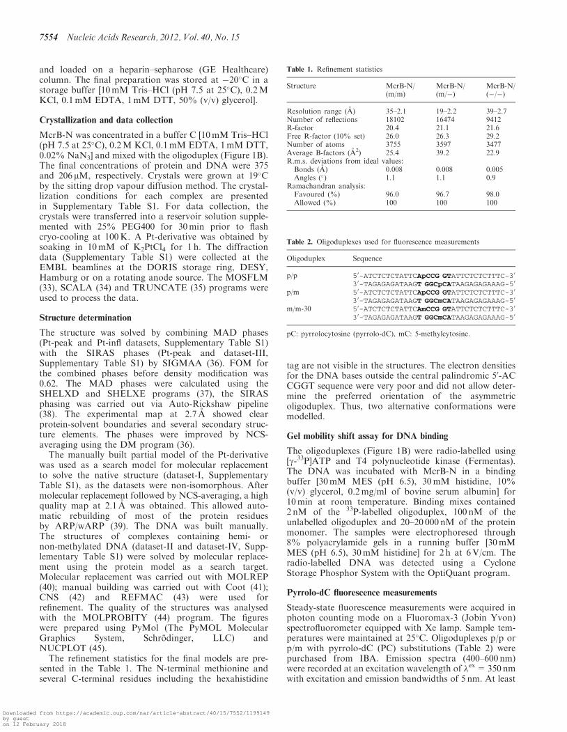
Figure 1. (A) Schematic representation of the McrBC restriction system. McrBC is composed of two subunits: McrB harbours an N-terminalDNA-binding domain (McrB-N) and GTP-ase motifs, while McrC contains a nuclease active site (20,22,23). (B) The oligoduplexes used for crys-tallization. The 50-AmC (50-AC) sequences are shown in bold. (C) Closer view of the McrB-N monomer bound to DNA. The ‘finger’ loop penetratinginto the minor groove is highlighted in orange. (D) Overall structure of the two McrB-N monomers bound to the di-methylated (m/m) oligoduplex.The DNA is bent by �29� and both the 5-methylcytosines are flipped out.
Nucleic Acids Research, 2012, Vol. 40, No. 15 7553
Downloaded from https://academic.oup.com/nar/article-abstract/40/15/7552/1199149by gueston 12 February 2018
and loaded on a heparin–sepharose (GE Healthcare)column. The final preparation was stored at �20�C in astorage buffer [10mM Tris–HCl (pH 7.5 at 25�C), 0.2MKCl, 0.1mM EDTA, 1mM DTT, 50% (v/v) glycerol].
Crystallization and data collection
McrB-N was concentrated in a buffer C [10mM Tris–HCl(pH 7.5 at 25�C), 0.2M KCl, 0.1mM EDTA, 1mM DTT,0.02% NaN3] and mixed with the oligoduplex (Figure 1B).The final concentrations of protein and DNA were 375and 206 mM, respectively. Crystals were grown at 19�Cby the sitting drop vapour diffusion method. The crystal-lization conditions for each complex are presentedin Supplementary Table S1. For data collection, thecrystals were transferred into a reservoir solution supple-mented with 25% PEG400 for 30min prior to flashcryo-cooling at 100K. A Pt-derivative was obtained bysoaking in 10mM of K2PtCl4 for 1 h. The diffractiondata (Supplementary Table S1) were collected at theEMBL beamlines at the DORIS storage ring, DESY,Hamburg or on a rotating anode source. The MOSFLM(33), SCALA (34) and TRUNCATE (35) programs wereused to process the data.
Structure determination
The structure was solved by combining MAD phases(Pt-peak and Pt-infl datasets, Supplementary Table S1)with the SIRAS phases (Pt-peak and dataset-III,Supplementary Table S1) by SIGMAA (36). FOM forthe combined phases before density modification was0.62. The MAD phases were calculated using theSHELXD and SHELXE programs (37), the SIRASphasing was carried out via Auto-Rickshaw pipeline(38). The experimental map at 2.7 A showed clearprotein-solvent boundaries and several secondary struc-ture elements. The phases were improved by NCS-averaging using the DM program (36).The manually built partial model of the Pt-derivative
was used as a search model for molecular replacementto solve the native structure (dataset-I, SupplementaryTable S1), as the datasets were non-isomorphous. Aftermolecular replacement followed by NCS-averaging, a highquality map at 2.1 A was obtained. This allowed auto-matic rebuilding of most of the protein residuesby ARP/wARP (39). The DNA was built manually.The structures of complexes containing hemi- ornon-methylated DNA (dataset-II and dataset-IV, Supp-lementary Table S1) were solved by molecular replace-ment using the protein model as a search target.Molecular replacement was carried out with MOLREP(40); manual building was carried out with Coot (41);CNS (42) and REFMAC (43) were used forrefinement. The quality of the structures was analysedwith the MOLPROBITY (44) program. The figureswere prepared using PyMol (The PyMOL MolecularGraphics System, Schrodinger, LLC) andNUCPLOT (45).The refinement statistics for the final models are pre-
sented in the Table 1. The N-terminal methionine andseveral C-terminal residues including the hexahistidine
tag are not visible in the structures. The electron densitiesfor the DNA bases outside the central palindromic 50-ACCGGT sequence were very poor and did not allow deter-mine the preferred orientation of the asymmetricoligoduplex. Thus, two alternative conformations weremodelled.
Gel mobility shift assay for DNA binding
The oligoduplexes (Figure 1B) were radio-labelled using[g-33P]ATP and T4 polynucleotide kinase (Fermentas).The DNA was incubated with McrB-N in a bindingbuffer [30mM MES (pH 6.5), 30mM histidine, 10%(v/v) glycerol, 0.2mg/ml of bovine serum albumin] for10min at room temperature. Binding mixes contained2 nM of the 33P-labelled oligoduplex, 100 nM of theunlabelled oligoduplex and 20–20 000 nM of the proteinmonomer. The samples were electrophoresed through8% polyacrylamide gels in a running buffer [30mMMES (pH 6.5), 30mM histidine] for 2 h at 6V/cm. Theradio-labelled DNA was detected using a CycloneStorage Phosphor System with the OptiQuant program.
Pyrrolo-dC fluorescence measurements
Steady-state Fuorescence measurements were acquired inphoton counting mode on a Fluoromax-3 (Jobin Yvon)spectrofluorometer equipped with Xe lamp. Sample tem-peratures were maintained at 25�C. Oligoduplexes p/p orp/m with pyrrolo-dC (PC) substitutions (Table 2) werepurchased from IBA. Emission spectra (400–600 nm)were recorded at an excitation wavelength of �ex=350 nmwith excitation and emission bandwidths of 5 nm. At least
Table 1. Refinement statistics
Structure McrB-N/(m/m)
McrB-N/(m/�)
McrB-N/(�/�)
Resolution range (A) 35–2.1 19–2.2 39–2.7Number of reflections 18102 16474 9412R-factor 20.4 21.1 21.6Free R-factor (10% set) 26.0 26.3 29.2Number of atoms 3755 3597 3477Average B-factors (A2) 25.4 39.2 22.9R.m.s. deviations from ideal values:
Bonds (A) 0.008 0.008 0.005Angles (�) 1.1 1.1 0.9
Ramachandran analysis:Favoured (%) 96.0 96.7 98.0Allowed (%) 100 100 100
Table 2. Oligoduplexes used for fluorescence measurements
Oligoduplex Sequence
p/p 50-ATCTCTCTATTCApCCG GTATTCTCTCTTTC-30
30-TAGAGAGATAAGT GGCpCATAAGAGAGAAAG-50
p/m 50-ATCTCTCTATTCApCCG GTATTCTCTCTTTC-30
30-TAGAGAGATAAGT GGCmCATAAGAGAGAAAG-50
m/m-30 50-ATCTCTCTATTCAmCCG GTATTCTCTCTTTC-30
30-TAGAGAGATAAGT GGCmCATAAGAGAGAAAG-50
pC: pyrrolocytosine (pyrrolo-dC), mC: 5-methylcytosine.
7554 Nucleic Acids Research, 2012, Vol. 40, No. 15
Downloaded from https://academic.oup.com/nar/article-abstract/40/15/7552/1199149by gueston 12 February 2018
four scans were averaged for each spectrum. The samplescontained 5 mM of McrB-N and 1 mM of DNA in 30mMMES (pH 6.5), 30mM histidine. A 5-fold excess of theprotein was used to ensure the complete binding of theDNA. Control spectra used for the background subtrac-tion corrections were collected under identical conditionsexcept that duplex m/m-30 was used instead of theFuorescent DNA. The Fuorescence emission value of thecorrected spectrum was determined at the emissionmaximum for each sample.
RESULTS
To understand the molecular mechanism for the 5mC rec-ognition by the McrB N-terminal domain we have solvedthree crystal structures of McrB-N in complex withdi-methylated, hemi-methylated and non-methylatedDNA, respectively. Since 50-AmCCGGT sequence isbound by McrB-N more tightly than other 50-RmC con-taining sequences (30), three different oligoduplexes con-taining the methylated (m/m and m/�) andnon-methylated (�/�) 50-ACCGGT sequence were usedfor crystallization experiments (Figure 1B). (i) In thedi-methylated oligoduplex (m/m) the first C within the50-ACCGGT sequence is methylated in both strands toyield two 50-AmC sites separated by 2 bp; (ii) in thehemi-methylated oligoduplex (m/�) the first C residuewithin the 50-ACCGGT sequence is methylated only inone DNA strand to generate a single 50-AmC site; (iii) inthe oligoduplex (�/�) there are two non-methylated 50-AC sites on the opposite DNA strands. McrB-N gavecrystals belonging to the same P212121 group in presenceof any of these DNA fragments. The structure of aplatinum derivative of the McrB-N/hemi-methylatedDNA complex was solved using both the anomalousand isomorphous signal (see ‘Experimental procedures’section for more details). The structures of McrB-Nbound to di-methylated, hemi-methylated andnon-methylated DNA were solved by molecular replace-ment to the resolutions of 2.1, 2.2 and 2.7 A, respectively.In all the structures there are two McrB-N molecules andone oligoduplex in the asymmetric unit. The proteincomponents of the three independent structures arehighly similar (r.m.s.d. of <1 A when comparing proteinmonomers). Here we will describe the structure ofMcrB-N bound to the di-methylated DNA and willdiscuss the differences between the three structures.
Overall structure of protein–DNA complex
The N-terminal recognition domain of McrB folds intothree a-helices and five b-strands with a topologicalorder: a1(4–18), b1(33–39), b2(49–54), b3(63–71), b4(75–83),a2(102–111), b5(121–128) and a3(134–153) (Figure 1C). Thefive b-strands are arranged into a single anti-parallelb-sheet. The N-terminal a1 and the C-terminal a3helices pack on the convex side of the b-sheet, while theconcave side is oriented towards the DNA. Three loopsthat emanate from the b-sheet grip the DNA from theminor groove side. In the asymmetric unit there are twoMcrB-N monomers bound to 50-AmC sites on different
strands within di-methylated DNA (m/m) (Figure 1D).In crystal, these two McrB-N monomers do not contacteach other. The DNA is heavily distorted, with both 5mCresidues flipped out of the DNA duplex and positioned inthe binding pockets within the protein. According to theanalysis by the CURVES program (46), the minor grooveis significantly wider in comparison to the canonicalB-DNA, and the oligoduplex is bent by an overall angleof 29� towards the major groove. The loop b1b2(40–48),which we named ‘finger’, protrudes into the widenedminor groove and the side chain of Tyr41 intercalatesinto the DNA base stack replacing the flipped out 50-methylcytosine (Figure 1C).Both the structure of the protein and the conformation
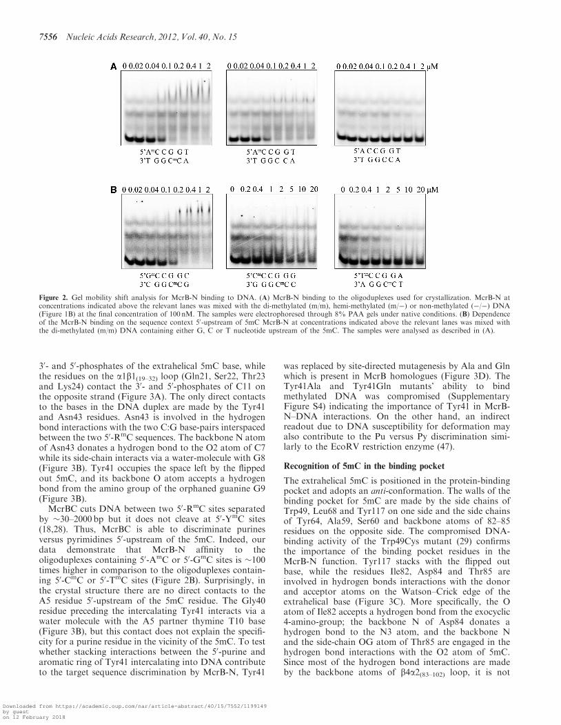
of the DNA are nearly identical in the complexes ofMcrB-N with hemi-methylated (m/�) and di-methylated(m/m) oligoduplexes (Supplementary Figure S1). In thecase of the hemi-methylated oligoduplex (m/�) there aretwo McrB-N monomers bound to separate 50-RmC/50-RCsites, the DNA is bent, and both the methylated andnon-methylated C bases are flipped out (we could not de-termine the preferred orientation of the oligoduplex (m/�)in the McrB-N–DNA complex, thus two alternative DNAconformations with the methyl-group positioned on dif-ferent DNA strands were modelled). According to thegel-mobility shift assay, McrB-N binds the hemi-methylated (m/�) and di-methylated (m/m) oligoduplexeswith the same affinity (Figure 2A). These results are ingood agreement with the biochemical data that DNAmethylated in one strand can be efficiently recognized bythe McrB protein (18,20). The stoichiometry of theMcrB-N–DNA complexes could not be determined inthe gel-shift assay since the complexes were purelyresolved.Surprisingly, in the structure of the McrB-N bound to
the non-methylated oligoduplex (�/�) two cytosineresidues adjacent to the A base are also flipped out. Theoverlay of the McrB-N structures bound to thedi-methylated (m/m) and non-methylated (�/�) oligodu-plexes does not show significant differences neither inprotein nor in DNA conformation (SupplementaryFigure S2). However, according to the gel mobility shiftassay, the lack of the methyl group on the cytosine de-creases the stability of the protein–DNA complex, sincethe McrB-N binding to the oligoduplex (�/�) is undetect-able in the gel (Figure 2A). One cannot exclude that thecomplex between McrB-N and oligoduplex (�/�) isstabilized by crystal packing forces.
Protein–DNA interactions
The contacts made by the two McrB-N monomers boundto the di-methylated oligoduplex (m/m) are identical(Figure 3A) therefore we will further discuss protein–DNA interactions made by one monomer. The McrB-Napproaches DNA from the minor groove side (Figure 1D)and three neighbouring loops a1b1(19–32), b1b2(40–48)(‘finger’) and b2b3(55–62) emanating from the b-sheet con-tribute amino acid residues for protein–DNA interactions.Ser38, Thr45, Ser46, Trp49, Glu58, Ala59 andSer60 make direct and water-mediated interactions with
Nucleic Acids Research, 2012, Vol. 40, No. 15 7555
Downloaded from https://academic.oup.com/nar/article-abstract/40/15/7552/1199149by gueston 12 February 2018
30- and 50-phosphates of the extrahelical 5mC base, whilethe residues on the a1b1(19–32) loop (Gln21, Ser22, Thr23and Lys24) contact the 30- and 50-phosphates of C11 onthe opposite strand (Figure 3A). The only direct contactsto the bases in the DNA duplex are made by the Tyr41and Asn43 residues. Asn43 is involved in the hydrogenbond interactions with the two C:G base-pairs interspacedbetween the two 50-RmC sequences. The backbone N atomof Asn43 donates a hydrogen bond to the O2 atom of C7while its side-chain interacts via a water-molecule with G8(Figure 3B). Tyr41 occupies the space left by the flippedout 5mC, and its backbone O atom accepts a hydrogenbond from the amino group of the orphaned guanine G9(Figure 3B).McrBC cuts DNA between two 50-RmC sites separated
by �30–2000 bp but it does not cleave at 50-YmC sites(18,28). Thus, McrBC is able to discriminate purinesversus pyrimidines 50-upstream of the 5mC. Indeed, ourdata demonstrate that McrB-N affinity to theoligoduplexes containing 50-AmC or 50-GmC sites is �100times higher in comparison to the oligoduplexes contain-ing 50-CmC or 50-TmC sites (Figure 2B). Surprisingly, inthe crystal structure there are no direct contacts to theA5 residue 50-upstream of the 5mC residue. The Gly40residue preceding the intercalating Tyr41 interacts via awater molecule with the A5 partner thymine T10 base(Figure 3B), but this contact does not explain the specifi-city for a purine residue in the vicinity of the 5mC. To testwhether stacking interactions between the 50-purine andaromatic ring of Tyr41 intercalating into DNA contributeto the target sequence discrimination by McrB-N, Tyr41
was replaced by site-directed mutagenesis by Ala and Glnwhich is present in McrB homologues (Figure 3D). TheTyr41Ala and Tyr41Gln mutants’ ability to bindmethylated DNA was compromised (SupplementaryFigure S4) indicating the importance of Tyr41 in McrB-N–DNA interactions. On the other hand, an indirectreadout due to DNA susceptibility for deformation mayalso contribute to the Pu versus Py discrimination simi-larly to the EcoRV restriction enzyme (47).
Recognition of 5mC in the binding pocket
The extrahelical 5mC is positioned in the protein-bindingpocket and adopts an anti-conformation. The walls of thebinding pocket for 5mC are made by the side chains ofTrp49, Leu68 and Tyr117 on one side and the side chainsof Tyr64, Ala59, Ser60 and backbone atoms of 82–85residues on the opposite side. The compromised DNA-binding activity of the Trp49Cys mutant (29) confirmsthe importance of the binding pocket residues in theMcrB-N function. Tyr117 stacks with the flipped outbase, while the residues Ile82, Asp84 and Thr85 areinvolved in hydrogen bonds interactions with the donorand acceptor atoms on the Watson–Crick edge of theextrahelical base (Figure 3C). More specifically, the Oatom of Ile82 accepts a hydrogen bond from the exocyclic4-amino-group; the backbone N of Asp84 donates ahydrogen bond to the N3 atom, and the backbone Nand the side-chain OG atom of Thr85 are engaged in thehydrogen bond interactions with the O2 atom of 5mC.Since most of the hydrogen bond interactions are madeby the backbone atoms of b4a2(83–102) loop, it is not
Figure 2. Gel mobility shift analysis for McrB-N binding to DNA. (A) McrB-N binding to the oligoduplexes used for crystallization. McrB-N atconcentrations indicated above the relevant lanes was mixed with the di-methylated (m/m), hemi-methylated (m/�) or non-methylated (�/�) DNA(Figure 1B) at the final concentration of 100 nM. The samples were electrophoresed through 8% PAA gels under native conditions. (B) Dependenceof the McrB-N binding on the sequence context 50-upstream of 5mC McrB-N at concentrations indicated above the relevant lanes was mixed withthe di-methylated (m/m) DNA containing either G, C or T nucleotide upstream of the 5mC. The samples were analysed as described in (A).
7556 Nucleic Acids Research, 2012, Vol. 40, No. 15
Downloaded from https://academic.oup.com/nar/article-abstract/40/15/7552/1199149by gueston 12 February 2018
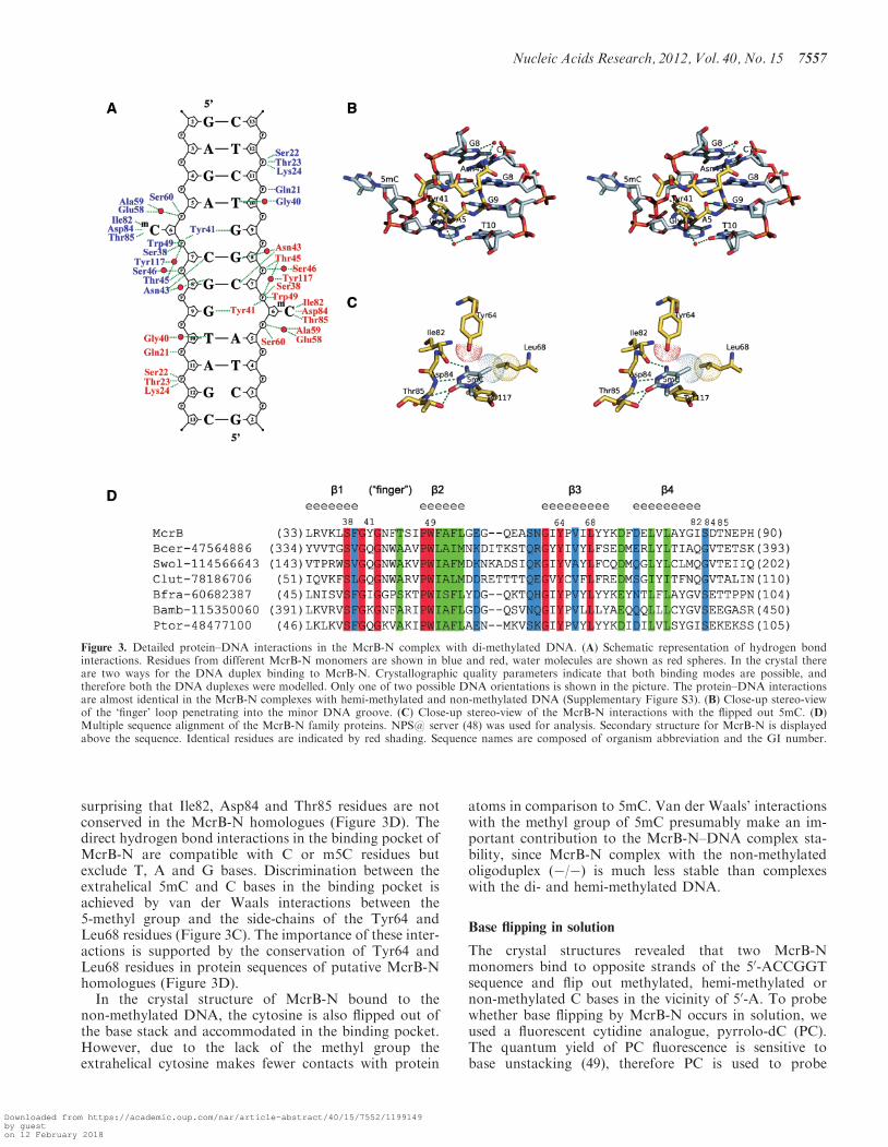
surprising that Ile82, Asp84 and Thr85 residues are notconserved in the McrB-N homologues (Figure 3D). Thedirect hydrogen bond interactions in the binding pocket ofMcrB-N are compatible with C or m5C residues butexclude T, A and G bases. Discrimination between theextrahelical 5mC and C bases in the binding pocket isachieved by van der Waals interactions between the5-methyl group and the side-chains of the Tyr64 andLeu68 residues (Figure 3C). The importance of these inter-actions is supported by the conservation of Tyr64 andLeu68 residues in protein sequences of putative McrB-Nhomologues (Figure 3D).
In the crystal structure of McrB-N bound to thenon-methylated DNA, the cytosine is also flipped out ofthe base stack and accommodated in the binding pocket.However, due to the lack of the methyl group theextrahelical cytosine makes fewer contacts with protein
atoms in comparison to 5mC. Van der Waals’ interactionswith the methyl group of 5mC presumably make an im-portant contribution to the McrB-N–DNA complex sta-bility, since McrB-N complex with the non-methylatedoligoduplex (�/�) is much less stable than complexeswith the di- and hemi-methylated DNA.
Base flipping in solution
The crystal structures revealed that two McrB-Nmonomers bind to opposite strands of the 50-ACCGGTsequence and flip out methylated, hemi-methylated ornon-methylated C bases in the vicinity of 50-A. To probewhether base flipping by McrB-N occurs in solution, weused a fluorescent cytidine analogue, pyrrolo-dC (PC).The quantum yield of PC fluorescence is sensitive tobase unstacking (49), therefore PC is used to probe
Figure 3. Detailed protein–DNA interactions in the McrB-N complex with di-methylated DNA. (A) Schematic representation of hydrogen bondinteractions. Residues from different McrB-N monomers are shown in blue and red, water molecules are shown as red spheres. In the crystal thereare two ways for the DNA duplex binding to McrB-N. Crystallographic quality parameters indicate that both binding modes are possible, andtherefore both the DNA duplexes were modelled. Only one of two possible DNA orientations is shown in the picture. The protein–DNA interactionsare almost identical in the McrB-N complexes with hemi-methylated and non-methylated DNA (Supplementary Figure S3). (B) Close-up stereo-viewof the ‘finger’ loop penetrating into the minor DNA groove. (C) Close-up stereo-view of the McrB-N interactions with the flipped out 5mC. (D)Multiple sequence alignment of the McrB-N family proteins. NPS@ server (48) was used for analysis. Secondary structure for McrB-N is displayedabove the sequence. Identical residues are indicated by red shading. Sequence names are composed of organism abbreviation and the GI number.
Nucleic Acids Research, 2012, Vol. 40, No. 15 7557
Downloaded from https://academic.oup.com/nar/article-abstract/40/15/7552/1199149by gueston 12 February 2018
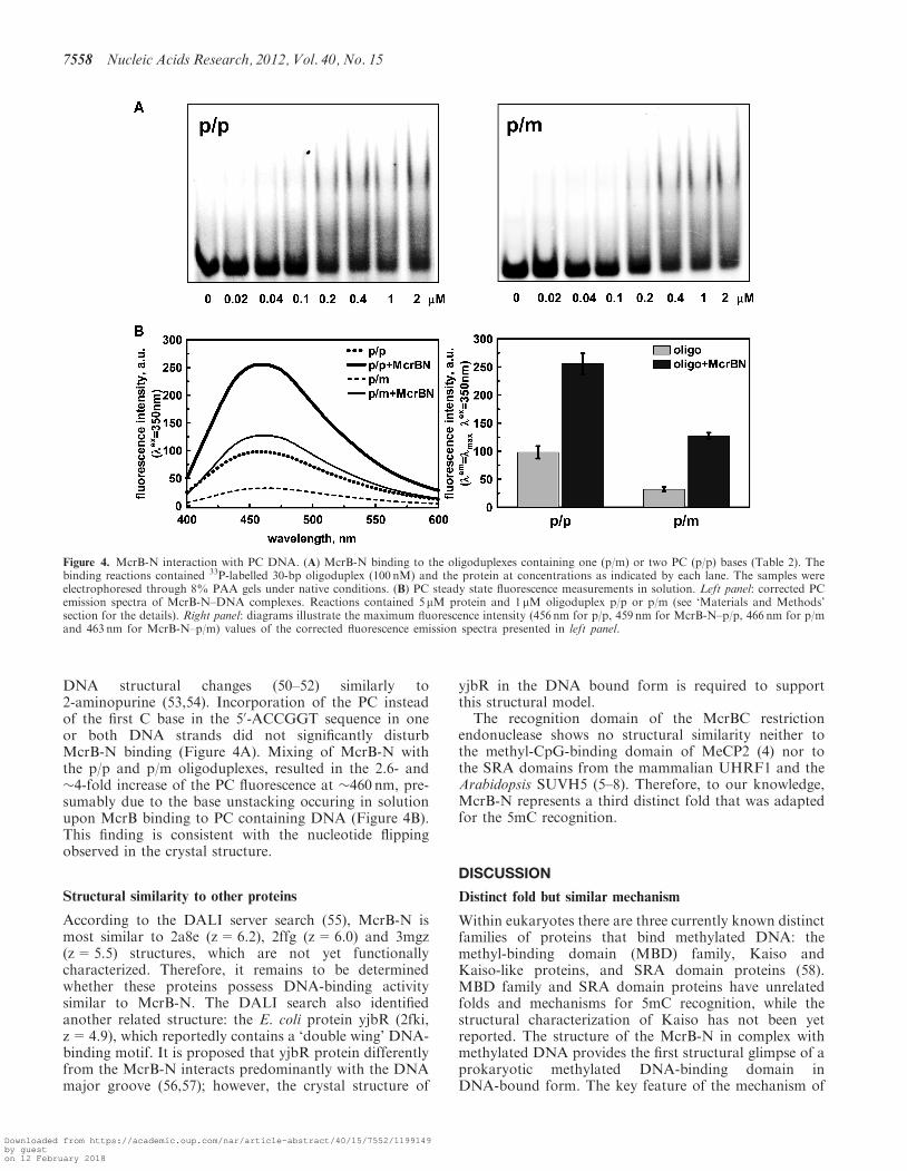
DNA structural changes (50–52) similarly to2-aminopurine (53,54). Incorporation of the PC insteadof the first C base in the 50-ACCGGT sequence in oneor both DNA strands did not significantly disturbMcrB-N binding (Figure 4A). Mixing of McrB-N withthe p/p and p/m oligoduplexes, resulted in the 2.6- and�4-fold increase of the PC fluorescence at �460 nm, pre-sumably due to the base unstacking occuring in solutionupon McrB binding to PC containing DNA (Figure 4B).This finding is consistent with the nucleotide flippingobserved in the crystal structure.
Structural similarity to other proteins
According to the DALI server search (55), McrB-N ismost similar to 2a8e (z=6.2), 2ffg (z=6.0) and 3mgz(z=5.5) structures, which are not yet functionallycharacterized. Therefore, it remains to be determinedwhether these proteins possess DNA-binding activitysimilar to McrB-N. The DALI search also identifiedanother related structure: the E. coli protein yjbR (2fki,z=4.9), which reportedly contains a ‘double wing’ DNA-binding motif. It is proposed that yjbR protein differentlyfrom the McrB-N interacts predominantly with the DNAmajor groove (56,57); however, the crystal structure of
yjbR in the DNA bound form is required to supportthis structural model.
The recognition domain of the McrBC restrictionendonuclease shows no structural similarity neither tothe methyl-CpG-binding domain of MeCP2 (4) nor tothe SRA domains from the mammalian UHRF1 and theArabidopsis SUVH5 (5–8). Therefore, to our knowledge,McrB-N represents a third distinct fold that was adaptedfor the 5mC recognition.
DISCUSSION
Distinct fold but similar mechanism
Within eukaryotes there are three currently known distinctfamilies of proteins that bind methylated DNA: themethyl-binding domain (MBD) family, Kaiso andKaiso-like proteins, and SRA domain proteins (58).MBD family and SRA domain proteins have unrelatedfolds and mechanisms for 5mC recognition, while thestructural characterization of Kaiso has not been yetreported. The structure of the McrB-N in complex withmethylated DNA provides the first structural glimpse of aprokaryotic methylated DNA-binding domain inDNA-bound form. The key feature of the mechanism of
Figure 4. McrB-N interaction with PC DNA. (A) McrB-N binding to the oligoduplexes containing one (p/m) or two PC (p/p) bases (Table 2). Thebinding reactions contained 33P-labelled 30-bp oligoduplex (100 nM) and the protein at concentrations as indicated by each lane. The samples wereelectrophoresed through 8% PAA gels under native conditions. (B) PC steady state fluorescence measurements in solution. Left panel: corrected PCemission spectra of McrB-N–DNA complexes. Reactions contained 5 mM protein and 1 mM oligoduplex p/p or p/m (see ‘Materials and Methods’section for the details). Right panel: diagrams illustrate the maximum Fuorescence intensity (456 nm for p/p, 459 nm for McrB-N–p/p, 466 nm for p/mand 463 nm for McrB-N–p/m) values of the corrected Fuorescence emission spectra presented in left panel.
7558 Nucleic Acids Research, 2012, Vol. 40, No. 15
Downloaded from https://academic.oup.com/nar/article-abstract/40/15/7552/1199149by gueston 12 February 2018
5mC recognition by the N-terminal domain of McrB is thebase flipping.
The structure analysis shows that three different stepsensure a unique discrimination of the 5mC against otherbases by McrB-N. First, the methylated cytosine is flippedout of the double helix and positioned in the well definedbinding pocket within the protein. The size of the pocketpre-selects pyrimidines against purines as a purine basewould not fit in. Second, direct readout of the donorand acceptor atoms on the Watson–Crick edge of theflipped out base discriminates cytosine against thymine.The pattern of the hydrogen bond donors and acceptorsprovided by the three amino acid residues within thebinding pocket of McrB-N is compatible to that of C or5mC but not of other heterocyclic bases in their typicalconformation. Finally, van der Waals interactions of theconserved binding pocket residues Tyr64 and Leu68residues with the methyl group discriminates 5mCagainst cytosine.
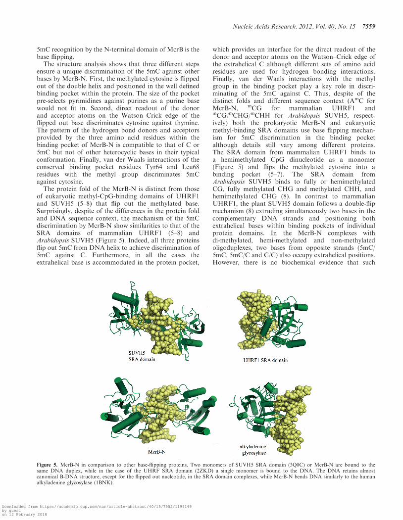
The protein fold of the McrB-N is distinct from thoseof eukaryotic methyl-CpG-binding domains of UHRF1and SUVH5 (5–8) that flip out the methylated base.Surprisingly, despite of the differences in the protein foldand DNA sequence context, the mechanism of the 5mCdiscrimination by McrB-N show similarities to that of theSRA domains of mammalian UHRF1 (5–8) andArabidopsis SUVH5 (Figure 5). Indeed, all three proteinsflip out 5mC from DNA helix to achieve discrimination of5mC against C. Furthermore, in all the cases theextrahelical base is accommodated in the protein pocket,
which provides an interface for the direct readout of thedonor and acceptor atoms on the Watson–Crick edge ofthe extrahelical C although different sets of amino acidresidues are used for hydrogen bonding interactions.Finally, van der Waals interactions with the methylgroup in the binding pocket play a key role in discri-minating of the 5mC against C. Thus, despite of thedistinct folds and different sequence context (AmC forMcrB-N, mCG for mammalian UHRF1 andmCG/mCHG/mCHH for Arabidopsis SUVH5, respect-ively) both the prokaryotic McrB-N and eukaryoticmethyl-binding SRA domains use base flipping mechan-ism for 5mC discrimination in the binding pocketalthough details still vary among different proteins.The SRA domain from mammalian UHRF1 binds toa hemimethylated CpG dinucleotide as a monomer(Figure 5) and flips the methylated cytosine into abinding pocket (5–7). The SRA domain fromArabidopsis SUVH5 binds to fully or hemimethylatedCG, fully methylated CHG and methylated CHH, andhemimethylated CHG (8). In contrast to mammalianUHRF1, the plant SUVH5 domain follows a double-flipmechanism (8) extruding simultaneously two bases in thecomplementary DNA strands and positioning bothextrahelical bases within binding pockets of individualprotein domains. In the McrB-N complexes withdi-methylated, hemi-methylated and non-methylatedoligoduplexes, two bases from opposite strands (5mC/5mC, 5mC/C and C/C) also occupy extrahelical positions.However, there is no biochemical evidence that such
Figure 5. McrB-N in comparison to other base-flipping proteins. Two monomers of SUVH5 SRA domain (3Q0C) or McrB-N are bound to thesame DNA duplex, while in the case of the UHRF SRA domain (2ZKD) a single monomer is bound to the DNA. The DNA retains almostcanonical B-DNA structure, except for the flipped out nucleotide, in the SRA domain complexes, while McrB-N bends DNA similarly to the humanalkyladenine glycosylase (1BNK).
Nucleic Acids Research, 2012, Vol. 40, No. 15 7559
Downloaded from https://academic.oup.com/nar/article-abstract/40/15/7552/1199149by gueston 12 February 2018
‘double flip’ mechanism would be important for McrBCfunction. Most likely, ‘double flip’ results from independ-ent interactions between two McrB-N monomers and two50-AC targets in the palindromic 50-ACCGGT sequence.Furthermore, differently from the SUVH5 and UHRF1SRA domains that do not disturb B-DNA conformationexcept of base flipping (5–8), McrB-N bends DNA nearlyby 30�. In this respect McrB-N mechanistically resemblesthe human alkyladenine glycosylase that approachesDNA form the minor groove side, flips out thedamaged base and severely distorts DNA conformation(59) (Figure 5). Intriguingly, ROS1 DNA glycosylasewhich catalyses active DNA demethylation also usesbase flipping to discriminate 5mC (60).
Implications for McrBC function
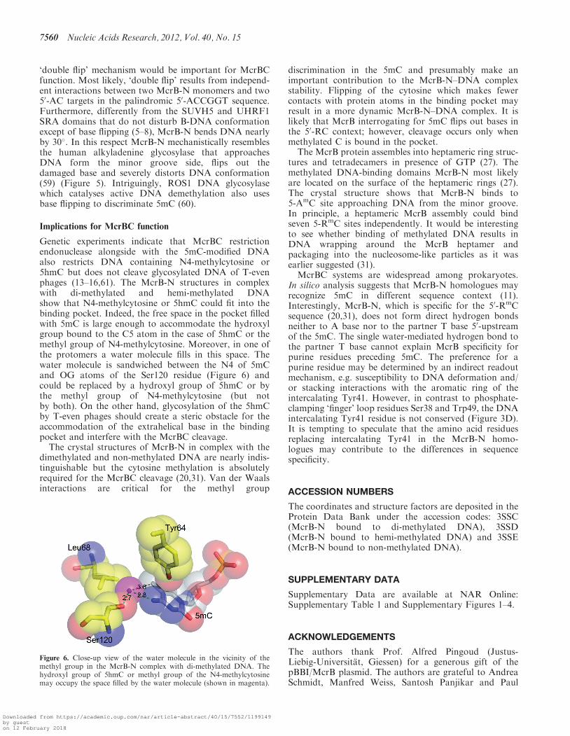
Genetic experiments indicate that McrBC restrictionendonuclease alongside with the 5mC-modified DNAalso restricts DNA containing N4-methylcytosine or5hmC but does not cleave glycosylated DNA of T-evenphages (13–16,61). The McrB-N structures in complexwith di-methylated and hemi-methylated DNAshow that N4-methylcytosine or 5hmC could fit into thebinding pocket. Indeed, the free space in the pocket filledwith 5mC is large enough to accommodate the hydroxylgroup bound to the C5 atom in the case of 5hmC or themethyl group of N4-methylcytosine. Moreover, in one ofthe protomers a water molecule fills in this space. Thewater molecule is sandwiched between the N4 of 5mCand OG atoms of the Ser120 residue (Figure 6) andcould be replaced by a hydroxyl group of 5hmC or bythe methyl group of N4-methylcytosine (but notby both). On the other hand, glycosylation of the 5hmCby T-even phages should create a steric obstacle for theaccommodation of the extrahelical base in the bindingpocket and interfere with the McrBC cleavage.The crystal structures of McrB-N in complex with the
dimethylated and non-methylated DNA are nearly indis-tinguishable but the cytosine methylation is absolutelyrequired for the McrBC cleavage (20,31). Van der Waalsinteractions are critical for the methyl group
discrimination in the 5mC and presumably make animportant contribution to the McrB-N–DNA complexstability. Flipping of the cytosine which makes fewercontacts with protein atoms in the binding pocket mayresult in a more dynamic McrB-N–DNA complex. It islikely that McrB interrogating for 5mC flips out bases inthe 50-RC context; however, cleavage occurs only whenmethylated C is bound in the pocket.
The McrB protein assembles into heptameric ring struc-tures and tetradecamers in presence of GTP (27). Themethylated DNA-binding domains McrB-N most likelyare located on the surface of the heptameric rings (27).The crystal structure shows that McrB-N binds to5-AmC site approaching DNA from the minor groove.In principle, a heptameric McrB assembly could bindseven 5-RmC sites independently. It would be interestingto see whether binding of methylated DNA results inDNA wrapping around the McrB heptamer andpackaging into the nucleosome-like particles as it wasearlier suggested (31).
McrBC systems are widespread among prokaryotes.In silico analysis suggests that McrB-N homologues mayrecognize 5mC in different sequence context (11).Interestingly, McrB-N, which is specific for the 50-RmCsequence (20,31), does not form direct hydrogen bondsneither to A base nor to the partner T base 50-upstreamof the 5mC. The single water-mediated hydrogen bond tothe partner T base cannot explain McrB specificity forpurine residues preceding 5mC. The preference for apurine residue may be determined by an indirect readoutmechanism, e.g. susceptibility to DNA deformation and/or stacking interactions with the aromatic ring of theintercalating Tyr41. However, in contrast to phosphate-clamping ‘finger’ loop residues Ser38 and Trp49, the DNAintercalating Tyr41 residue is not conserved (Figure 3D).It is tempting to speculate that the amino acid residuesreplacing intercalating Tyr41 in the McrB-N homo-logues may contribute to the differences in sequencespecificity.
ACCESSION NUMBERS
The coordinates and structure factors are deposited in theProtein Data Bank under the accession codes: 3SSC(McrB-N bound to di-methylated DNA), 3SSD(McrB-N bound to hemi-methylated DNA) and 3SSE(McrB-N bound to non-methylated DNA).
SUPPLEMENTARY DATA
Supplementary Data are available at NAR Online:Supplementary Table 1 and Supplementary Figures 1–4.
ACKNOWLEDGEMENTS
The authors thank Prof. Alfred Pingoud (Justus-Liebig-Universitat, Giessen) for a generous gift of thepBBI/McrB plasmid. The authors are grateful to AndreaSchmidt, Manfred Weiss, Santosh Panjikar and Paul
Figure 6. Close-up view of the water molecule in the vicinity of themethyl group in the McrB-N complex with di-methylated DNA. Thehydroxyl group of 5hmC or methyl group of the N4-methylcytosinemay occupy the space filled by the water molecule (shown in magenta).
7560 Nucleic Acids Research, 2012, Vol. 40, No. 15
Downloaded from https://academic.oup.com/nar/article-abstract/40/15/7552/1199149by gueston 12 February 2018
Tucker (EMBL Hamburg) for their expert assistance atthe beamlines. The authors thank Giedre Tamulaitiene,Lena Manakova, Dmitrij Golovenko and GiedriusSasnauskas for valuable discussions.
FUNDING
Funding for open access charge: European Social Fundunder the Global Grant measure [project R100].
Conflict of interest statement. None declared.
REFERENCES
1. Bird,A. (2002) DNA methylation patterns and epigenetic memory.Genes Dev., 16, 6–21.
2. Tost,J. (2010) DNA methylation: an introduction to the biologyand the disease-associated changes of a promising biomarker.Mol. Biotechnol., 44, 71–81.
3. Henderson,I.R. and Jacobsen,S.E. (2007) Epigenetic inheritance inplants. Nature, 447, 418–424.
4. Ho,K.L., McNae,I.W., Schmiedeberg,L., Klose,R.J., Bird,A.P.and Walkinshaw,M.D. (2008) MeCP2 binding to DNA dependsupon hydration at methyl-CpG. Mol. Cell, 29, 525–531.
5. Arita,K., Ariyoshi,M., Tochio,H., Nakamura,Y. andShirakawa,M. (2008) Recognition of hemi-methylated DNA bythe SRA protein UHRF1 by a base-flipping mechanism. Nature,455, 818–821.
6. Avvakumov,G.V., Walker,J.R., Xue,S., Li,Y., Duan,S.,Bronner,C., Arrowsmith,C.H. and Dhe-Paganon,S. (2008)Structural basis for recognition of hemi-methylated DNA by theSRA domain of human UHRF1. Nature, 455, 822–825.
7. Hashimoto,H., Horton,J.R., Zhang,X., Bostick,M., Jacobsen,S.E.and Cheng,X. (2008) The SRA domain of UHRF1 flips5-methylcytosine out of the DNA helix. Nature, 455, 826–829.
8. Rajakumara,E., Law,J.A., Simanshu,D.K., Voigt,P.,Johnson,L.M., Reinberg,D., Patel,D.J. and Jacobsen,S.E. (2011)A dual flip-out mechanism for 5mC recognition by theArabidopsis SUVH5 SRA domain and its impact on DNAmethylation and H3K9 dimethylation in vivo. Genes Dev., 25,137–152.
9. Iyer,L.M., Abhiman,S. and Aravind,L. (2011) Natural history ofeukaryotic DNA methylation systems. Prog. Mol. Biol. Transl.Sci., 101, 25–104.
10. Bickle,T.A. and Kruger,D.H. (1993) Biology of DNA restriction.Microbiol. Rev., 57, 434–450.
11. Ishikawa,K., Fukuda,E. and Kobayashi,I. (2010) Conflictstargeting epigenetic systems and their resolution by cell death:novel concepts for methyl-specific and other restriction systems.DNA Res., 17, 325–342.
12. Warren,R.A. (1980) Modified bases in bacteriophage DNAs.Annu. Rev. Microbiol., 34, 137–158.
13. Hattman,S. and Fukasawa,T. (1963) Host-induced modification ofT-even phages due to defective glucosylation of their DNA. Proc.Natl Acad. Sci. USA, 50, 297–300.
14. Shedlovsky,A. and Brenner,S. (1963) A chemical basis for thehost-induced modification of T-even bacteriophages. Proc. NatlAcad. Sci. USA, 50, 300–305.
15. Raleigh,E.A., Trimarchi,R. and Revel,H. (1989) Genetic andphysical mapping of the mcrA (rglA) and mcrB (rglB) loci ofEscherichia coli K-12. Genetics, 122, 279–296.
16. Luria,S.E. and Human,M.L. (1952) A nonhereditary, host-inducedvariation of bacterial viruses. J. Bacteriol., 64, 557–569.
17. Hiom,K. and Sedgwick,S.G. (1991) Cloning and structuralcharacterization of the mcrA locus of Escherichia coli.J. Bacteriol., 173, 7368–7373.
18. Sutherland,E., Coe,L. and Raleigh,E.A. (1992) McrBC: amultisubunit GTP-dependent restriction endonuclease.J. Mol. Biol., 225, 327–348.
19. Roberts,R.J., Vincze,T., Posfai,J. and Macelis,D. (2010)REBASE–a database for DNA Restriction and modification:enzymes, genes and genomes. Nucleic Acids Res., 38, D234–D236.
20. Kruger,T., Wild,C. and Noyer-Weidner,M. (1995) McrB: aprokaryotic protein specifically recognizing DNA containingmodified cytosine residues. EMBO J., 14, 2661–2669.
21. Dila,D., Sutherland,E., Moran,L., Slatko,B. and Raleigh,E.A.(1990) Genetic and sequence organization of the mcrBC locus ofEscherichia coli K-12. J. Bacteriol., 172, 4888–4900.
22. Pieper,U. and Pingoud,A. (2002) A mutational analysis of thePD. . .D/EXK motif suggests that McrC harbors the catalyticcenter for DNA cleavage by the GTP-dependent restrictionenzyme McrBC from Escherichia coli. Biochemistry, 41,5236–5244.
23. Pieper,U., Brinkmann,T., Kruger,T., Noyer-Weidner,M. andPingoud,A. (1997) Characterization of the interaction between therestriction endonuclease McrBC from E. coli and its cofactorGTP. J. Mol. Biol., 272, 190–199.
24. Neuwald,A.F., Aravind,L., Spouge,J.L. and Koonin,E.V. (1999)AAA+: A class of chaperone-like ATPases associated with theassembly, operation, and disassembly of protein complexes.Genome Res., 9, 2743.
25. Pieper,U., Schweitzer,T., Groll,D.H., Gast,F.U. and Pingoud,A.(1999) The GTP-binding domain of McrB: more than just avariation on a common theme? J. Mol. Biol., 292, 547–556.
26. White,S.R. and Lauring,B. (2007) AAA+ATPases: achievingdiversity of function with conserved machinery. Traffic, 8,1657–1667.
27. Panne,D., Muller,S.A., Wirtz,S., Engel,A. and Bickle,T.A. (2001)The McrBC restriction endonuclease assembles into a ringstructure in the presence of G nucleotides. EMBO J., 20,3210–3217.
28. Stewart,F.J. and Raleigh,E.A. (1998) Dependence of McrBCcleavage on distance between recognition elements. Biol. Chem.,379, 611–616.
29. Panne,D., Raleigh,E.A. and Bickle,T.A. (1999) The McrBCendonuclease translocates DNA in a reaction dependent on GTPhydrolysis. J. Mol. Biol., 290, 49–60.
30. Gast,F.U., Brinkmann,T., Pieper,U., Kruger,T., Noyer-Weidner,M. and Pingoud,A. (1997) The recognition of methylatedDNA by the GTP-dependent restriction endonuclease McrBCresides in the N-terminal domain of McrB. Biol. Chem., 378,975–982.
31. Stewart,F.J., Panne,D., Bickle,T.A. and Raleigh,E.A. (2000)Methyl-specific DNA binding by McrBC, amodification-dependent restriction enzyme. J. Mol. Biol., 298,611–622.
32. Pieper,U., Schweitzer,T., Groll,D.H. and Pingoud,A. (1999)Defining the location and function of domains of McrB bydeletion mutagenesis. Biol. Chem., 380, 1225–1230.
33. Leslie,A.G. (2006) The integration of macromolecular diffractiondata. Acta Crystallogr. D Biol. Crystallogr., 62, 48–57.
34. Evans,P. (2006) Scaling and assessment of data quality. ActaCrystallogr. D Biol. Crystallogr., 62, 72–82.
35. French,G.S. and Wilson,K.S. (1978) On the treatment of negativeintensity observations. Acta Crystallogr., A34, 517–525.
36. CCP4. (1994) The CCP4 suite: programs for proteincrystallography. Acta Crystallogr. D Biol. Crystallogr., 50,760–763.
37. Sheldrick,G.M. (2008) A short history of SHELX. ActaCrystallogr. A, 64, 112–122.
38. Panjikar,S., Parthasarathy,V., Lamzin,V.S., Weiss,M.S. andTucker,P.A. (2005) Auto-Rickshaw: an automated crystalstructure determination platform as an efficient tool for thevalidation of an X-ray diffraction experiment. Acta Crystallogr.D Biol. Crystallogr., 61, 449–457.
39. Morris,R.J., Perrakis,A. and Lamzin,V.S. (2002) ARP/wARP’smodel-building algorithms. I. The main chain. Acta Crystallogr.D Biol. Crystallogr., 58, 968–975.
40. Vagin,A. and Teplyakov,A. (2010) Molecular replacement withMOLREP. Acta Crystallogr. D Biol. Crystallogr., 66, 22–25.
41. Emsley,P. and Cowtan,K. (2004) Coot: model-building tools formolecular graphics. Acta Crystallogr. D Biol. Crystallogr., 60,2126–2132.
Nucleic Acids Research, 2012, Vol. 40, No. 15 7561
Downloaded from https://academic.oup.com/nar/article-abstract/40/15/7552/1199149by gueston 12 February 2018
42. Brunger,A.T., Adams,P.D., Clore,G.M., DeLano,W.L., Gros,P.,Grosse-Kunstleve,R.W., Jiang,J.S., Kuszewski,J., Nilges,M.,Pannu,N.S. et al. (1998) Crystallography & NMR system: a newsoftware suite for macromolecular structure determination. ActaCrystallogr. D Biol. Crystallogr., 54, 905–921.
43. Murshudov,G.N., Vagin,A.A. and Dodson,E.J. (1997)Refinement of macromolecular structures by themaximum-likelihood method. Acta Crystallogr. D Biol.Crystallogr., 53, 240–255.
44. Davis,I.W., Leaver-Fay,A., Chen,V.B., Block,J.N., Kapral,G.J.,Wang,X., Murray,L.W., Arendall,W.B. 3rd, Snoeyink,J.,Richardson,J.S. et al. (2007) MolProbity: all-atom contacts andstructure validation for proteins and nucleic acids. Nucleic AcidsRes., 35, W375–W383.
45. Luscombe,N.M., Laskowski,R.A. and Thornton,J.M. (1997)NUCPLOT: a program to generate schematic diagrams ofprotein-nucleic acid interactions. Nucleic Acids Res., 25,4940–4945.
46. Lavery,R. and Sklenar,H. (1988) The definition of generalizedhelicoidal parameters and of axis curvature for irregular nucleicacids. J. Biomol. Struct. Dyn., 6, 63–91.
47. Martin,A.M., Sam,M.D., Reich,N.O. and Perona,J.J. (1999)Structural and energetic origins of indirect readout in site-specificDNA cleavage by a restriction endonuclease. Nat. Struct. Biol., 6,269–277.
48. Combet,C., Blanchet,C., Geourjon,C. and Deleage,G. (2000)NPS@: network protein sequence analysis. Trends Biochem. Sci.,25, 147–150.
49. Berry,D.A., Jung,K., Wise,D.S., Sercel,D.A., Pearson,W.H.,Mackie,H., Randolph,J.B. and Somers,R.L. (2004) Pyrrolo-dCand pyrrolo-C: fluorescent analogs of cytidine and 20-deoxycytidine for the study of oligonucleotides. Tetrahedron Lett.,45, 2457–2461.
50. Liu,C. and Martin,C.T. (2001) Fluorescence characterization ofthe transcription bubble in elongation complexes of T7 RNApolymerase. J. Mol. Biol., 308, 465–475.
51. Dash,C., Rausch,J.W. and Le Grice,S.F. (2004) Usingpyrrolo-deoxycytosine to probe RNA/DNA hybrids containing
the human immunodeficiency virus type-1 30 polypurine tract.Nucleic Acids Res., 32, 1539–1547.
52. Tinsley,R.A. and Walter,N.G. (2006) Pyrrolo-C as a fluorescentprobe for monitoring RNA secondary structure formation. RNA,12, 522–529.
53. Holz,B., Klimasauskas,S., Serva,S. and Weinhold,E. (1998)2-Aminopurine as a fluorescent probe for DNA base flipping bymethyltransferases. Nucleic Acids Res., 26, 1076–1083.
54. Tamulaitis,G., Zaremba,M., Szczepanowski,R.H., Bochtler,M. andSiksnys,V. (2007) Nucleotide flipping by restriction enzymesanalyzed by 2-aminopurine steady-state fluorescence. NucleicAcids Res., 35, 4792–4799.
55. Holm,L., Kaariainen,S., Rosenstrom,P. and Schenkel,A. (2008)Searching protein structure databases with DaliLite v.3.Bioinformatics, 24, 2780–2781.
56. Li,N., Sickmier,E.A., Zhang,R., Joachimiak,A. and White,S.W.(2002) The MotA transcription factor from bacteriophage T4contains a novel DNA-binding domain: the ‘double wing’ motif.Mol. Microbiol., 43, 1079–1088.
57. Singarapu,K.K., Liu,G., Xiao,R., Bertonati,C., Honig,B.,Montelione,G.T. and Szyperski,T. (2007) NMR structure ofprotein yjbR from Escherichia coli reveals ‘double-wing’ DNAbinding motif. Proteins, 67, 501–504.
58. Defossez,P.A. and Stancheva,I. (2010) Biological functions ofmethyl-CpG-binding proteins. Prog. Mol. Biol. Transl. Sci., 101,377–398.
59. Lau,A.Y., Scharer,O.D., Samson,L., Verdine,G.L. andEllenberger,T. (1998) Crystal structure of a humanalkylbase-DNA repair enzyme complexed to DNA: mechanismsfor nucleotide flipping and base excision. Cell, 95, 249–258.
60. Ponferrada-Marin,M.I., Parrilla-Doblas,J.T., Roldan-Arjona,T.and Ariza,R.R. (2011) A discontinuous DNA glycosylase domainin a family of enzymes that excise 5-methylcytosine. Nucleic AcidsRes., 39, 1473–1484.
61. Raleigh,E.A. and Wilson,G. (1986) Escherichia coli K-12 restrictsDNA containing 5-methylcytosine. Proc. Natl Acad. Sci. USA,83, 9070–9074.
7562 Nucleic Acids Research, 2012, Vol. 40, No. 15
Downloaded from https://academic.oup.com/nar/article-abstract/40/15/7552/1199149by gueston 12 February 2018