the recording of jugular venous & carotid arterial pulses
TRANSCRIPT

The Recording of Jugular The Recording of Jugular Venous & Carotid Arterial Venous & Carotid Arterial
PulsesPulses

Dr. AbeerDr. Abeer Cardiovascular PracticalCardiovascular Practical 22
Objectives:Objectives:
To be able toTo be able to:: identify,identify, understand the events causing the understand the events causing the
different waves of the JVP & CP tracings.different waves of the JVP & CP tracings.

Dr. AbeerDr. Abeer Cardiovascular PracticalCardiovascular Practical 33
A. The Carotid Arterial PulseA. The Carotid Arterial Pulse
MethodMethod::
1.1. Subject lies quietly on a couch.Subject lies quietly on a couch.2.2. Feel CAP on medial side of Feel CAP on medial side of
sternomastoid muscle.sternomastoid muscle.3.3. Apply transducer over CA using Apply transducer over CA using
soft rubber band & connect it to soft rubber band & connect it to recorder.recorder.

Dr. AbeerDr. Abeer Cardiovascular PracticalCardiovascular Practical 44
• Pulse:
Record of pressure changes created
by ejection of blood from LV into
already full aorta & is propagated as a
wave over the vessel wall.

Dr. AbeerDr. Abeer Cardiovascular PracticalCardiovascular Practical 55
Recorded CAP graph:Recorded CAP graph:
Anacrotic limbAnacrotic limb:: - - Record during maximum ejection phase Record during maximum ejection phase of ventricular systole.of ventricular systole. Dicrotic notch (Incisura)Dicrotic notch (Incisura):: - - Due to closure of aortic valve.Due to closure of aortic valve. Dicrotic waveDicrotic wave:: - - Due to elastic recoil of arterial wall.Due to elastic recoil of arterial wall. Dicrotic limbDicrotic limb:: (descending)(descending)

Dr. AbeerDr. Abeer Cardiovascular PracticalCardiovascular Practical 66

Dr. AbeerDr. Abeer Cardiovascular PracticalCardiovascular Practical 77
Cardiac Cycle duration = 0.8 sec.Cardiac Cycle duration = 0.8 sec. Ventricular systole = 0.3 sec.Ventricular systole = 0.3 sec. Ventricular diastole = 0.5 sec.Ventricular diastole = 0.5 sec.

Dr. AbeerDr. Abeer Cardiovascular PracticalCardiovascular Practical 88
Aortic StenosisAortic Stenosis:: - - Slow rising pulse, small volume, late systolic peak.Slow rising pulse, small volume, late systolic peak. Shock or dehydrationShock or dehydration:: - - weak or thready pulse, due to weak or thready pulse, due to volume. volume. Aortic RegurgitationAortic Regurgitation:: - - Collapsing pulse (water hummer). Collapsing = Collapsing pulse (water hummer). Collapsing = Diastolic leak back to Lt. ventricle. Rapid up stroke (Diastolic leak back to Lt. ventricle. Rapid up stroke ( stroke volume & stroke volume & pulse wave). pulse wave). HypertensionHypertension:: - - Bounding pulse, due to good volume.Bounding pulse, due to good volume. PregnancyPregnancy:: (N)(N) - due to good volume.- due to good volume.
Clinical abnormalities:Clinical abnormalities:

Dr. AbeerDr. Abeer Cardiovascular PracticalCardiovascular Practical 99
B. The Jugular Venous PulseB. The Jugular Venous Pulse
MethodMethod::1.1. Subject performs Valsalva manoeuvre (deep Subject performs Valsalva manoeuvre (deep
inspiration followed by forced expiration inspiration followed by forced expiration against closed glottis), internal jugular vein will against closed glottis), internal jugular vein will be prominent.be prominent.
2.2. Choose position on the IJV away from CA.Choose position on the IJV away from CA.
3.3. Place pulse transducer over the vein & keep it Place pulse transducer over the vein & keep it in position with self adhesive plaster.in position with self adhesive plaster.
4.4. Connect to recorder.Connect to recorder.

Dr. AbeerDr. Abeer Cardiovascular PracticalCardiovascular Practical 1010
Pressure changes in RA can be recorded from IJV as there are no valves between them.The EJV can’t be relied because it:1. has valves, 2. ? obstructed by facial & muscular layers through which it passes.
JVP in:1. Rt. Sided heart failure.2. Fluid overload.
Dr. AbeerDr. Abeer Cardiovascular PracticalCardiovascular Practical 1111
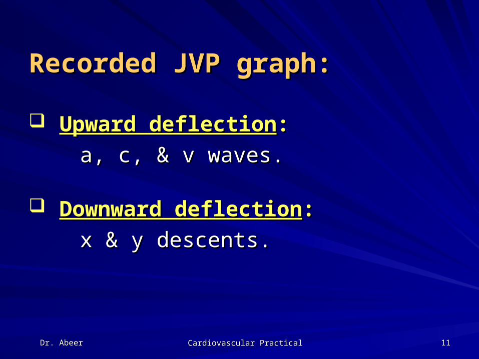
Recorded JVP graph:Recorded JVP graph:
Upward deflectionUpward deflection::
a, c, & v waves.a, c, & v waves.
Downward deflectionDownward deflection::
x & y descents.x & y descents.

Dr. AbeerDr. Abeer Cardiovascular PracticalCardiovascular Practical 1212
Causes of these waves:Causes of these waves:
‘‘a’ wavea’ wave: : RA contraction.RA contraction. ‘‘c’ wavec’ wave:: Bulging of TV into RA during Bulging of TV into RA during
isovolumetric contraction phase.isovolumetric contraction phase. ‘‘v’ wavev’ wave:: RA press due to filling of atrium RA press due to filling of atrium
with blood, (venous return.)with blood, (venous return.) ‘‘x’ descentx’ descent:: Downward displacement of TV Downward displacement of TV
during rapid ejection phase.during rapid ejection phase. ‘‘y’ descenty’ descent:: Rapid blood flow from RA to RV. Rapid blood flow from RA to RV.

Dr. AbeerDr. Abeer Cardiovascular PracticalCardiovascular Practical 1313

Dr. AbeerDr. Abeer Cardiovascular PracticalCardiovascular Practical 1414
Q. How to identify JVP tracing?Q. How to identify JVP tracing?
1.1. First identify ‘v’ wave, you will find two First identify ‘v’ wave, you will find two descents ‘x’ & ‘y’ on either side of ‘v’.descents ‘x’ & ‘y’ on either side of ‘v’.
2. The ‘a’ & ‘c’ wave precede the ‘x’ 2. The ‘a’ & ‘c’ wave precede the ‘x’ descent.descent.

Dr. AbeerDr. Abeer Cardiovascular PracticalCardiovascular Practical 1515
Clinical abnormalities:Clinical abnormalities: ‘‘a’ wavea’ wave:: • Prominent:Prominent: 1. RV hypertrophy (1. RV hypertrophy ( resist of filling) resist of filling)
2. Pulmonary stenosis.2. Pulmonary stenosis. 3. Pulmonary hypertension.3. Pulmonary hypertension. 4. Tricuspid stenosis.4. Tricuspid stenosis.• Absence:Absence: Atrial fibrillation, TR.Atrial fibrillation, TR.
• Cannon wave:Cannon wave: Complete AV block, atrial flutter, Complete AV block, atrial flutter,
ventricular extrasystole.ventricular extrasystole. ‘‘c’ wavec’ wave: : Prominent in TR; absent in const.peric.Prominent in TR; absent in const.peric.
‘‘v’ wavev’ wave: : Prominent in constrictive pericarditis.Prominent in constrictive pericarditis.

Dr. AbeerDr. Abeer Cardiovascular PracticalCardiovascular Practical 1616
Thank You