the regulation of drosophila inhibitor of apoptosis protein - deep blue
TRANSCRIPT

THE REGULATION OF DROSOPHILA INHIBITOR
OF APOPTOSIS PROTEIN I
IN APOPTOSIS AND CELL SURVIVAL
by
Hui Li
A dissertation submitted in partial fulfillment of the requirements for the degree of
Doctor of Philosophy (Molecular Cellular and Developmental Biology)
in The University of Michigan 2009
Doctoral Committee: Associate Professor Kenneth M. Cadigan, Chair Professor Steven E Clark Professor Eva L Feldman Professor Ormond A MacDougald Associate Professor Jianming Li

The path to glory is always rugged
天将降大任于斯人也,必先苦其心志,劳其筋骨,饿其体肤,
空乏其身,行拂乱其所为,所以动心忍性,增益其所不能.
—孟子

© Hui Li
All Rights Reserved
2009

ii
To my mother and husband

iii
ACKNOWLEDGEMENTS
It has been six years since I came to Ann Arbor to pursue my PhD degree. I
would like to express my gratitude to all the people I met during this period. It has been
a great journey.
First and foremost, I would like to thank my advisor, Dr. Ken Cadigan, for his
continuous guidance, wholehearted support and enthusiastic encouragement. Ken has
tremendous influence on me both inside and outside science. He has taught me an
incredible amount about how to do research, how to envision 'the big picture' and then
work out the details, and how to be a logical, critical and creative thinker, all of which are
a truly invaluable treasure for me.
I am extremely grateful to the members of my thesis committee, Dr. Steve Clark,
Dr. Eva Feldman, Dr. Ormond Macdougald and Dr. Jianming Li, for their insightful,
thought provoking and constructive advice, which has greatly improved the quality of my
dissertation work.
I would like to thank all current and previous Cadigan lab members for providing
such an intellectual stimulating, supportive and enjoyable environment. I owe my
gratitude to my friend and classmate, Yunyun Ni, who took the six-year journey together
with me to achieve every milestone along the way, supporting me professionally and
personally, and sharing openly with me happeniess and sadness. I am grateful to have

iv
such a true friend in the lab. I would like to thank Dr. Ju Guan, who I worked with
during my first year on the Dco project, and Dr. Jennifer Kennell and undergraduate
student Liyu Tan, who also helped me with the Dco and Gish project. Thanks to
Chandan Bhambhani and Dave Parker, my current and previous baymates, and Yan Liu,
who shared a big bench with me. They always had interesting things to share with me,
making my life in the lab very delightful. I also want to thank Mikyung Chang, JinHee
Chang and Jennifer Kennell for their help and support. They were always so
considerate and caring.
Thanks to our neighbors, Collins Lab, Csankovszki Lab and Raymond Lab, for
their generosity for reagents, equipments and advice, as well as all the talks in the
hallway and happy hours. During my first two years here, our lab didn’t have neighbors,
therefore, I really cherish our lovely third floor corner right now. I want to thank Dr.
Clark for giving me the opportunity to rotate in his lab, and all people in Clark lab.
I would like to thank Dr. Santhadevi Jeyabalan, who I worked for as Graduate
Student Instructor for three terms. She really helped me grow up as a confident
instructor and start to love teaching. Special thanks to Dr. Diane Spillane, who I really
should have known personally much earlier than when we taught Introductory Biology
together. She generously helped me in the most difficult and depressing moment I had
in the past several years.

v
I appreciate all the useful discussions on my projects with people in several joint
group meetings our lab participated in.
Thanks to Ms. Mary Carr, who is always ready to help. She made my life here
much easier. Thanks to Sobocinski Gregg for helping me with confocal microscopy.
Also, thanks to all people in MCDB office.
I would like to sincerely thank my husband Weifeng Ye and my mom for their
unconditional love and unwavering support. I am extremely grateful to all my friends
for making my journey here so much enjoyable and rewarding.

vi
TABLE OF CONTENTS
DEDICATION.................................................................................................................... ii
ACKNOWLEDGEMENTS............................................................................................... iii
LIST OF FIGURES ........................................................................................................... xi
LIST OF TABLES........................................................................................................... xiv
CHAPTER I GENERAL INTRODUCTION................................................................... 1
Apoptosis in multi-cellular organism........................................................................ 1
Living with Death ........................................................................................... 1
Programmed cell death is an actively regulated cell death process ................ 1
Apoptosis has been implicated in development and adult homeostasis ......... 3
Apoptosis machinery is evolutionarily conserved .......................................... 4
Apoptosis regulation in Drosophila .......................................................................... 7
Apoptosis happens throughout fly development............................................. 8
Apoptosis at the perimeter of pupal eye ......................................................... 9
The apoptotic core machinery is composed of Dark, Dronc and Drice........ 12
Inhibitor of Apoptosis Protein (IAP) is an essential inhibitor of apoptosis............ 15
DIAP1 is an essential inhibitor of apoptosis in Drosophila ......................... 18
DIAP1 regulates caspases through several mechanisms .............................. 19
Activation of apoptosis by RHG proteins............................................................... 22
Liberation Model—RHG proteins antagonize DIAP1 functions through their binding to BIR domains........................................................................ 23

vii
Degradation Model—RHG proteins regulate DIAP1 level through ubiquitination and degradation ..................................................................... 25
RHG proteins activate apoptosis through other mechanisms ....................... 27
Coordination between cell proliferation and Apoptosis ......................................... 30
The Hippo pathway mediates the coordination between cell death and proliferation................................................................................................... 30
Apoptosis induces compensatory proliferation............................................. 31
Caspases have other non-apoptotic functions ............................................... 33
Rationale and specific aims .................................................................................... 35
Figures and Tables .................................................................................................. 37
Reference ................................................................................................................ 45
Chapter II RHG PROTEIN MEDIATED APOPTOSIS IN THE PUPAL EYE OF DROSOPHILA: DOWN-REGULATION OF DIAP1..................................... 57
Abstract ................................................................................................................... 57
Introduction............................................................................................................. 58
Materials and Methods............................................................................................ 63
Drosophila Genetics ..................................................................................... 63
Immunostainings and TUNEL...................................................................... 64
Microscopy and image analysis .................................................................... 65
Results..................................................................................................................... 66
DIAP1 is down-regulated post-transcriptionally in peripheral ommatidia that undergo apoptosis. ................................................................................. 66
DIAP1 down-regulation is independent of effector caspase activity but requires Wg signaling as well as Hid, Grim and Rpr. .................................. 67
Apoptosis can occur in the absence of DIAP1 down-regulation in peripheral ommatidia. ................................................................................... 69
Down-regulation of DIAP1 in the perimeter ommatidia depends on its BIR1 and BIR2 domains, but is independent of its RING domain............... 70
Removal of Dronc and Drak partially suppresses DIAP1 down-regulation. 72
Discussion............................................................................................................... 73

viii
The degradation vs. the liberation model...................................................... 73
Role of the RING domain in DIAP1 down-regulation ................................. 75
Role of the BIR domains in DIAP1 down-regulation................................... 76
Role of Dark and Dronc in DIAP1 down-regulation .................................... 77
Acknowledgements................................................................................................. 78
References............................................................................................................... 90
CHAPTER III THE DROSOPHILA CASEIN KINASE I EPSILON DISCS OVERGROWN PROMOTES CELL SURVIVAL VIA ACTIVATION OF DIAP1 EXPRESSION ..................................................................................... 94
Abstract ................................................................................................................... 94
Introduction............................................................................................................. 95
Materials and methods ............................................................................................ 98
Fly strains...................................................................................................... 98
Generation of dco clones ............................................................................ 100
Immunostainings, TUNEL and in situ hybridization.................................. 100
Microscopy and image analysis .................................................................. 101
Results................................................................................................................... 101
Dco preferentially suppresses Hid induced cell death in the eye................ 101
dco mutant imaginal discs have elevated apoptosis.................................... 104
Dco activates DIAP1 expression in the wing imaginal disc ....................... 105
dco mutant clones are significantly smaller than their twins...................... 109
The E3 ubiquitin ligase activity of DIAP1 is not required for its regulation by Dco........................................................................................ 110
dco mutant clones have normal Wg and Dpp signaling ............................. 111
Discussion............................................................................................................. 112
Dco promotes cell survival by activating DIAP1 expression ..................... 112
The role of the IAP antagonists Hid, Grim and Rpr ................................... 114
Dco may promote cell growth/proliferation ............................................... 116

ix
Relation of Dco to Wg and other pathways affecting PCD/proliferation... 117
CKIδ/ε in other systems.............................................................................. 119
Acknowledgements............................................................................................... 119
Figures................................................................................................................... 121
References............................................................................................................. 133
CHAPTER IV EXPLORATION OF THE REGULATION MECHANISM OF APOPTOSIS AND THE WINGLESS PATHWAY BY DROSOPHILA CASEIN KINASE I FAMILY PROTEINS ................................................... 138
Abstract ................................................................................................................. 138
Introduction........................................................................................................... 139
Introduction to Casein Kinase I family....................................................... 139
Apoptosis and CKI...................................................................................... 140
Wingless signaling and CKI ....................................................................... 142
Materials and methods .......................................................................................... 144
Cell Culture, RNAi treatment, DNA transfection, and conditional media treatment ..................................................................................................... 144
Cell survival assay and flow cytometry ...................................................... 146
Real-time quantitative RT-PCR (qRT-PCR) .............................................. 146
Fly Genetics ................................................................................................ 147
Immunostainings and Western blotting ...................................................... 148
Whole-mount staining and microscopy ...................................................... 148
Results................................................................................................................... 149
Exploration of the anti-apoptosis function of Dco in S2 cells .................... 149
Exploration of potential redundant partner of Dco ..................................... 150
Exploration of the redundancy between Gish and Dco .............................. 152
Exploration of Dco’s function in the Wingless signaling pathway ........... 153
Attempted identification of Dco substrates by the Shocat substrate screen 156
Discussion............................................................................................................. 159
x
How does Dco regulate DIAP1 protein levels? .......................................... 159
Putative function of Gish in DIAP1 regulation........................................... 160
Dco as a positive regulator of Wg signaling pathway ................................ 161
Does Dco have a redundant partner? .......................................................... 162
Attempted identification of Dco kinase substrates ..................................... 163
Acknowledgements............................................................................................... 164
Figures................................................................................................................... 166
Reference .............................................................................................................. 180
CHAPTER V CONCLUDING REMARKS................................................................... 183
Summary of contributions..................................................................................... 183
RHG protein mediated apoptosis in the pupal eye of Drosophila: down-regulation of DIAP1 ......................................................................... 183
The Drosophila casein kinase Iε/δ Discs overgrown promotes cell survival via activation of DIAP1 expression .............................................. 185
Future directions ................................................................................................... 187
Exploration of the apoptosis regulation via the ubiquitination pathway .... 187
DIAP1 regulation in other developmental apoptosis .................................. 189
Characterization of Gilgamesh (Gish) in DIAP1 regulation....................... 191
Identification of Dco substrates .................................................................. 193
Prospects ............................................................................................................... 195
Reference .............................................................................................................. 197

xi
LIST OF FIGURES
Figure 1.1 Ultrastructural features of cell death by apoptosis .......................................... 37
Figure 1.2 A schematic Drosophila Ommatidium............................................................ 38
Figure 1.3 Periphery ommatidia apoptosis triggered by Wg ............................................ 39
Figure 1.4 The projection of receptor axons from a single ommatidium ......................... 40
Figure 1.5 Schematic representation of IAP family members in mammal and fly........... 41
Figure 1.6 Fly and mammalian RHG proteins.................................................................. 42
Figure 1.7 Regulators of caspase-dependent cell death in Drosophila............................. 43
Figure 2. 1 DIAP1 is down-regulated post-transcriptionally in peripheral ommatidia that undergo apoptosis...................................................................................... 79
Figure 2. 2 DIAP1 is down-regulated in peripheral ommatidia that undergo apoptosis .. 80
Figure 2.3 Down-regulation of DIAP1 is inhibited in peripheral ommatidia where Wg signaling is blocked by overexpression of GPI-fz2................................... 81
Figure 2.4 Hid, Grim and Rpr contribute to DIAP1 down-regulation in peripheral ommatidia ......................................................................................................... 82
Figure 2.5 The absence of DIAP1 down-regulation attenuates the progression of apoptosis ........................................................................................................... 83
Figure 2.6 Apoptosis can occur in the absence of DIAP1 down-regulation in peripheral ommatidia........................................................................................ 84
Figure 2.7 Down-regulation of DIAP1 in the peripheral ommatidia depends on its Bir1 and Bir2 domains ..................................................................................... 85
Figure 2.8 Downregulation of DIAP1 in peripheral ommatidia is independent of its RING domain ................................................................................................... 86
Figure 2.9 Dronc and Dark are required for DIAP1 down-regulation.............................. 87
Figure 2.10 Removal of Hid, Rpr and Grim blocks DIAP1 down-regulation in peripheral ommatidia........................................................................................ 88
Figure 3.1 Ectopic expression of dco suppresses the small eye phenotype induced by overexpression of the proapoptotic genes hid, rpr and grim .......................... 121

xii
Figure 3.2 Ectopic expression of dco suppresses the small eye phenotype induced by a Ras/MAPK resistant form of Hid ................................................................ 122
Figure 3.3 RNAi inhibition of dco enhances Hid-mediated apoptosis in the eye........... 123
Figure 3.4 Kinase activity is required for dco to suppress Hid-mediated apoptosis in the eye............................................................................................................. 124
Figure 3.5 dco mutant wing discs have elevated cell death and growth delay that is largely suppressed by removing one copy of hid, rpr and grim ................. 125
Figure 3.6 Dco regulates DIAP1 protein levels in the wing imaginal disc..................... 126
Figure 3.7 Inhibition of dco has little effect on hid, grim or rpr transcript levels .......... 127
Figure 3.8 dco mutant clones are rescued by inhibition of caspases and exhibit increased Drice activation and decreased DIAP1 protein, but not DIAP-lacZ........................................................................................................................ 128
Figure 3.9 Mitotic clones of dco in a Minute/+ background result in loss of DIAP1, but only modest Drice activation.................................................................... 129
Figure 3.10 Hid, Grim and Rpr do not regulate DIAP1 expression in the wing disc and DIAP1 E3 ligase activity is not required for Dco regulation of DIAP1.. 130
Figure 3.11 The dying cells in dco mutant wing discs are basally extruded and express Wg ..................................................................................................... 131
Figure 3.12 The dco mutant cells have unaffected Wg and Dpp signaling .................... 132
Figure 4.1 Casein Kinase I family members in Drosophila ........................................... 166
Figure 4.2 Model of the Wg signaling pathway and proposed phosphorylation events by CKIs ............................................................................................... 167
Figure 4.3 Dco suppresses Hid induced cell death in S2 cells........................................ 168
Figure 4.4 Dco does not regulate DIAP1 protein level in S2 cells ................................. 169
Figure 4.5 Dco does not phosphorylate DIAP1 in vitro ................................................. 170
Figure 4.6 Suppression of Hid induced small eye phenotype by CKI family members. 171
Figure 4.7 Identification of Gish as putative redundant partner of Dco in DIAP1 regulation........................................................................................................ 172
Figure 4.8 dco gish double null mutant clones exhibit no change on DIAP1 level........ 173
Figure 4.9 Exploration of the ability of Gish to rescue DcoRNAi phenotypes .............. 174
Figure 4.10 Dco is a positive regulator of the Wg signaling pathway in Kc cells.......... 175

xiii
Figure 4.11 Dco regulates the Wg pathway upstream of Axin CKIα and Arm in Kc cells ........................................................................................................... 176
Figure 4.12 Exploration of the ability of Dco to regulate Arm stability......................... 177
Figure 4.13 Gish is not the redundant partner of Dco in the Wg pathway regulation .... 178
Figure 4.14 Attempted identification of Dco substrate by the Shocat Substrate Screen 179

xiv
LIST OF TABLES
Table 1.1 Core apoptosis machinery is concerved from the worm, fly to mammal ......... 44
Table 2.1 Comparison of DIAP1 protein levels between periphery and internal ommatidia in different genetic backgrounds .................................................... 89

1
CHAPTER I
GENERAL INTRODUCTION
APOPTOSIS IN MULTI-CELLULAR ORGANISM
Living with Death
As in Taolism, the interconnected and interdependent balance between Yin and
Yang achieves the ultimate path leading to a greater whole in nature. In an analogous
fashion in biology, with Yin presenting death, and Yang presenting life, the ancient
philosophy nicely describes how multi-cellular organisms balance the death and survival
of cells, i.e., by achieving dynamic equilibrium between signals that drive cellular
destruction and promote proliferation and differentiation (Lam and Greenberg 2000;
Baehrecke 2002). In short, death shapes life during development, and death ensures life
in adulthood.
Programmed cell death is an actively regulated cell death process
The destruction of cells was first observed during the seventeenth century, but
physiological cell death was not clearly recognized as a normal component of life until

2
the middle of the twentieth century by A. Glücksmann (Glucksmann 1951). Studies
beginning from 1950s conceptualized the term ‘Programmed Cell Death’ (PCD) which
was originally used to describe a series of events that culminate in the death of a cell.
Later on, PCD has been endued with a broad meaning for naturally occurring cell death
that is carried out in an actively regulated process with defined morphological features
(Baehrecke 2002; Vaux 2002; Zakeri and Lockshin 2008). PCD is distinct from
necrosis which is induced by acute external cellular injury and provokes the leaking of
the cell contents and inflammation (Baehrecke 2002; Gilbert 2003).
One major form of PCD in multi-cellular organisms is apoptosis. Coined in
1970s by Kerr and colleagues, the term ‘apoptosis’ was adopted from a Greek word
meaning the process of leaves falling from trees or petals falling from flowers (Baehrecke
2002; Gilbert 2003). Apoptosis involves a series of biochemical events that lead to a
step-wise morphological changes, involving chromosomal DNA fragmentation, nucleus
and cytoplasm condensation, membrane blebbing, followed by cell fragmentation and
phagocytosis by macrophages or neighboring cells (Figure 1.1) (Baehrecke 2002; Gilbert
2003; Lodish 2007). Apoptosis eliminates cells that are in excess or dangerous to
achieve normal development and maintain homeostasis.
Autophagy is another abundant form of PCD with different molecular mechanisms
and morphological changes (Levine and Kroemer 2008; Kourtis and Tavernarakis 2009).
Although several studies indicate autophagy shares some key machinery with apoptosis,

3
e.g. during Drosophila midgut destruction in metamorphosis, the two processes are still
considered to be distinct (Baehrecke 2002; Baehrecke 2003; Leulier et al. 2006; Kroemer
et al. 2009).
Apoptosis has been implicated in development and adult homeostasis
Apoptosis is a normal part of development. Classical examples include the
sculpting of digits in vertebrate limb bud (Zuzarte-Luis and Hurle 2002), the deletion of
larval tissues during metamorphosis in amphibians and insects (in conjunction with
autophagy) (Fox 1975; Shi and IshizuyaOka 1996; Baehrecke 2002), the elimination of
male mouse mammary tissues (Kratochwil and Schwartz 1976), and the removal of
excess neurons during the development of neuron system (Klambt et al. 1991; Robinow
et al. 1993). Apoptosis also removes cells damaged, e.g. to DNA (Abrams et al. 1993;
MacCallum et al. 1996), and cells that are potentially dangerous, e.g., lymphocytes that
produce self-reactive receptors (Krammer 2000). In summary, apoptosis contributes
significantly ensuring the appropriate shape, cell number and functions of developing
organisms (Baehrecke, 2002; Gilbert, 2003).
In adulthood, apoptosis is essential for balancing proliferation, selection of the
immune repertoire and defense of infected cells. An average adult human loses as many
as 1011 cells via apoptosis each day, which means the accumulated mass that each person
eliminates throughout a year roughly equals to the weight of the entire body (Gilbert
2003). Not surprisingly, deregulation of apoptosis has severe consequences.

4
Inappropriate apoptosis is reported to associate with degenerative neurological diseases,
stoke, cardiac ischaemia and immune suppression that is implicated with AIDS; whereas
suppression of naturally occurring apoptosis is linked with autoimmune diseases and
cancers (Baehrecke 2002; Green and Evan 2002; Zakeri and Lockshin 2008).
Tremendous progress has been made in understanding the mechanism by which
apoptosis is carried out and regulated; now apoptosis is widely believed to be the result of
an evolutionarily conserved, dedicated cell destruction process regulated by a wide range
of signals.
Apoptosis machinery is evolutionarily conserved
The core apoptosis machinery and mechanisms of regulation are evolutionarily
conserved from the nematode Caenorhabditis elegans, the fruit fly Drosophila
melanogaster, to mammals (Table 1.1).
The main executioner of apoptosis is a family of cysteine proteases called caspases.
Caspases are highly selective proteases that preferentially cleave after Asp residues
(Thornberry et al. 1997). As is true for most proteases, caspases are synthesized as
enzymatically inactive zymogens, and they are activated by proteolytic processing. Based
on their pro-domains as well as inherent substrate specificity, caspases can be divided into
long pro-domain initiator caspases and short pro-domain effector caspases. Upon
stimulation by upstream death signals, initiator caspases are activated first, leading to the
cleavage and activation of effector caspases, which in turn process hundreds of cellular
5
proteins, resulting in a series well defined morphological changes that characterize
apoptosis (Hengartner 2000; Salvesen and Abrams 2004).
In mammals, the caspase cascade can be initiated through two major branches, the
intrinsic mitochondria pathway and external death receptor pathway. Intrinsic death
stimuli trigger the release of cytochrome c from mitochondria, leading to the activation of
Apaf-1 and its consequent oligomerization into an initiator caspase activation platform
called the apoptosome. The apoptosome in turn recruits and facilities the activation of
caspase-9 (Liu et al. 1996; Kluck et al. 1997; Li et al. 1997; Danial and Korsmeyer 2004).
In the death receptor pathway, ligand-bound death receptors recruit the adaptor proteins
Fadd, inducing the formation of a Death Inducing Signaling Complex (DISC). DISC
recruits and activates initiator caspases, such as caspase-8 and caspase-10 (Danial and
Korsmeyer 2004; Salvesen and Abrams 2004). The initiator caspases of both pathways
cleave and thereby activate caspase 3 and caspase 7, therefore, the two pathways converge
at the level of effector caspase activation (Danial and Korsmeyer 2004; Salvesen and
Abrams 2004).
The comparison between the two pathways reveals a common theme that the
activation of initiator caspases requires the formation of multimeric activation complexes
with adaptor proteins as scaffolds. The protein-protein interaction modules that mediate
the formation of those complexes are the Caspase Recruitment Domain (CARD) in Apaf-1
and caspase-9, and the Death Effector Domains (DED) in Fadd and caspase-8 / -10

6
(Hengartner 2000; Salvesen and Abrams 2004). CARD and DED share little sequence
identity, but fold into very similar three-dimensional structures, suggesting they may
derive from a common ancestral domain (Hofmann 1999; Yan and Shi 2005).
The fact that C. elegans has only one layer of caspase, CED-3, supports the idea that
hierarchical activation of caspases was added to the ancient pathway during evolution to
optimize the apoptosis system. The advantages of a caspase cascade may be threefold.
First, it amplifies the apoptotic signal by generating substantial amounts of active effector
caspases. Second, it provides more regulatory points before the final commitment to
irreversible cell death. Third, it allows initiator caspases to be specialized to sense
different types of upstream signals, yet integrate signals at the common execution phase at
the level of effector caspases. Indeed, phylogenetic and statistic analyses suggest that
effector caspases largely remain unchanged, while more initiator caspases and initial death
signals are continuously recruited during the evolution of apoptosis pathway (Wang and
Gu 2001).
Caspases is also subjected to negative regulation by endogenous inhibitors of
caspases, the Inhibitor of Apoptosis Protein (IAP) family. The most extensively
characterized IAPs include mammalian X-linked IAP (XIAP) (Duckett et al. 1996; Uren et
al. 1996), c-IAP1 and c-IAP2 (Rothe et al. 1995), Survivin (Ambrosini et al. 1997) and
Drosophila IAP1(DIAP1) (Hay et al. 1995; Vaux and Silke 2005; Srinivasula and Ashwell
2008). A significant body of evidence has shown that IAPs directly bind to and inhibit the

7
activation and activity of caspases through several distinct mechanisms (Hay et al. 1995;
Vaux and Silke 2005; Srinivasula and Ashwell 2008). The anti-apoptotic function of
IAPs is in turn antagonized by a group of pro-apoptotic proteins including SMAC/Diablo
(Du et al. 2000; Verhagen et al. 2000) and Omi/HtrA2 (Suzuki et al. 2001) in mammals and
Reaper (Rpr) (White et al. 1994; White et al. 1996), Head involution defective (Hid)
(Grether et al. 1995) ,Grim (Chen et al. 1996), Sickle (Christich et al. 2002; Srinivasula et
al. 2002; Wing et al. 2002), Jafrac-2 (Tenev et al. 2002) and dOmi (Khan et al. 2008) in
flies, which are collectively referred to as RHG proteins. The regulation of IAPs is one of
the important decisions for cells poised between life and death. Studies on DIAP1 and fly
RHG proteins have contributed significantly to our current understanding of apoptosis
regulation. The following discussion and the focus of the dissertation will be directed at
the regulation of DIAP1 in both apoptotic and living cells in Drosophila.
APOPTOSIS REGULATION IN DROSOPHILA
The fruit fly Drosophila is a superb model system to explore the mechanisms by
which apoptosis is carried out and regulated. First, apoptosis happens, and has been
characterized, throughout fly development and adulthood, providing diverse and highly
accessible research subjects. Secondly, the core machinery and regulation mechanism
have been shown to be evolutionarily conserved, allowing similar findings to be extended
to other model organisms. Third, the fly system is well known for its rich genetic and

8
molecular resources. Therefore, the fruit fly system provides outstanding opportunities to
enrich our understanding on apoptosis regulation.
Apoptosis happens throughout fly development
Apoptosis happens throughout fly development. For instance, during
embryogenesis, most of midline glial cells are eliminated by apoptosis to achieve a well
circuited central neural system (Sonnenfeld and Jacobs 1995; Zhou et al. 1995). During
metamorphosis, steroid hormone ecdysone triggers apoptosis and autophagy to remove the
embryonic neural system, larval midgut, salivary glands and some other tissues that are no
longer needed during adulthood (Jiang et al. 1997; Baehrecke 2003). In addition,
apoptosis occurs during the development of fly compound eye in larva and pupal stages.
The fly retina provides a useful model to understand apoptosis events and to
elucidate their physiological importance. The adult fly compound eye is composed of
roughly 750 repeating hexagonal clusters called ommatidia. Each ommatidium consists
of eight photoreceptors surrounded by pigment cells, bristle and cone cells (Figure 1.2).
This highly organized structure is achieved by selective recruitment plus several waves of
apoptosis which removes extra cells during larval and pupal stages (Kickson and Hafen
1993; Wolff and Ready 1993; Brachmann and Cagan 2003). At early larval life, eye disc
is a sheet of undifferentiated cells which, later on, are specified into different cell fates via
active recruitment of photoreceptors, cone cells, primary pigment cells and finally
secondary and tertiary pigment cells. After the specification of photoreceptor cell clusters,
9
the undifferentiated cells furthest from these clusters undergo apoptosis (Cagan and Ready
1989; Bate and Arias 1993; Wolff and Ready 1993).
Another wave of apoptosis happens after the primary pigment cells adopt their cell
fate around 20 hours after pupurium formation (APF, 25 ºC) (Cagan and Ready 1989; Bate
and Arias 1993; Wolff and Ready 1993). During the following 16 hours, apoptosis
eliminates approximately 2000 cells, which is about one third of the unspecified
inter-ommatidia population at the time. The remaining cells are then sorted into
secondary and tertiary pigment cells that resolve the precise inter-ommatidia lattice.
There is a later occurrence of apoptosis that removes periphery ommatidia during
mid-pupation, as discussed in detail below. These waves of apoptosis allow the precise
rearrangement of ommatidia into a highly organized honeycomb-like structure, while
imperfect alignment of ommatidia presumably affects the ability of the fly to see (Cagan
and Ready 1989; Wolff and Ready 1991; Bate and Arias 1993; Kickson and Hafen 1993;
Wolff and Ready 1993; Brachmann and Cagan 2003).
Apoptosis at the perimeter of pupal eye
Apoptosis eliminates about 80 to 100 ommatidia at the edge of the eye during
mid-pupation (Wolff and Ready 1991; Bate and Arias 1993; Wolff and Ready 1993; Lin et
al. 2004). These perimeter ommatidia often lack the full component of photoreceptors
and support cells (Figure 1.3 A-H) (Lin et al. 2004).
10
What is physiological importance of this wave of apoptosis? The answer may lie in
the precise organization of the fly retina and underlying lamina. The fly lamina is
composed of an array of columns called cartridges, and each cartridge is positioned right
underneath one retinal ommatidia. Interestingly, the six outer photoreceptors (R1-R6) of
an ommatidium do not project axons into the underlying cartridge; rather, the bundle of six
axons twists and diverges while it descends towards the lamina, and eventually each axon
froms synapses with six distinct neighboring cartridges surrounding the underlying one
(Figure 1.4). The process creates an interlocking network of interwoven receptor
connections across the entire lamia, where each ommatidia form synapses with six
cartridges in lamina. Such a precise network presumably fulfills the functional
requirement of retinotopy of the fly compound eye (Meinertzhagen and Hanson 1993; Lin
et al. 2004).
However, this strategy leads to a dilemma at the edge of the eye: on one hand, the
precise arrangement described above requires one more circle of cartridges than ommatidia,
so that ommatidia at outer rim have enough cartridges on the lamina to innervate; on the
other hand, cartridges cannot be formed without the induction by the overlying ommatidia
during larva and early pupae stage. It seems likely that flies solve the problem by first
forming an extra circle of ommatidia, which induce the formation of underlying cartridges,
and then eliminating these ommatidia through apoptosis after their mission is fulfilled. In
short, perimeter ommatidia apoptosis is speculated to allow the precise synapse formation

11
between photoreceptors of the retina and cartridges of lamina (Meinertzhagen and Hanson
1993; Lin et al. 2004).
The molecular mechanism that triggers this apoptosis event has been partially
uncovered by previous studies. It has been demonstrated that the secreted glycoprotein
Wingless (Wg) elevates the expression of the RHG proteins Hid, Grim and Rpr, triggering
apoptosis in perimeter ommatidia (Figure 1.4 I )(Lin et al. 2004). How Wg induces the
RHG expression is unclear, although the snail family of transcription factors have been
proposed to play a role in the process (Lim and Tomlinson 2006).
Perimeter ommatidia apoptosis serves as an outstanding system to study apoptotic
mechanisms. First, the entire ommatidia are eliminated wholesale, rather than cell-by-cell
elimination as in inter-ommatidia and embryonic midline cells apoptosis. It makes
observation much easier, and does not require imaging techniques with single cell
resolution. Secondly, ommatidia that are destined to die have a definite location; virtually,
every ommatidia row ends in one of them (Wolff and Ready 1993). And Wg expression
truthfully marks ommatidia that are destined to die (Lin et al. 2004). Therefore, cell fates
can be predicted precisely, allowing early observations even before apoptosis takes places.
In addition, apoptosis only eliminates part of the tissue, rather than the whole tissue as in
larval midgut and salivary gland cell death. It is extremely useful for the observation of
DIAP1 protein level changes during apoptosis, since interior ommatidia that continue to
survive serve as natural internal controls. Furthermore, it has been demonstrated that the
12
core apoptosis machinery is required for the event (Lin et al. 2004; Mendes et al. 2006).
All the reasons stated above make the periphery apoptosis an attractive model system to
study naturally occurring apoptosis, and Chapter II in this dissertation presents one of the
examples.
The apoptotic core machinery is composed of Dark, Dronc and Drice
The Drosophila genome encodes seven caspases, including three putative initiator
caspases, Dronc (Dorstyn et al. 1999), Dredd (Chen et al. 1998) and Strica (Vernooy et al.
2000; Doumanis et al. 2001), and four putative effector caspase, Drice (Fraser and Evan
1997), Dcp-1(Song et al. 1997), Decay (Dorstyn et al. 1999) and Damm (Harvey et al.
2001). Among the seven caspases, the function of initiator caspase Dronc and effector
caspase Drice are well established as critical factors for fly apoptosis (Salvesen and
Abrams 2004; Hay and Guo 2006; Steller 2008). The activation of Dronc requires
adaptor protein Dark (Drosophila Apaf-1 related killer) (Kanuka et al. 1999; Rodriguez et
al. 1999; Zhou et al. 1999). Dark, Dronc and Drice initiate many, but not all, cell death in
Drosophila (Hay and Guo 2006).
Dronc, the only CARD domain containing caspase in fly, is essential for bringing
about apoptosis. Dronc is ubiquitously expressed during early development and
dramatically upregulated at the transcription level by steroid hormone ecdyson in salivary
glands and midgut before histolysis of these tissues during metamorphosis (Dorstyn et al.
1999). Animals that lacking dronc show reduced cell death during the developmental of
13
embryo, eye, wing, central nervous system and larval salivary gland, and in response to
multiple stress conditions such as DNA damage (Chew et al. 2004; Daish et al. 2004;
Waldhuber et al. 2005; Xu et al. 2005; Mendes et al. 2006). Similar results are also
obtained by overexpression of the dominant-negative version of Dronc and dronc RNAi in
flies or cultured embryonic cell line S2 cells (Hawkins et al. 2000; Meier et al. 2000; Quinn
et al. 2000; Igaki et al. 2002; Muro et al. 2002; Muro et al. 2004; Kiessling and Green 2006;
Leulier et al. 2006). The exact activation mechanism of Dronc is still under debate, but it
is clear that the process is Dark-dependent.
Dark is the fly homologue of mammalian Apaf-1(Kanuka et al. 1999; Rodriguez et
al. 1999; Zhou et al. 1999). Several loss of function analyses support its critical role in
apoptosis. Reduced level of Dark by RNAi or in animals bearing dark hypomorphic
alleles leads to decreased cell death in several contexts, including, embryo, larval brain,
and central nervous system (Kanuka et al. 1999; Rodriguez et al. 1999; Srivastava et al.
2007). Both strong and null alleles of Dark blocks most, but not all, developmental
apoptosis (Mills et al. 2006; Srivastava et al. 2007), including perimeter ommatidia
apoptosis in developing eye (Mendes et al. 2006). Biochemical analysis and structural
studies illustrated that Dark can interact with Dronc through its CARD domain to from a
high-molecular-weight complex, a structure resembling the mammalian apoptosome,
further supporting its function as a adaptor protein for Dronc activation (Quinn et al. 2000;
Dorstyn et al. 2002; Yu et al. 2006; Dorstyn and Kumar 2008).

14
The effector caspase Drice is also important for many apoptotic events. As the case
with Dronc mutants, loss of Drice results in reduced cell death in many contexts. Drice
and another effector caspase Dcp-1 have partially overlapping functions to execute
developmental apoptosis and apoptosis in response to stress such as X-irradiation (Leulier
et al. 2006; Mendes et al. 2006; Muro et al. 2006; Xu et al. 2006). In contrast, Dcp-1 null
alleles are healthy and fertile (Xu et al. 2006). The only cell death phenotype reported for
Dcp-1 mutant is the lack of germline cell death during mid-oogenesis in response to
nutrient deprivation. The comparison may imply Drice as a predominant effector caspase
in the fly.
Drice is known to be cleaved and thereby activated by Dronc (Hawkins et al. 2000;
Meier et al. 2000). Depletion of Drice inhibits Dronc-dependent cell death, suggesting
Drice is genetically downstream of Dronc (Fraser and Evan 1997; Muro et al. 2004). The
antibodies that recognize the cleaved, activated form of Drice, referred to as active-Drice
antibody, are widely used to label apoptotic cells during development as well as cells
exposed to other apoptotic stimuli (Yoo et al. 2002; Yu et al. 2002; Muro et al. 2006; Xu et
al. 2006).
Clearly, Dark, Dronc, and Drice do not present the whole picture of the apoptosis
execution in Drosophila. A number of apoptotic events have been shown to be
independent of the three core factors in embryos and during metamorphosis (Chew et al.
2004; Daish et al. 2004; Xu et al. 2005; Xu et al. 2006; Srivastava et al. 2007), and some
15
animals lacking Dronc or Dark even survive to adulthood (Xu et al. 2005; Srivastava et al.
2007). It is likely that other relatively uncharacterized caspases, i.e. Strica, Damm, Decay
and apoptosis regulators, i.e. adaptor proteins, may participate in apoptotic events.
Another possibility is that other forms of cell death contribute to cell elimination in flies, as
it is known that autophagy happens during metamorphosis (Jiang et al. 1997; Baehrecke
2003). Nonetheless, for the purpose of this dissertation, the simplified linear pathway that
involves Dark dependent-activation of Dronc and consequent activation of Drice is used as
a working model to represent the apoptosis machinery. This core machinery is subjected
to negative regulation of DIAP1 which provides the last line of defense against
inappropriate apoptosis.
INHIBITOR OF APOPTOSIS PROTEIN (IAP) IS AN ESSENTIAL INHIBITOR OF APOPTOSIS
IAPs were first identified by Miller and colleagues in baculoviruses where they
inhibit apoptosis in virally infected insect cells (Crook et al. 1993). Since then, many IAP
family members have been identified in yeast, nematodes, flies and higher vertebrates
(Figure 1.5). The majority of IAPs can bind and inhibit caspases, preventing inappropriate
apoptosis (Vaux and Silke 2005; Srinivasula and Ashwell 2008). In Drosophila, DIAP1
is absolutely essential for cell survival, as loss of DIAP1 results in rapid caspase-mediated
cell death (Wang et al. 1999; Goyal et al. 2000; Lisi et al. 2000). Human IAPs, including
16
XIAP, c-IAP1, c-IAP2 and survivin, have been implicated in apoptosis related diseases
such as cancer, and served as promising drug targets for anti-cancer therapy (LaCasse et al.
2008). Consistently, when over-expressed, mammalian IAPs XIAP, c-IAP1 and c-IAP2,
inhibit apoptosis effectively by directly binding to and neutralizing caspases (Srinivasula
and Ashwell 2008). Intriguingly, mice carrying mutants of XIAP, c-IAP1 and cIAP2 lack
apoptosis-related phenotypes, which might be due to the functional redundancy of the eight
IAPs identified by far in mouse (Ditzel and Meier 2002; Srinivasula and Ashwell 2008).
IAPs are characterized by the presence of the Bacularvirus IAP Repeat (BIR) motif
which is a sequence of about 70 amino acids holding a zinc ion via one histidine and three
cysteine residues (Hinds et al. 1999; Sun et al. 1999). BIR motif mediates the interaction
of IAPs with caspases and other pro-apoptotic factors such as RHG proteins, providing the
basis for several models explaining how IAPs inhibit capases and how the inhibition is
antagonized by RHGs in turn. These models will be further discussed in following
sections.
Many of the IAPs also contain a C-terminal RING (Really Interesting New Gene)
domain, which functions as an E3-uibquitin ligase in ubiquitination pathway. The first
evidence supporting the importance of IAP’s E3 ligase activity came from genetic studies
on DIAP1, where point mutations that disrupt this activity resulted in an embryonic
lethality in homozygotes, and affected cell death in different tissues in heterozygous flies
(Wang et al. 1999; Lisi et al. 2000). Later, Yang et. al. conducted a breakthrough study in

17
thymocytes, illustrating that XIAP and c-IAP1 undergo auto-ubiquitination and
consequent proteasomal degradation in a RING domain dependent manner, and the
application of proteasome inhibitors prevents thymocyte death (Yang et al. 2000). Since
then, significant efforts have been devoted to explore the roles of RING domain and
ubiquitination in apoptosis regulation; however, it is extremely difficult to synthesize a
simple model without controversy, probably due to the remarkable complexity of the
regulation mechanisms.
The first level of complexity lies in the ultimate consequence of the ubiquitination by
IAPs: cis-ubiquitination of IAP proteins themselves is pro-apoptotic, whereas
trans-ubiquitination of substrates such as caspases or RHG proteins is anti-apoptotic. It is
widely assumed that the relative levels of the two apparently opposite effects are clearly
under regulatory control in living and apoptotic cells, which, however, remains to be
elucidated.
Another level of complexity is that both degradative and nondegradative
ubiquitination have been implicated in the regulation of apoptosis. Generally speaking,
polyubiquitin chains assembled through lysine K48 of ubiquitin are a potent signal of
ubiquitination-proteasome pathway, whereas polyubiquitin chains assembled through
other lysine residues or monoubiquitin do not lead to degradation, rather they lead to a
range of outcomes, including modification of protein activity (Vaux and Silke 2005).
IAPs have been documented to mediate all the ubiquitination forms mentioned above

18
(Huang et al. 2000; Suzuki et al. 2001; Wilson et al. 2002; Tenev et al. 2005), raising the
question what the intrinsic and/or extrinsic forces are that determine the forms of
ubiquitination mediated by IAPs.
A further complexity is due to the potential crosstalk between IAPs and other E3
ligases. Accumulated evidence has brought into light some IAP interacting multi-subunit
E3 ligase complexes (Hays et al. 2002; Wing et al. 2002; Ditzel et al. 2003), suggesting the
existence of RING-independent ubiquitination. As it is ture for IAPs, the ubiquitination
by these E3 complexes could be degratative or non-degratative, and the consequences
could be pro- or anti-apoptotic, which makes the system more complicated (Hays et al.
2002; Wing et al. 2002).
To reveal the physiological functions and molecular mechanisms of IAPs in
apoptosis regulaiton, intensive studies have been done on mammalian XIAP, c-IAP1, and
cIAP2 and fly DIAP1. The role of IAPs in preventing inappropriate apoptosis is best
illustrated by DIAP1.
DIAP1 is an essential inhibitor of apoptosis in Drosophila
DIAP1, the product of the thread (th) gene (Hay et al. 1995), is essential in
preventing inappropriate apoptosis in Drosophila. DIAP1 homozygous mutants die at
very early stages of embryogenesis, with dramatically elevated levels of apoptosis (Wang
et al. 1999; Goyal et al. 2000; Lisi et al. 2000). Loss of DIAP1 elicits cell death in variety
of contexts, including homozygous clones in the germline cells and eye tissues, and RNAi

19
treated larval tissue, developing eye and cultured cells (Igaki et al. 2002; Muro et al. 2002;
Zimmermann et al. 2002; Yokokura et al. 2004; Leulier et al. 2006). Consistent with the
loss of function data, numerous lines of evidence have shown that overexpression of
DIAP1 suppresses ectopic cell death induced by pro-apoptotic factors (Hay and Guo
2006).
The primary function of DIAP1 is to inhibit caspase activity. First of all, apoptosis
caused by loss of DIAP1 can be suppressed by ectopic expression of the caspase inhibitor
p35 and by reducing the levels of Dark, Dronc, Drice and Dcp-1 (Igaki et al. 2002; Muro et
al. 2002; Yoo et al. 2002; Zimmermann et al. 2002; Huh et al. 2004; Muro et al. 2004; Xu et
al. 2005; Leulier et al. 2006; Muro et al. 2006; Xu et al. 2006). Overexpression of DIAP1
also reduces cell death induced by ectopic expression of activated versions of caspases
(Wang et al. 1999; Hawkins et al. 2000; Meier et al. 2000). The same results were also
obtained in yeast, a heterologous system thought to be unlikely to contain other
counterparts of core death regulators of Drosophila (Wang et al. 1999; Hawkins et al.
2000). Finally, biochemical analyses illustrate that DIAP1 indirectly bind to and inhibit
caspases through several distinct mechanisms.
DIAP1 regulates caspases through several mechanisms
The BIR1 domain of DIAP1 binds strongly with active Drice via the IAP-binding
motif (IBM), and the interaction is critically important for apoptosis regulation. Cells
with the interaction compromised, resulting from mutations on either IAP or Drice,

20
showed elevated apoptosis (Goyal et al. 2000; Lisi et al. 2000; Tenev et al. 2005). The
exact mechanism by which DIAP1 inhibits Drice is unclear, although it has been proposed
recently that the interaction between DIAP1 and Drice may lead to non-degradative
ubiquitination of Drice, and the ubiquitin conjugation sterically interferes with substrate
entry (Ditzel et al. 2008).
It should be noted that the binding by DIAP1 doesn’t inhibit the catalytically activity
of Drice. In fact, DIAP1 is cleaved at Asp20 by Drice upon the interaction (Tenev et al.
2005). The new N-terminal site created by the cleavage has been suggested to recruit the
N-end rule ubiquitination machinery, leading to DIAP1 degradation, the process of which
is referred to as the N-end rule degradation (Ditzel et al. 2003; Herman-Bachinsky et al.
2007). This mechanism would potentially remove any resistance from DIAP1, allowing
apoptosis to proceed efficiently; however the physiological relevance of this positive
feedback loop has yet to be established.
Interestingly, the IBM domain of Drice is only exposed after the removal of the
pro-domain of the caspase, indicating effector caspases and DIAP1 are invisible to each
other in absence of the Drice activation (Ditzel et al. 2003; Hay and Guo 2006; Ditzel et al.
2008). This, at some level, emphasizes the importance of the inhibition of initiator
caspase Dronc by DIAP1 in preventing inappropriate apoptosis.
Accumulated evidence supports a working model that DIAP1 binds to Dronc,
promoting Dronc ubiquitination through its RING domain. DIAP1 BIR2 domain

21
interacts with Dronc via a 12-residue sequence, which is structurally similar with a
traditional IBM motif (Chai et al. 2003). Dronc mutants with disrupted IBM-like
structure are unable to bind DIAP1 and show enhanced killing ability (Chai et al. 2003).
DIAP1 has been shown to polyubiquitylate Dronc in vitro (Wilson et al. 2002; Chai et al.
2003), and the ubiquitination may lead to proteasome degradation, as elevated Dronc
levels have been reported in cells harboring DIAP1 Ring domain mutants (Ryoo et al.
2004). Intriguingly, a recent study unveiled a new feedback inhibitory loop where Dronc
and Dark lower their protein levels mutually, and the inter-inhibition is dependent on
DIAP1 RING domain function (Shapiro et al. 2008), which indicates that DIAP1 may
function at the apoptosome level to prevent uncontrolled Dronc activation.
Several interesting observations discussed below may help to supplement the current
working model about Dronc regulation. One observation is that Dronc can cleave DIAP1
at Glu205 resulting one fragment containing BIR1 and the other containing BIR2 and
RING domain (Yan et al. 2004; Muro et al. 2005). Mutation of Glu205 affects the ability
of DIAP1 to interact with processed Dronc but not full the length version in vitro.
However, little is known about the physiological relevance of the cleavage, and Chapter II
in this dissertation may provide a clue to the issue. In addition, the IBM-like 12-residue
sequence that mediates DIAP1-Dronc interaction is located upstream of the documented
sites for Dronc processing, which raises the possibility that catalytically active Dronc is not
inhibited through this binding, if it is regulated by DIAP1 at all (Chai et al. 2003; Hay and

22
Guo 2006). Moreover, the ubiquitination by DIAP1 has been reported not to result in
Dronc degradation (Ditzel and Meier 2002; Vaux and Silke 2005; Lee et al. 2009). All the
observations above suggest that the mechanism by which DIAP1 regulates Dronc has only
been partially uncovered by far, and needs further exploration.
It seems that regulating caspases through distinct mechanisms is a common theme
shared within the IAP family. For example, XIAP regulates caspase-7 (an effector
caspase) by binding to the catalytic groove and functioning as a bona fide enzyme inhibitor,
while it has been shown to poly-ubiquitinate and thereby degrade caspase-3, another
effector caspase (Suzuki et al. 2001). XIAP inhibits initiator caspase 9 via binding which
exerts steric hindrance to prevent apoptosome formation, maintaining caspase 9 in an
inactive form. Besides, c-IAP2 is capable to mono-ubiquitylate caspase-3 and 7 in vitro
(Huang et al. 2000), unveiling another mechanisms. Although IAPs may have different
modes of action, the net outcome is the same: IAPs exert strong inhibition on caspases and
apoptosis. This inhibition has to be counteracted to allow the occurrence of apoptosis, and
RHG proteins are the key antagonists.
ACTIVATION OF APOPTOSIS BY RHG PROTEINS
RHG proteins are critical endogenous death activators. In Drosophila, the deletion
of the genomic region that contains rpr, hid and girm (the H99 defeciency) blocks almost
all cell death during embryogenesis, providing compelling evidence that these proteins act

23
together to regulate developmental apoptosis (White et al. 1994). Overexpression of any
one of the fly RHG proteins is sufficient to induce rapid caspase-dependent cell death in a
number of fly tissues and cultured cells, as well as several heterologues system such as
mammalian cells (White et al. 1994; Grether et al. 1995; Chen et al. 1996; Wing et al. 1998;
Christich et al. 2002; Claveria et al. 2002; Srinivasula et al. 2002; Tenev et al. 2002; Wing
et al. 2002; Olson et al. 2003; Claveria et al. 2004; Khan et al. 2008). Both loss of
function and gain of function analyses strongly suggest that RHG proteins are partially
redundant in the fly (White et al. 1994; Grether et al. 1995; Wing et al. 1998). Because of
their critical role to elicit apoptosis, rigorous studies have been conducted to elucidate the
mechanisms of action. The liberation model and degradation model for RHG action are
two popular competing models.
Liberation Model—RHG proteins antagonize DIAP1 functions through their binding to BIR domains
An important mechanism that RHG proteins employ is to antagonize DIAP1 through
direct binding. RHGs share very little similarity except for a short N-terminal IBM motif,
which binds to the BIR domains of DIAP1. The binding competitively disrupts the
BIR-IBM interaction between DIAP1and caspases, releasing the caspases to trigger
apoptosis, which is commonly referred to as the liberation model.
The IBM motif is a hydrophobic short peptide, which has to be exposed at the
N-terminal of the protein to mediate protein interactions (Figure 1.6). Cytoplasmic

24
proteins Hid, Rpr, Grim and Sickle expose their IBM domains after the removal of the
initial methionine, which is thought to happen shortly after translation. Therefore, these
proteins are subject to exquisite transcriptional regulation. Other RHGs are synthesized
with N-terminal localization sequences, the removal of which exposes their IBM motif
upon import to corresponding compartments. In response to death stimuli, they are
released from the sequestering environment to antagonize IAPs. RHGs belonging to this
group include Endoplasmic Reticulum (ER) localized Jafrac2 and mitochondrial localized
dOmi in Drosophila, as well as Samc/Diablo and Omi/Htra2 in mammals.
Evidence supporting the libration model comes from a variety of assays. Structure
analyses revealed that the IBM motif of RHGs occupies a conserved surface groove on
BIR domains, which, remarkably, is also the binding site for caspases, and that the
unlocked N-terminal Alanine (Ala) is critical to anchor IBM on the groove (Yan and Shi
2005). In addition, in vitro binding assays illustrated that RHG proteins disrupted the
interaction between DIAP1 and caspases in a domain specific manner (Figure 1.6)
(Zachariou et al. 2003). For instance, Hid binds to BIR2, not the BIR1 domain, with high
affinity, thus it can only displace Dronc sequestered by DIAP1 BIR2, but not Drice bound
by DIAP1 BIR1 domain (Zachariou et al. 2003). Moreover, deletions of IBM domain of
RHGs have been shown to abolish the ability of Hid, Grim and Rpr to liberate caspases
from DIAP1, and even the single mutation of Ala1 is found to greatly diminish the binding
to DIAP1. Therefore it is not surprising that these mutations dramatically reduced RHGs’

25
capability to trigger cell death in fly tissues and in cultured cells. Conversely, mutations
in DIAP1 BIR domains, for example, th6-3s with a BIR1 domain mutation (G88S) and
th23-4s with a BIR2 mutation (G269S), exhibit reduced binding affinity with IBM,
conferring cells that harbor the mutations the resistance to RHG induced cell death (Goyal
et al. 2000). Consistent with the fly data, similar structural biology and biochemistry data
on Smac/Diablo and Omi/HtrA2 also support the liberation model.
Degradation Model—RHG proteins regulate DIAP1 level through ubiquitination and degradation
While the liberation model offers a simple scenario for the pro-apoptotic function of
RHGs, it does not address the function of the RING domain in apoptosis. The
observations that auto-ubiquitination of DIAP1 can be stimulated by RHG proteins have
led to the view that the primary way RHG proteins promote apoptosis is by stimulating the
RING-dependent auto-ubiquitination and degradation of DIAP1, releasing caspases to
initiate the apoptotic program. This mechanism of RHG proteins’ action is referred to as
the degradation model.
Evidence supporting the degradation model can be summarized as follows.
Overexpression of Hid, Rpr and Grim in cultured cells and several fly tissues, including
embryos, developing wing and eye, stimulates apoptosis which is accompanied by
degradation of DIAP1 (Hays et al. 2002; Ryoo et al. 2002; Yoo et al. 2002; Yokokura et al.
2004). RHG proteins have also been shown to trigger DIAP1 auto-ubiquitination in vitro

26
(Ryoo et al. 2002; Yoo et al. 2002) and in Xenopus egg extracts (Holley et al. 2002). In
addition, RING domain mutants in DIAP1 that disrupt the E3 ligase activity can suppress
the RHG-dependent degradation of DIAP1 in many contexts (Hays et al. 2002; Holley et al.
2002; Ryoo et al. 2002; Yoo et al. 2002). One major concern of the above mentioned
studies is that they relied on over-expression assays in living tissues as well as reductionist
biochemical approaches, causing uncertainty whether RHG-mediated degradation of
DIAP1 occurs under physiological conditions and whether it is required for apoptosis
initiation.
It should be noted that there is considerable controversy concerning the degradation
model. Most studies have found that overexpression of RHGs can promote DIAP1
ubiquitination and down-regulation, but the effects with Hid and Rpr are variable in
different fly tissues, leading to the suggestion that not all RHGs promote DIAP1
degradation (Hays et al. 2002; Ryoo et al. 2002). In addition, the importance of the RING
domain in mediating DIAP1 down-regulation has not been universally observed in vivo
(Wilson et al. 2002; Yoo et al. 2002). In some cases, RING domain mutants enhanced
rather than suppressed apoptosis induced by RHG overexpression (Wilson et al. 2002).
Moreover, there is no agreement in the reported in vitro studies that DIAP1
auto-ubiquitination leads to its own degradation (Ryoo et al. 2002; Yoo et al. 2002).
Rather, it has been suggested that auto-ubiquitination attenuates DIAP’s E3-ligase activity
towards caspases (Herman-Bachinsky et al. 2007). Furthermore, suppression of DIAP1
27
degradation in cultured cells with a proteasome inhibitor failed to inhibit apoptosis
(Yokokura et al. 2004), questioning the physiological importance of DIAP1 degradation.
The controversy is likely due to the complexity of DIAP1 regulation in different tissues, as
well as the inherent limitations of the overexpression and biochemical approaches used
thus far.
Which function of RHG is essential for apoptosis initiation, the liberation of
caspases or degradation of DIAP1? One hypothesis proposes that RHG-mediated DIAP1
degradation appears to happen after RHG’s binding to DIAP1, and by then caspases have
already been liberated from DIAP1. Under this assumption, DIAP1 degradation might
not initiate apoptosis per se, rather,the total removal of DIAP1 would provide a fail-safe
mechanism that prevents any DIAP1-caspase re-association, allowing apoptosis to proceed
efficiently. Studies in Chapter II in this dissertation will provide new insights to address
this debate and other questions around degradation model.
RHG proteins activate apoptosis through other mechanisms
Multiple lines of evidence suggest that RHG proteins may have other pro-apoptotic
activities in addition to the two models discussed above.
First, Grim and Rpr may promote apoptosis by inhibiting general translation. The
underlying mechanism is not clear, yet Rpr is identified as the first factor that binds directly
to the 40S ribosomal subunit, modulating start-codon recognition during ribosome
scanning along mRNA (Colon-Ramons 2006). General inhibition of protein synthesis

28
presumably affects short-lived proteins more than relatively stable ones. DIAP1 is
reported to have a short half-life of approximately 30min (Holley et al. 2002; Yoo et al.
2002), which is much shorter than pro-apoptotic factors, for instance Dronc which has a
approximately 3hr half-life (Yoo et al. 2002). Therefore, the general inhibition by Grim
and Rpr may tip the balance between anti- and pro-apoptotic factors towards the latter,
which ultimately leads to cell death.
In addition, Hid and Rpr might promote apoptosis by inducing mitochondria
disruption. It is reported that Hid and Rpr rapidly localize to mitochondria after synthesis,
resulting in changes in mitochondria ultrastructure and permeabilization. Inhibition of the
mitochondria changes or disruption of Hid and Rpr mitochondria localization suppresses
apoptosis in fly cultured cells (Abdelwahid et al. 2007). This is consistent with
observations that overexpression of Rpr or Grim stimulate cytochrome c release in
mammalian cells (Claveria et al. 2002; Olson et al. 2003; Claveria et al. 2004). Besides,
the mitochondria localization of RHGs is dependent on a C-terminal GH3 domain for Rpr
and Grim, and on the C-termini of Hid. Deletions of GH3 domains of Rpr or Grim, or Hid
C-termini have been shown to attenuate their ability to induce apoptosis; conversely,
overexpression of Rpr or Grim lacking IBM is able to kill fly and mammalian cells
(Claveria et al. 2002; Olson et al. 2003; Claveria et al. 2004; Abdelwahid et al. 2007; Freel
et al. 2008). Furthermore, it is known that loss of Dark partially suppresses RHG-induced
apoptosis in flies (Rodriguez et al. 1999). One interpretation of these results is that RHGs

29
act through Dark, as the mammalian counterpart of Dark, Apaf-1, is well established to
associate with mitochondria disruption during apoptosis initiation. Overall, these data
support the idea that RHGs might function through mitochondria.
However, several concerns about the mitochondria hypothesis are definitely worth
mentioning. Most important of all, all evidence above is based on massive
over-expression of Hid, Rpr or Grim, which raises a considerable concern about the
physiological significance of these functions. Moreover, even if part of RHG’s function is
associated with mitochondria, it might not be independent of IAP antagonism for three
reasons: first, it is suggested mitochondria localization is important for promoting DIAP1
degradation in cultured cells (Freel et al. 2008); meanwhile, GH3 domain of Rpr is also
required for its ability to inhibit IAP (Claveria et al. 2002); and the mitochondria disruption
caused by Rpr and Hid is caspase-dependent (Abdelwahid et al. 2007). Finally, despite
intense scrutiny over the past 10 years, the relationship between mitochondria changes and
apoptosis onset in Drosophila remains a puzzle (Oberst et al. 2008). Nevertheless, RHG’s
function through mitochondria remains an interesting hypothesis, and deserves further
exploration.
30
COORDINATION BETWEEN CELL PROLIFERATION AND APOPTOSIS
The development and survival of multi-cellular organisms are dependent on the
dynamic equilibrium between cell proliferation, differentiation and death. A growing
body of evidence illuminates these processes are intimately linked via several pathways.
The Hippo pathway mediates the coordination between cell death and proliferation
The Hippo pathway has recently emerged as a signaling pathway that regulates
organ size by coordinating cell proliferation and apoptosis (reviewed in Harvey and
Tapon 2007). The coordination is mediated by a complex composed of two
serine/threonine kinases, Hippo (Harvey et al. 2003; Pantalacci et al. 2003; Udan et al.
2003; Wu et al. 2003) and Warts (Justice et al. 1995; Xu et al. 1995), and a scaffold
protein Salvador (Kango-Singh et al. 2002; Tapon et al. 2002). Once activated, Hippo
directly binds to and phosphorylates Warts (Pantalacci et al. 2003; Wu et al. 2003), which
in turn phosphorylates and thereby inhibits a transcriptional co-activator Yorkie (Huang
et al. 2005), leading to the suppression of putative target genes, notably the anti-apoptotic
factors diap1 and microRNA bantam, the cell-cycle regulator cyclin E (Huang et al. 2005;
Nolo et al. 2006; Thompson and Cohen 2006). The microRNA Bantam inhibits
apoptosis by down-regulating Hid translation (Brennecke et al. 2003). Not surprisingly,
loss of Hippo, Warts or Salvador leads to dramatic tissue-overgrowth resulting from
excessive proliferation and suppression of apoptosis (Xu et al. 1995; Kango-Singh et al.
2002; Tapon et al. 2002; Harvey et al. 2003; Pantalacci et al. 2003; Udan et al. 2003; Wu
31
et al. 2003). Studied initially in Drosophila, the Hippo pathway has been shown to be
conserved evolutionarily (Harvey and Tapon 2007).
Many questions about the Hippo pathway remain unclear. For example, what are
the upstream regulators of the Hippo complex? Recent genetic analyses revealed that
Merlin, Expanded and the atypical cadherin Fat may be involved in the process, yet the
link between these upstream factors and the Hippo complex is still missing (Bennett and
Harvey 2006; Cho et al. 2006; Hamaratoglu et al. 2006; Silva et al. 2006; Willecke et al.
2006). Genetic studies also suggested that the fly casein kinase Iε, known as Discs
overgrown (Dco), might function in the Hippo pathway (Cho et al. 2006; Mao et al.
2006). This is interesting as research in our lab (as in Chapter III) also reported the role
of Dco in coordinating proliferation and apoptosis via activation of DIAP1, yet the
DIAP1 regulation by Dco is post-transcriptional (Guan et al. 2007). Perhaps, as a
pleiotropic protein, Dco may help to achieve the equilibrium of proliferation and cell
death via different pathways.
Apoptosis induces compensatory proliferation
In addition to the Hippo pathway, it seems the coordination can be achieved
through active communication between apoptotic cells and their surviving neighbors.
Cell loss owing to stress or injury can induce proliferation of the surrounding cells, a
process known as compensatory proliferation (Huh et al. 2004; Ryoo et al. 2004).
Remarkably, it allows the organism to compensate a big loss of cells (up to 60% of cell

32
loss has been reported) and develop into tissues with normal pattern and size (reviewed in
Domingos and Steller 2007; Fan and Bergmann 2008).
What is the underlying mechanism of compensatory proliferation? Recent studies
revealed that caspases in apoptotic cells activate morphogenetic factors which stimulate
the cell division of surrounding cells. For example, it has been reported that in the
developing wing and eye discs, the initiator caspase Dronc activates the expression of
Wingless (Wg), a Drosophila member of the Wnt family of secreted molecules, and
decapentaplegic (Dpp), a Drosophila member of the TGFβ family, through mechanisms
that are poorly understood (Huh et al. 2004; Ryoo et al. 2004; Kondo et al. 2006; Wells et
al. 2006). Wg and Dpp move across the cell boarders to stimulate the proliferation of
neighbor cells. Given that the function of Dronc in compensatory proliferation has been
shown to be independent of effector caspases, this activity of Dronc is believed to be
distinct from its apoptotic function (Ryoo et al. 2004; Kondo et al. 2006).
Very recently, the effector caspases Drice and Dcp-1 have been suggested to
promote compensatory proliferation via activation of the Hedgehog (Hh) signaling
pathway (Fan and Bergmann 2008). One intriguing explanation proposed to address the
differences between the Dronc- and effector caspases-mediated compensatory
proliferation is centered on the developmental potential of tissues studied, i.e., the first
type seems engaged in proliferating tissues, whereas the second type in differentiating
tissues (Fan and Bergmann 2008).
33
Although the precise mechanisms remain elusive, the studies of compensatory
proliferation shed new light on how multi-cellular organisms orchestra cell death and
proliferation to achieve normal development and homeostasis.
Caspases have other non-apoptotic functions
It has emerged in the last several years that caspases and apoptotic factors play
essential roles in non-apoptotic contexts. In addition to mediating compensatory
proliferation, caspases and their regulators have been implicated in the regulation of cell
differentiation and cell shape.
Caspases can modify the shape of cells by destroying only part of the cells without
resulting in apoptosis. This function of caspases is employed during dendrite pruning, a
process needed to remodel the fly larval neuronal system to its adult form (Kuo et al.
2006; Williams et al. 2006). During dendrite pruning, caspases promote the local
degeneration of dendrites, while the neural cell body is unaffected. The activation of
Dronc and degradation of DIAP1 mediated by UbcD1, an E2 ubiquitn-conjugaing
emzyme, are suggested to be required for dendrite pruning (Kuo et al. 2006; Williams et
al. 2006). Another example is the local activation of caspases which is required to
remove cytoplasm from developing spermatids during spermatid individualization
(Arama et al. 2003). Multiple classical apoptotic factors Dronc, Drice, Dark, Dredd,
dFadd, Dcp-1 and Hid have been suggested indispensible in this case (Arama et al. 2003;
Huh et al. 2004; Arama et al. 2006; Muro et al. 2006; Arama et al. 2007).

34
Caspases also regulate cell differentiation, specifically, the specification of neural
precursor cells in Drosophila. The macrochaete in the scutellum of the adult notum is
derived from a single sensory organ precursor (SOP) cell (Kanuka et al. 2005).
Inhibition of Dark and Dronc, and, conversely, activation of DIAP1 have been illustrated
to convert extra cells into the SOP fate, leading to more macrochaetes (Kanuka et al.
2005; Mendes et al. 2006).
In addition, Dronc, Dark and DIAP1 are also reported to regulate the migration of
border cells during fly oogenesis, as well as mediate the innate immunity in response to
the infection of Gram-negative bacteria (Hultmark 2003; Tanji and Ip 2005; Domingos
and Steller 2007).
Notably, each cell type discussed above can be induced to undergo
caspase-dependent apoptosis; however, caspase activities do not trigger cell death during
these processes, which strongly indicates the caspase activity is tightly restricted in
non-apoptotic contexts. How is the restriction of caspase activity achieved? This
outstanding question remains poorly understood at this point, while some studies may
provide clues to it. During the SOP specification, the Drosophila IKK-related kinase
(dmIKKε) has been shown to directly bind and phosphorylate DIAP1, promoting DIAP1
degradation. Loss of dmIKKε function results in up-regulation of DIAP1 and extra
macrochaetes, and by contrast, no effects on cell death (Kuranaga et al. 2006). The
finding of dmIKKε leads to a hypothesis that dmIKKε, and potentially other proteins,
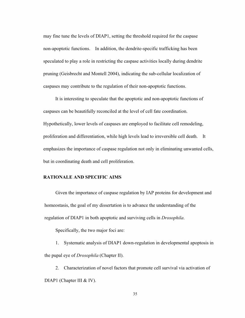
35
may fine tune the levels of DIAP1, setting the threshold required for the caspase
non-apoptotic functions. In addition, the dendrite-specific trafficking has been
speculated to play a role in restricting the caspase activities locally during dendrite
pruning (Geisbrecht and Montell 2004), indicating the sub-cellular localization of
caspases may contribute to the regulation of their non-apoptotic functions.
It is interesting to speculate that the apoptotic and non-apoptotic functions of
caspases can be beautifully reconciled at the level of cell fate coordination.
Hypothetically, lower levels of caspases are employed to facilitate cell remodeling,
proliferation and differentiation, while high levels lead to irreversible cell death. It
emphasizes the importance of caspase regulation not only in eliminating unwanted cells,
but in coordinating death and cell proliferation.
RATIONALE AND SPECIFIC AIMS
Given the importance of caspase regulation by IAP proteins for development and
homeostasis, the goal of my dissertation is to advance the understanding of the
regulation of DIAP1 in both apoptotic and surviving cells in Drosophila.
Specifically, the two major foci are:
1. Systematic analysis of DIAP1 down-regulation in developmental apoptosis in
the pupal eye of Drosophila (Chapter II).
2. Characterization of novel factors that promote cell survival via activation of
DIAP1 (Chapter III & IV).
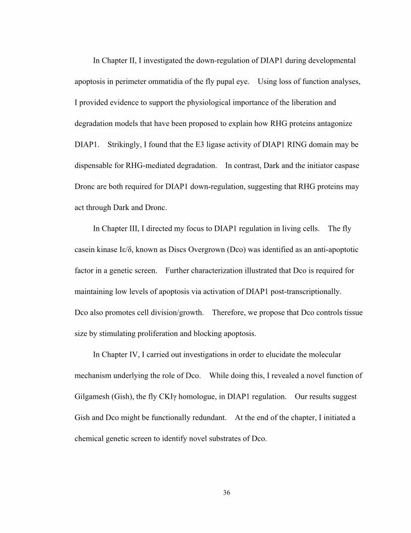
36
In Chapter II, I investigated the down-regulation of DIAP1 during developmental
apoptosis in perimeter ommatidia of the fly pupal eye. Using loss of function analyses,
I provided evidence to support the physiological importance of the liberation and
degradation models that have been proposed to explain how RHG proteins antagonize
DIAP1. Strikingly, I found that the E3 ligase activity of DIAP1 RING domain may be
dispensable for RHG-mediated degradation. In contrast, Dark and the initiator caspase
Dronc are both required for DIAP1 down-regulation, suggesting that RHG proteins may
act through Dark and Dronc.
In Chapter III, I directed my focus to DIAP1 regulation in living cells. The fly
casein kinase Iε/δ, known as Discs Overgrown (Dco) was identified as an anti-apoptotic
factor in a genetic screen. Further characterization illustrated that Dco is required for
maintaining low levels of apoptosis via activation of DIAP1 post-transcriptionally.
Dco also promotes cell division/growth. Therefore, we propose that Dco controls tissue
size by stimulating proliferation and blocking apoptosis.
In Chapter IV, I carried out investigations in order to elucidate the molecular
mechanism underlying the role of Dco. While doing this, I revealed a novel function of
Gilgamesh (Gish), the fly CKIγ homologue, in DIAP1 regulation. Our results suggest
Gish and Dco might be functionally redundant. At the end of the chapter, I initiated a
chemical genetic screen to identify novel substrates of Dco.

37
FIGURES AND TABLES
Figure 1.1
Figure 1.1 Ultrastructural features of cell death by apoptosis. (A) Schematic drawings illustrating the progression of morphologic changes observed in apoptotic cells. (B-C) Photomicrographs comparing a normal cell (B) and apoptotic cell (C). Clearly visible in the latter are dense spheres of compacted chromatin as the nucleus begins to fragment. (Adapted from Molecular Cell Biology, Lodish et al. 2007)
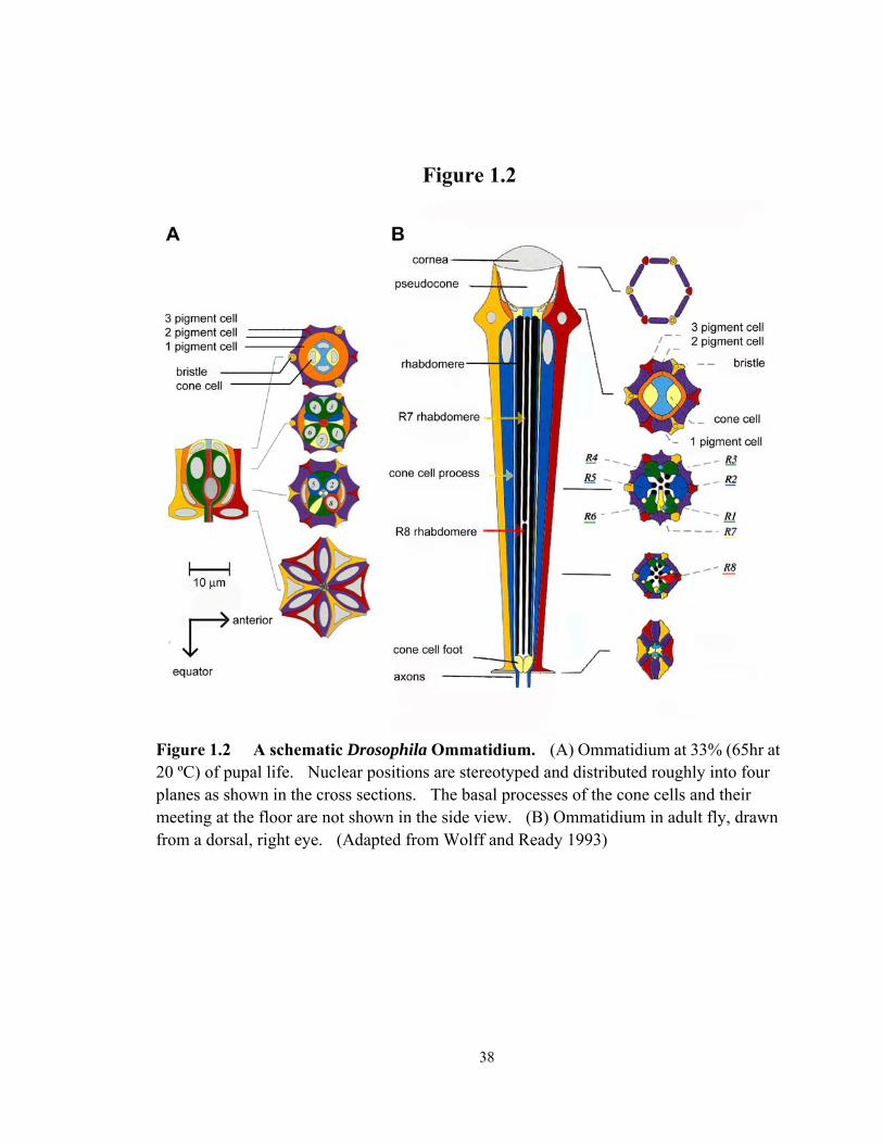
38
Figure 1.2
Figure 1.2 A schematic Drosophila Ommatidium. (A) Ommatidium at 33% (65hr at 20 ºC) of pupal life. Nuclear positions are stereotyped and distributed roughly into four planes as shown in the cross sections. The basal processes of the cone cells and their meeting at the floor are not shown in the side view. (B) Ommatidium in adult fly, drawn from a dorsal, right eye. (Adapted from Wolff and Ready 1993)
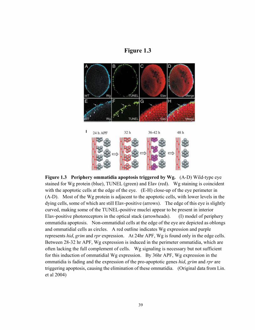
39
Figure 1.3
Figure 1.3 Periphery ommatidia apoptosis triggered by Wg. (A-D) Wild-type eye stained for Wg protein (blue), TUNEL (green) and Elav (red). Wg staining is coincident with the apoptotic cells at the edge of the eye. (E-H) close-up of the eye perimeter in (A-D). Most of the Wg protein is adjacent to the apoptotic cells, with lower levels in the dying cells, some of which are still Elav-positive (arrows). The edge of this eye is slightly curved, making some of the TUNEL-positive muclei appear to be present in interior Elav-positive photoreceptors in the optical stack (arrowheads). (I) model of periphery ommatidia apoptosis. Non-ommatidial cells at the edge of the eye are depicted as oblongs and ommatidial cells as circles. A red outline indicates Wg expression and purple represents hid, grim and rpr expression. At 24hr APF, Wg is found only in the edge cells. Between 28-32 hr APF, Wg expression is induced in the perimeter ommatidia, which are often lacking the full complement of cells. Wg signaling is necessary but not sufficient for this induction of ommatidial Wg expression. By 36hr APF, Wg expression in the ommatidia is fading and the expression of the pro-apoptotic genes hid, grim and rpr are triggering apoptosis, causing the elimination of these ommatidia. (Original data from Lin. et al 2004)

40
Figure 1.4
Figure 1.4 The projection of receptor axons from a single ommatidium. The axons of R1-R6 (labeled as 1-6) of a single ommatidium twist 180 degree as they descend to the lamina before diverging to their respective cartridges. R1-R6 in all the adjacent ommatidia also contribute axons to these cartridges through identical projections repeated in an overlapping mosaic, so that each cartridge receives not one but six receptor terminals. Therefore, an interlocking network of interwoven receptor connections is created across the entire lamina. The central cells R7 and R8 (not visible) share a single rhabdome, and their long visual fibers penetrate the lamina without terminating. (Adapted from Meinertzhagen and Hanson 1993)
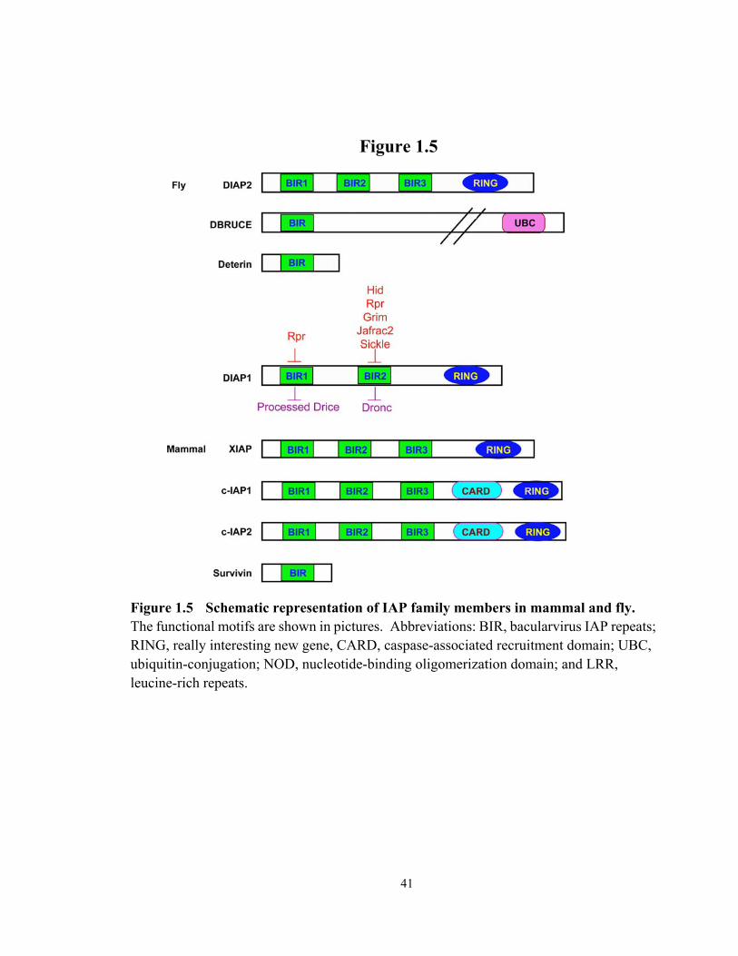
41
Figure 1.5
Figure 1.5 Schematic representation of IAP family members in mammal and fly. The functional motifs are shown in pictures. Abbreviations: BIR, bacularvirus IAP repeats; RING, really interesting new gene, CARD, caspase-associated recruitment domain; UBC, ubiquitin-conjugation; NOD, nucleotide-binding oligomerization domain; and LRR, leucine-rich repeats.

42
Figure 1.6
Figure 1.6 Fly and mammalian RHG proteins. RHG proteins are subjected to transcriptional and post-transcriptional regulation (left). RHG proteins and caspases contain IBM and IBM-like (in Dronc) motif, which provide molecular basis for the competitive binding to IAPs (right). (Adapted from Hay and Guo 2006)
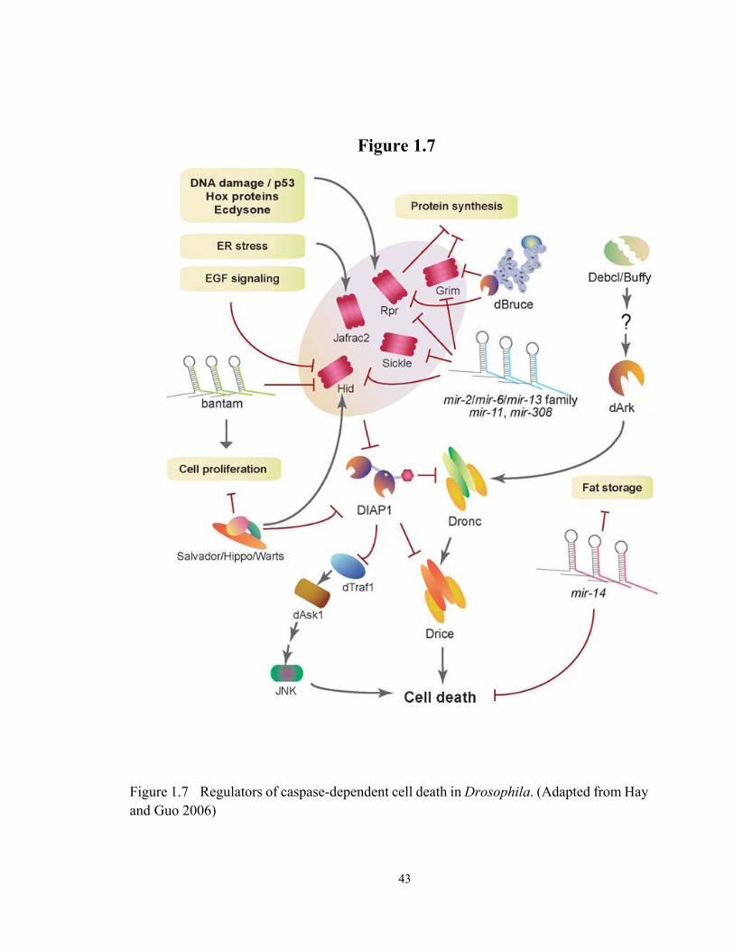
43
Figure 1.7
Figure 1.7 Regulators of caspase-dependent cell death in Drosophila. (Adapted from Hay and Guo 2006)

44
Table 1.1
Table 1.1 Core apoptosis machinery is concerved from the worm, fly to mammal. The conserved apoptosis regulation network is summarized on top of the table. The key factors from the worm, fly and mammal are listed in the table. (* Fly caspases Strica is listed as initiator caspase based on its long pro-domain. However, phylogenetic analysis based on the catalytic domain shows clustering with the effector caspases (Salvesen and Abrams 2004)).
45
REFERENCE
Abdelwahid, E., T. Yokokura, R. J. Krieser, S. Balasundaram, W. H. Fowle and K. White (2007). "Mitochondrial disruption in Drosophila apoptosis." Developmental Cell 12(5): 793-806.
Abrams, J. M., K. White, L. I. Fessler and H. Steller (1993). "PROGRAMMED CELL-DEATH DURING DROSOPHILA EMBRYOGENESIS." Development 117(1): 29-43.
Ambrosini, G., C. Adida and D. C. Altieri (1997). "A novel anti-apoptosis gene, survivin, expressed in cancer and lymphoma." Nature Medicine 3(8): 917-21.
Arama, E., J. Agapite and H. Steller (2003). "Caspase activity and a specific cytochrome c are required for sperm differentiation in Drosophila." Developmental Cell 4(5): 687-97.
Arama, E., M. Bader, G. E. Rieckhof and H. Steller (2007). "A ubiquitin ligase complex regulates caspase activation during sperm differentiation in Drosophila." Plos Biology 5(10): 2270-87.
Arama, E., M. Bader, M. Srivastava, A. Bergmann and H. Steller (2006). "The two Drosophila cytochrome C proteins can function in both respiration and caspase activation." Embo Journal 25(1): 232-43.
Baehrecke, E. H. (2002). "How death shapes life during development." Nature Reviews Molecular Cell Biology 3(10): 779-87.
Baehrecke, E. H. (2003). "Autophagic programmed cell death in Drosophila." Cell Death and Differentiation 10(9): 940-45.
Bate, M. and A. Arias (1993). The development of Drosophila melanogaster. New York, Cold Spring Harbor Laboratory Press.
Bennett, F. C. and K. F. Harvey (2006). "Fat cadherin modulates organ size in Drosophila via the Salvador/Warts/Hippo signaling pathway." Current Biology 16(21): 2101-10.
Brachmann, C. B. and R. L. Cagan (2003). "Patterning the fly eye: the role of apoptosis." Trends in Genetics 19(2): 91-96.
Brennecke, J., D. R. Hipfner, A. Stark, R. B. Russell and S. M. Cohen (2003). "bantam encodes a developmentally regulated microRNA that controls cell proliferation and regulates the proapoptotic gene hid in Drosophila." Cell 113(1): 25-36.
Cagan, R. L. and D. F. Ready (1989). "THE EMERGENCE OF ORDER IN THE DROSOPHILA PUPAL RETINA." Developmental Biology 136(2): 346-62.
Chai, J. J., N. Yan, J. R. Huh, J. W. Wu, W. Y. Li, B. A. Hay, et al. (2003). "Molecular mechanism of Reaper-Grim-Hid-mediated suppression of DIAP1-dependent Dronc ubiquitination." Nature Structural Biology 10(11): 892-98.
Chen, P., W. Nordstrom, B. Gish and J. M. Abrams (1996). "grim, a novel cell death gene in Drosophila." Genes & Development 10(14): 1773-82.

46
Chen, P., A. Rodriguez, R. Erskine, T. Thach and J. M. Abrams (1998). "DREDD, a novel effector of the apoptosis activators REAPER, GRIM, and HID in Drosophila." Developmental Biology 201(2): 202-16.
Chew, S. K., F. Akdemir, P. Chen, W. J. Lu, K. Mills, T. Daish, et al. (2004). "The apical caspase dronc governs programmed and unprogrammed cell death in Drosophila." Developmental Cell 7(6): 897-907.
Cho, E., Y. Feng, C. Rauskolb, S. Maitra, R. Fehon and K. D. Irvine (2006). "Delineation of a Fat tumor suppressor pathway." Nature Genetics 38(10): 1142-50.
Christich, A., S. Kauppila, P. Chen, N. Sogame, S. I. Ho and J. M. Abrams (2002). "The damage-responsive Drosophila gene sickle encodes a novel IAP binding protein similar to but distinct from reaper, grim, and hid." Current Biology 12(2): 137-40.
Claveria, C., E. Caminero, C. Martinez, S. Campuzano and M. Torres (2002). "GH3, a novel proapoptotic domain in Drosophila Grim, promotes a mitochondrial death pathway." Embo Journal 21(13): 3327-36.
Claveria, C., C. Martinez and M. Torres (2004). "A Bax/Bak-independent mitochondrial death pathway triggered by Drosophila Grim GH3 domain in mammalian cells." Journal of Biological Chemistry 279(2): 1368-75.
Crook, N. E., R. J. Clem and L. K. Miller (1993). "AN APOPTOSIS-INHIBITING BACULOVIRUS GENE WITH A ZINC FINGER-LIKE MOTIF." Journal of Virology 67(4): 2168-74.
Daish, T. J., K. Mills and S. Kumar (2004). "Drosophila caspase DRONC is required for specific developmental cell death pathways and stress-induced apoptosis." Developmental Cell 7(6): 909-15.
Danial, N. N. and S. J. Korsmeyer (2004). "Cell death: Critical control points." Cell 116(2): 205-19.
Ditzel, M., M. Broemer, T. Tenev, C. Boiduc, T. V. Lee, K. T. G. Rigbolt, et al. (2008). "Inactivation of Effector Caspases through Nondegradative Polyubiquitylation." Molecular Cell 32(4): 540-53.
Ditzel, M. and P. Meier (2002). "IAP degradation: decisive blow or altruistic sacrifice?" Trends in Cell Biology 12(10): 449-52.
Ditzel, M., R. Wilson, T. Tenev, A. Zachariou, A. Paul, E. Deas, et al. (2003). "Degradation of DIAP1 by the N-end rule pathway is essential for regulating apoptosis." Nature Cell Biology 5(5): 467-73.
Domingos, P. M. and H. Steller (2007). "Pathways regulating apoptosis during patterning and development." Current Opinion in Genetics & Development 17(4): 294-99.
Dorstyn, L., P. A. Colussi, L. M. Quinn, H. Richardson and S. Kumar (1999). "DRONC, an ecdysone-inducible Drosophila caspase." Proceedings of the National Academy of Sciences of the United States of America 96(8): 4307-12.
Dorstyn, L. and S. Kumar (2008). "A biochemical analysis of the activation of the Drosophila caspase DRONC." Cell Death and Differentiation 15(3): 461-70.

47
Dorstyn, L., S. Read, D. Cakouros, J. R. Huh, B. A. Hay and S. Kumar (2002). "The role of cytochrome c in caspase activation in Drosophila melanogaster cells." Journal Of Cell Biology 156(6): 1089-98.
Doumanis, J., L. Quinn, H. Richardson and S. Kumar (2001). "STRICA, a novel Drosophila melanogaster caspase with an unusual serine/threonine-rich prodomain, interacts with DIAP1 and DIAP2." Cell Death and Differentiation 8(4): 387-94.
Du, C. Y., M. Fang, Y. C. Li, L. Li and X. D. Wang (2000). "Smac, a mitochondrial protein that promotes cytochrome c-dependent caspase activation by eliminating IAP inhibition." Cell 102(1): 33-42.
Duckett, C. S., V. E. Nava, R. W. Gedrich, R. J. Clem, J. L. VanDongen, M. C. Gilfillan, et al. (1996). "A conserved family of cellular genes related to the baculovirus iap gene and encoding apoptosis inhibitors." Embo Journal 15(11): 2685-94.
Fan, Y. and A. Bergmann (2008). "Apoptosis-induced compensatory proliferation. The Cell is dead. Long live the Cell!" Trends in Cell Biology 18(10): 467-73.
Fan, Y. and A. Bergmann (2008). "Distinct mechanisms of apoptosis-induced compensatory proliferation in proliferating and differentiating tissues in the Drosophila eye." Developmental Cell 14(3): 399-410.
Fox, H. (1975). "ASPECTS OF TAIL MUSCLE ULTRASTRUCTURE AND ITS DEGENERATION IN RANA-TEMPORARIA." Journal of Embryology and Experimental Morphology 34(AUG): 191-207.
Fraser, A. G. and G. I. Evan (1997). "Identification of a Drosophila melanogaster ICE/CED3-related protease, drICE." Embo Journal 16(10): 2805-13.
Freel, C. D., D. A. Richardson, M. J. Thomenius, E. C. Gan, S. R. Horn, M. R. Olson, et al. (2008). "Mitochondrial localization of reaper to promote inhibitors of apoptosis protein degradation conferred by GH3 domain-lipid interactions." Journal of Biological Chemistry 283(1): 367-79.
Geisbrecht, E. R. and D. J. Montell (2004). "A role for Drosophila IAP1-mediated caspase inhibition in Rac-dependent cell migration." Cell 118(1): 111-25.
Gilbert, S. F. (2003). Developmental Biology. Sunderland, Massachusetts, Sinauer Associates, Inc.
Glucksmann, A. (1951). "CELL DEATHS IN NORMAL VERTEBRATE ONTOGENY." Biological Reviews of the Cambridge Philosophical Society 26(1): 59-86.
Goyal, L., K. McCall, J. Agapite, E. Hartwieg and H. Steller (2000). "Induction of apoptosis by Drosophila reaper, hid and grim through inhibition of IAP function." Embo Journal 19(4): 589-97.
Green, D. R. and G. I. Evan (2002). "A matter of life and death." Cancer Cell 1(1): 19-30. Grether, M. E., J. M. Abrams, J. Agapite, K. White and H. Steller (1995). "THE HEAD
INVOLUTION DEFECTIVE GENE OF DROSOPHILA-MELANOGASTER FUNCTIONS IN PROGRAMMED CELL-DEATH." Genes & Development 9(14): 1694-708.

48
Guan, J., H. Li, A. Rogulja, J. D. Axelrod and K. M. Cadigan (2007). "The Drosophila casein kinase I epsilon/delta Discs overgrown promotes cell survival via activation of DIAP1 expression." Developmental Biology 303(1): 16-28.
Hamaratoglu, F., M. Willecke, M. Kango-Singh, R. Nolo, E. Hyun, C. Y. Tao, et al. (2006). "The tumour-suppressor genes NF2/Merlin and Expanded act through Hippo signalling to regulate cell proliferation and apoptosis." Nature Cell Biology 8(1): 27-U9.
Harvey, K. and N. Tapon (2007). "The Salvador-Warts-Hippo pathway - an emerging tumour-suppressor network." Nature Reviews Cancer 7(3): 182-91.
Harvey, K. F., C. M. Pfleger and I. K. Hariharan (2003). "The Drosophila Mst ortholog, hippo, restricts growth and cell proliferation and promotes apoptosis." Cell 114(4): 457-67.
Harvey, N. L., T. Daish, K. Mills, L. Dorstyn, L. M. Quinn, S. H. Read, et al. (2001). "Characterization of the Drosophila caspase, DAMM." Journal of Biological Chemistry 276(27): 25342-50.
Hawkins, C. J., S. J. Yoo, E. P. Peterson, S. L. Wang, S. Y. Vernooy and B. A. Hay (2000). "The Drosophila caspase DRONC cleaves following glutamate or aspartate and is regulated by DIAP1, HID, and GRIM." Journal of Biological Chemistry 275(35): 27084-93.
Hay, B. A. and M. Guo (2006). "Caspase-dependent cell death in Drosophila." Annual Review of Cell and Developmental Biology 22: 623-50.
Hay, B. A., D. A. Wassarman and G. M. Rubin (1995). "Drosophila homologs of baculovirus inhibitor of apoptosis proteins function to block cell death." Cell 83(7): 1253-62.
Hays, R., L. Wickline and R. Cagan (2002). "Morgue mediates apoptosis in the Drosophila melanogaster retina by promoting degradation of DIAP1." Nature Cell Biology 4(6): 425-31.
Hengartner, M. O. (2000). "The biochemistry of apoptosis." Nature 407(6805): 770-76. Herman-Bachinsky, Y., H. D. Ryoo, A. Ciechanover and H. Gonen (2007). "Regulation of
the Drosophila ubiquitin ligase DIAP1 is mediated via several distinct ubiquitin system pathways." Cell Death and Differentiation 14(4): 861-71.
Hinds, M. G., R. S. Norton, D. L. Vaux and C. L. Day (1999). "Solution structure of a baculoviral inhibitor of apoptosis (IAP) repeat." Nature Structural Biology 6(7): 648-51.
Hofmann, K. (1999). "The modular nature of apoptotic signaling proteins." Cellular and Molecular Life Sciences 55(8-9): 1113-28.
Holley, C. L., M. R. Olson, D. A. Colon-Ramos and S. Kornbluth (2002). "Reaper eliminates IAP proteins through stimulated IAP degradation and generalized translational inhibition." Nature Cell Biology 4(60): 439-44.
Huang, H. K., C. A. P. Joazeiro, E. Bonfoco, S. Kamada, J. D. Leverson and T. Hunter (2000). "The inhibitor of apoptosis, cIAP2, functions as a ubiquitin-protein ligase
49
and promotes in vitro monoubiquitination of caspases 3 and 7." Journal of Biological Chemistry 275(35): 26661-64.
Huang, J. B., S. Wu, J. Barrera, K. Matthews and D. J. Pan (2005). "The Hippo signaling pathway coordinately regulates cell proliferation and apoptosis by inactivating Yorkie, the Drosophila homolog of YAP." Cell 122(3): 421-34.
Huh, J. R., M. Guo and B. A. Hay (2004). "Compensatory proliferation induced by cell death Drosophila wing disc requires activity of the apical cell death caspase dronc in a nonapoptotic role requires." Current Biology 14(14): 1262-66.
Huh, J. R., S. Y. Vernooy, H. Yu, N. Yan, Y. G. Shi, M. Guo, et al. (2004). "Multiple apoptotic caspase cascades are required in nonapoptotic roles for Drosophila spermatid individualization." Plos Biology 2(1): 43-53.
Hultmark, D. (2003). "Drosophila immunity: paths and patterns." Current Opinion in Immunology 15(1): 12-19.
Igaki, T., Y. Yamamoto-Goto, N. Tokushige, H. Kanda and M. Miura (2002). "Down-regulation of DIAP1 triggers a novel Drosophila cell death pathway mediated by dark and DRONC." Journal of Biological Chemistry 277(26): 23103-06.
Jiang, C. G., E. H. Baehrecke and C. S. Thummel (1997). "Steroid regulated programmed cell death during Drosophila metamorphosis." Development 124(22): 4673-83.
Justice, R. W., O. Zilian, D. F. Woods, M. Noll and P. J. Bryant (1995). "THE DROSOPHILA TUMOR-SUPPRESSOR GENE WARTS ENCODES A HOMOLOG OF HUMAN MYOTONIC-DYSTROPHY KINASE AND IS REQUIRED FOR THE CONTROL OF CELL-SHAPE AND PROLIFERATION." Genes & Development 9(5): 534-46.
Kango-Singh, M., R. Nolo, C. Tao, P. Verstreken, P. R. Hiesinger, H. J. Bellen, et al. (2002). "Shar-pei mediates cell proliferation arrest during imaginal disc growth in Drosophila." Development 129(24): 5719-30.
Kanuka, H., E. Kuranaga, K. Takemoto, T. Hiratou, H. Okano and M. Miura (2005). "Drosophila caspase transduces Shaggy/GSK-3 beta kinase activity in neural precursor development." Embo Journal 24(21): 3793-806.
Kanuka, H., K. Sawamoto, N. Inohara, K. Matsuno, H. Okano and M. Miura (1999). "Control of the cell death pathway by Dapaf-1, a Drosophila Apaf-1/CED-4-related caspase activator." Molecular Cell 4(5): 757-69.
Khan, F. S., M. Fujioka, P. Datta, T. Fernandes-Alnemri, J. B. Jaynes and E. S. Alnemri (2008). "The interaction of DIAP1 with dOmi/HtrA2 regulates cell death in Drosophila." Cell Death and Differentiation 15(6): 1073-83.
Kickson, B. and E. Hafen (1993). Genetic Dissection of Eye Development in Drosophila. New York, Cold Spring Harbor Laboratory Press.
Kiessling, S. and D. R. Green (2006). "Cell survival and proliferation in Drosophila S2 cells following apoptotic stress in the absence of the APAF-1 homolog, ARK, or downstream caspases." Apoptosis 11(4): 497-507.
50
Klambt, C., J. R. Jacobs and C. S. Goodman (1991). "THE MIDLINE OF THE DROSOPHILA CENTRAL-NERVOUS-SYSTEM - A MODEL FOR THE GENETIC-ANALYSIS OF CELL FATE, CELL-MIGRATION, AND GROWTH CONE GUIDANCE." Cell 64(4): 801-15.
Kluck, R. M., E. BossyWetzel, D. R. Green and D. D. Newmeyer (1997). "The release of cytochrome c from mitochondria: A primary site for Bcl-2 regulation of apoptosis." Science 275(5303): 1132-36.
Kondo, S., N. Senoo-Matsuda, Y. Hiromi and M. Miura (2006). "DRONC coordinates cell death and compensatory proliferation." Molecular and Cellular Biology 26(19): 7258-68.
Kourtis, N. and N. Tavernarakis (2009). "Autophagy and cell death in model organisms." Cell Death and Differentiation 16(1): 21-30.
Krammer, P. H. (2000). "CD95's deadly mission in the immune system." Nature 407(6805): 789-95.
Kratochwil, K. and P. Schwartz (1976). "TISSUE INTERACTION IN ANDROGEN RESPONSE OF EMBRYONIC MAMMARY RUDIMENT OF MOUSE - IDENTIFICATION OF TARGET TISSUE FOR TESTOSTERONE." Proceedings of the National Academy of Sciences of the United States of America 73(11): 4041-44.
Kroemer, G., L. Galluzzi, P. Vandenabeele, J. Abrams, E. S. Alnemri, E. H. Baehrecke, et al. (2009). "Classification of cell death: recommendations of the Nomenclature Committee on Cell Death 2009." Cell Death and Differentiation 16(1): 3-11.
Kuo, C. T., S. J. Zhu, S. Younger, L. Y. Jan and Y. N. Jan (2006). "Identification of E2/E3 ubiquitinating enzymes and caspase activity regulating Drosophila sensory neuron dendrite pruning." Neuron 51(3): 283-90.
Kuranaga, E., H. Kanuka, A. Tonoki, K. Takemoto, T. Tomioka, M. Kobayashi, et al. (2006). "Drosophila IKK-related kinase regulates nonapoptotic function of caspases, via degradation of IAPs." Cell 126(3): 583-96.
LaCasse, E. C., D. J. Mahoney, H. H. Cheung, S. Plenchette, S. Baird and R. G. Korneluk (2008). "IAP-targeted therapies for cancer." Oncogene 27(48): 6252-75.
Lam, E. and J. Greenberg (2000). "Cell death: the 'Yin' path in the balancing act of the life cycle - Preface." Plant Molecular Biology 44(3): VII-viii.
Lee, T. V., S. M. Srinivasula and A. Bergmann (2009). The initiator caspase Dronc is not subject to proteasome-mediated degradation. The 50th Annual Drosophila Research Conference. Chicago.
Leulier, F., P. S. Ribeiro, E. Palmer, T. Tenev, K. Takahashi, D. Robertson, et al. (2006). "Systematic in vivo RNAi analysis of putative components of the Drosophila cell death machinery." 13(10): 1663-74.
Leulier, F., P. S. Ribeiro, E. Palmer, T. Tenev, K. Takahashi, D. Robertson, et al. (2006). "Systematic in vivo RNAi analysis of putative components of the Drosophila cell death machinery." Cell Death and Differentiation 13(10): 1663-74.

51
Levine, B. and G. Kroemer (2008). "Autophagy in the pathogenesis of disease." Cell 132(1): 27-42.
Li, P., D. Nijhawan, I. Budihardjo, S. M. Srinivasula, M. Ahmad, E. S. Alnemri, et al. (1997). "Cytochrome c and dATP-dependent formation of Apaf-1/caspase-9 complex initiates an apoptotic protease cascade." Cell 91(4): 479-89.
Lim, H. Y. and A. Tomlinson (2006). "Organization of the peripheral fly eye: the roles of Snail family transcription factors in peripheral retinal apoptosis." Development 133(18): 3529-37.
Lin, H. V., A. Rogulja and K. M. Cadigan (2004). "Wingless eliminates ommatidia from the edge of the developing eye through activation of apoptosis." Development 131(10): 2409-18.
Lisi, S., I. Mazzon and K. White (2000). "Diverse Domains of THREAD/DIAP1 Are Required to Inhibit Apoptosis Induced by REAPER and HID in Drosophila." Genetics 154(2): 669-78.
Liu, X. S., C. N. Kim, J. Yang, R. Jemmerson and X. D. Wang (1996). "Induction of apoptotic program in cell-free extracts: Requirement for dATP and cytochrome c." Cell 86(1): 147-57.
Lodish, H. (2007). Molecular Cell Biology, Macmillan. MacCallum, D. E., T. R. Hupp, C. A. Midgley, D. Stuart, S. J. Campbell, A. Harper, et al.
(1996). "The p53 response to ionising radiation in adult and developing murine tissues." Oncogene 13(12): 2575-87.
Mao, Y. P., C. Rauskolb, E. Cho, W. L. Hu, H. Hayter, G. Minihan, et al. (2006). "Dachs: an unconventional myosin that functions downstream of Fat to regulate growth, affinity and gene expression in Drosophila." Development 133(13): 2539-51.
Meier, P., J. Silke, S. J. Leevers and G. I. Evan (2000). "The Drosophila caspase DRONC is regulated by DIAP1." Embo Journal 19(4): 598-611.
Meinertzhagen, I. and T. Hanson (1993). The Development of the Optic Lobe. New York, Cold Spring Harbor Laboratory Press.
Mendes, C. S., E. Arama, S. Brown, H. Scherr, M. Srivastava, A. Bergmann, et al. (2006). "Cytochrome c-d regulates developmental apoptosis in the Drosophila retina." Embo Reports 7(9): 933-39.
Mills, K., T. Daish, K. F. Harvey, C. M. Pfleger, I. K. Hariharan and S. Kumar (2006). "The Drosophila melanogaster Apaf-1 homologue ARK is required for most, but not all, programmed cell death." Journal Of Cell Biology 172(6): 809-15.
Muro, I., D. L. Berry, J. R. Huh, C. H. Chen, H. X. Huang, S. J. Yoo, et al. (2006). "The Drosophila caspase Ice is important for many apoptotic cell deaths and for spermatid individualization, a nonapoptotic process." Development 133(17): 3305-15.
Muro, I., B. A. Hay and R. J. Clem (2002). "The Drosophila DIAP1 protein is required to prevent accumulation of a continuously generated, processed form of the apical caspase DRONC." Journal of Biological Chemistry 277(51): 49644-50.

52
Muro, I., J. C. Means and R. J. Clem (2005). "Cleavage of the apoptosis inhibitor DIAP1 by the apical caspase DRONC in both normal and apoptotic Drosophila cells." Journal of Biological Chemistry 280(19): 18683-88.
Muro, I., K. Monser and R. J. Clem (2004). "Mechanism of Dronc activation in Drosophila cells." Journal of Cell Science 117(21): 5035-41.
Nolo, R., C. M. Morrison, C. Y. Tao, X. W. Zhang and G. Halder (2006). "The bantam microRNA is a target of the hippo tumor-suppressor pathway." Current Biology 16(19): 1895-904.
Oberst, A., C. Bender and D. R. Green (2008). "Living with death: the evolution of the mitochondrial pathway of apoptosis in animals." Cell Death and Differentiation 15(7): 1139-46.
Olson, M. R., C. L. Holley, E. C. Gan, D. A. Colon-Ramos, B. Kaplan and S. Kornbluth (2003). "A GH3-like domain in reaper is required for mitochondrial localization and induction of IAP degradation." Journal of Biological Chemistry 278(45): 44758-68.
Pantalacci, S., N. Tapon and P. Leopold (2003). "The Salvador partner Hippo promotes apoptosis and cell-cycle exit in Drosophila." Nature Cell Biology 5(10): 921-27.
Quinn, L. M., L. Dorstyn, K. Mills, P. A. Colussi, P. Chen, M. Coombe, et al. (2000). "An essential role for the caspase dronc in developmentally programmed cell death in Drosophila." Journal of Biological Chemistry 275(51): 40416-24.
Robinow, S., W. S. Talbot, D. S. Hogness and J. W. Truman (1993). "PROGRAMMED CELL-DEATH IN THE DROSOPHILA CNS IS ECDYSONE-REGULATED AND COUPLED WITH A SPECIFIC ECDYSONE RECEPTOR ISOFORM." Development 119(4): 1251-59.
Rodriguez, A., H. Oliver, H. Zou, P. Chen, X. D. Wang and J. M. Abrams (1999). "Dark is a Drosophila homologue of Apaf-1/CED-4 and functions in an evolutionarily conserved death pathway." Nature Cell Biology 1(5): 272-79.
Rothe, M., M. G. Pan, W. J. Henzel, T. M. Ayres and D. V. Goeddel (1995). "The TNFR2-TRAF signaling complex contains two novel proteins related to baculoviral-inhibitor of apoptosis proteins." Cell 83(7): 1243-52.
Ryoo, H. D., A. Bergmann, H. Gonen, A. Ciechanover and H. Steller (2002). "Regulation of Drosophila IAP1 degradation and apoptosis by reaper and ubcD1." Nature Cell Biology 4(6): 432-38.
Ryoo, H. D., T. Gorenc and H. Steller (2004). "Apoptotic cells can induce compensatory cell proliferation through the JNK and the wingless signaling pathways." Developmental Cell 7(4): 491-501.
Salvesen, G. S. and J. M. Abrams (2004). "Caspase activation - stepping on the gas or releasing the brakes? Lessons from humans and flies." Oncogene 23(16): 2774-84.
Shapiro, P. J., H. H. Hsu, H. Jung, E. S. Robbins and H. D. Ryoo (2008). "Regulation of the Drosophila apoptosome through feedback inhibition." Nature Cell Biology 10(12): 1440-U134.

53
Shi, Y. B. and A. IshizuyaOka (1996). Biphasic intestinal development in amphibians: Embryogenesis and remodeling during metamorphosis. Current Topics in Developmental Biology, Vol 32. 32: 205-35.
Silva, E., Y. Tsatskis, L. Gardano, N. Tapon and H. McNeill (2006). "The tumor-suppressor gene fat controls tissue growth upstream of expanded in the Hippo signaling pathway." Current Biology 16(21): 2081-89.
Song, Z. W., K. McCall and H. Steller (1997). "DCP-1, a Drosophila cell death protease essential for development." Science 275(5299): 536-40.
Sonnenfeld, M. J. and J. R. Jacobs (1995). "APOPTOSIS OF THE MIDLINE GLIA DURING DROSOPHILA EMBRYOGENESIS - A CORRELATION WITH AXON CONTACT." Development 121(2): 569-78.
Srinivasula, S. M. and J. D. Ashwell (2008). "IAPs: What's in a name?" Molecular Cell 30(2): 123-35.
Srinivasula, S. M., P. Datta, M. Kobayashi, J. W. Wu, M. Fujioka, R. Hegde, et al. (2002). "sickle, a novel Drosophila death gene in the reaper/hid/grim region, encodes an IAP-inhibitory protein." Current Biology 12(2): 125-30.
Srivastava, M., H. Scherr, M. Lackey, D. Xu, Z. Chen, J. Lu, et al. (2007). "ARK, the Apaf-1 related killer in Drosophila, requires diverse domains for its apoptotic activity." Cell Death and Differentiation 14(1): 92-102.
Steller, H. (2008). "Regulation of apoptosis in Drosophila." Cell Death Differ 15(7): 1132-38.
Sun, C. H., M. L. Cai, A. H. Gunasekera, R. P. Meadows, H. Wang, J. Chen, et al. (1999). "NMR structure and mutagenesis of the inhibitor-of-apoptosis protein XIAP." Nature 401(6755): 818-22.
Suzuki, Y., Y. Imai, H. Nakayama, K. Takahashi, K. Takio and R. Takahashi (2001). "A serine protease, HtrA2, is released from the mitochondria and interacts with XIAP, inducing cell death." Molecular Cell 8(3): 613-21.
Suzuki, Y., Y. Nakabayashi and R. Takahashi (2001). "Ubiquitin-protein ligase activity of X-linked inhibitor of apoptosis protein promotes proteasomal degradation of caspase-3 and enhances its anti-apoptotic effect in Fas-induced cell death." Proceedings of the National Academy of Sciences of the United States of America 98(15): 8662-67.
Tanji, T. and Y. T. Ip (2005). "Regulators of the Toll and Imd pathways in the Drosophila innate immune response." Trends in Immunology 26(4): 193-98.
Tapon, N., K. F. Harvey, D. W. Bell, D. C. R. Wahrer, T. A. Schiripo, D. A. Haber, et al. (2002). "salvador promotes both cell cycle exit and apoptosis in Drosophila and is mutated in human cancer cell lines." Cell 110(4): 467-78.
Tenev, T., A. Zachariou, R. Wilson, M. Ditzel and P. Meier (2005). "IAPs are functionally non-equivalent and regulate effector caspases through distinct mechanisms." Nature Cell Biology 7(1): 70-+.

54
Tenev, T., A. Zachariou, R. Wilson, A. Paul and P. Meier (2002). "Jafrac2 is an IAP antagonist that promotes cell death by liberating Dronc from DIAP1." Embo Journal 21(19): 5118-29.
Thompson, B. J. and S. M. Cohen (2006). "The hippo pathway regulates the bantam microRNA to control cell proliferation and apoptosis in Drosophila." Cell 126(4): 767-74.
Thornberry, N. A., T. A. Ranon, E. P. Pieterson, D. M. Rasper, T. Timkey, M. GarciaCalvo, et al. (1997). "A combinatorial approach defines specificities of members of the caspase family and granzyme B - Functional, relationships established for key mediators of apoptosis." Journal of Biological Chemistry 272(29): 17907-11.
Twomey, C. and J. V. McCarthy (2005). "Pathways of apoptosis and importance in development." Journal of Cellular and Molecular Medicine 9(2): 345-59.
Udan, R. S., M. Kango-Singh, R. Nolo, C. Y. Tao and G. Halder (2003). "Hippo promotes proliferation arrest and apoptosis in the Salvador/Warts pathway." Nature Cell Biology 5(10): 914-20.
Uren, A. G., M. Pakusch, C. J. Hawkins, K. L. Puls and D. L. Vaux (1996). "Cloning and expression of apoptosis inhibitory protein homologs that function to inhibit apoptosis and/or bind tumor necrosis factor receptor-associated factors." Proceedings of the National Academy of Sciences of the United States of America 93(10): 4974-78.
Vaux, D. L. (2002). "Apoptosis timeline." Cell Death and Differentiation 9(4): 349-54. Vaux, D. L. and J. Silke (2005). "IAPs, RINGs and ubiquitylation." Nature Reviews
Molecular Cell Biology 6(4): 287-97. Verhagen, A. M., P. G. Ekert, M. Pakusch, J. Silke, L. M. Connolly, G. E. Reid, et al.
(2000). "Identification of DIABLO, a mammalian protein that promotes apoptosis by binding to and antagonizing IAP proteins." Cell 102(1): 43-53.
Vernooy, S. Y., J. Copeland, N. Ghaboosi, E. E. Griffin, S. J. Yoo and B. A. Hay (2000). "Cell death regulation in Drosophila: Conservation of mechanism and unique insights." Journal Of Cell Biology 150(2): F69-F75.
Waldhuber, M., K. Emoto and C. Petritsch (2005). "The Drosophila caspase DRONC is required for metamorphosis and cell death in response to irradiation and developmental signals." Mechanisms of Development 122(7-8): 914-27.
Wang, S. L., C. J. Hawkins, S. J. Yoo, H. A. J. Muller and B. A. Hay (1999). "The Drosophila caspase inhibitor DIAP1 is essential for cell survival and is negatively regulated by HID." Cell 98(4): 453-63.
Wang, Y. F. and X. Gu (2001). "Functional divergence in the caspase gene family and altered functional constraints: Statistical analysis and prediction." Genetics 158(3): 1311-20.
Wells, B. S., E. Yoshida and L. A. Johnston (2006). "Compensatory proliferation in Drosophila imaginal discs requires dronc-dependent p53 activity." Current Biology 16(16): 1606-15.
55
White, K., M. E. Grether, J. M. Abrams, L. Young, K. Farrell and H. Steller (1994). "GENETIC-CONTROL OF PROGRAMMED CELL-DEATH IN DROSOPHILA." Science 264(5159): 677-83.
White, K., E. Tahaoglu and H. Steller (1996). "Cell killing by the Drosophila gene reaper." Science 271(5250): 805-07.
Willecke, M., F. Hamaratoglu, M. Kango-Singh, R. Udan, C. L. Chen, C. Y. Tao, et al. (2006). "The fat cadherin acts through the hippo tumor-suppressor pathway to regulate tissue size." Current Biology 16(21): 2090-100.
Williams, D. W., S. Kondo, A. Krzyzanowska, Y. Hiromi and J. W. Truman (2006). "Local caspase activity directs engulfment of dendrites during pruning." Nature Neuroscience 9(10): 1234-36.
Wilson, R., L. Goyal, M. Ditzel, A. Zachariou, D. A. Baker, J. Agapite, et al. (2002). "The DIAP1 RING finger mediates ubiquitination of Dronc and is indispensable for regulating apoptosis." Nature Cell Biology 4(6): 445-50.
Wing, J. P., J. S. Karres, J. L. Ogdahl, L. Zhou, L. M. Schwartz and J. R. Nambu (2002). "Drosophila sickle is a novel grim-reaper cell death activator." Current Biology 12(2): 131-35.
Wing, J. P., B. A. Schreader, T. Yokokura, U. Q. Wang, P. S. Andrews, N. Huseinovic, et al. (2002). "Drosophila Morgue is an F box/ubiquitin conjugase domain protein important for grim-reaper mediated apoptosis." Nature Cell Biology 4(6): 451-56.
Wing, J. P., L. Zhou, L. M. Schwartz and J. R. Nambu (1998). "Distinct cell killing properties of the Drosophila reaper, head involution defective, and grim genes." Cell Death and Differentiation 5(11): 930-39.
Wolff, T. and D. Ready (1993). Pattern Fromation in the Drosophila Retina. New York, Cold Spring Harbor Laboratory Press.
Wolff, T. and D. F. Ready (1991). "CELL-DEATH IN NORMAL AND ROUGH EYE MUTANTS OF DROSOPHILA." Development 113(3): 825-&.
Wu, S., J. B. Huang, J. X. Dong and D. J. Pan (2003). "hippo encodes a Ste-20 family protein kinase that restricts cell proliferation and promotes apoptosis in conjunction with salvador and warts." Cell 114(4): 445-56.
Xu, D., Y. Wang, R. Willecke, Z. Chen, D. Ding and A. Bergmann (2006). "The effector caspases drICE and dcp-1 have partially overlapping functions in the apoptotic pathway in Drosophila." Cell Death and Differentiation 13(10): 1697-706.
Xu, D. B., Y. Li, M. Arcaro, M. Lackey and A. Bergmann (2005). "The CARD-carrying caspase Dronc is essential for most, but not all, developmental cell death in Drosophila." Development 132(9): 2125-34.
Xu, T. A., W. Y. Wang, S. Zhang, R. A. Stewart and W. Yu (1995). "IDENTIFYING TUMOR SUPPRESSORS IN GENETIC MOSAICS - THE DROSOPHILA LATS GENE ENCODES A PUTATIVE PROTEIN-KINASE." Development 121(4): 1053-63.

56
Yan, N. and Y. G. Shi (2005). "Mechanisms of apoptosis through structural biology." Annual Review of Cell and Developmental Biology 21: 35-56.
Yan, N., J. W. Wu, J. J. Chai, W. Y. Li and Y. G. Shi (2004). "Molecular mechanisms of DrICE inhibition by DIAP1 and removal of inhibition by Reaper, Hid and Grim." Nature Structural & Molecular Biology 11(5): 420-28.
Yang, Y., S. Y. Fang, J. P. Jensen, A. M. Weissman and J. D. Ashwell (2000). "Ubiquitin protein ligase activity of IAPs and their degradation in proteasomes in response to apoptotic stimuli." Science 288(5467): 874-77.
Yokokura, T., D. Dresnek, N. Huseinovic, S. Lisi, E. Abdelwahid, P. Bangs, et al. (2004). "Dissection of DIAP1 functional domains via a mutant replacement strategy." Journal of Biological Chemistry 279(50): 52603-12.
Yoo, S. J., J. R. Huh, I. Muro, H. Yu, L. J. Wang, S. L. Wang, et al. (2002). "Hid, Rpr and Grim negatively regulate DIAP1 levels through distinct mechanisms." Nature Cell Biology 4(6): 416-24.
Yu, S. Y., S. J. Yoo, L. H. Yang, C. Zapata, A. Srinivasan, B. A. Hay, et al. (2002). "A pathway of signals regulating effector and initiator caspases in the developing Drosophila eye." Development 129(13): 3269-78.
Yu, X. C., L. Wang, D. Acehan, X. D. Wang and C. W. Akey (2006). "Three-dimensional structure of a double apoptosome formed by the Drosophila Apaf-1 related killer." Journal of Molecular Biology 355(3): 577-89.
Zachariou, A., T. Tenev, L. Goyal, J. Agapite, H. Steller and P. Meier (2003). "IAP-antagonists exhibit non-redundant modes of action through differential DIAP1 binding." Embo Journal 22(24): 6642-52.
Zakeri, Z. and R. A. Lockshin (2008). Cell death: History and future. Programmed Cell Death in Cancer Progression and Therapy. Dordrecht, Springer. 615: 1-11.
Zhou, L., H. Hashimi, L. M. Schwartz and J. R. Nambu (1995). "PROGRAMMED CELL-DEATH IN THE DROSOPHILA CENTRAL-NERVOUS-SYSTEM MIDLINE." Current Biology 5(7): 784-90.
Zhou, L., Z. W. Song, J. Tittel and H. Steller (1999). "HAC-1, a Drosophila homolog of APAF-1 and CED-4 functions in developmental and radiation-induced apoptosis." Molecular Cell 4(5): 745-55.
Zimmermann, K. C., J. E. Ricci, N. M. Droin and D. R. Green (2002). "The role of ARK in stress-induced apoptosis in Drosophila cells." Journal of Cell Biology 156(6): 1077-87.
Zuzarte-Luis, V. and J. M. Hurle (2002). "Programmed cell death in the developing limb." International Journal of Developmental Biology 46(7): 871-76.

57
CHAPTER II
RHG PROTEIN MEDIATED APOPTOSIS IN THE PUPAL EYE
OF DROSOPHILA: DOWN-REGULATION OF DIAP1
ABSTRACT
Drosophila Inhibitor of apoptosis protein 1 (DIAP1) is an essential negative
regulator of apoptosis in fly cells. DIAP1 binds to caspases and inhibits their activity.
RHG proteins, e.g., Head Involution defective (Hid), Grim and Reaper (Rpr), can induce
apoptosis by binding to DIAP1 and liberating the bound caspases. In addition, some RHG
proteins can induce DIAP1 degradation. These studies are based on in vivo
overexpression or in vitro biochemical assays, raising questions about which mechanism
(liberation or degradation) is more physiologically relevant. In this report, we
demonstrate significant down-regulation of DIAP1 protein in perimeter ommatidia
undergoing apoptosis in the pupal eye. This regulation was dependent on Hid, Grim and
Rpr. Interestingly, a partial reduction in RHG proteins suppressed DIAP1 degradation to
undetectable levels. Under these conditions, apoptosis was initiated at the normal time,
but was markedly reduced thereafter. These results argue that both liberation of caspases
58
and degradation of DIAP1 are required for timely initiation and progression of apoptosis
in this tissue. In addition, we provide evidence that the E3 ligase activity of DIAP1’s
RING domain is dispensable for RHG-mediated degradation. In contrast, the Drosophila
Apaf-1 related killer (Dark) and the initiator caspase Dronc are both required for DIAP1
down-regulation, suggesting that RHG proteins may act through Dark and Dronc.
INTRODUCTION
Apoptosis is an evolutionarily conserved cell death process that is essential for
normal development and adult homeostasis. The key executioners of apoptosis are a
family of cysteine proteases called caspases. Once activated, initiator caspases cleave and
activate downstream effector caspases, which in turn proteolyze hundreds of cellular
proteins, leading to cell death (Hengartner 2000; Hay and Guo 2006). In the fruit fly
Drosophila melanogaster, the initiator caspase Drosophila interleukin-IB-converting
enzyme (Dronc) and effector caspases Drosophila Nedd-2-like caspase (Drice) and death
caspase-1 (Dcp-1) are core components of the apoptosis machinery(Hengartner 2000; Hay
and Guo 2006).
Cells tightly regulate caspase activation and consequent apoptosis through several
mechanisms. In vertebrates, the so-called “intrinsic pathway” initiates cell death through
the release of cytochrome c from the intra-mitochondrial compartment. The released
cytochrome c binds to a protein called Apaf-1, and this complex promotes activation of
59
caspase 9, an initiator caspase(Hengartner 2000; Salvesen and Abrams 2004). While the
role of cytochrome c in fly apoptosis is still controversial(Mendes et al. 2006; Oberst et al.
2008), the Apaf-1 homolog Dark is required for activation of the initiator caspase
Dronc(Hay and Guo 2006).
Cells also employ factors called Inhibitor of Apoptosis proteins (IAPs) to bind and
inhibit caspases, thereby preventing the inappropriate onset of the apoptotic
program(Salvesen and Abrams 2004; Hay and Guo 2006; Srinivasula and Ashwell 2008).
IAPs are characterized by the presence of one or more Bacularvirus IAP Repeat (BIR)
domains which mediate the interaction with caspases and other pro-apoptotic
factors(Srinivasula and Ashwell 2008). The importance of IAPs in preventing
inappropriate apoptosis is highlighted by the phenotype of Drosophila IAP1 (DIAP1).
Loss of DIAP1 leads to global caspase-dependent apoptosis in multiple fly tissues and
cultured cells, demonstrating that DIAP1 is an essential inhibitor of cell death in the fly
(Hay and Guo 2006; Steller 2008).
The anti-apoptotic function of IAPs is antagonized by a group of pro-apoptotic
proteins including SMAC/Diablo and Omi/HtrA2 in mammals and Reaper (Rpr), Head
involution defective (Hid), Grim, Sickle and Jafrac-2 in flies, collectively referred to as
RHG proteins(Salvesen and Abrams 2004; Hay and Guo 2006; Srinivasula and Ashwell
2008). In Drosophila, the deletion of the genomic region containing hid, grim and rpr
(the Df(3L)H99 deletion) prevents almost all developmental apoptosis in embryos(White

60
et al. 1994). Consistent with a positive role in promoting cell death, over-expression of
RHG proteins induces massive apoptosis in many developmental contexts in flies(Hay and
Guo 2006).
A major mechanism that RHG proteins employ to antagonize IAPs is through direct
binding. RHGs share very little similarity except for a short N-terminal motif, called the
IAP Binding Motif (IBM), which binds to the BIR domains of IAPs. RHG protein
binding to IAPs competitively disrupts the IAP-caspase interaction, releasing caspases to
trigger apoptosis. Evidence for displacement of IAPs from caspases by RHGs comes
from studies of mammalian SMAC/DIABLO and Omi/HtrA2 as well as Hid and Rpr in
flies (Du et al. 2000; Verhagen et al. 2000; Suzuki et al. 2001; Chai et al. 2003; Zachariou
et al. 2003; Hay and Guo 2006). This mechanism to explain RHG protein action is
commonly referred to as the liberation model.
While the liberation model offers a simple explanation of the pro-apoptotic function
of RHGs, it does not take into account the fact that many IAPs contain a C-terminal RING
domain that can function as an E3-ubiquitin ligase(Yang et al. 2000; Suzuki et al. 2001;
Wilson et al. 2002; Yoo et al. 2002). The RING domain function of DIAP1 has been
implicated both in ubiquitination and proteasomal degradation of the initiator caspase
Dronc and in auto-ubiquitination of DIAP1(Hays et al. 2002; Holley et al. 2002; Ryoo et al.
2002; Wilson et al. 2002; Yoo et al. 2002). The auto-ubiquitination of DIAP1 can be
stimulated by Rpr, Hid and Grim in a cell-context-dependent manner(Hays et al. 2002;

61
Holley et al. 2002; Ryoo et al. 2002; Wilson et al. 2002; Yoo et al. 2002). These data
have led to the view that the primary way that RHG proteins promote apoptosis is by
stimulating the RING-dependent auto-ubiquitination and degradation of DIAP1, releasing
caspases to initiate the apoptotic program. This mechanism of RHG protein action is
referred to as the degradation model.
Evidence supporting the degradation model of RHG antagonism of DIAP1 can be
summarized as follows. Overexpression of RHG proteins in several fly tissues stimulates
apoptosis and degradation of DIAP1(Hays et al. 2002; Ryoo et al. 2002; Yoo et al. 2002).
In vitro, RHG proteins can trigger DIAP1 auto-ubiquitination(Ryoo et al. 2002; Yoo et al.
2002). In addition, RING domain mutants in DIAP1 that disrupt E3 ligase activity can
suppress the RHG-dependent degradation of DIAP1 in some contexts(Hays et al. 2002;
Holley et al. 2002; Ryoo et al. 2002; Yoo et al. 2002). One major drawback of the above
mentioned studies is that they relied on over-expression assays in living tissues as well as
reductionist biochemical approaches. While these are powerful for exploring molecular
mechanism, they do not address whether RHG-mediated degradation of DIAP1 occurs
under physiological conditions and whether this regulation contributes to the initiation of
apoptosis.
There is considerable controversy concerning the degradation model. Most studies
have found that overexpression of RHGs can promote DIAP1 ubiquitination and
down-regulation, but the effects with Hid and Rpr are variable in different fly tissues,

62
leading to the suggestion that not all RHGs promote DIAP1 degradation(Hays et al. 2002;
Ryoo et al. 2002). In addition, the importance of the RING domain in mediating DIAP1
downregulation has not been universally observed in vivo(Wilson et al. 2002; Yoo et al.
2002). In some cases, RING domain mutants enhanced rather than suppressed apoptosis
induced by RHG overexpression(Wilson et al. 2002). Moreover, there is no agreement in
the reported in vitro studies that DIAP1 auto-ubiquitination leads to its own
degradation(Ryoo et al. 2002; Yoo et al. 2002). Rather, it has been suggested that
auto-ubiquitination serves to attenuate DIAP’s E3-ligase activity towards
caspases(Herman-Bachinsky et al. 2007). Furthermore, suppression of DIAP1
degradation in cultured cells with a proteasome inhibitor failed to inhibit apoptosis
(Yokokura et al. 2004). The controversy is likely due to the complexity of DIAP1
regulation in different tissues, as well as the inherent limitations of overexpression and
biochemical approaches.
In this report, we address the physiological relevance of the DIAP1 degradation
model by examining naturally occurring apoptosis of ommatidia at the perimeter of the
pupal fly eye(Lin et al. 2004). This apoptotic event is triggered by the expression of
RHG proteins and is dependent on Dronc and effector caspases (Lin et al. 2004; Mendes et
al. 2006). We provide evidence, for the first time, that DIAP1 degradation occurs during
developmental apoptosis. Hid, Grim and Rpr all contribute to this regulation.
Interestingly, a partial reduction in Hid, Grim and Rpr activity reduced DIAP1 degradation
63
to undetectable levels, but apoptosis was still initiated at the normal time, though it then
proceeded with much slower than normal kinetics. These results strongly suggest that the
liberation model for RHG function contributes significantly in perimeter ommatidial
apoptosis. We favor a model where the combination of degradation of DIAP1 and
disruption of DIAP1-caspase complexes allows apoptosis to proceed in a timely fashion at
the periphery of the pupal fly eye.
Our results also demonstrate that the E3 ligase activity of RING domain of DIAP1 is
dispensable for RHG-mediated degradation. Instead we find that the Apaf-1 homolog
Dark and the initiator caspase Dronc are required to achieve the full extent of DIAP1
degradation. These results suggest that RHG proteins may act through Dark and Dronc to
promote DIAP1 down-regulation.
MATERIALS AND METHODS
Drosophila Genetics
Fly stocks were maintained on standard medium at 25°C. The following fly strains
were used. Oregon R was our wild type stock. UAS-p35 was from the Bloomington Stock
Center (Indiana University). The stocks hidX14, Df(3f)H99, Df(3f)X25, and Df(3f)XR38
were from Kristin White (Massachusetts General Hospital). droncI29 P[FRT, w+]2A ,
P[FRT,hs-neo]42D darkG8 and P[GMR-Gal4] on the third chromosome were from Andreas
Bergmann (M. D. Anderson Cancer Center). Eye-FlpT11 and Eye-FlpT12 were from Barry
64
Dickson (I.M.P., Vienna). UAS-GPI-fz2 was previously described(Cadigan et al. 1998;
Lin et al. 2004). The DIAP (th) alleles thj5c8, th6B, th33-1s, th6-3s, th23-4s were from Kristin
White and John Abrams (UT, Southwestern). Pupal developmental ages are expressed as
hours after puparium formation (APF) at 25°C with white pre-pupae defined as 0 hour APF.
Clonal analyses in the eye were performed as previously described(Lin et al. 2004) using
Eye-FlpT11 for darkG8 and Eye-FlpT12 for th alleles, droncI24 and Df(3f) H99.
Immunostainings and TUNEL
Immunostainings were performed as previously described(Lin et al. 2004). DIAP1
antibodies were mouse anti-DIAP1(1:200) (from B. Hay) and rabbit anti-DIAP1 (1:200)
(from K. White), with the mouse antibody being used throughout this report. Other
primary antibodies were rat anti-ELAV (1:100) (Developmental Studies Hybridoma Bank)
and rabbit anti-β-galactosidase (1:500) (Cappel, ICN, Costa Mesa, CA). Cy3- and
Cy5-conjugated (Jackson Immunochemicals) and Alexa Fluor 488-conjugated (Molecular
Probes) secondary antibodies were applied. To perform co-staining of mouse anti-DIAP1
and mouse anti-Wg (1:100) (Developmental Studies Hybridoma Bank), samples were first
incubated sequentially with anti-DIAP1 and anti-mouse secondary antibody. After
washing off unbound secondary antibody, the samples were incubated with normal mouse
serum and unconjugated anti-mouse Fab (1:200) (Jackson Immunochemicals) to ensure
that all the mouse anti-DIAP1 antibody was blocked. The samples were then incubated
with anti-Wg and Cy5-conjugated anti-mouse IgG1(Jackson Immunochemicals) and

65
processed as normal. The TUNEL assay was performed using the Cell Death Detection
kit (Fluorescein; Roche Diagnostics, Indianapolis, IN) as previously described(Lin et al.
2004).
Microscopy and image analysis
All fluorescent pictures were obtained with Leica confocal microscope DM6000 B
(Leica) and processed in Adobe Photoshop CS. For DIAP1 fluorescence pixel intensity
(PI) analysis, each ommatidium was traced manually around one ELAV positive cell
cluster. Perimeter ommatidia were identified via Wg immunostaining, and the adjacent
internal ommatidia lacking Wg signal were selected for comparison. Raw PI values were
calculated by the Mean Intensity function of Analyze Particle tool in Scion Image (Scion
Corporation, MD). All Raw PI values were normalized with the average PI values of
internal ommatidia. Normalized PI values of all ommatidia from multiples discs were
subjected to calculation of average, standard error of mean (S.E.M.) and a two-tailed
Student’s t-test. More than 20 perimeter/internal ommatidia pairs from at least 4 eyes
were quantified for each experimental condition. Normalized PI values were shown with
plus/minus S.E.M.. Differences were considered significant at P < 0.05.

66
RESULTS
DIAP1 is down-regulated post-transcriptionally in peripheral ommatidia that undergo apoptosis.
The fly compound eye is composed of approximately 800 repeated optical units
called ommatidia(Wolff and Ready 1993). During mid-pupation, the ommatidia at the
perimeter of the developing eye undergo apoptosis, starting around 36 hr after puparium
formation (APF). This process is complete by 48 hr APF (Lin et al. 2004). This
programmed cell death is controlled by Wingless (Wg) (Lin et al. 2004), a member of the
Wnt family of signaling molecules(Cadigan 2008). Wg is expressed in the outer rim of
the eye throughout pupal stages, and it is also activated in the cone cells of the peripheral
ommatidia that are destined to die starting around 28 hr APF (Lin et al. 2004; Lim and
Tomlinson 2006). Therefore, Wg immunostaining is used in this report to mark the
perimeter ommatidia that are beginning to undergo apoptosis (e.g., Figure 2.1a).
We found that DIAP1 levels in the perimeter ommatidia are down-regulated
preceding the onset of apoptosis. At 24 hr APF, all ommatidia throughout the pupal eye
had similar levels of DIAP1 (data not shown). By 33 hr APF, there are lower DIAP1
levels in many peripheral ommatidia (Figure 2.2a-d). At 36hr APF, all Wg-positive
ommatidia had less DIAP1 than ommatidia in the interior (Figure 2.1c). This
down-regulation is also observed at later stages as apoptosis progresses (Figure 2.2e-g).
The same results were also obtained by using an independent polyclonal antibody (Figure
2.2i-l), confirming that DIAP1 is down-regulated in developmental apoptosis.

67
To quantify the difference in DIAP1 levels between ommatidia that are undergoing
apoptosis and interior ommatidia, the Mean Pixel Intensity (PI) values for the DIAP1
immunostaining in ommatidia were determined. The area of an ommatidia was defined
by co-staining with ELAV, a marker for photoreceptors(Oneill et al. 1994). The PI of the
Wg-positive perimeter ommatidia are expressed as a percentage of the PI of the adjacent
Wg-negative ommatidia (the Normalized Mean Pixel Intensity (NPI) in Table 2.1). This
analysis revealed that the level of DIAP1 signal in the ommatidia undergoing apoptosis at
36 hr APF was 47.6% of that found in interior ommatidia (Table 2.1).
The possibility that the observed down-regulation of DIAP1 protein levels was due
to decreased DIAP1 transcription was examined in flies carrying the DIAP1-lacZ allele
thj5c8. These flies contain an enhancer trap insert in the DIAP1 locus, placing the LacZ
reporter under the control of the DIAP1 regulatory region (Hay et al. 1995; Ryoo et al.
2002). DIAP1-lacZ eyes displayed no difference in LacZ levels between the peripheral
ommatidia and interior ones (Figure 2.1e). These results argue that the down-regulation
of DIAP1 expression in perimeter ommatidia occurs at the post-transcriptional level.
DIAP1 down-regulation is independent of effector caspase activity but requires Wg signaling as well as Hid, Grim and Rpr.
The initiation of programmed cell death occurs through the activation of effector
caspases (Hengartner 2000). These proteases cleave a wide variety of cellular substrates
to promote the apoptotic cell program(Hengartner 2000). In addition, it has been

68
suggested that the cleavage of DIAP1 by effector caspases exposes a motif to recruit the
N-end-rule-associated ubiquitination machinery and accelerate the degradation of DIAP1
(Ditzel et al. 2003). This raises the possibility that the down-regulation of DIAP1
observed in the pupal eye is effector caspase-dependent. This question was addressed by
examining the expression of DIAP1 in eyes where effector caspase activity is inhibited by
the bacularvirus protein p35. Flies containing a GMR-Gal4 (expressing Gal4 in all eye
cell types) and UAS-p35 transgene did not show any perimeter ommatidia apoptosis (Lin
et al. 2004) (data not shown). Despite the block in apoptosis, the down-regulation of
DIAP1 still occurred at a similar level as in wild type (Figure 2.1g,h), with a NPI of 44.4%
(Table 2.1). These results indicate that the lower DIAP1 levels in perimeter ommatidia
occurred independently of effector caspase activity and is not an indirect result of the
apoptotic cell program.
The expression of Wg in peripheral ommatidia triggers apoptosis by activating the
expression of the RHG genes hid, grim and rpr(Lin et al. 2004). Expression of a GPI-fz2
transgene, known to inhibit Wg signaling(Cadigan et al. 1998; Lin et al. 2004) , resulted in
a complete block in DIAP1 down-regulation (Figure 2.3). To test if Hid, Grim and Rpr
are also required for DIAP1 down-regulation, we examined eyes from flies that were
heterozygous for hid (Figure 2.4a-d), rpr (Figure 2.4e-h), hid and grim (Figure 2.4i-l), and
hid and rpr (Figure 2.4m-o). DIAP1 down-regulation was partially suppressed in all four
backgrounds, and the NPI values obtained for the perimeter ommatidia in each background
69
were significantly higher than that of wild-type (Figure 2.4p and Table 2.1). Flies
heterozygous for the H99 deficiency (which removes hid, grim and rpr) blocked DIAP1
down-regulation (Figure 2.5a-d). The NPI in H99/+ perimeter ommatidia was not
significantly different from internal ommatidia (97.3%, P>0.5). Taken together, these
results indicate that Hid, Grim and Rpr are major factors contributing to DIAP1
down-regulation. The fact that removal of even one copy of hid or rpr caused a
significant reduction in DIAP1 down-regulation indicates that the system is very sensitive
to the amount of RHG protein present.
Apoptosis can occur in the absence of DIAP1 down-regulation in peripheral ommatidia.
RHG proteins are required for apoptosis (Lin et al. 2004) and down-regulation of
DIAP1 in perimeter ommatidia (Figure 2.4; Figure 2.5a-f; Table 2.1). Loss of DIAP1
results in a dramatic increase in apoptosis in several fly tissues (Hay et al. 1995; Wang et al.
1999; Goyal et al. 2000; Lisi et al. 2000; Yokokura et al. 2004; Hay and Guo 2006). This
suggests a model where RHG-dependent down-regulation of DIAP1 is the major cause of
apoptosis in the peripheral ommatidia. However, these correlations do not rule out the
competing model of RHG action, where RHGs liberate caspases from DIAP1
independently of DIAP1 down-regulation.
Reducing hid, grim and rpr activity by 50% (H99/+ flies) results in no detectable
down-regulation of DIAP1 (Figure 2.5a-d; Table 2.1). Despite the absence of DIAP1
70
down-regulation, perimeter apoptosis was initiated at similar level as in wild type eyes, as
judged by TUNEL staining at 36 hr APF (Figure 2.5g,h). Therefore, apoptosis in the
peripheral ommatidia can still occur in the absence of DIAP1 down-regulation, perhaps by
RHG proteins displacing DIAP1 bound to caspases, liberating them for apoptosis
initiation.
Although apoptosis is initiated at the normal time in H99 heterozygotes,
programmed cell death proceeds much more slowly compared to with wild-type controls.
TUNEL staining is much lower at 39, 42 and 45 hr APF in H99 heterozygotes (Figure
2.5k,l, and Figure 2.6). In H99/+ eyes at 45 hr APF, TUNEL-positive ommatidia still
contain unfragmented ELAV-positive photoreceptors (Figure 2.5 j), which is rarely
observed in wild type eyes (Figure 2.5 i). This indicates that apoptosis has not progressed
to the terminal stages in the H99 heterozygotes and strongly argues that down-regulation of
DIAP1 is required for the timely execution of programmed cell death.
Down-regulation of DIAP1 in the perimeter ommatidia depends on its BIR1 and BIR2 domains, but is independent of its RING domain
RHG proteins interact with DIAP1 through direct interactions between the IBM
domain of RHGs and the BIR1 and BIR2 domains of DIAP1 (Chai et al. 2003; Zachariou
et al. 2003; Hay and Guo 2006). The requirement for this interaction for DIAP1
down-regulation in the pupal eye was examined by utilizing DIAP1 mutants carrying point
mutations in the BIR1 or BIR2 domains. The DIAP1 alleles th6-3s (G88S) and th23-4s

71
(G269S) encode proteins with dramatically reduced binding to RHG proteins (Goyal et al.
2000; Zachariou et al. 2003). In peripheral ommatidia that are homozygous for either
th6-3s or th23-4s, the degree of down-regulation was significantly reduced compared to wild
type eyes (Figure 2.7, Table 2.1). These results suggest that the IBM-BIR domain
interaction of RHGs and DIAP1 contributes to DIAP1 down-regulation in perimeter
ommatidia.
DIAP1 contains a RING domain at its C-terminus that can function as an
E3-ubiquitin ligase and promote auto-ubiquitination and degradation (Yang et al. 2000;
Suzuki et al. 2001; Wilson et al. 2002; Yoo et al. 2002). To explore whether the
down-regulation of DIAP1 in peripheral ommatidia is mediated by its RING domain, we
induced clones of the RING domain mutation th6B. The C412Y mutation in th6B removes
one of the cysteines that coordinate zinc in the RING domain, and abolishes the E3 ligase
activity in in vitro ubiquitination assays (Lisi et al. 2000; Yoo et al. 2002). Homozygous
clones of th6B in the eye failed to survive to mid-pupal stage even in a GMR-p35
background, presumably due to lack of degradation of caspases. Therefore, these mosaic
eyes contained a mixture of wild type cells and cells that are heterozygous for th6B (Figure
2.8a). In internal ommatidia, cells harboring the th6B allele had higher DIAP1 levels than
wild type cells (Figure 2.8c, ** denotes +/+ cells and * denotes th6B/+ cells). This
suggests that loss of the RING domain results in stabilization the DIAP1 protein, due to
loss of constitutively occurring RING domain-dependent auto-ubiquitination.

72
Surprisingly, this stabilized DIAP1 is still down-regulated in perimeter ommatidia (Figure
2.8c, arrowheads).
Consistent with the th6B clonal analysis, flies that are heterozygous for th6B or th22-8,
another allele that abolishes E3 ligase activity (Ryoo et al. 2002), also showed DIAP1
down-regulation in peripheral ommatidia (Figure 2.8 e-h, Table 2.1). These results
support a model where RHG-dependent down-regulation of DIAP1 in peripheral apoptotic
cells does not require auto-ubiquitination.
Removal of Dronc and Drak partially suppresses DIAP1 down-regulation.
If auto-ubiquitination is not responsible for DIAP1 down-regulation, what is? As
part of our efforts to uncover this mechanism, we assessed the requirement of other
pro-apoptotic components that are required for perimeter ommatidia cell death. These
include Dark, the fly Apaf-1 homologue, and Dronc, an initiator caspase that is activated
by Dark (Hay and Guo 2006). droncI29 and darkG8 are strong loss-of-function or null
alleles that are compromised for apoptosis (Xu et al. 2005; Srivastava et al. 2007).
Interestingly, clones of droncI29 or darkG8 exhibit incomplete down-regulation of DIAP1
(Figure 2.9; Table 2.1). These results indicate that Dark and Dronc are required for
maximal down-regulation of DIAP1 and suggests that RHG may act through these factors.
73
DISCUSSION
Most developmental apoptosis in Drosophila is dependent on the Rpr, Hid and
Grim(Hay and Guo 2006). These proteins are proposed to initiate apoptosis by inducing
DIAP1 degradation, referred to as the degradation model. However, little is known about
the physiological relevance of this model. In this report, we analyzed the regulation of
DIAP1 protein levels during naturally occurring apoptosis at the perimeter of the fly pupal
eye. Our data showed, for the first time, that DIAP1 is down-regulated
post-transcriptionally by Hid, Grim and Rpr during developmental apoptosis (Figures
2.1-4). This allowed us to systematically examine how DIAP1 levels are regulated by
pro-apoptotic proteins under physiological conditions, and how the RING and BIR
domains of DIAP1 contribute to this down-regulation.
The degradation vs. the liberation model
RHG proteins have been proposed to activate apoptosis by promoting DIAP1
down-regulation (degradation model) or by disrupting DIAP1 binding to caspases
(liberation model). When DIAP1 down-regulation was inhibited in H99 heterozygous
eyes, apoptosis was still initiated with similar intensity at 36hr APF (Figure 2.5 g, h),
suggesting that RHG proteins are able to trigger apoptosis without down-regulating DIAP1.
This is consistent with a previous report in fly cultured cells that blocking DIAP1
degradation did not alter the initiation of apoptosis by Hid or Rpr overexpression
74
(Yokokura et al. 2004). However, the absence of DIAP1 down-regulation clearly
attenuated the progression apoptosis intensity (Figure 2.5k,l and Figure 2.6). This
supports the physiological importance of DIAP1 degradation in apoptosis. This is
consistent with data in mammalian thymocytes, where proteasome inhibitors prevented
thymocyte death, presumably by suppressing XIAP and c-IAP1 degradation(Yang et al.
2000). Taken together, our data support a model where both the degradation and
liberation models are required for RHG-induced apoptosis to occur with normal kinetics.
Our finding that a 50% reduction in Hid Grim and Rpr gene activity blocks DIAP1
down-regulation indicates that this regulation is a consequence of high level of RHGs.
We speculate that when Wg signaling first activates RHG expression, the low RHG protein
levels initiate apoptosis via the liberation model. As RHG levels increase,
down-regulation of DIAP1 also occurs, facilitating the progression of apoptosis to
completion.
In addition to the degradation model, Grim and Rpr have also been suggested to
reduce DIAP1 levels by suppressing global translation (Holley et al. 2002; Yoo et al. 2002).
While we provide evidence that DIAP1 down-regulation occurs post-transcriptionally in
the pupal eye, we cannot rule out that decreased translation contributes to the
down-regulation. Because Hid has not been shown to regulate translation, we believe that
the DIAP1 down-regulation is at least partially due to degradation, if not entirely.
75
Role of the RING domain in DIAP1 down-regulation
The RING domain has been proposed to mediate the auto-ubiquitination and
consequent degradation of DIAP1 during apoptosis (Hay and Guo 2006; Steller 2008).
Our results showed that the RING domain mutant th6B had elevated levels of DIAP1 in
internal ommatidia (Figure 2.8c), suggesting that the E3 ligase activity of the RING
domain contributes to the regulation of DIAP1 stability in these cells, presumably through
auto-ubiquitination. Strikingly, the RING domain mutant was still down-regulated in
peripheral ommatidia as wild type proteins (Figure 2.8c-h). These results suggest that
auto-ubiquitination regulates DIAP1 levels in most ommatidial cells, but it is not required
for down-regulation of DIAP1 in the apoptotic cells at the periphery.
One possibility is that DIAP1 degradation at the eye perimeter is mediated by other
E3 ligases. Intriguingly, SkpA, an SCF (SKP-Cullin-F-box) multiunit E3 ligase complex
component, has been implicated in Rpr- and Grim-induced apoptosis (Wing et al. 2002).
SkpA has been shown to interact with the F-box protein Morgue, and Morgue can bind to
DIAP1 and promote its degradation (Hays et al. 2002; Wing et al. 2002). These results
suggest that an SCF E3 ligase complex could target DIAP1 for degradation in peripheral
ommatidia, but this remains to be tested.
It has also been suggested that effector caspases cleave DIAP1 at its NH2 terminus,
which exposes a binding motif for UBR-domain-bearing class of NH2-end-rule E3 ligases
(Ditzel et al. 2003; Ditzel et al. 2008). However, we believe this class of E3 ligases is
76
irrelevant in our system, because our experiments were conducted in presence of p35,
which blocks the activity of effector caspases. Our results are consistent with those
showing that p35 does not block the DIAP1 down-regulation caused by overexpression of
RHGs (Ryoo et al. 2002; Yoo et al. 2002).
Role of the BIR domains in DIAP1 down-regulation
Both the BIR1 and BIR2 domains are required for DIAP1 down-regulation (Figure
2.7). Since these domains are required for interaction with RHGs in vitro (Zachariou et al.
2003; Hay and Guo 2006), this suggests that direct RHG-DIAP1 binding is required for
DIAP1 down-regulation in perimeter ommatidia.
Our studies have also revealed an unexpected level of regulation of DIAP1 levels in
the interior ommatidia of the pupal eye. A BIR2 mutation (th23-4s) resulted in increased
DIAP1 in internal ommatidia (Figure 2.7e). Likewise, mutations in the RING domain of
DIAP1 also caused a large increase in DIAP1 protein levels in these ommatidia (Figure
2.8c). In contrast, even though dronc and H99 (hid, grim, rpr) mutant clones suppressed
DIAP1 down-regulation at the perimeter, they had no effect on DIAP1 levels in the interior
ommatidia (Figure 2.9g and Figure 2.10). These results indicate a requirement for the
BIR2 and RING domains of DIAP1 in regulating expression levels that is independent of
Hid, Grim, Rpr and Dronc (which are crucial for regulation at the periphery). The
mechanism of this regulation remains to be discovered.

77
Role of Dark and Dronc in DIAP1 down-regulation
Our results demonstrate that Dronc is required to achieve the full extent of DIAP1
down-regulation in peripheral ommatidia, (Figure 2.9e-h). Previous reports showed that
active Dronc cleaves DIAP1 between the BIR1 and BIR2 domains in cultured fly cells as
well as in vitro (Yan et al. 2004; Muro et al. 2005). We speculate that in the perimeter
ommatidia of the pupal eye, Dronc cleavage of DIAP1 makes a significant contribution to
DIAP1 down-regulation.
In addition to Dronc, we also found that Dark is partially required for DIAP
down-regulation (Figure 2.9a-d). It is known that loss of Dark partially suppresses
RHG-induced apoptosis in flies(Rodriguez et al. 1999). One interpretation of these
results is that RHGs act through Dark to activate Dronc, leading to DIAP1 degradation.
Indeed, Hid, Grim and Rpr have all been reported to localize to the mitochrondria (Haining
et al. 1999; Claveria et al. 2002; Olson et al. 2003; Abdelwahid et al. 2007) and in the case
of Rpr, this localization is important for promoting DIAP1 degradation in cultured cells
(Freel et al. 2008). Interestingly, DIAP1 has also recently been shown to promote
down-regulation of Dark and Dronc (Shapiro et al. 2008). This raises the possibility that
RHGs could trigger a positive feedback loop where RHG binding to DIAP1 causes
increased Dark and Dronc levels, which lead to DIAP degradation, thereby leading to even
higher levels of Dark and Dronc. Further studies examining the levels of Dark and Dronc
in the perimeter ommatidia will be required to explore this model.

78
ACKNOWLEDGEMENTS
I’d like thank Bruce Hay, Kristin White, Andreas Bergmann, Barry Dickson and
John Abrams for providing fly stocks and antibodies. Thanks to Gregg Sobocinski for
help with confocal microscopy. I am grateful to Eva Feldman and Steven Clark for
discussion and advice, and also want to thank Yunyun Ni, Chandan Bhambhani, Hilary
Archbold and other members of the Cadigan lab for critical reading and helpful
discussions. This work was supported by NIH grant GM08894 to K. M. C.
This chapter has been submitted for publication as:
H. E. Li, and K. M. Cadigan “RHG protein mediated apoptosis in the pupal eye of Drosophila: down-regulation of DIAP1”
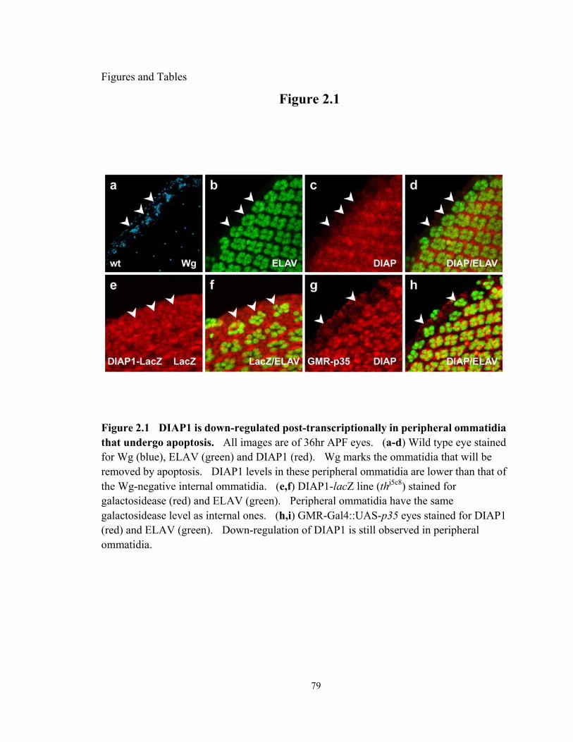
79
Figures and Tables
Figure 2.1
Figure 2.1 DIAP1 is down-regulated post-transcriptionally in peripheral ommatidia that undergo apoptosis. All images are of 36hr APF eyes. (a-d) Wild type eye stained for Wg (blue), ELAV (green) and DIAP1 (red). Wg marks the ommatidia that will be removed by apoptosis. DIAP1 levels in these peripheral ommatidia are lower than that of the Wg-negative internal ommatidia. (e,f) DIAP1-lacZ line (thj5c8) stained for galactosidease (red) and ELAV (green). Peripheral ommatidia have the same galactosidease level as internal ones. (h,i) GMR-Gal4::UAS-p35 eyes stained for DIAP1 (red) and ELAV (green). Down-regulation of DIAP1 is still observed in peripheral ommatidia.

80
Figure 2.2
Figure 2.2 DIAP1 is down-regulated in peripheral ommatidia that undergo apoptosis. Wild type eye stained for Wg (blue), ELAV (green) and DIAP1 (red) at 33hr (a-d), 42hr (e-h) and 36hr (i-l). Wg marks the ommatidia that will be removed by apoptosis. DIAP1 levels start to decrease at some ommatidia at 33hr, and stay lower than that of the Wg-negative internal ommatidia at 42hr. (i-l) DIAP1 down-regulation is also observed at 36hr in wild type eye stained with an independent anti-DIAP1 antibody from K. White (KW) (i).

81
Figure 2.3
Figure 2.3 Down-regulation of DIAP1 is inhibited in peripheral ommatidia where Wg signaling is blocked by overexpression of GPI-fz2. GMR-Gal4::UAS-GPI-fz2 eyes stained for DIAP1 (red) and ELAV (green).

82
Figure 2.4
Figure 2.4 Hid, Grim and Rpr contribute to DIAP1 down-regulation in peripheral ommatidia. All images are of 36hr APF eyes. Eyes stained for Wg (blue), ELAV (green) and DIAP1 (red) from hidX14/+ (a-d), Df(3f)XR38/+ (which removes one copy of rpr) (e-h), Df(3f)X25/+ (which removes one copy of hid and grim) (i-l), and a hidX14/ Df(3f)XR38 trans-heterozygote (m-o). DIAP1 down-regulation in peripheral ommatidia (arrowheads) is partially suppressed in all four genetic backgrounds. Quantification of DIAP1 protein levels in peripheral ommatidia are summarized in bar graph (p) and Table 2.1. Protein levels are presented as Normalized Mean Pixel Intensity (NPI), and shown as percentage of NPI of internal ommatidia in each genetic background. Two-tailed Student’s t-test was performed between perimeter ommatidia of different background, and ** denotes p < 0.001.

83
Figure 2.5
Figure 2.5 Apoptosis can occur in the absence of DIAP1 down-regulation in peripheral ommatidia. (a-f) Eyes from Df(3f)H99/+ (which removes one copy of hid, rpr and grim) stained for Wg (blue), ELAV (green) and DIAP1 (red) at 36hr APF (a-d) and 39hr APF (e-f). No significant difference in DIAP1 levels were observed between peripheral (arrowheads) and internal ommatidia, as shown in the representative samples (c, e) and summarized in Table1. (g-l) TUNEL staining of wild type (g, i, k) and Df(3f)H99/+ (h, j, l) at 36hr APF (g-h) and at 45hr APF (i-l). TUNEL signal is comparable between wild type and Df(3f)H99/+ at 36 hr, while the TUNEL signal is much greater in wild type at 45hr. At this time, H99 heterozygous eyes still contain unfragmented ELAV-positive photoreceptors compared to the TUNEL-positive cells in wild type (arrowheads in i, j).
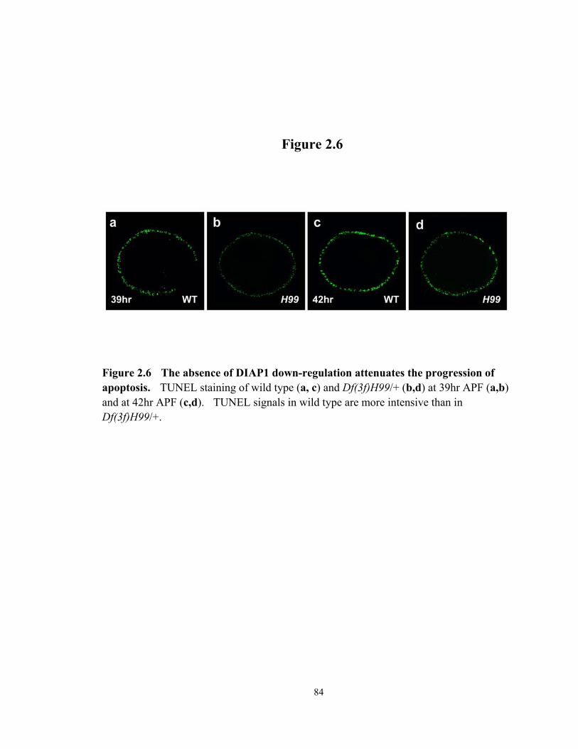
84
Figure 2.6
Figure 2.6 The absence of DIAP1 down-regulation attenuates the progression of apoptosis. TUNEL staining of wild type (a, c) and Df(3f)H99/+ (b,d) at 39hr APF (a,b) and at 42hr APF (c,d). TUNEL signals in wild type are more intensive than in Df(3f)H99/+.
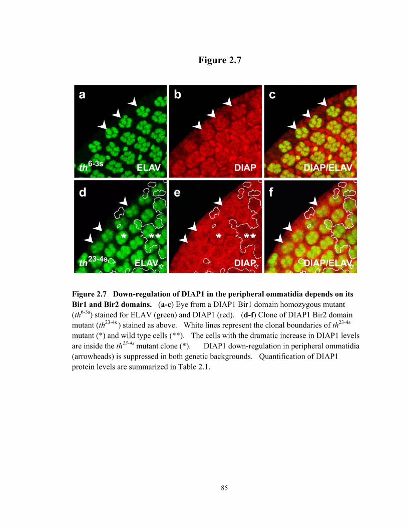
85
Figure 2.7
Figure 2.7 Down-regulation of DIAP1 in the peripheral ommatidia depends on its Bir1 and Bir2 domains. (a-c) Eye from a DIAP1 Bir1 domain homozygous mutant (th6-3s) stained for ELAV (green) and DIAP1 (red). (d-f) Clone of DIAP1 Bir2 domain mutant (th23-4s ) stained as above. White lines represent the clonal boundaries of th23-4s
mutant (*) and wild type cells (**). The cells with the dramatic increase in DIAP1 levels are inside the th23-4s mutant clone (*). DIAP1 down-regulation in peripheral ommatidia (arrowheads) is suppressed in both genetic backgrounds. Quantification of DIAP1 protein levels are summarized in Table 2.1.
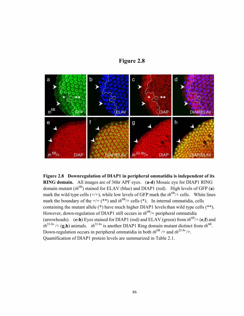
86
Figure 2.8
Figure 2.8 Downregulation of DIAP1 in peripheral ommatidia is independent of its RING domain. All images are of 36hr APF eyes. (a-d) Mosaic eye for DIAP1 RING domain mutant (th6B) stained for ELAV (blue) and DIAP1 (red). High levels of GFP (a) mark the wild type cells (+/+), while low levels of GFP mark the th6B/+ cells. White lines mark the boundary of the +/+ (**) and th6B/+ cells (*). In internal ommatidia, cells containing the mutant allele (*) have much higher DIAP1 levels than wild type cells (**). However, down-regulation of DIAP1 still occurs in th6B/+ peripheral ommatidia (arrowheads). (e-h) Eyes stained for DIAP1 (red) and ELAV (green) from th6B/+ (e,f) and th22-8s /+ (g,h) animals. th22-8s is another DIAP1 Ring domain mutant distinct from th6B. Down-regulation occurs in peripheral ommatidia in both th6B /+ and th22-8s /+. Quantification of DIAP1 protein levels are summarized in Table 2.1.

87
Figure 2.9
Figure 2.9 Dronc and Dark are required for DIAP1 down-regulation. All images are of 36hr APF eyes. Clones of darkG8 (a-d) and droncI29 (e-h) stained for ELAV (blue) and DIAP1 (red). Clones are marked by lack of clonal maker GFP (a, e), and white lines represent the boundaries. DIAP1 down-regulation in peripheral ommatidia (arrowheads) is partially suppressed in dronc and dark clones. Quantification of DIAP1 protein levels are summarized in Table 2.1.

88
Figure 2.10
Figure 2.10 Removal of Hid, Rpr and Grim blocks DIAP1 down-regulation in peripheral ommatidia. Clones of Df(3f)H99 stained for ELAV (green) and DIAP1 (red). White lines represent the boundaries of H99 mutant cells (*) and wild type cells (**). DIAP1 down-regulation does not occur in H99 peripheral ommatidia. Note also that loss of H99 has no detectable effect on DIAP1 levels in internal ommatidia.
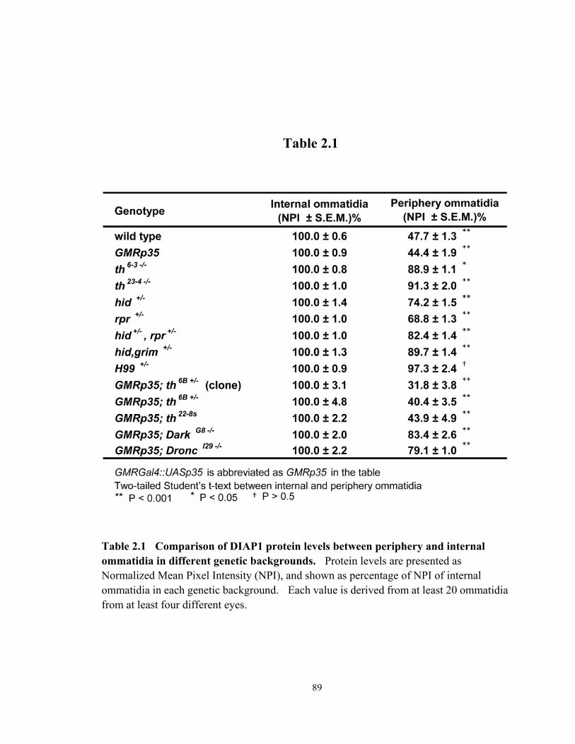
89
Table 2.1
Table 2.1 Comparison of DIAP1 protein levels between periphery and internal ommatidia in different genetic backgrounds. Protein levels are presented as Normalized Mean Pixel Intensity (NPI), and shown as percentage of NPI of internal ommatidia in each genetic background. Each value is derived from at least 20 ommatidia from at least four different eyes.
90
REFERENCES
Abdelwahid, E., T. Yokokura, R. J. Krieser, S. Balasundaram, W. H. Fowle and K. White (2007). "Mitochondrial disruption in Drosophila apoptosis." Developmental Cell 12(5): 793-806.
Cadigan, K. M. (2008). "Wnt-beta-catenin signaling." Current Biology 18(20): R943-R47. Cadigan, K. M., M. P. Fish, E. J. Rulifson and R. Nusse (1998). "Wingless repression of
Drosophila frizzled 2 expression shapes the wingless morphogen gradient in the wing." Cell 93(5): 767-77.
Chai, J. J., N. Yan, J. R. Huh, J. W. Wu, W. Y. Li, B. A. Hay, et al. (2003). "Molecular mechanism of Reaper-Grim-Hid-mediated suppression of DIAP1-dependent Dronc ubiquitination." Nature Structural Biology 10(11): 892-98.
Claveria, C., E. Caminero, C. Martinez, S. Campuzano and M. Torres (2002). "GH3, a novel proapoptotic domain in Drosophila Grim, promotes a mitochondrial death pathway." Embo Journal 21(13): 3327-36.
Ditzel, M., M. Broemer, T. Tenev, C. Boiduc, T. V. Lee, K. T. G. Rigbolt, et al. (2008). "Inactivation of Effector Caspases through Nondegradative Polyubiquitylation." Molecular Cell 32(4): 540-53.
Ditzel, M., R. Wilson, T. Tenev, A. Zachariou, A. Paul, E. Deas, et al. (2003). "Degradation of DIAP1 by the N-end rule pathway is essential for regulating apoptosis." Nature Cell Biology 5(5): 467-73.
Du, C. Y., M. Fang, Y. C. Li, L. Li and X. D. Wang (2000). "Smac, a mitochondrial protein that promotes cytochrome c-dependent caspase activation by eliminating IAP inhibition." Cell 102(1): 33-42.
Freel, C. D., D. A. Richardson, M. J. Thomenius, E. C. Gan, S. R. Horn, M. R. Olson, et al. (2008). "Mitochondrial localization of reaper to promote inhibitors of apoptosis protein degradation conferred by GH3 domain-lipid interactions." Journal of Biological Chemistry 283(1): 367-79.
Goyal, L., K. McCall, J. Agapite, E. Hartwieg and H. Steller (2000). "Induction of apoptosis by Drosophila reaper, hid and grim through inhibition of IAP function." Embo Journal 19(4): 589-97.
Haining, W. N., C. Carboy-Newcomb, C. L. Wei and H. Steller (1999). "The proapoptotic function of Drosophila Hid is conserved in mammalian cells." Proceedings of the National Academy of Sciences of the United States of America 96(9): 4936-41.
Hay, B. A. and M. Guo (2006). "Caspase-dependent cell death in Drosophila." Annual Review of Cell and Developmental Biology 22: 623-50.
Hay, B. A., D. A. Wassarman and G. M. Rubin (1995). "Drosophila homologs of baculovirus inhibitor of apoptosis proteins function to block cell death." Cell 83(7): 1253-62.

91
Hays, R., L. Wickline and R. Cagan (2002). "Morgue mediates apoptosis in the Drosophila melanogaster retina by promoting degradation of DIAP1." Nature Cell Biology 4(6): 425-31.
Hengartner, M. O. (2000). "The biochemistry of apoptosis." Nature 407(6805): 770-76. Herman-Bachinsky, Y., H. D. Ryoo, A. Ciechanover and H. Gonen (2007). "Regulation of
the Drosophila ubiquitin ligase DIAP1 is mediated via several distinct ubiquitin system pathways." Cell Death and Differentiation 14(4): 861-71.
Holley, C. L., M. R. Olson, D. A. Colon-Ramos and S. Kornbluth (2002). "Reaper eliminates IAP proteins through stimulated IAP degradation and generalized translational inhibition." Nature Cell Biology 4(60): 439-44.
Lim, H. Y. and A. Tomlinson (2006). "Organization of the peripheral fly eye: the roles of Snail family transcription factors in peripheral retinal apoptosis." Development 133(18): 3529-37.
Lin, H. V., A. Rogulja and K. M. Cadigan (2004). "Wingless eliminates ommatidia from the edge of the developing eye through activation of apoptosis." Development 131(10): 2409-18.
Lisi, S., I. Mazzon and K. White (2000). "Diverse Domains of THREAD/DIAP1 Are Required to Inhibit Apoptosis Induced by REAPER and HID in Drosophila." Genetics 154(2): 669-78.
Mendes, C. S., E. Arama, S. Brown, H. Scherr, M. Srivastava, A. Bergmann, et al. (2006). "Cytochrome c-d regulates developmental apoptosis in the Drosophila retina." Embo Reports 7(9): 933-39.
Muro, I., J. C. Means and R. J. Clem (2005). "Cleavage of the apoptosis inhibitor DIAP1 by the apical caspase DRONC in both normal and apoptotic Drosophila cells." Journal of Biological Chemistry 280(19): 18683-88.
Oberst, A., C. Bender and D. R. Green (2008). "Living with death: the evolution of the mitochondrial pathway of apoptosis in animals." Cell Death and Differentiation 15(7): 1139-46.
Olson, M. R., C. L. Holley, E. C. Gan, D. A. Colon-Ramos, B. Kaplan and S. Kornbluth (2003). "A GH3-like domain in reaper is required for mitochondrial localization and induction of IAP degradation." Journal of Biological Chemistry 278(45): 44758-68.
Oneill, E. M., I. Rebay, R. Tjian and G. M. Rubin (1994). "THE ACTIVITIES OF 2 ETS-RELATED TRANSCRIPTION FACTORS REQUIRED FOR DROSOPHILA EYE DEVELOPMENT ARE MODULATED BY THE RAS/MAPK PATHWAY." Cell 78(1): 137-47.
Rodriguez, A., H. Oliver, H. Zou, P. Chen, X. D. Wang and J. M. Abrams (1999). "Dark is a Drosophila homologue of Apaf-1/CED-4 and functions in an evolutionarily conserved death pathway." Nature Cell Biology 1(5): 272-79.
Ryoo, H. D., A. Bergmann, H. Gonen, A. Ciechanover and H. Steller (2002). "Regulation of Drosophila IAP1 degradation and apoptosis by reaper and ubcD1." Nature Cell Biology 4(6): 432-38.

92
Salvesen, G. S. and J. M. Abrams (2004). "Caspase activation - stepping on the gas or releasing the brakes? Lessons from humans and flies." Oncogene 23(16): 2774-84.
Shapiro, P. J., H. H. Hsu, H. Jung, E. S. Robbins and H. D. Ryoo (2008). "Regulation of the Drosophila apoptosome through feedback inhibition." Nature Cell Biology 10(12): 1440-U134.
Srinivasula, S. M. and J. D. Ashwell (2008). "IAPs: What's in a name?" Molecular Cell 30(2): 123-35.
Srivastava, M., H. Scherr, M. Lackey, D. Xu, Z. Chen, J. Lu, et al. (2007). "ARK, the Apaf-1 related killer in Drosophila, requires diverse domains for its apoptotic activity." Cell Death and Differentiation 14(1): 92-102.
Steller, H. (2008). "Regulation of apoptosis in Drosophila." Cell Death and Differentiation 15(7): 1132-38.
Suzuki, Y., Y. Imai, H. Nakayama, K. Takahashi, K. Takio and R. Takahashi (2001). "A serine protease, HtrA2, is released from the mitochondria and interacts with XIAP, inducing cell death." Molecular Cell 8(3): 613-21.
Suzuki, Y., Y. Nakabayashi and R. Takahashi (2001). "Ubiquitin-protein ligase activity of X-linked inhibitor of apoptosis protein promotes proteasomal degradation of caspase-3 and enhances its anti-apoptotic effect in Fas-induced cell death." Proceedings of the National Academy of Sciences of the United States of America 98(15): 8662-67.
Verhagen, A. M., P. G. Ekert, M. Pakusch, J. Silke, L. M. Connolly, G. E. Reid, et al. (2000). "Identification of DIABLO, a mammalian protein that promotes apoptosis by binding to and antagonizing IAP proteins." Cell 102(1): 43-53.
Wang, S. L., C. J. Hawkins, S. J. Yoo, H. A. J. Muller and B. A. Hay (1999). "The Drosophila caspase inhibitor DIAP1 is essential for cell survival and is negatively regulated by HID." Cell 98(4): 453-63.
White, K., M. E. Grether, J. M. Abrams, L. Young, K. Farrell and H. Steller (1994). "Genetic-control of programmed cell-death in Drosophila." Science 264(5159): 677-83.
Wilson, R., L. Goyal, M. Ditzel, A. Zachariou, D. A. Baker, J. Agapite, et al. (2002). "The DIAP1 RING finger mediates ubiquitination of Dronc and is indispensable for regulating apoptosis." Nature Cell Biology 4(6): 445-50.
Wing, J. P., B. A. Schreader, T. Yokokura, U. Q. Wang, P. S. Andrews, N. Huseinovic, et al. (2002). "Drosophila Morgue is an F box/ubiquitin conjugase domain protein important for grim-reaper mediated apoptosis." Nature Cell Biology 4(6): 451-56.
Wolff, T. and D. Ready (1993). Pattern Fromation in the Drosophila Retina. New York, Cold Spring Harbor Laboratory Press.
Xu, D. B., Y. Li, M. Arcaro, M. Lackey and A. Bergmann (2005). "The CARD-carrying caspase Dronc is essential for most, but not all, developmental cell death in Drosophila." Development 132(9): 2125-34.

93
Yan, N., J. W. Wu, J. J. Chai, W. Y. Li and Y. G. Shi (2004). "Molecular mechanisms of DrICE inhibition by DIAP1 and removal of inhibition by Reaper, Hid and Grim." Nature Structural & Molecular Biology 11(5): 420-28.
Yang, Y., S. Y. Fang, J. P. Jensen, A. M. Weissman and J. D. Ashwell (2000). "Ubiquitin protein ligase activity of IAPs and their degradation in proteasomes in response to apoptotic stimuli." Science 288(5467): 874-77.
Yokokura, T., D. Dresnek, N. Huseinovic, S. Lisi, E. Abdelwahid, P. Bangs, et al. (2004). "Dissection of DIAP1 functional domains via a mutant replacement strategy." Journal of Biological Chemistry 279(50): 52603-12.
Yoo, S. J., J. R. Huh, I. Muro, H. Yu, L. J. Wang, S. L. Wang, et al. (2002). "Hid, Rpr and Grim negatively regulate DIAP1 levels through distinct mechanisms." Nature Cell Biology 4(6): 416-24.
Zachariou, A., T. Tenev, L. Goyal, J. Agapite, H. Steller and P. Meier (2003). "IAP-antagonists exhibit non-redundant modes of action through differential DIAP1 binding." Embo Journal 22(24): 6642-52.

94
CHAPTER III
THE DROSOPHILA CASEIN KINASE I EPSILON DISCS
OVERGROWN PROMOTES CELL SURVIVAL VIA
ACTIVATION OF DIAP1 EXPRESSION
ABSTRACT
The proper number of cells in developing tissues is achieved by coordinating cell
division with apoptosis. In Drosophila, the adult wing is derived from wing imaginal
discs, which undergo a period of growth and proliferation during larval stages without
much programmed cell death. In this report, we demonstrate that the Drosophila casein
kinase Iε/δ, known as Discs overgrown (Dco), is required for maintaining this low level of
apoptosis. Expression of dco can suppress the apoptotic activity of Head involution
defective (Hid) in the developing eye. Loss of dco in the wing disc results in a dramatic
reduction in expression of the caspase inhibitor DIAP1 and a concomitant activation of
caspases. The regulation of DIAP1 by Dco occurs by a post-transcriptional mechanism
95
that is independent of hid. Mutant clones of dco are considerably smaller than controls
even when apoptosis is inhibited, suggesting that Dco promotes cell division/growth in
addition to its role in cell survival. The dco phenotype cannot be explained by defects
Wingless (Wg) signaling. We propose that Dco coordinates tissue size by stimulating cell
division/growth and blocking apoptosis via activation of DIAP1 expression.
INTRODUCTION
Apoptosis, or programmed cell death (PCD), is a tightly regulated mechanism of cell
death that is essential to animal development and homeostasis of adult organs (Jacobson et
al., 1997; Meier et al., 2000). In many developmental contexts, PCD is required for the
removal of excess cells during morphogenesis. Apoptosis also serves as a quality control
mechanism to eliminate abnormal or damaged cells from the organism. In addition, there
is a link between cell growth/proliferation and PCD. Cells stimulated to grow and divide
often also have an increased propensity for undergoing apoptosis (Abrams, 2002; Green
and Evan, 2002). This relationship has been suggested to act as a “fail-safe” system, to
prevent unwanted expansion of cell populations. Inhibition of this system is thought to
occur during the multi-step process of oncogenesis (Evan and Vousden, 2001; Green and
Evan, 2002; Hanahan and Weinberg, 2000).
PCD is executed by a family of cysteine proteases called caspases. Caspases are
synthesized as inactive zymogens and are processed to their active form to cleave cellular

96
substrates, thus promoting the apoptotic pathway. Caspases and many of their regulatory
components, such as the Bcl2 family proteins, are evolutionarily conserved from
nematodes to mammals (Kaufmann and Hengartner, 2001). However, additional
strategies have evolved in insects and higher organisms to provide greater flexibility of
regulation. One example is the Inhibitor of Apoptosis (IAP) family of proteins. IAPs
can bind to caspases, inhibiting their processing, catalytic activity or the ability of active
caspase to bind to substrates (Deveraux and Reed, 1999; Tenev et al. 2005). IAPs can also
facilitate caspase degradation by targeting them for ubiquitination via their E3 ubiquitin
ligase activity (Wilson et al., 2002; Yang et al., 2000).
Studies of a Drosophila member of the IAP family, DIAP1, encoded by the thread
(th) locus, have provided critical insights into the function and regulation of IAPs. DIAP1
can be bound and inhibited by Head involution defective (Hid), Grim and Reaper (Rpr)
(Hay, 2000). Overexpression of these pro-apoptotic factors can down regulate DIAP1
levels by promoting its degradation via the ubiquitin ligase activity of DIAP1 as well as
inhibiting DIAP1 translation (Hays et al., 2002; Holley et al., 2002; Ryoo et al., 2002; Yoo
et al., 2002). Once caspases are activated, they can cleave DIAP1, stimulating its
ubiquitination and degradation (Ditzel et al., 2003). This multilevel regulation of DIAP1
reflects the critical role it plays in cell survival, as evidenced by the massive activation of
caspases and apoptosis observed in DIAP1 mutants (Ryoo et al., 2004; Yoo et al., 2002).
97
The connection between cell growth/proliferation and cell death and pattern
formation has been extensively studied in the Drosophila wing imaginal discs (Edgar,
1999; Serrano and O'Farrell, 1997; Tapon et al., 2001a). During the three larval stages,
the wing discs undergo a rapid phase of cell growth and proliferation with very low levels
of PCD (Milan et al., 1997). The Drosophila homologue of Casein Kinase Iε/δ (CKIε/δ),
known as discs overgrown/double time (dco), was shown to be essential for imaginal tissue
development (Jursnich et al., 1990; Zilian et al., 1999). Weak dco mutants exhibit a lag in
disc growth, while stronger alleles have small or severely degenerated discs. Mitotic
clones of dco null mutant cells in the wing disc did not grow or survive when induced
during first or second larval stages, though small clones could be observed if induced at
later stages (Zilian et al., 1999). Electron micrographs of dco mutant discs demonstrated
the presence of basally extruded cells with nuclear condensation, suggesting that PCD was
occurring. Based on these observations it was concluded that dco is required for cell
proliferation and/or survival through an undetermined mechanism.
In this report we demonstrate that Dco is an essential cell survival factor in the wing
imaginal disc. We initially identified Dco as a potent suppressor of Hid induced cell death
in the eye. This Hid antagonism requires Dco kinase activity. Loss of dco in the wing
results in massive PCD, which is a major cause of the small disc phenotype seen in dco
mutants. When apoptosis is inhibited, dco mutant wings are normally patterned,
suggesting that dco’s effect on cell survival is not an indirect consequence of abnormal cell

98
specification. We demonstrate that dco mutant cells have a dramatic post-transcriptional
reduction of DIAP1 protein levels, which is sufficient to explain the increase in PCD.
This regulation of DIAP1 is independent of hid and not does appear to require the E3 ligase
activity of DIAP1. We also provide results suggesting that Dco promotes cell
growth/proliferation in the wing disc.
CKI family members, including CKIδ and CKIε, have been implicated in several
developmentally important signaling pathways. In Drosophila, dco has been shown to act
as a positive regulator of planar cell polarity in the larval eye and pupal wing (Strutt et al.,
2006; Klein et al., 2006). In fly cell culture and the wing imaginal disc, dco has been
shown to be required for Wnt/Wingless (Wg) signaling (Cong et al., 2004; Klein et al.,
2006; Zhang et al., 2006). Wg signaling is known to promote cell survival in the wing
imaginal discs (Giraldez and Cohen, 2003; Johnston and Sanders, 2003). However, we
find that cells homozygous for a null dco allele have normal Wg signaling in the
developing wing. Our data support a model where Dco promotes cell survival by
activating DIAP1 protein levels independently of Wg signaling.
MATERIALS AND METHODS
Fly strains
The following fly strains were used. Oregon R was our wild type stock. The
GMR-hid10, GMR-hid1M, GMR-rpr2, GMR-rpr5, GMR-grim2 and GMR-grim3 (Chen et al.,
99
1996; Rodriguez et al., 1999) stocks were from J. Abrams. GMR-hidala5 (Bergmann et al.,
1998) was from A. Bergmann. UAS-lacZ (Brand and Perrimon, 1993), UAS-p35
(Mergliano and Minden, 2003), enGal4 (Neufeld et al., 1998) and ptcGal4 (Johnson et al.,
1995) and thj5c8 (Hay et al., 1995) were from the Bloomington Stock Center. UAS-dco
(on the X chromosome; Price and Kalderon, 2002) was from M. Young. A wild-type
UAS-dco stock and UAS-dcoD132N mutant (Klein et al., 2006) were obtained from M.
Mlodzik and a UAS-dco and UAS-dcoK38R (Strutt et al., 2006) was provided by D. Strutt.
GMRGal4 (Freeman, 1996) was from M. Freeman. The hid05014and hidX14 stocks (Grether
et al., 1995) were from K. White, while the Df(3L)H99 and UAS-DIAP1 stocks (Hay et al.,
1995) were from B. Hay. The dco mutant alleles dcoP915, dcoP1396 and dcole88 (Zilian et al.,
1999) were from M. Noll. The th6B allele (Lisi et al., 2000) was from K. White. FRT82B
P[w+, Ubi-GFPnls] and FRT82B P[w+, armlacZ] (Tapon et al., 2001b) were from I.
Hariharan while all other chromosomes for clonal analysis, yw flp122 , FRT82B (Xu et al.,
1993) were from the Bloomington Stock Center. The UAS-dcoRNAi transgene was
constructed by cloning an inverted repeat of the C-terminal non-kinase domain coding
sequence using the PCR primers 5'CCAGTCTAGACAGCGGCCGCAGGACG
GAGCGGAC3' and 5'GCTATTTAGATCTAAACCGCAAGGGGTGCGGTGG3' into
the pWIZ vector (Lee and Carthew, 2003). Transgenic flies were generated following the
standard P element methodology.

100
Generation of dco clones
To generate dco clones with p35 expressed in the posterior domain of the wing disc,
the fly strain yw, flp122; enGal4; FRT82B P[armlacZ]/TM6 was crossed to w, UAS-p35;
FRT82Bdcole88. For the DIAP-lacZ experiment, the following strain yw,flp122; enGal4;
FRT82B P[w+, Ubi-GFPnls]/TM6 was crossed to UASp35; thj5c8, FRT82Bdcole88/TM6.
The progenies were heat shocked during early 2nd instar larval stage for 1 hour at 37°C.
The larvae were dissected and fixed with 4% formaldehyde 60 to 72 hours later.
Immunostainings, TUNEL and in situ hybridization
Immunostainings were performed as previously described (Parker et al., 2002).
Mouse anti-DIAP1 (1:200) and rabbit anti-active Drice (1:2,000) (Yoo et al., 2002) were
both provided by B. Hay. Guinea pig anti-Sen (1:5000) was made by immunization with
GST-Sen as previously described (Nolo et al., 2000). Rabbit anti-Dll (1:200) was from J.
Kohtz and rabbit anti-Spalt (1:100) (Kuhnlein et al., 1994) was from R. Schuh and B.
Mollereau. Rabbit anti-β-galactosidase (Cappel, ICN, Costa Mesa, CA) and mouse
anti-β-galactosidase (Sigma, St. Louis, MO) were both used at 1:500. Rabbit
anti-phospho-Histone 3 (1:150) was from Upstate (Lake Placid, NY). Monoclonal
anti-En (1:20) and anti-Wg (1:100) were from the Developmental Studies Hybridoma
Bank (Iowa City, IA). Cy3- and Alexa Fluor 488-conjugated secondary antibodies were
from Jackson Immunochemicals (West Grove, PA) and Molecular Probes (Eugene, OR),
respectively. The TUNEL assay was performed using the Cell Death Detection kit
101
(Roche Diagnostics, Indianapolis, IN) as previously described (Lin et al., 2004). In situ
hybridization detection of hid, grim and rpr transcripts was performed as previously
described (Lin et al., 2004). Pupal wings were stained as described (Axelrod, 2001) with
Alexa568 phalloidin (Molecular Probes) at a concentration of 6.25U/ml.
Microscopy and image analysis
The adult eyes were photographed on a Leica M10 microscope as previously
described (Parker et al., 2002). All fluorescent pictures were obtained with a Ziess
Axiophot coupled to a Zeiss LSM510 confocal apparatus. The pixel values of the clone
and twin areas in Figure 3.8 were calculated using the histogram tool in Photoshop 6.
RESULTS
Dco preferentially suppresses Hid induced cell death in the eye.
dco was identified in an EP misexpression screen (Rorth et al., 1998) for genes that
interact with the Wg signaling pathway. When placed under the control of the
eye-specific GMR promoter, Wg causes a severe reduction in eye size (Parker et al., 2002).
This is due in part to an increase in apoptosis in the late larval/early pupal eye (Lin et al.
2006). We expected that some EP lines that could suppress the GMR/wg phenotype
would be inhibitors of PCD. One such example was EP(3)3280, where the P-element is
inserted into the first intron of dco (data not shown). Similar suppression of GMR/wg was

102
obtained with a UAS-dco line, confirming that dco was responsible for the effect.
Misexpression of dco in several contexts outside the eye revealed no effect on Wg
signaling (data not shown). However, expression of dco in the eye strongly suppressed
the activity of the pro-apoptotic factor Hid (Figure 3.1B). dco had more subtle effects on
the small eye phenotype caused by GMR-rpr (Figure 3.1D, F) and moderately suppressed
GMR-grim (Figure 3.1H). Because hid, grim and rpr are expressed by the heterologous
GMR promoter, the suppression is not due to transcriptional inhibition of these
pro-apoptotic genes.
It has been previously shown that ectopic expression of activated Ras preferentially
suppresses Hid induced cell death in the eye (Bergmann et al., 1998; Kurada and White,
1998). However, the ability of activated Ras to suppress the GMR-hid small eye
phenotype was blocked when five potential MAP kinase phosphorylation sites were
mutated to alanine (GMR-hidala5) (Bergmann et al., 1998). Both EP(3)3280 and
UAS-dco were able to suppress GMR-hidala5 to the same extent as wild type GMR-hid
(Figure 3.2B and data not shown), suggesting that dco overexpression is not working
through a Ras/MAP kinase pathway.
To determine whether endogenous dco regulates apoptosis in the eye, we reduced
dco expression using RNAi via a dco hairpin construct (UAS-dcoRNAi). Expression of this
hairpin with the GMR-Gal4 driver resulted in a roughened eye phenotype but did not
reduce overall eye size (Figure 3.3B). However, dcoRNAi enhanced the small eye

103
phenotype of an intermediate GMR-hid1M construct (Figure 3.3 C, D). dcoRNAi had much
smaller effects on GMR-rpr and GMR-grim constructs (data not shown). These data
indicate that endogenous dco influences the level of apoptosis caused by misexpression of
hid.
To examine whether the kinase activity of Dco is required for Hid antagonism, two
mutant forms of dco were co-expressed with hid in the eye. dcoD132N is predicted to
compromise ATP binding (Peters et al., 1999; Zhu et al., 1998) while dcoK38R contains a
mutation within the catalytic site (Peters et al., 1999; DeMaggio et al., 1992; Fish et al.,
1995). Both mutants can act as dominant negatives for CKI function (Zhu et al., 1998;
Peters et al., 1999; McKay et al., 2001; Klein et al., 2006; Strutt et al., 2006). However,
DcoD132N was clearly able to suppress the GMR-hid small eye phenotype (Figure 3.4B),
while DcoK38R had the opposite effect. The K38R form of Dco enhanced both a
moderate/strong GMR-hid (Figure 3.4D) and a weaker GMR-hid transgene (Figure 3.4F).
When expressed by GMRGal4 in an otherwise wild-type eye, DcoK38R caused a roughening
of the eye, but did not effect eye size (data not shown). Because the D132N mutation has
been reported to reduce, but not eliminate CKI kinase activity (Zhu et al., 1998), the data
suggest that a partially active Dco can still suppress Hid-mediated apoptosis but some Dco
kinase activity is required for antagonizing Hid activity.
104
dco mutant imaginal discs have elevated apoptosis
Although dco overexpression and RNAi knockdown effect the ability of Hid to
promote apoptosis, mitotic clones homozygous for a dco null allele (dcole88) are readily
detectable in late larval eye discs and mid-pupal eyes (data not shown). dco mutant clones
are not recovered in the adult eye, although some scarring near the twin spots was seen
(data not shown). We have not been able to detect increased apoptosis in the larval or
pupal clones. Apparantly, cells lacking dco are eliminated sometime in late pupal
development.
To further explore whether dco is required for suppressing PCD, we examined
several other imaginal discs. Using the TUNEL DNA fragmentation assay, we compared
the level of apoptosis in imaginal discs from wild type and hypomorphic dco mutants
(dcoP915/P1395). Throughout third instar larval development, TUNEL positive cells in the
wing were detected at low levels in wild type, as shown in Figure 3.5A & D. In contrast,
we found a dramatic increase in TUNEL signal in the dco mutant wing discs (Figure 3.5B,
E). Note that the dco hypomorphs are developmentally delayed, and the discs were
normalized for developmental stage by examining the pattern of Wg expression. Elevated
TUNEL signal was also observed in the leg and haltere discs from dco mutants, but no
increase was observed in eye imaginal discs (data not shown).
To determine whether the elevated PCD we observe is related to hid, grim and/or rpr
function, we lowered the dose of these genes in a dco mutant background. Removal of
105
one copy of hid, grim and rpr (using the Df(3L)H99 deficiency) caused a significant
reduction of TUNEL signal in dco mutants (Figure 3.5E, F). Removal of one copy of hid
alone did not significantly reduce the level of apoptosis, but a dramatic reduction was
achieved by removing both copies of hid (data not shown). These data suggest that in dco
mutant discs endogenous hid, grim and/or rpr are required for the elevated cell death.
As reported previously, dcoP915/P1395 mutant animals have a prolonged larval phase
with slow imaginal disc growth (Zilian et al., 1999). These animals die in late larval or
early pupal stages. We found that the decrease in cell death in H99, dcoP915/+, dcoP1395
discs was correlated with marked acceleration in disc growth and larval development (from
8-10 days to 5-6 days). These H99/+, dcoP915/P1395 animals have increased survival into late
pupal stages, and the veination pattern of the majority of these animals are similar to
wildtype (Figure 3.5G, H). Occasional escapers survive to adulthood displaying normal
legs and wrinkled or blistered wings (data not shown). These results suggest that the
developmental delay in disc growth and larval development seen in dco mutants is largely
caused by elevated apoptosis. In addition, the data in Figure 3.5H indicate that patterning
of the wing is normal in dco mutants when PCD is reduced.
Dco activates DIAP1 expression in the wing imaginal disc
Both overexpression and loss of function studies with dco suggest that it encodes an
anti-apoptotic factor in Drosophila imaginal tissues. Our initial observation that
overexpression of dco differentially suppresses GMR-hid and GMR-rpr is reminiscent of

106
DIAP1 alleles containing mutations in its RING domain (Lisi et al., 2000; Wilson et al.,
2002). It has been shown that these DIAP1 mutants lack ubiquitin ligase activity and may
be present at higher levels due to stabilization of DIAP1. These findings prompted us to
look at DIAP1 protein levels when dco expression is elevated (with UAS-dco) or reduced
(UAS-dcoRNAi). Both UAS transgenes were driven by patched (ptc)-Gal4, which is active
in a stripe on the anterior side of the anterior/posterior boundary of the wing imaginal disc
(Johnson et al., 1995).
Overexpression of dco led to a moderate increase of DIAP1 protein levels in the ptc
expression domain (Figure 3.6B). Conversely, DIAP1 expression was decreased upon
RNAi-mediated knockdown of dco (Figure 3.6C). Using an antibody specific for the
activated form of the effector caspase Drice (Yoo et al., 2002), we found that dco
knockdown also caused a marked increased in Drice activation (Figure 3.6E). Z-axis
projection of ptc-Gal4/UAS-dcoRNAi wing discs reveals that the Drice-positive cells are
found basally in the epithelial sheet (Figure 3.6G) and that DIAP1 levels are low both
apically and basally (Figure 3.6H). Loss of DIAP1 is known to cause apoptosis (Ryoo et
al. 2004; Wang et al. 1999; Yoo et al. 2002), and the data suggest that Dco promotes cell
survival by maintaining DIAP1 expression.
To determine whether hid was required for Dco regulation of DIAP1, the
ptc-Gal4/UAS-dcoRNAi experiments were repeated in a hid mutant background. An
obvious decrease in DIAP1 levels was still observed (Figure 3.6D) indicating that Dco can

107
activate DIAP1 independently of hid. The reduction in DIAP1 was also seen in a H99/+
background (data not shown). In contrast, the activation of Drice by dcoRNAi was greatly
suppressed in hid mutants (Figure 3.6F) or H99 heterozygotes (data not shown). This is
consistent with the results from dco hypomorphic mutants (Figure 3.5C, F).
To rule out the possibility that Dco regulates the transcript levels of hid, grim or rpr,
in situ hybridization was performed on ptc-Gal4/UAS-dcoRNAi wing imaginal discs. hid
expression was not detectably effected (Figure 3.7A), but a small reduction in grim
expression was sometimes observed within the Ptc domain (Figure 3.7B) while a small
increase in rpr transcripts was also consistently seen (Figure 3.7C). These minor
differences were not observed in wild-type discs (data not shown). We consider it highly
unlikely that these minor and contradictory differences can explain the dramatic effect
observed on DIAP1 protein levels.
We also examined the phenotype of dco null (dcole88) mutant cells induced by
mitotic recombination. As reported previously (Zilian et al., 1999), clones of this allele
were not obtained when recombination was induced before third larval instar (data not
shown). This experiment was repeated in discs where the baculovirus caspase inhibitor
p35 was constantly expressed in the posterior compartment via engrailed (en)-Gal4. As
shown in Figure 3.8A, dco mutant clones were not detected in the anterior compartment
(lacking p35), but were found in the posterior compartment expressing p35. Twin spots
were visible in both compartments, indicating that mitotic recombination had occurred.

108
These data indicate that caspase-dependent PCD contributes significantly to the loss of
cells in dco mutant clones.
Consistent with a role of Dco in promoting cell survival by maintaining DIAP1
levels, we found elevated active Drice and decreased DIAP1 in dco clones (Figure 3.8C-H).
The activation of Drice is largely cell autonomous, though we occasionally see some
additional signal adjacent to the clones (arrows in Figure3.8C-E). We speculate that this
might represent dying cells engulfed by surrounding cells or macrophages. It has been
reported in fly embryos that phagocytosis of cells containing activated caspases is not
blocked by p35 expression (Mergliano and Minden, 2003). The dco null clones have
decreased DIAP1 levels regardless of the area in the wing discs (hinge, pouch, notum)
where the clones are found, indicating that Dco regulates DIAP1 throughout the wing
discs.
When dco clones were generated in a Minute/+ background, a strong decrease in
DIAP1 levels were observed, but only a small increase in activated Drice was seen, most
prominently in the basal area of the clone (Figure 3.9). This suggests that cell competition
is involved in promoting PCD in dco mutant cells, though not in the primary effect on
DIAP1 protein expression.
To examine whether this activation of DIAP1 by dco is at the transcriptional level,
we looked at the expression of a lacZ reporter under the control of the DIAP1 control
109
region (DIAP1-lacZ) in dco clones and found that is was not altered (Figure 3.8I-K). This
suggests that Dco regulates DIAP1 by a post-transcriptional mechanism.
dco mutant clones are significantly smaller than their twins
A role for Dco in cell proliferation was suggested by the size of dco clones in the
presence of the caspase inhibitor p35, which were consistently smaller than the twin spots
(e.g., Figure 3.8A). When this was quantitated, we found that the dco clones were on
average 4 times smaller than the twin spots in the posterior compartment (Figure 3.8B).
Because the twins and clones originate from the two daughter cells of a single cell that
underwent mitotic recombination, they grow for the same period of time in a similar
location. Therefore the approximate 1:4 ratio of clone/twin size in dco mutants suggests a
defect in the rate of growth/proliferation. To examine this further, we performed bromo
deoxyuridine incorporation and immunostaining for phospho-Histone 3, which label cells
that are undergoing DNA replication or mitosis, respectively. Neither analysis revealed
any obvious differences between the clones and surrounding tissues (data not shown).
These techniques are probably not sensitive enough to reveal subtle differences in cell
proliferation that could contribute to the reduced clone size after several generations of cell
division. The cells and nuclei in the clones also appear to be of similar size and shape as
those in the surrounding tissue, suggesting that reduced cell size cannot account for the
small size of dco clones.

110
The E3 ubiquitin ligase activity of DIAP1 is not required for its regulation by Dco
The C-terminus of DIAP1 contains a RING domain that has intrinsic E3 ubiquitin
ligase activity which can promote its own degradation via auto-ubiquitination.
Mutations that abolish this activity suppress Hid induced cell death but do not affect or
enhance Rpr killing (Lisi et al., 2000; Wilson et al., 2002; Yokokura et al., 2004). This is
consistent with the differential suppression of Hid and Rpr by ectopic expression of Dco
(Figure 3.1A-F). It is conceivable that Dco regulates DIAP1 level through modulating the
auto-ubiquitination process.
To explore the relationship between Dco and DIAP1 E3 ligase activity, we examined
whether a point mutation in one of the essential cysteine residues in the RING domain (th6B)
disrupted the decrease in DIAP1 protein levels upon RNAi depletion of dco. One
complication of monitoring DIAP1 levels in the RING domain mutant is the possibility
that the mutant DIAP1 proteins are unusually stable compared to wild-type, due to lack of
degradation by Hid or perhaps Rpr/Grim, since there are reports that these factors can also
effect DIAP1 stability (Hays et al. 2002; Ryoo et al. 2002). However, mitotic clones of
Df(3L)H99 did not contain higher levels of DIAP1 immunostaining in the wing imaginal
disc (Figure 3.10A-C). This was true of clones located throughout the wing pouch and
notum (data not shown). While much of the tissue surrounding these clones are
heterozygous for the hid, grim and rpr deficiency, the +/+ twins clones also had similar
levels of DIAP1 staining compared to the H99 clones (data not shown). These data

111
indicate that under normal conditions, endogenous Hid, Grim and Rpr do not significantly
regulate the expression of DIAP1 in the wing.
As previously described, expression of dcoRNAi in the ptc domain of the wing disc
results in a significant decrease in DIAP1 expression (Figure 3.6C) and an increase in
Drice activation (Figure 3.6E). In a th6B/+ background, similar results were obtained
(Figure 10D-E), suggesting that the effect of Dco on DIAP1 protein levels does not require
DIAP1’s E3 ligase activity.
dco mutant clones have normal Wg and Dpp signaling
CKIε has been proposed to act both as a positive and negative regulator of Wnt
signaling (Price, 2006). A positive role for Dco in Wg signaling has recently been
demonstrated in cultured Drosophila S2 cells (Cong et al., 2004) and wing imaginal discs
(Klein et al., 2006; Zhang et al., 2006). Wg signaling regulates cell proliferation and
promotes cell survival in the wing imaginal discs (Giraldez and Cohen, 2003; Johnston and
Sanders, 2003). Therefore, the phenotypes we have observed in dco mutant clones could
be explained by a reduction in Wg signaling. However, we find that two targets of Wg
signaling in wing imaginal discs, Senseless (Sen) and Distal-less (Dll) (Parker et al., 2002)
are unaffected in cells (expressing p35) that are homozygous for the dco null mutation
(Figure 3.12A-F). We conclude that the cell survival function of Dco in wing imaginal
discs is not linked to the Wg pathway and that Dco is dispensable for Wg signaling under
the conditions used in our study.
112
Like Wg, Dpp signaling has also been shown to promote cell proliferation and cell
survival in the wing imaginal disc (Martin-Castellanos and Edgar, 2002; Moreno et al.,
2002). CKIε has been shown to influence TGF-β signaling in mammalian cell culture,
both negatively and positively, depending on the assay (Waddell et al. 2004). However,
Dpp signaling appears to be normal in cells lacking dco, as judged by expression of the
Dpp target Spalt (Figure 3.12G-I).
DISCUSSION
Dco promotes cell survival by activating DIAP1 expression
Dco is required for suppression of PCD in several developing tissues.
Hypomorphic dco mutants have elevated apoptosis in the wing, leg and haltere discs
(Figure 3.5 and data not shown). Reduction of dco with either RNAi or with mitotic
clones of a null allele also caused activation of the effector caspase Drice in wing discs
(Figure3.6E, 3.8D). In the eye disc and early pupal eye, clones of the null dco allele did
not result in obvious activation of Drice or PCD (data not shown). However, reduction of
endogenous dco with RNAi did enhance the ability of Hid to promote cell death in the eye
(Figure 3.3C, D), indicating that Dco promotes cell survival in this tissue as well. It is
possible that more subtle phenotype in the eye is caused by other CKI family members
acting redundantly with Dco in this tissue.
In the wing disc, the loss of dco resulted in a dramatic reduction in DIAP1
expression (Figure 3.6, 3.8, 3.10). The loss of DIAP1 in either fly embryos or the wing

113
imaginal discs leads to massive activation of caspases and cell death (Ryoo et al., 2004;
Wang et al., 1999; Yoo et al., 2002). Therefore, the reduction in DIAP1 levels observed in
dco mutants is sufficient to explain the elevated caspase activation and apoptosis in these
cells. The reduction in DIAP1 expression is not a secondary consequence of activating
the apoptotic machinery, since a strong reduction in DIAP1 levels was observed in the
presence of a caspase inhibitor (Figure 3.8G), or in mutant backgrounds where active
caspases are barely detectable (Figure 3.6D, F). These results argue that reduction in dco
leads to a loss of DIAP1 expression, which results in caspase activation and PCD.
Though our data indicate that Dco activation of DIAP1 is important to suppress
apoptosis, the possibility existed that this regulation was due to non-specific effects of
mispatterning in dco mutants. However, the phenotype of H99, dcoP915/+, dcoP1395
mutants argues strongly against this scenario. Heterozygosity of H99 suppressed the
elevated PCD in the wing (Figure 3.5C, F), shortened the developmental lag observed in
dco hypomorphic mutants and increased the lethal phase of these mutants from early
pupation to late pupal stages, with some escapers eclosing. The flies that emerged had
normally patterned legs, but the pattern of the wings was difficult to analyze due to lack of
unfolding and blistering. However, the veination pattern of the pupal wings (prior to
folding) appeared normal (Figure 3.5H). This data, combined with the fact that Dpp and
Wg signaling appear normal in dco null clones (Figure 3.12) strongly suggests that Dco
does not have a significant role in patterning, but rather is a direct repressor of apoptosis.

114
The role of the IAP antagonists Hid, Grim and Rpr
dco interacts genetically with hid, grim and rpr in the eye (Figure 3.1-4) and the
wing (Figure 3.5-6), suggesting a link between Dco and these pro-apoptotic factors. Dco
appears to regulate DIAP1 at a post-transcriptional level (Figure 3.8J) and it has been
reported that DIAP1 is inhibited by Hid, Grim and Rpr at both the translational level and by
increased protein turnover (Hays et al., 2002; Holley et al., 2002; Ryoo et al., 2002; Yoo et
al., 2002). This raises the possibility that Dco could activate DIAP1 expression through
repression of one or more of these IAP inhibitors. Alternatively, Dco could activate
DIAP1 though a pathway that acts in parallel to Hid, Grim and Rpr.
If Dco regulates DIAP expression by repressing IAP inhibitor activity, it cannot
work solely through Hid. The elevated caspase activation and PCD observed upon
reduction of dco gene activity is greatly suppressed in a hid mutant background (Figure
3.6F and data not shown). However, DIAP1 levels are still reduced with dco RNAi in hid
mutants (Figure 3.6D). The apoptotic phenotype in dco mutant wing discs is suppressed
by removal of one copy of hid, grim and rpr (Figure 3.5C,F), but not by removal of one
copy of hid (data not shown). Therefore Dco would have to act by inhibiting Hid and
Grim and/or Rpr. While Dco overexpression most effectively suppresses Hid activity
(Figure 3.1B), it also moderately suppresses Grim (Figure 3.1H) and has a small effect on
Rpr-induced PCD (Figure 3.1F).

115
If Dco regulates DIAP1 in parallel to the IAP inhibitors, then how to explain the
suppression of caspase activation and PCD in dco mutants when hid is mutated (Figure
3.6F) or in Df(3L)H99 heterozygotes (Figure 3.5C, F)? These data can be explained by a
balance between negative regulators of apoptosis (Dco) and positive inputs
(Hid/Grim/Rpr). Reduction of dco lowers DIAP1 levels, promoting caspase activation,
however, the IAP inhibitors are known to bind to DIAP1 and sequester it from its normal
function of caspase inhibition (Hay, 2000). So reduction of these DIAP1 inhibitors allows
the lower levels of DIAP1 in the dco mutants to bind to caspases and prevent their
activation.
In addition to binding to DIAP1 and inhibiting its ability to repress caspases, Hid,
Grim and Rpr have been shown to down regulate DIAP1 expression (Hays et al., 2002;
Holley et al., 2002; Ryoo et al., 2002; Yoo et al., 2002). Interestingly, we found that
clones lacking hid, grim and rpr had no effect on DIAP1 protein levels in the wing disc
(Figure 3.10A-C). These results suggest that under normal conditions, these IAP
inhibitors are not major regulators of DIAP1 expression in the wing imaginal disc. A
more informative experiment would be to examine DIAP1 expression in clones lacking hid,
grim, rpr and dco, to determine whether the decrease in DIAP1 requires the three
pro-apoptotic factors. Attempts to perform this experiment have thus far not been
possible, due to technical difficulties.

116
Hid, Grim and Rpr have been suggested to downregulate DIAP1 though its intrinsic
E3 ubiquitin ligase activity (Hays et al., 2002; Ryoo et al., 2002; Yoo et al., 2002).
However, a mutation that disrupts enzymatic activity (th6B) is still downregulated in
dcoRNAi depleted cells (Figure 3.10D-F). This argues against DIAP1 auto-ubiquitination
playing an important role in Dco regulation of DIAP1. This experiment could only be
performed in a th6B heterozygous background, because th6B clones do not survive/grow.
Dco may promote cell growth/proliferation
In addition to its role in promoting cell survival, our clonal analysis indicates that
dco may promote cell growth/proliferation. In the presence of p35 caspase inhibitor, dco
mutant clones are about four times smaller than their twins (Figure 3.8B). One
explanation for this is that p35 does not completely suppress the elevated apoptosis in the
dco clone. However, the UAS-p35 transgene used is able to fully suppress a strong
GMR-hid phenotype in the eye, and no TUNEL positive cells are observed in our dco
clones in the presence of p35 (data not shown).
The conclusion that dco mutant cells grow/divide slower than controls is tempered
by several caveats. Recent findings show that apoptotic cells in the wing disc can
stimulate their neighbors to proliferate (de la Cova et al., 2004; Huh et al., 2004; Moreno
and Basler, 2004; Ryoo et al., 2004). This phenomenon has been observed in cells
expressing p35, suggesting that caspase activation but not PCD itself generates the
proliferative signal (Huh et al., 2004; Ryoo et al., 2004; Perez-Garijo et al. 2005).
117
Induced expression of Wg in the dying cells has been proposed to be part of the mitogenic
signal for this compensatory proliferation (Ryoo et al., 2004). While we observed
substantial ectopic Wg staining in dco mutant wing discs (Figure 3.11), ectopic Wg was
not observed in the p35 expressing dco mutant clones (n=30, data not shown). Despite
this, our current data cannot rule out that the low clone/twin ratio we observe for dco clones
in Figure 3.8B could at least be partially due to increased proliferation of the twins.
Relation of Dco to Wg and other pathways affecting PCD/proliferation
In our study, no defect in Wg signaling was observed in mitotic clones for a dco null
allele in wing imaginal discs (Figure 3.12A-F). A similar result has been recently
reported, though a hypomorphic dco allele was used (Strutt et al., 2006). In contrast,
another recent study generated large clones with a strong dco allele (dcodbt-P) in a Minute/+
background, and reported no activation of caspases and a loss of Sens and adult wing
margin, two readouts of Wg signaling (Klein et al., 2006). We have made dcole88 clones
with the Minute technique, and find that they are infrequent and similar in size to the ones
reported in Figure 3.8. The few at the dorsal/ventral boundary expressed Sens normally
(data not shown), while clones throughout the wing pouch had a dramatic reduction in
DIAP1 expression and only a small increase in activated Drice (Figure 3.9). Another
report generating very large dcole88 clones using MS1096-Gal4/UAS-FLP, Minute and p35
expression (Zhang et al. 2006) observed a strong decrease in Sens expression. This report
also found evidence that other CKI family members could promote Wg signaling (Zhang et

118
al., 2006). Clone size and redundancy/compensation may explain the differences between
our results and those of Klein et al. (2006) and Zhang et al. (2006). However, the primary
phenotype observed when dco is removed under the conditions used in this study is a
decrease in DIAP1 levels.
In our misexpression studies in the eye, Dco was able to suppress the ability of a
Ras/MAPK resistant version of Hid (Bergmann et al., 1998) to induce apoptosis (Figure
3.2). At least in the eye, Dco appears to control cell survival independently from
Ras/MAPK signaling.
The phenotype of dco is similar to that of yorkie, a nuclear protein that is required for
DIAP1 expression and proliferation (Huang et al., 2005). Yorkie is inhibited by a
complex containing the Lats and Hippo kinases, and the adaptor protein Salvador (Sav).
Clones mutant for lats, sav or hippo display an opposite phenotype to dco, i.e., dramatic
increases in proliferation and a block in PCD (see Edgar, 2006 for a review). Double
mutant clones for sav and dco do not survive/grow (data not shown), suggesting that Dco
may act downstream of the Wts/Sav/Hippo complex. However, the expression of
DIAP1-lacZ is upregulated in hippo clones (Wu et al., 2003) and decreased in yorkie clones
(Huang et al., 2005) but we observe no effect on this transgene in dco clones (Figure
3.8I-K). In addition, Wts/Sav/Hippo represses Cyclin E expression, but Dco does not
(data not shown). Therefore, we consider it unlikely that Dco acts with Wts/Sav/Hippo in
a simple linear pathway. Hippo has also been reported to phosphorylate DIAP1, leading

119
to instability (Harvey et al., 2003; Pantalacci et al., 2003), so the possibility remains that
Dco may interact with Hippo in this regard.
CKIδ/ε in other systems
Our data supports a model where Dco promotes cell survival in fly imaginal discs by
maintaining expression of DIAP1 through a novel pathway. Do casein kinase Iδ/ε family
members have similar functions in other organisms? The yeast CKI gene Hrr25 is
involved in DNA repair, cell growth and viability (Hoekstra et al., 1991). In mammalian
cell lines, CKIε plays a protective role against apoptosis induced by extrinsic death signals
(Desagher et al., 2001; Izeradjene et al. 2004). This cell survival activity was correlated
with CKIε phosphorylation of the pro-apoptotic factor Bid (Desagher et al., 2001).
Inhibition of CKIδ induces apoptosis in trophoblast cells (Stoter et al. 2005). Further
studies will be required to determine whether there are commonalities between these
different systems. The ability of Dco to inhibit apoptosis and possibly promote cell
proliferation makes it an attractive candidate for functioning as an oncogene in
tumorigenesis.
ACKNOWLEDGEMENTS
We would like to thank all the researchers who provided the reagents described in
the Methods section, especially M. Noll for the dco stocks, J. Abrams for the GMR-hid,

120
GMR-rpr and GMR-grim stocks, K. White for the hid alleles, H. Steller, K. White and B.
Hay for th alleles. Special thanks to D. Strutt and M. Mlodzik for providing UAS-dco
mutant stocks shortly after publication. Thanks also to J. Kennell for generating one of
the UAS-dcoRNAi stocks used in this study. We also would like to thank B. Hay for
DIAP1 and active Drice antibodies, P. Meier for DIAP1 antibody, L. Saez and M. Young
for the Dco antibody and the UAS-dco stock, and H. Richardson for the CycE antibodies.
This work was supported by NIH grants RO1 GM59846 and RO1 CA95869 to K.M.C.
This chapter was published as:
Guan, J., H. Li, A. Rogulja, J. D. Axelrod and K. M. Cadigan (2007). "The Drosophila casein kinase I epsilon/delta Discs overgrown promotes cell survival via activation of DIAP1 expression." Developmental Biology 303(1): 16-28.
In this project, I generated the data as below:
Figure 3.3; Figure 3.4; Figure3.6 G-I; Figure 3.7; Figure 3.9, Figure 3.10

121
FIGURES
Figure 3.1
Figure 3.1. Ectopic expression of dco suppresses the small eye phenotype induced by overexpression of the proapoptotic genes hid, rpr and grim. Micrographs are of adult female eyes. All flies shown contain GMR-Gal4 and either UAS-lacZ (A, C, E, J) or UAS-dco (B, D, F, H). They contain two copies of GMR-hid1M (A, B), one copy of GMR-rpr2 (C, D), GMR-rpr5 (E, F) or a copy each of GMR-grim2 and GMR-grim3 (G, H). Overexpression of dco strongly suppresses GMR-hidIM and other GMR-hid strains (data not shown), while suppression of GMR-rpr and GMR-grim is weaker and variable.

122
Figure 3.2
Figure 3.2. Ectopic expression of dco suppresses the small eye phenotype induced by a Ras/MAPK resistant form of Hid. Micrographs are of adult female eyes. Flies contain GMR-Gal4, GMR-hidala5 and either UAS-lacZ (A) or UAS-dco (B). Overexpression of dco strongly suppresses the GMR-hidala5 phenotype.
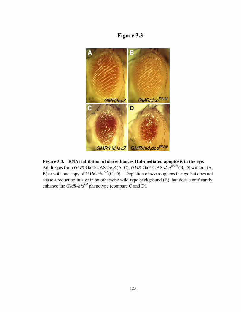
123
Figure 3.3
Figure 3.3. RNAi inhibition of dco enhances Hid-mediated apoptosis in the eye. Adult eyes from GMR-Gal4/UAS-lacZ (A, C), GMR-Gal4/UAS-dcoRNAi (B, D) without (A, B) or with one copy of GMR-hid1M (C, D). Depletion of dco roughens the eye but does not cause a reduction in size in an otherwise wild-type background (B), but does significantly enhance the GMR-hidIM phenotype (compare C and D).

124
Figure 3.4
Figure 3.4. Kinase activity is required for dco to suppress Hid-mediated apoptosis in the eye. Adult eyes from GMR-Gal4, GMR-hidala5 and either UAS-GFP (A), UAS-dco (B), UAS-dcoD132N (C) or UAS-dcoK38R (D). While the ATP-binding mutant dcoD132N
suppresses Hid activity as well as wild-type dco, the kinase dead mutant K38R enhances the small eye phenotype. (E, F) Enhancement was also observed with a weaker GMR-hid transgene. GMR-Gal4, GMR-hid1M and either UAS-GFP (E) or UAS-dcoK38R (F). Wild type dco and dcoD132N strongly suppressed GMR-hid1M (data not shown).
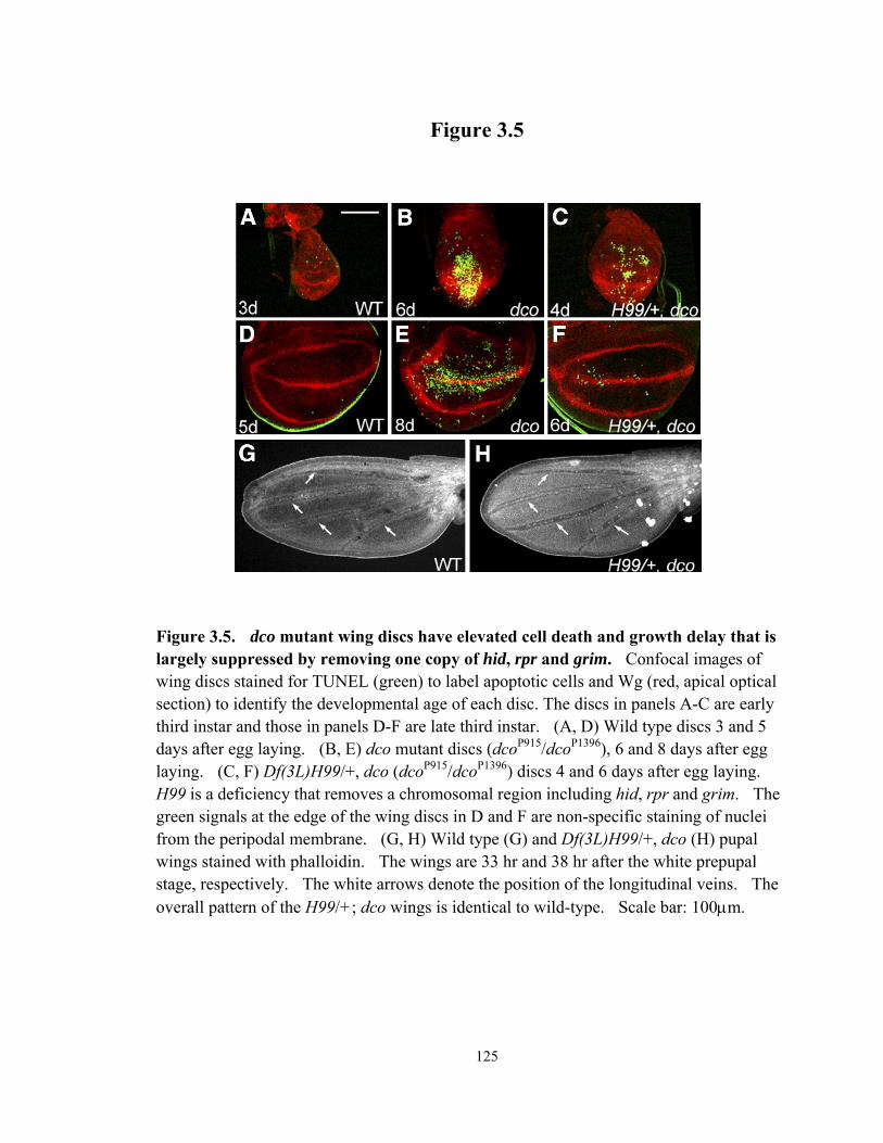
125
Figure 3.5
Figure 3.5. dco mutant wing discs have elevated cell death and growth delay that is largely suppressed by removing one copy of hid, rpr and grim. Confocal images of wing discs stained for TUNEL (green) to label apoptotic cells and Wg (red, apical optical section) to identify the developmental age of each disc. The discs in panels A-C are early third instar and those in panels D-F are late third instar. (A, D) Wild type discs 3 and 5 days after egg laying. (B, E) dco mutant discs (dcoP915/dcoP1396), 6 and 8 days after egg laying. (C, F) Df(3L)H99/+, dco (dcoP915/dcoP1396) discs 4 and 6 days after egg laying. H99 is a deficiency that removes a chromosomal region including hid, rpr and grim. The green signals at the edge of the wing discs in D and F are non-specific staining of nuclei from the peripodal membrane. (G, H) Wild type (G) and Df(3L)H99/+, dco (H) pupal wings stained with phalloidin. The wings are 33 hr and 38 hr after the white prepupal stage, respectively. The white arrows denote the position of the longitudinal veins. The overall pattern of the H99/+; dco wings is identical to wild-type. Scale bar: 100μm.

126
Figure 3.6
Figure 3.6. Dco regulates DIAP1 protein levels in the wing imaginal disc. Confocal images of late third instar larval wing discs stained for DIAP1 in red (A-D,H,I) and activated Drice in green (E-G,I). ptcGal4 is expressed at an anterior strip next to the anterior-posterior boundary (indicated by arrows in B-D). All discs are oriented anterior to the left. (A) Wild type. (B) ptc-Gal4/UAS-dco. (C, E) ptc-Gal4/UAS-dcoRNAi. (D, F) ptc-Gal4/UAS-dcoRNAi; hid05014/hidX14. (G-I) Z axis projection of ptc-Gal4/UAS-dcoRNAi disc showing basal localization of Drice positive cells. Arrows indicate the reduction in DIAP1 staining in the ptc expression domain.
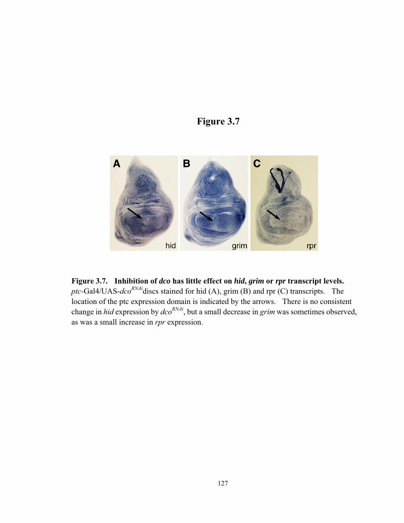
127
Figure 3.7
Figure 3.7. Inhibition of dco has little effect on hid, grim or rpr transcript levels. ptc-Gal4/UAS-dcoRNAidiscs stained for hid (A), grim (B) and rpr (C) transcripts. The location of the ptc expression domain is indicated by the arrows. There is no consistent change in hid expression by dcoRNAi, but a small decrease in grim was sometimes observed, as was a small increase in rpr expression.
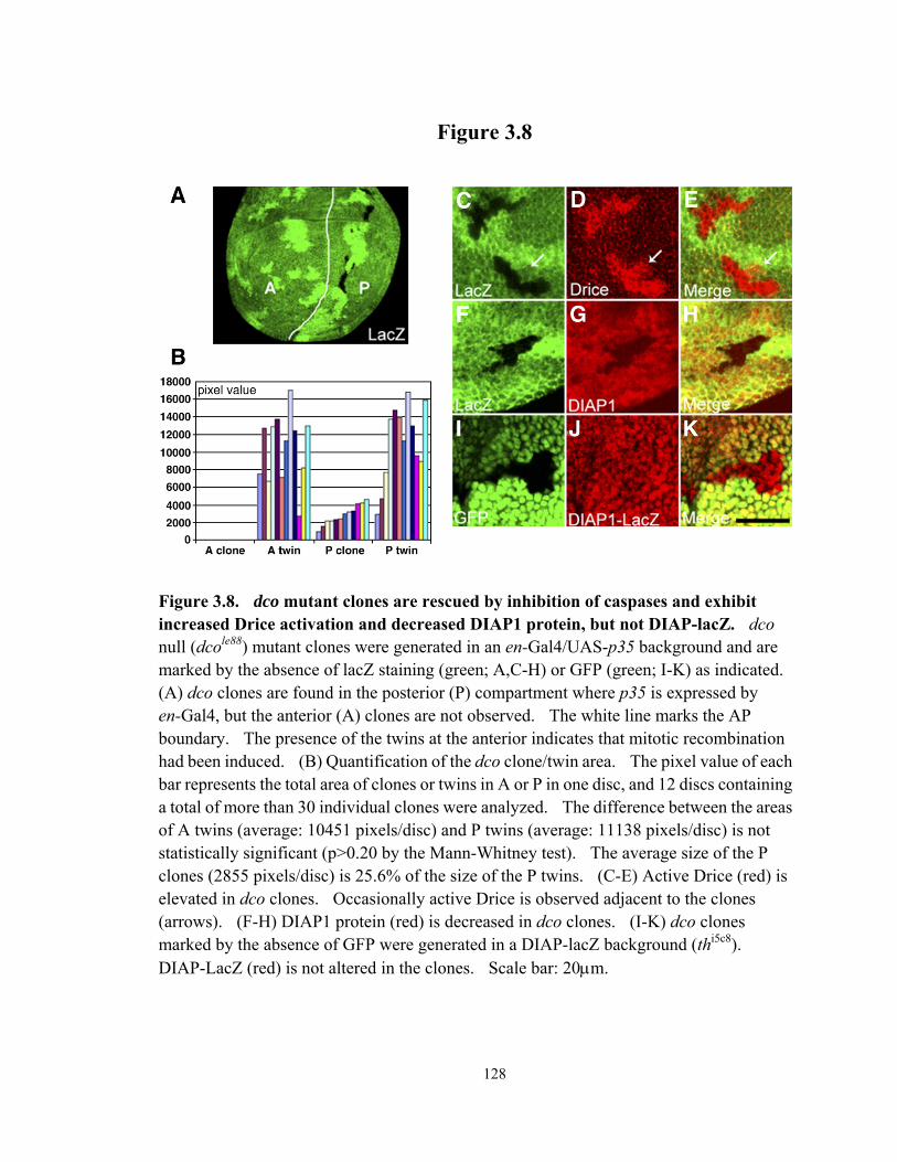
128
Figure 3.8
Figure 3.8. dco mutant clones are rescued by inhibition of caspases and exhibit increased Drice activation and decreased DIAP1 protein, but not DIAP-lacZ. dco null (dcole88) mutant clones were generated in an en-Gal4/UAS-p35 background and are marked by the absence of lacZ staining (green; A,C-H) or GFP (green; I-K) as indicated. (A) dco clones are found in the posterior (P) compartment where p35 is expressed by en-Gal4, but the anterior (A) clones are not observed. The white line marks the AP boundary. The presence of the twins at the anterior indicates that mitotic recombination had been induced. (B) Quantification of the dco clone/twin area. The pixel value of each bar represents the total area of clones or twins in A or P in one disc, and 12 discs containing a total of more than 30 individual clones were analyzed. The difference between the areas of A twins (average: 10451 pixels/disc) and P twins (average: 11138 pixels/disc) is not statistically significant (p>0.20 by the Mann-Whitney test). The average size of the P clones (2855 pixels/disc) is 25.6% of the size of the P twins. (C-E) Active Drice (red) is elevated in dco clones. Occasionally active Drice is observed adjacent to the clones (arrows). (F-H) DIAP1 protein (red) is decreased in dco clones. (I-K) dco clones marked by the absence of GFP were generated in a DIAP-lacZ background (thi5c8). DIAP-LacZ (red) is not altered in the clones. Scale bar: 20μm.

129
Figure 3.9
Figure 3.9 Mitotic clones of dco in a Minute/+ background result in loss of DIAP1, but only modest Drice activation. A apical (A-D) and basal (E-H) optical slice through a clone of dcole88 stained for DIAP1 (B,F) and active Drice (C,G). Clonal boundaries were marked with GFP (A,D). A strong reduction in DIAP1 is observed in the clones, while only a modest elevation of active Drice was observed in the basal portion of the clones. Note that there is appreciable Drice activation outside of the clones in the basal portion of the disc.
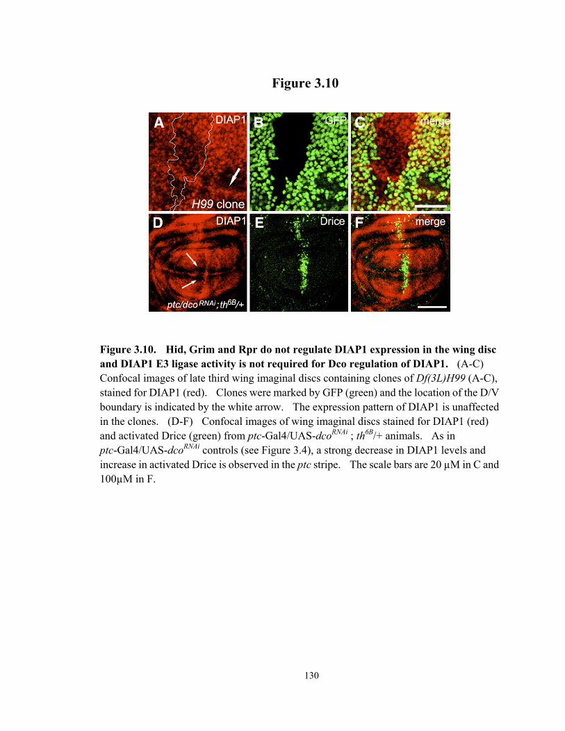
130
Figure 3.10
Figure 3.10. Hid, Grim and Rpr do not regulate DIAP1 expression in the wing disc and DIAP1 E3 ligase activity is not required for Dco regulation of DIAP1. (A-C) Confocal images of late third wing imaginal discs containing clones of Df(3L)H99 (A-C), stained for DIAP1 (red). Clones were marked by GFP (green) and the location of the D/V boundary is indicated by the white arrow. The expression pattern of DIAP1 is unaffected in the clones. (D-F) Confocal images of wing imaginal discs stained for DIAP1 (red) and activated Drice (green) from ptc-Gal4/UAS-dcoRNAi ; th6B/+ animals. As in ptc-Gal4/UAS-dcoRNAi controls (see Figure 3.4), a strong decrease in DIAP1 levels and increase in activated Drice is observed in the ptc stripe. The scale bars are 20 µM in C and 100µM in F.

131
Figure 3.11
Figure 3.11 The dying cells in dco mutant wing discs are basally extruded and express Wg. TUNEL (green) labels dying cells and Wg is stained in red. All four panels are confocal images of the same wing disc from a dcoP915/dcoP1396 larva. (A, C) Basal focal plane. (B, D) Apical focal plane. Wg retains a relatively normal pattern in the apical view but is ectopically expressed in the dying cells at the basal epithelium. Scale bar: 100μm.

132
Figure 3.12
Figure 3.12. The dco mutant cells have unaffected Wg and Dpp signaling. Confocal images of late third instar wing imaginal discs containing dcole88 null clones, generated as described in Figure 3.5. Clones are marked by the lack of lacZ staining (green) and the Wg targets Sens and Dll and Dpp target Spalt are stained in red. Sens (A-C) and Dll (D-F) and Spalt (G-I) expression are normal in dco clones.
133
REFERENCES
Abrams, J.M., 2002. Competition and compensation: Coupled to death in development and cancer. Cell 110, 403-406.
Axelrod, J.D., 2001. Unipolar membrane association of Dishevelled mediates Frizzled planar cell polarity signaling. Genes Dev. 15, 1182-1187.
Bergmann, A., Agapite, J., McCall, K., Steller, H., 1998. The Drosophila gene hid is a direct molecular target of Ras-dependent survival signaling. Cell 95, 331-341.
Brand, A., Perrimon, N., 1994. Targeted gene expression as a means of altering cell fates and generating dominant phenotypes. Development 118, 401-415.
Chen. P., Nordstrom, W, Gish, B., Abrams, J.M. 1996. grim, a novel cell death gene in Drosophila. Genes Dev. 10, 1773-1782.
Cong, F., Schweizer, L., Varmus, H., 2004. Casein kinase Iepsilon modulates the signaling specificities of dishevelled. Mole. Cell. Biol. 24, 2000-2011.
de la Cova, C., Abril, M., Bellosta, P., Gallant, P., Johnston, L.A., 2004. Drosophila Myc regulates organ size by inducing cell competition. Cell 117, 107-116.
DeMaggio, A.J., Lindberg, R.A., Hunter, T., Hoekstra, M.F. 1992. The budding yeast HRR25 gene product is a casein kinase I isoform. Proc. Natl. Acad. Sci. USA 89, 7008-7012.
Desagher, S., Osen-Sand, A., Montessuit, S., Magnenat, E., Vilbois, F., Hochmann, A., Journot, L., Antonsson, B., Martinou, J.C., 2001. Phosphorylation of bid by casein kinases I and II regulates its cleavage by caspase 8. Mol. Cell 8, 601-611.
Deveraux, Q.L., Reed, J.C., 1999. IAP family proteins--suppressors of apoptosis. Genes Dev. 13, 239-252.
Ditzel, M., Wilson, R., Tenev, T., Zachariou, A., Paul, A., Deas, E., Meier, P., 2003. Degradation of DIAP1 by the N-end rule pathway is essential for regulating apoptosis. Nat. Cell Biol. 5, 467-473.
Edgar, B.A., 1999. From small flies come big discoveries about size control. Nat. Cell Biol. 1, E191-E193.
Edgar, B.A., 2006. From cell structure to transcription: Hippo forges a new path. Cell 124, 267-273.
Evan, G.I., Vousden, K.H., 2001. Proliferation, cell cycle and apoptosis in cancer. Nature 411, 342-348.
Fish, K.J., Cegielska, A., Getman, M.E., Landes, G.M., Virshup, D.M., 1995. Isolation and Characterization of Human Casein Kinase Iε (CKI), a Novel Member of the CKI Gene Family. J. Biol. Chem. 270, 14875-14883.
Freeman, M., 1996. Reiterative use of the EGF receptor triggers differentiation of all cell types in the Drosophila eye. Cell 87, 651-660.

134
Gerether, M.E., Abrams, J.M., Agapite, J., White, K., Steller, H. 1995. The head involution defective gene of Drosophila melanogaster functions in programmed cell death. Genes Dev. 9, 1694-1708.
Giraldez, A.J., Cohen, S.M. 2003. Wingless and Notch signaling provide cell survival cues and control cell proliferation during wing development. Development 130, 6533-6543.
Green, D.R., Evan, G.I., 2002. A matter of life and death. Cancer Cell 1, 19-30. Hanahan, D., Weinberg, R.A., 2000. The hallmarks of cancer. Cell 100, 57-70. Harvey, K.F., Pfleger, C.M., Hariharan, I.K., 2003. The Drosophila Mst ortholog, hippo,
restricts growth and cell proliferation and promotes apoptosis. Cell 114, 457-467. Haung, J., Wu, S., Barrera, J., Matthews, K., Pan, D., 2005. The Hippo Signaling Pathway
Coordinately Regulates Cell Proliferation and Apoptosis by Inactivating Yorkie, the Drosophila Homolog of YAP. Cell 122, 421-434.
Hay, B.A., Wassarman, D.A., Rubin G.M. 1995. Drosophila homologs of baculovirus inhibitor and apoptosis proteins function to block cell death. Cell 83, 1253-1262.
Hay, B.A., 2000. Understanding IAP function and regulation: a view from Drosophila. Cell Death Differ. 7, 1045-1056.
Hays, R., Wickline, L., Cagan, R., 2002. Morgue mediates apoptosis in the Drosophila melanogaster retina by promoting degradation of DIAP1. Nat. Cell Biol. 4, 425-431.
Hoekstra, M.F., Liskay, R.M., Ou, A.C., Demaggio, A.J., Burbee, D.G., Heffron, F., 1991. Hrr25, a putative protein-kinase from budding yeast - association with repair of damaged DNA. Science 253, 1031-1034.
Holley, C.L., Olson, M.R., Colon-Ramos, D.A., Kornbluth, S. 2002. Reaper eliminates IAP proteins through stimulated IAP degradation and generalized translational inhibition. Nat. Cell Biol. 4, 439-444.
Huh, J.R., Guo, M., Hay, B.A., 2004. Compensatory proliferation induced by cell death in the Drosophila wing disc requires activity of the apical cell death caspase Dronc in a nonapoptotic role. Curr. Biol. 14, 1262-1266.
Izeradjene, K., Douglas, L., Delaney, A.B., Houghton, J.A., 2004. Casein Kinase I attenuates Tumor Necrosis Factor-related apoptosis-inducing ligand-induced apoptosis by regulating the recruitment of Fas-associated death domain and procaspase-8 to the death-inducing signal complex. Cancer Res. 64, 8036-8044.
Jacobson, M.D., Weil, M., Raff, M.C., 1997. Programmed cell death in animal development. Cell 88, 347-354.
Johnson, R.L., Grenier, J.K., Scott, M.P., 1995. patched overexpression alters wing disc size and pattern: transcriptional and post-transcriptional effects on hedgehog targets. Development 121, 4161-4170.
Johnston, L.A., Sanders, A. L., 2003. Wingless promotes cell survival but constrains growth during Drosophila wing development. Nat. Cell Biol. 5, 827-833.
Jursnich, V.A., Fraser, D.E., Held, L.I.J., Ryerse, J., Bryant, P.J., 1990. Defective gap-junctional communication associated with imaginal disc overgrowth and

135
degeneration caused by mutations of the dco gene in Drosophila. Dev. Biol. 140, 413-429.
Kaufmann, S.H., Hengartner, M.O., 2001. Programmed cell death: alive and well in the new millennium. Trends Cell Biol. 11, 526-534.
Klein, T.J., Jenny A., Djiane, A, Mlodzik, M., 2006. CKIε/discs overgrown promotes both Wnt/b-catenin and Fz/PCP signaling in Drosophila. Curr. Biol. 16, 1337-1343.
Kuhnlein, R.P., Frommer, G., Friedrich, M., Gonzalez-Gaitan, M., Weber, A., Wagner-Bernholz, J.F., Gehring, W.J., Jackle, H., Schuh, R. 1994. splat encodes an evolutionarily conserved zinc finger protein of novel structure which provides homeotic gene function in the head and tail region of the Drosophila embryo. EMBO J. 13, 168-179.
Kurada, P., White, K., 1998. Ras promotes cell survival in Drosophila by downregulating hid expression. Cell 95, 319-329.
Lee, Y.S., Carthew, R.W., 2003. Making a better RANi vector for Drosophila: use of intron spacers. Methods 30, 322-329.
Lin, H.V., Rogulja, A., Cadigan, K.M., 2004. Wingless eliminates ommatidia from the edge of the developing eye through activation of apoptosis. Development 131, 2409-2418.
Lisi, S., Mazzon, I., White, K., 2000. Diverse domains of THREAD/DIAP1 are required to inhibit apoptosis induced by REAPER and HID in Drosophila. Genetics 154, 669-678.
Martin-Castellanos, C., Edgar, B.A., 2002. A characterization of the effects of Dpp signaling on cell growth and proliferation in the Drosophila wing. Development 129, 1003-1013.
McKay, R.M., Peters, J.M., Graff, J., 2001. The Casein Kinase I Family: Roles in Morphogenesis. Dev. Biol. 235, 378-387.
Meier, P., Finch, A., Evan, G., 2000. Apoptosis in development. Nature 407, 796-801. Mergliano, J., Minden, J.S., 2003. Caspase-independent cell engulfment mirrors cell death
pattern in Drosophila embryos. Development 130, 5779-5789. Milan, M., Campuzano, S., Garcia-Bellido, A., 1997. Developmental parameters of cell
death in the wing disc of Drosophila. Proc. Natl. Acad. Sci. USA 94, 5691-5696. Moreno, E., Basler, K., Morata, G. 2002. Cells compete for Decapentaplegic survival
factor to prevent apoptosis in Drosophila wing development. Nature 416, 755-759. Moreno, E., Basler, K., 2004. dMyc transforms cells into super-competitors. Cell 117,
117-129. Neufeld, T.P., de la Cruz, A.F., Johnston, L.A., Edgar, B.A. 1998. Coordination of growth
and cell division in the Drosophila wing. Cell 93, 1183-1193. Nolo, R., Abbott, L.A., Bellen, H.J., 2000. Senseless, a Zn finger transcription factor, is
necessary and sufficient for sensory organ development in Drosophila. Cell 102, 349-362.
136
Pantalacci, S., Tapon, N., Leopold, P., 2003. The Salvador partner Hippo promotes apoptosis and cell-cycle exit in Drosophila. Nat. Cell Biol. 5, 921-927.
Parker, D.S., Jemison, J., Cadigan, K.M., 2002. Pygopus, a nuclear PHD-finger protein required for Wingless signaling in Drosophila. Development 129, 2565-2576.
Perez-Garijo, A., Martin, F.A., Struhl, G., Morata, G., 2005. Dpp signaling and the induction of neoplastic tumors by caspase-inhibited apoptotic cells in Drosophila. Proc. Natl. Acad. Sci. USA 102, 17664-17669.
Peters, J.M., McKay, R.M., McKay, J.P., Graff, J.M., 1999. Casein kinase I transduces Wnt signals. Nature 401, 345-350.
Price, M.A., Kalderon, D., 2002. Proteolysis of the Hedgehog signaling effector Cubitus interruptus requires phosphorylation by Glycogen Synthase Kinase 3 and Casein Kinase I. Cell 108, 823-835.
Price, M.A., 2006. CKI, there's more than one: casein kinase I family members in Wnt and Hedgehog signaling. Genes Dev. 20, 399-410.
Rodriguez, A., Oliver, H., Zou, H., Chen, P., Abrams, J., 1999. Dark is a Drosophila homologue of Apaf-1/CED-4 and functions in an evolutionarily conserved death pathway. Nat. Cell Biol. 1, 272-279.
Rorth, P., Szabo, K., Bailey, A., Laverty, T., Rehm, J., Rubin, G.M., Weigmann, K., Milan, M., Benes, V., Ansorge, W., Cohen, S.M., 1998. Systematic gain-of-function genetics in Drosophila. Development 125, 1049-57.
Ryoo, H.D., Bergmann, A., Gonen, H., Ciechanover, A., Steller, H., 2002. Regulation of Drosophila IAP1 degradation and apoptosis by reaper and ubcD1. Nat. Cell Biol. 4, 432-438.
Ryoo, H.D., Gorenc, T., Steller, H., 2004. Apoptotic cells can induce compensatory cell proliferation through the JNK and the wingless signaling pathways. Dev. Cell 7, 491-501.
Serrano, N., O'Farrell, P.H., 1997. Limb morphogenesis: connections between patterning and growth. Curr. Biol. 7, R186-R195.
Stoter, M., Bamberger, A.M., Aslan, B., Kurth, M., Speidel, D., Loning, T., Frank, H.G., Kaufmann, P., Lohler, J., Henne-Bruns, D., Deppert, W., Knippschild, U., 2005. Inhibition of casein kinase I delta alters mitotic spindle formation and induces apoptosis in trophoblast cells. Oncogene 24, 7964-7975.
Strutt, H., Price, M.A., Strutt, D., 2006. Planar Polarity is Positively Regulated by Casein Kinase Iε in Drosophila. Curr. Biol. 16, 1329-1336.
Tapon, N., Moberg, K.H., Hariharan, I.K., 2001a. The coupling of cell growth to the cell cycle. Curr. Opin. Cell Biol. 13, 731-737.
Tapon, N., Ito, N., Dickson, B.J., Treisman, J.E., Hariharan, I.K., 2001b. The Drosophila tuberous sclerosis complex gene homologs restrict cell growth and cell proliferation. Cell 105, 345-355.

137
Tenev, T., Zachariou, A., Wilson, R., Ditzel, M., Meier, P., 2005. IAPs are functionally non-equivalent and regulate effector caspases through distinct mechanisms. Nat. Cell Biol. 7, 70-77.
Waddell, D.S., Liberati, N.T., Guo, X., Frederick, J.P., Wang, X.F., 2004. Casein Kinase Iε plays a functional role in the Transforming Growth Factor-β signaling pathway. J. Biol. Chem. 279, 29236-29246.
Wang, S.L., Hawkins, C.J., Yoo, S.J., Muller, H.A.J., Hay, B.A., 1999. The Drosophila caspase inhibitor DIAP1 is essential for cell survival and is negatively regulated by HID. Cell 98, 453-463.
Wilson, R., Goyal, L., Ditzel, M., Zachariou, A., Baker, D.A., Agapite, J., Steller, H., Meier, P., 2002. The DIAP1 RING finger mediates ubiquitination of Dronc and is indispensable for regulating apoptosis. Nat. Cell Biol. 4, 445-450.
Wu, S., Huang, J.B., Dong, J.X., Pan, D.J., 2003. hippo encodes a Ste-20 family protein kinase that restricts cell proliferation and promotes apoptosis in conjunction with salvador and warts. Cell 114, 445-456.
Xu, T., Rubin, G.M., 1993. Analysis of genetic mosaics in developing and adult Drosophila tissues. Development 117, 1223-1237.
Yang, Y., Fang, S., Jensen, J.P., Weissman, A.M., Ashwell, J.D., 2000. Ubiquitin protein ligase activity of IAPs and their degradation in proteasomes in response to apoptotic stimuli. Science 288, 874-877.
Yokokura, T., Dresnek, D., Huseinovic, N., Lisi, S., Abdelwahid, E., Bangs, P., White, K., 2004. Dissection of DIAP1 functional domains via a mutant replacement strategy. J. Biol. Chem. 279, 52603-52612.
Yoo, S.J., Huh, J.R., Muro, I., Yu, H., Wang, L.J., Wang, S.L., Feldman, R.M.R., Clem, R.J., Muller, H.A.J., Hay, B.A., 2002. Hid, Rpr and Grim negatively regulate DIAP1 levels through distinct mechanisms. Nat. Cell Biol. 4, 416-424.
Zhang, L., Jia, J., Wang, B., Amanai, K., Wharton, K.A., Jiang, J., 2006. Regulation of wingless signaling by the CKI family in Drosophila limb development. Dev. Biol. 299, 221-237.
Zhu J., Shibasaki, F., Price, R., Guillernot, J.C., Yano, T., Dotsch, V., Wagner, G., Ferrara, P., McKeon, F., 1998. Intramolecular masking of nuclear import signal on NF-AT4 by Casein Kinase I and MEKK1. Cell 93, 851-861.
Zilian, O., Frei, E., Burke, R., Brentrup, D., Gutjahr, T., Bryant, P.J., Noll, M., 1999. double-time is identical to discs overgrown, which is required for cell survival, proliferation and growth arrest in Drosophila imaginal discs. Development 126, 5409-5420.

138
CHAPTER IV
EXPLORATION OF THE REGULATION MECHANISM OF
APOPTOSIS AND THE WINGLESS PATHWAY BY
DROSOPHILA CASEIN KINASE I FAMILY PROTEINS
ABSTRACT
The Casein Kinase I (CKI) family of proteins has been implicated in a variety of
cellular processes, including apoptosis and the Wingless (Wg) signaling pathway. The
Drosophila CKIε/δ homologue, known as Discs Overgrown (Dco), has been demonstrated
to regulate apoptosis via activation of the endogenous caspase inhibitor DIAP1 in vivo, yet
the molecular mechanisms remain to be elucidated. In this report, we explored the
molecular mechanism of Dco in cultured cells. We conducted two small scale screens of
Drosophila CKI members in the adult fly eye and cultured cells. The screens and further
characterization revealed a novel function of Gilgamesh (Gish), the fly CKIγ homologue,
in DIAP1 regulation. Our results suggest Gish and Dco might be functionally redundant.
In order to address the controversy around the role of Dco in Wg signaling, we extended
our study into the Wg pathway. Our results strongly support Dco acting as a positive
regulator of the pathway. We also initiated a chemical genetic screen to identify novel
substrates of Dco.

139
INTRODUCTION
Introduction to Casein Kinase I family
The Casein Kinase I protein family (CKI) is a large family of monomeric
serine/threonine protein kinases which contain a highly conserved central kinase domain
(53–98% identical), flanked by diverse amino- and carboxyl-termini (Vielhaber and
Virshup 2001). CKI members have been implicated in a diversity of cellular progresses,
including the Wnt/Wingless (Wg) signaling pathway, apoptosis, circadian rhythm, cell
division and membrane transport (reviewed in Vielhaber and Virshup 2001; Knippschild et
al. 2005).
Family members of CKI are found in eukaryotic organisms, from yeast to mammals.
So far, seven mammalian CKI isoforms (α, β, γ1, γ2, γ3, δ and ε) have been characterized.
The examinations of substrate specificity revealed a substrate recognition motif
S/T(P)-X1-2-S/T or Dn-X1-2-S/T, where S/T(P) indicates phosphor-serine or threonine, X is
any amino acid, and D is aspartic acid (Vielhaber and Virshup 2001). As this motif is
relatively common, the substrate selection is thought to be regulated via subcellular
localization and docking sites in substrates (Price 2006).
In the fruitfly Drosophila melanogaster, eight family members have been predicted
based on sequence homology (Figure 4.1A). Among them, CKIα (Santos et al. 1996), the
Drosophila CKIγ Gilgamesh (Gish) (Hummel et al. 2002), and the Drosophila CKIε/δ
Discs overgrown (Dco) (Kloss et al. 1998), have been characterized in multiple cellular
140
processes (Figure 4.1B) (Vielhaber and Virshup 2001; Knippschild et al. 2005). Dco is
also known as double-time (DBT), and as the name implies, it plays an important role in
circadian rhythm (Knippschild et al. 2005). Interestingly, all of the three isoforms have
been implicated in the Wg pathway (discussed in details below) (Price 2006). Recently,
we reported that Dco regulates apoptosis (Guan et al. 2007)(Chapter III of this
dissertation).
Apoptosis and CKI
Apoptosis is a highly conserved cell death process that is essential for development
and adult homeostasis. The main executioners of apoptosis are called caspases, whose
proteolytic activity results in cleavage of numerous cellular substrates, leading to cell death.
Synthesized as zymogens, caspases are subject to tight regulation at the activation level by
several pathways, including the intrinsic mitochondria pathway and extrinsic death
receptor pathway (Hengartner 2000; Salvesen and Abrams 2004). There is another level
of caspase regulation, carried out by the Inhibitor of Apoptosis Proteins (IAP), which bind
to caspases to prevent their activation and/or enzymatic activity (Srinivasula and Ashwell
2008). IAPs are in turn inhibited by a class of proteins termed RHG proteins, e.g. Hid in
Drosophila, and Smac in mammals. Expression of RHG proteins can rapidly induce
apoptosis by antagonizing Drosophila IAP1(DIAP1) in the fly (reviewed in Salvesen and
Abrams 2004; Hay and Guo 2006; Steller 2008).
141
Data from our laboratory demonstrated that Dco promotes cell survival via the
activation of DIAP1 during larval stages (Guan et al. 2007). Expression of Dco blocks
Hid-dependent apoptosis in the developing eye, whereas loss of Dco leads to a dramatic
reduction in expression of DIAP1 and activation of caspases (Guan et al. 2007). Since the
observation cannot be explained by Dco’s activity in other pathways, we believe that we
have uncovered a novel mechanism of DIAP1 regulation.
Several questions remained unanswered about the underlying mechanism by which
Dco regulates DIAP1 activation. First, what is the substrate of Dco in apoptosis
regulation? As discussed in Chapter III, Dco probably regulates DIAP1
post-transcriptionally in a kinase activity-dependent manner (Guan et al. 2007). The
simplest scenario would be that Dco phosphorylates and thereby stabilizes DIAP1. In fact,
several putative CKI substrate recognition motifs are found in DIAP1, and DIAP1 protein
levels have been reported to be regulated via phosphorylation (Kuranaga et al. 2006).
However, our attempts to show DIAP1 being the substrate of Dco was negative. In order
to identify the substrate of Dco in apoptosis regulation, we initiated a chemical genetic
screen.
Dco has been shown to regulate DIAP1 protein levels in wing imaginal discs,
however the same effect has not been observed in eye discs (Guan et al. 2007). One
hypothesis is that the loss of Dco in eye discs is compensated by a redundant partner,
presumably another CKI. Therefore, efforts have been made to identify CKI family
142
members that contribute to apoptosis regulation, and Gish appeared as a promising
candidate, as discussed in this chapter.
Wingless signaling and CKI
The Drosophila Wingless (Wg) protein belongs to the Wnt family of secreted
glycoproteins that are important for a wide range of developmental events throughout the
animal kingdom (Cadigan and Nusse 1997; Cadigan 2008). A major Wg downstream
player is cytoplasmic Armadillo (Arm), the homologue of mammalian β-catenin. In the
absence of Wg signaling (Figure 4.2A), Arm is constitutively expressed, but the
cytoplasmic pool of Arm is continuously phosphorylated and subsequently degraded by a
degradation complex, which contains two kinases GSK3β and CKIα, scaffold proteins
Axin and APC, and protein phosphatase 2A (PP2A). Upon Wg stimuli (Figure 4.2B), the
signal crosses the plasma membrane through the Frizzled receptors Frizzleds (Fz&Fz2)
and the members of the low-density-lipoprotein receptor-related family protein Arrow (in
mammals LRP5 and LRP6) to inactivate the degradation complex. Disheveled (Dsh)
mediates the signal transduction from receptors to the degradation complex via
mechanisms that are not well understood (Cadigan and Liu 2006). The stabilized Arm
then translocates into the nucleus, where it interacts with the transcription factors TCF, and
converts TCF from a transcriptional repressor into an activator, leading to activation of Wg
target genes (Cadigan 2008).

143
The Drosophila CKI members, CKIα, Gish and Dco have been implicated in the Wg
pathway regulation in the past decade. Three key phosphorylation events have been
described. Phosphorylation of Arm by CKIα, which primes the consequent
phosphorylation by GSK3β, leads to Arm degradation (Amit et al. 2002; Liu et al. 2002).
In the presence of Wg signaling, Gish has been recently demonstrated to phosphorylate
Arrow, which contributes to the recruitment of Axin to membrane, leading to the
inactivation of the degradation complex (Davidson et al. 2005; Zeng et al. 2005). In
addition, there are several reports suggest that the phosphorylation of Dsh by Dco is an
important event to activate the pathway; however, the mechanisms still remain a mystery
(reviewed in Wharton 2003; Price 2006). The three events have been shown conserved
from flies to mammals.
Several other functions of CKIε have also been suggested, which leads to the
controversy about the molecular mechanism of CKIε in regulating the Wnt/Wg pathway
(reviewed in Vielhaber and Virshup 2001; Knippschild et al. 2005; Price 2006). Briefly,
both positive and negative roles have been proposed, and some genetic data suggests that
Dco/CKIε is not required for Wg/Wnt signaling in some tissues. Making the situation
more complex, several components throughout the Wnt/Wg pathway, i.e. Disheveled,
APC, Axin, Arm/β-catenin, PP2A and TCF have been suggested to be modified by
Dco/CKI ε (Figure 4.2). To clarify this puzzle in a physiological context, we are
exploring the role of Dco in the Drosophila embryonic cell line Kc167 (Kc) in this chapter.

144
MATERIALS AND METHODS
Cell Culture, RNAi treatment, DNA transfection, and conditional media treatment
Drosophila embryonic cell lines were cultured at room temperature in Schneider's
Drosophila media (Invitrogen) containing Penicillin/ Streptomycin and 10% FBS for S2
cells and 5% FBS for Kc cells.
RNAi-mediated gene knockdowns were performed as described (Fang et al. 2006;
Liu et al. 2008). Double-strand RNAs with a typical length of 400–700 bp were
synthesized using the MEGAscript T7 in vitro transcription kit (Ambion). The sequences
of the PCR primers for the dsRNA synthesis are as follows:
dco 3’ UTR 5’TAATACGACTCACTATAGGGCGTTGAATGTATCCAAGCGGCAGG,
3’TAATACGACTCACTATAGGGTAGGTTACAATGTGGGTGCCTTGC;
gish –5’TAATACGACTCACTATAGGGCTAGACGGTGTACAATGTAGTCCA,
3’ TAATACGACTCACTATAGGGCGGTGTGTTTGTAGTTGGT;
CKIa –5’ TAATACGACTCACTATAGGGCGTGGATGCGTGGATGTAAA,
3’ TAATACGACTCACTATAGGGGATGAGAGTGGGTGAGTGTGT;
CG7094-1–5’ TAATACGACTCACTATAGGGCCACTCCAACATGAAGTAGT,
3’ TAATACGACTCACTATAGGGGCAGGGAACAGATTCTGGAA;
CG7094-2–5’ TAATACGACTCACTATAGGGCGTGATGCTTAGTCCGAGATA,
3’ TAATACGACTCACTATAGGGGGCAGTAAAGTCACAA;
CG2577–5’ TAATACGACTCACTATAGGGCGATAAGCCCAACTACGACT ,

145
3’ TAATACGACTCACTATAGGGGTTGTGAGGGGAGTAATTCT;
CG12147-1–5’ TAATACGACTCACTATAGGGGTGGCAACAATAGATCGCCA ,
3’ TAATACGACTCACTATAGGGGGTCCATCACCATGACATT;
CG12147-2–5’ TAATACGACTCACTATAGGGCAGATGCCCTGTCTTTGA ,
3’ TAATACGACTCACTATAGGGCCAAGTTTCCACGGTATCGA;
CG9962-1–5’ TAATACGACTCACTATAGGGGAACGACTACGAACTGGA,
3’ TAATACGACTCACTATAGGGCAGTAACTCCATGACCAGCA;
CG9962-2–5’ TAATACGACTCACTATAGGGGAACAGGCATGTGCCACAA ,
3’ TAATACGACTCACTATAGGGCAGCAAACTCAAGAAGGTGCA;
CG11533-2–5’ TAATACGACTCACTATAGGGGTGCATCTAAGCCACTTGAGT,
3’ TAATACGACTCACTATAGGGGCACTGTTCTGGAGCCTATA.
Transient transfections were performed with Fugene 6 (Roche Applied Science)
according to the manufacturer’s instruction. pMT-Hid-flag, pAc-GFP and
pAc-DIAP1-flag were from P. Meier lab. pAc-Gish-flag was from C. Niehrs lab.
pAc-Dco-flag was cloned from Dco cDNA. pAc-Arm* was previously described (Fang
et al. 2006). Mutant forms of Dco, including DcoK38R, M82G, M82A, M82V and M82C
were generated using QuikChange○R Site-Directed Mutagenesis Kit (Strategene, Catalog
#200518) using pAc-Dco-flag as the template. Hid is under the control of a copper
inducible metallothionein promoter. The expression of Hid was induced 24hr after
transfection by the application of 0.7mM CuSO4 at the final concentration. When both
146
transfection and RNAi were performed, cells were typically treated with RNAis for 4 days,
diluted to 106 cells/ml and transfected with plasmids.
Wg-CM and control medium were prepared as described (Fang et al. 2006; Liu et al.
2008). 106 Kc cells were treated with 400 μl of Wg-CM for 5 hr prior to harvesting.
Cell survival assay and flow cytometry
A cell survival assay was employed to quantitatively describe cell death intensity
(Goyal et al. 2000). GFP proteins were co-expressed with other proteins to label
transfected cells. The transfection efficiency is around 10-15% for S2 cells and 15-20%
for Kc cells. The percentage of GFP positive cells over total cell numbers was utilized to
reflect cell survival rate. Cell death is induced by ectopic expression of Hid as described
above. The cell numbers were counted either manually or by flow cytometry.
Flow cytometry was performed 60hr after the induction of Hid expression. Cells
were spinned down at 1500rpm for 2min, and resuspended in 0.5ml 0.4% formaldehyde for
30min at room temperature. Then cells were spinned down and wash with sterile PBS
twice, and stayed at 4 degree for 12hr before be filtered into the Falcon tubes for analysis.
Real-time quantitative RT-PCR (qRT-PCR)
qRT-PCR was performed as previously described (Fang et al. 2006). Briefly,
Trizol (Invitrogen) was used to extract total RNA from 1-5×10⁶ cells. Reverse
transcription was performed using Stratascript reverse transcriptase (Stratagene) followed

147
by qPCR analysis using an iCycler iQ real-time PCR detection system. β-tubulin was
used to normalize transcript levels. qPCR primers were designed by using the online
program Primer3. Primers for nkd and CG6234 were described previously (Fang et al.
2006). Primers for dco to detect dco RNAi knockdown efficiency was designed across
the 3’ boundary of dcoUTR RNAi targeted region, i.e. 5’ primer
(GCATATGCCACACAGCAAGG) is upstream of the boundary, while 3’
(TGCACGTGGTAATCGCAATC) is downstream.
Fly Genetics
The following fly strains were used. Oregon R was our wild type stock. The
GMR-hidIM and GMR-hid10 stocks were from J. Abrams. GMR-hidala5 was from A.
Bergmann. UAS-lacZ, UAS-p35, dppGal4 and ptcGal4 and thj5c8 were from the
Bloomington Stock Center. UASgish, UASCG2577, UASCG7094, UASCG9962 and
UASCG12147 were from J. Jiang. GMRGal4 was from M. Freeman. The Df(3L)H99 and
UAS-DIAP1 stocks were from B. Hay. The dco mutant allele dcole88 were from M. Noll.
The FRT82B P[w+, Ubi-GFPnls] was from I. Hariharan. The gish null allele mutant
gishnull was generated by Dr. Jennifer Kennell in Cadigan lab. All other reagents for
clonal analysis, ywflp122 , FRT82B, FRT82B P[w+, armlacZ] were from the Bloomington
Stock Center. The UASdco RNAi-J transgene on the third chromosome was generated by
mobilizing the transgene from UASdco RNAi (Guan et al. 2007).

148
Immunostainings and Western blotting
Immunostainings were performed as previously described (Lin et al. 2004).
Antibodies used were mouse anti-DIAP1(1:200) (from B. Hay) and rabbit
anti-β-galactosidase (1:500) (Cappel, ICN, Costa Mesa, CA). Cy3 conjugated (Jackson
Immunochemicals) and Alexa Fluor 488-conjugated (Molecular Probes) secondary
antibodies were applied after primary antibody.
Western blotting was performed as standard protocol. Antibodies used include
anti-Flag (M2) (1:5000)(Sigma), anti-anti-DIAP1 (1:1000) (from B. Hay), rat anti-Dco
(1:1000)(from M. Young), and mouse anti-tubulin (1:5000) (Sigma). HRP-anti-rabbit
IgG and HRP-anti-mouse IgG are used as secondary antibody (Amersham Bioscience).
Signal was detected using Amersham ECL Plus Western Blotting Detection Reagents kit
(GE Healthcare).
Whole-mount staining and microscopy
The adult eye, notum and wing were photographed on a Leica M10 microscope as
previously described (Parker et al., 2002). All fluorescent pictures were obtained with a
Leica confocal microscope DM6000 B (Leica) and processed in Adobe Photoshop CS.
149
RESULTS
Exploration of the anti-apoptosis function of Dco in S2 cells
The Drosophila embryonic cell line S2 has been widely utilized to explore apoptosis
regulation mechanism. Ectopic expression of pro-apoptotic protein Hid has been shown
to rapidly trigger apoptosis, which can be suppressed by over-expression of DIAP1 in S2
cells (Goyal et al. 2000). This potentially provides a promising system to elucidate the
mechanism by which Dco suppresses apoptosis.
Our results of cell survival assays illustrated that the anti-apoptotic function of Dco
can be recapitulated in S2 cells. Over-expression of Hid reduces the survival rate to 25%
of that in control cells, and the reduction was effectively suppressed by DIAP1 (Figure 4.3
A, B), consistent with previous reports (Goyal et al. 2000). In this system, Dco partially
suppresses Hid-induced cell death (Figure 4.3 A, B); conversely, dominant negative
version of Dco, i.e. DcoK38R, and dco RNAi enhances Hid-induced cell death (Figure 4.3
C, D), supporting the anti-apoptotic function of Dco in S2 cells.
Previous report showed that Dco depletion led to DIAP1 down-regulation in fly
wing imaginal discs (Guan et al. 2007). However, no detectable reduction of DIAP1 was
observed in S2 cells that were either treated with dco RNAi (Figure 4.4 A) or transfected
with the dominant negative mutant DcoK38R (Figure 4.4B). DIAP1 is a short-lived
protein that is degraded via the ubiquitination-proteasome pathway. As expected, DIAP1
was very sensitive to the treatment of the proteasome inhibitor lactacystin (LAC) and the

150
ribosomal inhibitor cycloheximide (CHX), supported by the results that LAC stabilized
DIAP1 while CHX reduced it. Even in these sensitized backgrounds, dco RNAi didn’t
influence DIAP1 level. In addition, no dco RNAi induced reduction of endogenous
DIAP1 was detected in another fly embryonic cell line Kc167, or the wing imaginal
disc-derived cell line Clone8 (Data not shown). Similar results were obtained when
exogenous DIAP1 was introduced into cells by transfection (Data not shown).
The possibility that Dco may phosphorylate and thereby enhance DIAP1 stability
was examined by in vitro phosphorylation, where bacterially produced MBP-DIAP1 was
used as substrate, and kinases were expressed and immunoprecipitated (IP) from S2 cells.
In our system, wild type Dco was able to phosphorylate casein, a known substrate of Dco,
while the kinase dead mutant DcoK38R showed dramatically reduced activity. However,
no detectable phosphorylation of DIAP1 was observed in the same assay (Figure 4.5). In
addition, the attempts to detect the interaction between Dco and DIAP1 in S2 cell lysates
via co-IP were unsuccessful (date not shown).
Overall, although Dco shows anti-apoptotic activity in S2 cells, its role in regulating
DIAP1 levels was not established in cultured cells, which might be due to the existence of
potential redundant partners.
Exploration of potential redundant partner of Dco
Dco belongs to the casein kinase I family, composed of eight members in
Drosophila (Figure 4.1 A). Given that redundancies among CKI family members have

151
been reported repeatedly in different signaling pathways (Vielhaber and Virshup 2001;
Price 2006), small scale screens were conducted in the fly eye and S2 cells to determine
whether other CKI members are potential redundant partners of Dco.
It has been previously shown that expression of Dco strongly suppresses the small
eye phenotype induced by wild type hid alleles hid10, hidIM, and a Ras/MAPK resistant
form hidala5 (Guan et al. 2007). Thus Dco redundant partner(s) are expected to show
similar suppression. In the screen, hid alleles and UAS lines of CKI members were under
the control of an eye-specific promoter GMR. The screen results were summarized in
Figure 4.6B, and the results with hidala5 were shown as examples in Figure 4.6A. Our data
showed that Gish, the fly homologue of casein kinase Iγ, was able to suppress all three
alleles, and thereby selected as a putative Dco redundant partner for further
characterization.
Another screen conducted in cultured cells also indicated Gish as a redundant
partner based on its ability to down-regulate DIAP1 (Figure 4.7). In the screen, Kc cells
that stably expressing exogenous DIAP1-flag (DIAP1-Kc) were treated with
double-stranded RNA targeting one of the eight CKI family members, and the levels of
DIAP1-flag were examined. It is worth mentioning here that all eight CKIs are expressed
in S2 cells, judged by the existence of the corresponding cDNA generated from the cell
lysate. Cells treated with gish RNAi showed lower DIAP1 levels compared with control
cells (Figure 4.7A). The down-regulation was further confirmed with the same cell line

152
(Figure 4.7C) and with endogenous DIAP1 in S2 cells (Figure 4.7B). Consistently,
over-expression of Gish stabilized ectopically expressed DIAP1 (Figure 4.7E). Overall,
results in the fly eye and cultured cells indicate Gish as a putative anti-apoptotic factor
working through DIAP1
It should be noted that the effects of DIAP1 down-regulation by gish RNAi were
variable, and sometimes not reproducible (data not shown). Thus, the ability of Gish to
regulate DIAP1 stability definitely requires further validation.
Exploration of the redundancy between Gish and Dco
To test the hypothesis that redundancy may cause the lack of DIAP1 reduction in the
developing eye and culture cells where Dco is reduced, we simultaneously reduced the
levels of Dco and its putative redundant partner Gish in both S2 cells (Figure 4.7 D) and
eye imaginal discs (Figure 4.8). gish RNAi alone decreased the level of DIAP1 in S2 cells,
yet no synergetic effect was observed in cells with both Dco and Gish depleted (Figure 4.7
D). Negative result was also obtained in eye discs where double mutant cells of dco and
gish didn’t show lower levels of DIAP1. Interestingly, double mutant clones were
considerably smaller than dco single mutant clones, indicating cell survival was affected in
absence of gish, which is consistent with its putative anti-apoptotic role (Figure 4.8).
To explore whether Gish is a redundant partner of Dco, we examined the ability of
Gish to rescue dco RNAi induced defects in the adult notum (Figure 4.9 A), adult wing
(Figure 4.9 B) and the larval wing imagnial disc (Figure 4.9 C). UAS-dco RNAi driven
153
by dpp-Gal4 resulted in less macrochaetes in the scutellum of the adult notum, which was
partially reversed by over-expression of gish. And over-expression of gish alone led to
more macrochaetes (Figure 4.9 A). The macrochaete phenotype caused by dco RNAi was
not suppressed in a H99 heterozygous background, where the proapoptotic factors Hid Rpr
and Grim were reduced (Figure 4.9 A). This suggests the macrochaete phenotype is not a
consequence of increased apoptosis due to elevated RHG proteins. In the fly adult wing,
dco RNAi distorted the vein pattern, which was unexpectedly enhanced by gish expression,
and this phenotype was not attenuated in the H99 heterozygous background (Figure 4.9B).
The wing imaginal discs are the tissue where the function of Dco to regulate DIAP1 was
originally reported, thus the inspection of this phenotype would directly address the
question whether Gish is a redundant partner of Dco in apoptosis regulation. Preliminary
results showed no consistent suppression of DIAP1 reduction by gish (Figure 4.9 C),
arguing against the redundancy.
Exploration of Dco’s function in the Wingless signaling pathway
Dco and its vertebrate homologue CKIε have been implicated in Wnt/ Wg signaling
in the past decade, however, the molecular mechanism still remains controversial. To
clarify the function of Dco/CKIε in a physiological context, we are exploring the role of
Dco in the Drosophila embryonic cell line Kc167 (Kc), mainly using a loss of function
approach.

154
Kc cells have been well established as a system to study the Wg signaling pathway in
Cadigan lab, as they are responsive to Wg signaling, judged by luciferase reporter genes
and several endogenous target genes (Fang et al. 2006). Readouts used in this study
include a luciferase reporter gene, SuperTopFlash (STF), which is composed of 12 copies
of TCF binding sites, and the transcript levels of two endogenous genes, nkd and CG6234 ,
which has been demonstrated as direct target genes of Wg signaling (Fang et al. 2006).
Wg conditional medium (Wg-CM) can activate all three readouts effectively, compared
with control medium (Figure 4.8 A, B, D).
In order to address the requirement of Dco in Wg signaling, we inhibited
endogenous Dco by IC261, a CKIε specific inhibitor (Mashhoon et al. 2000), or RNAi.
The treatment of IC261, suppressed Wg CM induced activation of STF reporters (Figure
4.10 A). Cells depleted for dco with RNAi showed 3-4 fold reduction of Wg signaling, as
judged by the activation of STF reporters (Figure 4.10B), and the transcript levels of nkd
(Figure 4.10C) and CG6234 (Figure 4.10D), which strongly support Dco as a positive
regulator of Wg signaling. Consistently, expression of Dco, not DcoK38R, activated STF
in a dosage dependent manner (Figure 4.10E), indicating that the kinase activity is required
for Dco to stimulate Wg signaling. To illustrate the specificity of the RNAi treatment, a
rescue experiment was performed as follows. Dco was knocked down by RNAi that
specifically targets the 3’UTR, which presumably would not inhibit Dco that were
expressed from an exogenous plasmid only containing the coding region. Exogenous Dco
155
was transfected at such a low amount (10ng) that it, on its own, could not activate Wg
signaling (Figure 4.10 E, F). Under these conditions, exogenous Dco partially rescued the
suppression caused by dco RNAi (Figure 4.10 F), supporting the specificity of dco RNAi.
The lack of full rescue may be due to the relatively low levels of exogenous Dco. Overall,
our results support the positive role of Dco in the Wg pathway, at least in Kc cells.
Where does Dco function in the Wg pathway? We conducted epistasis analyses to
address this question. The Wg pathway can be stimulated remarkably by over-expresion
of a stabilized form of Arm (Arm*) and by knocking down two negative regulators Axin
and CKIa, which are components of the Arm degradation complex (Figure 4.11). Dco
knockdown did not reproducibly suppress the activation of STF reporters induced by any
of the three approaches, (Figure 4.11 and data not shown), placing Dco upstream of Arm
and the Arm degradation complex.
To explore the possibility that Dco positively regulates Wg signaling by stabilizing
Arm, the protein levels of Arm were compared between dco RNAi and control RNAi
treated cells which are stimulated by Wg CM. The treatment of dco RNAi effectively
reduced dco transcripts to approximately 10% of its normal level, leading to 4-5 fold
suppression of the nkd activation in presence of Wg signaling (Figure 4.12 A).
Unexpectedly, in the same cells, no reduction of total Arm (Figure 4.12 B) or nuclear Arm
levels (Figure 4.12 C) were reproducibly observed, indicating Dco might not regulate Arm
protein levels or Arm nuclear translocation. These results apparently contradict to the

156
epistasis analyses, and reconciliation of all the data requires a better understanding of the
pathway (see discussion).
To characterize the functions of all CKI isoforms in Wg signaling, cells were
depleted for a single CKI member, or together with dco. gish RNAi attenuated Wg
activation, and interestingly it showed an additive effect with dco RNAi (Figure 4.13 A).
Gish/CKIγ has been reported to phosphorylate the Wnt co-receptor Arrow/LRP6,
promoting the disassociation of the Arm degradation complex (Davidson et al. 2005; Zeng
et al. 2005), which is distinct from all documented functions of Dco/CKIε. As Dco/CKIε
has been proposed to regulate Dsh, we speculate the additive effect may be due to the
paralleled functions of Dsh and Arrow in traducing the Wg signal from membrane to
downstream. Consistently, Dco expression didn’t rescue Wg suppression by gish RNAi,
nor vice versa, arguing against Gish as the redundant partner of Dco in the Wg pathway.
Attempted identification of Dco substrates by the Shocat substrate screen
We have shown that the kinase activity is required for Dco to regulate apoptosis and
the Wg pathway (Figure 3.4, Figure 4. 10E). Therefore, the identification of its novel
substrates could undoubtedly expand our knowledge of the kinase and the regulation
mechanism.
To identify the novel substrates of Dco, we have employed a chemical genetic screen
developed by Shokat and colleagues, using an engineered kinase with larger ATP binding
pocket and a bulky ATP analogue, which is referred to as the Shokat screen thereafter

157
(Shah et al. 1997; Liu et al. 1998). This method has been shown to specifically label direct
substrates of a particular serine/threonine protein kinase in a mixture of cellular proteins,
and the application of the Shokat screen has successfully identified many novel substrates
of several kinases belonging to different families (Elphick et al. 2007).
In the Shokat screen, the ATP binding pocket in the kinase of interest is modified to
accept an ATP analogue with a bulky substitution at the N-6 position of the purine ring
(Figure 4.14A) (Shah et al. 1997; Liu et al. 1998). Specifically, one or more conserved
residue that comes into close contact with the N-6 position of ATP is mutated to a smaller
amino acid residue. Then, ATP analogues with different N-6 substitutions are screened to
identify the one that the mutant kinase can transfer most efficiently. Thereby, the mutant
kinase, but not other cellular kinases, can utilize a particular N-6 modified ATP to
phosphorylate substrates. Theoretically, when radio-labeled ATP analog is used in a
kinase reaction with the mutant kinase and cell lysates, only direct substrates of the kinase
become radio-labeled and consequently identified.
To apply the Shokat screen to Dco, we first examined the structure of CKI ATP
binding pocket. The crystal structure of Dco has not been solved, but several structures of
CKI family members in other organisms have been documented in Protein Data Bank
(PDB). Among them, the structure of CKI from Schizosaccharomyces pombe (PDB ID:
1CSN) (Xu et al. 1995) was utilized as a model to represent Dco, mainly because the

158
protein was co-crystalized with ATP, which is extremely useful to identify amino acids
that in close contact with N-6 position.
In order to engineer the ATP pocket so that it can accomendate N-6 modified ATP,
the following criteria were summarized based on published examples (Shah et al. 1997;
Liu et al. 1998; Eblen et al. 2003; Hindley et al. 2004): 1) the candidate mutation residue(s)
should locate in the ATP pocket, but should NOT be an essential residue to transfer the
phosphate; 2) the site should not be a charged residue; 3) the candidate residue should be in
close proximity with N-6 position; specifically the distance between the nearest atom of the
candidate residue and ATP N6 should be around 5.5 Å; 4) the distance between the
backbone of the candidate residue and ATP N6 atom should be 7.5 Å or above, so that
when the candidate residue is mutated to glysine, the space should be big enough to
accommodate N6-modified ATP analogues; 5) the residue should be located in the area
where the N-6 substitution is predicted to occupy. A careful inspection revealed that I85
was the only residue in CKI that meets all criteria (Figure 4.14 B). Sequence alignment
showed that it corresponds to M82 of Dco.
Dco M82 was mutated to G, A, V or C, the four amino acids with shorter side chains.
As in published examples, the mutant kinases should be able to utilize wild type ATP,
indicating they may be functional in vivo. However, all of the four mutants showed very
low catalytic activity, if any, as judged by the STF activation assay (Figure 4.14C), raising
doubt about the feasibility of this approach.

159
DISCUSSION
How does Dco regulate DIAP1 protein levels?
Previous genetic studies have demonstrated the anti-apoptotic role of Dco via
DIAP1 activation (Guan et al. 2007), yet the underlying molecular mechanism remains to
be elucidated. The Drosophila S2 cell line has been nicely established as a useful system
to study apoptosis mechanism, and the core apoptosis machinery in Drosophila, including
Dark, Dronc, Drice and DIAP1, has been demonstrated to mediate apoptosis regulation in
S2 cells (Hay and Guo 2006). Therefore, we took advantage of S2 cells to explore the
molecular mechanism of Dco.
We have provided evidence that the anti-apoptotic function of Dco in vivo can be
recapitulated in S2 cells (Figure 4.3). However, DIAP1 protein levels seem not under the
control of Dco in S2 cells (Figure 4.4). These data reminded us of the situation in the fly
developing eye, where Dco regulates Hid-induced apoptosis in both overexpression
(Figure 3. 1, 3.2) and loss of function analyses (Figure 3. 3), yet loss of Dco does not lead to
any detectable modification of DIAP1 levels (Figure 4.8). The similarity might imply
some cell-specific mechanism that we still don’t understand.
Our results also showed that Dco did not phospharylate DIAP1 in vitro (Figure 4.5).
It is possible that bacterially produced DIAP1 may lack certain modifications that are
indispensable for Dco phosphorylation, given that CKI members often require a so called
‘priming’ phosphorylation, which means substrates have to be phosphorylated by another

160
kinase beforehand so as to be recoganized by CKIs (Vielhaber and Virshup 2001).
Alternatively, DIAP1 could simply not be the direct substrate of Dco. Instead, the
function of Dco might be mediated by other factor(s), for instance, RHG proteins which
directly regulate DIAP1, or by multiple components composing an unidentified pathway.
The identification of Dco’s substrate would help uncover the molecular mechanism.
Putative function of Gish in DIAP1 regulation
Our results may have uncovered a novel function of Gish in DIAP1 regulation, as we
illustrated that depletion of gish caused DIAP1 down-regulation in S2 cells (Figure 4.7).
What is the physiological importance of the regulation? The macrochaete phenotype
might provide a clue (Figure 4.9). It has been proposed that the number of macrochaetes
is determined by a nonapoptotic function of caspases, and proper DIAP1 levels set a
threshold for the caspase activity in this context (Kanuka et al. 2005; Kuranaga et al. 2006).
By integrating the above information, we speculate that Gish might play a role in
maintaining proper DIAP1 levels and consequently regulating the non-apoptotic function
of caspase.
It is quite possible that Gish regulates DIAP1 in apoptotic contexts, which is
supported by the ability of Gish to suppress Hid-induced small eye phenotype (Figure 4.6).
Nevertheless, the putative anti-apoptotic function of Gish has yet to be established.
Whether gish mutant cells show elevated apoptosis levels in vivo and whether Gish
suppresses Hid induced cell death in S2 cells are among the first questions to be addressed.
161
It should be noted that the lack of reproducibility is a concern with the DIAP1 level
changes upon gish depletion in S2 cells. Although the effects have been observed several
times in independent experiments, they did display variations ranging from dramatic
effects to almost none, sometimes even within the same experimental set. In Figure 4.7, it
is also clear that the levels of the loading control Tubulin fluctuated for uncertain reasons.
While this could be due to technical reasons, it seems possible that gish RNAi treatment
might affect cell healthiness, which may be associated with the putative anti-apoptotic role
of Gish, although no reproducible effects on cell survival were observed (data not shown).
In conclusion, Gish’s ability to modulate DIAP1 levels needs further validation with
careful monitoring of cell conditions.
Dco as a positive regulator of Wg signaling pathway
Through both loss and gain of function analyses, our results in S2 cells clearly
suggest that Dco regulates the Wg signaling pathway positively (Figure 4.10). However,
how Dco regulates Wg signaling remains inconclusive. Epistasis analysis indicates that
Dco functions upstream of Arm (Figure 4.11), while the fact that Dco reduction did not
result in a reproducible decrease of Arm levels argues against the upstream position of
Gish (Figure 4.12). Curiously, the latter observation provokes an interesting question
whether the protein level of Arm correlates with the activation fold of target genes in a
linear manner. Take the result in Figure 4.12 as an example, the question can be
elaborated as whether cells with the 5-fold reduction of nkd transcripts harbor a 5-fold
162
reduction of Arm protein level. If not, is it possible that the change of Arm level might be
much below the detection threshold of Western blotting, a semi-quantative approach?
Another explanation to the paradox is that the negative regulation by dco RNAi becomes
undetectable when Arm is accumulated to an unnaturally high level, as in cells treated with
axin RNAi, CKIa RNAi and Arm*. In this scenario, the epistasis analysis may be
somewhat misleading. Also, Dco may regulate Arm activity rather than its stability.
Thus far, it still remains inconclusive how Dco regulates the Wg pathway. We are still
open to the possibility that Dco regulates several components throughout the Wg pathway.
Does Dco have a redundant partner?
In signal transduction pathways, many crucial factors are frequently associated with
functional redundancy, with many examples of redundant isoforms of kinases (Kafri et al.
2009). Numerous examples reveal that typical redundant partners have partially
overlapped functions, exhibit either spatially or temporally different expression patterns,
show synergistic effects when expresses together, and moreover, one can compensate the
other’s loss (Kafri et al. 2009). These shared attributes have been widely referred to as
functional redundancy.
According to the definition stated in the paragraph above, we carefully inspected if
Dco and Gish are redundant partners in apoptosis regulation. So far, no synergistic effects
on DIAP1 levels have been observed when dco and gish are simultaneously depleted in S2
cells or eye discs. Gish does not rescue DIAP1 downregulation caused by the loss of Dco

163
in wing discs. Although Gish partially rescues the loss of macrochaetes resulting from
Dco depletion, the defects may not be apoptosis-related (Kuranaga et al. 2006). These
data strongly argue against the redundancy for Dco and Gish in apoptosis regulation.
However, we are still open to this possibility, mainly because Gish has been shown as a
suppressor of the Hid induced small eye phenotype, and as a putative regulator of DIAP1,
which are functions shared by Dco. It would be interesting to examine if gish mutant cells
display low DIAP1 levels in wing discs as dco mutants, and if expression of Dco can
suppress the decrease of DIAP1 resulted from Gish knockdown in S2 cells. It is also
possible that Dco and Gish act redundantly in non-apoptotic contexts. Ultimately,
experiments elucidating the molecular mechanisms, for instance, the kinase substrates and
consequence of phosphorylation, would help address this issue conclusively.
Attempted identification of Dco kinase substrates
In the efforts to identify direct substrates of Dco, we initiated a genetic chemical
screen, referred to as the Shokat screen. We engineered the ATP binding pocket which
potentially enlarges the space to accommodate N-6 substitutions of ATP. The M82 site
we chose by examining the protein structure is also listed as the only recommended site by
Shokat lab, the information of which we obtained afterwards. Unfortunately, all the
DcoM82 mutants showed low enzymatic activities in functional assays carried out in S2
cells. Three speculations, if validated, may potentially rescue the application of the
Shokat screen for Dco. First of all, although DcoM82 mutants are not efficient users of
164
wild type ATP, they might be able to transfer the bulky ATP analogue better to
phosphorylate substrates. Secondly, as M82V and M82C mutants showed limited activity
in vivo, it is possible that when applied with a considerable amount of ATP in vitro, they
may be able to phosphorylate enough substrates to the level for detection. Thirdly, it is
possible that mutations of other residues located away from the ATP binding sites would
modify the overall three-dimensional protein structure and partially rescue Dco’s catalytic
activity, as kindly suggested by David L. Akey, a structural biologist at the University of
Michigan. Detailed structural analysis might provide clues to this possibility.
ACKNOWLEDGEMENTS
I would like to thank Dr. Ju Guan for initiating this project, generating Figure 4.4A
and collaborating with me to generate Figure 4.3 and Figure 4.5. I’d like to thank Dr.
Ming Fang for the initial analysis on the fly CKI family members, and Figure 4.1 is
modified from the phylogenetic tree generated by him. I also want to thank Dr. Jennifer
Kennel for discussion and advice on the Gish project. Dr. Kennel first reported the
inhibition of Hid induced small eye phenotype by EP676, and generated dcoRNAi-J for
Figure 4.9 and gishnull for Figure 4.10.
I’d like to thank all members from Cadigan lab who made a tremendous effort to
establish the Kc cell system to study the Wg signaling pathway. I’d like to thank B. Hay,
K. White, A. Bergmann, P. Meier, C. Niehrs, J. Jiang, I. Hariharan, M. Young and J.

165
Abrams for providing fly stocks and antibodies. Thanks to David L. Akey for the
insightful advice about the Shokat screen. Thanks to Gregg Sobocinski for help with
confocal microscopy. Thanks to the flow cytometry facility at the University of
Michigan.

166
FIGURES
Figure 4.1
Figure 1.1 Casein Kinase I family members in Drosophila. (A) Phylogenetic tree of eight CKI family members in Drosophila. (B) Schematic presentation of CKIα, Dco and Gish. The lengths of proteins are labeled on the left. Grey boxes represent the conserved kinase domains.

167
Figure 4.2
Figure 4.2 Model of the Wg signaling pathway and proposed phosphorylation events by CKIs. (A) In the absence of Wg ligand, Arm is polyubiquitinated and degraded. This degradation requires Axin and APC, and requires phosphorylation of Arm by CKIα ○a and GSK3β ○G . (B) Upon binding of Wg to its coreceptors dFz and Arrow, Arrow becomes phosphorylated by CKIγ○γ(Gish in the fly), and Dsh becomes phosphorylated by CKIε ○ε (Dco in the fly). This recruits Axin to the receptor complex and stabilizes it, leading to the disassociation of the degradation complex which results in Arm stabilization. Arm is accumulated and migrates to the nucleus where it complexes with TCF to activate expression of Wg-target genes. The above mentioned key phosphorylation events are labeled in the figure as P. In addition, other phosphorylation events by CKIε are proposed according to studies in the fly and mammalian cells. The positive roles as presented in red ○ε include (1) CKIε binds and phosphorylates mammalian Tcf3, leading to the enhanced interaction between TCF and β-catenin (mammalian homologue of Arm); (2) CKIε phosphorylates PP2A, leading to reduced affinity of PP2A with axin and β-catenin which facilitates the disassociation of degradation complex. The negative roles presented in black ○ε include (1) CKIε binds and phosphorylates Axin and APC to reinforce the degradation complex; (2) CKIε works redundantly to phosphorylate β-catenin; (3) CKIε phosphorylates LEF-1, a mammalian TCF family member, leading to the disruption of LEF-1and β-catenin complex. (Price 2006; Cadigan 2008)

168
Figure 4.3
Figure 4.3 Dco suppresses Hid induced cell death in S2 cells. (A-B) Dco suppresses Hid induced cell death. Vectors indicated in the figure were cotranfected with GFP. 24hr after transfection, cells were treated with copper to induce Hid expression. 48 hr later, cells were inspected for the percentage of GFP positive cells by manual cell counting (A) or flow cytometry (B). The percentage of GFP positive cells is normalized to that in control cells, and presented as cell survival rate (control cells is 100%). (C) The kinase activity of Dco is required for its anti-apoptotic function. DcoK38R is a kinase dead mutant, and it has comparable expression levels as wild type. (D) Dco depletion enhances Hid induced cell death. The amount of Hid vector transfected in (D) is much less than that in (A-C) to provide a sensitized backgroud.

169
Figure 4.4
Figure 4.4 Dco does not regulate DIAP1 protein levels in S2 cells. (A) Dco knockdown does not regulate DIAP1 protein level. Cells were incubated with control or dco RNAi for 4 days and subsequently treated with carrier DMSO (-), the ribosomal inhibitor cycloheximide (CHX) and the proteosome inhibitor Lactacystin (LAC) for 2 hours. Cells were harvested and analyzed by SDS-PAGE followed by western blotting for DIAP1 and Dco. A background band in the DIAP1 blot was used as a loading control. (B) Overexpression of either wild type Dco or the dominant negative version (KR) does not affect exogenous DIAP1 level. DIAP1-flag, Dco-flag or DcoK38R-flag were transfected as indicated. Three days after transfection, cells were harvested and analyzed by western blotting for DIAP1 and Dco using anti-flag antibody (M2 antibody). Endogenous tubulin was used as loading control.

170
Figure 4.5
Figure 4.5 Dco does not phosphorylate DIAP1 in vitro. Wild type Dco, but not kinase dead mutant DcoK38R, effectively phosphorylates casein, a known substrate of casein kinase I. No phosphorylation of MBP-DIAP1 (indicated by arrows) by wild type Dco was detected. Both wild type and kinase dead version of Dco were immuno- precipitated with anti-flag (M2) antibody from S2 cells transfected with corresponding vectors. Dco and DcoK38R were eluted with flag peptide for immediate use in in vitro phosphorylation assay. MBP-DIAP1 was bacterially produced and used as substrates. Casein pre-treated with phosphotase was used as positive controls for kinase activities.

171
Figure 4.6
Figure 4.6 Suppression of Hid induced small eye phenotype by CKI family members. (A) Ectopic expression of CKI family members suppresses the small eye phenotype induced by hidala5, a Ras/MAPK resistant form of Hid. hidala5 and UAS-CKIs were expressed driven by GMR-Gal4. (B) Summary of the suppression of Hid induced phenotypes. hid10 and hidIM express wild type protein with different strength, resulting in a severe and intermediate small eye phenotypes respectively.

172
Figure 4.7
Figure 4.7 Identification of Gish as putative redundant partner of Dco in DIAP1 regulation. (A) RNAi based screen of CKIs reveals the ability of Gish to decrease DIAP1 level. Cells stably expressing DIAP1-flag (DIAP1-Kc) were treated with dsRNA targeting corresponding CKIs for 4 days. DIAP1 was detected by anti-flag (M2) antibody. RNAis target gish, CKIa (a), CG7094 (3-1,3-2), CG2577 (4-1), CG12147 (5-1, 5-2), CG9962 (7-1, 7-2) and CG11533 (8-2) as indicated. (B, C) Depletion of gish reduces DIAP1 protein level in S2 cells (B) or DIAP1-Kc cells (C). Cells were treated with RNAis as indicated for 4 days. Cells in (C) were subsequently treated with carrier DMSO (-), the ribosomal inhibitor cycloheximide (CHX) and the proteosome inhibitor MG132 for 5 hours before harvesting. DIAP1 levels were detected by either a-DIAP1 antibody (B) or a-flag (C). (D) Simultaneous knockdown of Dco and Gish do not show synergistic effects. Cells were treated with either single or double RNAis targeting dco (D) and gish (G) for 4 days before harvesting. (E) Overexpression of Gish increases DIAP1 levels. Cells were transfected with vectors expressing DIAP1-flag and Gish and harvested after three days. DIAP1 levels were detected by anti-flag antibody.

173
Figure 4.8
Figure 4.8 dco gish double null mutant clones exhibit no change on DIAP1 level. Confocal images of late third instar larval eye and antenna discs stained for DIAP1 in red (B,E,H) and the clonal marker LacZ in green (A,D,G). Clones analyzed are wild type (A-C), dcole88 (D-F) and dcole88, gishnull double mutants(G-I). Clones are indicated by the lack of LacZ staining. The clones in dcole88, gishnull double mutants are smaller than those in dcole88, which are smaller than wild type.

174
Figure 4.9
Figure 4.9 Exploration of the ability of Gish to rescue DcoRNAi phenotypes. (A) Overexpression of Gish partially suppresses the reduction of macrochaetea in the scutellum of the adult notum induced by dco RNAi. Photos are of notums from wild type, dpp-Gal4;UAS-gish, dpp-Gal4;dcoRNAi-J, dpp-Gal4/UASgish;UASdcoRNAi-J, and dpp-Gal4;dcoRNAi-J,Df(3L)H99/- as indicated. Over-expression of Gish results in more macrochaetes, while dco RNAi causes less. Reduction of pro-apoptotic factors Hid, Rpr and Grim by Df(3L)H99 deficiency does not reverse the less macrochaete phenotype by dco RNAi, suggesting the phenotype may not be a consequence of increased apoptosis. (B) Over-expression of Gish enhances the vain pattern defects of adult wings. Genotypes are the same as in (A). (C) Preliminary data shows no producible rescue of DIAP1 reduction resulting from dco depletion by over-expression of Gish. Photos are of late third instar wing imaginal discs from patchedGal4;UASdcoRNAi-J and patchedGal4/UASgish; UASdcoRNAi-J. DIAP1 level reduction in pathched domain is indicated by arrows.

175
Figure 4.10
Figure 4.10 Dco is a positive regulator of the Wg signaling pathway in Kc cells. (A-D) Inhibition of Dco suppresses the activation of Wg signaling reporter SuperTopFlush (STF) (A,C), Wg endogenous target genes nkd and CG6234. Cells were treated with DMSO or the CKIε specific inhibitor IC261 for 2 hours (A), or with control or dco RNAi for 4 days (B-D) before the treatment of control or Wg media for 4 h before harvesting. (E) Dosage dependent activation of STF reporters by overexpression of Dco. (F) Expression of Dco rescues the suppression of STF reporter activation resulting from dco RNAi. 10ng and 30ng were chosen because they showed minimal activation when expressed alone. STF activation (A,C,E,F) was measured by standard luciferase assays, while nkd and CG6234 activation was reflected by their transcripts level, judged by quantitative RT-PCR.

176
Figure 4.11
Figure 4.11 Dco regulates the Wg pathway upstream of Axin CKIα and Arm in Kc cells. dco RNAi does not suppress the STF activation induced by stabilized Arm (Arm*) (A), axin RNAi (B) and CKIα RNAi (C). (A) Cells were treated with RNAi for 4 days before the transfection of empty vectors (EV) or vectors expressing Arm*, and harvested 48hrs later. (B,C) Cells were treated with RNAi as indicated for 5 days before harvesting.

177
Figure 4.12
Figure 4.12 Exploration of the ability of Dco to regulate Arm stability. Cells were treated with RNAi for 4 days and with control or Wg media for 5 hours. After harvesting, cells were split into three portions for analyses of dco and nkd transcript levels (A), endogenous total Arm protein levels (B) and nuclear Arm levels (C). Tubulin and a predominately nuclear protein dOTT were used as loading controls respectively. dco RNAi effectively deceases dco transcript levels, leading to the reduction of nkd activation. However, no reproducible reduction of Arm or nuclear Arm levels were detected in this and several other similar experiment sets.

178
Figure 4.13
Figure 4.13 Gish is not the redundant partner of Dco in the Wg pathway. (A) RNAi-based screen for redundant partners of Dco in the Wg pathway. Simultaneous knockdown of dco and gish shows additive effects to suppress STF activation. (B-C) Gish and Dco fail to rescue the other’s loss. Cells were treated with corresponding RNAi for 4 days, and transfected with indicated vectors. 48 hours later, cells were incubated with either control or Wg mediate for 5 hour, followed by standard luciferase assays.
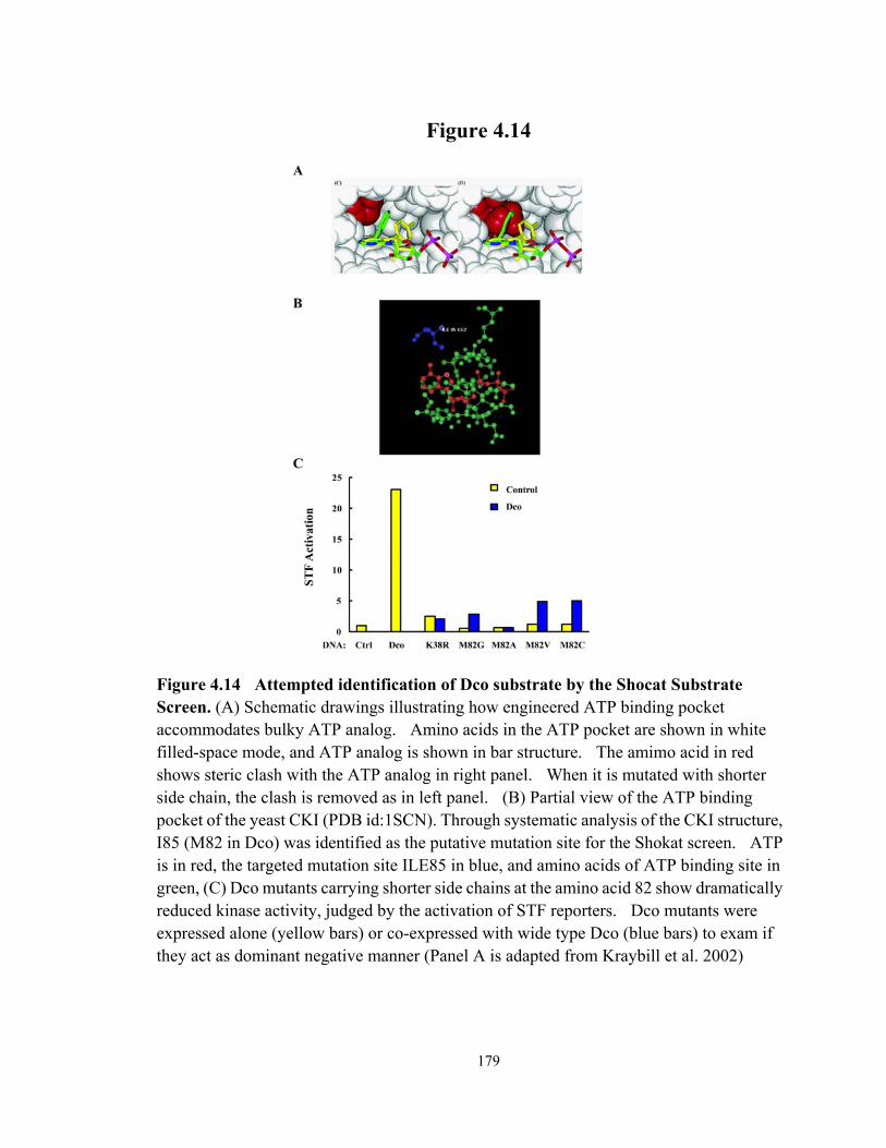
179
Figure 4.14
Figure 4.14 Attempted identification of Dco substrate by the Shocat Substrate Screen. (A) Schematic drawings illustrating how engineered ATP binding pocket accommodates bulky ATP analog. Amino acids in the ATP pocket are shown in white filled-space mode, and ATP analog is shown in bar structure. The amimo acid in red shows steric clash with the ATP analog in right panel. When it is mutated with shorter side chain, the clash is removed as in left panel. (B) Partial view of the ATP binding pocket of the yeast CKI (PDB id:1SCN). Through systematic analysis of the CKI structure, I85 (M82 in Dco) was identified as the putative mutation site for the Shokat screen. ATP is in red, the targeted mutation site ILE85 in blue, and amino acids of ATP binding site in green, (C) Dco mutants carrying shorter side chains at the amino acid 82 show dramatically reduced kinase activity, judged by the activation of STF reporters. Dco mutants were expressed alone (yellow bars) or co-expressed with wide type Dco (blue bars) to exam if they act as dominant negative manner (Panel A is adapted from Kraybill et al. 2002)

180
REFERENCE
Amit, S., A. Hatzubai, Y. Birman, J. S. Andersen, E. Ben-Shushan, M. Mann, et al. (2002). "Axin-mediated CKI phosphorylation of beta-catenin at Ser 45: a molecular switch for the Wnt pathway." Genes & Development 16(9): 1066-76.
Cadigan, K. M. (2008). "Wnt-beta-catenin signaling." Current Biology 18(20): R943-R47. Cadigan, K. M. and Y. I. Liu (2006). "Wnt signaling: complexity at the surface." Journal of
Cell Science 119(3): 395-402. Cadigan, K. M. and R. Nusse (1997). "Wnt signaling: a common theme in animal
development." Genes & Development 11(24): 3286-305. Davidson, G., W. Wu, J. L. Shen, J. Bilic, U. Fenger, P. Stannek, et al. (2005). "Casein
kinase 1 gamma couples Wnt receptor activation to cytoplasmic signal transduction." Nature 438(7069): 867-72.
Eblen, S. T., N. V. Kumar, K. Shah, M. J. Henderson, C. K. W. Watts, K. M. Shokat, et al. (2003). "Identification of novel ERK2 substrates through use of an engineered kinase and ATP analogs." Journal of Biological Chemistry 278(17): 14926-35.
Elphick, L. M., S. E. Lee, V. Gouverneur and D. J. Mann (2007). "Using chemical genetics and ATP analogues to dissect protein kinase function." Acs Chemical Biology 2(5): 299-314.
Fang, M., J. Li, T. Blauwkamp, C. Bhambhani, N. Campbell and K. M. Cadigan (2006). "C-terminal-binding protein directly activates and represses Wnt transcriptional targets in Drosophila." Embo Journal 25(12): 2735-45.
Goyal, L., K. McCall, J. Agapite, E. Hartwieg and H. Steller (2000). "Induction of apoptosis by Drosophila reaper, hid and grim through inhibition of IAP function." Embo Journal 19(4): 589-97.
Guan, J., H. Li, A. Rogulja, J. D. Axelrod and K. M. Cadigan (2007). "The Drosophila casein kinase I epsilon/delta Discs overgrown promotes cell survival via activation of DIAP1 expression." Developmental Biology 303(1): 16-28.
Hay, B. A. and M. Guo (2006). "Caspase-dependent cell death in Drosophila." Annual Review of Cell and Developmental Biology 22: 623-50.
Hengartner, M. O. (2000). "The biochemistry of apoptosis." Nature 407(6805): 770-76. Hindley, A. D., S. Park, L. L. Wang, K. Shah, Y. L. Wang, X. C. Hu, et al. (2004).
"Engineering the serine/threonine protein kinase Raf-1 to utilise an orthogonal analogue of ATP substituted at the N-6 position." Febs Letters 556(1-3): 26-34.
Hummel, T., S. Attix, D. Gunning and S. L. Zipursky (2002). "Temporal control of glial cell migration in the Drosophila eye requires gilgamesh, hedgehog, and eye specification genes." Neuron 33(2): 193-203.
Kafri, R., M. Springer and Y. Pilpel (2009). "Genetic Redundancy: New Tricks for Old Genes." Cell 136(3): 389-92.
181
Kanuka, H., E. Kuranaga, K. Takemoto, T. Hiratou, H. Okano and M. Miura (2005). "Drosophila caspase transduces Shaggy/GSK-3 beta kinase activity in neural precursor development." Embo Journal 24(21): 3793-806.
Kloss, B., J. L. Price, L. Saez, J. Blau, A. Rothenfluh, C. S. Wesley, et al. (1998). "The Drosophila clock gene double-time encodes a protein closely related to human casein kinase I epsilon." Cell 94(1): 97-107.
Knippschild, U., A. Gocht, S. Wolff, N. Huber, J. Lohler and M. Stoter (2005). "The casein kinase I family: participation in multiple cellular processes in eukaryotes." Cellular Signalling 17(6): 675-89.
Kraybill, B. C., L. L. Elkin, J. D. Blethrow, D. O. Morgan and K. M. Shokat (2002). "Inhibitor scaffolds as new allele specific kinase substrates." Journal of the American Chemical Society 124(41): 12118-28.
Kuranaga, E., H. Kanuka, A. Tonoki, K. Takemoto, T. Tomioka, M. Kobayashi, et al. (2006). "Drosophila IKK-related kinase regulates nonapoptotic function of caspases, via degradation of IAPs." Cell 126(3): 583-96.
Lin, H. V., A. Rogulja and K. M. Cadigan (2004). "Wingless eliminates ommatidia from the edge of the developing eye through activation of apoptosis." Development 131(10): 2409-18.
Liu, C. M., Y. M. Li, M. Semenov, C. Han, G. H. Baeg, Y. Tan, et al. (2002). "Control of beta-catenin phosphorylation/degradation by a dual-kinase mechanism." Cell 108(6): 837-47.
Liu, Y., K. Shah, F. Yang, L. Witucki and K. M. Shokat (1998). "Engineering Src family protein kinases with unnatural nucleotide specificity." Chemistry & Biology 5(2): 91-101.
Liu, Y. I., M. V. Chang, H. E. Li, S. Barolo, J. L. Chang, T. A. Blauwkamp, et al. (2008). "The chromatin remodelers ISWI and ACF1 directly repress Wingless transcriptional targets." Developmental Biology 323(1): 41-52.
Mashhoon, N., A. J. DeMaggio, V. Tereshko, S. C. Bergmeier, R. Egli, M. F. Hoekstra, et al. (2000). "Crystal structure of a conformation-selective casein kinase-1 inhibitor." Journal of Biological Chemistry 275(26): 20052-60.
Price, M. A. (2006). "CKI, there's more than one: casein kinase I family members in Wnt and Hedgehog signaling." Genes & Development 20(4): 399-410.
Salvesen, G. S. and J. M. Abrams (2004). "Caspase activation - stepping on the gas or releasing the brakes? Lessons from humans and flies." Oncogene 23(16): 2774-84.
Santos, J. A., E. Logarinho, C. Tapia, C. C. Allende, J. E. Allende and C. E. Sunkel (1996). "The casein kinase 1 alpha gene of Drosophila melanogaster is developmentally regulated and the kinase activity of the protein induced by DNA damage." Journal of Cell Science 109: 1847-56.
Shah, K., Y. Liu, C. Deirmengian and K. M. Shokat (1997). "Engineering unnatural nucleotide specificity for Rous sarcoma virus tyrosine kinase to uniquely label its

182
direct substrates." Proceedings of the National Academy of Sciences of the United States of America 94(8): 3565-70.
Srinivasula, S. M. and J. D. Ashwell (2008). "IAPs: What's in a name?" Molecular Cell 30(2): 123-35.
Steller, H. (2008). "Regulation of apoptosis in Drosophila." Cell Death Differ 15(7): 1132-38.
Vielhaber, E. and D. M. Virshup (2001). "Casein kinase I: From obscurity to center stage." Iubmb Life 51(2): 73-78.
Wharton, K. A. (2003). "Runnin' with the Dvl: Proteins that associate with Dsh/Dvl and their significance to Wnt signal transduction." Developmental Biology 253(1): 1-17.
Xu, R. M., G. Carmel, R. M. Sweet, J. Kuret and X. D. Cheng (1995). "CRYSTAL-STRUCTURE OF CASEIN KINASE-1, A PHOSPHATE-DIRECTED PROTEIN-KINASE." Embo Journal 14(5): 1015-23.
Zeng, X., K. Tamai, B. Doble, S. T. Li, H. Huang, R. Habas, et al. (2005). "A dual-kinase mechanism for Wnt co-receptor phosphorylation and activation." Nature 438(7069): 873-77.

183
CHAPTER V
CONCLUDING REMARKS
SUMMARY OF CONTRIBUTIONS
Apoptosis is an evolutionarily conserved, actively regulated cell death process that
plays an essential role during development and in adulthood. Apoptosis is subject to the
negative regulation of Inhibitor of Apoptosis Proteins (IAPs). The function of IAPs has to
be antagonized to allow the occurrence of apoptosis, while the activation of IAPs promotes
cell survival. The goal of my dissertation research was to advance the understanding of
IAP regulation during development. Specifically, the two major foci were: a. systematic
analysis of Drosophila IAP1 (DIAP1) down-regulation in developmental apoptosis in the
fly pupal eye; and b. characterization of novel factors that promote cell survival via
activation of DIAP1.
RHG protein mediated apoptosis in the pupal eye of Drosophila: down-regulation of DIAP1
Drosophila Inhibitor of apoptosis protein 1 (DIAP1) is an essential negative
regulator of apoptosis in fly cells. DIAP1 binds to caspases and inhibits their activity.
The antagonism of DIAP1 by RHG proteins, e.g., Head Involution defective (Hid), Grim
184
and Reaper (Rpr), is a central theme in cells committed to apoptosis. Previous studies
revealed two major hypotheses, generally referred to as the liberation model and
degradation model, which explain how RHG proteins counteract DIAP1’s anti-apoptotic
function. The liberation model proposes that RHGs induce apoptosis through binding to
DIAP1 and replacing the caspases, whereas the degradation model emphasizes the ability
of RHGs to induce DIAP1 degradation, releasing the inhibition on caspases. However,
these two models were mainly based on in vivo overexpression or in vitro biochemical
assays, provoking an important question which mechanism (liberation or degradation) is
more physiologically relevant.
In this dissertation, I demonstrated significant down-regulation of DIAP1 levels in
naturally occurring apoptosis in the perimeter ommatidia of the fly pupal eye. Further
characterization revealed that the down-regulation is post-transcriptional and independent
of effector caspase activity. Using a loss of function approach, I illustrated that the
regulation was dependent on Hid, Grim and Rpr. This is the first time that RHG protein
dependent DIAP1 down-regulation has been illustrated in naturally occurring apoptosis.
My results also help to resolve the debate around the two competing models. I
showed that a partial reduction in RHG proteins suppressed DIAP1 degradation to
undetectable levels. Under these conditions, apoptosis was surprisingly initiated at the
normal time with comparable strength, but was markedly reduced thereafter. These
results argue that the liberation of caspases plays a significant role in apoptosis initiation,

185
whereas the degradation of DIAP1 is required for timely progression of apoptosis in this
tissue.
In addition, I provided evidence that the E3 ligase activity of DIAP1’s RING domain
is dispensable for RHG-mediated degradation. The data was unexpected as
auto-ubiquitination and consequent degradation has been proposed as an important
approach to eliminate DIAP1. In contrast, I showed the Drosophila Apaf-1 related killer
(Dark) and the initiator caspase Dronc are both required for DIAP1 down-regulation,
suggesting that RHG proteins may act through Dark and Dronc.
In summary, by studying DIAP1 down-regulation under physiological conditions, I
provided new insights regarding the mechanism underlying DIAP1 antagonism in
apoptotic cells.
The Drosophila casein kinase Iε/δ Discs overgrown promotes cell survival via activation of DIAP1 expression
The development and survival of multi-cellular organisms are dependent on the
dynamic equilibrium between cell proliferation and death. For tissue to achieve fast
growth, apoptosis has to be kept at low levels while cell division needs to be promoted.
Through a genetic screen, the Drosophila casein kinase Iε/δ, known as Discs overgrown
(Dco), was identified as a negative regulator of apoptosis. Further characterized by Dr. Ju
Guan in the Cadigan lab, Dco has been demonstrated to promote cell survival via activation
of DIAP1.

186
In order to reveal the regulation mechanism, I demonstrated Dco mutants don’t
affect the levels of Hid, Grim and Rpr mRNA, suggesting the regulation is probably at the
protein level. I also demonstrated that the kinase activity is required for Dco to suppress
apoptosis.
To uncover the underlying molecular mechanism, I established a cell culture system
using fly embryonic cell line S2 to study apoptosis and DIAP1 regulation. I showed that
the anti-apoptotic role of Dco could be recapitulated in S2 cells, however no influence on
DIAP1 levels was observed under a variety of conditions. One attractive hypothesis to
explain the absence of DIAP1 regulation was the existence of redundant partners, which
possibly is another casein kinase I (CKI).
To identify redundant partners of Dco in apoptosis regulation, I conducted two small
scale screens in the fly eye and cultured cells by knocking down expression of the other fly
CKI members. Both screens indicated Gilgamesh (Gish), a homologue of mammalian
CKIγ, can influence apoptosis. Characterization of Gish showed that it may
down-regulate DIAP1 in S2 cells, and partially rescue some phenotypes caused by dco
depletion in the fly. Further analyses are required to establish the anti-apoptotic role of
Gish as well as the redundancy of Dco and Gish.
In summary, via studies on Dco and Gish, I have made contribution to understand
how DIAP1 function is reinforced to secure the survival fate of cells. Further studies on

187
Dco and Gish can potentially uncover a novel regulation pathway that coordinates cell
survival and apoptosis.
FUTURE DIRECTIONS
Exploration of the apoptosis regulation via the ubiquitination pathway
The RING domain has been proposed to mediate the auto-ubiquitination and
consequent degradation of DIAP1 during apoptosis (Hay and Guo 2006; Steller 2008).
Surprisingly, our results suggest the E3 ligase activity of DIAP1’s RING domain may be
dispensable for RHG-mediated degradation.
One concern with my experimental data is that I examined DIAP1 downregulation in
flies that were heterozygous for the RING domain mutant th6B. Mammalian IAPs have
been reported to dimerize through the RING domains (Vaux and Silke 2005), raising the
possibility that wild type DIAP1 present in the heterozygous cells may ubiquitinate the
RING domain mutant in trans, leading to the consequent degradation. To address this
concern, we need to overcome the technical difficulty to generate homozygous clones of
th6B. I am currently using MarcM system to achieve the goal, and the fly stocks needed
for the experiments are under construction. If the down-regulation of DIAP1 is still
observed in cells homozygous for th6B, it will strongly reinforce our argument that
DIAP1’s RING domain is dispensable for RHG-mediated degradation. Conversely, if
downregulation is not observed when both copies of DIAP1 have RING domain mutations,

188
this would suggest that DIAP1 may employ a similar dimerization mechanism as its
mammalian counterparts. In this case, it would be interesting to conduct biochemical
analyses to illustrate the interaction and map the corresponding interaction domain. If the
interaction domain is the RING domain as for mammalian IAPs, the physiological
importance of the putative dimerization can be easily tested in flies heterozygous for the
RING domain deletion th33-1s (Wilson et al. 2002).
If the RING domain is confirmed to be dispensable, it is likely that DIAP1
degradation in apoptotic cells is mediated by other E3 ligases. Intriguingly, SkpA, an
SCF (SKP-Cullin-F-box) multiunit E3 ligase complex component, has been implicated in
Rpr- and Grim-induced apoptosis (Wing et al. 2002). SkpA has been shown to interact
with an F-box protein Morgue, and Morgue can bind to DIAP1 and promote its degradation
(Hays et al. 2002; Wing et al. 2002). It suggests that an SCF E3 ligase complex could
target DIAP1 for degradation in peripheral ommatidia. One approach to test this
possibility is to examine DIAP1 down-regulation in ommatidia depleted of skpA or morgue
by RNAi. UAS-skpA RNAi and UAS-morgue RNAi have been obtained from Vienna
Drosophila RNAi Center, and they can suppress the small eye phenotype induced by
GMRGal4; UAS-Rpr (data not shown). Preliminary data showed that DIAP1
down-regulation is partially suppressed by skpA and morgue RNAi. Further validation is
needed to support the hypothesis that DIAP1 down-regulation is facilitated via the
trans-ubiquitination by the SkpA containing multi-unit E3 complex.

189
One interesting question is how DIAP1 directs its E3 ligase activity toward different
substrates, including Dronc, RHG proteins and DIAP1 itself. A simple scenario which
has been suggested is that DIAP1 ubiquitinates Dronc in living cells, while it ubiquitinates
itself in apoptotic cells (Chai et al. 2003). Our results clearly suggest the physiological
situation is far more complicated, as we found E3 ligase activity of the RING domain
contributes to the DIAP1 stability in cells that have a survival fate, whereas it seems
dispensable in apoptotic cells. It would be interesting to examine the stability of Dronc in
both apoptotic and living cells carrying th6B. This information will be helpful to
understand how the E3 ligase activity of DIAP1 is regulated differently in cells with
different fates.
In addition, several ubiquitination pathway factors have been implicated in apoptosis
regulation, including the E1 ubiquitin-activating enzyme Uba1 (Wing et al. 2002), the E2
ubiquitin-conjugating enzyme UbcD1(Ryoo et al. 2002), the de-ubiquitinating enzyme Fat
facets (Wing et al. 2002), as well as SkpA and Mogure. To address the physiological
relevance of these factors in apoptosis, it would be informative to observe whether the
DIAP1 stability and the occurrence of apoptosis in perimeter ommatidia are affected in
flies with these factors either mutated or depleted by RNAi.
DIAP1 regulation in other developmental apoptosis
In Chapter II, I utilized the naturally occuring apoptosis in the developing pupal eye
as a model system to address the physiological importance of several popular models about

190
DIAP1 regulation mechanisms. I obtained several unexpected results which are
inconsistent with some of the tested models. This is not totally surprising, since the
discrepancy is likely due to the inherent limits of in vitro and over-expression analyses that
these models are based on. From this perspective, my data illustrates the significance of
studies using naturally occurring apoptosis as model systems.
I am currently developing another system that is based on developmental apoptosis
during embryogenesis, where most of the developing midline glial cells are eliminated by
apoptosis to achieve a well circuited central neural system (Sonnenfeld and Jacobs 1995;
Zhou et al. 1995). Hid and Rpr have been proposed to cooperate with each other in a
synergistic fashion to achieve this apoptosis (Zhou et al. 1997). It would be interesting to
see if DIAP1 is regulated differently during the midline glial apoptosis compared to the
pupal eye.
To outline the midline cells, I will express CD8-GFP under the control of a CNS
midline precursor cell specific promoter (Bloomington Stock No.9150). Preliminary data
illustrated the midline glial apoptosis happens between stage 11 to stage 14 of the fly
embryo. However, because cells with survival fates cannot be distinguished from those
committed to die, it is hard to judge whether DIAP1 is down-regulated in apoptotic cells
(data not shown). To address this issue, I will ectopically express the caspase inhibitor
p35 to produce “undead cells”. If DIAP1 is down-regulated during the midline cell
apoptosis, lower DIAP1 levels would be observed in some of the GFP positive cells, which

191
presumably would have died without p35. Positive data would extand the physiological
importance of DIAP1 degradation during apoptosis, while negative results would be
equally interesting as it might indicate that the fly may employ tissue-specific mechanisms
to regulate DIAP1 and apoptosis.
Another event that can be potentially developed as a model system to study DIAP1
regulation is the inter-ommatidia cell death that removes extra secondary and tertiary
pigment cells in the early pupal eye (Cagan and Ready 1989; Brachmann and Cagan 2003).
DIAP1 staining of pupal eyes at 24hr APF shows a highly ordered pattern with different
intensities of DIAP1 in inter-ommatidia areas, indicating DIAP1 levels might be regulated
at this stage (data not shown). To specifically label pigment cells, homothorax will be
used as a marker (Mendes et al. 2006).
Studies in these two apoptosis events may further confirm our results in Chapter II,
and may potentially provide new mechanistic insights on how RHG proteins trigger
apoptosis via DIAP1 regulation.
Characterization of Gilgamesh (Gish) in DIAP1 regulation
In Chapter III, our results suggest that Gish may regulate DIAP1 stability in S2 cells.
Before further characterization, DIAP1 down-regulation resulting from the depletion of
gish needs to be confirmed. After validation, it would be interesting to determine the
physiological importance of this regulation. Our results illustrated that over-expression of
Gish causes extra macrochaetes in the scutellum of the adult notum, a phenotype

192
considered as a consequence of decreased non-apoptotic activity of capases. Therefore, I
speculate Gish may regulate DIAP1 in both apoptotic and non-apoptotic contexts.
To explore the anti-apoptotic function of Gish, the levels of DIAP1, active-Drice and
TUNEL will be tested in clones of gishnull in wing imaginal discs. Elevated TUNEL and
active-Drice would suggest the role of Gish in promoting cell survival via activation of
DIAP1. If gishnull cells can not survive to the third instar larval stage, p35 would be
expressed to block apoptosis, and in this case only active-Drice level, but not TUNEL,
would be informative. One caveat about active-Drice is that it is also activated in
non-apoptotic contexts, although the level of Drice activation has been reported to be
considerably low (Kuranaga et al. 2006). Meanwhile, the cell survival assay can be
performed in S2 cells to show the ability of Gish to suppress Hid-induced cell death.
To test the possibility that Gish regulates DIAP1 in a non-apoptotic context, I would
start with the macrochaete phenotype. Previous studies illustrated that DIAP1
over-expression resulted in extra SOP cells labeled by anti-senseless antibody, ultimately
causing the extra macrochaete phenotype (Kuranaga et al. 2006). It would be interesting
to examine the number of SOP cells in wing discs of dppGal4;UASgish, which showed
extra macrochaetes in adult previously (Figure 4.9A). Scabrous-Gal4; UASgish would be
tested too, as Scabrous-Gal4 drives expression in proneural clusters of the wing discs, and
Scabrous-Gal4; UASDIAP1 shows extra macrochaetes in adult (Kuranaga et al. 2006). If
Gish over-expression leads to more SOP cells, loss of function analyses through either

193
RNAi knockdown or mutant clones would be performed to show the physiological
relevance. If positive results are obtained for the non-apoptotic role, it would be
interesting to examine the genetic interaction between Gish and dmIKKε, the known
DIAP1 regulator during SOP specification (Kuranaga et al. 2006).
At the same time, biochemical assays would be conducted to elucidate the molecular
mechanism, for instance, co-immnoprecipitation between DIAP1 and Gish, and in vitro
kinase assay of Gish with DIAP1 as the putative substrate.
Identification of Dco substrates
In Chapter IV, I initiated a chemical genetic screen, referred to as the Shokat screen,
to identify the substrates of Dco. I engineered the ATP binding pocket which potentially
enlarges the space to accommodate N-6 substitutions of ATP. Unfortunately, all the
DcoM82 mutants show low enzymatic activities in a functional assay carried out in S2
cells (Figure 4.14), causing me to abandon this approach at that time.
I think it is worthwhile considering resuming the Shokat screen, as there are some
possibilities to potentially produce a functional Dco mutant for the screen. First of all,
DcoM82 mutants cannot use normal ATP, but they might be able to utilize N-6 modified
ATP analogues. Thus, we may apply multiple N-6 modified ATP analogues to DcoM82
mutants in in vitro kinase assays to identify the best match of ATP and mutants that leads to
acceptable kinase activity. In addition, DcoM82V and M82C show some residual kinase
activities in the functional assay (Figure 4.14C). Perhaps a high concentration of the right

194
ATP analogue in in vitro kinase assay with DcoM82V or M82C would enable an
acceptable level of kinase activity to generate enough phosphorylated proteins for
detection. Finally, I am also interested in developing a strategy to rescue the kinase
activity by mutating one or two amino acids that are located away from the ATP binding
pocket. With helps from an experienced structural biologist, we can evaluate the
feasibility of this idea.
In the most optimistic scenario that one of the approaches rescues the Shokat screen,
I would like to propose following experiments. First of all, it would be very informative if
any known apoptotic factors or DIAP1 regulators are identified from the screen. In this
case, the genetic and molecular interactions between Dco and these factors will be
investigated after the phosphorylation is confirmed. Meanwhile, other factors will be
subject to an RNAi-based functional screen, in order to narrow down to those who affect
cell survival and/or DIAP1 stability. Positive picks will be bacterially produced, and
verified as Dco substrates in in vitro kinase assay. The confirmed factors will be
examined whether they can either phenocopy Dco or show the opposite effects in a series
of experiments in S2 cells and fly tissues. Finally, the phosphorylation sites will be
mapped and consequently mutated to illustrate the importance of the phosphorylation in
apoptosis regulation.
Dco/CKIε substrates are missing in several other pathways, for example the Hippo
pathway. Thus, if any known factors in these pathways are identified from the screen,

195
they will be of interests too. Presumably, the success of the screen can be easily extended
to other CKI members, for example, Gish.
PROSPECTS
At the very beginning of this dissertation, I emphasized that life and death are two
opposite yet interconnected and interdependent forces in multi-cellular organisms. From
the perspective of my dissertation, it is interesting to speculate that DIAP1 serves as a
coordinator of the two forces, as the activation of DIAP1 promotes cell survival, while the
inhibition leads to cell death.
Our knowledge of DIAP1 regulation in apoptosis has accumulated for over a decade.
With all great information in hand, I think it is time to synthesize all the information in the
context of physiological importance. Luckily, my dissertation study takes a small step
towards that direction. In addition, I think the studies on DIAP1 regulation under
physiologal conditions hold the promise to uncover how cells control caspases to pursue
either apoptotic or non-apoptotic function.
As major regulators of DIAP1, RHG proteins have been established as essential
players to initiate apoptosis in Drosophila. I think the putative connection of RHG
proteins with mitochondria, as well as their potential role to trigger Dark-dependent Dronc
activation will continue to attract considerable attention in the near future. As Hid, Grim
and Rpr are elegantly regulated at the transcription level, I would like to predict that

196
studying the cis- and trans-regulatory elements of RHG genes and how they integrate a
variety of death and survival signals may become a new focus of the field.
To take the speculation one step further, I would like to envision that the regulation
of IAPs in living cells is far more complex than what we have known: the IAP family
should not be a lonely fighter against caspases, and there may be a network of factors
supporting and regulating their roles. Our limited understanding of Dco might serve as an
example to show how much is missing with our current knowledge on this issue. Future
investigations on the regulation of IAP proteins in both apoptosis and cell survival would
advance our understanding on how multi-cellular organisms balance the death and
survival of cells.
197
REFERENCE
Brachmann, C. B. and R. L. Cagan (2003). "Patterning the fly eye: the role of apoptosis." Trends in Genetics 19(2): 91-96.
Cagan, R. L. and D. F. Ready (1989). "THE EMERGENCE OF ORDER IN THE DROSOPHILA PUPAL RETINA." Developmental Biology 136(2): 346-62.
Chai, J. J., N. Yan, J. R. Huh, J. W. Wu, W. Y. Li, B. A. Hay, et al. (2003). "Molecular mechanism of Reaper-Grim-Hid-mediated suppression of DIAP1-dependent Dronc ubiquitination." Nature Structural Biology 10(11): 892-98.
Hay, B. A. and M. Guo (2006). "Caspase-dependent cell death in Drosophila." Annual Review of Cell and Developmental Biology 22: 623-50.
Hays, R., L. Wickline and R. Cagan (2002). "Morgue mediates apoptosis in the Drosophila melanogaster retina by promoting degradation of DIAP1." Nature Cell Biology 4(6): 425-31.
Kuranaga, E., H. Kanuka, A. Tonoki, K. Takemoto, T. Tomioka, M. Kobayashi, et al. (2006). "Drosophila IKK-related kinase regulates nonapoptotic function of caspases, via degradation of IAPs." Cell 126(3): 583-96.
Lin, H. V., A. Rogulja and K. M. Cadigan (2004). "Wingless eliminates ommatidia from the edge of the developing eye through activation of apoptosis." Development 131(10): 2409-18.
Mendes, C. S., E. Arama, S. Brown, H. Scherr, M. Srivastava, A. Bergmann, et al. (2006). "Cytochrome c-d regulates developmental apoptosis in the Drosophila retina." Embo Reports 7(9): 933-39.
Ryoo, H. D., A. Bergmann, H. Gonen, A. Ciechanover and H. Steller (2002). "Regulation of Drosophila IAP1 degradation and apoptosis by reaper and ubcD1." Nature Cell Biology 4(6): 432-38.
Sonnenfeld, M. J. and J. R. Jacobs (1995). "APOPTOSIS OF THE MIDLINE GLIA DURING DROSOPHILA EMBRYOGENESIS - A CORRELATION WITH AXON CONTACT." Development 121(2): 569-78.
Steller, H. (2008). "Regulation of apoptosis in Drosophila." Cell Death and Differentiation 15(7): 1132-38.
Vaux, D. L. and J. Silke (2005). "IAPs, RINGs and ubiquitylation." Nature Reviews Molecular Cell Biology 6(4): 287-97.
Wilson, R., L. Goyal, M. Ditzel, A. Zachariou, D. A. Baker, J. Agapite, et al. (2002). "The DIAP1 RING finger mediates ubiquitination of Dronc and is indispensable for regulating apoptosis." Nature Cell Biology 4(6): 445-50.
Wing, J. P., B. A. Schreader, T. Yokokura, U. Q. Wang, P. S. Andrews, N. Huseinovic, et al. (2002). "Drosophila Morgue is an F box/ubiquitin conjugase domain protein important for grim-reaper mediated apoptosis." Nature Cell Biology 4(6): 451-56.

198
Zhou, L., H. Hashimi, L. M. Schwartz and J. R. Nambu (1995). "PROGRAMMED CELL-DEATH IN THE DROSOPHILA CENTRAL-NERVOUS-SYSTEM MIDLINE." Current Biology 5(7): 784-90.
Zhou, L., A. Schnitzler, J. Agapite, L. M. Schwartz, H. Steller and J. R. Nambu (1997). "Cooperative functions of the reaper and head involution defective genes in the programmed cell death of Drosophila central nervous system midline cells." Proceedings of the National Academy of Sciences of the United States of America 94(10): 5131-36.