the respiratory system · the respiratory system learning strategy in thischapter wewill consider...
TRANSCRIPT
1The respiratory system
Learning strategy
In this chapter we will consider the essential ‘must know’ facts and concepts of therespiratory system. Our main strategy would involve an exploration of these keyprinciples by following several clinical scenarios.
The first scenario, an asthma attack will introduce us to the anatomy of therespiratory system. A consideration of the pathophysiology of asthma, will lead toa review of the immune system and the mechanism of Type 1 hypersensitivity.
Breathing difficulties will lead us to consider the mechanism of breathing and lungcompliance. Lung volumes and capacities will be discussed as we consider the lungfunction tests of the asthmatic patient. We will also review the key drugs used inasthma treatment.
A second scenario – COPD – leads us to a consideration of acidosis and alkalosis. Wewill also discuss some key respiratory infections and the important concepts of V/Qmismatch and dead space. Finally we will consider gas exchange, oxygen and carbondioxide saturation curves and the central and peripheral control of respiration.
Throughout, we will also consider the pathophysiological mechanisms of several keydisease states involving the respiratory system, which will, in addition to high-lighting the key pathophysiological principles, further reinforce basic principles ofanatomy, physiology and pharmacology relevant to the respiratory system.
Try to answer the questions and try to complete the Learning Tasks. The TriggerBoxes should be used as a guide for further reading and revision. At the end of thischapter you should have a sound understanding of the key facts and conceptsunderlying the respiratory system.
Integrated Medical Sciences by Shantha Perera, Stephen Anderson, Ho Leung and Rousseau Gama# 2007 John Wiley & Sons, Ltd ISBN: 978470016589 (HB) 978470016596 (PB)
COPYRIG
HTED M
ATERIAL

Zoe’s breathing difficulties . . .
It happened again on Boxing Day. Around 5pm Zoe was sitting on her bed reading whenshe started to become breathless. Breathing was always her ‘problem’ and Zoe couldn’tunderstand it.
‘After all it’s supposed to be such a simple thing to do isn’t it?’ she had asked Mary. ‘It’ssupposed to be automatic isn’t it? I mean you just breathe in and out. So why is it such aneffort sometimes?’
Zoe took a puff out of her blue inhaler. She wondered if her problems had somethingto do with the fact that she had been a premature baby and that that she had to bedelivered at 33 weeks, by caesarean section.
‘Maybe my lungs weren’t developed properly’ she thought.
What is in Zoe’s blue inhaler?
Although Zoe was a premature baby, she didn’t have any problems and grew into ahealthy child. Had she been born around, say, 26 weeks she would have had seriousproblems because her respiratory system would have been underdeveloped.
So, let us begin by reviewing the key stages in the development of the respiratorysystem. First, in terms of origins, the epithelium of the nasopharynx, trachea, bronchi,bronchioles and alveoli are derived from endoderm. The associated cartilage and muscleare mesodermal in origin.
You are expected to know the embryological origins of key anatomical structures.Construct a table listing themainderivativesof endoderm,ectodermandmesoderm.
So what are the key embryological events in the development of the respiratory system?The respiratory system starts off as an outgrowth of the foregut. In the 4th week theoesophagotracheal septum separates the foregut into the respiratory diverticulum (lungbud) and oesophagus (Figure 1.1). The bud elongates and then branches into two. Eachof these two new buds will become the primary bronchus of each lung.
What happens if the diverticulum fails to separate completely from the foregut
What is a TOF (tracheo-oesophageal fistula)?
The left lung bud develops into two secondary bronchi and eventually forms two lobes;the right bud forms three secondary bronchi and three lobes. The tertiary bronchi createthe bronchopulmonary segments.
Gas exchange between blood and air in the primitive alveoli is possible in the seventhmonth of gestation. Lung growth after birth is mainly due to an increase in the numberof respiratory bronchioles and alveoli and not due to an increase in size of alveoli. Newalveoli are formed for at least 10 years of postnatal life.
What do we mean by the term ‘gas exchange’?
Before birth, the lungs are filled with fluid containing surfactant mainly made up ofdipalmitoyl phosphatidylcholine, which is produced by type II epithelial cells. When
2 CH1 THE RESPIRATORY SYSTEM

respiration begins, lung fluid is reabsorbed but leaves a surfactant coating. If Zoe wasborn around 26 weeks, her surfactant levels would have been low. She would suffer fromrespiratory distress syndrome (RDS). Her lungs would be difficult to expand and duringdeflation her alveoli would collapse. Surfactant decreases the alveolar surface tensionand helps the alveoli to expand more easily.
What are type I and II pneumocytes? Where do you find them?
Mothers of premature babies are treated with steroids. Why?
What treatment can be given to a 28-week premature baby having difficultyinflating its lungs?
Trigger box Respiratory distress syndrome (RDS)
Deficiency of surfactant causes alveolar collapse and poor gas exchange.
Majority of infants born before 28 weeks develop RDS within 4 hours of birth.
Features: Tachypnoea, cyanosis, diaphragm, subcostal and intercostal retraction, grunting.
CXR: Reticulogranular appearance with air bronchograms.
Treatment: Glucocorticoids to mother, exogenous surfactant, oxygen, continuous
positive airway pressure (CPAP), artificial ventilation.
Respiratorydiverticulumoesophagus
lung buds
tertiary bronchi
left primary bronchus
right primary bronchus
left secondary bronchi
right secondary bronchi
left inferior
lobe
right inferior
lobe
right middle lobe
right superior
lobe
left superior
lobe
lung budslung buds
tertiary bronchitertiary bronchi
left primary bronchus
right primary bronchus
left secondary bronchi
right secondary bronchi
left primary bronchus
right primary bronchus
left secondary bronchi
right secondary bronchi
left inferior
lobe
right inferior
lobe
right middle lobe
right superior
lobe
left superior
lobe
left inferior
lobe
right inferior
lobe
right middle lobe
right superior
lobe
left superior
lobe
Figure 1.1 Development of the respiratory system. Adapted from Tortora andGrabowski (2003),principles of Anatomy and Physiology, 10th edition, John Wiley & Sons Inc. p.842
ZOE’S BREATHING DIFFICULTIES . . . 3

Next let us consider the gross anatomy of the respiratory system. Figure 1.2 shows theimportant anatomical structures you need to know.
Note that the right lung has three lobes; the left has two. Each lung lobe is made up ofbronchopulmonary segments. Label the oblique and horizontal fissures. The right mainbronchus is straighter and shorter than the left main bronchus. This helps to explain why Zoe’sbrother John, at age 4, got a small peanut stuck in the right main bronchus when he inhaled it.
When Zoe’s great-uncle Arthur suffered from a really nasty bout of pulmonarytuberculosis the surgeons had to remove several of his bronchopulmonary segments.This was not too difficult because each bronchopulmonary segment is served by its ownarteries and vein and is partitioned from other segments by connective tissue.
Define what is meant by the terms (a) ‘respiratory bronchiole’ and (b) ‘terminalbronchiole’.
Let us look at the other main structures that make up the respiratory system. Importantstructures to know include the nasopharynx, oropharynx, larynx, glottis and trachea.The blood supply of the lungs consists of the pulmonary arteries that run with theairways, the bronchial arteries that branch off from the aorta, and the pulmonary veinsthat run in the connective tissue septa.
What kind of blood (oxygenated, deoxygenated) is found in these differentvessels?
Bronchi have cartilage whereas bronchioles do not. They both have smoothmuscle – what is the relevance of these facts to asthma?
Lung connective tissue contains lots of elastic tissue – what is the significanceof this elasticity?
epiglottis
lobes of right lung
right bronchus
cartilage ringtrachea
larynx
aorta
right atrium
Figure 1.2 Anatomy of the respiratory system. Adapted from Mackean (1969), Introduction toBiology, 4th edition, John Murray, London, p.69.
4 CH1 THE RESPIRATORY SYSTEM

The lungs are covered by visceral pleura. This is separated from the parietal pleura whichcovers the inside of the chest wall by the interpleural space.
What is pleurisy
Trigger box Pleural effusions
Transudate: (protein <30 g/L; LDH <200 iu/l):
Congestive heart failure (CHF), hypothyroidism, nephrotic syndrome
Exudate: (protein >30 g/L; LDH >200 iu/l):
Pneumonia, carcinoma, tuberculosis (TB), pulmonary infarct.
Can detect clinically if >500mL; by CXR >300mL.
Findings: Reduced chest movements, stony dull percussion, decreased breath sounds,
reduced vocal resonance. Blunting of costophrenic angle on CXR.
Treatment: drain exudates, treat underlying cause of transudate.
Sclerosing agents to reduce recurrent malignant pleural effusions.
Three years ago Dr Smith, Zoe’s GP, had told her that she had asthma. Zoe was also toldthat this was related to her tendency to suffer from allergies. Also, colds, he stated, canlead to an asthma attack – and she got plenty of those especially in winter.
‘The problem is with your immune system’ Dr Smith said.‘What’s wrong with my immune system?’ Zoe asked. ‘Isn’t it supposed to defend me,
zap these nasty bugs?’‘Your immune system is reacting inappropriately to certain antigens’ replied Dr
Smith and then went on to explain how her immune system was causing her asthma andallergic reactions.
This leads us to introduce the basics of the immune system, which needs to beunderstood in order to appreciate the pathophysiology of Zoe’s asthma. This importantsystem will be considered in detail in Chapter 10 but Figure 1.3 will help to explain whatis meant by appropriate and inappropriate immune responses.
Note the central role of the Th cell and the Tc response that can eliminate viruses. Thisis an appropriate immune response. Type I hypersensitivity on the other hand, is aninappropriate immune response brought about by the generation of IgE reaginicantibodies against allergens, which leads to mast cell degranulation and the release ofmediators that give rise to inflammation and the asthmatic symptoms. Goodpasture’ssyndrome is a type II hypersensitivity reaction affecting the lung. A type III diseaseaffecting the lung is hypersensitivity pneumonitis and an important type IV hypersen-sitivity disease affecting the lung is tuberculosis.
ZOE’S BREATHING DIFFICULTIES . . . 5

Trigger box Hypersensitivity reactions
Type I
IgE.
Primary and secondary mediators from mast cells, basophils.
Asthma, allergic rhinitis, eczema, urticaria, food allergies, systemic anaphylaxis.
Type II
Cyotoxic.
IgG against cell surface antigens – complement-mediated damage.
Blood group incompatabilities in transfusion, autoimmune haemolytic anaemia (AHA),
erythroblastosis fetalis, Goodpasture’s syndrome.
Type III
Antigen/antibody (Ag/Ab) complexes – complement activation, neutrophil infiltration.
Arthus reaction, serum sickness, vasculitis, glomerulonephritis, systemic lupus
erythematosus (SLE), rheumatoid arthritis (RA), hypersensitivity pneumonitis.
Type IV
Cell mediated.
Th1 cells release cytokines – macrophage, T-cell activation –tissue damage.
Contact dermatitis, TB.
-negitnallec gnitneserp
T replehllec
histocompatibilitymajorcomplex II
peptideT cell receptor
4DC
detavitca T repleh
sllec
detavitcasllec B
cixototycsllec T
cytokines
anti-pathogenresponse
hostdamage
cytotoxicity
anti-pathogenresponse
autoreactivecells and
autoimmunity
normal immune response, antibody production and anti-pathogen response
andautoantibodiesautoimmunity
IgE reaginicantibody
production and hypersensitivity
Figure 1.3 Appropriate (open arrows) and inappropriate (closed arrows) immune responses
6 CH1 THE RESPIRATORY SYSTEM
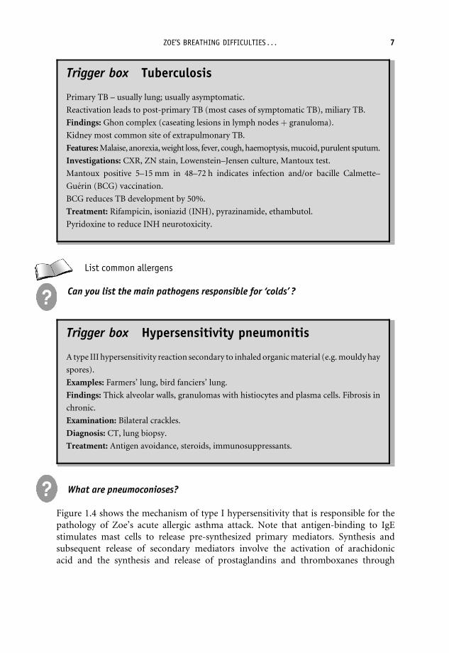
Trigger box Tuberculosis
Primary TB – usually lung; usually asymptomatic.
Reactivation leads to post-primary TB (most cases of symptomatic TB), miliary TB.
Findings: Ghon complex (caseating lesions in lymph nodesþ granuloma).
Kidney most common site of extrapulmonary TB.
Features: Malaise, anorexia, weight loss, fever, cough, haemoptysis, mucoid, purulent sputum.
Investigations: CXR, ZN stain, Lowenstein–Jensen culture, Mantoux test.
Mantoux positive 5–15 mm in 48–72 h indicates infection and/or bacille Calmette–
Guerin (BCG) vaccination.
BCG reduces TB development by 50%.
Treatment: Rifampicin, isoniazid (INH), pyrazinamide, ethambutol.
Pyridoxine to reduce INH neurotoxicity.
List common allergens
Can you list the main pathogens responsible for ‘colds’ ?
Trigger box Hypersensitivity pneumonitis
A type III hypersensitivity reaction secondary to inhaled organic material (e.g. mouldy hay
spores).
Examples: Farmers’ lung, bird fanciers’ lung.
Findings: Thick alveolar walls, granulomas with histiocytes and plasma cells. Fibrosis in
chronic.
Examination: Bilateral crackles.
Diagnosis: CT, lung biopsy.
Treatment: Antigen avoidance, steroids, immunosuppressants.
What are pneumoconioses?
Figure 1.4 shows the mechanism of type I hypersensitivity that is responsible for thepathology of Zoe’s acute allergic asthma attack. Note that antigen-binding to IgEstimulates mast cells to release pre-synthesized primary mediators. Synthesis andsubsequent release of secondary mediators involve the activation of arachidonicacid and the synthesis and release of prostaglandins and thromboxanes through
ZOE’S BREATHING DIFFICULTIES . . . 7

cyclooxygenase pathway and leukotrienes C4 and D4 (termed slow reacting substance ofanaphylaxis (SRS-A)) through the lipoxygenase pathway.
What is anaphylaxis? Can you describe the mechanisms underlying systemicanaphylaxis? How is this condition treated?
At the time of her diagnosis, Zoe had been given a skin prick test to confirm her allergicstatus. She was inoculated with a series of allergens including grass pollen and dust miteextracts. She got a classic wheal and flare reaction after 20 minutes but was surprisedwhen the reaction reappeared around 5 hours later. Immediate reactions occur withinminutes of allergen exposure and are mediated principally by the mast cell granulecontents (primary mediators). Some 5–8 hours after the immediate reaction hassubsided, a second reaction – the late-phase reaction – occurs due to the release ofadditional secondary mediators including cytokines. Tables 1.1 and 1.2 show the key
Ca2+
primary mediators, e.g. histamine, 5-HT.secondary mediators, e.g. prostaglandins.
allergenIgE antibodymast cell
granules
degranulation
+
prostaglandins leukotrienes
arachidonic acid
phospholipids
cyclo-oxygenase
lipoxgyenase
phospholipase A2
Ca2+
primary mediators, e.g. histamine, 5-HT.secondary mediators, e.g. prostaglandins.
allergenIgE antibodymast cell
granules
degranulation
++
prostaglandins leukotrienes
arachidonic acid
phospholipids
cyclo-oxygenase
lipoxgyenase
phospholipase A2
prostaglandins leukotrienes
arachidonic acid
phospholipids
cyclo-oxygenase
lipoxgyenase
phospholipase A2
Figure 1.4 Type 1 hypersensitivity�
Table 1.1 Key primary mediators and their effects
Mediator Characteristics and effects
Histamine Basic amine stored in granules of mast cells and basophils.
Contracts non-vascular smooth muscle, vasodilatation,
increased vascular permeability, enhanced mucus secretion,
prostaglandin secretion in the lung, chemokinesis
Serotonin Increased vascular permeability, smooth muscle contraction
ECF A Eosinophil chemotaxis
NCF-A Neutrophil chemotaxis
�Note that cross-linking of IgE by allergen leads to receptor cross-linkage. This leads to a transient elevation ofcAMP, activation of protein tyrosine kinases, methylation of membrane phospholipids and an influx of calciumwhich causes fusion of granules with plasma membrane and release of primary mediators into extracellularenvironment. Secondary mediators are synthesized.
8 CH1 THE RESPIRATORY SYSTEM

primary and secondary mediators that lead to the inflammatory reaction seen in type Ihypersensitivity.
Cytokines are small proteins (5–20 kDa) that are released from cells and act in asimilar way as hormones, affecting cellular behaviour. Cytokines allow cells of theimmune system to communicate with each other to modulate immune responses.Cytokines are important in mediating many different types of immune responses.Table 1.3 shows the functions of some key cytokines.
Table 1.2 Key secondary mediators and their effects
Mediator Characteristics and effects
Cytokines Stimulate and amplify Th2 cell responses (IL-4, IL-13),promote eosinophil production and activation (IL-3, IL-5,GM-CSF) and promote inflammation (TNF-a).
Prostaglandin E2 Causes vasodilatation and potentiates increased vascularpermeability produced by histamine.
Leukotrienes C4 and D4 Causes smooth muscle contraction, increased vascularpermeability and mucus secretion.
Platelet-activating factor Synthesized from phospholipid. Causes platelet andneutrophil activation, increased vascular permeabilityand smooth muscle contraction.
Major basic protein Triggers histamine release from mast cells.and eosinophil peroxidase Tissue damage.
Bradykinin Nonapeptide formed from kininogen. Causes vasodilatation,increased vascular permeability and stimulation of painnerve endings.
Th2, T helper 2; IL, interleukin; GM-CSF, granulocyte–macrophage colony-stimulating factor; TNF-a, tumournecrosis factor-a.
Table 1.3 Some key cytokines and their function
Cytokine Function
IL-1 Stimulates T- and B-cell proliferation and is a pyrogenIL-2 Stimulates T- and B-cell proliferation and activates natural killer cellsIL-3 Stimulates B memory cellsIL-4 Stimulates plasma cell formation, IgE synthesis and activates B cellsIL-5 Stimulates plasma cell secretion of IgA and IgM, stimulates B cells and eosinophilsIL-6 Induces B-cell differentiation into plasma cells and induces T-cell proliferation
and activationIFN-a Inhibits viral replicationIFN-b Inhibits viral replicationIFN-g Stimulates monocytes and macrophages and decreases viral replicationTNF-a Cytotoxic to tumour cells, cachexiaTNF-b Cytotoxic and increase phagocytosis
ZOE’S BREATHING DIFFICULTIES . . . 9

Describe the following terms as applied to cytokine action:
AutocrineParacrineEndocrineRedundancyAntagonismPleiotrophySynergy.
What are the four signs of inflammation?
Which cells are found in sites of acute and chronic inflammation?
Can you define the following: triggers, inducers, intrinsic and extrinsic asthma.
The ‘blue’ inhaler didn’t seem to work for Zoe and she knew what this meant. She washeading for another major asthma attack. Zoe called out to Mary around 7pm. Maryheard her and raced upstairs to find Zoe breathing rapidly, gasping for breath andwheezing. She was sitting at her desk, all hunched up; she could barely speak.
What is the normal respiratory rate for young adults?
Mary called out to John who took one look at Zoe and decided to take her to theEmergency Doctor. Since both John and Mary had been drinking, they had to call a taxi!Zoe was breathing with great difficulty. This distressing scene leads us to consider themechanics of breathing shown in Figure 1.5.
Note that during inspiration as the chest wall expands (external intercostals) and thediaphragm moves down the two pleurae are moved apart. This causes a more negative
riadellepxe
expiration(relaxed/domed diaphragm
with ribs moving downwards and inwards)
airdrawn
intrachea
rib
pleural fluid
lungs
diaphragm
spine
inspiration(contracted/flattened
diaphragm with ribs moving upwards and outwards)
Figure 1.5 The mechanism of breathing. Adapted from Mackean (1969), Introduction toBiology, 4th edition, John Murray, London, p.101.
10 CH1 THE RESPIRATORY SYSTEM

intrapleural pressure (subatmospheric) to develop. This increase in negative intra-pleural pressure overcomes the natural elasticity (elastic recoil) of the lungs and thesurface tension of the inner alveoli lining and the lungs inflate, the negative alveolarpressure drawing air into the lungs. Intercostal nerves innervate the intercostal musclesand the diaphragm is innervated by the phrenic nerve (C3, C4, C5).
Why is a head injury that causes damage to spinal cord above C3, C4 or C5potentially fatal?
The accessory muscles, sclani, sternocleidomastoids and pectoralis are used in forcedinspiration. Remember how Zoe is sitting during her acute asthma attack – she isutilizing her accessory muscles.
The diaphragm is the most important structure involved in breathing. In severeasthmatics, airway obstruction causes air trapping, which tends to hyperinflate thelungs. This causes a barrel-shaped thoracic cavity and a flattening of the diaphragm,which impairs its movement during breathing and leads to shortness of breath.
Some years later Max, Debbie’s boyfriend was rushed into the ED after being stabbed inthe chest. His left lung collapsed because air was getting into the pleural cavity and wasunable to leave because his shirt was stuck to his chest. This is called a tension pneumo-thorax and a chest tube had to be inserted to remove the trapped air and reinflate his lung.
Describe other types of pneumothorax.Define atelectasis.
Let us now consider the important lung feature of compliance. Compliance is a measureof how expandable the lungs are for a given change in pressure. Zoe’s lung compliance isnearly normal. On the other hand Ted, Grandma Irene’s brother living down the streetwho suffers from emphysema has an increased lung compliance. Figure 1.6 showscompliance in different conditions and how compliance can be calculated.
lung
vol
ume
‘V’ (
L)
fibrosis
emphysema
asthma
normal
CL = ∆VL/∆P
8
6
4
2
0
0 10 20 30 40transpulmonary pressure ‘P’ (cm H2O)
Figure 1.6 Compliance. Adapted from Berne and Levy (1996) Principles of Physiology, 3rdedition, Mosby
ZOE’S BREATHING DIFFICULTIES . . . 11
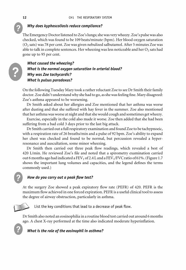
Why does kyphoscoliosis reduce compliance?
The Emergency Doctor listened to Zoe’s lungs; she was very wheezy. Zoe’s pulse was alsochecked, which was found to be 109 beats/minute (bpm). Her blood oxygen saturation(O2 sats) was 78 per cent. Zoe was given nebulized salbutamol. After 5 minutes Zoe wasable to talk in complete sentences. Her wheezing was less noticeable and her O2 sats hadgone up to 95 per cent.
What caused the wheezing?
What is the normal oxygen saturation in arterial blood?
Why was Zoe tachycardic?
What is pulsus paradoxus?
On the following Tuesday Mary took a rather reluctant Zoe to see Dr Smith their familydoctor. Zoe didn’t understand why she had to go, as she was feeling fine. Mary disagreed:Zoe’s asthma appeared to be worsening.
Dr Smith asked about her allergies and Zoe mentioned that her asthma was worseafter dusting and that she suffered with hay fever in the summer. Zoe also mentionedthat her asthma was worse at night and that she would cough and sometimes get wheezy.
Exercise, especially in the cold also made it worse. Zoe then added that she had beensuffering from a bad cold 3 days prior to the last big attack.
Dr Smith carried out a full respiratory examination and found Zoe to be tachypnoeic,with a respiration rate of 26 breaths/min and a pulse of 92 bpm. Zoe’s ability to expandher chest was checked and found to be normal, but percussion revealed a hyper-resonance and auscultation, some minor wheezing.
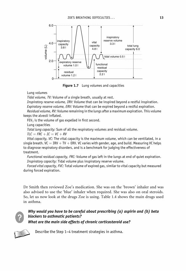
Dr Smith then carried out three peak flow readings, which revealed a best of420 L/min. He reviewed Zoe’s file and noted that a spirometry examination carriedout 6 months ago had indicated a FEV1 of 2.4 L and a FEV1/FVC ratio of 61%. (Figure 1.7shows the important lung volumes and capacities, and the legend defines the termscommonly used.)
How do you carry out a peak flow test?
At the surgery Zoe showed a peak expiratory flow rate (PEFR) of 420. PEFR is themaximum flow achieved in one forced expiration. PEFR is a useful clinical tool to assessthe degree of airway obstruction, particularly in asthma.
List the key conditions that lead to a decrease of peak flow.
Dr Smith also noted an eosinophilia in a routine blood test carried out around 6 monthsago. A chest X-ray performed at the time also indicated moderate hyperinflation.
What is the role of the eosinophil in asthma?
12 CH1 THE RESPIRATORY SYSTEM
Dr Smith then reviewed Zoe’s medication. She was on the ‘brown’ inhaler and wasalso advised to use the ‘blue’ inhaler when required. She was also on oral steroids.So, let us now look at the drugs Zoe is using. Table 1.4 shows the main drugs usedin asthma.
Why would you have to be careful about prescribing (a) aspirin and (b) betablockers to asthmatic patients?
What are the main side effects of chronic corticosteroid use?
Describe the Step 1–4 treatment strategies in asthma.
residualvolume 1.2 l
expiratory reserve volume 1.0 l
tidal volume 0.5 l
inspiratorycapacity
3.8 l
vital capacity
4.8 l
inspiratoryreserve volume
3.3 l
functional residualcapacity
2.2 l
total lungcapacity 6.0
l
6.0
4.0
2.0
0
volu
me
(L)
Figure 1.7 Lung volumes and capacities
Lung volumes
Tidal volume, TV: Volume of a single breath, usually at rest.
Inspiratory reserve volume, IRV: Volume that can be inspired beyond a restful inspiration.
Expiratory reserve volume, ERV: Volume that can be expired beyond a restful expiration.
Residual volume, RV: Volume remaining in the lungs after a maximum expiration. This volumekeeps the alveoli inflated.
FEV1 is the volume of gas expelled in first second.
Lung capacities
Total lung capacity: Sum of all the respiratory volumes and residual volume.
TLC ¼ FRC þ IC ¼ VC þ RV
Vital capacity, VC: The vital capacity is the maximum volume, which can be ventilated, in asingle breath. VC ¼ IRVþ TVþ ERV. VC varies with gender, age, and build. Measuring VC helpsto diagnose respiratory disorders, and is a benchmark for judging the effectiveness oftreatment.
Functional residual capacity, FRC: Volume of gas left in the lungs at end of quiet expiration.
Inspiratory capacity: Tidal volume plus inspiratory reserve volume.
Forced vital capacity, FVC: Total volume of expired gas, similar to vital capacity but measuredduring forced expiration.
ZOE’S BREATHING DIFFICULTIES . . . 13

Grandma’s bad chest . . .
With the Christmas and New Year holidays over, the Sickalotts returned to their usualroutines. Zoe’s wheezing was much less pronounced. Things however took a turn for theworse in the bungalow down the road.
Grandma Irene suffered from chronic obstructive pulmonary disease (COPD) andwas under the care of Dr Blunt at Hope Hospital. Dr Blunt was also looking after Ted,Irene’s brother who was also suffering from COPD. For the last few months GrandmaIrene was finding it harder and harder to breathe. But not being one to complain, shehad carried on with her routines, which mainly centred on looking after Albert.However, her cough was getting worse and she became breathless on minimal exertion.During Mary’s daily visit, on the 8th of January, Albert told Mary that Irene’s coughingwas getting a lot worse and that she might have a raised temperature. Mary wanted totake Irene to see Dr Smith. Irene declined, stating that she always got like this in thewinter. It was no big deal.
Around midnight that same day, John and Mary were woken up by the telephoneringing incessantly. On the other end of the line was a rather shaky Albert saying thatIrene had taken a turn for the worse.
John and Mary rushed over to the bungalow. Irene was sitting in her chair in theliving room, attached to her oxygen, with Albert standing next to her and lookingvery distressed. John called an ambulance and the paramedics arrived 10 minuteslater. They found Irene gasping for breath. She looked cyanotic and was using heraccessory muscles for breathing. Irene was given nebulized salbutamol and taken tohospital.
Table 1.4 The main drugs used in asthma
Beta agonists Salbutamol, albuterol, salmeterol (long acting). Theyrelax airway smooth muscles (via b2 adrenoceptors).Adverse effect: tachycardia (via b1 adrenoceptors).
Corticosteroids Beclomethasone, prednisolone. They prevent productionof leukotrienes from arachidonic acid by blockingphospholipase A2. Drug of choice in status asthmaticus
Antileukotrienes Zileuton – blocks synthesis of lipoxygenaseZafirlukast – blocks leukotriene receptors
Methylxanthines Theophylline – bronchodilation by inhibitingphosphodiesterase involved in degrading cAMP
Muscarinic antagonists Ipratropium – blocks muscarinic receptors preventingbronchoconstriction
Cromolyn (cromoglycate) Prevents release of mediators from mast cells. Effectiveonly as prophylactic
Anti IgE-monoclonal antibody Neutralizes IgE
14 CH1 THE RESPIRATORY SYSTEM

At the hospital Irene was examined by one of the duty doctors. He noted themild cyanosis, the jugular venous distension (JVD) and end expiratory wheezing. He alsonoted high-pitched bronchial sounds on the left side, crackles and dullness to percussion.Irene’s chest showed moderate hyperinflation and her nicotine-stained fingers were notedto be clubbed. Her respiration rate was 45 bpm, her BP was 160/95 and she had atemperature of 39�C. Irene was responsive only to repeated verbal commands. Hertreatment started with nebulized salbutamol, i.v. steroids and i.v. ampicillin.
What is JVD?
What is the most likely cause of Irene’s increased temperature?
What is the significance of dullness to percussion, high pitched bronchialsounds and finger clubbing?
What is the significance of mentioning Irene’s nicotine stained fingers?
Why was Irene treated with ampicillin? What is the mechanism of action ofampicillin?
The duty doctor ordered some arterial blood gases (ABGs), which indicated hypoxaemia,hypercapnia (hypercarbia) and mild respiratory acidosis. A chest X-ray was also ordered.
You should be able to explain the mechanisms involved in respiratory acidosis andalkalosis. Respiratory acidosis is seen when arterial PaCO2 rises above normal (e.g. inIrene’s COPD when ventilation was impaired). Conversely, overbreathing, such asduring Zoe’s acute asthma attack, can cause more CO2 to be blown off, giving rise toa respiratory alkalosis. Table 1.5 shows the causes of respiratory and metabolic acidosisand alkalosis and also shows the compensatory reactions.
Table 1.5 Acidosis and alkalosis
Condition Causes Compensatory mechanism
Respiratory acidosis Increased PaCO2 and decreasedpH due to hypoventilationcaused by any lung,neuromuscular or physicalcause of respiratory failure.
Increased renal excretion ofHþ ions and increasedreabsorption of HCO3
� ions.
Respiratory alkalosis Decreased PaCO2 and increasedpH due to hyperventilation.
Decreased renal excretion ofHþ ions and decreasedreabsorption of HCO3
� ions.
Metabolic acidosis Decreased HCO3� ion concentration
and decreased pH caused byexcessive HCO3
� ion loss orincreased Hþ production.
Hyperventilation to increaseCO2 excretion and reducecarbonic acid concentration.
Metabolic alkalosis Increased HCO3� ion concentration
and increased pH caused byexcessive Hþ ion loss.
Hypoventilation to reduce CO2
excretion and increasecarbonic acid concentration.
GRANDMA’S BAD CHEST . . . 15

What are the typical arterial blood gas findings in (a) acute asthma and (b)acute COPD?
List other causes that lead to respiratory acidosis and alkalosis.
By next morning, Irene was much better. The doctor noted her sputum pot and requ-ested a sputum culture. He also requested lung function tests which revealed a FEV1 of1.1 L and a FEV1/FVC ratio of 43 per cent.
Irene, Ted and Zoe suffer from obstructive lung diseases. In these diseases theFEV1/FVC ratio is <80 per cent, in contrast to restrictive diseases where the ratio is>80 per cent.
Trigger box Key obstructive and restrictive lung diseases
1. Obstructive lung diseases
(FEV1/FVC <80%; increased TLC; increased FRC; increased RV).
Chronic bronchitis.
Emphysema.
Asthma.
Bronchiectasis.
2. Restrictive lung diseases
(FEV1/FVC >80%; decreased VC; decreased TLC).
Sarcoidosis.
Diffuse interstitial pulmonary fibrosis.
Scoliosis.
Neuromuscular disease (e.g. polio).
Myasthenia gravis.
Trigger box Diffuse interstitial pulmonary fibrosis
Most common restrictive lung disease.
More common in the elderly.
Pathology: Thick alveolar walls – cystic spaces.
Findings: Shallow rapid breathing, cough.
Reticular pattern/honeycomb pattern on CXR (in severe disease).
Treatment: Supportive, steroids.
What is the aetiology of this disease?
16 CH1 THE RESPIRATORY SYSTEM
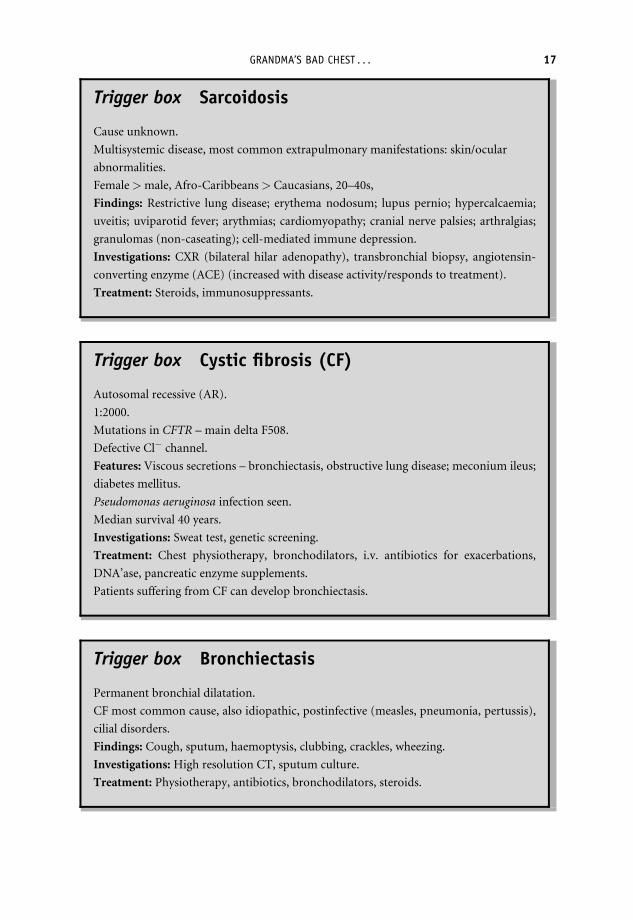
Trigger box Sarcoidosis
Cause unknown.
Multisystemic disease, most common extrapulmonary manifestations: skin/ocular
abnormalities.
Female>male, Afro-Caribbeans> Caucasians, 20–40s,
Findings: Restrictive lung disease; erythema nodosum; lupus pernio; hypercalcaemia;
uveitis; uviparotid fever; arythmias; cardiomyopathy; cranial nerve palsies; arthralgias;
granulomas (non-caseating); cell-mediated immune depression.
Investigations: CXR (bilateral hilar adenopathy), transbronchial biopsy, angiotensin-
converting enzyme (ACE) (increased with disease activity/responds to treatment).
Treatment: Steroids, immunosuppressants.
Trigger box Cystic fibrosis (CF)
Autosomal recessive (AR).
1:2000.
Mutations in CFTR – main delta F508.
Defective Cl� channel.
Features: Viscous secretions – bronchiectasis, obstructive lung disease; meconium ileus;
diabetes mellitus.
Pseudomonas aeruginosa infection seen.
Median survival 40 years.
Investigations: Sweat test, genetic screening.
Treatment: Chest physiotherapy, bronchodilators, i.v. antibiotics for exacerbations,
DNA’ase, pancreatic enzyme supplements.
Patients suffering from CF can develop bronchiectasis.
Trigger box Bronchiectasis
Permanent bronchial dilatation.
CF most common cause, also idiopathic, postinfective (measles, pneumonia, pertussis),
cilial disorders.
Findings: Cough, sputum, haemoptysis, clubbing, crackles, wheezing.
Investigations: High resolution CT, sputum culture.
Treatment: Physiotherapy, antibiotics, bronchodilators, steroids.
GRANDMA’S BAD CHEST . . . 17

Irene’s chest X-ray showed left lower lobe consolidation suggesting lobar pneumonia.This was confirmed by the sputum culture, which revealed Streptococcus pneumoniae.So, Irene was suffering from a chest infection with underlying COPD.
People like Irene who suffer from COPD also suffer from frequent pulmonary infe-ctions like pneumonia. The pathogens causing pneumonia depend on the age of thepatient, as shown in Table 1.6.
Trigger box Pneumonia
Inflammation of lung tissue.
Findings: Pyrexia, cough, sputum, pleurisy, dyspnoea, consolidation, pleural rub, pleural
effusion, confusion (elderly).
Blood cultures positive in 20% cases even if sputum is negative: indicates poor prognosis.
Risk factors: Smoking, alcoholism, lung disease, immunosuppression, chronic disease.
Investigations: CXR, FBC (WBC > 15� 109/L suggests bacterial infection), cold agglu-
tinins in Mycoplasma pneumoniae, raised urea and hypoalbunimia¼ severe pneumonia,
arterial blood gases (ABG) (PaO2 <8 kPa/hypercarbia¼ severe pneumonia).
Main pathogens
1. Community acquired: Streptococcus pneumoniae (70%). Mycoplasmapneumoniae. (15%), Chlamydia spp. (7%).
2. Hospital acquired: Gram-negative bacteria (50%).
Treatment:
Community acquired – amoxicillin.
Penicillin allergics – erythromycin or azithromycin.
Staphylococcus aureus – flucloxacillin.
Physiotherapy.
Complications: Lung abscess, empyema.
Mortality 25% in elderly.
Table 1.6 Age of patient and the pathogens causing pneumonia
Age Pathogens
Neonates Group B streptococcus, E. coli.Children Respiratory syncytial virus, Mycoplasma pneumoniae,
Chlamydia pneumoniae, Streptococcus pneumoniaeYoung adults Mycoplasma pneumoniaeOlder adults & elderly Streptococcus pneumoniae
18 CH1 THE RESPIRATORY SYSTEM

Irene has chronic bronchitis, her brother Ted has emphysema. These diseases are part ofa group of conditions (together with chronic asthma) that make up COPD. Most COPDsufferers are smokers.
Trigger box COPD
Decreased FEV1; decreased FEV1/FVC; increased TLC; increased RV.
Chronic bronchitis/emphysema.
Most are smokers.
Chronic bronchitis (blue bloater):
Features: Productive cough >3/12 for 2 consecutive years, hypercarbia, hypoxia,
hyper inflated lungs.
Emphysema (pink puffer):
Centriolobular (smoking); panlobular (a1 antitrypsin deficiency).
Features: Dyspnoea, decreased breath sounds, hypercarbia, hypoxia, bullae.
Treatment: O2, beta agonists, ipratropium, steroids, antibiotics.
Can you describe the significance of a1-antitrypsin to emphysema?
Smokers are also at a high risk of developing lung cancer. Study the Trigger Boxon lung cancer.
Trigger box Lung cancer
Most common malignant tumour.
Types:
1. Non small cell (70–80%)
Squamous cell carcinoma (40%), large cell carcinoma (25%) adenocarcinoma (10%).
2. Small cell (20–30%).
Causes: Smoking, asbestos.
Features: Cough, chest pain, hemoptysis, finger clubbing, Pancoast’s tumour, Horner’s
syndrome, superior vena cava (SVC) obstruction.
GRANDMA’S BAD CHEST . . . 19
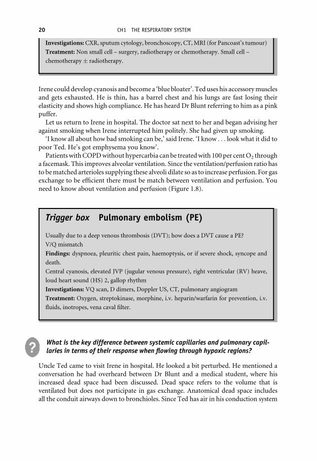
Investigations: CXR, sputum cytology, bronchoscopy, CT, MRI (for Pancoast’s tumour)
Treatment: Non small cell – surgery, radiotherapy or chemotherapy. Small cell –
chemotherapy� radiotherapy.
Irene could develop cyanosis and become a ‘blue bloater’. Ted uses his accessory musclesand gets exhausted. He is thin, has a barrel chest and his lungs are fast losing theirelasticity and shows high compliance. He has heard Dr Blunt referring to him as a pinkpuffer.
Let us return to Irene in hospital. The doctor sat next to her and began advising heragainst smoking when Irene interrupted him politely. She had given up smoking.
‘I know all about how bad smoking can be,’ said Irene. ‘I know . . . look what it did topoor Ted. He’s got emphysema you know’.
Patients with COPD without hypercarbia can be treated with 100 per cent O2 througha facemask. This improves alveolar ventilation. Since the ventilation/perfusion ratio hasto be matched arterioles supplying these alveoli dilate so as to increase perfusion. For gasexchange to be efficient there must be match between ventilation and perfusion. Youneed to know about ventilation and perfusion (Figure 1.8).
Trigger box Pulmonary embolism (PE)
Usually due to a deep venous thrombosis (DVT); how does a DVT cause a PE?
V/Q mismatch
Findings: dyspnoea, pleuritic chest pain, haemoptysis, or if severe shock, syncope and
death.
Central cyanosis, elevated JVP (jugular venous pressure), right ventricular (RV) heave,
loud heart sound (HS) 2, gallop rhythm
Investigations: VQ scan, D dimers, Doppler US, CT, pulmonary angiogram
Treatment: Oxygen, streptokinase, morphine, i.v. heparin/warfarin for prevention, i.v.
fluids, inotropes, vena caval filter.
What is the key difference between systemic capillaries and pulmonary capil-laries in terms of their response when flowing through hypoxic regions?
Uncle Ted came to visit Irene in hospital. He looked a bit perturbed. He mentioned aconversation he had overheard between Dr Blunt and a medical student, where hisincreased dead space had been discussed. Dead space refers to the volume that isventilated but does not participate in gas exchange. Anatomical dead space includesall the conduit airways down to bronchioles. Since Ted has air in his conduction system
20 CH1 THE RESPIRATORY SYSTEM

and lots of destroyed alveoli, which do not participate in gas exchange, he has lots ofdead space! Physiological dead space includes alveoli that are ventilated but notperfused, which occurs in patients suffering with pulmonary emboli.
Note: alveolar ventilation rate¼ (tidal vol – dead space)� respiratory rate
Dead space is increased in artificial respiration when the patient is connectedto tubing. What physiological effects will this have on the patient’s tidalvolume and respiratory rate?
After talking about dead space, Dr Blunt had gone on to describe things that caused Tedto experience even more fear.
‘The loss of functional alveoli in emphysema,’ he had said, ‘will reduce diffusioncapacity and therefore impair gas exchange. You can also see this in pulmonaryoedema.’
normalVA/Q = 1
A
reduced perfusionVA/Q > 1e.g. PE
B
reduced ventilationVA/Q < 1
e.g. COPD
C
reduced ventilation andvasoconstriction ofpulmonary artery
VA/Q = 1
D
increased ventilation andvasodilatation ofpulmonary artery
VA/Q = 1
E
Figure 1.8 Ventilation and perfusion. ‘D’ and ‘E’ shows how vasodilatation or vasoconstrictioncorrects a VA/Q mismatch
The V/Q ratio¼ alveolar ventilation rate/pulmonary blood flow
If there is no ventilation, Va/Q¼ 0
For example you get perfusion without ventilation when an alveolus is full of liquid as insevere pneumonia. This will reduce arterial PaO2.
If there is no perfusion, Va/Q¼1Here the alveoli are ventilated but not perfused, for example due to a pulmonary embolus.
Remember pulmonary vessels constrict in poorly ventilated regions.
A pulmonary embolus is a medical emergency and causes a severe V/Q mismatch.
GRANDMA’S BAD CHEST . . . 21

Figure 1.9 shows changes in PaO2 and PaCO2 as blood moves from metabolicallyactive tissues to the lungs and back.
What drives the movement of these gases from tissue to venous blood, fromvenousblood toalveoli, fromalveoli toarterial bloodand fromarterial blood totissue?
Why is PaO2 in systemic arterial blood lower than that in pulmonary veins?
Which hormone increases RBC formation? Where is it produced?
List the causes of pulmonary oedema.
How is diffusion capacity measured?
What conditions reduce diffusion capacity?
Zoe’s oxygen saturation at the height of her asthma attack was 78 per cent. Aftertreatment it rose to 98 per cent. You need to know about haemoglobin (Hb) and O2
saturation curves. O2 carrying capacity of the blood is proportional to the Hb concen-tration in the alveoli. Hb has four O2 binding sites. The shape of the dissociation curve is
atmospheric air:PO2 = 160 mmHgPCO2 = 0.03 mmHg
expired air:PO2 = 120 mmHgPCO2 = 28 mmHg
alveolar air:PO2 = 104 mmHgPCO2 = 40 mmHg
pulmonary vein blood:PO2 = 104 mmHgPCO2 = 40 mmHg
PP
precapillary blood:O2 = 100 mmHg
CO2 = 40 mmHg
postcapillary blood:PO2 = 40 mmHgPCO2 = 45 mmHg
pulmonary artery blood:PO2 = 40 mmHgPCO2 = 45 mmHg
tissues:PO2 = 40 mmHgPCO2 = 45 mmHg
O2
Figure 1.9 Gas exchange. Adapted from Sherwood (2004) Human Physiology from Cells toSystem, 5th edition, Thomson, Brooks/Cole.
22 CH1 THE RESPIRATORY SYSTEM
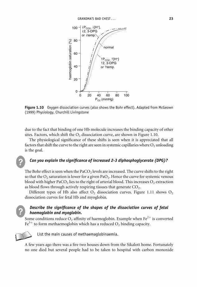
due to the fact that binding of one Hb molecule increases the binding capacity of othersites. Factors, which shift the O2 dissociation curve, are shown in Figure 1.10.
The physiological significance of these shifts is seen when it is appreciated that allfactors that shift the curve to the right are seen in systemic capillaries where O2 unloadingis the goal.
Can you explain the significance of increased 2-3 diphosphoglycerate (DPG)?
The Bohr effect is seen when the PaCO2 levels are increased. The curve shifts to the rightso that the O2 saturation is lower for a given PaO2. Hence the curve for systemic venousblood with higher PaCO2 lies to the right of arterial blood. This increases O2 extractionas blood flows through actively respiring tissues that generate CO2.
Different types of Hb also affect O2 dissociation curves. Figure 1.11 shows O2
dissociation curves for fetal Hb and myoglobin.
Describe the significance of the shapes of the dissociation curves of fetalhaemoglobin and myoglobin.
Some conditions reduce O2 affinity of haemoglobin. Example when Fe2þ is convertedFe3þ to form methaemoglobin which has a reduced O2 binding capacity.
List the main causes of methaemaglobinaemia.
A few years ago there was a fire two houses down from the Sikalott home. Fortunatelyno one died but several people had to be taken to hospital with carbon monoxide
100
80
60
40
20
0
haem
oglo
bin
satu
ratio
n (%
)
40 60 80 1000 20 PO2 (mmHg)
normal
PCO2, [H+],
2, 3-DPGor temp.
PCO2, [H+],2, 3-DPG
or temp.
Figure 1.10 Oxygen dissociation curves (also shows the Bohr effect). Adapted from McGeown(1999) Physiology, Churchill Livingstone
GRANDMA’S BAD CHEST . . . 23

poisoning. CO binds with greater affinity to haemoglobin than O2 and forms carbox-yhemoglobin, as seen in Figure 1.12.
Why do patients suffering from CO poisoning appear a pink - red colour?
Let us now consider CO2 transport.CO2 is transported in three forms:
1. As HCO3 (60–70 per cent). (See Figure 1.13)
2. In carbamino groups - mostly binding to Hb (20–30 per cent).
3. As dissolved gas (10 per cent).
COHb75%COHb50%COHb25%COHb10%COHb0%
100
80
60
40
20
0
O2
satu
ratio
n of
ava
ilabl
e ha
emog
lobi
n (%
)
0 20 40 60 80 100PO2 (mmHg)
Figure 1.12 The effects of carbon monoxide on the oxygen carrying capacity of blood. Adaptedfrom www.coheadquarters.com/cohaldane1.htm
myoglobin
etal haemoglobinf
haemoglogindulta
100
80
60
40
20
0
O2
satu
ratio
n (%
)
0 20 40 60 80 100PO2 (mmHg)
Figure 1.11 Dissociation curves for different oxygen carriers. Adapted from McGeown (1999)Physiology, Churchill Livingstone
24 CH1 THE RESPIRATORY SYSTEM

What is the chloride shift
CO2 dissociation curves (not shown) show higher CO2 concentrations at a given PaCO2
when compared to the situation in the O2 dissociation curve. This is because CO2 ismore soluble. Also when comparing to the O2 dissociation curve, CO2 has a narrowerphysiological range and no plateau.
The Haldane effect describes how the CO2 dissociation curve shifts down and to theleft when the PaO2 is increased. Hence the CO2 concentration drops at a given PaCO2.The Haldane effect increases the uptake of CO2 from respiring tissues. The Haldaneeffect is due to deoxygenated Hb being able to carry more CO2 in the carbamino formand because it is a more effective pH buffer than oxyhaemoglobin. The Haldane effect isshown in Figure 1.14.
Whilst in hospital Ted found out that someone in the next room was having an eventougher time. He overheard Dr Blunt explaining- to his student – that ‘‘this fellow was
deoxygenatedvenous blood
oxygenated arterial blood
dissolved CO2
70
60
50
40
30
20
10
040 50 60 700 10 20 30
PCO2 (mmHg)
CO
2(m
l/dl)
Figure 1.14 The Haldane effect. Adapted from McGeown (1999) Physiology, ChurchillLivingstone
OC 2 H + 2O H2 OC 3 H+ OCH + 3−
H+ bH+ − bHH
Cl−CO2 chlorideshift
cinobracesardyhna
Figure 1.13 Carbon dioxide metabolism in the erythrocyte. Adapted from Ganong (1991)Review of Medical Physiology,15th edition, Appleton & Lange
GRANDMA’S BAD CHEST . . . 25
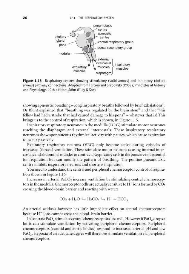
showing apneustic breathing – long inspiratory breaths followed by brief exhalations’’.Dr Blunt explained that ‘‘breathing was regulated by the brain stem’’ and that ‘‘thisfellow had had a stroke that had caused damage to his pons’’ – whatever that is! Thisbrings us to the control of respiration, which is shown, in Figure 1.15.
Inspiratory respiratory neurones in the medulla (DRG) stimulate motor neuronesreaching the diaphragm and external intercostals. These inspiratory respiratoryneurones show spontaneous rhythmical activity with pauses, which cause expirationto occur passively.
Expiratory respiratory neurons (VRG) only become active during episodes ofincreased (forced) ventilation. These stimulate motor neurons causing internal inter-costals and abdominal muscles to contract. Respiratory cells in the pons are not essentialfor respiration but can modify the pattern of breathing. The pontine pneumotaxiccentre inhibits inspiratory neurons and shortens inspiration.
You need to understand the central and peripheral chemoreceptor control of respira-tion shown in Figure 1.16.
Increases in arterial PaCO2 increase ventilation by stimulating central chemorecep-tors in the medulla. Chemoreceptor cells are actually sensitive to Hþ ions formed by CO2
crossing the blood–brain barrier and reacting with water:
CO2 þH2O ! H2CO3 ! Hþ þHCO�3
An arterial acidosis however has little immediate effect on central chemoreceptorsbecause Hþ ions cannot cross the blood–brain barrier.
In contrast PaO2 stimulate central chemoreceptors less well. However if PaO2 drops alot it can stimulate ventilation by activating peripheral chemoreceptors. Peripheralchemoreceptors (carotid and aortic bodies) respond to increased arterial pH and lowPaO2. Hypoxia of an adequate degree will therefore stimulate ventilation via peripheralchemoreceptors.
pons
apneusticcentre
pneumotaxiccentre
pituitarygland
dorsal respiratory group
ventral respiratory group
almedul
expiratorymuscles
externalintercostalmuscles
diaphragm
inspiratorymuscles
Figure 1.15 Respiratory centres showing stimulatory (solid arrows) and inhibitory (dottedarrows) pathway connections. Adapted from Tortora and Grabowski (2003), Principles of Antomyand Physiology, 10th edition, John Wiley & Sons
26 CH1 THE RESPIRATORY SYSTEM

Irene has hypoxic drive because her PaCO2 is chronically elevated due to her COPD.Her central chemoreceptors have become unresponsive and her oxygen sensitiveperipheral chemoreceptors now respond to lowered PaO2.
You have to be careful giving her 100 per cent O2 because this can remove the stimulusfor her breathing.
What happens if the spinal cord in transected just below the pons?
Describe the main changes seen in respiratory physiology in (a) high alti-tude, (b) vigorous exercise
arterial PCO2
PCO2 + pH in cerebrospinal fluid
centralchemoreceptorsof the medulla
(70% of response)
peripheralchemoreceptors:aortic + carotid bodies (30% of
response)
ventilation (removing CO2)
respiratory centresmedullary
arterial Pnormalized CO2 and pH
arterial PO2
centralchemoreceptors
peripheralchemoreceptors:
ventilation
respiratory centresmedullary
arterial Pnormalized O2
(no effect)
Figure 1.16 Central and peripheral chemoreceptor control of breathing
GRANDMA’S BAD CHEST . . . 27
