the retinoblastoma protein associates with the …genesdev.cshlp.org/content/7/4/555.full.pdfthe...
TRANSCRIPT
The retinoblastoma protein associates with the protein phosphatase type 1 catalytic subunit Tim Durfee, 1'2's Kathleen Becherer, 3 Phang-Lang Chen, 1 Shiou-Hwei Yeh, ~ Yanzhu Yang, ~ April E. Kilburn, a Wen-Hwa Lee, 1 and Stephen J. Elledge 3-s
~Center for Molecular Medicine/Institute of Biotechnology, University of Texas Health Science Center at San Antonio, San Antonio, Texas 78245 USA; 2Graduate Program in Molecular Pathology, University of Califomia-San Diego, LaJolla, California 92093 USA; 3Department of Biochemistry and 4Institute for Molecular Genetics, Baylor College of Medicine, Houston, Texas 77030 USA
The retinoblastoma protein {pll0 nB) interacts with many cellular proteins in complexes potentially important for its growth-suppressing [unction. We have developed and used an improved version of the yeast two-hybrid system to isolate human cDNAs encoding proteins able to bind pl l0 RB. One clone encodes a novel type 1 protein phosphatase catalytic subunit (PP-la2), which differs from the originally defined PP-lc~ by an amino-terminal l 1-amino-acid insert. In vitro-binding assays demonstrated that PP-lc~ isoforms preferentially bind the hypophosphorylated form of p l l 0 RB. Moreover, similar p l l0 RB sequences are required for binding PP-lc~2 and SV40 large T antigen. Cell cycle synchrony experiments revealed that this association occurs from mitosis to early Gv The implications of these findings on the regulation of both proteins are discussed.
[Key Words: Two-hybrid system; protein-protein interaction; retinoblastoma; protein phosphatase 1; cell-cycle regulation]
Received December 18, 1992; revised version accepted February 9, 1993.
The retinoblastoma susceptibility gene (Rb) was the first tumor suppressor gene to be cloned and characterized (Friend et al. 1986; Lee et al. 1987a). Inactivation of both alleles of this gene has been found in all retinoblastomas examined, and additional studies have implicated Rb mutations in the development of a variety of tumors (for review, see Bookstein and Lee 19911. Introduction of a wild-type copy of this gene into Rb- tumor cells sup- presses their tumorigenicity in nude mice (Huang et al. 1988; Bookstein et al. 1990; Takahashi et al. t9911, pro- vidmg further evidence that the cloned gene has properties consistent with those predicted for a tumor suppressor.
The Rb protein {p110 RB) is a nuclear protein of 110 kD [Lee et al. 1987bl, and is phosphorylated on both serine and threonine residues (Buchkovich et al. 1989; Ludlow et al. 1989; Shew et al. 1989). These modifications are thought to be important regulators of p110 RB and have been shown to occur in a cell cycle-dependent manner (Buchkovich et al. 1989; Chen et al. 1989; DeCaprio et al. 1989}. Cells in early G1 phase of the cycle contain exclusively unphosphorylated or underphosphorylated p l l 0 ~ . At an as yet undefined point later in GI, the protein is hyperphosphorylated [pp110 r~) and exists in this state until M phase, although the level of phospho- rylation does fluctuate slightly. Several studies have im-
SCorresponding authors.
plicated the cyclin-dependent kinase (cdkt family of ki- nases as enzymes responsible for this phosphorylation {Lees et al. 1991; Lin et al. 1991). Dephosphorylation of pp l l0 gB occurs in mitosis, probably initiated during anaphase [Ludlow et al. 1993). Recent studies have shown that both type I and 2A protein phosphatases can dephosphorylate pp l l0 RB in vitro, and strongly suggest that type 1 protein phosphatase is critical for this event in vivo [Alberts et al. 1993; Ludlow et al. 1993}.
The mechanism by which pl I0 m~ suppresses tumori- genicity remains unknown. However, a possible expla- nation emerged with the discovery that p l l 0 Rs physi- cally associates with oncoproteins of many DNA tumor viruses, namely, the adenovirus EIA protein {Whyte et al. 1988), the SV40 T antigen {DeCaprio et al. 19881, and the E7 protein of human papillomavirus 16 (HPVl61 (Dy- son et al. 1989}. p110 ~ sequences required for these in- teractions were mapped to two large domains in the car- boxy-terminal portion of the protein (Hu et al. 1990; Huang et al. 19901. These domains are affected in all known naturally occurring mutant Rb proteins, suggest- ing an important role for this region of the protein in tumor suppression (for review, see Bookstein and Lee 1991}. In addition, T antigen specifically binds the un- phosphorylated form of the Rb protein, implying that this isoform is important in cell growth control [Ludlow et al. 19891. One possibility, by analogy to the viral pro-
GENES & DEVELOPMENT 7:555-569 �9 1993 by Cold Spring Harbor Laboratory Press ISSN 0890-9369/93 $5.00 555
Cold Spring Harbor Laboratory Press on March 1, 2020 - Published by genesdev.cshlp.orgDownloaded from
Durfee et al.
teins, is that this region of p 110 Ra associates with cellu- lar factors and, through these complexes, asserts its tu- mor-suppressing function. It has been shown that the carboxy-terminal half of p 110 RB is capable of complexing with a variety of cellular proteins using Rb protein affin- ity columns (Kaelin et al. 1991; Lee et al. 1991). Subse- quently, using a protein-screening method, a number of potential pll0ga-binding proteins have been identified; these include RBP-1 and RBP-2 (Defeo-Jones et al. 1991), a 46-kD protein (Huang et al. 1991), the E2F transcrip- tion factor (Helin et al. 1992; Kaelin et al. 1992; Shah et al. 1992), and several other proteins (Shah et al. 1992}. The c-myc and N-myc proteins {Rustgi et al. 1991), the Cdc2 kinase (Hu et al. 1992}, and the activating tran- scription factor 2 (ATF-2) (Kim et al. 1992) were also identified as likely p110RB-associating proteins by the investigation of logical candidates in vitro. However, it is likely that many unknown targets of Rb interaction remain, and a thorough understanding of Rb function requires their identification.
To further our understanding of the role of Rb in tu- mor suppression, an in vivo strategy was employed to identify human proteins capable of physically associat- ing with p110 RB. The yeast two-hybrid system devised by Fields and Song (1989) was chosen to provide a phys- iological environment in which to detect potential inter- actions involving the Rb protein. Recently, this system has been used to screen eDNA libraries for clones encod- ing proteins capable of binding to a protein of interest (Chien et al. 1991; Dalton and Treisman 1992; Yang et al. 1992). Here, we describe several improvements to this approach and the application of this modified technique to isolate eDNA clones encoding proteins that interact with the carboxy-terminal portion of p 110 RB. One clone
isolated in this screen encodes a novel protein phos- phatase type 1 catalytic subunit (PP-lct2). The type 1 class of serine/threonine phosphatases is important for many cellular processes, including cell cycle regulation (for review, see Cohen 1989; Cyert and Thomer 1989). The cloning and characterization of this phosphatase is described, as well as its association with p110 RB and the possible regulatory implications of this interaction.
R e s u l t s
S t r a t e g y
To screen for cDNAs encoding proteins able to interact with p110 RB, the general scheme outlined in Figure 1A was employed. First, two sets of fusion proteins are con- structed; one generates a hybrid between sequences for the DNA-binding domain of the yeast transcription fac- tor Gal4 (amino acids 1-147; Keegan et al. 1986) and a portion of the Rb protein. A second expression plasmid contains sequences for the Gal4 activation domain II (amino acids 768-881; Ma and Ptashne 1987a}, fused to a eDNA library generated from human lymphocytes. As first demonstrated with Gal4--Gal80 interactions {Ma and Ptashne 1988} and later generalized by Fields and Song (1989), if the two proteins expressed in yeast are able to interact, the resulting complex will regain the ability to activate transcription from promoters contain- ing Gal4-binding sites, upstream activating sequence from G A L l (UASG).
A new yeast strain, Y153, has been constructed that provides a dual selection system to efficiently screen eDNA expression libraries for clones interacting with a
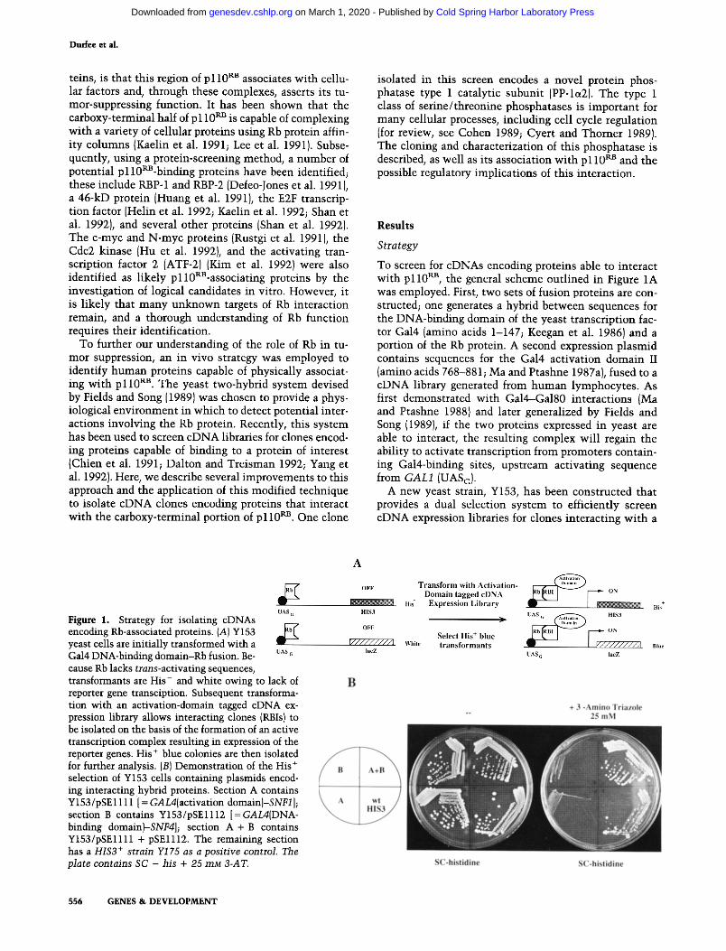
Figure 1. Strategy for isolating cDNAs encoding Rb-associated proteins. (A) Y153 yeast cells are initially transformed with a Gal4 DNA-binding domain-Rb fusion. Be- cause Rb lacks trans-activating sequences, transformants are His- and white owing to lack of reporter gene transciption. Subsequent transforma- tion with an activation-domain tagged cDNA ex- pression library allows interacting clones (RBIs) to be isolated on the basis of the formation of an active transcription complex resulting in expression of the reporter genes. His + blue colonies are then isolated for further analysis. (BI Demonstration of the His + selection of Y153 cells containing plasmids encod- ing interacting hybrid proteins. Section A contains Y153/pSE1111 [ = GAL4(activation domain)-SNF1 ]; section B contains Y153/pSE1112 [ = GAL4iDNA- binding domain~SNF4]; section A + B contains Y153/pSE1111 + pSE1112. The remaining section has a HIS3 + strain Y175 as a posi t ive control. The plate contains SC - his + 25 mM B-AT.
556 GENES & DEVELOPMENT
Cold Spring Harbor Laboratory Press on March 1, 2020 - Published by genesdev.cshlp.orgDownloaded from
The retinob|astoma protein and P P - l a
protein of interest. The strain carries two chromosomal- ly located reporter genes whose expression is regulated by Gal4. First, the Escherichia coli lacZ gene is under the control of the GALl promoter, and its usefulness in this system has been described (Fields and Song 1989}. A sec- ond reporter, the selectable HIS3 gene, was chosen be- cause very low levels of the enzyme [imidazole glycerol phosphate (IGP) dehydratase] are required for prototro- phy. To provide Gal4 control, the HIS3 regulatory se- quences have been replaced by the GALl UASG. Because Y153 is deleted for gal4 (and its negative regulator gal80), expression of both reporters should be off in the absence of exogenous Gal4. However, the GALI-HIS3 fusion has residual HIS3 expression sufficient to allow growth without exogenous histidine, even in the absence of Gal4. This can be overcome by growing cells in the pres- ence of 25 mM 3-aminotriazole (3-AT}, a chemical inhib- itor of IGP dehydratase, which restores histidine aux- otrophy (Kishore and Shah 1988). The low requirement for His3 protein makes this selection very sensitive such that proteins that only weakly interact can be selected. To screen for associated proteins, Y153 cells expressing a protein of interest fused to the Gal4 DNA-binding do- main are transformed with an activation domain-tagged cDNA library. Interacting hybrids are isolated by select- ing for His + prototrophs and subsequently screened for B-galactosidase {B-gal) activity. This secondary screen eliminates His + revertants and plasmids bearing the HIS3 gene of the organism from which the library is derived. Colonies that are His + and blue are considered positives and are isolated for further analysis.
A second advantage of the HIS3 selection/lacZ screen combination involves the elimination of false positives. We and others (P. Bartel and S. Fields, pets. comm.) have observed that a class of false positives appear in these library screens that depend on both plasmids but that will activate other nonspecific fusions bound to a DNA- binding domain. These positives are often transcription factors that are thought to access the promoter DNA adjacent to the target protein when overproduced. Be- cause the two different reporter promoters, HIS3 and GALl, share only a small region of DNA sequences in common (150 bp that should mostly be protected by the binding of target protein fusions), this class of false pos- itives will be largely diminished.
The efficacy of the His selection in identifying inter- acting proteins was tested using fusions to SNF1 and SNF4, two proteins known to physically associate in vivo and whose interaction can be detected using the two-hybrid system (Fields and Song 19891. Simultaneous expression of both hybrid proteins is required to generate His prototrophy under selective conditions (Fig. 1B). Sur- prisingly, the HIS3 transcription produced by the SNF1- SNF4 interaction provided more resistance to 3-AT than the wild-type HIS3 gene itself, indicating the potential for interacting hybrids to increase expression above wild- type levels, thus increasing the sensitivity of the selec- tion. This selection was also found to work efficiently directly following cotransformation of the two test plas- raids.
The Rb protein can specifically interact with SV40 large T in yeast
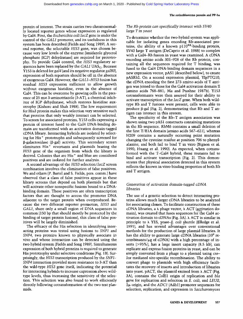
To determine whether the two-hybrid system was appli- cable for isolating genes encoding Rb-associated pro- teins, the ability of a known p l l0~-binding protein, SV40 large T antigen (DeCaprio et al. 1988) to complex with a Gal4-Rb fusion in yeast was examined. A eDNA encoding amino acids 301-928 of the Rb protein, con- taining all the sequences required for T binding, was fused to the Gal4 DNA-binding domain sequences in a new expression vector, pAS 1 (described below), to create pASRb2. On a second expression plasmid, YIpPTG10, the eDNA encoding the first 273 amino acids of T anti- gen was joined to those for the Gal4 activation domain II (amino acids 768-881; Ma and Ptashne 1987b). Y153 cotransformants were then assayed for their ability to activate transcription of the IacZ gene. When both wild- type Rb and T fusions were present, cells were able to produce ~-gal [Fig. 2), demonstrating that Rb and T an- tigen can interact in this system.
The specificity of the Rb-T antigen association was shown using two pAS 1 constructs containing mutations in the Rb sequence. RbM6 contains a small deletion of the first T/E1A domain (amino acids 567-621), whereas H209 contains a naturally occurring point mutation changing the cysteine residue at position 706 to phenyl- alanine, and both fail to bind T in vitro (Bignon et al. 1990; Huang et al. 1990). As expected, when cotrans- formed with the T-Gal4 hybrid, these mutants fail to bind and activate transcription (Fig. 2). This demon- strates that physical association detected in this system reflects the known in vitro-binding properties of both Rb and T antigen.
Generation of activation domain-tagged cDNA libraries
The use of a genetic selection to detect interacting pro- teins allows much larger cDNA libraries to be analyzed for associating clones. To facilitate construction of these cDNA libraries, a k phage vector, k ACT (ac__.~tivation do- main), was created that fuses sequences for the Gal4 ac- tivation domain to cDNAs {Fig. 3A). k ACT is similar in principle to k YES, zeast-E, coli shuttle (Elledge et al. 1991), and has several advantages over conventional methods for the production of large plasmid libraries. It has the ability to generate large eDNA libraries (109 re- combinants/~g of cDNA) with a high percentage of in- serts (>95%), has a large insert capacity (8.5 kb), can replicate and express fusion proteins in yeast, and can be simply converted from a phage to a plasmid using cre-- Iox mediated site-specific recombination. The ability to convert phage to plasmids with high efficiency facili- tates the recovery of inserts and introduction of libraries into yeast, pACT, the plasmid excised from k ACT (Fig. 3A), contains the ColE1 origin of replication and bla gene for replication and selection in E. coli, and LEU2, 2~ origin, and the ADC1 (Adhl)promoter sequences for selection, replication, and expression in Saccharomyces
GENES & DEVELOPMENT 557
Cold Spring Harbor Laboratory Press on March 1, 2020 - Published by genesdev.cshlp.orgDownloaded from
Durfee e t al.
Figure 2. Rb and SV40 T antigen interaction in yeast. (A) Fusion constructs used to test Rb and T association in yeast. The Rb2 fusion protein contains the Gal4 DNA-binding domain {amino acids 1-147, black hatched boxl joined to a carboxy-terminal fragment of Rb. The T/E1A-binding domains of Rb are shown as stippled boxes. RbM6 is a deletion affecting the first T/EIA domain; RbH209 is a point mutation affecting the second domain. Neither mutant is able to bind T in vitro. The Gal4 trans-activation domain (amino acids 768-881; white hatched box) was fused to the amino terminus of T antigen (solid box). {B) Determination of ~-gal activity in transformed yeast cells. Y153 was transformed with various plasmids as indicated, and ~-gal activity was determined by the colony lift method and quantitated using an ONPG assay. Three independent colonies per transformation were used for each ONPG deter- mination.
cerevisiae. The ADC promoter drives expression of a hy- brid protein consisting of the SV40 large T antigen nu- clear localization signal and sequences encoding the ac- tivation domain II of Gal4. Fused to GAL4 at amino acid 881 is a polylinker containing an XhoI site into which cDNAs are inserted.
A human eDNA library was constructed in k ACT using mRNA prepared from Epstein-Barr virus (EBV)- transformed human peripheral lymphocytes. The library contained 1.1 x l0 s total recombinants with >95% in- serts. It was amplified and converted to plasmid form, and plasmid DNA was prepared in bulk for yeast trans- formation.
A companion plasmid, pAS1, was constructed to facil- itate creation of target protein fusions with the Gal4 DNA-binding domain (for details of construction, see Fig. 3B and Materials and methods). This plasmid con- tains TRP1, 2 ~ origin, and the ADC1 promoter driving expression of the Gal4 DNA-binding domain (amino ac- ids 1-147; Keegan et al. 1986) fused to a polylinker. The Gal4 derivative is tagged with the hemagglutinin {HAl epitope recognized by a commercially available mono- clonal antibody (mAb 12CA5) (Baboo, Richmond, CA). The polylinker contains several useful cloning sites, in- cluding NcoI and NdeI, that each have an in-frame ATG codon in their recognition sequences.
Screening for cellular proteins that interact with the Rb protein
Tumor suppression by the Rb protein likely requires as- sociation with cellular factors through sequences in the carboxy-terminal half of the protein. To screen for hu- man proteins able to interact with this portion of p110 RB, Y153 containing pASRb2 was transformed with the k ACT human lymphocyte eDNA library and trans- formants were subjected to the screening procedure de- scribed above (Fig. 1). A total of - 2 million transfor-
mants were placed under selection (Fig. 4B). Of the trans- formants spread on synthetic complete (SC)-His, Leu, Trp plus 25 mM 3-AT plates, -1% grew into colonies within 3-5 days. Many colonies were very small but were nevertheless included in the count. It is likely that the use of higher 3-AT levels would further reduce the background; however, we chose to use a lower level so that weak interactions would also be detected. At 25 mM 3-AT, the selection behaves more like a 100-fold enrich- ment.
After selection, transformants were screened for their ability to produce [3-galactosidase using a filter lift assay (Breeden and Nasmyth 1985). One hundred thirty-nine His + colonies were also blue in this assay (Fig. 4B). In general, larger His + colonies were much more likely tO be blue than the small colonies. These His + blue colo- nies were considered positives in the initial screen and were used for additional studies.
Identification of the positive clones dependent on Rb hybrid expression
To test further whether the phenotype observed in the original screen was reproducible and dependent on the Rb hybrid, library-derived plasmids were selectively re- covered by virtue of the yeast LEU2 gene carried on those plasmids to complement a leuB6 mutation present in the E. coIi strain, JA226 {Tschumper and Carbon 1980). Plasmids isolated were then used to transform Y153 either alone or with three test fusions in pAS1. The test fusions were pASRb2, pAS/N-Rb (containing eDNA for amino acids 1-300 of pll0Pa3}, and pAS/SNF1 (pSE1112), which contains the yeast SNF1 gene. Trans- formants were assayed for [3-gal activity, and those show- ing activity only in the presence of pASRb2 were con- sidered positive {Fig. 4A). Of the 139 original isolates, 28 of the recovered plasmids induced the expression of lacZ and did so only in the presence of pASRb2 (Fig. 4B). This
558 GENES & DEVELOPMENT
Cold Spring Harbor Laboratory Press on March 1, 2020 - Published by genesdev.cshlp.orgDownloaded from
The retinoblastoma protein and PP- le
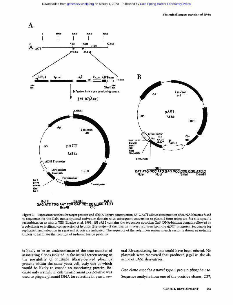
Figure 3. Expression vectors for target protein and eDNA library construction. (A) k ACT allows construction of eDNA libraries fused to sequences for the Gal4 transcriptional activation domain with subsequent conversion to plasmid form using cre--lox site-specific recombination as with h YES [Elledge et al. 1991 I. {B) pAS1 contains the sequences encoding Gal4 DNA-binding domain followed by a polylinker to facilitate construction of hybrids. Expression of the fusions in yeast is driven from the ADC1 promoter. Sequences for replication and selection in yeast and E. coli are indicated. The sequence of the polylinker region in each vector is shown as in-frame triplets to facilitate the creation of in-frame fusion proteins.
is likely to be an underestimate of the true number of associating clones isolated in the initial screen owing to the possibility of multiple library-derived plasmids present within the same yeast cell, only one of which would be likely to encode an associating protein. Be- cause only a single E. coli transformant per positive was used to prepare plasmid DNA for retesting in yeast, sev-
eral Rb-associating fusions could have been missed. No plasmids were recovered that produced ~-gal in the ab- sence of pASI derivatives.
One clone encodes a novel type I protein phosphatase
Sequence analysis from one of the positive clones, C27,
GENES & DEVELOPMENT 559
Cold Spring Harbor Laboratory Press on March 1, 2020 - Published by genesdev.cshlp.orgDownloaded from

D u d e e et al.
Figure 4. Determination of Rb dependency and screening re- sults. (A) Assessing the dependency of the Gal4-Rb2 fusion for transcriptional activation by positive clones isolated in the ini- tial screen. Library-derived plasmids from positive clones, iso- lated by passage through E. coli, were used to transform Y153 alone or with Gal4 DNA-binding domain {DBD) test fusions carried on pAS1. DBD fusions were N-Rb {Rb amino acids 1-3001, the yeast SNFI gene, and the Rb2 construct used in the initial screen. Transformants were assayed for the presence of B-gal activity using the colony filter lift method (Breeden and Nasmyth 1985). The pattern of B-gal-positive (solid bars) and [3-gal-negative {white bars) outcomes is dependent on the class of the plasmid isolated. Class X 1 represents Rb2-dependent clones chosen for further analysis. (B) Summary of the screening and rescreening results. The number of total transformants was estimated by plating an aliquot of each transformation on media selecting only for the presence of the plasmids [SC-trp--leu). The approximate number of His +-positive colonies was deter- mined following plating of the transformation on SC - Trp, Leu, His + 25 mM 3-AT. The presence of f3-gal activity was detected using the colony filter lift method (Breeden and Nasmyth 1985). Rb dependency was determined as described inA.
revealed that the eDNA insert encoded a protein with a predicted molecular mass of 38.6 kD and was identical to the PP-I~ catalytic subunit (Barker et al. 1990) with two exceptions. First, an 11-amino-acid insert was found in the amino terminus starting at amino acid 18 [Fig. 5). This insertion is located at some distance from a highly conserved region (amino acids 60-130) that likely serves as the active site for the phosphatase (Bollen and Stal- marts 1992), and thus should not interfere with the en- zymatic function of the protein. Second, the 5'-untrans- lated region was different from the published sequence. Although the authenticity of the differences seen at the 5' end need to be verified further by analysis of multiple
isolates, the insert seen in the coding region is likely real, as both flanking sequences correspond to PP-la. These findings suggest the potential for alternative splic- ing in the generation of the phosphatase messages. On the basis of the identity between the two human clones, we have named the C27 encoded isozyme PP-la2.
Both PP-1 a isoforms bind unphosphorylated p l 10 RB in vitro
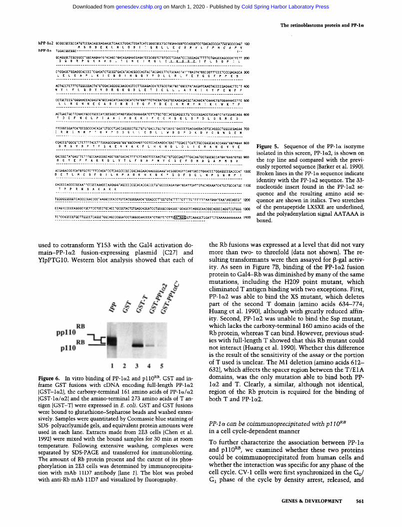
To confirm and extend the binding data obtained in yeast, we then expressed PP-1~2 as a glutathione S-trans- ferase (GST) fusion protein in E. coli (Smith and Johnson 1988). To test the ability of this protein to bind p110 RB in vitro, a GST-PP-la2-containing affinity matrix was in- cubated with human cell extracts from 2E3 cells, a WERI (Rb +)reconsti tuted cell line (Chen et al. 1992). Follow- ing extensive washing, proteins were eluted and run on a SDS-PAGE gel and Western blotted, and the blot was probed with an anti-Rb antibody, 11D7 (Shan et al. 1992). GST-PP-lc~2 was able to complex with p110 ~u3, as was the GST-T antigen (GST-T) control, whereas a matrix of GST alone was not (Fig. 6, lanes 2-4t. This result estab- lishes that the PP-I~2 protein can interact with the na- tive, full-length Rb protein.
The sequence identity of these isoforms outside of the amino-terminal insert suggested that PP-lc~ should also bind p110 R~. To test this possibility, a common carboxy- terminal region (amino acids 181-342 of PP-la21 was fused to GST and the resultant protein was used for the in vitro-binding assay. As expected, this truncated pro- tein was still able to bind the Rb protein (Fig. 6, lane 5) albeit with lower affinity than the full-length protein. Nonetheless, this confirms the idea that both PP-lc~ iso- forms can bind p 110 gB.
Both PP-lc~ isoforms interact preferentially with the unphosphorylated form of p l l 0 RB {Fig. 6) analogous to the binding characteristics of T antigen (Ludlow et al. 1989). This was confirmed using completely unphospho- rylated Rb protein produced in E. coli, which was also able to bind GST-PP-la2 in vitro (data not shown). Al- though the ability of PP-l~x to bind unphosphorylated Rb protein is clear, the possibility that the enzyme binds to the phosphorylated form of p110 ~J3 and catalyzes the re- moval of phosphate from the protein cannot be ruled out. In support of this hypothesis, it has been shown recently that PP1 is capable of dephosphorylating pp l l 0 ~ in vitro [Alberts et al. 1992; Ludlow et al. 1993).
Similar regions of Rb protein are required for binding PP-l a2 and T antigen
The p110 RB sequences required for binding PP-1~2 were examined to determine whether they coincided with the T/E1A-binding domains defined previously (Hu et al. 1990; Huang et al. 1990]. To accomplish this, a deletion set that had originally been used to delineate the T/E1A domains was employed (Huang et al. 1990). Several car- boxy-terminal deletion mutants, as well as the H209 point mutant, were subcloned into pAS1 (Fig. 7A) and
560 GENES & DEVELOPMENT
Cold Spring Harbor Laboratory Press on March 1, 2020 - Published by genesdev.cshlp.orgDownloaded from
The retinoblastoma protein and PP-la
Figure 5. Sequence of the PP-la isozyme isolated in this screen, PP-lct2, is shown on the top line and compared with the previ- ously reported sequence (Barker et al. 1990). Broken lines in the PP-la sequence indicate identity with the PP-la2 sequence. The 33- nucleotide insert found in the PP-lot2 se- quence and the resulting amino acid se- quence are shown in italics. Two stretches of the pentapeptide LXSXE are underlined, and the polyadenylation signal AATAAA is boxed.
used to cotransform Y153 wi th the Gal4 activation do- m a i n - P P - l a 2 fusion-expressing plasmid (C271 and YIpPTG10. Western blot analysis showed that each of
Figure 6. In vitro binding of PP-la2 and p l l0 ~. GST and in- frame GST fusions with cDNA encoding full-length PP-la2 [GST-la2), the carboxy-terminal 161 amino acids of PP-lot/a2 {GST-ledc~2] and the amino-terminal 273 amino acids of T an- tigen [GST-T) were expressed in E. coll. GST and GST fusions were bound to glutathione-Sepharose beads and washed exten- sively. Samples were quantitated by Coomassie blue staining of SDS-polyacrlyamide gels, and equivalent protein amounts were used in each lane. Extracts made from 2E3 cells (Chen et al. 1992) were mixed with the bound samples for 30 rain at room temperature. Following extensive washing, complexes were separated by SDS-PAGE and transferred for immunoblotting. The amount of Rb protein present and the extent of its phos- phorylation in 2E3 cells was determined by immunoprecipita- tion with mAb 11D7 antibody (lane 1). The blot was probed with anti-Rb mAb 11D7 and visualized by fluorography.
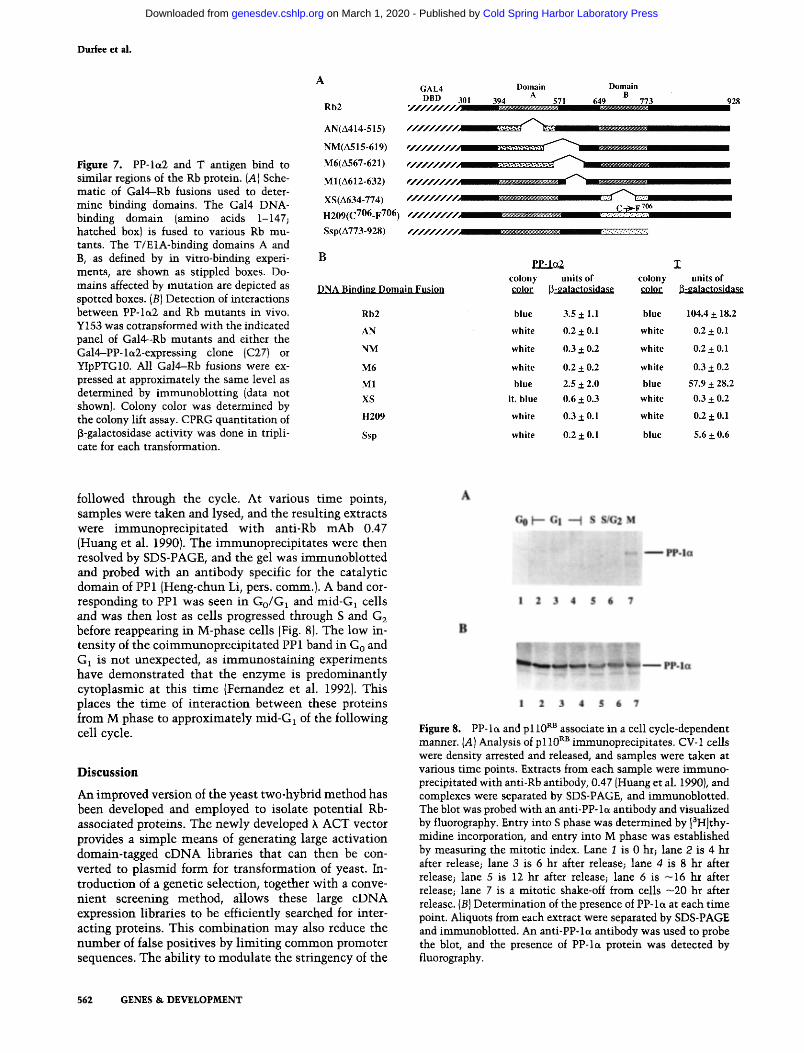
the Rb fusions was expressed at a level that did not vary more than two- to threefold (data not shown). The re- sulting transformants were then assayed for f~-gal activ- ity. As seen in Figure 7B, binding of the PP- la2 fusion protein to Gal4--Rb was d iminished by m a n y of the same mutations, including the H209 point mutant , which e l iminated T antigen binding wi th two exceptions. First, PP- la2 was able to bind the XS mutant , which deletes part of the second T domain (amino acids 634--774; Huang et al. 1990), al though wi th greatly reduced affin- ity. Second, PP-loL2 was unable to bind the Ssp mutant , which lacks the carboxy-terminal 160 amino acids of the Rb protein, whereas T can bind. However, previous stud- ies wi th full-length T showed that this Rb mutan t could not interact (Huang et al. 1990). Whether this difference is the result of the sensit ivi ty of the assay or the portion of T used is unclear. The M1 deletion (amino acids 612- 632), which affects the spacer region between the T/E1A domains, was the only muta t ion able to bind both PP- l a2 and T. Clearly, a similar, although not identical, region of the Rb protein is required for the binding of both T and PP-la2.
PP-1 a can be co immunoprec ip i ta t ed wi th p l I 0 a~ in a cell cycle-dependent manner
To further characterize the association between PP- la and p l l 0 RB, we examined whether these two proteins could be coimmunoprecipi ta ted from h u m a n cells and whether the interaction was specific for any phase of the cell cycle. CV-1 cells were first synchronized in the Go/ G1 phase of the cycle by density arrest, released, and
GENES & DEVELOPMENT 561
Cold Spring Harbor Laboratory Press on March 1, 2020 - Published by genesdev.cshlp.orgDownloaded from
Dudee et ai.
Figure 7. PP-Iet2 and T antigen bind to similar regions of the Rb protein. {A] Sche- matic of Gal4-Rb fusions used to deter- mine binding domains. The Gal4 DNA- binding domain (amino acids 1-147; hatched box) is fused to various Rb mu- tants. The T/E1A-binding domains A and B, as defined by in vitro-binding experi- ments, are shown as stippled boxes. Do- mains affected by mutation are depicted as spotted boxes. (B) Detection of interactions between PP-la2 and Rb mutants in vivo. Y153 was cotransformed with the indicated panel of Gal4--Rb mutants and either the Gal4-PP-let2-expressing clone {C27) or YIpPTG10. All Gal4-Rb fusions were ex- pressed at approximately the same level as determined by immunoblotting (data not shown). Colony color was determined by the colony lift assay. CPRG quantitation of [3-galactosidase activity was done in tripli- cate for each transformation.
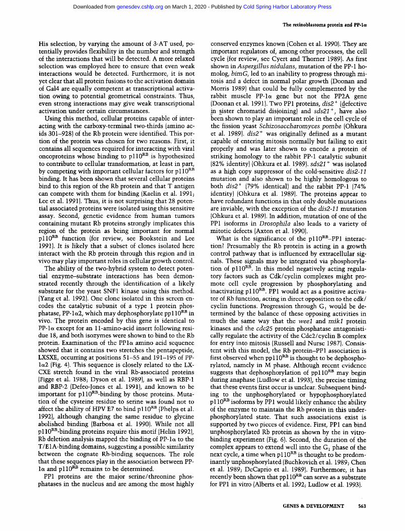
followed through the cycle. At various t ime points, samples were taken and lysed, and the resulting extracts were immunoprecipi ta ted wi th anti-Rb mAb 0.47 tHuang et al. 1990). The immunoprecipi ta tes were then resolved by SDS-PAGE, and the gel was immunoblot ted and probed wi th an antibody specific for the catalytic domain of PP1 (Heng-chun Li, pers. comm.). A band cor- responding to PP1 was seen in Go/G 1 and mid-G1 cells and was then lost as cells progressed through S and G2 before reappearing in M-phase cells (Fig. 8). The low in- tensity of the coimmunoprecipi ta ted PP1 band in G O and G~ is not unexpected, as immunos ta in ing experiments have demonstrated that the enzyme is predominant ly cytoplasmic at this t ime (Fernandez et al. 1992}. This places the t ime of interact ion between these proteins from M phase to approximately mid-G~ of the following cell cycle.
D i s c u s s i o n
An improved version of the yeast two-hybrid method has been developed and employed to isolate potential Rb- associated proteins. The newly developed ~ ACT vector provides a s imple means of generating large activation domain-tagged cDNA libraries that can then be con- verted to p lasmid form for transformation of yeast. In- troduction of a genetic selection, together wi th a conve- nient screening method, allows these large cDNA expression libraries to be efficiently searched for inter- acting proteins. This combinat ion may also reduce the number of false positives by l imi t ing common promoter sequences. The abil i ty to modulate the stringency of the
Figure 8. PP-la and p110 ~ associate in a cell cycle-dependent manner. (A) Analysis of p110 a~ immunoprecipitates. CV-1 cells were density arrested and released, and samples were taken at various time points. Extracts from each sample were immuno- precipitated with anti-Rb antibody, 0.47 [Huang et al. 1990), and complexes were separated by SDS-PAGE, and immunoblotted. The blot was probed with an anti-PP-1 a antibody and visualized by fluorography. Entry into S phase was determined by [~H]thy- midine incorporation, and entry into M phase was established by measuring the mitotic index. Lane I is 0 hr; lane 2 is 4 hr after release; lane 3 is 6 hr after release; lane 4 is 8 hr after release; lane 5 is 12 hr after release; lane 6 is -16 hr after release; lane 7 is a mitotic shake-off from cells -20 hr after release. (B) Determination of the presence of PP-la at each time point. Aliquots from each extract were separated by SDS-PAGE and immunoblotted. An anti-PP-1 et antibody was used to probe the blot, and the presence of PP-la protein was detected by fluorography.
562 GENES & DEVELOPMENT
Cold Spring Harbor Laboratory Press on March 1, 2020 - Published by genesdev.cshlp.orgDownloaded from
The retinoblastoma protein and PP-le~
His selection, by varying the amount of 3-AT used, po- tentially provides flexibility m the number and strength of the interactions that will be detected. A more relaxed selection was employed here to ensure that even weak interactions would be detected. Furthermore, it is not yet clear that all protein fusions to the activation domain of Gal4 are equally competent at transcriptional activa- tion owing to potential geometrical constraints. Thus, even strong interactions may give weak transcriptional activation under certain circumstances.
Using this method, cellular proteins capable of inter- acting with the carboxy-terminal two-thirds (amino ac- ids 301-928} of the Rb protein were identified. This por- tion of the protein was chosen for two reasons. First, it contains all sequences required for interacting with viral oncoproteins whose binding to p110 Rn is hypothesized to contribute to cellular transformation, at least in part, by competing with important cellular factors for p 110 Rs binding. It has been shown that several cellular proteins bind to this region of the Rb protein and that T antigen can compete with them for binding (Kaelin et al. 1991~ Lee et al. 1991). Thus, it is not surprising that 28 poten- tial associated proteins were isolated using this sensitive assay. Second, genetic evidence from human tumors containing mutant Rb proteins strongly implicates this region of the protein as being important for normal p l l 0 RB function {for review, see Bookstein and Lee 19911. It is likely that a subset of clones isolated here interact with the Rb protein through this region and in vivo may play important roles in cellular growth control.
The ability of the two-hybrid system to detect poten- tial enzyme-substrate interactions has been demon- strated recently through the identification of a likely substrate for the yeast SNF1 kinase using this method. IYang et al. 19921. One clone isolated in this screen en- codes the catalytic subunit of a type 1 protein phos- phatase, PP- la2, which may dephosphorylate pp 110 RS in vivo. The protein encoded by this gene is identical to pp-loL except for an 11-amino-acid insert following resi- due 18, and both isozymes were shown to bind to the Rb protein. Examination of the PPlct amino acid sequence showed that it contains two stretches the pentapeptide, LXSXE, occurring at positions 51-55 and 191-195 of PP- la2 (Fig. 4). This sequence is closely related to the LX- CXE stretch found in the viral Rb-associated proteins [Figge et al. 1988~ Dyson et al. 1989}, as well as RBP-1 and RBP-2 (Defeo-lones et al. 1991), and known to be important for p110~-binding by those proteins. Muta- tion of the cysteine residue to serine was found not to affect the ability of HPV E7 to bind p110 RB (Phelps et al. 1992}, although changing the same residue to glycine abolished binding IBarbosa et al. 1990). While not all p ll0aS-binding proteins require this motif {Helin 19921, Rb deletion analysis mapped the binding of PP-1 a to the T/EiA-binding domains, suggesting a possible similarity between the cognate Rb-binding sequences. The role that these sequences play in the association between PP- Is and p 110 ru3 remains to be determined.
PP1 proteins are the maior serine/threonine phos- phatases in the nucleus and are among the most highly
conserved enzymes known (Cohen et al. 1990}. They are important regulators of, among other processes, the cell cycle (for review, see Cyert and Thorner 1989}. As first shown in Aspergillus nidulans, mutation of the PP-1 ho- molog, bimG, led to an inability to progress through mi- tosis and a defect in normal polar growth (Doonan and Morris 1989) that could be fully complemented by the rabbit muscle PP-la gene but not the PP2A gene (Doonan et al. 1991}. Two PP1 proteins, dis2 + (defective in sister chromatid disjoining} and sds21 +, have also been shown to play an important role in the cell cycle of the fission yeast Schizosaccharomyces pombe (Ohkura et al. 19891. dis2 + was originally defined as a mutant capable of entering mitosis normally but failing to exit properly and was later shown to encode a protein of striking homology to the rabbit PP-1 catalytic subunit (82% identity} {Ohkura et al. 1989). sds21 + was isolated as a high copy suppressor of the cold-sensitive dis2-11 mutation and also shown to be highly homologous to both dis2 + {79% identical) and the rabbit PP-1 [74% identity} [Ohkura et al. 1989). The proteins appear to have redundant functions in that only double mutations are inviable, with the exception of the dis2-11 mutation {Ohkura et al. 1989}. In addition, mutation of one of the PPl isoforms in Drosophila also leads to a variety of mitotic defects [Axton et al. 1990).
What is the significance of the pll0RB--PP1 interac- tion? Presumably the Rb protein is acting in a growth control pathway that is influenced by extracellular sig- nals. These signals may be integrated via phosphoryla- tion of p l l 0 Rn. In this model negatively acting regula- tory factors such as Cdk/cyclin complexes might pro- mote cell cycle progression by phosphorylating and inactivating p110 gB. PP1 would act as a positive activa- tor of Rb function, acting in direct opposition to the cdk/ cyclin functions. Progression through G1 would be de- termined by the balance of these opposing activities in much the same way that the wee1 and m i k l protein kinases and the cdc25 protein phosphatase antagonisti- cally regulate the activity of the Cdc2/cyclin B complex for entry into mitosis (Russell and Nurse 1987}. Consis- tent with this model, the Rb protein-PPl association is first observed when pp 110 kB is thought to be dephospho- rylated, namely in M phase. Although recent evidence suggests that dephosphorylation of pp110 RB may begin during anaphase (Ludlow et al. 1993), the precise timing that these events first occur is unclear. Subsequent bind- ing to the unphosphorylated or hypophosphorylated p110 R~ isoforms by PP1 would likely enhance the ability of the enzyme to maintain the Rb protein in this under- phosphorylated state. That such associations exist is supported by two pieces of evidence. First, PP 1 can bind unphosphorylated Rb protein as shown by the in vitro- binding experiment (Fig. 6). Second, the duration of the complex appears to extend well into the G1 phase of the next cycle, a time when p110 ~ is thought to be predom- inantly unphosphorylated [Buchkovich et al. 1989~ Chen et al. 1989~ DeCaprio et al. 1989}. Furthermore, it has recently been shown that pp110 ~ can serve as a substrate for PP1 in vitro (Alberts et al. 1992; Ludlow et al. 1993}.
GENES & DEVELOPMENT 563
Cold Spring Harbor Laboratory Press on March 1, 2020 - Published by genesdev.cshlp.orgDownloaded from
Dudee et al.
Currently, it is hypothesized that the unphosphory- lated form of the Rb protein is active in growth suppres- sion. This notion is supported by several lines of evi- dence. Biochemically, T antigen and cellular factors such as E2F bind preferentially to the unphosphorylated form of the Rb protein (Ludlow et al. 1989; Chellappan et al. 1991), and it is this form of the protein that is found tethered to the "nuclear structure," whereas phosphory- lated and mutant forms of the protein are not (Mittnach and Weinberg 1991; Templeton 1992). Functionally, mi- croinjection of unphosphorylated Rb protein early in G1 is capable of inhibiting progression of cells through the cell division cycle (Goodrich et al. 19911. Thus, tempo- rally the PPI-p110 rib association also correlates with the presence of an active form of the Rb protein in the cell and may be required for the dephosphorylation event at the end of the cell cycle that regenerates this isoform. These observations suggest that this complex is impor- tant for the p110RB function in vivo.
A second, though not mutually exclusive, explanation is that p110 RB regulates PP1. PP1 activity and subcellu- lar localization is known to be modulated by its associ- ation with other cellular factors. For example, mamma- lian PP1 is found complexed with a 161-kD glycogen- binding protein that directs and stimulates PP 1 activity to proteins involved in glycogenolysis and glycogen syn- thesis (Cohen 1989). Also, the enzyme is negatively reg- ulated by protein inhibitors 1 and 2 (Cohen 1989), as well as newly defined nuclear inhibitors (Beullens et al. 1992). In S. pombe, dis2 + immunocomplexes have been re- ported to contain additional proteins potentially in- volved in its regulation (Kinoshita et al. 1990). One can- didate for such a regulator, the sds22 + gene product, isolated as a suppressor of the dis2-11 mutation, has been shown to positively modulate PP1 activity possibly through direct interaction with one of the PP1 proteins (Ohkura and Yanagida 1991). Whether the Rb protein can serve as a regulator and direct PP1 activity to specific substrates during the mid-M to early G, period is an intriguing possibility. In this connection the tethering of unphosphorylated Rb protein to the nuclear structure (Mittnach and Weinberg 1991; Templeton 1992) may be important for the subcellular localization of PP1, which is also found in a largely insoluble form in nuclear prep- arations (Beullens et al. 19921.
The phosphorylation of cellular proteins is recognized as an important regulatory mechanism governing a vari- ety of cellular processes (for review, see Hunter 1987). Protein phosphatases are now emerging as critical en- zymes in a wide range of cellular events as well. The inhibition of PP1 activity (as well as PP2A} by the potent tumor-promoting factor, okadaic acid, has prompted some to hypothesize that PP 1 activity may play a role in maintaining normal restraints on cell growth (Barker et al. 1990). Consistent with that, microinjection of PP1 into cells in G1 phase blocks progression into S phase, similar to the Rb protein [Alberts et al. 19921. The PPla association with p110 P'B may provide an important link in understanding these phenomenon. Conversely, cells with PP-loL mutations that specifically disrupt its inter-
action with p110 aB might be expected to have a trans- formed phenotype because of inappropriate Rb protein phosphorylation or similar effects on other proteins re- quiring an active phosphatase-pll0 ~a3 complex. The chromosomal location of the PPla gene, 11q13, is asso- ciated with the etiology of certain cancers (Barker et al. 1990), although the involvement (if any) by the enzyme in these neoplasias is unknown. A thorough understand- ing of the influences imparted by this interaction should yield important information on how these proteins reg- ulate cell growth.
Materials and m e t h o d s
Bacterial and yeast strains
E. coli JM107 [endA1, gyrA96, thi, hsdR17, supE44, relA1, h(lac--proAB), {F', traD36, proAB +, lacI a lacZhM 15) {Yannisch- Perron et al. 1985), and DH5 (F-, recA1, endA1, hsdR17, supE44, thil, gyrA, re/A 1] were the transformation recipient for all plasmid constructions. JA226 (hsdR, hsdM, leuB6, topl 1, thi, recBC, strR) (the gift of Merle Hoekstra, ICOS, Seattle, WA] was used to recover expression plasmids from yeast. The E. coli B strain BL21-LysS (Studier et al. 1990) was used for the expres- sion of GST fusion proteins. Y153, MATa leu2-3,I12, ura3-52, trpl-901, his3-A200, ade2-101, gal4A gal80A URA3::GAL--lacZ, LYS2::GAL--HIS3. The GAL-HIS3 fusion was introduced into Y153 by two-step gene replacement by integration of pBM1499 IURA3 GAL-HIS3)[Flick and Johnston 1990} into YJO-Z (MATa leu2-3,112, ura3-52, trpl-901, his3-A200, ade2-101, gal4a gal80A GAL-lacZI [Leuther and Johnston 1992) to create Y152. YJO-Z contains a GAL-lacZ reporter integrated at an unknown chromosomal site. The integration of pBM1499 is tar- geted adjacent to the LYS2 locus by cleavage with StuI, which cleaves in plasmid-bome LYS2 sequences before transformation and was selected by Ura prototrophy. The second recombina- tion event was selected by growth on plates supplemented with 5-fluoro-orotic acid as described (Flick and Johnston 1990) and screening for the His + phenotype. A second GAL--lacZ fusion gene was introduced into this background to increase the sen- sitivity to X-gal staining. The GAL-lacZ reporter contained on the YIp plasmid, pRY171 (URA3 GAL-lacZ) (Yocum et al. 1984) was introduced by selection for Ura prototrophy to create Y 153.
Media, enzymes, assays, and genetic methods
For drug selections, Luria broth (LB) plates were supplemented with ampicitlin (50 ~g/ml). Minimal media plates for E. coil, lacking leucine and containing ampicillin were used. Yeast YEPD and SC media were prepared as described (Rose et al. 1990). Restriction endonucleases, E. coli DNA polymerase I large fragment, T4 polynucleotide kinase, T4 DNA polymerase, and T4 DNA ligase were purchased from New England Biolabs. Deoxyribonucleotides and ATP were purchased from P-L Bio- chemicals. Drugs were purchased from Sigma.
Construction of X ACT
pSEll07 was constructed in several steps. First the HpaI-SalI LEU2-containing fragment from YEp13 was inserted into YEp24 cleaved with StuI and SalI, replacing the URA3 gene, to create pSE1101. The large Sa1I-PvuII fragment of pSE1101 was ligated in a three-way ligation to an 844-bp SphI-BamHI frag- ment from pBTM146 (P. Bartel and S. Fields, unpubl.], in which SphI was made blunt by T4 polymerase, and a 435-bp BamHI-
564 GENES & DEVELOPMENT
Cold Spring Harbor Laboratory Press on March 1, 2020 - Published by genesdev.cshlp.orgDownloaded from
The retinoblastoma protein and PP-Ia
Sail fragment of pBTM116, to create pSE1106. The 844-bp frag- ment contains the ADCI promoter driving a fusion protein con- sisting of the nuclear localization sequence from SV40 T anti- gen fused to the activation domain of GAL4 starting at amino acid 768. The 435-bp fragment contains the ADC1 transcrip- tional terminator. A SalI-XhoI fragment from pSE998 contain- ing two synthetic lox sites flanking a NotI site was inserted into the SalI site of pSE1106 to create pSE1107, pSE1107 was linear- ized with NotI and inserted into NotI-cleaved ), TRP arms, which are similar to h YES arms, to make k ACT.
cDNA library construction in A ACT
cDNA was made by standard methods using the procedure of Gubler and Hoffman (1983) employing AMV reverse tran- scriptase. After the second-strand reaction, the cDNA in 400 p,1 was spermine precipitated by the addition of 22 txl of 100 mM spermine, incubated on ice for 30 rain, pelleted for 15 rain in an Eppendorf centrifuge, washed three times for 30 rain each with 1 ml of spermine wash buffer [70% EtOH, 10 mM Mg(Ac)2, 0.3 M NaAc at pH 7] and once with 1 ml of 70% EtOH (Hoopes and McClure 1981}. eDNA was resuspended in 50 }xl of TE buffer and made flush by treatment with T4 DNA polymerase under the conditions suggested by the supplier, followed by the addi- tion of 5 ~l of 0.5 M EDTA and phenol/chloroform extractions, and ethanol precipitation, cDNA, approximately 4 txg, was re- suspended in 7 ~1 of TE buffer and was ligated to 2 ~g of an adaptor in a total volume of 10 ~1 at 4~ overnight. The se- quence of the kinased adaptors was 5'-GGCCTTCGTGGCC-3' (top strand}, and 5'-CGAGGCCACGAAGGCC-3' bottom strand}. After ligation, 170 ~1 of TE, 20 ~1 of 1 M KC1, and 10 lxl of 100 mM spermine were added, incubated on ice for 30 min and precipitated and washed as described above. Adapted eDNA was resuspended in 20 ~1 of TE buffer and run on a 1% low melting point agarose gel. cDNA of 600 bp and longer was gel purified for ligation into ~, ACT arms. cDNA (0.1 ~gj was ligated to 2 ~g of T-filled k ACT plasmid DNA, prepared as described previously {Elledge et al. 1991} in a volume of 4 jxl at 4~ over- night and packaged using one Gigapack Gold packaging extract (Stratagene, La Jolla, CA). Total recombinants (1.1 x 10 a) were obtained. Phage libraries were amplified on LE392.
Automatic subcloning conversion of the human cDNA li- brary in k ACT into plasmid was accomplished by incubation of 109 phage with 2 ml of a fresh overnight culture of BNN132 in 10 mM MgCl2 for 30 rain at 30~ without shaking. Two milli- liters of LB was added, and cells were incubated with shaking for 1 hr at 30~ Cells were then plated on ten 150-ram LB plates with 50 txg/ml of ampicillin and incubated at 37~ overnight. Ap * cells were scraped from these plates, added to 3 liters of terrific broth (Maniatis et al. 1982} with ampicillin, and grown to stationary phase. Plasmid DNA was prepared and purified using CsC1 density gradients by standard methods (Maniatis et al. 1982).
Construction of pASI
The ADC-GAL4(1-147)-ADC terminator fragment from pMA424 (Ma and Ptashne 1987a} was cloned into pIC20H {Marsh et al. 1984] by a three-way ligation between gel-purified fragments including the EcoRV-XhoI ADC1 promoter and XhoI-SphI GAL4--ADC terminator fragments from pMA424 and SphI-EcoRV-cleaved pIC20H to create pi l l . An EcoRI- BamHI double-stranded oligonucleotide encoding an HA epitope tag and several cloning sites was fused to the GAL4 DNA-binding domain of EcoRI-BamHI-cut pil l . The sequence of this oligonucleotide is GAA TTC ATG GCT TAC CCA TAC
GAT GTT CCA GAT TAC GCT AGC TTG GGT GGT CAT ATG GCC ATG GAG GCC CCG GGG ATC C and has the following restriction sites that are unique in pAS 1: EcoRI, NcleI, NcoI, SfiI, SmaI, BamHI. The sequence is presented as in-frame triplets. The NcoI and NdeI sites contain in-frame ATG codons that are particularly useful for fusing full-length cDNAs gener- ated by polymerase chain reaction (PCR} to the DNA-binding domain encoding sequences of GAL4.
Rb and T expression plasmids
pASRb2 was created as follows: The HindIII site in p44-1 (Huang et al. 1990) was converted to XhoI by Klenow fill-in and ligation of XhoI linkers to create p44-3. This plasmid was then cut with EcoRI, the site was filled in with Klenow, and 10-bp BamHI linkers were added. Following BamHI and XhoI diges- tion, the 1.8-kb fragment containing the 3' end of the Rb cDNA was subcloned into pSG424 {Fields and Jang 1990) creating pS- GRb2. The Rb fragment was then excised by BamHI-XbaI and cloned into pUC 18. The resulting clone, pUCRb2.66, was then cut with BarnHI and SalI, and the Rb-containing fragment was subcloned into pAS1 creating pASRb2. The same fragment was cloned into BKS[ + ) creating pBKRb266.
Rb deletion mutants were subcloned as EcoRI-HindlII frag- ments from existing plasmids {Huang et al. 1990) into BKS( + ). EcoRI-SalI fragments from the resulting clones replaced Rb se- quences in pBKRb266. Finally, BamHI-SalI fragments were subcloned into pAS1. The H209 mutation was removed from pAg-H209 (Bignon et al. 1990) as a 139-bp DralII-NdeI fragment and used to replace the homologous region in pUCRb266. The 1.8-kb BamHI-SalI fragment from the resultant plasmid was subcloned into pAS 1, and the mutation was verified by sequenc- ing.
YIpPTG10 was constructed as follows: The KpnI fragment from pNY7 (the gift of S. Fields, State University of New York, Stony Brook), containing sequences for the Gal4 trans-activa- tion domain II and the ADH1 transcriptional terminator, was subcloned into BKS( + I. This vector was cleaved with HindlII and the 823-bp HindlII fragment from Y62-25-2 encoding T an- tigen amino acids 1-273 fused in-frame with the GAL4 se- quences resulting in pTG25. A SacI-EcoRI fragment containing the ADCI promoter from YC-DE2 (Beiei and Young 19821 was then inserted into pTG25 upstream of the T-GAL4 fusion. This plasmid, pPTG7, was cleaved with XbaI and BamHI, and the insert was subcloned into YEpl3, resulting in YIpPTG10. The plasmid was linearized at the unique SacI site in the LEU2 gene preceding yeast transformation to increase the integration fre- quency.
Library screening
Y153 was transformed to Trp prototrophy with pASRb2 by the method of Scheistl and Geitz {1989}. A single colony was grown in SC - Trp medium and transformed with library DNA using total yeast RNA as carrier. Aliqouts were taken from each trans- formation mix before plating and used to determine the trans- formation efficiency by plating on SC media lacking tlyptophan and leucine. The transformation mix was then plated on 15-cm petri dishes containing SC media lacking tryptophan, leucine, and histidine but including 25 mM 3-AT (Sigma], and incubated at 30~ for 3-5 days. His + colonies were then screened for B-gal activity using a filter lift assay (Breeden and Nasmyth I985). Colonies were transferred onto nitrocellulose filters, permeabi- tized by freezing in liquid nitrogen (1 minl, and thawed at room temperature. Filters were then overlaid on Whatman 3MM pa- per saturated with a LacZ-X-gal solution {Breeden and Nasmyth
GENES & DEVELOPMENT 565
Cold Spring Harbor Laboratory Press on March 1, 2020 - Published by genesdev.cshlp.orgDownloaded from
Dur|ee et al.
1985; 8 ml/15-cm dish} and incubated at 30~ The time re- quired for color development ranged from 30 rain to overnight. Colonies corresponding to positives in this screen were then patched onto a master plate and analyzed further.
Recovery of plasmids from yeast
Total DNA from yeast was prepared according to the method of Hoffman and Winston {19871 and used to transform JA226 via electroporation using a Bio-Rad GenePulser according to the manufacturer's specifications. Transformations were plating on minimal media lacking leucine and containing ampicillin.
Sequence analysis
C27 and related subclones were sequenced using dideoxy NTPs and Sequenase 2.0 according to the manufacturer's specifica- tions (U.S. Biochemical}. Sequence analysis and homology searches were performed using DNASTAR software (DNAS- TAR, Inc., Madison, WII.
Quantitation of [3-gal activity in yeast
Cultures of 2.5 ml were grown in the appropriate selecting me- dia to OD6o o of 1.0-1.2. Cells were then prepared and perme- abilized as described (Guarente 1983). For quantitation using o-nitrophenyl-~-D-galactoside (ONPG), standard conditions were used [Guarente 1983). When using chlorophenyl-red-13-D- galactopyranoside (CPRG; Boehringer Mannheim), the same procedure was followed, except cell pellets were resuspended in 900 I~1 of H buffer 1100 mM HEPES, 150 mM NaC1, 2 mM MgC12, 1% BSA at pH 7.0), 100 ~1 of 50 mM CPRG was added following permeabilization, and the amount of liberated CPR was deter- mined by ODsz4.
Construction of GST fusions, protein preparation, and in vitro binding
To construct GST-PP-la2, the plasmid C27 was partially di- gested with BgllI and the 1.4-kb insert fragment was subcloned into the BamHI site pGEX-3X. GST-PP-Ia was created by sub- cloning the 850-bp fragment following complete digestion of C27 with BgllI into pGEX-3X. GST-T was made by cutting Y62-25-2 with HindlII, blunt-ending with Klenow, and subclon- ing the 823-bp fragment into pGEX-3X cut with SmaI. Expres- sion of GST fusion proteins was induced with 0.2 mM IPTG. Cells were centrifuged at 10K for 5 rain, and the resultant pellet resuspended in buffer A [150 mM NaC1, 5 mM DTT, 2 mM EDTA, 50 mM Tris (pH 8.0), 1 mM phenylmethylsulfonyl fluo- ride (PMSFI, 8 ~g of leupeptin, and 8 ~g of antipain]. Four mil- ligrams of lysozyme was added, and the cells were held at 4~ for 30 rain. Four hundred microliters of 10% NP-40 was added, and the cells were lysed by sonication. Cell debris was removed by centrifugation (10K for 30 mini, and the supernatant was added to glutathione-coated beads.
The in vitro-binding assay was performed as follows. Extracts made from 2 x l 0 6 2E3 cells {Chen et al. 1992) were incubated with beads containing 2-3 txg of GST or GST fusion proteins in lysis buffer [50 raM, Tris (pH 7.4), 150 mM NAG1, 5 mM EDTA, 0.1% NP-40, 50 mM NaF, 1 mM PMSF, 1 ~g of leupeptin/ml, 1 ~g antipain/ml] for 30 min at room temperature. Complexes were washed extensively with lysis buffer, boiled in loading buffer, and run on 7.5% SDS-polyacrylamide gels. Gels were transferred to immobilon membranes and immunoblotted with anti-Rb mAb 11DT. Following the addition of an alkaline phos- phatase-conjugated secondary antibody, bound Rb protein was
visualized with 5-bromo-4-chloro-3-indolylphosphate toluidin- ium and nitro blue tetrazolium {BCIP, NBT; Promega, Madison, wII.
Cell cycle synchrony and immunoprecipitatdon
CV-1 cells were grown to confluence and maintained in 10% fetal calf serum (FCS) for 2 weeks. Cells were then split 1 : 2 and grown in 10% FCS. Aliqouts were lysed in 500 ~1 of lysis 250 buffer [50 mM TRIS {PH 7.4), 250 MM NAG1, 5 mM EDTA, 0.1% NP-40, 50 mM NaF, 1 mM PMSF, 1 ~g of leupeptin/ml, 1 ~g of antipain/ml] and cleared by centrifugation {14K for 2 rain at room temperature). The supernatant was diluted in 400 ~1 of lysis 0 buffer (same as lysis 250, except lacking NaC1; 850 ~1 was immunoprecipitated with anti-Rb polyclonal antibody 0.47 (Huang et al. 1990), and 50 ~1 was directly assayed for PP-let content by Western blotting with an antibody raised against the catalytic subunit of PP-le~ prepared from bovine heart (kindly provided by Heng-chun Li, Mt. Sinai Medical School, NY). Im- munoprecipitates were separated by SDS-PAGE and irnmuno- blotted using the same anti-PP-let antibody. Proteins were vi- sualized using BCIP/NBT. Using an independently prepared anti-PP1 antibody {Brautigan et al. 19851, identical results to those in Figure 8 were obtained (data not shownl.
A c k n o w l e d g m e n t s
We thank D. Goodrich, M. Kuroda, and S. Sazer for comments on the manuscript. We thank Stanely Fields and Paul Bartel (State University of New York, Stony Brook), Joachim Schnier (University of Califomia, Davis], Heng-chun Li, David Brauti- gan (Brown Universityl, K. Leuther and S. Johnston (Southwest- em Medical Center), M. Johnston (Washington University}, and J. Wade Harper and James Shero (Baylor College of Medicine) for many helpful discussions and gifts of reagents and plasmids. We also thank John Wilson in whose laboratory pAS1 was con- structed. This work was supported by National Institutes of Health (NIH} grants GM-44664 and HG-00463 to S.J.E., and grants from NIH 05758 and the Council for Tobacco Research to W.H.L.S.J.E. is a Pew Scholar in the Biomedical Sciences. T.D. is a predoctoral student of the Molecular Pathology Pro- gram-University of California at San Diego, and is supported by a training grant from the National Eye Institute. W.H.L. is the A.P. McDermitt Chair Professor.
The publication costs of this article were defrayed in part by payment of page charges. This article must therefore be hereby marked "advertisement" in accordance with 18 USC section 1734 solely to indicate this fact.
References
Alberts, A.S., A.M. Thorbum, S. Shenolikar, M.C. Mumby, and J.R. Feramisco. 1993. Regulation of cell cycle progression and the nuclear affinity of the retinoblastoma protein by protein phosphatases. Proc. Natl. Acad. Sci. 90: 388-392.
Axton, V., J.M. Dombradi, P.T.W. Cohen, and D.M. Glover. 1990. One of the protein phosphatase 1 isozymes in Drosphilia is essential for mitosis. Ceil 63: 33-46.
Barbosa, C., M.S. Edmonds, C. Fisher, J.T. Schiller, D.R. Lowy, and K.H. Vousden. 1990. The region of the HPV E7 oncopro- tein homologous to adenovirus E1A and SV40 large T anti- gen contains separate domains for Rb binding and casein kinase II phosphorylation. EMBO J. 9: 153-160.
Barker, H.M., T.A. Jones, E.F.d.C.e. Silva, N.K. Spurr, D. Sheer, and P.T.W. Cohen. 1990. Localization of the gene encoding a
566 GENES & DEVELOPMENT
Cold Spring Harbor Laboratory Press on March 1, 2020 - Published by genesdev.cshlp.orgDownloaded from
The retinoblastoma protein and PP-lot
type 1 protein phosphatase catalytic subunit to human chro- mosome band 11q13. Genomics 7: 159-166.
Beier, D.R. and E.T. Young. 1982. Characterization of a regula- tory region upstream of the ADR2 locus of S. cerevisiae. Nature 300: 724-728.
Beullens, M., A.V. Eynde, W. Stalmans, and M. Bollen. 1992. The isolation of novel inhibitory polypeptides of protein phosphatase 1 from bovine thymus nuclei. J. Biol. Chem. 267: 16538-16544.
Bignon, Y.-I., I--Y. Shew, D. Rappolee, S.L. Naylor, E.Y.-H.P. Lee, I. Schnier, and W.-H. Lee. 1990. A single Cys706 to Phe substitution in the retinoblastoma protein causes the loss of binding to SV40 T antigen. Cell Growth Differ. 1: 647-651.
Bollen, M. and W. Stalmans. 1992. The structure, role, and reg- ulation of type 1 protein phosphatases. CRC Crit. Rev. Bio- chem. Mol. Biol. 27:227-281.
Bookstein, R. and W.-H. Lee. 1991. Molecular genetics of the retinoblastoma suppressor gene. CRC Crit. Rev. Oncogene- sis. 2:211-227.
Bookstein, R., J.-Y. Shew, P.-L. Chen, P. Scully, and W.-H. Lee. 1990. Suppression of tumorigenicity of human prostate car- cinoma cells by replacing a mutated RB gene. Science 247: 712-715.
Brautigan, D.L., C.L. Shiner, and P.A. Gruppuso. 1985. Phos- phorylase phosphatase catalytic subunit: evidence that the Mr 33,000 enzyme fragment is derived from a native protein of M r 70,000. J. Biol. Chem. 260: 4295--4302.
Breeden L. and K. Nasmyth. 1985. Regulation of the yeast HO gene. Cold Spring Harbor Syrup. Quant. Biol. 50: 643-650.
Buchkovich, K., L.A. Duffy, and E. Harlow. 1989. The retino- blastoma protein is phosphorylated during specific phases of the cell cycle. Cell 58: 1097-1105.
Chellappan, S., S. Hiebert, M. Mudryj, J.M. Horowitz and J. R. Nevins. 1991. The E2F transcription factor is a cellular target for the RB protein. Cell 65: 1053-1061.
Chen, P.-L., P. Scully, J.-Y. Shew, J.Y.-J. Wang, and W.-H. Lee. 1989. Phosphorylation of the retinobtastoma gene product is modulated during the cell cycle and cellular differentiation. Cell 58:1193-1198.
Chen, P.-L., Y. Chen, B. Shan, R. Bookstein, and W.H. Lee. 1992. Stability of retinoblastoma gene expression determines the tumorigenicity of reconstituted retinoblastoma cells. Cell Growth Differ. 3: 119-125.
Chien, C.T., P.L. Bartel, R. Sternglanz, and S. Fields. 1991. The two-hybrid system: A method to identify and clone genes for proteins that interact with a protein of interest. Proc. Natl. Acad. Sci. 88: 9578-9582.
Cohen, P. 1989. The structure and regulation of protein phos- phatases. Annu. Rev. Biochem. 58: 453-508.
Cohen, P.T.W., N.D. Brewis, V. Hughes, and D.J. Mann. 1990. Protein serine/threonine phosphatases; an expanding fam- ily. FEBS Lett. 268: 355-359.
Cyert, M.S. and J. Thomer. 1989. Putting it on and taking it off: Phosphoprotein phosphatase involvement in cell cycle reg- ulation. Cell 57: 891-893.
Dalton, S. and R. Treisman. 1992. Characterization of SAP-l, a protein recruited by serum response factor to the c-los serum response element. Cell 68: 597-612.
DeCaprio, J.A., J.W. Ludlow, J. Figge, J.-Y. Shew, C.-M. Huang, W.-H. Lee, E. Marsillo, E. Paucha, and D.M. Livingston. 1988. SV40 large tumor antigen forms a specific complex with the product of the retinoblastoma susceptibility gene. Cell 54: 275-283.
DeCaprio, J.A., J.W. Ludlow, D. Lynch, Y. Furukawa, J. Griffin, H. Piwnica-Worms, C.-M. Huang, and D.M. Livingston. 1989. The product of the retinoblastoma susceptibility gene
has properties of a cell cycle regulatory element. Cell 58: 1085-1095.
Defeo-lones, D., P.S. Huang, R.E. Jones, K.M. HaskelI, G.A. Vuocolo, M. Hanobik, H.E. Huber, and A. Oliff. 1991. Clon- ing of cDNAs foI cellular proteins that bind to the retino- blastoma gene product. Nature 352: 251-254.
Doonan, I.H. and N.R. Morris. 1989. The b/raG gene of As- pergillus nidulans, required foi completion of anaphase, en- codes a homolog of mammalian phosphoprotein phos- phatase 1. Cell 57: 987-996.
Doonan, J.H., C. MacKintosh, S. Osmani, P. Cohen, G. Bai, E.Y.C. Lee, and N.R. Morris. 1991. A eDNA encoding rabbit muscle protein phosphatase la complements the Aspergillus cell cycle mutant, MmGll . I. Biol. Chem. 266: 18889- 18894.
Dyson, N., P.M. Howley, K. Munger, and E. Harlow. 1989. The human papilloma virus-16 E7 oncoprotein is able to bind to the retinoblastoma gene product. Science 243: 934-937.
Elledge, S.J., J.T. Mulligan, S.W. Ramer, M. Spottswood, and R.W. Davis. 1991. ),YES: A multifunctional eDNA expres- sion vector for the isolational of genes by complementation of yeast and Escherichia coli mutations. Proc. Natl. Acad. Sci. 88: 1731-1735.
Fernandez, A., D.L. Brautigan, and N.J.C. Lamb. 1992. Protein phosphatase type 1 in mammalian mitosis: Chromosomal localization and involvement in mitotic exit. ]. Cell. Biol. 116: 1421-1430.
Fields, S. and S.K. Jang. 1990. Presence of a potent transcription activating sequence in the p53 protein. Science 249: 1046- 1051.
Fields, S. and O.K. Song. 1989. A novel genetic system to detect protein-protein interactions. Nature 340: 245-246.
Figge, l., T. Webster, T. Smith, and E. Paucha. 1988. Prediction of similar transforming regions in simian virus 40 large T, adenovirus E1A and myc oncoproteins. J. Virol. 62: 1814- 1818.
Flick, J. and M. Johnston. 1990. Two systems of glucose repres- sion at the GALI promoter in Saccharomyces cerevisiae. Mol. Cell. Biol. 10: 4757-4769.
Friend, S.H., R. Bernards, S. Rogelj, R.A. Weinberg, J.M. Rapa- port, D.M. Albert, and T.P. Dryja. 1986. A human DNA seg- ment with properties of the gene that predisposes to retino- blastoma and osteosarcoma. Nature 323: 643-646.
Goodrich, D.W., N.P. Wang, Y.-W. Qian, E.Y.-H.P. Lee, and W.- H. Lee. 1991. The retinoblastoma gene product regulates progression through the G1 phase of the cell cycle. Cell 67: 293-302.
Guarente, L. 1983. Yeast promoters and lacZ fusions designed to study expression of cloned genes in yeast. Methods Enzy- tool. 101: 181-191.
Gubler, U. and B. Hoffman. 1983. A simple and very efficient method for generating eDNA libraries. Gene 25: 263-272.
Helin, K., J.A. Lees, M. Vidal, N. Dyson, E. Harlow, and A. Fattaey. 1992. A eDNA encoding a pRB-binding protein with the properties of the transcription factor E2F. Cell 70: 337- 350.
Hoffman, C.S. and F. Winston. 1987. A ten-minute DNA prep- aration from yeast efficiently releases autonomous plasmids for transformation of Escherichia coli. Gene 57: 267-272.
Hoopes, B.C. and W.R. McClure. 1981. Studies on the selectiv- ity of DNA precipitation by spermine. Nucleic Acids Res. 9: 5493-5504.
Hu, Q., N. Dyson, and E. Harlow. 1990. The regions of the retinoblastoma protein needed for binding to the adenovirus E1A or SV40 large T antigen are common sites for muta- tions. EMBO ]. 9:1147-1155.
GENES & DEVELOPMENT 567
Cold Spring Harbor Laboratory Press on March 1, 2020 - Published by genesdev.cshlp.orgDownloaded from
Duffee et al.
Hu, Q., J.A. Lees, K.J. Buchkovieh, and E. Harlow. 1992. The retinoblastoma protein physically associates with the hu- man cdc2 kinase. Mol. Cell. Biol. 12: 971-980.
Huang, H.-J.S., J.-K. Yee, J.-Y. Shew, P.-L. Chen, R. Bookstein, T. Friedmann, E.Y.-H.P. Lee, and W.-H. Lee. 1988. Suppression of the neoplastic phenotype by replacement of the retino- blastoma gene product in human cancer cells. Science 242: 1563-1566.
Huang, S., N.P. Wang, B.Y. Tseng, W.-H. Lee, and E.Y.-H.P. Lee. 1990. Two distinct and frequently mutated regions of reti- noblastoma protein are required for binding to SV40 T anti- gen. EMBO J. 9: 1815-1822.
Huang, S., W.-H. Lee, and E.Y.-H.P. Lee. 1991. Identification of a cellular protein that competes with SV40 T antigen for binding to the retinoblastoma gene product. Nature 350: 160-162.
Hunter, T. 1987. A thousand and one protein kinases. Cell 50: 823-829.
Kaelin, W.GJ., D.C. Pallas, J.A. DeCaprio, F.J. Kaye, and D.M. Livingston. 1991. Identification of cellular proteins that can interact specifically with the T/E1A binding region of the retinoblastoma gene product. Celt 64: 521-532.
Kaelin, W.G.J., W. Krek, W.R. Sellers, J.A. DeCaprio, F. Ajchen- baum, C.S. Fuchs, T. Chittenden, Y. Li, P.J. Farnham, M.A. Blanar, D.M. Livingston, and E.K. Flemington. 1992. Expres- sion cloning of a eDNA encoding a retinoblastoma-binding protein with E2F-like properties. Cell 70:351-364 .
Keegan, L., G. Gill, and M. Ptashne. 1986. Separation of DNA binding from the transcription-activating function of a eu- karyotic regulatory protein. Science 231: 699-704.
Kim, S.J., S. Wagner, F. Liu, M.A. O'Reilly, P.D. Robbins, and M.R. Green. 1992. Retinoblastoma gene product activates expression of the human TGF-~2 gene through transcription factor ATF-2. Nature 358: 331-334.
Kinoshita, N., H. Ohkura, and M. Yanagida. 1990. Distinct, es- sential roles of typel and 2A protein phosphatases in the control of the fission yeast cell division cycle. Ceil 63: 405- 415.
Kishore, G.M. and D.M. Shah. 1988. Amino acid biosynthesis inhibitors as herbicides. Annu. Rev. Biochem. 57: 627-663.
Lee, W.-H., R. Bookstein, F. Hong, L.-J. Young, J.-Y. Shew, and E.Y.-H.P. Lee. 1987a. Human retinoblastoma susceptibility gene: Cloning, identification, and sequence. Science 235: 1394-1399.
Lee, W.-H., J.-Y. Shew, F. Hong, T. Sery, L.A. Donoso, L.J. Young, R. Bookstein, and E.Y.-H.P. Lee. 1987b. The retino- blastoma susceptibility gene product is a nuclear phos- phoprotein associated with DNA binding activity. Nature 329: 642-645.
Lee, W.-H., R.E. Hollingsworth, Y.-W. Qian, P.-L. Chen, F. Hong, and E.Y.-H.P. Lee. 1991. RB protein as a cellular "cor- ral" for growth-promoting proteins. Cold Spring Harbor Syrup. Quant. Biol. 56: 211-217.
Lees, J.A., K.J. Buchkovich, D.R. Marshak, C.W. Anderson, and E. Harlow. 1991. the retinoblastoma protein is phosphory- lated on multiple sites by human cdc2. EMBO J. 10: 4279- 4290.
Leuther, K.K. and S.A. Johnston. 1992. Nondissociation of GAL4 and GAL80 in vivo after galactose induction. Science 256: 1333-1335.
Lin, B.T.-Y., S. Gruenwald, A.O. Morla, W.H. Lee, and J.Y.J. Wang. 1991. Retinoblastoma cancer suppressor gene product is a substrate of the ceil cycle regulator cdc2 kinase. EMBO J. 10: 857-864.
Ludlow, J.W., J.A. DeCaprio, C.-M. Huang, W.-H. Lee, E. Paucha, and D.M. Livingston. 1989. SV40 large T antigen
binds preferentially to an underphosphorylated member of the retinoblastoma susceptibility gene family. Cell 56: 57- 65.
Ludlow, ].W., C.L. Glendening, D.M. Livingston, and J.A. De- Caprio. 1993. Specific enzymatic dephosphorylation of the retinoblastoma protein. Mol. Cell Biol. 13: 367-372.
Ma, J. and M. Ptashne. 1987a. Deletion analysis of GAL4 defines two transcriptional activating segments. Cell 48: 847-853.
- - . 1987b. A new class of yeast transcriptional activators. Cell 51:113-119.
~ . 1988. Converting a eukaryotic transcriptional inhibitor into an activator. Cell 55: 443-446.
Maniatis, T., E.F. Fritsch, and J. Sambrook. 1982. Molecular cloning: A laboratory manual. Cold Spring Harbor Labora- tory, Cold Spring Harbor, New York.
Marsh, J.L., M. Erfle, and E.F. Wykes. 1984. The pIC plasmid and phage vectors with versatile multiple cloning sites for re- combinant selection by insertional inactivation. Gene 32: 481-485.
Mittnach, S. and R.A. Weinberg. 1991. G1/S phosphorylation of the retinoblastoma protein is associated with an altered af- finity for the nuclear compartment. Ceil 6 5 : 3 8 1 - 3 9 3 .
Ohkura, H. and M. Yanagida. 1991. S. pombe gene sds22 + es- sential for a midmitotic transition encodes a leucine-rich repeat protein that positively modulates protein phos- phatase-1. Cell 64: 149-157.
Ohkura, H., N. Kinoshita, S. Miyatani, T. Toda, and M. Yan- agida. 1989. The fission yeast dis2 + gene required for chro- mosome disjoining encodes one of two putative type 1 pro- tein phosphatases. Cell 57: 997-1007.
Phelps, W.C., K. Munger, C.L. Yee, ].A. Barnes, and P.M. How- ley. 1992. Structure-function analysis of the human papillo- mavims type 16 E7 oncoprotein. J. Virol. 66: 2418-2427.
Rose, M.D., F. Winston, and P. Hieter. 1990. Methods in yeast genetics. A laboratory course manual. Cold Spring Harbor Laboratory Press, Cold Spring Harbor, New York.
Russell, P. and P. Nurse. 1987. Negative regulation of mitosis by weel +, a gene encoding a protein kinase homolog. Cell 49: 559-567.
Rustgi, A.K., N. Dyson, and R. Bernards. 1991. Amino-terminal domains of c-myc and N-myc proteins mediate binding to the retinoblastoma gene product. Nature 352: 541-544.
Schiestl, R.H. and R.D. Gietz. 1989. High efficiency transfor- mation of intact yeast cells using single stranded nucleic acids as a carrier. Curl Genet. 16: 339-346.
Shan, B., X. Zhu, P.-L. Chen, T. Durfee, Y. Yang, D. Sharp, and W.-H. Lee. 1992. Molecular cloning of cellular genes encod- ing retinoblastoma-associated proteins: Identification of a gene with properties of the transcription factor E2F. Mol. Celt. Biol. 12: 5620--5631.
Shew, J.-Y., N. Ling, X. Yang, O. Fodstad, and W.H. Lee. 1989. Antibodies detecting abnormalities of the retinoblastoma susceptibility gene product {ppl l0 W) in oesteosarcoma and synovial sarcomas. Oncogene Res. 1: 205-214.
Smith, D.B. and K.S. Johnson. 1988. Single-step purification of polypeptides expressed in Escherichia coti as fusions with glutathione S-transferase. Gene 67: 31-40.
Studier, F.W., A.H. Rosenberg, J.J. Dunn, and J.W. Dubendorff. 1990. Use of T7 RNA polymerase to direct expression of cloned genes. Methods Enzymol. 185: 60--89.
Takahashi, P.R., T. Hashimoto, X. Hong-ji, S.-X. Hu, T. Matsui, T. Miki, H. Bigo-Marshall, S.A. Aaronson, and W.F. Bene- dict. 1991. The retinoblastoma gene functions as a growth and tumor suppressor in human bladder carcinoma cells. Proc. Natl. Acad. Sci. 88: 5257-5261.
Templeton, D.J. 1992. Nuclear binding of purified retinoblasto-
568 GENES & DEVELOPMENT
Cold Spring Harbor Laboratory Press on March 1, 2020 - Published by genesdev.cshlp.orgDownloaded from
The retinoblastoma protein and PP-I~
ma gene product is determined by cell cycle-regulated phos- phorylation. Mol. Cell. Biol. 12: 435-443.
Tschumper, G. and J. Carbon. 1980. Sequence of a yeast DNA fragment containing a chromosomal replicator and the TRP1 gene. Gene 10: 157-166.
Whyte, P., K.I. Buchkovich, J.M. Horowitz, S.H. Friend, M. Ray- buck, R.A. Weinberg, and E. Harlow. 1988. Association be- tween an oncogene and an anti-oncogene: The adenovirus E1A proteins bind to the retinoblastoma gene product. Na- ture 334: 124-129.
Yang, X., E.J.A. Hubbard, and M. Carlson. 1992. A protein ki- nase substrate identified by the two-hybrid system. Science 257: 680-682.
Yannisch-Perron, C., J. Vieira, and J. Messing. 1985. Improved M13 phage cloning vectors and host strains: Nucleotide se- quences of the M13mp18 and pUC19 vectors. Gene 33: 103- 119.
Yocum, R.R., S. Hanely, R. West, and M. Ptashne. 1984. Use of lacZ fusions to delimit regulatory elements of the inducible divergent GALI-GALIO promoter in Saccharomyces cerevi- siae. Mol. Cell. Biol. 4: t895--1998.
GENES & DEVELOPMENT 569
Cold Spring Harbor Laboratory Press on March 1, 2020 - Published by genesdev.cshlp.orgDownloaded from
10.1101/gad.7.4.555Access the most recent version at doi: 7:1993, Genes Dev.
T Durfee, K Becherer, P L Chen, et al. type 1 catalytic subunit.The retinoblastoma protein associates with the protein phosphatase
References
http://genesdev.cshlp.org/content/7/4/555.full.html#ref-list-1
This article cites 82 articles, 27 of which can be accessed free at:
License
ServiceEmail Alerting
click here.right corner of the article or
Receive free email alerts when new articles cite this article - sign up in the box at the top
Copyright © Cold Spring Harbor Laboratory Press
Cold Spring Harbor Laboratory Press on March 1, 2020 - Published by genesdev.cshlp.orgDownloaded from