the road to recovery: post-stroke rehabilitation · move a limb repeatedly, ... the patient’s...
TRANSCRIPT

The Road to Recovery: Post-Stroke Rehabilitation
Contact Hours: 2
First Published: January 23, 2012
Course Expires: August 7, 2015
Copyright© 2012 by RN.com
All Rights Reserved
Reproduction and distribution of these materials is
prohibited without the express written
authorization of RN.com

Acknowledgments
RN.com acknowledges the valuable contributions of…
… The National Institute of Neurological Disorders and Stroke (2011).
… Nadine Salmon, RN, BSN, is the Education Support Specialist for RN.com. Nadine earned her
BSN from the University of the Witwatersrand, Johannesburg, South Africa. She worked as a
midwife in Labor and Delivery, an RN in Postpartum units and Antenatal units and then moved
to the United Kingdom, where she worked as a medical surgical nurse. After coming to the US
in 1997, Nadine worked in obstetrics and became a Board Certified Lactation Consultant.
Nadine was the Clinical Pre Placement Manager for the International Nurse Staffing division
before moving to RN.com. Nadine is currently pursuing a master’s degree in Nursing
Leadership.
Disclaimer
RN.com strives to keep its content fair and unbiased. The author(s), planning committee, and
reviewers have no conflicts of interest in relation to this course. There is no commercial support
being used for this course. Participants are advised that the accredited status of RN.com does
not imply endorsement by the provider or ANCC of any commercial products mentioned in this
course.
There is no "off label" use of drugs or products discussed in this course.
You may find that both generic and trade names are used in courses produced by RN.com. The
use of trade names does not indicate any preference of one trade named agent or company
over another. Trade names are provided to enhance recognition of agents described in the
course.
Note: All dosages given are for adults unless otherwise stated. The information
on medications contained in this course is not meant to be prescriptive or
all-encompassing. You are encouraged to consult with physicians and
pharmacists about all medication issues for your patients.

Purpose
The purpose of this continuing nursing education course is to provide healthcare professionals
with information about stroke rehabilitation.
The course provides an overview of the purpose of stroke rehabilitation and the nurses role in
rehabilitating the post-stroke patient.
Learning Objectives
After successful completion of this course, you will be able to:
1. Identify the goals of post-stroke rehabilitation.
2. Discuss the importance of early initiation of post-stroke rehabilitation.
3. List 4 specific roles of the rehabilitation nurse.
4. Identify 5 types of stroke disabilities and discuss their management principles.
5. Review pain assessment and management in the post-stroke patient.

Introduction
In the United States more than 700,000 people suffer a stroke each year, and approximately
two-thirds of these individuals survive and require rehabilitation (Post-Stroke Rehabilitation
Fact Sheet, 2008).
The goals of rehabilitation are to help survivors become as independent as possible and to
attain the best possible quality of life.
Even though rehabilitation does not "cure" the effects of stroke in that it does not reverse brain
damage, rehabilitation can substantially help people achieve the best possible long-term
outcome.
The Concept of Post-Stroke Rehabilitation
Rehabilitation helps stroke survivors relearn skills that are lost when part of the brain is
damaged.
For example, these skills can include coordinating leg movements in order to walk or carrying
out the steps involved in any complex activity.
Rehabilitation also teaches survivors new ways of performing tasks to circumvent or
compensate for any residual disabilities. Individuals may need to learn how to bathe and dress
using only one hand, or how to communicate effectively when their ability to use language has
been compromised.
There is a strong consensus among rehabilitation experts that the most important element in
any rehabilitation program is carefully directed, well-focused, repetitive practice.

When Does Stroke Rehabilitation Begin?
Rehabilitative therapy begins in the acute care hospital after the person’s overall condition has
been stabilized, often within 24 to 48 hours after the stroke.
The first steps involve promoting independent movement because many individuals are
paralyzed or seriously weakened. Patients are prompted to change positions frequently while
lying in bed and to engage in passive or active range of motion exercises to strengthen their
stroke-impaired limbs.
“Passive” range-of-motion exercises are those in which the therapist actively helps the patient
move a limb repeatedly, whereas “active” exercises are performed by the patient with no
physical assistance from the therapist.
Depending on many factors, including the extent of the initial injury, patients may progress
from sitting up and being moved between the bed and a chair to standing, bearing their own
weight, and walking with or without assistance.
The Road to Recovery
The road to recovery after a stroke is a long and bumpy one, but much progress can be made
along the way.
A multidisciplinary team approach to rehabilitation will ensure coordination of services and a
holistic approach. At the core of the multi-disciplinary team is the Rehab Nurse.
Rehabilitation nurses work together with allied healthcare professionals (speech therapists,
occupational therapists, and physical therapists) to help patients progress towards achieving
maximal functional independence (MFI). Beginning to re-acquire the ability to carry out the
basic activities of daily living represents the first stage in a stroke survivor's return to
independence.
For some stroke survivors, rehabilitation is an ongoing process to maintain and refine skills over
a long period of time.

The Role of the Rehab Nurse
Nurses specializing in post-stroke rehabilitation help survivors relearn how to carry out the
basic activities of daily living. They also educate survivors about routine healthcare, such as
following a medication schedule, skin care, moving in and out of a bed, and special needs for
people with diabetes.
Rehabilitation nurses also work with survivors to reduce risk factors that may lead to a second
stroke, and provide training for caregivers.
Nurses are closely involved in helping stroke survivors manage personal care issues, such as
bathing and controlling incontinence. Most stroke survivors regain their ability to maintain
continence, often with the help of strategies learned during rehabilitation. These strategies
include strengthening pelvic muscles through special exercises and following a timed voiding
schedule.

Post-Stroke Assessment
On admission to a post-stroke rehabilitation unit, the nurse should complete a thorough initial
assessment that includes the completion of a full medical, family, and social history.
The medical history includes a review and documentation of the level of consciousness (LOC)
and cognitive status of the patient. During the medical assessment, the nurse can also identify
risk factors for stroke recurrence, and develop a plan of care to minimize the occurrence of
these risk factors. Motor function and muscle tone, including mobility, needs to be assessed, as
well as sensory and cognitive impairment. The emotional needs of both the patient and the
family need to be evaluated and documented as well.
According to the Veterans Association [VA] & the Department of Defense [DoD] Clinical Practice
Guidelines (2010), an initial screening should be performed by an appropriately trained
provider within the first 24 hours of admission to determine baseline residual functioning.
Daily assessments of functional ability are also recommended, with an emphasis on the
assessment of Activities of Daily Living (ADL).
In addition, the VA & DoD (2010) strongly recommend that the National Institutes of Health
Stroke Scale (NIHSS) be used at the time of admission, or at least within the first 24 hours
following presentation (See Appendix Two).
The NIHSS provides valuable prognostic information and can predict long-term outcomes
(Kwiatkowski et al., 1999 in Summers et al., 2009). Patients with an NIHSS score of <10 have a
much more favorable outcome at 1 year than patients with an NIHSS score of >20
Clinicians are encouraged to use standardized, validated assessment instruments to evaluate
the patient’s stroke-related impairments and functional status, and the assessment results be
used to assess probability of outcome, determine the appropriate level of care, and develop
appropriate interventions.

Determining the Extent of Disability
The types and degrees of disability that follow a stroke depend upon the extent and location of
the brain injury.
• It is difficult to compare one individual’s disability to another, since every stroke can
damage slightly different parts and amounts of the brain.
Classification of Post-Stroke Disabilities
Generally, stroke can cause five types of disabilities:
1. Paralysis or problems controlling movement’
2. Sensory disturbances
3. Problems using or understanding language
4. Problems with thinking and memory
5. Emotional disturbance

Post-Stroke Disabilities
The following are in depth explanations of five of the most comment post-stroke disabilities.
Disabilities Involving Motor Control:
Paralysis is one of the most common disabilities resulting from stroke.
The paralysis is usually on the side of the body opposite the side of the brain damaged
by stroke, and may affect the face, an arm, a leg, or the entire side of the body.
This one-sided paralysis is called hemiplegia if it involves complete inability to move or,
known as hemiparesis, if it is less than total weakness.
Stroke patients with hemiparesis or hemiplegia may have difficulty with everyday
activities such as walking or grasping objects.
Some stroke patients have problems with dysphagia (difficulty swallowing), due to
damage to the part of the brain that controls the muscles for swallowing.
Damage to the cerebellum (lower area of the brain) can affect the body's ability to
coordinate movement, resulting in ataxia (inability to co-ordinate movements).
Sensory Disabilities:
Stroke patients may lose the ability to feel, touch, pain, temperature, or position. Sensory
deficits also may hinder the ability to recognize objects that patients are holding and can even
be severe enough to cause loss of recognition of one's own limb.
Some stroke patients experience pain, numbness, or odd sensations of tingling or prickling in
paralyzed or weakened limbs, a symptom known as paresthesias.
The loss of urinary continence is fairly common immediately after a stroke and often results
from a combination of sensory and motor deficits. Stroke survivors may lose the ability to sense
the need to urinate or the ability to control bladder muscles. Some may lack enough mobility to
reach a toilet in time. Loss of bowel control or constipation also may occur.
Permanent incontinence after a stroke is uncommon, but even a temporary loss of bowel or
bladder control can be emotionally difficult for stroke survivors.

Post-Stroke Disabilities (cont.)
Language Difficulties:
At least one-fourth of all stroke survivors experience language impairments, involving
the ability to speak, write, and understand spoken and written language.
A stroke-induced injury to any of the brain's language-control centers can severely
impair verbal communication. The dominant centers for language are in the left side of
the brain for right-handed individuals and many left-handers as well. Damage to a
language center located on the dominant side of the brain, known as Broca's area,
causes expressive aphasia. People with this type of aphasia have difficulty conveying
their thoughts through words or writing. They lose the ability to speak the words they
are thinking and to put words together in coherent, grammatically correct sentences.
In contrast, damage to the language center (Wernicke’s area) of the brain, results in
receptive aphasia. People with this condition have difficulty understanding spoken or
written language and often have incoherent speech. People with global aphasia lose
nearly all their linguistic abilities.
Thinking & Memory Challenges:
Stroke can cause damage to parts of the brain responsible for memory, learning, and
awareness.
Stroke survivors may have dramatically shortened attention spans or may experience
deficits in short-term memory. Individuals also may lose their ability to make plans,
comprehend meaning, learn new tasks, or engage in other complex mental activities.
Two fairly common deficits resulting from stroke are:
1. Anosognosia: An inability to acknowledge the reality of the physical
impairments resulting from stroke.
2. Neglect: The loss of the ability to respond to objects or sensory stimuli located
on the stroke-impaired side.
Stroke survivors who develop apraxia (loss of ability to carry out a learned purposeful
movement) cannot plan the steps involved in a complex task and act on them in the
proper sequence.
Stroke survivors with apraxia also may have problems following a set of instructions.
Apraxia appears to be caused by a disruption of the subtle connections that exist
between thought and action.

Post-Stroke Disabilities (cont.)
Emotional Disturbances:
Many people who survive a stroke feel fear, anxiety, frustration, anger, sadness, and a
sense of grief for their physical and mental losses. These feelings are a natural response
to the psychological trauma of stroke.
Some emotional disturbances and personality changes are caused by the physical
effects of brain damage. Clinical depression, which is a sense of hopelessness that
disrupts an individual's ability to function, appears to be the emotional disorder most
commonly experienced by stroke survivors.
Signs of clinical depression include sleep disturbances, a radical change in eating
patterns that may lead to sudden weight loss or gain, lethargy, social withdrawal,
irritability, fatigue, self-loathing, and suicidal thoughts.
Post-stroke depression can be treated with antidepressant medications and
psychological counseling. Early therapy can shorten the rehabilitation process, lead to
more rapid recovery, and save healthcare costs.
Post-Stroke Cognitive Impairment
Patients with multiple areas of cognitive impairment may benefit from a variety of cognitive
retraining approaches that may involve multiple disciplines.
The Veterans Association & Department of Defense Clinical Practice Guidelines (2010)
recommend the use of training to develop compensatory strategies for memory deficits in post-
stroke patients who have mild short term memory deficits.
Patients can be given cognitive re-training if any of the following conditions are present
(Veterans Association & Department of Defense, 2010):
• Attention deficits
• Visual deficits
• Memory impairment
• Executive function and problem-solving difficulties
Pharmacological Therapies for Cognitive Impairment
In patients with vascular dementia or vascular cognitive impairment, healthcare providers may
consider using acetylcholinesterase inhibitors (AChEIs), such as galantamine, donepezil, and
rivastigmine, in dosages and frequency used for Alzheimer’s disease (The Veterans Association
& Department of Defense, 2010).
In addition, the NMDA (non-competitive N-methyl-D-aspartate) receptor inhibitor known as
memantine (Namenda) is useful in post-stroke patients with vascular dementia or vascular
cognitive impairment (The Veterans Association & Department of Defense, 2010).
The use of conventional or atypical antipsychotics for dementia-related psychosis or behavioral
disturbance should be used with caution for short term, acute changes.
The use of centrally acting adrenergic receptor agonists (clonidine) and antagonists (prazosin)
as antihypertensive medications are NOT recommended for stroke patients because of their
potential to impair recovery (The Veterans Association & Department of Defense, 2010).
Post-Stroke Motor Impairment
It is strongly recommended that a comprehensive motor recovery program is initiated early on
in stroke rehabilitation. A motor recovery program should incorporate multiple interventions,
emphasizing progressive difficulties, repetition, and functional task practice (The Veterans
Association & Department of Defense, 2010).
Interventions for motor recovery (including improving ambulation) should include
cardiovascular exercise, fitness and strengthening, and strength training.

Management of Motor Impairment
Consider active and passive ROM prolonged stretching programs to decrease the risk of
contracture development (night splints, tilt table) in the early period following stroke. Joint
movement and positioning needs to be carefully monitored during rehabilitation to prevent the
development of maladaptive activity patterns.
Spasticity can be minimized by employing antispastic positioning, range of motion exercises,
stretching, and splinting. Contractures may need to be treated using splinting, serial casting, or
surgical correction (The Veterans Association & Department of Defense, 2010).
Pharmacological management of motor impairment may include the use of oral agents such as
tizanidine and oral baclofen for spasticity, especially if the spasticity is associated with pain,
poor skin hygiene or decreased function.
Tizanidine should be used specifically for chronic stroke patients (The Veterans Association &
Department of Defense, 2010).
Botulinum toxin has been shown to be effective in treating spasticity that is painful, impairs
function, reduces the ability to participate in rehabilitation, or compromises proper positioning
or skin care (The Veterans Association & Department of Defense, 2010).

Balance & Posture: Lower Extremities
Patients demonstrating balance impairments following stroke should be provided a balance
training program.
For lower extremity (gait) impairment, treadmill training is recommended together with other
task-specific practice and exercise training. However, the patient should first be assessed for
any cardiac risk factors that may pose a risk for treadmill training.
The use of partial body weight support for treadmill training (partial BWSTT) can be used for up
to 40% of an individual’s weight.
Ankle foot orthoses (AFO) can also be used for patients with foot drop, to prevent foot drop
and improve knee stability during walking.
Functional electrical stimulation (FES) is another useful adjunctive treatment for patients with
impaired muscle contraction, specifically for patients with impaired gait due to ankle/knee
motor impairment. FES can be utilized for individuals with acute or chronic deficits after stroke.
Transcutaneous electrical nerve stimulation (TENS) is another effective adjunctive treatment for
enhancing recovery of gait function after stroke.

Balance & Posture: Upper Extremities
Upper extremity functional recovery should consist of the practice of functional tasks,
emphasizing progressive difficulty and repetition. Treatment should be tailored to the
individual patients, considering the intervention that is most appropriate, engaging to the
patient, accessible, and available. Strength training is also a useful therapy.
Additional adjunct therapies to improve upper extremity balance and posture include:
• Constraint-Induced Movement Therapy (CIMT) for individuals with at least 10
degrees of extension in two fingers, the thumb, and the wrist.
• Robot-assisted movement therapy as an adjunct to conventional therapy in
patients with deficits in arm function to improve motor skill at the joints trained.
• Functional Electrical Stimulation (FES) therapy is another option for patients who
have impaired upper extremity muscle contraction, specifically with patients
with elbow/wrist motor impairment, or shoulder subluxation.
Post-Stroke Complications
Medical complications can cause problems after an acute stroke, and may present potential
barriers to optimal recovery and worsen long-term outcomes.
Many of the complications described are potentially preventable or treatable if recognized
early on in the post-stroke rehabilitation period.
The stroke rehabilitation nurse is trained to identify possible risks of complications by screening
for:
• Aspiration risk and dysphagia
• Malnutrition and dehydration
• Compromised skin integrity and risk for pressure ulcers
• Risk of deep vein thrombosis (DVT)
• Bowel and bladder dysfunction
• Alteration in sensation and pain

Aspiration Risk & Dysphagia
According to the Veterans Association [VA] & the Department of Defense [DoD] Clinical Practice
Guidelines (2010), an initial swallowing screening should be performed by an appropriately
trained provider within the first 24 hours of admission to determine the risk of aspiration:
• Low risk for aspiration: Patients who are cooperative, able to talk, voluntarily
cough, swallow saliva and pass a simple swallowing screening test (water).
• High risk for aspiration: Patients who are non–cooperative; failed the simple
swallowing screening test (wet hoarse voice or coughing are noted or volume of
water consumed is below population norms); or have a history of swallowing
problems, aspiratio, or dysphagia.
If screening results indicate that the patient is at high risk for dysphagia, oral food and fluids
should be withheld from the patient (the patient should be Nil by mouth) and the patient
should be referred to a speech-language pathologist (SLP) for further evaluation and
management.
Dysphagia (difficulty swallowing) following stroke is common and present in about 30%-60% of
patients with acute stroke (AHRQ Evidence Report in Carnaby-Mann, Lenius & Crary, 2007).
The Agency for Healthcare Research and Quality (AHRQ) estimates that dysphagia resulting
from stroke and neurological deficit affects approximately 300,000-600,000 persons each year
in the USA (Carnaby-Mann et al., 2007).
Dysphagia following stroke is associated with an increased risk of complications such as
(Carnaby-Mann et al., 2007):
• Aspiration pneumonia
• Dehydration
• Increased mortality and co-morbidity
• Poorer long-term outcome and greater healthcare costs
For some patients, dysphagia can be a permanent condition requiring long-term tube feeding
(Mann et al. in Carnaby-Mann et al., 2007).

Did You Know?
Swallowing disorders, also called dysphagia, can occur at different stages in the swallowing
process (American Speech Language Hearing Association, 2011):
Oral Phase:
• Sucking, chewing, and moving food or liquid into the throat
Pharyngeal Phase:
• Starting the swallowing reflex, squeezing food down the throat, and closing off
the airway to prevent food or liquid from entering the airway (aspiration) or to
prevent choking
Esophageal Phase:
• Relaxing and tightening the openings at the top and bottom of the feeding tube
in the throat (esophagus) and squeezing food through the esophagus into the
stomach
Signs & Symptoms of Dysphagia
General signs of dysphagia may include (American Speech Language Hearing Association, 2011):
• Coughing during or right after eating or drinking
• Wet or gurgled sounding voice during or after eating or drinking
• Extra effort or time needed to chew or swallow
• Food or liquid leaking from the mouth or getting stuck in the mouth
• Recurring pneumonia or chest congestion after eating
• Weight loss or dehydration from not being able to eat enough

Diagnosis of Dysphagia
A speech-language pathologist (SLP) who specializes in swallowing disorders can evaluate
individuals who are experiencing problems eating and drinking. The SLP will:
• Take a careful history of medical conditions and symptoms
• Examine the strength and movement of the muscles involved in swallowing
• Observe feeding to see posture, behavior, and oral movements during eating
and drinking
• Perform special tests to evaluate swallowing, such as a modified barium swallow
or an endoscopic assessment, such as videofluoroscopy and videoendoscopy
Management of Dysphagia
Current American Stroke Association (ASA) stroke management guidelines recommend
completion of a comprehensive clinical assessment for any stroke patient suspected to have
dysphagia.
Management depends on the cause, symptoms, and type of swallowing disorder and may
include (American Speech Language Hearing Association, 2011):
• Specific swallowing treatment (e.g., exercises to improve muscle movement)
• Positions or strategies to help the individual swallow more effectively
• Specific food and liquid textures that are easier and safer to swallow
• Consider the use of feeding tubes to prevent or reverse the effects of
malnutrition in patients who are unable to safely eat and those who may be
unwilling to eat
• Oral supplementation may be considered for patients who are safe with oral
intake but do not receive sufficient quantities to meet their nutritional
requirements

Malnutrition & Dehydration
Food and fluid intake should be monitored in all patients, and body weight should be
determined regularly.
Avoiding malnutrition and dehydration requires treating the specific problems that interfere
with intake, providing assistance in feeding if needed, consistently offering fluid by mouth to
dysphagic patients and catering to the patient's food preferences. If intake is not maintained,
feeding by a feeding gastrostomy may be necessary (Veterans Association & Department of
Defense, 2010).
Patients at high risk for or who have problems with nutrition should receive counseling, along
with their family/caregiver, from a Registered Dietitian upon discharge regarding healthy diet
and food choices.
Compromised Skin Integrity & Pressure Ulcers
It is recommended that a thorough assessment of skin integrity be completed upon admission
and monitored at least daily thereafter.
The risk for skin breakdown should be assessed using a standardized assessment tool, such as
the Braden Scale (See Appendix One).

Management of Fragile Skin & Pressure Ulcers
The nurse plays an important role in protecting the skin and maintaining skin integrity in the
post-stroke rehabilitation period.
The use of proper positioning, turning and transferring techniques can prevent the buildup of
pressure on any one area, and maintain healthy circulation and perfusion. The judicious use of
barrier sprays and lubricants can also assist in protecting the integrity of the skin.
It is recommended that special mattresses, protective dressings and padding be used in the
stroke rehabilitation period to avoid skin injury due to maceration, friction, or excessive
pressure (Veterans Association & Department of Defense, 2010).

Risk of Deep Vein Thrombosis
Deep venous thrombosis (DVT) is a common and preventable complication after a stroke.
Although the treatment of DVT is simple and straightforward, its
prevention remains controversial (Zorowitz, Smout, Gassaway &
Horn, 2005).
All post-stroke patients should be closely assessed and monitored
for DVT formation. Signs and symptoms of a DVT include: pain,
swelling, redness, warmth in the affected extremity, and superficial
veins may be engorged. The most serious complication of a DVT is
that the clot could dislodge and travel to the lungs, causing a
pulmonary embolism (PE). DVT is a medical emergency, so, all limb
swellings, however trivial, should be regarded as a DVT until proven
otherwise.
Risk factors include mobility status, congestive heart failure (CHF),
obesity, prior DVT or pulmonary embolism, limb trauma, or long
bone fracture.
A deep vein thrombosis of
the right leg. Note the
redness & swelling.
Image provided by Heilman, J. (n.d.),
under the terms of the GNU Free
Documentation License.

Management of Deep Vein Thrombosis
All patients should be mobilized, as soon as possible, and subcutaneous low-dose low molecular
weight heparin (LMWH) should be used to prevent DVT in patients with ischemic stroke and leg
weakness with impaired mobility. Note that the use of heparin is contraindicated in
hemorrhagic stroke.
Platelet count should be monitored regularly. The use of graduated compression stockings, or
an intermittent pneumatic compression device is a useful adjunct to heparin for non-
ambulatory patients or as an alternative to heparin for patients in whom anticoagulation is
contraindicated.
Bowel & Bladder Dysfunction
Problems with bladder and bowel function are common but distressing for stroke survivors.
Urinary and bowel incontinence may occur temporarily in the early post-stroke recovery phase,
or may be permanently affected.
A structured and comprehensive assessment of bladder function in acute stroke patients
includes:
• Assessment of urinary retention through the use of a bladder scanner or an in-
and-out catheterization.
• Measurement of urinary frequency, volume, and control.
• Assessment for presence of dysuria (painful urination).
Consider removal of the indwelling catheter within 48 hours to avoid increased risk of urinary
tract infection; however, if a catheter is needed for a longer period, it should be removed as
soon as possible, and the use of a silver alloy-coated urinary catheter is recommended.
An individualized bladder training program (such as pelvic floor muscle training in women)
should be developed and implemented for patients who are incontinent of urine. The use of
prompted voiding in stroke patients with urinary incontinence has also been found to be
effective.
For patients suffering from constipation or bowel incontinence, a bowel management program
can also be implemented.

Risk of Falls
Falls are the number one medical complication after acute stroke (Weerdesteyn, van
Duijnhoven & Geurts, 2008). Stroke survivors are at high risk for falls in all post-stroke stages,
which has severe consequences, both physically and psychosocially. Individuals with stroke
have an increased risk for hip fractures, and balance and gait deficiencies.
Furthermore, the high fall risk for individuals with stroke is not only present in the acute phase,
but it remains a considerable health concern throughout the post-stroke life span
(Weerdesteyn, van Duijnhoven & Geurts, 2008).
All post-stroke patients should have a complete fall risk assessment done regularly, using an
established fall risk assessment tool. Fall prevention precautions should be implemented for all
patients identified to be at risk for falls while they are in the hospital.
The patient, family, and caregiver should be educated on fall prevention measures, both in the
hospital setting and in the home environment.

Seizures
Seizures are among the most common neurologic sequel of stroke, and about 10% of all stroke
patients experience seizures, from stroke onset until several years later (Silverman, Restrepo &
Mathews, 2002).
Management
A baseline ECG will provide useful information to guide management in the post-stroke patient
who experiences a seizure and treat patients with post-stroke epilepsy with anti-epileptic
medications, as prescribed.
Leviteracetam and lamotrigine are the first-line anticonvulsants for post-stroke seizure and
epilepsy in elderly patients or in younger patients requiring anticoagulants (Veterans
Association & Department of Defense, 2010).
Prophylactic treatment with an anti-epilepsy drug is not indicated in patients without a
seizure after a stroke.

Post-Stoke Pain
Stroke survivors frequently have a variety of chronic pain syndromes resulting from stroke-
induced damage to the nervous system (neuropathic pain).
In some stroke patients, pathways for sensation in the brain are damaged, causing the
transmission of false signals that result in the sensation of pain in a limb or side of the body that
has the sensory deficit. The most common of these pain syndromes is called “thalamic pain
syndrome” (caused by a stroke to the thalamus, which processes sensory information from the
body to the brain), which can be difficult to treat even with medications.
Finally, some pain that occurs after stroke is not due to nervous system damage, but rather to
mechanical problems caused by the weakness from the stroke. Patients who have a seriously
weakened or paralyzed arm commonly experience moderate to severe pain that radiates
outward from the shoulder. Most often, the pain results from lack of movement in a joint that
has been immobilized for a prolonged period of time and the tendons and ligaments around the
joint become fixed in one position. This is commonly called a "frozen" joint; and passive
movement (manipulation of the joint by a therapist) of the limb is essential to keep the joint
mobile, to facilitate movement if and when voluntary motor strength returns.
Alterations in Sensation & Pain
Central post-stroke pain results from damage to the thalamus, which can occur during a stroke
(NursingTimes.net, 2006).
The thalamus is responsible for translating impulses from receptors around the body into
sensations of pain, temperature and touch.
Central post-stroke pain may not appear for some time after the initial stroke or, in some cases,
until months after. This feature of the pain can mean that sometimes it is not initially
associated with the original stroke.
Approximately 5% of people who have a stroke will experience this type of pain (Nursing
Times.net, 2006).

Pain Assessment
An assessment of pain should be conducted regularly, using a pain assessment rating scale. A
thorough pain assessment considers the following factors:
• Location of pain
• Quality of pain
• Quantity of pain
• Duration of pain
• Intensity of pain
• Aggravating or relieving factors
The benefits of pain control with possible adverse effects of medications on an individual’s
ability to participate in and benefit from rehabilitation need to be carefully evaluated.
When possible, the psychological aspects of pain should be addressed, and alternative and
complimentary therapies employed to extend pain relief measures.
Pain Management
Musculoskeletal pain syndromes can respond to correcting the underlying condition such as
reducing spasticity or preventing or correcting joint subluxation (partial disclocation of a joint),
and non-steroidal anti-inflammatory drugs (NSAIDs) may also be useful in treating
musculoskeletal pain.
Neuropathic pain usually responds best to agents that reduce the activity of abnormally
excitable peripheral or central neurons. Opioids and other medications that can impair
cognition should be used with caution.
Centrally acting analgesics can cause confusion and deterioration of cognitive performance and
interfere with the rehabilitation process (Veterans Association & Department of Defense,
2010).

Providing Support
Patients and caregivers should be educated throughout the rehabilitation process to address
the patient’s rehabilitation needs, expected outcomes, procedures, and treatment, as well as
appropriate follow-up in the home/community.
Patient and caregiver education should be provided in both interactive and written formats,
and caregivers should be trained to provide a variety of treatment options based on patients’
specific needs, cognitive capability, and local resources.
Comprehensive discharge planning should be done prior to discharge to ensure that all
necessary support services are available as needed.

Case Study
The following case study (adapted from Mathews, 2009) highlights the nursing responsibilities
in caring for the post-stroke rehabilitation patient.
Mr. X is a 63 year old Caucasian male admitted to an acute care rehab facility following an acute
CVS (cerebrovascular accident), with residual symptoms of right-sided facial weakness and
dysphasia (speech impairment).
On admission to the Rehabilitation Unit, his disabilities are:
1. Receptive and expressive dysphasia
2. Right-sided weakness, with paralysis in the right upper limb
On admission, Mr. X scored a 7 on the National Institute of Health’s Stroke Scale (NIHSS). This
indicates that he will have a much more favorable outcome at 1 year than patients with a NIHSS
score of >20 (Kwiatkowski et al., 199 in Summers et al., 2009).
Pain Management:
During rehabilitation Mr. X develops severe spasticity in his affected upper limb.
Spasticity is characterized by increased muscle tone (hypertonicity) caused by hyper-
excitability of the stretch reflex following the stroke (Mathews, 2009). It is a painful and
debilitating condition, which causes loss of function, and may limit the return of
function.
Nursing staff are responsible for ensuring that Mr X's hemiplegiec right arm and
shoulder are correctly supported during transfers and mobility, by utilizing a snug-fitting
arm sling. This will prevent further subluxation in the right shoulder joint. The right arm
should always be supported by pillows to maintain natural alignment.
Nurses are also responsible for administering analgesia as needed (PRN) to reduce
discomfort and maximize Mr. X's active participation in his rehabilitation program.
Uncontrolled pain is also a contributing factor to depression post-stroke (Frese et al.
2006 in Mathews, 2009).

Case Study (cont.)
Psychosocial Management:
Depression is common after stroke, affecting up to 50% of people (Brandstater, 2005 in
Mathews, 2009) and is associated with decreased quality of life and increased frequency of
suicide.
Mr. X is withdrawn, tearful, and anxious during therapy, and these emotions are limiting his
active participation in Activities of Daily Living (ADL). This is leading to frustration and distress as
he realizes that his communication is limited as well as his ability to function on a daily basis.
The night staff report he was having difficulty sleeping, and day staff have noticed a decline in
Mr. X’s oral intake. Research has shown a significant correlation between post-stroke fatigue,
depression, and sleeping disorders (Mathews, 2009).
An antidepressant is started and Mr. X is monitored for initial worsening of the depression for
the first 14 days after the antidepressant is started. Nursing staff continued to promote and
encourage Mr. X's independence with ADL's by providing adequate time to perform tasks and
positive reinforcement and praise for what he is able to achieve.
Bladder & Bowel Training:
An assessment by nursing staff found no evidence of a urinary tract infection (UTI) or
urinary retention. However, Mr. X developed nocturnal incontinence when he became
depressed.
In addition, Mr. X was having difficulty using a urinal in bed, due to the hyperspasicity in
his dominant arm. This type of incontinence is termed functional incontinence as it is
caused by a problem outside the urinary tract (Stevens, 2008 in Mathews, 2009).
Nursing staff placed an absorbent sheet on the bed and provided Mr. X with a non-spill
urinal. In time Mr. X was able to use the urinal without spillage.
At times, Mr. X was also having difficulty with constipation. A high fiber diet plan was
developed by the nutritionist, and a daily exercise plan designed by the Physical
Therapist helped to keep Mr. X’s bowel movements regular.

Case Study (cont.)
Patient Education:
Kendall, Deacon-Crouch, and Raymond (2007), in Mathews (2009), state that lack of
education regarding medication management, and the resulting poor understanding
and confusion, results in increased mortality and morbidity, and a higher rate of re-
admission into hospital, especially in the elderly.
Toward the end of Mr. X’s post-stroke rehabilitation, the nursing staff conducted a
medication education assessment to determine the extent to which the patient will be
able to manage his medications and care independently after discharge.
It was determined that Mr. X will not be competent to safely manage his own
medications, and a discharge plan was formulated to include Mrs. X, who will be the
patient’s primary caregiver.
The nursing staff began an in-depth training program, teaching Mrs. X how to draw up
and administer Mr. X’s medications and side effects and possible complications to
monitor for.

Case Study (cont.)
Discharge Planning:
The National Stroke Foundation's Rehabilitation and Recovery Guidelines (2005) state
that before discharge the person and their caregiver should have the opportunity to
discuss their post-discharge needs.
Fitzpatrick and Dawber (2008) (in Summers et al., 2009) state that when health and
social care services work together to facilitate a smooth return home, it can assist in
helping people recover quickly and prevent unnecessary readmissions.
Mr. X’s nurses developed a comprehensive discharge plan that included information
about the services available after discharge, such as domiciliary services for hygiene
assistance and respite care.
A complete list of completed referrals for post-discharge visits and services was
documented and explained to Mr. and Mrs. X prior to discharge. It is the responsibility
of the nursing staff to ensure that all services, medications, follow up appointments,
equipment, and transport home are in place before the patient is discharged.
Mr. X was eventually discharged home. He was accepted for admission to the Transition
Care Program, which provides ongoing therapy and support, with the aim of restoring as
much independence in activities of daily living (ADL) as possible.
Conclusion
Rehabilitation nurses play a vital role in the care and recovery of the post-stroke patient.
Rehabilitation nurses work toward restoring maximum functional ability in the post-stroke
patient and educating the patient and the family in managing residual limitations and potential
complications.

Resources
American Speech-Language-Hearing Association (ASHA)
http://www.asha.org
Tel: 800-638-8255
American Stroke Association: A Division of American Heart Association
http://www.strokeassociation.org
Tel: 888-4STROKE (478-7653)
National Aphasia Association
http://www.aphasia.org
Tel: 212-267-2814 / 800-922-4NAA (4622)
Fax: 212-267-2812
National Institute of Child Health and Human Information Resource Center
http://www.nichd.nih.gov
Tel: 800-370-2943 / 888-320-6942 (TTY)
National Rehabilitation Information Center (NARIC)
http://www.naric.com
Tel: 301-459-5900 / 301-459-5984 / 800-346-2742 (TTY)
National Stroke Association
http://www.stroke.org
Tel: 303-649-9299 / 800-STROKES (787-6537)

Appendix One: Braden Assessment Scale
The Braden Scale is a tool that was developed by Braden and Bergstrom, to assess a patient's
risk of developing a pressure ulcer, and assesses six criteria:
1. Sensory Perception: This parameter measures a patient's ability to detect and
respond to discomfort or pain that is related to pressure on parts of their body.
2. Moisture: Excessive and continuous skin moisture can compromise skin integrity
by causing the skin tissue to become macerated and therefore be at risk for
epidermal erosion.
3. Activity level: Limited physical activity can encourage atrophy of muscles.
4. Ability to Adjust Body Position Independently: This assesses the physical
competency to move.
5. Nutrition
6. Friction and Shear: Assesses the amount of assistance a client needs to move
and the degree of sliding on beds of chairs that they experience.
Scoring with the Braden Scale:
Each category is rated on a scale of 1 to 4, excluding 'friction and shear‘, which is rated on a 1-3
scale. This combines for a possible total of 23 points, with a higher score meaning a lower risk
of developing a pressure ulcer and vice-versa. An adult with a score below 18 is considered to
have a high risk for developing a pressure ulcer.
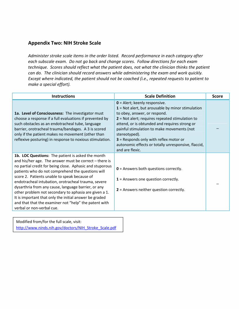
Appendix Two: NIH Stroke Scale
Administer stroke scale items in the order listed. Record performance in each category after
each subscale exam. Do not go back and change scores. Follow directions for each exam
technique. Scores should reflect what the patient does, not what the clinician thinks the patient
can do. The clinician should record answers while administering the exam and work quickly.
Except where indicated, the patient should not be coached (i.e., repeated requests to patient to
make a special effort).
Instructions Scale Definition Score
1a. Level of Consciousness: The investigator must
choose a response if a full evaluations if prevented by
such obstacles as an endotracheal tube, language
barrier, orotracheal trauma/bandages. A 3 is scored
only if the patient makes no movement (other than
reflexive posturing) in response to noxious stimulation.
0 = Alert; keenly responsive.
1 = Not alert, but arousable by minor stimulation
to obey, answer, or respond.
2 = Not alert; requires repeated stimulation to
attend, or is obtunded and requires strong or
painful stimulation to make movements (not
stereotyped).
3 = Responds only with reflex motor or
autonomic effects or totally unresponsive, flaccid,
and are flexic.
_
1b. LOC Questions: The patient is asked the month
and his/her age. The answer must be correct – there is
no partial credit for being close. Aphasic and stuporous
patients who do not comprehend the questions will
score 2. Patients unable to speak because of
endotracheal intubation, orotracheal trauma, severe
dysarthria from any cause, language barrier, or any
other problem not secondary to aphasia are given a 1.
It is important that only the initial answer be graded
and that that the examiner not “help” the patent with
verbal or non-verbal cue.
0 = Answers both questions correctly.
1 = Answers one question correctly.
2 = Answers neither question correctly.
_
Modified from/for the full scale, visit:
http://www.ninds.nih.gov/doctors/NIH_Stroke_Scale.pdf

Appendix Two: NIH Stroke Scale (cont.)
Instructions Scale Definition Score
1c. LOC Commands: The patient is asked to open and
close the eyes and then to grip and release the non-
paretic hand. Substitute another one step command if
the hands cannot be used. Credit is given if an
unequivocal attempt is made but not completed due to
weakness. If the patient does not respond to
command, the task should be demonstrated to him or
her (pantomime), and the result scored (i.e., follows
none, one or two commands). Patients with trauma,
amputation, or other physical impediments should be
given suitable one-step commands. Only the first
attempt is scored.
0 = Performs both tasks correctly.
1 = Performs one task correctly.
2 = Performs neither task correctly.
_
2. Best Gaze: Only horizontal eye movements will be
tested. Voluntary or reflexive (oculocephalic) eye
movements will be scored, but caloric testing is not
done. If the patient has a conjugate deciation of the
eyes that can be overcome by voluntary or reflexive
activity, the score will be 1. If a patients has an isolated
peripheral nerve paresis (CN III, IV, or VI), score a 1.
Gaze is testable in all aphasic patients. Patients with
ocular trauma, bandages, pre-existing blindness, or
other disorder of visual acuity or fields should be tested
with reflexive movements, and a choice made by the
investigator. Establishing eye contact and then moving
about the patient from side to side will occasionally
clarify the presence of a partial gaze palsy.
0 = Normal.
1 = Partial gaze palsy; gaze is abnormal in one or
both eyes, but forced deviation or total gaze paresis
is not present.
2 = Forced deviation, or total gaze paresis not
overcome by the oculocephalic maneuver.
_
3. Visual: Visual fields (upper and lower quandrants)
are tested by confrontation, using finger counting or
visual threat, as appropriate. Patients may be
encouraged, but if they look at the side of the moving
fingers appropriately, this can be scored as normal. If
there is unilateral blindness or enucleation, visual fields
in the remaining eye are scored. Score 1 only if a clear-
cut asymmetry, including quadrantanopia is found. If
patient is blind from any cause, score 3. Double
simultaneous stimulation is performed at this point. If
there is extinction, patient receives a 1, and the results
are used to respond to item 11.
3. Visual: Visual fields (upper and lower quandrants)
are tested by confrontation, using finger counting or
visual threat, as appropriate. Patients may be
encouraged, but if they look at the side of the
moving fingers appropriately, this can be scored as
normal. If there is unilateral blindness or
enucleation, visual fields in the remaining eye are
scored. Score 1 only if a clear-cut asymmetry,
including quadrantanopia is found. If patient is blind
from any cause, score 3. Double simultaneous
stimulation is performed at this point. If there is
extinction, patient receives a 1, and the results are
used to respond to item 11.
_

Appendix Two: NIH Stroke Scale (cont.)
Instructions Scale Definition Score
4. Facial Palsy: Ask – or use pantomime to encourage
– the patient to show teeth or raise eyebrows and close
eyes. Score symmetry of grimace in response to
noxious stimuli in the poorly responsive or non-
comprehending patient. If facial trauma/bandages,
orotracheal tube, tape or other physical barriers
obscure the face, these should be removed to the
extent possible.
0 = Normal symmetrical movements.
1 = Minor paralysis (flattened nasolabial fold,
asymmetry on smiling).
2 = Partial paralysis (total or near-total paralysis
of lower face).
3 = Complete paralysis of one or both sides
(absence of facial movement in the upper and
lower face).
_
5. Motor Arm: The limb is placed in the appropriate
position: extend the arms (palms down) 90 degrees (if
sitting) or 45 degrees (if supine). Drift is scored if the
arm falls before 10 seconds. The aphasic patient is
encouraged using urgency in the voice and pantomime
with the non-paretic arm. Each limb is tested in turn,
beginning with the non-paretic arm. Only in the case of
amputation or joint fusion at the shoulder, the
examiner should record the score as untestable (UN),
and clearly write the explanation for this choice.
0 = No drift; limb holds 90 (or 45) degrees for full
10 seconds.
1 = Drift; limb holds 90 (or 45) degrees, but drifts
down before the full 10 seconds; does not hit bed
or other support.
2 = Some effort against gravity; limb cannot get
to or maintain (if cued) 90 (or 45) degrees, drifts
down to bed, but has some effort against gravity.
3 = No effort against gravity; limb falls.
4 = No movement.
UN = Amputation or joint fusion, explain:
_________________
5a. Left Arm
5b. Right Arm
_
6. Motor Leg: The limb is placed in the appropriate
position: hold the leg at 30 degrees (always tested
supine). Drift is scored if the leg falls before 5 seconds.
The aphasic patient is encouraged using urgency in the
voice and pantomime, but not noxious stimulation.
Each limb is tested in turn, beginning with the non-
paretic leg. Only in the case of amputation or joint
fusion at the hip, the examiner should record the score
as untestable (UN), and clearly write the explanation
for this choice.
0 = No drift; leg holds 30-degree position for full 5
seconds.
1 = Drift; leg falls by the end of the 5 second
period, but does not hit bed.
2 = Some effort against gravity; leg falls to bed by
5 seconds, but has some effort against gravity.
3 = No effort against gravity; leg falls to bed
immediately.
4 = No movement.
UN = Amputation or joint fusion, explain:
_________________
5a. Left Leg
5b. Right Leg
_

Appendix Two: NIH Stroke Scale (cont.)
Instructions Scale Definition Score
7. Limb Ataxia: This item is aimed at finding evidence
of a unilateral cerebellar lesion. Test with eyes open.
In case of visual defect, ensure testing is done in intact
visual field. The finger-nose-finger and heel-shin tests
are performed on both sides, and ataxias is scored only
if present out of proportion to weakness. Ataxia is
absent in the patient who cannot understand or is
paralyzed. Only in the case of amputation or joint
fusion, the examiner should record the score as
untestable (UN), and clearly write the explanation for
this choice. In case of blindness, test by having the
patient touch nose from extended arm position.
0 = Absent.
1 = Present in one limb.
2 = Present in two limbs.
UN = Amputation or joint fusion, explain:
__________________
_
8. Sensory: Sensation or grimace to pinprick when
tested, or withdrawal from noxious stimulus in the
obtunded or aphasic patient. Only sensory loss
attributed to stroke is scored as abnormal and the
examiner should test as many body areas (arms [not
hands], legs, trunk, face) as needed to accurately check
for hemisensory loss. A score of 2, “severe or total
sensory loss,” should only be given when a severe or
total loss of sensation can be clearly demonstrated.
Stuporous and aphasic patients will, therefore,
probably score 1 or 0. The patient with brainstem
stroke who has bilateral loss of sensation is scored 2. If
the patient does not respond and is quadriplegic, score
2. Patients in a coma (item 1a=3) are automatically
given a 2 on this item.
0 = Normal; no sensory loss.
1 = Mild-to-moderate sensory loss; patient feels
pinprick is less sharp or is dull on the affected
side; or there is a loss of superficial pain with
pinprick, but patient is aware of being touched.
2 = Sever to total sensory loss; patient is not
aware of being touched in the face, arm, and leg.
_

Appendix Two: NIH Stroke Scale (cont.)
Instructions Scale Definition Score
9. Best Language: A great deal of information about
comprehension will be obtained during the preceding
sections of the examination. For this scale item, the
patient is asked to describe what is happening in the
attached picture, to name the items on the attached
naming sheet and to read from the attached list of
sentences*. Comprehension is judged from responses
here, as well as to all of the commands in the preceding
general neurological exam. If visual loss interferes with
the tests, ask the patient to identify objects placed in the
hand, repeat, and produce speech. The intubated
patient should be asked to write. The patient in a coma
(item 1a=3) will automatically score a 3 on this item. The
examiner must choose a score for the patient with
stupor or limited cooperation, but a score of 3 should be
used only if the patient is mute and follows no one-step
commands.
*For the full scale/naming sheet, visit:
http://www.ninds.nih.gov/doctors/NIH_Stroke_Scale.pdf
0 = No aphasia; normal.
1 = Mild-to-moderate aphasia; some obvious
loss of fluency or facility of comprehension,
without significant limitation on ideas expressed
or form of expression. Reduction of speech
and/or comprehension, however, makes
conversation about provided materials difficult
or impossible. For example, in conversation
about provided materials, examiner can identify
picture or naming card content from patient’s
response.
2 = Severe aphasia; all communication is
through fragmentary expression; great need for
inference, questioning, and guessing by the
listener. Range of information that can be
exchanged is limited; listener carries burden on
communication. Examiner cannot identify
materials provided from patient response.
3 = Mute, global aphasia; no usable speech or
auditory comprehension.
_
10. Dysarthria: If patient is thought to be normal, an
adequate sample of speech must be obtained by asking
patient to read or repeat words from the attached list*.
If the patient has severe aphasia, the clarity of
articulation of spontaneous speech can be rated. Only if
the patient is intubated or has other physical barriers to
producing speech, the examiner should record the score
as untestable (UN), and clearly write an explanation for
this choice. Do not tell the patient why he or she is
being tested.
0 = Normal.
1 = Mild-to-moderate dysarthria; patient slurs at
least some words and, at worst, can be
understood with some difficulty.
2 = Severe dysarthria; patient’s speech is so
slurred as to be unintelligible in the absence of
or out of proportion to any dysphasia, or is
mute/anarthric.
UN = Intubated or other physical barrier,
explain: _________________
_
11. Extinction and Inattention (formerly Neglect):
Sufficient information to identify neglect may be
obtained during the prior testing. If the patient has a
severe visual loss preventing double simultaneous
stimulation, and the cutaneous stimuli are normal, the
score is normal. If the patient has aphasia but does not
appear to attend to both sides, the score is normal. The
presence of visual spatial neglect or anosagnosia may
also be taken as evidence of abnormality. Since the
abnormality is scored only if present, the item is never
untestable.
0 = No abnormality.
1 = Visual, tactile, auditory, spatial, or personal
inattention or extinction to bilateral
simultaneous stimulation in one of the sensory
modalities.
2 = Profound hemi-inattention or extinction to
more than one modality; does not recognize
own hand or orients to only one side of space.
_

Appendix Three: Activities of Daily Living
Activities in ADL and IADL
ADL IADL
Mobility
• Bed mobility
• Wheelchair mobility
• Transfers
• Ambulation
• Stair climbing
Home Management
• Shopping
• Meal planning
• Meal preparation
• Cleaning
• Laundry
• Child care
Self-Care
• Dressing
• Self-feeding
• Toileting
• Bathing
• Grooming
Community Living Skills
• Money/financial management
• Use of public transportation
• Driving
• Shopping
• Access to recreational activities
Communication
• Writing
• Typing/computer use
• Telephoning
• Using special communication devices
Health Management
• Handling medication
• Knowing health risks
• Making medication appointments
Environmental Hardware
• Keys
• Faucets
• Light switches
• Windows/doors
Safety Management
• Fire safety awareness
• Ability to call 911
• Response to smoke detector
• Identification of dangerous situations
Modified from: Pedretti LQ. Occupational Therapy: Practice Skills for Physical Dysfunction. 4th
ed. St. Louis: Mosby; 1996.

References
American Speech Language Hearing Association (2011). Swallowing Disorders In Adults.
Retrieved November 28, 2011 from:
http://www.asha.org/public/speech/swallowing/SwallowingAdults.htm
Carnaby-Mann, G.,Lenius, k. & Crary, M. (2007). Update on Assessment and Management of
Dysphagia Post Stroke. Northeast Florida Medicine, 58 (2), p. 31-34. Retrieved November 22,
2011 from: http://www.dcmsonline.org/jax-
medicine/2007journals/StrokeTherapies/dysphagia.pdf
McGinnes, A., Easton,S., Williams, J. & Neville, J. (2009).The role of the community stroke
rehabilitation nurse. British Journal of Nursing, 19 (16), p. 1033 -1038.
Matthews, S. (2009). Interventions for Rehabilitation Post-Stroke and the Contribution of t he
Nursing Staff. Journal of the Australasian Rehabilitation Nurses' Association [JARNA],12 (3), p.7-
11.
National Institute of Neurological Disorders & Stroke [NINDS]. (2011). Post-Stroke Rehabilitation
Fact Sheet. Retrieved November 15, 2011 from:
http://www.ninds.nih.gov/disorders/stroke/poststrokerehab.htm
National Institute of Health (2011). Stroke Scale. Retrieved November 28, 2011 from:
http://www.ninds.nih.gov/doctors/NIH_Stroke_Scale.pdf
National Stroke Association (2009). Stroke Facts: Recovery After Stroke: Bladder & Bowel
Function. Retrieved November 20, 2011 from:
http://www.stroke.org/site/DocServer/NSAFactSheet_BowelandBladder.pdf?docID=984
Nursing Times.net (2006). Central post-stroke pain.Retrieved November 15, 2011 from:
http://www.nursingtimes.net/nursing-practice-clinical-research/central-post-stroke-
pain/203228.article
Silverman, L.,Restrepo, L. & Mathews, G. (2002). Poststroke Seizures. Archives of Neurology, 59,
P. 195-201. Retrieved November 22, 2011 from: http://archneur.ama-
assn.org/cgi/content/full/59/2/195

References (cont.)
Summers, D., Leonard, A., Wentworth, D. et al. (2009). Comprehensive overview of nursing &
interdisciplinary care of the acute ischemic stroke patient. AHA Scientific Statement, 40, p.
2911-2944. Retrieved November 29, 2011 from:
http://stroke.ahajournals.org/content/40/8/2911.full
Veterans Association & Department of Defense (2010). Summary of Clinical Practice Guidelines
for the Management of Stroke Rehabilitation. Version 3.0. Retrieved online from: Full guideline
available at: http://www.healthquality.va.gov
Weerdesteyn,V.,de Niet,M.,van Duijnhoven,H. & Geurts,A. (2008).Falls in individuals with
stroke. Journal of Rehabilitation Research & Development, 45 (8), P. 1195–1214. Retrieved
November 28, 2011 from:
http://www.rehab.research.va.gov/jour/08/45/8/pdf/weerdesteyn.pdf
Zorowitz, R., Smout, J., Gassaway, J. & Horn, S. (2005). Prophylaxis for and treatment of deep
venous thrombosis after stroke: the Post-Stroke Rehabilitation Outcomes Project (PSROP).
Physical Medicine and Rehabilitation, 12, (4), p.1-10. Retrieved from:
http://www.ncbi.nlm.nih.gov/pubmed/16698732

Please Read
This publication is intended solely for the use of healthcare professionals taking this course, for
credit, from RN.com. It is designed to assist healthcare professionals, including nurses, in
addressing many issues associated with healthcare. The guidance provided in this publication is
general in nature, and is not designed to address any specific situation. This publication in no
way absolves facilities of their responsibility for the appropriate orientation of healthcare
professionals.
Hospitals or other organizations using this publication as a part of their own orientation
processes should review the contents of this publication to ensure accuracy and compliance
before using this publication. Hospitals and facilities that use this publication agree to defend
and indemnify, and shall hold RN.com, including its parent(s), subsidiaries, affiliates,
officers/directors, and employees from liability resulting from the use of this publication. The
contents of this publication may not be reproduced without written permission from RN.com.