the role of bacterial biofi lms in chronic infections · the role of bacterial biofi lms in...
TRANSCRIPT
ACTA PATHOLOGICA, MICROBIOLOGICA ET IMMUNOLOGICA SCANDINAVICA
The Role of Bacterial Biofi lms in Chronic Infections
T. Bjarnsholt
apms_121_s136_title.indd 1apms_121_s136_title.indd 1 4/9/2013 2:32:38 PM4/9/2013 2:32:38 PM

Denne afhandling er af Det Sundhedsvidenskabelige Fakultet ved Københavns Universitet antaget til offentligt at forsvares for den medicinske doktorgrad. København, den 31 maj kl 13:00, Haderup Auditoriet, Panum, Blegdamsvej 3B, 2200 København N.
Dekan Ulla Wewer
apms_121_s136_title.indd 2apms_121_s136_title.indd 2 4/9/2013 2:32:40 PM4/9/2013 2:32:40 PM

Absolutely no benefit can be derived from involving oneself with the natural sciences. One standsthere defenseless, with no control over anything. The researcher immediately begins to distractone with his details: now one is to go to Australia, now to the moon; now into an undergroundcave; now, by Satan, up the arse—to look for an intestinal worm; now the telescope must beused; now the microscope: who the devil can endure it?.*
Med Naturvidenskaberne kan det slet ikke hjælpe at indlade [sig]. Man staaer der værgeløs ogkan aldeles ikke controlere. Forskeren begynder strax at adsprede med sine Enkeltheder, nu skalman til Australien nu til Maanen, nu ned i en Hule under Jorden, nuFanden i Vold i Røven –efter en Indvoldsorm; nu skal Teleskopet bruges, nu Mikroskopet: hvo Satan kan holde det ud.
Søren Kierkegaard 1846
*Søren Kierkegaard: A Biography, by Joachim Garff, translated by Bruce H. Kirmmse. 867pages, Princeton University Press, ISBN: 978069112788

Contents
Preface iBasis of the thesis (articles I–XV) ii1. Introduction 1
1.1. The development of the biofilm era 21.2. What is a biofilm? 21.3. Chronic infections 3
2. Purpose of the studies 43. How biofilms are formed 4
3.1. Pseudomonas aeruginosa as a model biofilm 43.2. Why biofilms are formed 63.3. The scaffold (united we stand, divided we fall) 6
4. Biofilm-related phenotypes 84.1. Decreased antimicrobial susceptibility 84.2. Predator and phagocyte tolerance 94.3. Quorum sensing 9
5. Environmental biofilms 115.1. Submerged or flooded surfaces 115.2. Activated sludge 115.3. Rhizosphere 115.4. Oral biofilms 115.5. Intestinal biofilms 125.6. Other environmental biofilm habitats 13
6. Medical biofilms 136.1. Infections 136.2. Acute infection 146.3. Biofilm-related infections 14
6.3.1. Chronic wounds 146.3.2. Cystic fibrosis 156.3.3. Chronic otitis media 176.3.4. Tissue fillers 176.3.5. Additional chronic biofilm infections 19
7. Clinical microbiology: the need for diagnostics and treatments for biofilm infections 197.1. Diagnostics 197.2. Treatment 207.3. Prevention 20
8. Scientific extrapolations among environments 208.1. In vitro and in vivo biofilms 21
8.1.1. Mushroom-like structures 218.2. To surface or not to surface 228.3. How can biofilms be studied? 238.4. The opportunity of sociomicrobiology 25
8.4.1. Succession or opportunity 268.4.2. Low diversity and high pathogenicity 28
8.5. Diagnosing biofilms in infections 289. Discussion 31
10. Conclusion 3311. Future perspectives 3412. Reference list 3613. Summary in Danish 50
Cover image: Taking out the defender. The in vivo interaction between a Pseudomonas aeruginosabiofilm, on a silicone implant, and the responding polymorphonuclear leukocytes.SEM imaging depicts the interaction at day 1 post insertion of the implant in the peritoneal cav-
ity of a mouse. The leukocytes (yellow) are damaged with obvious cavities in the cell membraneand killed by the bacteria (cyan) following contact with the biofilm.

PREFACE
The work behind the present thesis was initiated during my PhD studies on bacterial communica-tion in biofilms and chronic infections. The study of laboratory biofilms and my subsequent inqui-ries into their impact in cystic fibrosis sparked my curiosity about how bacteria succeed in biofilmsand the role they play in chronic infections. My first grant application for the project ‘The role ofbiofilm in chronic infections’ was supported by the Lundbeck Foundation and the CarlsbergFoundation, which meant I was able to proceed and study this phenomenon thanks to both foun-dations. Throughout this process, I received help from and collaborated with many people towhom I am most grateful. First of all, I would like to thank Niels Høiby because I am so gratefulfor all your help, support, coaching, constant encouragement, and for inspiring me to write thisthesis. I also want to thank Michael Givskov for accepting me as a PhD student, your supportand for continuously sharing your laboratory facilities and the help of your staff. I would also liketo thank my good friend and colleague Peter Østrup Jensen, because we complement each otherfantastically, and without you I would not be where I am today.Another person who has helped me for many years is Anne Kirstine Nielsen and I am so grate-
ful for all your help in the laboratory. I would like to thank my close colleague and friend MortenAlhede for all his scientific help, inspiration, collaboration, and help with the present thesis. Forhelp with the present thesis, I would also like to thank Oana Ciofu, as well as for our fruitful col-laborations. I would like to thank Louise Dahl Christensen and Maria van Gennip (Alhede) forall their hard work with the implant model and experiments in general. For support and inspira-tion, I would like to thank many good friends and colleagues: Bill Costerton, Søren Molin, MetteBurmølle, Søren J. Sørensen, Claus Moser, Tim Holm Jakobsen, Claus Bøgelund Andersen, KlausQvortup, Klaus Kirketerp-Møller, Tanja Pressler, Christine Rønne Hansen, Trine Rolighed Thom-sen, Per Halkjær, Vibeke Rudkjøbing, Lise H Christensen, Steen Seier Poulsen, Nils Erik Samdal,Michael Tvede. Hans Petter Hougen, Henrik Calum, Matt Parsek, Mark Shirtliff, Marie Allesen-Holm, Claus Sternberg, Michael K€uhl, Preben Homøe, Helle Krogh Johansen, Tim Tolker-Niel-sen, and Gerd D€oring. In particular, I would like to thank my newly started research group ofKasper Nørskov Kragh, €Ozge Er, Majken Sønderholm, Stephanie Geisler Crone, Steffen RobertEickhardt-Sørensen, Morten Alhede, and Anne Kirstine Nielsen. We will rock and roll the future.Thank you guys.I would also like to thank Lars Christophersen, Ulla R Johansen, Lena Noerregaard, Katja
Bloksted, Tina Rathmann, Jette Pedersen, Margit Bæksted, and Tina Wassermann for their helpevery day. I would like to thank Peter Kindt Fridorff-Jens for his friendship and help in creatingour online biofilm course (www.biofilmcourse.ku.dk). The world of science is not always easy tonavigate and I would like to thank Charlotte Lex for her mentorship.I would like to thank the company AdvanDx for their support and constant supply of PNA
FISH probes. In addition, I would like to thank all the co-authors of the manuscripts included inthis thesis.I would like to thank Museum of Copenhagen and Marianne Bisballe, for giving me permission
to use their illustration of the ‘Drop of water’ by H.C. Andersen (Figure 23), Grazyna Hahn Poul-sen for the illustration of my river analogy (Figure 21) and Claus Lex for help understandingSøren Kierkegaard, and life in general.Finally, I would like to express my uttermost gratitude and love to my wonderful and beautiful
wife Marianne for all her support and help, my son Rasmus, my daugther Milla, my mother,father, Mette, Annie, Niels, and the rest of my fantastic family, because without you I amnothing.

BASIS OF THE THESIS
The main part of the thesis is based on the following papers.(They are marked as bold in the text.)
I. Jensen PØ, Bjarnsholt T, Phipps R, Rasmussen TB, Calum H, Christoffersen L, Moser C,Williams P, Pressler T, Givskov M, Høiby N. Rapid necrotic killing of polymorphonuclearleukocytes is caused by quorum-sensing-controlled production of rhamnolipid by Pseudo-monas aeruginosa. Microbiology 2007 May;153(Pt 5):1329-38.
II. Christensen LD, Moser C, Jensen PØ, Rasmussen TB, Christophersen L, Kjelleberg S, Ku-mar N, Høiby N, Givskov M, Bjarnsholt T. Impact of Pseudomonas aeruginosa quorumsensing on biofilm persistence in an in vivo intraperitoneal foreign-body infection model.Microbiology 2007 Jul;153:2312-20.
III. Bjarnsholt T, Kirketerp-Møller K, Kristiansen S, Phipps R, Nielsen AK, Jensen PØ, HøibyN, Givskov M. Silver against Pseudomonas aeruginosa biofilms. APMIS 2007;115:921–8.
IV. Bjarnsholt T, Kirketerp-Møller K, Jensen PØ, Madsen KG, Phipps R, Krogfelt K, HøibyN, Givskov M. Why chronic wounds won’t heal: a novel hypothesis. Wound Repair andRegeneration 2008 Jan–Feb;16(1):2-10.
V. Kirketerp-Møller K; Jensen PØ, Fazli M, Madsen KG, Pedersen J, Moser C, Tolker-Niel-sen T, Høiby N, Givskov M, Bjarnsholt T. Distribution, organization and ecology of bacte-ria in chronic wounds. J Clin Microbiol 2008 Aug;46(8):2717-22.
VI. Bjarnsholt T, Jensen PØ, Fiandaca MJ, Pedersen J, Hansen CR, Andersen CB, Pressler T,Givskov M, Høiby N. Pseudomonas aeruginosa biofilms in the respiratory tract of cysticfibrosis patients. Pediatr Pulmonol 2009 Jun;44(6):547-58.
VII. Bjarnsholt T, Tolker-Nielsen T, Givskov M, Janssen M, Christensen LH. Detection of bac-teria by fluorescence in situ hybridization in culture-negative soft tissue filler lesions. Der-matol Surg 2009;35:1620–1624.
VIII. Homøe P, Bjarnsholt T, Wessman M, Sørensen HC, Johansen HK. Morphological evidenceof biofilm formation in Greenlanders with chronic suppurative otitis media. Eur ArchOtorhinolaryngol 2009 Oct;266(10).
IX. Van Gennip M, Christensen LD, Alhede M, Phipps R, Jensen PØ, Christophersen L, PampSJ, Moser C, Mikkelsen PJ, Koh AY, Tolker-Nielsen T, Pier GB, Høiby N, Givskov M,Bjarnsholt T. Inactivation of the rhlA gene in Pseudomonas aeruginosa prevents rhamnoli-pid production, disabling the protection against polymorphonuclear leukocytes. APMIS2009 Jul;117(7):537-46.
X. Werth"en M, Henriksson L, Jensen PØ, Sternberg C, Givskov M, Bjarnsholt T. An in vitromodel of bacterial infections in wounds and other soft tissues. APMIS 2010 Feb;118(2):156-64.
XI. Bjarnsholt T, Jensen PØ, Jakobsen TH, Phipps R, Nielsen AK, Rybtke MT, Tolker-NielsenT, Givskov M, Høiby N, Ciofu O. Scandinavian cystic fibrosis study consortium. Quorumsensing and virulence of Pseudomonas aeruginosa during lung infection of cystic fibrosispatients. PLoS One 2010 Apr 12;5(4):e10115.
XII. Alhede M, Kragh K, Qvortrup K, Allesen-Holm M, van Gennip M, Christensen LD, Jen-sen PØ, Nielsen AK, Parsek M, Wozniak D, Molin S, Tolker-Nielsen T, Høiby N, GivskovM, Bjarnsholt T. Phenotypes of non-attached Pseudomonas aeruginosa aggregates resemblesurface attached biofilm. PLoS One 2011;6(11):e27943.
XIII. van Gennip M, Christensen LD, Qvortrup K, Alhede M, Jensen PØ, Høiby N, GivskovM, Bjarnsholt T. Interactions between polymorphonuclear leucocytes and Pseudomonasaeruginosa biofilms on silicone implants in vivo. Infect Immun. 2012 Aug;80(8):2601-7
IVX. Rudkjøbing VB, Thomsen TR, Alhede M, Kragh KN, Nielsen PH, Johansen UR, GivskovM, Høiby N, Bjarnsholt T. True microbiota involved in chronic lung infection of cystic

fibrosis patients found by culturing and 16S rRNA gene analysis. J Clin Microbiol 2011Dec;49(12):4352-5.
XV. Rudkjøbing VB, Thomsen TR, Alhede M, Kragh KN, Nielsen PH, Johansen UR, PoulsenSS, Givskov M, Høiby N, Bjarnsholt T. The microorganisms in chronically infected end-stage and non-end-stage cystic fibrosis patients. FEMS Immunol Med Microbiol. 2012Jul;65(2):236-44

The role of bacterial biofilms in chronic infections
THOMAS BJARNSHOLT
Københavns Universitet, København N, Denmark
Bjarnsholt T. The role of bacterial biofilms in chronic infections. APMIS 2013; 121 (Suppl. 136): 1–54.
Acute infections caused by pathogenic bacteria have been studied extensively for well over 100 years. Theseinfections killed millions of people in previous centuries, but they have been combated effectively by thedevelopment of modern vaccines, antibiotics and infection control measures. Most research into bacterialpathogenesis has focused on acute infections, but these diseases have now been supplemented by a new cat-egory of chronic infections caused by bacteria growing in slime-enclosed aggregates known as biofilms.Biofilm infections, such as pneumonia in cystic fibrosis patients, chronic wounds, chronic otitis media andimplant- and catheter-associated infections, affect millions of people in the developed world each year andmany deaths occur as a consequence. In general, bacteria have two life forms during growth and prolifera-tion. In one form, the bacteria exist as single, independent cells (planktonic) whereas in the other form,bacteria are organized into sessile aggregates. The latter form is commonly referred to as the biofilmgrowth phenotype. Acute infections are assumed to involve planktonic bacteria, which are generally treat-able with antibiotics, although successful treatment depends on accurate and fast diagnosis. However, incases where the bacteria succeed in forming a biofilm within the human host, the infection often turns outto be untreatable and will develop into a chronic state. The important hallmarks of chronic biofilm-basedinfections are extreme resistance to antibiotics and many other conventional antimicrobial agents, and anextreme capacity for evading the host defences. In this thesis, I will assemble the current knowledge on bio-films with an emphasis on chronic infections, guidelines for diagnosis and treatment of these infections,before relating this to my previous research into the area of biofilms. I will present evidence to support aview that the biofilm lifestyle dominates chronic bacterial infections, where bacterial aggregation is thedefault mode, and that subsequent biofilm development progresses by adaptation to nutritional and envi-ronmental conditions. I will make a series of correlations to highlight the most important aspects of bio-films from my perspective, and to determine what can be deduced from the past decades of biofilmresearch. I will try to bridge in vitro and in vivo research and propose methods for studying biofilms basedon this knowledge. I will compare how bacterial biofilms exist in stable ecological habitats and opportunis-tically in unstable ecological habitats, such as infections. Bacteria have a similar lifestyle (the biofilm) inboth habitats, but the fight for survival and supremacy is different. On the basis of this comparison, I willhypothesize how chronic biofilm infections are initiated and how bacteria live together in these infections.Finally, I will discuss different aspects of biofilm infection diagnosis. Hopefully, this survey of currentknowledge and my proposed guidelines will provide the basis and inspiration for more research, improveddiagnostics, and treatments for well-known biofilm infections and any that may be identified in the future.
Key words: Biofilms; chronic infections
Thomas Bjarnsholt, Københavns Universitet, SUND, Blegdamsvej 3b, 24.1, 2200 København N, Denmark.e-mail: [email protected]
1. INTRODUCTION
Bacterial growth is characterized by two phe-notypes, single cells (planktonic) or sessileaggregates. The later is commonly referred to
as the biofilm mode of growth. Many diver-gent definitions of bacterial biofilms exist, butall agree that biofilms are composed of multi-ple bacteria that form a consortium. The defi-nitions found in the literature differ mainly in
1
APMIS 121 (Suppl. 136): 1–51 © 2013 APMIS Published by Blackwell Publishing Ltd.DOI 10.1111/apm.12099

terms of whether cells have to be attached to asurface and whether bacteria form a structuredcommunity. The topic of this thesis is medicalmicrobiology and biofilm infections, so I havedefined a biofilm as follows:
A coherent cluster of bacterial cellsembedded in a matrix, which is moretolerant of most antimicrobials and hostdefences compared with planktonicbacterial cells (1).
1.1 The development of the biofilm era
Conventional microbiology from 1880 until themiddle of the twentieth century is popularlyreferred to as ‘the pure culture period’ (2).During this period, bacteria were viewed sim-ply as free-floating single cells, which are alsoreferred to as planktonic. Most studies of bac-terial characterization involved the propaga-tion of bacteria in liquid media in test tubes oron agar plates. This seems very peculiar givenour current knowledge, because it is estimatedthat <0.1% of the total microbial biomass ispresent as a planktonic phenotype (3, 4).The first observation of surface-associated
aggregated bacteria was made by Antonie vanLeeuwenhoek (5) in 1684 when he describedthe ‘animals’ present in the plaque on teeth.Photomicrographs of aggregating bacteriawere produced in 1933 by Henrici (6) and heobserved that ‘It is quite evident that for themost part water bacteria are not free floatingorganisms, but grow upon submerged sur-faces’. For the purpose of this thesis, micro-biology may be divided into two fields:environmental and medical. Environmentalmicrobiology acknowledged the aggregation ofbacteria almost 20 years before it was evenconsidered medical microbiology, with theexception of the field of odontology. Aggre-gates, or flocs, of bacteria have long been usedin wastewater treatment plants and the firstarticle to use the term biofilm was publishedby Rogovska et al. in Microbiology-USSR(MIKROBIOLOGIYA) during 1961 (7).Publications in the medical field began to
acknowledge clumps or heaps of bacteria in1977 (8), when Høiby described aggregates(heaps) of Pseudomonas aeruginosa in the lungs
of chronically infected cystic fibrosis (CF)patients. In 1978, Costerton et al. (9) describedthe presence of surface adhering bacteriaembedded in a ‘glycocalyx’ (matrix), and in1981 he used the term biofilm for the firsttime, to describe this phenomenon (10). Thephenomenon was reviewed and re-described in1987 by Costerton et al. (11) as a matrix-enclosed mode of growth.In 1993, the American Society for Microbiol-
ogy recognized that the biofilm growth pheno-type was relevant to microbiology (12). As aresult, the biofilm phenotype became increas-ingly accepted as an important bacterial trait.In 1999, Costerton et al. (13) defined a biofilmas ‘a structured community of bacterial cellsenclosed in a self produced polymeric matrix,adherent to a surface’. During the last 15 years,the literature on biofilms has increased dramati-cally in terms of publications (see Fig. 1) andthere are also numerous books on the subject.
1.2 What is a biofilm?
Biofilms have probably been present on theEarth since the first bacteria evolved. In medi-cal microbiology, biofilms are typicallyinvolved in chronic persistent infections. Com-mon bacterial infections were very seriousbefore the antibiotic era, when many peopledied of pneumonia and other acute infectionsthat are now easily cured using antibiotics.Since the development of antibiotics, the world
0
2000
4000
6000
8000
10000
12000
14000
16000
18000
20000
Pu
blic
aƟo
ns
Year
Biofilm publicaƟons in PubMed
Biofilm publicaƟons total
Biofilm publicaƟon per year
Fig. 1. Accumulated publications on biofilms andper year, derived from the search engine PubMed(http://www.ncbi.nlm.nih.gov/pubmed/).
2 © 2013 APMIS Published by Blackwell Publishing Ltd
BJARNSHOLT
has experienced an increase in slow-progress-ing infections with ‘lowgrade’ pathogenesiscompared with acute infections. These slow-progressing infections occur in all age groupswhere patients experience discomfort, fever,and other clinical signs of infection. However,bacteria were often not detected and the effectsof antibiotics were either very disappointing orabsent, resulting in persistent infections. Thebreakthrough in identifying the sources ofthese persistent infections was a series of in vi-tro and in vivo observations made in the 1980s(14). These observations showed that aggrega-tion of bacteria were the cause of slow-pro-gressing infections and Costerton referred tothis phenomenon as bacterial biofilms (9).However, biofilms and their extreme toleranceof antimicrobial agents had been discovered300 years earlier by van Leeuwenhoek (1684)(5). Van Leeuwenhoek observed that animals(bacteria) within the scurf (plaque) on teethwere more resistant to vinegar than animalsfound outside the plaque, which were killed.This is now known to be one of the majorhallmarks of biofilms, i.e., an extreme toler-ance of antimicrobial agents. It must to benoted that biofilm antibiotic tolerance shouldnot be confused with antibiotic resistancebecause, although bacteria within a biofilmtend to survive antibiotic treatment, theybecome susceptible to the treatment when thebiofilm is disrupted (15) (XII) (see section 4.1).Numerous in vitro and in vivo biofilm obser-
vations show that the causes of most persistentinfections are bacterial aggregates or biofilms.The bacteria in these aggregates are physicallyjoined together and they produce an extracel-lular matrix that contains many different typesof extracellular polymeric substances (EPS)including proteins, DNA and polysaccharides.These aggregates can withstand very highdoses of antibiotics that would kill planktoniccells. Their tolerance of host defences is alsodramatically increased. These characteristicsare contained in the biofilm definition pre-sented above. This definition differs in onerespect from most other biofilm definitionsbecause it no longer requires that a biotic orabiotic surface is a hallmark. Many chronicinfections involve surfaces such as infectionson implants, catheters, artificial heart valves,teeth and contact lenses. However, many
observations of non-surface-related infections,such as CF, otitis media, chronic wounds andchronic osteomyelitis, have found the samepatterns without a surface.In 2009, Høiby included ‘persisting pathol-
ogy’ (16) in the definition of chronic infections.
1.3. Chronic infections
Chronic infections have a slower progressionthan acute infections and their symptoms areoften vague (14) (often referred to as lowgrade). They are very difficult, if not impossi-ble, to cure with antibiotics (see section 4.1).Chronic inflammation is usually characterizedby an adaptive inflammatory response, whichis dominated by mononuclear leucocytes andIgG antibodies. In some chronic infections, theinflammatory response is characterized by achronic inflammatory response and continuousrecruitment of polymorphonuclear leucocytes(PMNs). The classic chronic infections beforethe antibiotic era included tuberculosis andleprosy, which slowly degrade the tissue andaffected organs (e.g., lungs) of patients, eventu-ally leading to death.Chronic infections can develop in patients
who suffer from diseases or conditions thatcause deficiencies in the primary defensive bar-riers (innate immunity). This includes disrup-tion of the anatomical (e.g., skin, mucousmembranes and cilia) and physiological (e.g.,temperature and low pH) inflammatory barri-ers, as well as phagocytic defects (e.g., PMNsand macrophages). These deficiencies can bedivided into congenital abnormalities, the pres-ence of foreign bodies and acquired chronicdiseases. The classic example is the chroniclung infection found in patients suffering fromthe genetic disorder CF (see section 6.3.2).These patients have a reduced volume of peri-ciliary fluid in the airways, which impairs thenormal mucociliary clearing of the paranasalsinuses and the lungs, facilitating persistentbacterial infections. The presence of foreignbodies can include artificial limbs and otherbody parts, and indwelling catheters (11, 13,17–19), while injected tissue fillers are alsonow being reported as a site of chronic infec-tions (VII). Acquired chronic diseases includediabetes mellitus, arteriosclerosis of the arteriesof legs and smoking-induced chronic obstruc-
© 2013 APMIS Published by Blackwell Publishing Ltd 3
THE ROLE OF BACTERIAL BIOFILM

tive pulmonary disease (COPD). Thesepatients may be prone to the development of anon-healing wound that is chronically infectedwith bacteria, or the chronic lung infectionsseen in patients suffering from COPD (20, 21).
2. PURPOSE OF THE STUDIES
Since my PhD studies, my scientific researchfocus has been the investigation of the occur-rence and persistence of biofilms in chronicinfections. One of the major aims of this thesiswas to compare the structural and physiologicalcharacteristics of the two major forms of biofilminfections: surface (biotic and abiotic)-relatedand non-surface-related biofilms. Anothermajor question was to elucidate the basis ofhow biofilms tolerate the immune systemattacks, i.e., the so-called ‘frustrated phagocyto-sis’. The third major purpose was to improvethe diagnosis of biofilm-related infections,where the classical microbiological culturemethods are inappropriate, so molecular probesand microscopy were applied to a wide varietyof samples from chronic human infections. Iinvestigated biofilm formation, structure andbiofilm responses to the immune system andantibacterial agents using in vitro methods (I,III, IX, X, XI, XII), which were confirmed within vivo animal models (I, II, IX, XIII). My ques-tions could not be answered entirely by usingavailable models, so I developed new in vitro(X) and in vivo (II, XIII) model systems. I haveused molecular probes and antibodies to ana-lyse the presence, organization and distributionof bacteria and biofilms in chronic infections,based on the identification of bacterial aggre-gates and their matrices. I have applied thesemethods to a large variety of chronic humaninfections including chronic P. aeruginosa infec-tions in CF patients (VI, XIV, XV), soft tissuefillers (VII), chronic otitis media infections(VIII) and chronic wounds (IV, V).
3. HOW BIOFILMS ARE FORMED
3.1. Pseudomonas aeruginosa biofilms as a biofilmmodel
Biofilm developmental processes have beenthoroughly studied using surface-based in vitro
systems (22–28). The most commonly studiedbacterium in this context is P. aeruginosa.Pseudomonas aeruginosa is a Gram-negativerod-shaped bacterium with virtually no specificgrowth requirements. It is a non-fermentativeorganism that is capable of growing with orwithout oxygen. The sequenced genome ofP. aeruginosa contains 5770 open readingframes and ~10% of its genes encode proteinsinvolved in regulatory processes, which makesit a very diverse and adaptable organism (29).The ability of P. aeruginosa to form biofilms
is thought to be one of its main survival strate-gies when infecting a host, and it is consideredto be an important pathogenicity trait (13, 30).The P. aeruginosa in vitro biofilm consists of
microcolonies encapsulated by EPS producedby the bacteria, although most of the biofilmis comprised of water channels that arethought to function as a distribution systemfor nutrients and oxygen (3, 31). An oxygengradient descends from the surface to the sub-stratum (32–34). Pseudomonas aeruginosa canform biofilms on virtually any surface and inany nutritional or environmental conditions.Classically, the in vitro, surface-based biofilm
developmental process can be divided into thefollowing different stages: (i) attachment, (ii)maturation and (iii) dispersion, as suggested bySauer et al. (24) and Klausen et al. (22). Themodel of in vitro surface biofilm developmenthas been subject to changes over the years. Anexperimental-based model of the formation ofin vitro biofilms is shown in Fig. 2 (22).As shown in Fig. 2, cells attach to the sur-
face and form the microcolonies of the biofilm.
Fig. 2. Formation of an in vitro surface-attachedPseudomonas aeruginosa biofilm. Initial attachment isfollowed by clonal growth where one subpopulationof irreversibly attached bacteria forms the base ofmicrocolonies and another subpopulation of non-attached bacteria move on their surface. These non-attached bacteria may eventually climb from the baseto form the caps of mushroom structures [adaptedfrom (22) with permission from the publisher].
4 © 2013 APMIS Published by Blackwell Publishing Ltd
BJARNSHOLT
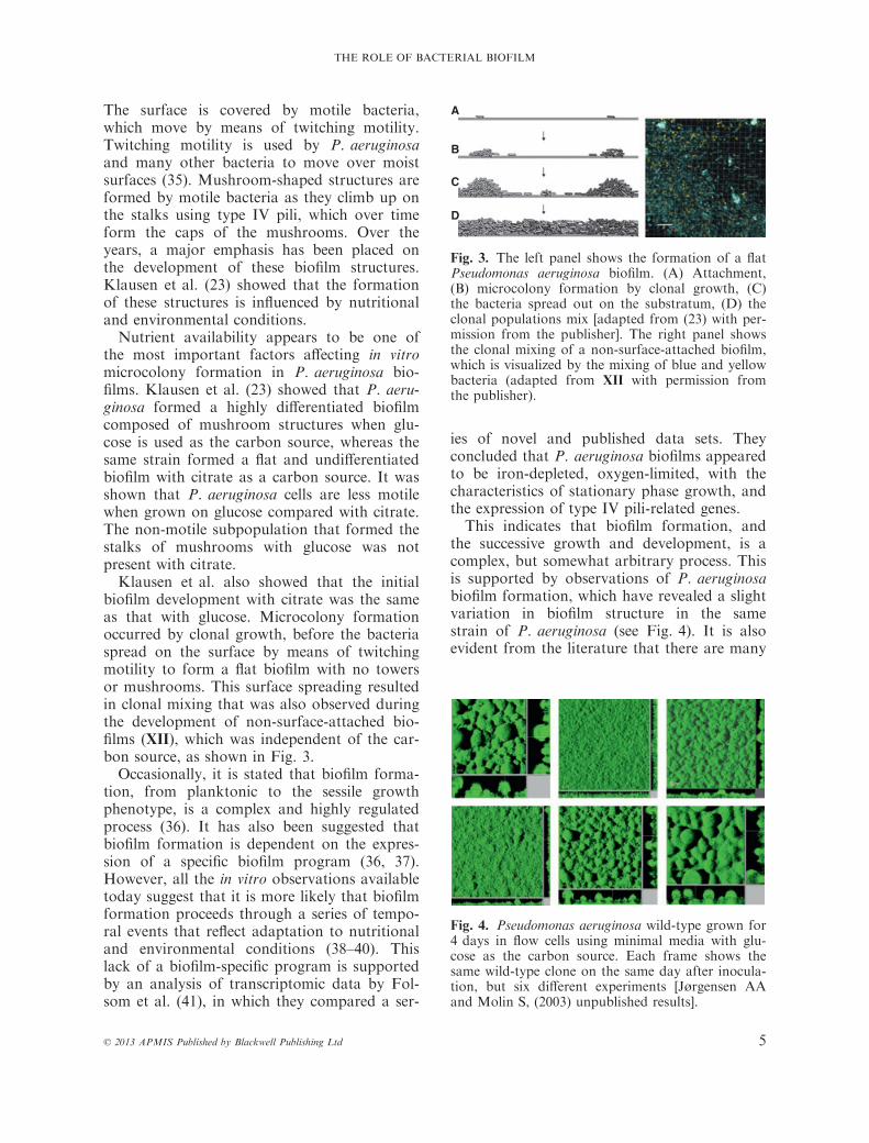
The surface is covered by motile bacteria,which move by means of twitching motility.Twitching motility is used by P. aeruginosaand many other bacteria to move over moistsurfaces (35). Mushroom-shaped structures areformed by motile bacteria as they climb up onthe stalks using type IV pili, which over timeform the caps of the mushrooms. Over theyears, a major emphasis has been placed onthe development of these biofilm structures.Klausen et al. (23) showed that the formationof these structures is influenced by nutritionaland environmental conditions.Nutrient availability appears to be one of
the most important factors affecting in vitromicrocolony formation in P. aeruginosa bio-films. Klausen et al. (23) showed that P. aeru-ginosa formed a highly differentiated biofilmcomposed of mushroom structures when glu-cose is used as the carbon source, whereas thesame strain formed a flat and undifferentiatedbiofilm with citrate as a carbon source. It wasshown that P. aeruginosa cells are less motilewhen grown on glucose compared with citrate.The non-motile subpopulation that formed thestalks of mushrooms with glucose was notpresent with citrate.Klausen et al. also showed that the initial
biofilm development with citrate was the sameas that with glucose. Microcolony formationoccurred by clonal growth, before the bacteriaspread on the surface by means of twitchingmotility to form a flat biofilm with no towersor mushrooms. This surface spreading resultedin clonal mixing that was also observed duringthe development of non-surface-attached bio-films (XII), which was independent of the car-bon source, as shown in Fig. 3.Occasionally, it is stated that biofilm forma-
tion, from planktonic to the sessile growthphenotype, is a complex and highly regulatedprocess (36). It has also been suggested thatbiofilm formation is dependent on the expres-sion of a specific biofilm program (36, 37).However, all the in vitro observations availabletoday suggest that it is more likely that biofilmformation proceeds through a series of tempo-ral events that reflect adaptation to nutritionaland environmental conditions (38–40). Thislack of a biofilm-specific program is supportedby an analysis of transcriptomic data by Fol-som et al. (41), in which they compared a ser-
ies of novel and published data sets. Theyconcluded that P. aeruginosa biofilms appearedto be iron-depleted, oxygen-limited, with thecharacteristics of stationary phase growth, andthe expression of type IV pili-related genes.This indicates that biofilm formation, and
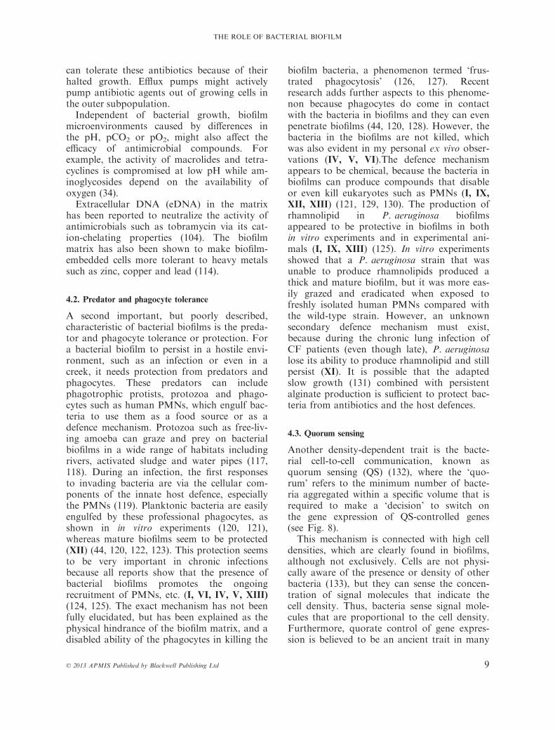
the successive growth and development, is acomplex, but somewhat arbitrary process. Thisis supported by observations of P. aeruginosabiofilm formation, which have revealed a slightvariation in biofilm structure in the samestrain of P. aeruginosa (see Fig. 4). It is alsoevident from the literature that there are many
A
B
C
D
Fig. 3. The left panel shows the formation of a flatPseudomonas aeruginosa biofilm. (A) Attachment,(B) microcolony formation by clonal growth, (C)the bacteria spread out on the substratum, (D) theclonal populations mix [adapted from (23) with per-mission from the publisher]. The right panel showsthe clonal mixing of a non-surface-attached biofilm,which is visualized by the mixing of blue and yellowbacteria (adapted from XII with permission fromthe publisher).
Fig. 4. Pseudomonas aeruginosa wild-type grown for4 days in flow cells using minimal media with glu-cose as the carbon source. Each frame shows thesame wild-type clone on the same day after inocula-tion, but six different experiments [Jørgensen AAand Molin S, (2003) unpublished results].
© 2013 APMIS Published by Blackwell Publishing Ltd 5
THE ROLE OF BACTERIAL BIOFILM

discrepancies as to how and when mature bio-film develop during laboratory experiments(24, 40, 42–48).These arbitrary growth patterns might be
caused by the increased mutation rates foundin biofilms, as suggested by Conibear et al.(49). This study found that mutation frequen-cies were elevated in the microcolony struc-tures, which explained the heterogeneity ofbiofilm development.A biofilm is thought to maintain equilibrium
via growth and dispersal. Dispersal is believedto occur either as single cells or as small mi-crocolonies that are torn from the biofilm, asshown in Fig. 5 (13, 50, 51). The mechanismof dispersion is not fully understood, but Cos-terton et al. (13) suggested that planktonic dis-persion may be a programmed process,whereas clusters are torn away by shear forces(Fig. 5) and/or by prophage-mediated celldeath, as proposed by Webb et al. (52).Dispersal has severe implications for medical
biofilms because it provides a mechanismwhereby biofilm bacteria can spread through-out an infected organ, or to the whole body.This is the mechanism whereby a chronicinfection can cause an acute blood streaminfection, which sometimes occurs in patientswith infected catheters and implants or thosesuffering from other biofilm infections such asendocarditis (13, 53–55). Remarkably, it doesnot occur in patients suffering from CF (seesection 6.3.2).
3.2. Why biofilms are formed
What drives bacteria to produce or form abiofilm? Four driving forces are depicted byJefferson (56) in Fig. 6 and they probably allapply.Initial aggregation is probably a default
mechanism whereby bacteria stick to each
other. Further biofilm formation progresses byadaptation to the available nutritional andenvironmental conditions.
3.3. The scaffold (united we stand, divided we fall)
Any type of aggregation demands a physicalattachment or attractive forces between indi-vidual particles within an aggregate, or theaggregate will disintegrate, and bacterial aggre-gates are no exception. It is generally believedthat bacteria are immobilized in aggregates bythe matrix or EPS components. Extracellularpolymeric substances consist of polysaccha-rides (57–59), extracellular DNA (60–63) andother macromolecular components such asproteins (64–66), lipids (67), biosurfactants(68, 69), flagella and pili (70–72). Thus, thematrix has been referred to as the ‘house ofbiofilm cells’ (73). The initial interactionamong bacteria, or between bacteria and a sur-face, is most often mediated via flagella and/orpili. Bacteria in biofilms are then encapsulatedin the EPS, which is either produced by thebacteria or sometimes additionally adaptedfrom the host. Extracellular polymeric sub-stances seems to constitute the scaffoldingcomponent for bacteria aggregating in the bio-film (44, 60, 74) and it acts as a scavenger of
Fig. 5. Dispersal of bacteria from a biofilm. Disper-sion can take place as single cells or clusters [adaptedfrom (13) with permission from the author].
Fig. 6. Cartoon by Jefferson (56) with permissionfrom the publisher, showing the four driving forcesunderlying biofilm formation.
6 © 2013 APMIS Published by Blackwell Publishing Ltd
BJARNSHOLT

free oxygen radicals (75), as well as bindingmany classes of antibiotics, such as aminogly-cosides (76). Apart from this, very little isknown about the biofilm matrix, and no com-plete biochemical profiles exist because differ-ent bacteria seem to produce different matrixcomponents.In most biofilm research, P. aeruginosa is
the main model organism. The main matrixcomponent that has been investigated inP. aeruginosa is undoubtedly polyanion poly-saccharide alginate (77, 78). Pseudomonas aeru-ginosa can produce vast amounts of alginateand its mucoid phenotype is most often associ-ated with CF (see section 6.3.2) (79, 80). Thedevelopment of in vitro biofilms by wild-typeP. aeruginosa does not seem to involve theproduction or a dependence on alginate as amatrix component (81). This is probably anartefact of in vitro systems, because wild-type,non-mucoid P. aeruginosa produce smallamounts of alginate when infecting experimen-tal animals (82).
Two other very important matrix compo-nents produced by P. aeruginosa biofilms arethe polysaccharides Pel and Psl. Psl consists ofrepeating D-mannose, D-glucose and L-rham-nose units, while Pel is rich in glucose (83, 84).Psl seems to be involved in all stages of biofilmdevelopment and it is a key scaffolding compo-nent in mature biofilms (59). Interestingly, Pslis not present in the centres of microcolonies,which is the location of dispersing planktonicbacteria. This suggests that dispersal, at leastin vitro, is a controlled and not a stochasticevent (59).The matrix can be considered the most
important property of a bacterial biofilm,because without it a biofilm would not exist.There is no consensus on the constitution orthe appearance of the matrix, but it is evidentthat its main role is to maintain bacterialassemblages. This was supported by a recentstudy, in which advanced electron microscopywas used to elucidate the appearance of thematrix (85). Figure 7 shows that the matrix
Fig. 7. Comparison of biofilm matrices obtained by standard scanning electron microscopy (SEM), Cryo-SEM and ESEM. The last two methods required no fixation of the sample so they provide a realistic view ofthe hydrated matrix [adapted from (85) with permission from the publisher].
© 2013 APMIS Published by Blackwell Publishing Ltd 7
THE ROLE OF BACTERIAL BIOFILM

connecting all the bacteria may be muchhydrated.Considerable evidence also suggests that the
matrix has many subsequent roles such asshielding against predators, phagocytes (65,86), (I, IX, VI, XII, XIII) and antibiotics (87,88), and in the formation of signalling net-works (89–91).
4. BIOFILM-RELATED PHENOTYPES
Although specialized organization and pro-grammed aggregation are debatable in the ori-gin of biofilms, bacterial aggregates have analtered phenotype compared with their plank-tonic counterparts.
4.1. Decreased antimicrobial susceptibility
The most important, and only truly consensualcharacteristic of bacterial biofilms (apart fromthe aggregation and matrix), is a decreasedsusceptibility to antimicrobial agents (44, 92–94) (III, XII). This decreased susceptibility hastwo aspects, tolerance and resistance. Toler-ance means that bacteria are not killed,although they are unable to grow in the pres-ence of the drug, whereas resistance allowsbacteria to grow in the presence of antibiotics.How is tolerance facilitated by the aggregationof bacteria in biofilms and the biofilm matrix,and what is the source of the conventionalresistance found in bacteria in biofilms?I believe it is very important to distinguish
between these two phenomena because all bac-teria can become resistant, irrespective of theirgrowth phenotypes, whereas only bacterialaggregates adapt by exhibiting biofilm toler-ance (XII). Both phenomena are equallyimportant and they may occur simultaneously,although the time perspective is different. Tol-erance may arise once a threshold number ordensity of bacteria has aggregated, whereasresistance will develop over time due to intrin-sic and external factors such as mutations.Most chronic infections imply countless bacte-rial divisions so the accumulation of resistanceis a serious problem, which is why toleranceand resistance are equally important forchronic infections in terms of treatment (16).Many bacteria are also naturally resistant to a
variety of antibiotics because of penetrationbarriers, efflux pumps or degrading enzymes.For example, P. aeruginosa produces b-lactam-ase, which inactivates many b-lactam antibiot-ics by cleavage (95). Several bacterial speciesare resistant to polymyxins because they havea modified LPS molecule (96). Other activeprocesses such as efflux pump systems have awide spectrum of activity against substratesincluding quinolones, tetracycline, chloramphe-nicol, trimethoprim, b-lactam antibiotics, b-lactamase inhibitors, detergents and solvents(97, 98). Like any bacteria, P. aeruginosa canacquire resistance from other bacteria via hori-zontal gene transfer or uptake (99, 100). How-ever, biofilm tolerance will be emphasized inthis thesis to facilitate the understanding of thespecial biofilm phenotype. ‘Normal’ resistancehas been described in detail in numerous origi-nal research and review articles (16, 101–103).Tolerance is caused by the following factors:
(i) the three-dimensional architecture, i.e., thepresence of several layers of bacteria promotesthe development of nutrient and oxygen gradi-ents (34) and slows down growth in the coreof the aggregate; and (ii) the matrix compo-nents can bind and/or neutralize antimicrobialagents (104).Differentiated growth within a biofilm aggre-
gate is documented in several publications (34,41, 105–107). These investigations demonstratethe presence of areas within a biofilm that areinhabited by stationary phase or even dormantbacteria. This minimizes the effect of most anti-biotics because they target active biological pro-cesses (92, 108–111). Slow growth is likely to becaused by nutrient- (112) and oxygen- (113) lim-iting gradients in the biofilm. Studies of entirein vitro biofilm transcriptomes reveal that thebulk of bacteria resemble a stationary phaseplanktonic culture (40, 41). Anaerobic growthalso seems to be favoured (40, 41). However,antimicrobial agents that target the membrane,such as colistin, heavy metals and chlorine,appear to have the opposite effect on in vitrobiofilms, where growing bacteria are more toler-ant (111, 114–116). This agrees with the discov-ery that in vitro P. aeruginosa biofilms containsubpopulations with differentiated growth. Theouter layer is most similar to exponentiallygrowing cells and is killed by conventional anti-biotics, whereas cells in the more central regions
8 © 2013 APMIS Published by Blackwell Publishing Ltd
BJARNSHOLT
can tolerate these antibiotics because of theirhalted growth. Efflux pumps might activelypump antibiotic agents out of growing cells inthe outer subpopulation.Independent of bacterial growth, biofilm
microenvironments caused by differences inthe pH, pCO2 or pO2, might also affect theefficacy of antimicrobial compounds. Forexample, the activity of macrolides and tetra-cyclines is compromised at low pH while am-inoglycosides depend on the availability ofoxygen (34).Extracellular DNA (eDNA) in the matrix
has been reported to neutralize the activity ofantimicrobials such as tobramycin via its cat-ion-chelating properties (104). The biofilmmatrix has also been shown to make biofilm-embedded cells more tolerant to heavy metalssuch as zinc, copper and lead (114).
4.2. Predator and phagocyte tolerance
A second important, but poorly described,characteristic of bacterial biofilms is the preda-tor and phagocyte tolerance or protection. Fora bacterial biofilm to persist in a hostile envi-ronment, such as an infection or even in acreek, it needs protection from predators andphagocytes. These predators can includephagotrophic protists, protozoa and phago-cytes such as human PMNs, which engulf bac-teria to use them as a food source or as adefence mechanism. Protozoa such as free-liv-ing amoeba can graze and prey on bacterialbiofilms in a wide range of habitats includingrivers, activated sludge and water pipes (117,118). During an infection, the first responsesto invading bacteria are via the cellular com-ponents of the innate host defence, especiallythe PMNs (119). Planktonic bacteria are easilyengulfed by these professional phagocytes, asshown in in vitro experiments (120, 121),whereas mature biofilms seem to be protected(XII) (44, 120, 122, 123). This protection seemsto be very important in chronic infectionsbecause all reports show that the presence ofbacterial biofilms promotes the ongoingrecruitment of PMNs, etc. (I, VI, IV, V, XIII)(124, 125). The exact mechanism has not beenfully elucidated, but has been explained as thephysical hindrance of the biofilm matrix, and adisabled ability of the phagocytes in killing the
biofilm bacteria, a phenomenon termed ‘frus-trated phagocytosis’ (126, 127). Recentresearch adds further aspects to this phenome-non because phagocytes do come in contactwith the bacteria in biofilms and they can evenpenetrate biofilms (44, 120, 128). However, thebacteria in the biofilms are not killed, whichwas also evident in my personal ex vivo obser-vations (IV, V, VI).The defence mechanismappears to be chemical, because the bacteria inbiofilms can produce compounds that disableor even kill eukaryotes such as PMNs (I, IX,XII, XIII) (121, 129, 130). The production ofrhamnolipid in P. aeruginosa biofilmsappeared to be protective in biofilms in bothin vitro experiments and in experimental ani-mals (I, IX, XIII) (125). In vitro experimentsshowed that a P. aeruginosa strain that wasunable to produce rhamnolipids produced athick and mature biofilm, but it was more eas-ily grazed and eradicated when exposed tofreshly isolated human PMNs compared withthe wild-type strain. However, an unknownsecondary defence mechanism must exist,because during the chronic lung infection ofCF patients (even though late), P. aeruginosalose its ability to produce rhamnolipid and stillpersist (XI). It is possible that the adaptedslow growth (131) combined with persistentalginate production is sufficient to protect bac-teria from antibiotics and the host defences.
4.3. Quorum sensing
Another density-dependent trait is the bacte-rial cell-to-cell communication, known asquorum sensing (QS) (132), where the ‘quo-rum’ refers to the minimum number of bacte-ria aggregated within a specific volume that isrequired to make a ‘decision’ to switch onthe gene expression of QS-controlled genes(see Fig. 8).This mechanism is connected with high cell
densities, which are clearly found in biofilms,although not exclusively. Cells are not physi-cally aware of the presence or density of otherbacteria (133), but they can sense the concen-tration of signal molecules that indicate thecell density. Thus, bacteria sense signal mole-cules that are proportional to the cell density.Furthermore, quorate control of gene expres-sion is believed to be an ancient trait in many
© 2013 APMIS Published by Blackwell Publishing Ltd 9
THE ROLE OF BACTERIAL BIOFILM

species, which was well established at an earlystage in the evolution of bacteria (134).The principle of signal-mediated gene
expression is common in Gram-positive andGram-negative bacteria, although the molecu-lar mechanisms and signal molecules differ.The first evidence of co-operative behaviouramong bacteria was described during the late1960s and early 1970s by Tomasz (135) andNealson et al. (136). Nealson et al. (136) stud-ied the biology of light-producing organelles indeep sea fish, where the light was produced bythe bacterium Vibrio fischeri in a cell density-dependent reaction. The bioluminescence ofV. fisheri originates from the expression of twoluciferase also known as the lux genes. Thegene products of these two genes increase rap-idly when the growth of the bacteria enters thelate exponential phase and the early stationaryphase of growth.The first reports of QS as a controller of vir-
ulence appeared in the mid-1990s (137). Lateranalyses of bacterial transcriptomes (138–140)and plant (141), nematode (142), and animalinfection studies (I, II) (44, 128, 138, 143, 144)have substantiated these reports. In particular,the regulation of rhamnolipid production byP. aeruginosa is well known to be regulated byQS (145, 146).Antibiotic tolerance also appears to be regu-
lated by QS, at least in part, although thecomplete mechanism has not been fully eluci-
dated (44, 138, 147–149). One link betweenantibiotic tolerance and QS is QS-regulatedeDNA release (60), because DNA is a chelatorof aminoglycosides (see section 4.1). Thismight explain why tobramycin tolerance ispartly QS-dependent and why treatment withQS inhibitors has a synergistic action with tob-ramycin (44, 150).N-acyl-L-homoserine lactone (AHL) con-
trolled systems in Gram-negative bacteria arethe best-studied examples of QS. These QSsystems control a wide range of functions inGram-negative bacteria (151, 152), such asplasmid conjugation in Agrobacterium tumefac-iens (153), virulence gene expression in Vibriocholerae, Burkholderia cepacia and P. aerugin-osa (I, II, IX, XI, XIII) (138, 154–156), antibi-otic production in Erwinia carotovora (152),and surface motility by means of swarming inSerratia liquefaciens, P. aeruginosa, andB. cepacia (151, 157–160). AHL signal mole-cules vary among bacteria and some bacteriaproduce more than one type of AHL molecule.They all exhibit the same basic structure, i.e.,an acyl chain of variable length typically with4–16 carbons, which in most cases are evennumbered (C4, C6, C8, etc.) (161).The general AHL QS controller is comprised
of an I gene encoding the AHL synthetase andan R gene encoding the receptor. During bac-terial growth, the signal molecule is producedby AHL synthetase. The signal molecules forman activated complex with the R receptor pro-tein, which in turn binds to specific regulatorsites upstream of the promoter. This bindingeither facilitates positive or negative regulationof target gene transcription. However, thissimple scenario applies to only a limited num-ber of Gram-negative bacteria. For example,QS in P. aeruginosa is composed of two AHLsystems encoded by lasR/lasI and rhlR/rhlI,and a quinolone signal pathway encoded bythe pqs genes and the PQS signal (162, 163).The entire hierarchy has additional regulatorylayers (162, 164). QS systems have also beenidentified in Gram-positive bacteria where,instead of AHL molecules, small peptides actas signalling molecules, which usually measure5–17 amino acids in length (165). The signal-ling peptides are products of oligopeptides thatare cleaved and processed within cells. Afterprocessing, the signalling peptides are exported
Fig. 8. The principle of quorum sensing (QS).Harmless bacteria do not express virulence factors.As the concentration of QS signal moleculesincreases with the bacterial density, the expressionof QS-regulated genes is initiated and virulence fac-tors are produced, which are excreted into the envi-ronment [adapted from (109) with permission fromthe publisher].
10 © 2013 APMIS Published by Blackwell Publishing Ltd
BJARNSHOLT

out of cells by active transportation. Thesecreted peptides then interact with transmem-brane receptors in two-component regulatorysystems, activating an intercellular response.The basis of this regulation is similar to AHLregulation, i.e., it depends on an increase inbacterial density that leads to an increase inthe peptide signal’s concentration. Examples ofGram-positive QS-controlled behaviourinclude: development of genetic competenceand sporulation in Bacillus subtilis (161) andvirulence expression in Enterococcus faecalis(166) and Staphylococcus aureus (167).
5. ENVIRONMENTAL BIOFILMS
In this thesis, I define environmental biofilmsas general ecosystems of aggregating bacteriathat are present in their natural habitat withan essential function. These can include bacte-rial aggregates that live as commensalism withthe human body without causing disease orthose found in environments such as the soil,submerged surfaces, flocs in wastewater treat-ment plants and plant rhizospheres.
5.1. Submerged or flooded surfaces
Bacterial biofilms may be found on all abioticsurfaces that are submerged in non-sterilewater. If a sterile surface is submerged intowater, such as seawater or fresh water, a bac-terial biofilm will form almost immediately(168, 169). One of the first intensive studies ofbacterial aggregation was performed in analpine stream (170). The authors identified thesessile bacteria and noted they were sur-rounded by a slimy substance, which theauthors hypothesized to be self-produced andimportant for the persistence of aggregates onthe surface. Complex communities are oftenobserved in fresh and marine waters. Differentbacterial species can interact within them,often in symbiosis, and these ecosystems aredriven by the capacity to use the availablenutrients and sunlight, or a lack of it (171).Thus, these complex biofilms are mixed speciesbiofilms (172). Bacterial colonization of sur-faces that are exposed to non-sterile liquids isa substantial problem affecting ship hulls, andindustrial pipelines such as oil pipelines and
fresh drinking water supplies. Freshwater pipe-lines are a particular problem because patho-gens such as P. aeruginosa and Legionellapneumophila grow readily within them and thiscan be an infection route in humans (173–175).
5.2. Activated sludge
Wastewater treatment often depends on bacte-ria that breakdown organic matter such asnitric oxides, phosphorus and other com-pounds, into a non-toxic biomass (173, 176).Wastewater treatment is a continuous processwhere the converted biomass, i.e., the sludge,is introduced into new batches of waste tobreakdown. Flocs or aggregates are formed inthe sludge, which consist of bacteria andorganic and inorganic compounds (177–179).The bacterial composition of flocs depends onthe nature of the wastewater. The bacterialaggregates are embedded in EPS, which keepsthe aggregate together and provides a stabileand protective environment (see section 3.3).Wastewater is very complex and many differ-ent symbiotic bacteria need to be present tobreakdown the individual components, i.e.,multispecies biofilms (177, 179, 180), as shownin Fig. 9.
5.3. Rhizosphere
Another environmental habitat for bacteria isthe thin soil layer adhering to plant roots thatremains when the loose soil is shaken away.This layer is known as the rhizosphere andbacteria can grow there in symbiosis with aplant (181). Beneficial microbiota in the rhizo-sphere promotes plant growth and it functionsas biocontrol to protect the plant from soil-borne pathogens (182, 183). If this microbiotais compromised, the protection of the plant islost and a harmful biofilm may form, leadingto pathogenesis in the plant (184). The synergybetween the plant and the microbiota in therhizosphere is based on mutual modification ofthe soil environment by water uptake, and therelease of organic materials and growth fac-tors. Different processes affect the nutrient andgas availability, so the bacterial compositionof rhizosphere biofilms is multispecies andhighly diverse (181).
© 2013 APMIS Published by Blackwell Publishing Ltd 11
THE ROLE OF BACTERIAL BIOFILM

5.4. Oral biofilms
Dental plaque is a classic example of syner-gism between mammals and bacteria. Dentalplaque was the first location in the humanbody were biofilms were described (185, 186).Forty years ago, researchers exploited electronmicroscopy to study the development andstructure of multispecies dental biofilms (187–189). Later, immunofluorescence and fluores-cence in situ hybridization (FISH) were com-bined with confocal laser scanning microscopy(CLSM) and used to explore the spatial distri-bution and population dynamics of the differ-ent bacteria found in the multispecies dentalmicrobiota (190). The teeth (natural and artifi-cial) are easily accessible, so the colonizationpattern has been thoroughly investigated (for areview, see (191, 192)).Oral bacteria colonizethe pellicle-coated tooth surfaces as single cellsand pairs. The initial stages are dominated bybacteria in various stages of cell division andmicrocolonies are formed as monolayers. Con-tinued cell division in these microcoloniesresults in the formation of multilayered bio-films. The early colonizers are dominated by
streptococci that may comprise up to 60–90%of the initial flora (193). The remaining bacte-ria are mainly made up of Gram-positive rods,predominantly Actinomyces (194). The com-plexity of the microbiota increases during thenext 48 h, as indicated by a high morphologi-cal diversity (195) (see Fig. 10). During healthyconditions, the microbiota of the oral cavityprovides a beneficial environment, but ecologi-cal shifts may occur within the microbial com-munity that result in the two major oraldiseases; dental caries and periodontal disease(196, 197).
5.5. Intestinal biofilms
The human gut is the next environment afterthe oral cavity, where commensal multispeciesbiofilms form. A wide range of bacterial spe-cies exists in the human intestine, which inter-act symbiotically with the host. The number ofbacterial species in the intestine has been esti-mated as ranging from 500 to 1000 (198),although only 20% of this number of specieshas been cultured. The microbiota of the intes-tines has numerous functional and beneficial
A B
Fig. 9. In situ hybridization of sections of sucrose-fed anaerobic granular sludge. The sections weresimultaneously hybridized with a fluorescein-labelledoligonucleotide probe that was universal for bacteria,EUB338, and a rhodamine-labelled specific probe forthe strain MPOB1, before being viewed by epifluores-cence microscopy with a fluorescein-specific (A) andrhodamine-specific (B) filter set. The photomicro-graphs show the outer layers of a granule. Variousmorphotypes of rods and cocci hybridized with thebacterial probe, although only the short rods presentin the microcolony in the top left corner of the micro-graphs were visualized by the MPOB1 probe.Bar = 10 mm [adapted from Harmsen et al. (179)with permission from the publisher].
Fig. 10. Examples of multispecies oral biofilms.Confocal laser scanning microscopy images of 24- to48-hours-old in situ biofilms. Biofilms were visualizedby fluorescence in situ hybridization using an all-bac-terium-specific EUB338 probe and a Streptococcus-specific STR405 probe simultaneously. Yellow–greenrepresents streptococci and red represents non-strep-tococci. The image on the left shows filamentousnon-streptococci (arrow), while that on the rightshows their partial concealment by streptococci(arrows). Scale bar for all images 10 lm [adaptedfrom (195) with permission from the publisher].
12 © 2013 APMIS Published by Blackwell Publishing Ltd
BJARNSHOLT
roles within the host. Indigestible dietary fibreis metabolized to short-chain fatty acids thatnourish the epithelium, promoting the absorp-tion of glucose by inducing the expression ofsodium/glucose transporters in the epithelium,thereby enhancing the storage of fat and thesynthesis of essential vitamins. The hostimmune system is stimulated by the microbiotaand the binding of pathogenic bacteria to theepithelium is competitively inhibited.Thus, the intestinal microbiota is a major
factor in human health and disease.If this healthy microbiota is disrupted by anti-
biotics, chemotherapy or a change in the diet,intestinal colonization by pathogenic bacteriaor viruses may occur, leading to disease (199).This is supported by the use of probiotics
because it has been shown that when beneficialmicrobiota are disturbed as in acute diarrhoea,the balance can be restored if the patient ingestsbeneficial bacteria (200). Another example isrelapsing Clostridium difficile antibiotic-associ-ated diarrhoea. Here, C. difficile has becomevery difficult to eradicate. However, it remainsunknown whether this is due to C. difficile dis-rupting the normal microbiota biofilm, biofilmproduction by C. difficile itself, or its ability tosurvive by producing endospores.It is clear that most beneficial enteric bacte-
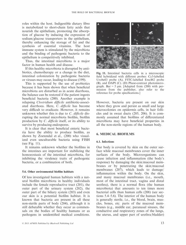
ria have the ability to produce biofilm, asshown by Zoetendal et al., (200) who visual-ized even unculturable bacteria using FISH(see Fig. 11).It remains unknown whether the biofilms in
the intestines are important for stabilizing thehomoeostasis of the intestinal microbiota, forinhibiting the virulence traits of pathogenicbacteria, or a combination of both.
5.6. Other environmental biofilm habitats
Of less investigated human habitats with a nat-ural biofilm microbiota in healthy individualsinclude the female reproductive tract (201), theouter part of the urinary system (202), theouter part of the biliary system (203) and evenour skin is a potential biofilm habitat. It isknown that bacteria are present in all thesenon-sterile parts of body (204), although it isstill debatable whether they occur as commen-sals on the bodies of healthy humans or aspathogens in unidentified medical conditions.
However, bacteria are present on our skinwhere they grow and persist as small and largemicrocolonies on epidermis cells, in hair folli-cles and in sweat ducts (205, 206). It is com-monly assumed that biofilms of differentiatedmicrobiota may have beneficial properties inall the non-sterile regions of the human body.
6. MEDICAL BIOFILMS
6.1. Infections
Our body is covered by skin on the outer sur-face while mucosal membranes cover the innersurfaces of the body. Microorganisms cancause infection and inflammation (the body’sresponse) by damaging the skin/mucosal mem-branes or by penetrating the skin/mucosalmembranes (207), which leads to damage/inflammation within the body. On the skin,and many mucosal membranes (i.e., mouth,most of the intestinal tract, vagina and distalurethra), there is a normal flora (the humanmicrobiota) that amounts to ten times morebacterial cells than human cells (208) (see sec-tions 5.4–5.6). The interior of the human bodyis generally sterile, i.e., the blood, brain, mus-cles, bones, etc. parts of the mucosal mem-branes (e.g., middle ear, paranasal sinuses, theconductive and respiratory zones of the lungs,the uterus, and upper part of urethra/bladder)
A B
C D
Fig. 11. Intestinal bacteria cells in a microscopicfield hybridized with different probes: Cy3-labelledUrobe63 probe (A), FITC-labelled Erec482 probe(B), and DAPI (C). (D) Phase-contrast photomicro-graph. Bar = 5 lm. [Adapted from (200) with per-mission from the publisher, also refer to thereference for probe specifications.]
© 2013 APMIS Published by Blackwell Publishing Ltd 13
THE ROLE OF BACTERIAL BIOFILM

do not harbour a permanent flora, but they aresometimes contaminated with a few bacteria,for example, by aspiration. However, thedefence mechanisms of the mucosal membranesrapidly remove these contaminants in healthyhumans without causing signs of inflammationor infection. If infection occurs without causingclinical symptoms (subclinical or silent infec-tion), an immune response may be detectedlater. If an infection causes clinical symptoms,for example, inflammation and fever, it may becured spontaneously by innate or adaptiveimmune response and/or by antibiotics,although sometimes it may be lethal. Suchinfections are known as acute and they can bedetected 1–2 weeks after the onset of an anti-body response. If the infection persists, despitethe immune response and antibiotic therapy, itis known as a chronic/persistent infection.
6.2. Acute infection
Acute infections have a rapid progression, butare normally relatively easy to treat with anti-biotics. This may be because the bacteria inacute infections are caused by planktonic bac-terial cells (single, small clusters or chains) (seesection 4.1). Acute infections are characterizedby an innate inflammatory response, which isdominated by PMNs. The vast majority ofhuman infections are acute and can be treatedeasily by general practitioners, for example,upper airway infections, skin and woundinfections, urinary tract infections, entericinfections and pneumonia. More severe andlife-threatening infections, such as sepsis, men-ingitis, and severe cases of pneumonia, are stillfairly easily treated at hospitals, if the infectionis diagnosed and treated in time.
6.3. Biofilm-related infections
Increasing evidence suggests that the chronicityof persistent bacterial infections is due to bac-terial biofilm formation, which contrasts withthe planktonic bacteria found in acute infec-tions (IV, V, VI, VII, VIII, XIII, XV) (11, 13,14, 209) as shown in Table 1. The longest rec-ognized biofilm infections are dental infections,such as caries and parodontitis (191, 210, 211)(see Table 1), although these are outside thescope of this thesis.
6.3.1. Chronic wounds – The global increase inobesity has been accompanied by a similarincrease in diabetes and cardiovascular dis-eases. These patients are particularly prone tothe development of chronic wounds, whichmay be colonized by a number of bacterialspecies (217, 238–240). My research has shownthat bacterial biofilms are present in chronicnon-healing wounds (IV, V) (217, 241) Figure12, although controversy persists as to whetherbiofilms have a role in the delayed healing ofchronic wounds (242). Most studies show thatthe deep dermal tissues of all chronic woundsharbour multiple bacterial species (239, 240,243–245). The most common bacterium foundin wounds is S. aureus, although P. aeruginosawas observed in more than half of the chronicwounds investigated (V) (239, 245). Further-more, P. aeruginosa-infected wounds were sig-nificantly larger in area than wounds withoutP. aeruginosa, while the presence of P. aeru-ginosa also seemed to delay or even preventthe healing process (246–248).Many chronic wounds will not heal, despite
aggressive treatment, and it was hypothesizedthat this was due to the presence of bacteria
Fig. 12. Biofilms of Pseudomonas aeruginosa in a chronic wound visualized using a specific peptide nucleicacid-fluorescence in situ hybridization probe (red) with confocal laser scanning microscopy. The right imageshows an enlargement of the middle image (adapted from V with permission from the publisher).
14 © 2013 APMIS Published by Blackwell Publishing Ltd
BJARNSHOLT

with a biofilm growth phenotype (249–252).The first direct microscopic evidence of bacte-rial biofilm involvement in chronic woundswas based on the direct microscopic identifica-tion of bacterial aggregates (IV) (217, 241).Three publications were published back-to-back in the same issue of the journal ‘WoundRepair and Regeneration’ during 2008. In mystudy (IV), I detected the presence of bacterialaggregates of S. aureus and P. aeruginosausing specific peptide nucleic acid (PNA) FISHprobes, in combination with CLSM. Peptidenucleic acid FISH allows the direct illuminat-ing of specific target cells within a sample. Thepresence of biofilms in chronic infectedwounds was highly debatable, so the observa-
tion of aggregated bacteria was consideredinsufficient evidence. To elucidate the biofilmgrowth phenotype that was present, I detectedthe EPS matrix by illuminating the alginatesurrounding P. aeruginosa. We also hypothe-sized that the presence of P. aeruginosa main-tained the wound in a chronic state, due to thecytolytic effects of the rhamnolipids producedby P. aeruginosa (see section 4.2). James et al.(217) demonstrated the elevated presence ofmicrobial aggregates in chronic wounds com-pared with acute wounds using scanning elec-tron microscopy (SEM).In a subsequent study, we collected and
examined chronic wound samples from 22 dif-ferent patients, all of whom were suspected to
Table 1. Visual identification of biofilms in chronic infections [modified and expanded from (238)]
Biofilm site Visualization method Reference
Dental plaque Light microscopyElectron microscopyElectron microscopyLight and Electron microscopyFISH
Hodson (211)Boyde and Lester (212)Theilade and Theilade (185)Listgarten (186)Dige et al. (195)
Periodontitis Electron microscopyLight microscopyFISH
Theilade (213)Berthold and Listgarten (214)Zijnge et al. (215)
Cystic fibrosis lung infections Light microscopyElectron microscopyFISH
Høiby (8),Lam et al. (216),VI
Chronic wounds FISHLight and electron microscopy
IVJames et al. (217)
Soft tissue fillers FISH VIIOtitis media FISH
FISHHall-Stoodley et al. (218)VIII
Implant associated Electron microscopyFISH
Marrie et al. (219),Waar et al. (220)
Catheter and shunt associated Electron microscopyElectron microscopyFluorescence microscopyFISH and electron microscopy
Marrie et al. (221)Marrie and Costerton (222)Stoodley et al. (223)Parsa et al. (224)
Chronic osteomyelitis Electron microscopyElectron microscopyLight and electron microscopy
Gristina et al. (225),Marrie and Costerton (226)Sedghizadeh et al. (90)
Chronic rhinosinusitis Electron microscopyElectron microscopyFISHFluorescence microscopy
Cryer et al. (227)Sanclement et al. (228)Sanderson et al. (229)Li et al. (230)
Endocarditis EchocardiographyElectron microscopy
Stewart et al. (231)Poyart et al. (232)
UTI Electron microscopyLight and electron microscopyLight microscopy
Nickel and Costerton (233)Nickel and Costerton (234)Reid et al. (235)
Contact lenses Electron microscopy Stapleton and Dart (236)Human gastrointestinal tract FISH Macfarlane and Dillon (237)
© 2013 APMIS Published by Blackwell Publishing Ltd 15
THE ROLE OF BACTERIAL BIOFILM

be infected by P. aeruginosa (V). The focus onP. aeruginosa was based on our hypothesisthat P. aeruginosa has a major role in chronicwounds (IV).The wound samples of the 22 different
patients were investigated using standardculturing methods and PNA FISH for thedirect identification of bacteria. Using stan-dard culturing methods, S. aureus was detectedin the majority of the wounds, whereasP. aeruginosa was observed less frequently. Bycontrast, PNA FISH showed that a large frac-tion of the wounds harboured biofilms ofP. aeruginosa that were embedded in thematrix alginate component. These microcolon-ies were detected within the wound bed,whereas S. aureus was detected on the surfaceof the wounds, if present. This was supportedby our subsequent observations (253) and astudy by Davis et al. (241), who demonstratedthat S. aureus forms microcolonies on the sur-face of the wound bed that were encased withextracellular matrix. I also participated in astudy that showed that bacteria were highlyheterogeneously distributed in these chronicinfected wounds (245).
6.3.2. Cystic fibrosis – Cystic fibrosis is themost common lethal inherited disease in Cauca-
sians (254). It is a monogenic, autosomal reces-sive multi-organ disease with a worldwideincidence of gene defects in the range of1:32 000 to 1:2000 live births (255). The geneticcause of CF was identified in 1989 as a defect inthe Cystic Fibrosis Transmembrane Conduc-tance Regulator (CFTR) gene, which is locatedon chromosome 7 (256–258). The CFTR defectcauses a decrease in epithelial chloride secretionand an increase in sodium absorption. In theCF lung, this results in dehydrated viscousmucus that is very difficult to clear mechani-cally, i.e., by coughing. The abnormal mucusviscosity is due to the chronic depletion of waterin the periciliary liquid layer and mucus (259,260). The non-inflammatory defence mecha-nism, i.e., mucociliary clearance, is impaired soinflammatory defence mechanisms are recruited(PMS, macrophages, IgG, etc.) giving rise toclinical symptoms, i.e., recurrent or chronic bac-terial lung infections (255, 261) (see Fig. 13).Since 1976, CF patients suffering from
chronic P. aeruginosa lung infection have suc-cessfully received intensive treatment with highconcentrations of antibiotics at the Copenha-gen CF Centre (i.e., maintenance treatmentand chronic suppressive treatment) (262, 263).Before 1976, only 50% of CF patients wouldsurvive 5 years of chronic P. aeruginosa lung
A B
C
Fig. 13. Schematic introduction to cystic fibrosis (CF). Frame C shows the thick dehydrated mucus thatallows bacteria to settle and form a chronic biofilm infection in the CF lung (adapted from: http://www.medi-cinenet.com/cystic_fibrosis/page3.htm).
16 © 2013 APMIS Published by Blackwell Publishing Ltd
BJARNSHOLT

infection (VI). Most CF patients now survivefor decades with chronic P. aeruginosa infec-tions (264).Despite the aggressive and intensive treat-
ment of chronic P. aeruginosa infections, thebacteria still persist in the CF lung. The inten-sive treatment delays and reduces the damagecaused by the chronic infection, but it cannotbe eradicated. CF patients experience a contin-uous degradation of lung tissue during chronicP. aeruginosa infections. This is caused byinfection and inflammatory processes where apronounced immune response leads to immunecomplex-mediated tissue destruction (VI).There is a consequent decline in lung function,which is the primary cause of death in CFpatients (265). Like chronic wounds, CF wasbelieved to be a biofilm disease (32, 79, 216).To investigate the true growth phenotype ofbacteria in the CF lung, I evaluated the distri-bution pattern of P. aeruginosa in the conduc-tive and respiratory zones of explanted lungsfrom chronic P. aeruginosa-infected CFpatients using PNA FISH (VI), just as I didwith the chronic wounds (IV). I also comparedintensively treated explanted lungs with autop-sies of non-intensively treated CF patients. Asexpected, the bacteria were mainly found inaggregates and only a few planktonic bacteriawere observed (see Fig. 14). It was evidentfrom my visual observations that bacteria weremainly localized in the conductive zone (theupper part of the lung with large and smallbronchi) if the patient was treated intensivelywith very few in the respiratory zone (thelower part, i.e., the alveoli). All of the aggre-gates were embedded in mucus plugs, althoughnot all of the mucus plugs contained bacteria.The mucus plugs containing bacteria variedgreatly in size and spatial orientation. By con-trast, bacteria were distributed throughout theentire lung in both the conductive and respira-tory zones in patients that had not been trea-ted intensively. In general, the inflammatoryresponse in the chronic P. aeruginosa-infectedCF lung was dominated by PMNs, and Iobserved a vast amount of PMNs surroundingthe aggregates. Bacteria did not adhere to theepithelial wall, demonstrating that the bacteriahad grown within the mucus rather thanadhering to the bronchial inner surface, as pre-viously suggested (32).
6.3.3. Chronic otitis media – The upper respira-tory tract consists of the, nose, paranasalsinuses, middle ear and throat, and it is fre-quently infected in children (266). Children aremore susceptible to infection of the middle earbecause their Eustachian tube is shorter and lessfunctional compared with the adult ear (267).Infection is most often initiated as an acute viralrespiratory infection followed by complicatingbacterial infections that may develop into otitismedia with effusion, a chronic suppurative bac-terial infection with mastoiditis, and even cho-lesteatomas, even if appropriate antibiotictreatment has been initiated (268).In the past, a bacterial cause of chronic
infections in the middle ear was difficult toconfirm because of culture-negative sampling.Recurrences or exacerbations were intriguingand difficult to explain, because bacteria werenot isolated. It was suggested that the infectionwas a local inflammatory reaction withoutbacteria. However, experiments using animalmodels demonstrated that bacterial biofilmscould cause these infections (269–271). Later,it was shown directly that treatment failure,culture-negative results and recurrent exacer-bations were due to bacteria that firmlyresided in biofilms (218, 272, 273).Recently, I was part of a morphological
investigation of bacterial biofilms in a high-risk population in Greenland (VIII). As withmy other biofilm studies, I used PNA FISH toelucidate whether bacteria were present in theaggregates. We observed aggregates that werepresent in pus discharged from the ears of fiveof the six (83%) children with chronic suppu-rative otitis media (CSOM) and we found evi-dence of biofilms in biopsies from the middleear in eight of the ten (80%) adults treated forCSOM (see Fig. 15) (VIII). These findingswere later confirmed in a controlled study ofhumans with CSOM in the USA (274).It is now widely accepted that bacterial bio-
films have a role in several chronic infectiousmiddle ear diseases (1, 218, 274–278).
6.3.4. Tissue fillers – An emerging problem isthe injection of foreign bodies in the form ofpolyacrylamide gel (soft tissue fillers) underthe skin for aesthetic purposes and remodellingafter trauma. Many different types of fillers
© 2013 APMIS Published by Blackwell Publishing Ltd 17
THE ROLE OF BACTERIAL BIOFILM

are available ranging from polymers to micro-particles, of which some are permanent andother are semi-permanent (279). Fillers aresupposedly cleared for toxicity and antigenic-ity, before they are permitted for human use(279). Fillers are injected subdermally and, aswith most foreign materials apart from noblemetals, they evoke an inflammatory responsewhere the intensity varies with the type of fil-ler. This inflammatory response is supposed tobe short term without complications, although
some fillers rely on inflammation to producethe filling effect (280). Most injections have nofurther complication, but an increasing num-ber of patients develop adverse events such asinflammatory swellings or nodules. If these areleft untreated, they often result in fistula for-mation and the discharge of pus and filler.Only a few years ago, it was assumed that
these reactions were caused by a foreign bodyreaction towards the injected filler. This wasdespite suspicions that the adverse reactions
A B C
D E F
G H I
Fig. 14. Visualization of Pseudomonas aeruginosa biofilms in the cystic fibrosis (CF) lung. Pseudomonas aeru-ginosa was found in the conductive zone, whereas very few bacteria were detected in the respiratory zonewhere they were phagocytosed. (A) Bacteria in biofilm within a bronchus visualized using Gram stain (CFmale, 41 years of age, chronic P. aeruginosa mucoid and non-mucoid infection for 28 years, 46 precipitatingantibodies, 114 two-week anti P. aeruginosa treatment courses). (B and C) HE staining of bacteria-filled bron-chiole. (D and E) Intraluminal P. aeruginosa biofilms surrounded by polymorphonuclear leucocytes visualizedusing peptide nucleic acid-fluorescence in situ hybridization, and DAPI. (F) Intact bronchi wall. (G and H)Increasing consolidation of the alveoli, and (I) single phagocytosed P. aeruginosa in the respiratory zone(adapted from VI with permission from the publisher).
18 © 2013 APMIS Published by Blackwell Publishing Ltd
BJARNSHOLT
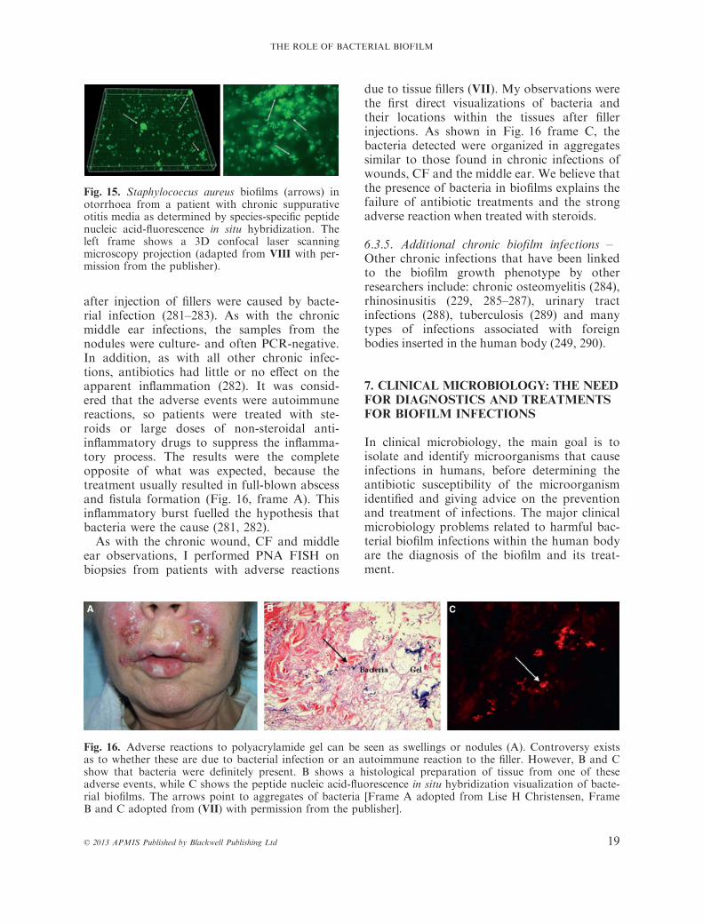
after injection of fillers were caused by bacte-rial infection (281–283). As with the chronicmiddle ear infections, the samples from thenodules were culture- and often PCR-negative.In addition, as with all other chronic infec-tions, antibiotics had little or no effect on theapparent inflammation (282). It was consid-ered that the adverse events were autoimmunereactions, so patients were treated with ste-roids or large doses of non-steroidal anti-inflammatory drugs to suppress the inflamma-tory process. The results were the completeopposite of what was expected, because thetreatment usually resulted in full-blown abscessand fistula formation (Fig. 16, frame A). Thisinflammatory burst fuelled the hypothesis thatbacteria were the cause (281, 282).As with the chronic wound, CF and middle
ear observations, I performed PNA FISH onbiopsies from patients with adverse reactions
due to tissue fillers (VII). My observations werethe first direct visualizations of bacteria andtheir locations within the tissues after fillerinjections. As shown in Fig. 16 frame C, thebacteria detected were organized in aggregatessimilar to those found in chronic infections ofwounds, CF and the middle ear. We believe thatthe presence of bacteria in biofilms explains thefailure of antibiotic treatments and the strongadverse reaction when treated with steroids.
6.3.5. Additional chronic biofilm infections –Other chronic infections that have been linkedto the biofilm growth phenotype by otherresearchers include: chronic osteomyelitis (284),rhinosinusitis (229, 285–287), urinary tractinfections (288), tuberculosis (289) and manytypes of infections associated with foreignbodies inserted in the human body (249, 290).
7. CLINICAL MICROBIOLOGY: THE NEEDFOR DIAGNOSTICS AND TREATMENTSFOR BIOFILM INFECTIONS
In clinical microbiology, the main goal is toisolate and identify microorganisms that causeinfections in humans, before determining theantibiotic susceptibility of the microorganismidentified and giving advice on the preventionand treatment of infections. The major clinicalmicrobiology problems related to harmful bac-terial biofilm infections within the human bodyare the diagnosis of the biofilm and its treat-ment.
Fig. 15. Staphylococcus aureus biofilms (arrows) inotorrhoea from a patient with chronic suppurativeotitis media as determined by species-specific peptidenucleic acid-fluorescence in situ hybridization. Theleft frame shows a 3D confocal laser scanningmicroscopy projection (adapted from VIII with per-mission from the publisher).
A B C
Fig. 16. Adverse reactions to polyacrylamide gel can be seen as swellings or nodules (A). Controversy existsas to whether these are due to bacterial infection or an autoimmune reaction to the filler. However, B and Cshow that bacteria were definitely present. B shows a histological preparation of tissue from one of theseadverse events, while C shows the peptide nucleic acid-fluorescence in situ hybridization visualization of bacte-rial biofilms. The arrows point to aggregates of bacteria [Frame A adopted from Lise H Christensen, FrameB and C adopted from (VII) with permission from the publisher].
© 2013 APMIS Published by Blackwell Publishing Ltd 19
THE ROLE OF BACTERIAL BIOFILM

7.1. Diagnostics
The initial challenge with all infections is toidentify the infecting organisms and the focusof the infection. This is usually not a problemfor acute infections because specialists in clini-cal microbiological departments benefit fromover 150 years of experience with microscopictechniques, as well as a wide variety of growthmedia and cultivation conditions. The firsttask is to obtain a sample for investigation,which is typically identified by the attendingmedical doctor. Depending on the sample, lab-oratory technicians make smears for Gramstaining and microscopy before selecting differ-ent growth media to use and whether the sam-ple should be propagated in solid or liquidmedia, incubated anaerobically or aerobically,the specific temperatures and duration. PCR isused routinely for some slow-growing or diffi-cult to grow bacteria (e.g., Mycobacteriumtuberculosis, Chlamydia species, and Legionellaspp.). After obtaining a pure bacterial culture,species are identified using different biochemi-cal and serological tests, or molecular-basedmethods such as PNA FISH, Matrix-assistedlaser desorption/ionization-Time of Flight(MALDI-TOF), or PCR amplification and16S rDNA sequencing.These methods are sometimes more problem-
atic with chronic infections (see Table 1). Anexception is CF (see section 6.3.2), where theeasily accessible purulent sputum or endolaryn-geal secretions can be obtained from patientson a regular basis and they will harbour bacte-ria if present. However, infections of thesinuses are difficult to access and CF patientsare believed to experience these before lunginfections (291). Other chronic infections, suchas the non-healing wounds and orthopaedicinfections, are even more problematic. Clearly,the wound bed is attached to the patient and itcannot readily be removed or homogenized forroutine diagnostics. The routine sampling hastypically used a swab or a biopsy, althoughboth approached might fail to sample the bac-teria because of their heterogeneous distribu-tion within the wound bed, as we havepreviously shown (V) (245, 253). A swab canonly collect bacteria found on the surface andnot the bacteria embedded in the wound bed(V) (253). Given the heterogeneous distribution
of bacteria, there is a risk that a ‘blind’ biopsywill fail to contain any bacteria (245).
7.2. Treatment
After the infecting organism or organisms havebeen identified, the next task is to treat theinfection. Importantly, biofilm infections can-not be treated in the same way as acute infec-tions, as described previously (see section 4.1).The most efficient treatment for a biofilminfection is to mechanically remove theinfected area or body part, e.g., dental infec-tions. This is sometimes possible if the focus isa catheter, an implant or an infected organthat is eligible for transplantation. However, itis not always possible or without risk of com-plications for patients. The two main strategiesfor preventing or suppressing bacterial biofilminfections are: (i) early aggressive antibiotictreatment before the biofilm is formed; (ii)chronic suppressive antibiotic treatment whenthe biofilm is established, if it cannot beremoved physically (16). As stated in the previ-ous sections, the administration of antibioticsto treat bacterial biofilms demands combina-tions of several different antibiotics (differenttargets, subpopulations and penetration) inhigh doses and for an extended period of time(16) because conventional resistance mecha-nisms will contribute to the intrinsic biofilmresistance mechanisms.Due to the risk of inducing traditional resis-
tance mechansims, it is advisable to avoidtreating the biofilm infection with an acuteinfection regime of single drugs in minimaldoses for short periods. This may induce fur-ther resistance and tolerance, and eventuallycreate opportunities for new bacteria to chron-ically infect the focus (16, 292).
7.3. Prevention
The most efficient means of combating biofilminfections is to prevent the infection in the firstplace. Normally, human skin acts as a barrierto prevent bacterial invasion of the body. Thisnatural barrier is compromised by surgery andthe insertion of implants. Thus, surgeons haveto exercise extreme caution when insertingimplants or injecting dermal fillers to avoid theintroduction of bacteria. Ultra-clean operating
20 © 2013 APMIS Published by Blackwell Publishing Ltd
BJARNSHOLT
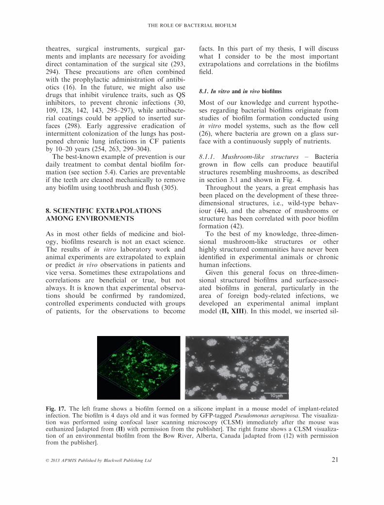
theatres, surgical instruments, surgical gar-ments and implants are necessary for avoidingdirect contamination of the surgical site (293,294). These precautions are often combinedwith the prophylactic administration of antibi-otics (16). In the future, we might also usedrugs that inhibit virulence traits, such as QSinhibitors, to prevent chronic infections (30,109, 128, 142, 143, 295–297), while antibacte-rial coatings could be applied to inserted sur-faces (298). Early aggressive eradication ofintermittent colonization of the lungs has post-poned chronic lung infections in CF patientsby 10–20 years (254, 263, 299–304).The best-known example of prevention is our
daily treatment to combat dental biofilm for-mation (see section 5.4). Caries are preventableif the teeth are cleaned mechanically to removeany biofilm using toothbrush and flush (305).
8. SCIENTIFIC EXTRAPOLATIONSAMONG ENVIRONMENTS
As in most other fields of medicine and biol-ogy, biofilms research is not an exact science.The results of in vitro laboratory work andanimal experiments are extrapolated to explainor predict in vivo observations in patients andvice versa. Sometimes these extrapolations andcorrelations are beneficial or true, but notalways. It is known that experimental observa-tions should be confirmed by randomized,controlled experiments conducted with groupsof patients, for the observations to become
facts. In this part of my thesis, I will discusswhat I consider to be the most importantextrapolations and correlations in the biofilmsfield.
8.1. In vitro and in vivo biofilms
Most of our knowledge and current hypothe-ses regarding bacterial biofilms originate fromstudies of biofilm formation conducted usingin vitro model systems, such as the flow cell(26), where bacteria are grown on a glass sur-face with a continuously supply of nutrients.
8.1.1. Mushroom-like structures – Bacteriagrown in flow cells can produce beautifulstructures resembling mushrooms, as describedin section 3.1 and shown in Fig. 4.Throughout the years, a great emphasis has
been placed on the development of these three-dimensional structures, i.e., wild-type behav-iour (44), and the absence of mushrooms orstructure has been correlated with poor biofilmformation (42).To the best of my knowledge, three-dimen-
sional mushroom-like structures or otherhighly structured communities have never beenidentified in experimental animals or chronichuman infections.Given this general focus on three-dimen-
sional structured biofilms and surface-associ-ated biofilms in general, particularly in thearea of foreign body-related infections, wedeveloped an experimental animal implantmodel (II, XIII). In this model, we inserted sil-
Fig. 17. The left frame shows a biofilm formed on a silicone implant in a mouse model of implant-relatedinfection. The biofilm is 4 days old and it was formed by GFP-tagged Pseudomonas aeruginosa. The visualiza-tion was performed using confocal laser scanning microscopy (CLSM) immediately after the mouse waseuthanized [adapted from (II) with permission from the publisher]. The right frame shows a CLSM visualiza-tion of an environmental biofilm from the Bow River, Alberta, Canada [adapted from (12) with permissionfrom the publisher].
© 2013 APMIS Published by Blackwell Publishing Ltd 21
THE ROLE OF BACTERIAL BIOFILM

icone implants pre-coated with P. aeruginosainto the peritoneal cavity of mice. The bacteriaon the implants formed a biofilm almostimmediately, which the mice were unable toclear. Ex vivo examination of these implantsdetected bacterial microcolonies on the siliconeimplants that resembled the microcoloniesfound in rivers (12), as shown in Fig. 17, aswell as the biofilms observed in chronicwounds (see Fig. 12) (IV, V) and CF (seeFig. 14) (VI), whereas no three-dimensionalstructures similar to in vitro mushroom struc-tures were observed.This discrepancy is probably attributable to
the design of the flow cell, because they weredesigned to allow continuous, non-invasiveand reproducible monitoring of controlled bio-film development. No other experimental orclinical infection settings would allow suchconditions. Grazers and phagocytes are present(see section 4.2) in most environmental andmedical biofilm habitats and there is a varyingflow (if any flow at all), as well as the presenceof pus, other excreted fluids, and many otherfactors. All of these dynamic and variable fac-tors combined make in vivo biofilms very dif-ferent from flow cell biofilms. However, flowcell biofilms have provided important clinicalinformation on biofilm physiology, althoughthey cannot be correlated directly with in vivobiofilms without further observational orexperimental evidence.In support of the flow cell, numerous studies
and observations using this method have beenextremely fruitful. Almost all hypothesesrelated to biofilms have originated from flowcell experiments, including antibiotic tolerance,predator and phagocyte tolerance, biofilm for-mation and biofilm dispersal.
8.2. To surface or not to surface
In the first definition provided by Costertonet al. (13), i.e., ‘a structured community ofbacterial cells enclosed in a self produced poly-meric matrix, adherent to a surface’, theauthors indicated the necessity of a surface.This was based on the slimy film layer com-posed of biomaterials found in alpine streams(170) and on submerged ship hulls (306, 307),i.e., biofilms. A surface is present in many ofthe infections that are now known to involve
biofilms (Table 1). However, bacterial aggre-gates also have been observed in other persis-tent infections such as chronic wounds andCF, which completely lack a surface (see sec-tions 6.3.1–6.3.4) (216–218) (IV, V, VI, VII,VIII, XV).Why should these aggregates, clumps or
heaps (8), be referred to as biofilms? Asexplained in the introduction, the most impor-tant hallmarks of bacterial biofilms are theaggregation of bacteria within a matrix (seesection 3.3) and the tolerance of antimicrobialagents (see section 4.1). It is evident from allobservations of bacteria in chronic infectionsthat bacteria are present in aggregates andthey persist despite antimicrobial chemother-apy. The only difference, and this is open todebate, is the apparent surface. I have neverbeen in any doubt about the lesser importanceof the surface, and that aggregation and toler-ance are the most crucial factors, because themajor differences between planktonic growthand biofilm growth are the biofilm matrix andthe gradients (e.g., oxygen, nutrient, generationtime, metabolic activity and antibiotic activity)from the surface to the bottom/centre of thebiofilm, whereas the surface at the bottom ofthe biofilm may simply be a surrogate for thegradients.To elucidate whether aggregated non-sur-
face-attached bacteria displayed the same phe-notypes as surface-attached bacteria, wecompared the matrix and antibiotic/phagocytetolerance in these two scenarios (XII). All ofthe features of the aggregated bacteria weresimilar to the flow cell biofilm, apart from thefirst layer of bacteria that was attached to theglass surface in the flow cells. All other charac-teristics were similar in both scenarios, includ-ing the aggregation, matrix and protectionagainst antibiotics and phagocytes, evenamong different subpopulations (XII)(Fig. 18).The most important feature is the build-up
of bacterial layers, which leads to QS initiationand nutritional and gaseous gradients withinthe biofilm. This initiates virulence factor pro-duction, such as rhamnolipid production (seesection 4.3), the emergence of subpopulationsand dormant bacteria.If the surface had an important role, the
standard biofilm phenotypes should be gener-
22 © 2013 APMIS Published by Blackwell Publishing Ltd
BJARNSHOLT

ated immediately. Interestingly, during theinvestigation of biofilm-enabled protectionagainst phagocytes, we observed that imma-ture wild-type P. aeruginosa biofilms were eas-ily eradicated when exposed to PMNs (XIII)(44). The same phenomenon was observed exvivo on hollow tubes that had been insertedinto the peritoneal cavity of mice (XIII). SEMshowed that single bacterial cells or smallgroups of surface-attached bacteria werehunted and phagocytosed by PMNs initially.A persistent biofilm did not develop until afterday 1, when the PMNs could no longer eradi-cate the bacteria and they were themselveskilled (see Fig. 19).Single layers of surface-attached bacteria are
easily phagocytosed, so it is difficult to imaginethat the surface has a role in biofilm-related
phenotypes. The only situation where a surfacemight play a role would be those that experi-ence shear forces or high flow rates, where thestochastic anchoring of a single bacteriumcould seed a population of bacteria that woulddevelop into a biofilm.
8.3. How should biofilms be studied?
We do not need to define a true biofilm, becauseall the bacterial aggregates with the distinct phe-notypes discussed here should be considered asbiofilms. However, we do need to considerwhich biofilm setup should be used to study agiven hypothesis or direct problem, based onthe origin of this hypothesis or problem. Thegoals should also be clear in biofilm research;for example, whether the aim is to test a hypoth-
A B
C D
Fig. 18. Scanning electron microscopy of surface- vs non-surface-attached Pseudomonas aeruginosa biofilms.(A) Aggregate harvested from a 48-hours-old stationary culture. (B) Details of a 3-day-old biofilm grown in aflow cell. (C) Details of 48-hours-old stationary aggregate. (D) Planktonic cells (OD600 = 0.5; adapted fromXII with permission from the publisher).
© 2013 APMIS Published by Blackwell Publishing Ltd 23
THE ROLE OF BACTERIAL BIOFILM

esis, such as attachment, or a specific applica-tion such as validating a drug against biofilms.One of the simplest methods for studying sur-face-attached biofilms is the microtitre plateassay (308), where bacterial attachment to thesurfaces of 96 wells is easily monitored by crys-tal violet staining of the entire biomass. This isa crude and not very reproducible assay, and itdoes not discriminate between live and deadcells, as well as aggregates that do not adhere tothe surface after washing (XII). Another sur-face-based method is the flow cell system. Thismethod is more reproducible and also facilitatesdiscrimination between live and dead bacteria.As with the microtitre plate assay, the weaknesswith this system is that host materials are miss-ing such as inflammatory components, pus andmucus. Examples may include biofilms inchronic wounds or the CF lung where no hardsurface is present, but there is a lot of pus,necrotic tissue, inflammatory cells, etc. Flowcells or microtitre plates could be used to testpreliminary questions such as whether a givendrug can kill the dormant subpopulation of thewound biofilm or whether the biofilm producesdensity-specific compounds. Flow cells not mic-rotitre plates should be used if the specific kill-
ing efficacy or dispersal is being monitoredduring the treatment, because the microtitreassay does not allow multiple time-point obser-vations. Observations using a flow cell setup tostudy specific biofilm behaviour may not bereadily extrapolated to an environment lackinga hard surface, such as wounds or the CF lung.Careful consideration should be required whenselecting the optimal experimental setup toavoid false negative and false positive results. Aclassical example is when the microtitre plateassay is used to screen for mutations that pre-vent bacteria from forming biofilms. We foundthat an equal amount of biomass was present assuspended aggregates in the wells as was foundon the inner surface of the microtitre plates,while bacteria that were unable to attach to theinner surfaces also formed aggregates (XII). It isparadoxical that if CF isolates were tested, allthe non-adhering cells would be classified asnon-biofilm formers, although no surface isavailable for adhesion in the CF lung. The non-biofilm formers can also form aggregates in vivowith the same virulence as their attachablecounterparts.The study of biofilms in these settings
requires a model that mimics the pus or mucus
Fig. 19. Scanning electron microscopy images of ex vivo interactions between the Pseudomonas aeruginosabiofilm and the host defences. The top row was on day 1, where the left picture shows the tube was cleanedof bacteria from the end, and the middle and right picture shows the phagocytosing polymorphonuclear leu-cocytes (PMNs). The bottom row shows the interactions on day 2, where the bacteria succeeded in forming aprotective biofilm that killed the PMNs (adapted from XIII with permission from the publisher).
24 © 2013 APMIS Published by Blackwell Publishing Ltd
BJARNSHOLT

where the bacteria grow. We developed amodel of bacterial biofilm growth in a semi-solid matrix of collagen (X), similar to thatfound in a wound bed or the mucopurulentpus of the CF lung. Using this model system,S. aureus and P. aeruginosa formed aggregateswithout adhering to a surface. The size andthe shape of the aggregates resemble thoseobserved in wound beds. These aggregates dis-played the same specialized phenotypes as theflow cell biofilms, i.e., matrix production andhigh tolerance of antibiotics. Liquid-suspendedaggregates were similar to surface-attachedbiofilms in terms of all their biofilm properties(XII). It is possible to add different inflamma-tory components to both these non-surfacemodels, such as PMNs and antibodies.However, these models cannot mimic the
complex interplay that exists with the actualhost defences, which is why animal modelsalso should be used when biofilm interactionswith the host are investigated. Many chronicdiseases have their own animal models, suchas CF (309–311), otitis media (312), chronicwounds (313) and our models of implant-related infections (II, XIII) [for a review of invivo biofilm models see (314)].Modelling all aspects of chronic human
infections may not be possible with a singleexperimental method. The goal must be toidentify and ask the right questions using theappropriate available models, or alternativelydeveloping new ones.
8.4. The opportunity of sociomicrobiology
Bacteria form biofilms everywhere and theyopportunistically exploit any available carbon,nutrient or energy resources for their meta-bolic processes. The literature shows that bac-teria can thrive almost anywhere and that thebiofilm growth phenotype is dominant (3, 4).As described in section 5, the intermixing ofdifferent bacterial species is natural wherecommensalism and synergies exist among spe-cies. Different species are mutually dependentin many environmental consortia because ofanabolic and catabolic specializations and theavailability of nutrients, oxygen, electron ac-ceptors, etc. Most in vitro biofilm research hasbeen performed using a single species,although investigations using multispecies and
complex consortia have increased because suchconditions are prevalent in systems such asdental biofilms (1, 195, 315).In human infections, especially chronic
infections, it is often possible to isolate severalbacterial species from the same infection.Wounds, in particular, appear to contain anextreme range of species (217, 238–240). Thechronic lung infection of CF patients has alsobeen suggested to be multispecies (316, 317).This infection diversity has been elucidatedusing traditional identification techniques andmolecular methods such as PCR and the newIBIS technology (318). IBIS is a combinationof PCR and MALDI-TOF mass spectrometry.This allows the identification of bacterial spe-cies and an estimation of the relative abun-dance of the different bacterial species present.These methods can be used to identify the spe-cies present in a sample, but they cannot iden-tify the orientations of each species ordetermine the species that contribute to patho-genesis. Direct microscopy can reveal the ori-entation and distribution, but it also fails toattribute pathogenesis.As with environmental biofilm consortia, it
is possible that the presence of many differentspecies is due to their synergistic survivalwithin a host. If this is the case, all the differ-ent species present, such as those found in achronic wound, should occur in close proxim-ity, or in co-aggregates, and they may possiblybe engaged in symbiotic relationships.Our approach to investigating chronic
infections for the potential presence of bio-films has applied traditional visualizationtechniques and novel methods such as PNAFISH and CLSM. The microscopic approachhas allowed us to visualize several differentbacteria within one tissue or sputum sample.To our surprise, our observations have shownthat although different bacterial species werepresent in the same sample, the majority ofthe aggregate was formed of a single species(see Figs 12, 14, 15) (IV, V, VI, VIII, XIV,XV) (1).Along these lines, only in extremely rare
cases have we observed mixed biofilms, oradjacent biofilms formed of different species inchronic infections. This is in spite of the use ofa universal eubacterial PNA FISH probe(designed to detect all bacteria) in combination
© 2013 APMIS Published by Blackwell Publishing Ltd 25
THE ROLE OF BACTERIAL BIOFILM

with specific PNA FISH probes for bacteriasuch as P. aeruginosa or S. aureus.In our study of the putative CF microbiota,
only single or dual species were detected usingmicroscopy and molecular methods (VI, XIV,XV), while few species other than P. aeruginosaand S. aureus were detected in wounds (IV, V).The CF lung is end-stage when eligible for
transplantation, so we further investigated thesegregation of species in sputum from non-end-stage CF patients that were known to beinfected with one to three bacterial or fungalspecies (XV). The microscopic results showedalmost complete segregation of the differentbacterial species within the sputum as seen inFig. 20.
8.4.1. Succession or opportunity – It is apparentthat chronic infections are often multispecies,but the majority of each aggregate predomi-nantly consists of only one bacterial speciesper aggregate in multispecies infections. Differ-ent bacterial species can form sovereign aggre-gates in the same infection, but in differentlocations or niches. Thus, although environ-mental biofilms are referred to as multispecies,this does not apply to the biofilms found inchronic infections, with the exception of dentalinfections and foreign body infections that cancommunicate with a body surface that has anormal flora, for example, catheters or stents.This difference in the bacterial organization
of environmental biofilms and infection bio-
A B
C D
Fig. 20. Peptide nucleic acid-fluorescence in situ hybridization visualization of bacterial aggregates/biofilms inexpectorated sputum from non-end-stage cystic fibrosis patients. Frames (A, C and D) show Pseudomonasaeruginosa (red) and other bacterial species (green) while frame (B) shows Staphylococcus aureus (red) andother bacteria (green). The images show that the biofilms of different species appear to be segregated fromeach other (adapted from XV with permission from the publisher).
26 © 2013 APMIS Published by Blackwell Publishing Ltd
BJARNSHOLT
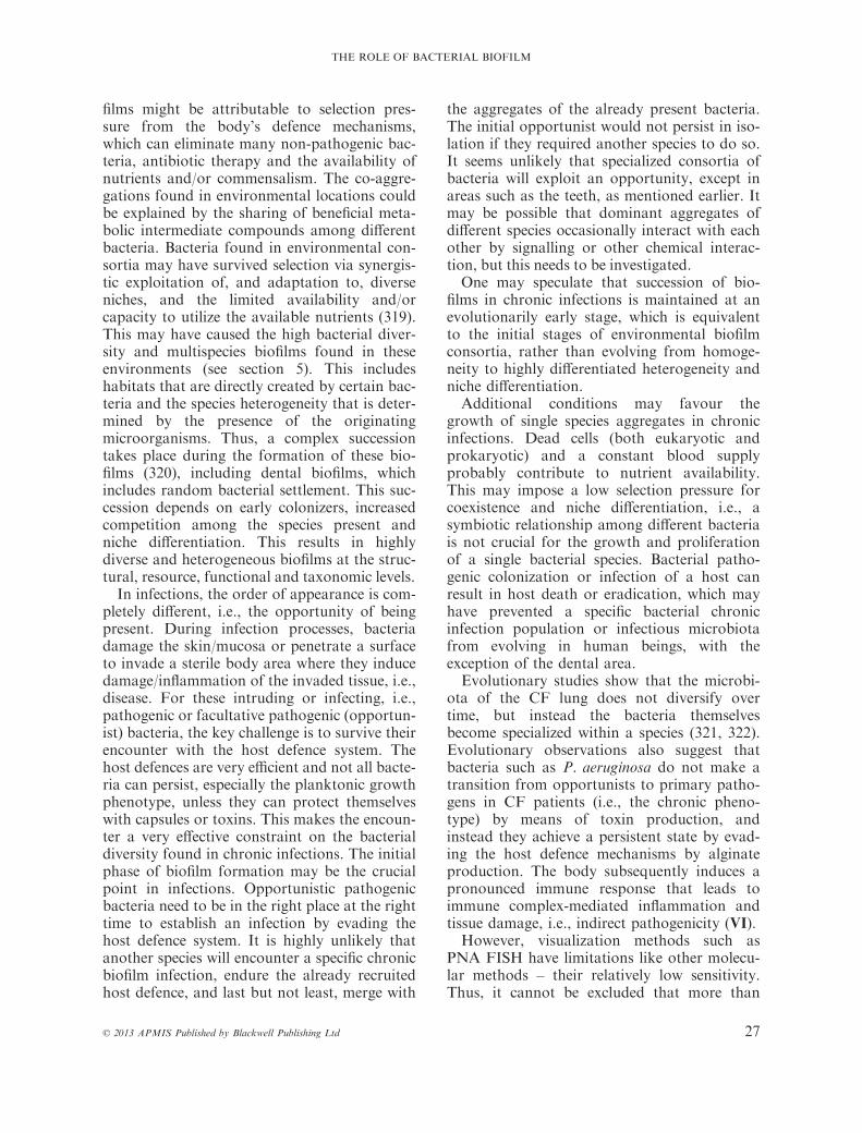
films might be attributable to selection pres-sure from the body’s defence mechanisms,which can eliminate many non-pathogenic bac-teria, antibiotic therapy and the availability ofnutrients and/or commensalism. The co-aggre-gations found in environmental locations couldbe explained by the sharing of beneficial meta-bolic intermediate compounds among differentbacteria. Bacteria found in environmental con-sortia may have survived selection via synergis-tic exploitation of, and adaptation to, diverseniches, and the limited availability and/orcapacity to utilize the available nutrients (319).This may have caused the high bacterial diver-sity and multispecies biofilms found in theseenvironments (see section 5). This includeshabitats that are directly created by certain bac-teria and the species heterogeneity that is deter-mined by the presence of the originatingmicroorganisms. Thus, a complex successiontakes place during the formation of these bio-films (320), including dental biofilms, whichincludes random bacterial settlement. This suc-cession depends on early colonizers, increasedcompetition among the species present andniche differentiation. This results in highlydiverse and heterogeneous biofilms at the struc-tural, resource, functional and taxonomic levels.In infections, the order of appearance is com-
pletely different, i.e., the opportunity of beingpresent. During infection processes, bacteriadamage the skin/mucosa or penetrate a surfaceto invade a sterile body area where they inducedamage/inflammation of the invaded tissue, i.e.,disease. For these intruding or infecting, i.e.,pathogenic or facultative pathogenic (opportun-ist) bacteria, the key challenge is to survive theirencounter with the host defence system. Thehost defences are very efficient and not all bacte-ria can persist, especially the planktonic growthphenotype, unless they can protect themselveswith capsules or toxins. This makes the encoun-ter a very effective constraint on the bacterialdiversity found in chronic infections. The initialphase of biofilm formation may be the crucialpoint in infections. Opportunistic pathogenicbacteria need to be in the right place at the righttime to establish an infection by evading thehost defence system. It is highly unlikely thatanother species will encounter a specific chronicbiofilm infection, endure the already recruitedhost defence, and last but not least, merge with
the aggregates of the already present bacteria.The initial opportunist would not persist in iso-lation if they required another species to do so.It seems unlikely that specialized consortia ofbacteria will exploit an opportunity, except inareas such as the teeth, as mentioned earlier. Itmay be possible that dominant aggregates ofdifferent species occasionally interact with eachother by signalling or other chemical interac-tion, but this needs to be investigated.One may speculate that succession of bio-
films in chronic infections is maintained at anevolutionarily early stage, which is equivalentto the initial stages of environmental biofilmconsortia, rather than evolving from homoge-neity to highly differentiated heterogeneity andniche differentiation.Additional conditions may favour the
growth of single species aggregates in chronicinfections. Dead cells (both eukaryotic andprokaryotic) and a constant blood supplyprobably contribute to nutrient availability.This may impose a low selection pressure forcoexistence and niche differentiation, i.e., asymbiotic relationship among different bacteriais not crucial for the growth and proliferationof a single bacterial species. Bacterial patho-genic colonization or infection of a host canresult in host death or eradication, which mayhave prevented a specific bacterial chronicinfection population or infectious microbiotafrom evolving in human beings, with theexception of the dental area.Evolutionary studies show that the microbi-
ota of the CF lung does not diversify overtime, but instead the bacteria themselvesbecome specialized within a species (321, 322).Evolutionary observations also suggest thatbacteria such as P. aeruginosa do not make atransition from opportunists to primary patho-gens in CF patients (i.e., the chronic pheno-type) by means of toxin production, andinstead they achieve a persistent state by evad-ing the host defence mechanisms by alginateproduction. The body subsequently induces apronounced immune response that leads toimmune complex-mediated inflammation andtissue damage, i.e., indirect pathogenicity (VI).However, visualization methods such as
PNA FISH have limitations like other molecu-lar methods – their relatively low sensitivity.Thus, it cannot be excluded that more than
© 2013 APMIS Published by Blackwell Publishing Ltd 27
THE ROLE OF BACTERIAL BIOFILM
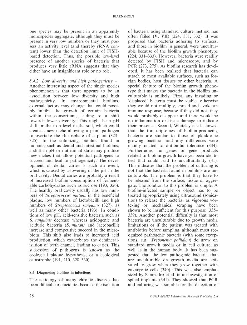
one species may be present in an apparentlymonospecies aggregate, although they must bepresent in very low numbers or they must pos-sess an activity level (and thereby rRNA con-tent) lower than the detection limit of FISH-based detection. Thus, the possible low-levelpresence of another species of bacteria thatproduces very little rRNA suggests that theyeither have an insignificant role or no role.
8.4.2. Low diversity and high pathogenicity –Another interesting aspect of the single speciesphenomenon is that there appears to be anassociation between low diversity and highpathogenicity. In environmental biofilms,external factors may change that could possi-bly inhibit the growth of certain bacteriawithin the consortium, leading to a shifttowards lower diversity. This might be a pHshift or the iron level in the soil, which couldcreate a new niche allowing a plant pathogento overtake the rhizosphere of a plant (323–325). In the colonizing biofilms found inhumans, such as dental and intestinal biofilms,a shift in pH or nutritional state may producenew niches that allow potential pathogens tosucceed and lead to pathogenicity. The devel-opment of dental caries is such an event,which is caused by a lowering of the pH in theoral cavity. Dental caries are probably a resultof increased biofilm consumption of ferment-able carbohydrates such as sucrose (193, 326).The healthy oral cavity usually has low num-bers of Streptococcus mutans in the beneficialplaque, low numbers of lactobacilli and highnumbers of Streptococcus sanguinis (327), aswell as many other bacteria (193). In condi-tions of low pH, acid-sensitive bacteria such asS. sanguinis decrease whereas acidogenic andaciduric bacteria (S. mutans and lactobacilli)increase and competitive succeed in the micro-biota. This shift also leads to increased acidproduction, which exacerbates the demineral-ization of teeth enamel, leading to caries. Thissuccession of pathogens is known as theecological plaque hypothesis, or a ecologicalcatastrophe (191, 210, 328–330).
8.5. Diagnosing biofilms in infections
The aetiology of many chronic diseases hasbeen difficult to elucidate, because the isolation
of bacteria using standard culture method hasoften failed (V, VII) (224, 331, 332). It wasproposed that bacteria adhering to surfaces,and those in biofilm in general, were uncultur-able because of the biofilm growth phenotype(224, 331–333). However, bacteria were readilydetected by FISH and microscopy, and byPCR (273, 275). As biofilm research has devel-oped, it has been realized that bacteria canattach to most available surfaces, such as for-eign bodies, host tissues or other bacteria. Aspecial feature of the biofilm growth pheno-type that makes the bacteria in the biofilm un-culturable is unlikely. First, any invading or‘displaced’ bacteria must be viable, otherwisethey would not multiply, spread and evoke animmune response, because if they did not, theywould probably disappear and there would beno inflammation or tissue damage to indicatetheir presence. Second, Whiteley et al. showedthat the transcriptomes of biofilm-producingbacteria are similar to those of planktonicgrowing bacteria, and any differences weremainly related to antibiotic tolerance (334).Furthermore, no genes or gene productsrelated to biofilm growth have yet been identi-fied that could lead to unculturability (41).This indicates that the problem of culturing isnot that the bacteria found in biofilms are un-culturable. The problem is that they have tobe released from the surface, tissue or aggre-gate. The solution to this problem is simple. Abiofilm-infected sample or object has to betreated appropriately using ultrasound (sonica-tion) to release the bacteria, as vigorous vor-texing or mechanical scraping have beenshown to be insufficient for this purpose (335–339). Another potential difficulty is that mostbacteria are unculturable due to growth medialimitations or if the patient was treated withantibiotics before sampling, although most rec-ognized pathogenic bacteria (with some excep-tions, e.g., Treponema pallidum) do grow onstandard growth media or in cell culture, aswell as in the human body. It has been sug-gested that the few pathogenic bacteria thatare unculturable on growth media are acti-vated to grow when they grow together witheukaryotic cells (340). This was also empha-sized by Sampedro et al. in an investigation ofspinal implants (341). They showed that PCRand culturing was suitable for the detection of
28 © 2013 APMIS Published by Blackwell Publishing Ltd
BJARNSHOLT
bacteria on spinal implants, if they were pres-ent in the host environment in sufficient num-bers to establish an infection (342).The problems of diagnosing bacteria in
chronic infections are far from being solved bysonication prior to culturing and PCR. Asshown in Table 2, each of these methods hasits advantages and limitations.In all techniques, i.e., culturing, PCR, IBIS
or microscopy, the major problem is the col-lection of the bacteria because of their hetero-geneous distribution (V) (245, 253) and strongattachment to the surfaces of catheters andimplants. One advantage of direct growth,and to some extent IBIS plexID (318), is thatbacterial susceptibility to antibiotics can beinvestigated if they grow or are identified.However, a pitfall is that antibiotic suscepti-bility is performed on bacteria in their plank-tonic form and such tests can be irrelevantbecause of biofilm-enabled tolerance (16) (seesection 4.1).The advantages of PCR are that this method
will detect bacteria independent of theirgrowth. The major disadvantage is that PCRdoes not discriminate between live and deadcells. Furthermore, the detection of a bacte-rium by PCR does not necessarily indicate thatit contributes to the pathogenesis of the infec-tion because it may be a contaminant. Micros-copy facilitates the direct visualization of theinfecting bacteria, the surrounding tissue andinflammatory cells (IV, V, VI, VII, VIII, XIII,XV). A disadvantage of microscopy is that
observation might prove difficult if only a fewbacteria are present.Specific criteria to aid the diagnosis of bio-
film infections have been published in recentyears. The first suggested criteria included: (i)bacteria adherent to a surface; (ii) direct obser-vation of a bacterial cell cluster in a matrix;(iii) confined to a particular location; and (iv)failure of antibiotics to clear the infection,despite using concentrations that would killbacteria in their planktonic state (343). Thesecriteria were then re-evaluated to include: (i)only associated with a surface, but not firmlyattached; (ii) a culture-negative result despite aclinically documented high suspicion of infec-tion; and (iii) direct observation of protectionagainst the host defences (344). This was fur-ther expanded by Høiby to include a distinc-tion between acute and chronic infections (seeTable 3) (16).Only a few universal diagnostic molecular
biofilm markers have been identified (345).Great efforts have been made, but a com-mon difficulty is the heterogeneity of bacte-rial biofilms. This has also been empathizedwhen designing vaccines against biofilms(345, 346).A further problem associated with the diffi-
culties of identifying bacteria comes withunderstanding the significance of the manymicroorganisms identified by increasingly sen-sitive molecular techniques.The problem of bacterial detection using
methods based on DNA/RNA, and even cul-
Table 2. Advantages and disadvantages of different methods used for identifying infectious bacteria inhumans
Method Advantages Pitfalls and difficulties
Culturing Bacterial presence is confirmedAntibiotic susceptibilityDirect quantification
Heterogeneous distributionFinding the focusPathogens vs contaminationBiofilms or planktonic samplescan be culture-negative
PCR and IBIS Fast results even when cultureis negative
Low cut-off
Heterogeneous distributionFinding the focusPathogens vs contaminationBiofilm or planktonic
Microscopy Biofilms are confirmedInteractions with tissuesInflammatory cellsResults even when culture-negative
Heterogeneous distributionFinding the focus
© 2013 APMIS Published by Blackwell Publishing Ltd 29
THE ROLE OF BACTERIAL BIOFILM

ture, is that a positive test result does not pro-vide any information on pathogenicity or itsabsence. This makes it very difficult to discrim-inate between pathogens, passive bystandersand contamination. Contamination is a partic-ular problem when using increasingly sensitivemethods. This has been evident when mappingthe CF microbiota, because the samples haveto pass through the upper respiratory tractwhich is colonized with many different bacteria(191, 192, 347). We aimed to overcome this byinvestigating explanted lungs (VI, XIV),although this had the disadvantage that onlyend-stage microbiota could be investigated (seesection 8.4).In 1884, during his investigation of the aeti-
ology of tuberculosis, Robert Koch proposedthat:
…it is necessary to obtain a perfect proofto satisfy oneself that the parasite and thedisease are not only correlated, butactually casually related, and the parasiteis the actual direct cause of the disease.This can only be done by completelyseparating the parasite from the diseasedorganism, and from all of the products ofthe disease which could be subscribed to adisease-inducing influence, and thenintroducing the isolated parasite intohealthy organisms and induce the disease anew with all its characteristic symptomsand properties.
The dilemma of biofilm-related infections isthat bacteria can be difficult to isolate and/orthat numerous species are identified. Thisdirect evidence has also been a problem withviral infections. In 1936, Rivers proposedthat: (i) a specific virus must be found associ-ated with a disease with a degree of regular-ity; and (ii) the virus must be shown to occurin the sick individual, not as an incidental oraccidental finding, but as the cause of the dis-ease under investigation (348). In the searchfor the causative agent of infectious mononu-cleosis, the Epstein–Barr virus was identifiedon immunological grounds alone, whichshowed beyond reasonable doubt that it wasthe agent (349–351). This led to the produc-tion of a set of factors to support immuno-logical proof that an agent caused a specificdisease (351).The use of specific immune responses has
also been used in the diagnostics of chronicinfections of the CF lung (352, 353) and endo-carditis (354, 355). In 1965, Hill proposed ninefactors (see Table 4) for determining a possiblecausal relationship between an infectiousorganism and a specific disease.These modified Koch’s criteria were devel-
oped further for chronic infections in CFpatients by Høiby (356).Using these tools and guidelines in combina-
tion with relevant animal models, we may beable to distinguish between the causativeagents (should be treated) of specific chronic
Table 3. Some general features of biofilm infections in humans compared with acute planktonic infectionsand superficial colonization/normal flora found on the skin and mucosal membranes. Bold indicates biofilm-specific features.
Features of biofilm infections Necessaryconditionfor biofilminfection
Also foundin acuteplanktonicinfection
Also found incolonization/normalflora on skin andmucosal membranes
Aggregates of bacteria embedded in a self-producedpolymer matrix
Yes No No/Yes
Tolerant of clinically relevant PK/PD* doses ofantibiotics, despite the susceptibility of planktoniccells
Yes No No/Yes
Tolerant of innate and adaptive immune responses Yes No No/Yes—unknownInflammation Yes Yes NoChronic infections Yes No NoForeign body-associated infections No Initial NoLocated on surfaces No Yes YesLocalized infection Yes Yes YesFocus of spreading or local exacerbation Yes Yes Yes
*PK/PD, pharmacokinetic/pharmacodynamic [adapted from (16)]
30 © 2013 APMIS Published by Blackwell Publishing Ltd
BJARNSHOLT

infections and irrelevant commensals (shouldnot be treated). It is crucial to make this dis-tinction to optimize the validity of new diag-nostic methods such as IBIS (318), or we willaccumulate an abundance of useless and mis-leading information.
9. DISCUSSION
The philosopher Søren Kirkegaard (see page3) was disdainful of scientists who crave to dis-sect nature and explore our world and its sur-roundings to find true meaning, which iscertainly thought-provoking. Is it necessary toexplore everything? Is it necessary to distin-guish between surface and non-surface-attached bacteria, and single and multispeciesbiofilms? The great explorers of our world sev-eral hundred years ago were curious, but theywere also driven by the great rewards andfame that new explorations would provide.Today, as always, science is driven by the pas-sion for fame and fortune, but also for newinsights and progress that makes a difference,e.g., to patients. In the science of biofilms, ithas been imperative for me to understand howto study biofilms, how they are formed, howthey are detected and identified, and how theyare treated when they cause an infection. Todo this, I knew initially that I had to use amicroscope to study the ‘small animals’ (bacte-ria) (5), pathogenic or commensal, and theirdetailed interactions, before applying othermethods based on these microscopic observa-tions. The classic example is Louis Pasteur(1822–1895) who observed and sketched theaggregations of bacteria that caused the acidi-
fication of wine, which led to his famous dis-covery of pasteurization.The relevance of my biofilm research was
stated by the National Institutes of Health’sProgram on Immunology of Biofilms (PA07-288), as follow:
One of the limitations of early approachesused to study infectious organisms indisease is that planktonic (pure freelysuspended cultures of) bacteria wereemployed. Although much information onthe immune response came from the studyof planktonic bacteria, it is now clear thatbacteria in the clinical environment livemore often as communities ofmicroorganisms (biofilms) than as singlecell suspensions.
My contribution to the growing field of bio-films may be divided into three categories. Themain category is additional evidence that thebiofilm growth phenotype is associated withchronic bacterial infections. The biofilm phe-notype found in chronic infections seems toprotect the bacteria, and biofilm formationcould be explained using a ‘united we stand –divided we fall’ paradigm. The denominator ofthese chronic infections are an extreme toler-ance and resistance to antimicrobial agentsand a capacity to evade the host defences (I,II, IV, V, VI, VII, VIII, IX, XI, XII, XIII)(65). The second category is the study of bio-film-enabled antimicrobial tolerance (III, X,XII) (16, 44, 128, 357). Interestingly, myresearch has shown that biofilm-enabled toler-ance of antibiotics is reversible, at least inmodel systems (XII). In theory, this means
Table 4. Hill’s epidemiologic criteria for causal associations [modified from (350)]
Causal criterion Causal association
Strength of association What is the relative risk?Consistency ofassociation
Is there agreement among repeated observations in different places, atdifferent times, using different methodology, by different researchers,under different circumstances?
Specificity of association Is the outcome unique to the exposure?Temporality Does exposure precede the outcome variable?Biological gradient Is there evidence of a dose–response relationship?Plausibility Does the causal relationship make biological sense?Consistency Is the causal association compatible with present knowledge of the disease?Experimentation Does controlled manipulation of the exposure variable change the outcome?Analogy Does the causal relationship conform to a previously described relationship?
© 2013 APMIS Published by Blackwell Publishing Ltd 31
THE ROLE OF BACTERIAL BIOFILM

that chronic biofilm infections can be treatedor prevented if the biofilm is dissolved or bac-teria are actively prevented from aggregating.The third category is the capacity of biofilmsto evade phagocytes such as PMNs. No solidbarrier appears to protect the bacteria and theprotection seems to be derived from excretedchemicals that kill, paralyse or divert phago-cytes (I, II, IX, XIII) (125).As with many other biofilm researchers,
P. aeruginosa has been my preferred modelorganism for biofilm research. Based on ourex vivo observations, this was an appropriatechoice for studying biofilm-related infectionsbecause it is involved in several chronic infec-tions (IV, V, VI) (253), which have much incommon. First of all, in vitro and in vivoexperimental P. aeruginosa biofilms excreterhamnolipids that provide protection fromphagocytes and grazers (see section 4.2) (I, IX,XIII) (44, 125, 358). Plenty of evidence indi-cates that rhamnolipids also have this protec-tive role in chronic human infections (359,360). Evidence of this major role came fromclinical isolates of P. aeruginosa that do notlose their capacity to produce rhamnolipidsover decades in chronic infections, althoughmany other toxins are lost (XI). PMN debrissuch as DNA and myeloperoxidase has alsobeen observed surrounding aggregates of exvivo samples of human infections similar tothat observed in in vitro and in vivo model sys-tems (V, VI, XIII). Interestingly, P. aeruginosamicrocolonies are connected to a massive accu-mulation of PMNs, although the PMNsappear to be prevented from penetrating themicrocolonies. This fatal attraction could becaused by the QS signal molecules of P. aeru-ginosa, because our in vitro experimentsshowed that they attract PMNs (I). Rhamnoli-pid production by P. aeruginosa is regulatedby QS (see section 4.3), while QS regulationdepends on the build-up of signal moleculesproduced by the bacteria. This is a cunningstrategy whereby P. aeruginosa avoids the cel-lular components of the host defences by wait-ing for the build-up of a critical mass beforeproducing its protection, the rhamnolipid (30).Pseudomonas aeruginosa then lures theapproaching PMNs to their death. This leadsto a chronic inflammatory condition, the con-tinuous influx and dominant presence of neu-
trophils, and the efflux of intracellulardegradation enzymes from the dead neutroph-ils, such as reactive oxygen species and matrixmetalloproteases (MMPs) (361–363).This is also our hypothesis of chronic wounds,
where P. aeruginosa arrests the wound in achronic inflammatory state (IV) (361–364).Many similarities exist between chronic infec-tions of the CF lung and wounds, i.e., the persis-tent influx of PMNs, elevated MMPs and animbalance of several cytokines (254, 299, 300,365, 366). These observations may explain thepreviously reported impairment of host cells inchronic infections, and why the balance is tippedeven further away from healing. A negative feed-back loop might further increase the release ofdestructive enzymes from incoming PMNs.Another interesting similarity between
chronic wounds and CF is that the chronicP. aeruginosa infection is preceded by intermit-tent colonization/infection with other bacteria,especially S. aureus (304, 367, 368). Whetherthis is a real phenomenon or simply a coinci-dence is not yet known, but it might be specu-lated that bacteria such as S. aureus induce thehost defences to a state that favours P. aeru-ginosa (synergism). Another possible factoraffecting this phenomenon is that S. aureusdoes not present a therapeutic problem in CFand chronic wounds. It may be hypothesizedthat the eradication of S. aureus creates a vac-uum and an opportunity for bacteria such asP. aeruginosa.In fact, chronic infections appear to depend
on a preceding compromising event, becausehealthy people do not appear to acquire chronicinfections. First of all, the primary defences ofthe human body have to be compromised (seesection 5.3). Second, a stochastic event such as aviral infection, stress, intoxication or acuteinfection is probably required to dampen oroccupy the host response, thereby allowing bac-teria to settle in protective biofilms. These eventsmight be compared to the obstruction of a river,which at some point will cause flooding of thesurrounding dry land, as shown in Fig. 21.On the basis of my in vitro and in vivo
observations of bacterial aggregates, I believeit is clear that bacteria aggregate as a defaultand that subsequent biofilm development pro-gresses via adaptations to nutritional and envi-ronmental conditions.
32 © 2013 APMIS Published by Blackwell Publishing Ltd
BJARNSHOLT
As stated previously, bacterial biofilms existin environmental ecological habitats andopportunistic ecological habitats, such as infec-tions. The lifestyle is the same, except thatinfectious biofilms are formed in places wherea preceding microbiota was absent so the fightfor survival and supremacy is different. Theconsequence is that chronic infections aredominated by monospecies biofilms, althoughthe infections are most likely multispecies,whereas environmental biofilms are dominatedby multispecies (V, XIV, XV) (1, 217, 239,240, 245, 253).
10. CONCLUSION
My goal many years ago was to elucidate therole of biofilms in chronic infection. I have
studied biofilms in infections and in the labo-ratory, how they are formed, how they persistwith antibiotics, how to diagnose them andhow they can be treated. I soon discoveredthat, like all other natural phenomena andprocesses, biofilms appear very difficult andcomplex when viewed from a distance. How-ever, my microscopic examinations of biofilmshave shown that all the interactions and path-ways are simple. The biofilm era has beendominated by a tendency to complicate theprocesses of biofilm formation, regulation andprotection. However, based on my in vitro andin vivo observations of aggregating bacteria, Ibelieve that biofilm formation and persistenceis less complex than postulated in the past,although we still have many unanswered ques-tions. I propose a simplistic view of biofilmsbased on a ‘united we stand – divided we fall’
A B
C D
Fig. 21. Frame (A) shows the river in natural balance. Frame (B) is an event that compromises the primarydefences (e.g., insertion of implants or catheters, or thickened mucus in the cystic fibrosis airway) as shownby the presence of a rock in the river. In itself, this event does not cause any persistent pathology, but bacte-ria are allowed to settle and persist on the rock (implant etc.) if natural predators (phagocytes = hostdefences) are impaired or sluggish as a consequence of pollution (viral infection etc.). A persistent biofilm isformed, Frame (C). This persistent biofilm eventually causes the river to flood (e.g., severe acute infections orimpaired lung function), Frame (D). The only solution is to remove the rock, which symbolizes the removalof the catheter, amputation of limbs or the transplantation of organs to prevent the death of the patient.
© 2013 APMIS Published by Blackwell Publishing Ltd 33
THE ROLE OF BACTERIAL BIOFILM
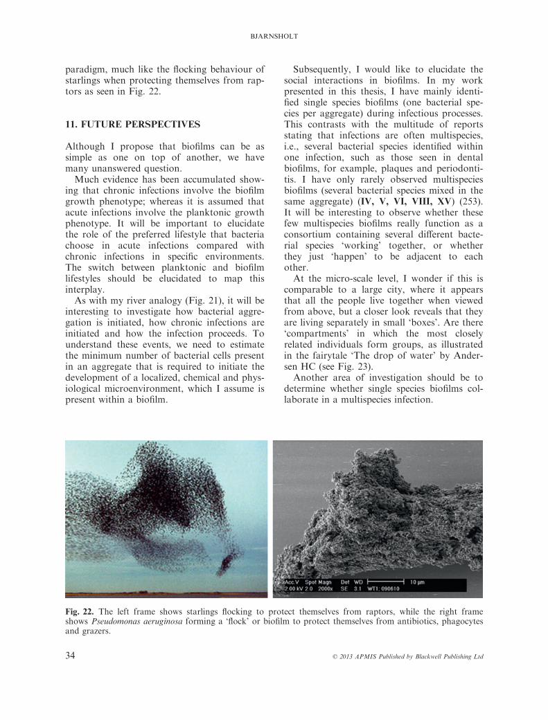
paradigm, much like the flocking behaviour ofstarlings when protecting themselves from rap-tors as seen in Fig. 22.
11. FUTURE PERSPECTIVES
Although I propose that biofilms can be assimple as one on top of another, we havemany unanswered question.Much evidence has been accumulated show-
ing that chronic infections involve the biofilmgrowth phenotype; whereas it is assumed thatacute infections involve the planktonic growthphenotype. It will be important to elucidatethe role of the preferred lifestyle that bacteriachoose in acute infections compared withchronic infections in specific environments.The switch between planktonic and biofilmlifestyles should be elucidated to map thisinterplay.As with my river analogy (Fig. 21), it will be
interesting to investigate how bacterial aggre-gation is initiated, how chronic infections areinitiated and how the infection proceeds. Tounderstand these events, we need to estimatethe minimum number of bacterial cells presentin an aggregate that is required to initiate thedevelopment of a localized, chemical and phys-iological microenvironment, which I assume ispresent within a biofilm.
Subsequently, I would like to elucidate thesocial interactions in biofilms. In my workpresented in this thesis, I have mainly identi-fied single species biofilms (one bacterial spe-cies per aggregate) during infectious processes.This contrasts with the multitude of reportsstating that infections are often multispecies,i.e., several bacterial species identified withinone infection, such as those seen in dentalbiofilms, for example, plaques and periodonti-tis. I have only rarely observed multispeciesbiofilms (several bacterial species mixed in thesame aggregate) (IV, V, VI, VIII, XV) (253).It will be interesting to observe whether thesefew multispecies biofilms really function as aconsortium containing several different bacte-rial species ‘working’ together, or whetherthey just ‘happen’ to be adjacent to eachother.At the micro-scale level, I wonder if this is
comparable to a large city, where it appearsthat all the people live together when viewedfrom above, but a closer look reveals that theyare living separately in small ‘boxes’. Are there‘compartments’ in which the most closelyrelated individuals form groups, as illustratedin the fairytale ‘The drop of water’ by Ander-sen HC (see Fig. 23).Another area of investigation should be to
determine whether single species biofilms col-laborate in a multispecies infection.
Fig. 22. The left frame shows starlings flocking to protect themselves from raptors, while the right frameshows Pseudomonas aeruginosa forming a ‘flock’ or biofilm to protect themselves from antibiotics, phagocytesand grazers.
34 © 2013 APMIS Published by Blackwell Publishing Ltd
BJARNSHOLT

These perspectives are more or less basic sci-ence. The main challenge will be to identifybacteria that have a role and to exclude thosethat have none. It will also be an ongoingchallenge to develop tools for the rapid andefficient diagnosis of chronic infections. Noone has yet identified a molecule or productthat is produced by all the bacteria within abiofilm, not to mention by all bacteria. In CFpatients with chronic P. aeruginosa lung infec-tions, the presence of alginate in the sputum(369) and the growth of the mucoid phenotypeindicates biofilm infection (80). Anotherapproach could be to identify a host-producedmarker such as antibodies or interleukins. Theproblem is that most chronic infections evokea localized inflammatory response that mightnot be measurable in the circulating blood.Until now, a general antibody response hasbeen very useful for detecting P. aeruginosa
biofilm infections in the lungs of CF patients(352, 370–373), while specific antibodies toalginate can also be used (78, 374) in endocar-ditis (354, 355).Finally, great benefits will be derived from
an optimized and efficient treatment regime forbiofilm infections. If mechanical disruption ofthe biofilm at the site of infection was possible,local high concentrations of antibiotics wouldprobably be effective. Thus, matrix-degradingenzymes could prove useful, again in combina-tion with antibiotics, as seen with the DNasetreatment of CF patients (375). Anotherapproach may be the use of anti-virulencedrugs, such as drugs that inhibit QS regula-tion, which could inhibit the production of vir-ulence factors, for example, rhamnolipids. Thisapproach is actually used in CF patients whereazithromycin fulfils these criteria (376).Directly targeting virulence factors with neu-
Fig. 23. The drop of water magnifies the individuals in the picture of Copenhagen as I have magnified thebacteria in biofilms by microscopy. Picture reproduced with permission from the Museum of Copenhagen andMarianne Bisballe.
© 2013 APMIS Published by Blackwell Publishing Ltd 35
THE ROLE OF BACTERIAL BIOFILM

tralizing agents such as antibodies is yetanother possibility, although this may not beuseful because CF patients with chronicP. aeruginosa lung infection produce neutraliz-ing antibodies for many toxins produced bythese bacteria (374, 377–379).A completely opposite approach would be
to prevent bacteria from settling into biofilmsor killing their planktonic phenotype beforethey switch to the protective biofilm pheno-type. This strategy has already proven itsworth for many years, because the earlyaggressive treatment of lung infections in CFpatients has postponed the onset of chronicinfections and prolonged the life of CFpatients by decades (263, 264, 299, 300, 302,304, 380, 381).One thing that seems certain is that although
we have started to unravel the mechanismsbehind the biofilm phenotype, we have justscratched the surface.
CONFLICT OF INTEREST
The author declares no conflicts of interest.
12. REFERENCES
1. Burmolle M, Thomsen TR, Fazli M, Dige I,Christensen L, Homoe P, et al. Biofilms inchronic infections - a matter of opportunity -monospecies biofilms in multispecies infec-tions. FEMS Immunol Med Microbiol2010;59:324–36.
2. Atlas RM, Bartha R. Microbial Ecology, 4thed. Menlo Park, CA: Benjamin/CummingsScience Publishing, 1997.
3. Costerton JW, Lewandowski Z, Caldwell DE,Korber DR, Lappin-Scott HM. Microbial bio-films. Annu Rev Microbiol 1995;49:711–45.
4. Potera C. Studying slime. Environ Health Per-spect 1998;106:A604–6.
5. van Leeuwenhoek A. Microscopical observa-tions about animals in the scurf of the teeth.Philos Trans R Soc Lond B Biol Sci 1684;14:568–74.
6. Henrici AT. Studies of freshwater bacteria. JBacteriol 1933;25:2132.
7. Rogovska T, Lazareva MF. Microbiologicalcharacteristics of biofilm purifying hydrogensulphide containing sewage. 1961.
8. Høiby N. Pseudomonas aeruginosa infection incystic fibrosis. University of Copenhagen, 1977.
9. Costerton JW, Geesey GG, Cheng KJ. Howbacteria stick. Sci Am 1978;238:86–95.
10. McCoy WF, Bryers JD, Robbins J, CostertonJW. Observations of fouling biofilm forma-tion. Can J Microbiol 1981;27:910–7.
11. Costerton JW, Cheng KJ, Geesey GG, LaddTI, Nickel JC, Dasgupta M, et al. Bacterialbiofilms in nature and disease. Annu RevMicrobiol 1987;41:435–64.
12. Costerton JW, Lewandowski Z, DeBeer D,Caldwell D, Korber D, James G. Biofilms, thecustomized microniche. J Bacteriol1994;176:2137–42.
13. Costerton JW, Stewart PS, Greenberg EP.Bacterial biofilms: a common cause of persis-tent infections. Science 1999;284:1318–22.
14. Costerton JW. Replacement of acute planc-tonic by chronic biofilm diseases. In: Eckey C,editor. The Biofilm Primer. New York:Springer, 2007;129–61.
15. Bayles KW. The biological role of death andlysis in biofilm development. Nat Rev Micro-biol 2007;5:721–6.
16. Hoiby N, Bjarnsholt T, Givskov M, Molin S,Ciofu O. Antibiotic resistance of bacterial bio-films. Int J Antimicrob Agents 2010;35:322–32.
17. Anwar H, van Biesen T, Dasgupta M, LamK, Costerton JW. Interaction of biofilm bac-teria with antibiotics in a novel in vitro che-mostat system. Antimicrob Agents Chemother1989;33:1824–6.
18. Donlan RM. Biofilms: microbial life on sur-faces. Emerg Infect Dis 2002;8:881–90.
19. Zimmerli W, Trampuz A. Implant-associatedinfection. In: Bjarnsholt T, Moser C, JensenPO, Høiby N, editors. Biofilm Infections.New York: Springer, 2010: 91–110.
20. Brusselle GG, Joos GF, Bracke KR. Newinsights into the immunology of chronicobstructive pulmonary disease. Lancet2011;378:1015–26.
21. Maraffi T, Piffer F, Cosentini R. Prophylacticantibiotic therapy in chronic obstructive pul-monary disease. Ther Adv Respir Dis2010;4:135–42.
22. Klausen M, aes-Jorgensen A, Molin S,Tolker-Nielsen T. Involvement of bacterialmigration in the development of complexmulticellular structures in Pseudomonasaeruginosa biofilms. Mol Microbiol 2003;50:61–8.
23. Klausen M, Heydorn A, Ragas P, LambertsenL, aes-Jorgensen A, Molin S, et al. Biofilmformation by Pseudomonas aeruginosa wildtype, flagella and type IV pili mutants. MolMicrobiol 2003 Jun;48:1511–24.
24. Sauer K, Camper AK, Ehrlich GD, CostertonJW, Davies DG. Pseudomonas aeruginosadisplays multiple phenotypes during develop-
36 © 2013 APMIS Published by Blackwell Publishing Ltd
BJARNSHOLT

ment as a biofilm. J Bacteriol 2002;184:1140–54.
25. Alhede M, Kragh KN, Qvortrup K, lesen-Holm M, van GM, Christensen LD, et al.Phenotypes of non-attached Pseudomonasaeruginosa aggregates resemble surfaceattached biofilm. PLoS ONE 2011;6:e27943.
26. Christensen BB, Sternberg C, Andersen JB,Palmer RJ Jr, Nielsen AT, Givskov M, et al.Molecular tools for study of biofilm physiol-ogy. Methods Enzymol 1999;310:20–42.
27. Christensen BB, Haagensen JA, Heydorn A,Molin S. Metabolic commensalism and compe-tition in a two-species microbial consortium.Appl Environ Microbiol 2002;68:2495–502.
28. Gjermansen M, Ragas P, Sternberg C, MolinS, Tolker-Nielsen T. Characterization of star-vation-induced dispersion in Pseudomonasputida biofilms. Environ Microbiol2005;7:894–906.
29. Stover CK, Pham XQ, Erwin AL, MizoguchiSD, Warrener P, Hickey MJ, et al. Completegenome sequence of Pseudomonas aeruginosaPA01, an opportunistic pathogen. Nature2000;406:959–64.
30. Bjarnsholt T, Givskov M. The role of quorumsensing in the pathogenicity of the cunningaggressor Pseudomonas aeruginosa. Anal Bio-anal Chem 2006;387(2):409–14.
31. Sandt C, Smith-Palmer T, Pink J, Brennan L,Pink D. Confocal Raman microspectroscopyas a tool for studying the chemical heteroge-neities of biofilms in situ. J Appl Microbiol2007;103:1808–20.
32. Worlitzsch D, Tarran R, Ulrich M, SchwabU, Cekici A, Meyer KC, et al. Effects ofreduced mucus oxygen concentration in air-way Pseudomonas infections of cystic fibrosispatients. J Clin Invest 2002;109:317–25.
33. Kolpen M, Hansen CR, Bjarnsholt T, MoserC, Christensen LD, van GM, et al. Polymor-phonuclear leucocytes consume oxygen in spu-tum from chronic Pseudomonas aeruginosapneumonia in cystic fibrosis. Thorax2010;65:57–62.
34. Walters MC III, Roe F, Bugnicourt A,Franklin MJ, Stewart PS. Contributions ofantibiotic penetration, oxygen limitation, andlow metabolic activity to tolerance of Pseudo-monas aeruginosa biofilms to ciprofloxacinand tobramycin. Antimicrob Agents Chemo-ther 2003;47:317–23.
35. Semmler AB, Whitchurch CB, Mattick JS. Are-examination of twitching motility in Pseu-domonas aeruginosa. Microbiology 1999;145(Pt 10):2863–73.
36. O’Toole G, Kaplan HB, Kolter R. Biofilmformation as microbial development. AnnuRev Microbiol 2000;54:49–79.
37. Sauer K. The genomics and proteomics ofbiofilm formation. Genome Biol 2003;4:219.
38. Purevdorj B, Costerton JW, Stoodley P. Influ-ence of hydrodynamics and cell signaling onthe structure and behavior of Pseudomonasaeruginosa biofilms. Appl Environ Microbiol2002;68:4457–64.
39. Reimmann C, Ginet N, Michel L, Keel C,Michaux P, Krishnapillai V, et al. Geneticallyprogrammed autoinducer destruction reducesvirulence gene expression and swarming motil-ity in Pseudomonas aeruginosa PAO1. Micro-biology 2002;148(Pt 4):923–32.
40. Hentzer M, Eberl L, Givskov M. Transcrip-tome analysis of Pseudomonas aeruginosabiofilm development: anaerobic respirationand iron limitation. Biofilms 2005;2:37–61.
41. Folsom JP, Richards L, Pitts B, Roe F, Ehr-lich GD, Parker A, et al. Physiology of Pseu-domonas aeruginosa in biofilms as revealedby transcriptome analysis. BMC Microbiol2010;10:294.
42. Lee B, Haagensen JA, Ciofu O, Andersen JB,Høiby N, Molin S. Heterogeneity of biofilmsformed by nonmucoid Pseudomonas aerugin-osa isolates from patients with cystic fibrosis.J Clin Microbiol 2005;43:5247–55.
43. Davies DG, Parsek MR, Pearson JP, IglewskiBH, Costerton JW, Greenberg EP. Theinvolvement of cell-to-cell signals in the devel-opment of a bacterial biofilm. Science1998;280:295–8.
44. Bjarnsholt T, Jensen PO, Burmolle M, Hent-zer M, Haagensen JA, Hougen HP, et al.Pseudomonas aeruginosa tolerance to tobra-mycin, hydrogen peroxide and polymorphonu-clear leukocytes is quorum-sensing dependent.Microbiology 2005;151(Pt 2):373–83.
45. Stoodley P, Sauer K, Davies DG, CostertonJW. Biofilms as complex differentiated com-munities. Annu Rev Microbiol 2002;56:187–209.
46. Kreft JU, Bonhoeffer S. The evolution ofgroups of cooperating bacteria and the growthrate versus yield trade-off. Microbiology2005;151(Pt 3):637–41.
47. Kreft JU. Biofilms promote altruism. Microbi-ology 2004;150(Pt 8):2751–60.
48. Monds RD, O’Toole GA. The developmentalmodel of microbial biofilms: 10 years of aparadigm up for review. Trends Microbiol2009;17:73–87.
49. Conibear TC, Collins SL, Webb JS. Roleof mutation in Pseudomonas aeruginosabiofilm development. PLoS ONE 2009;4:e6289.
50. Stoodley P, Wilson S, Hall-Stoodley L, BoyleJD, Lappin-Scott HM, Costerton JW. Growthand detachment of cell clusters from mature
© 2013 APMIS Published by Blackwell Publishing Ltd 37
THE ROLE OF BACTERIAL BIOFILM

mixed-species biofilms. Appl Environ Micro-biol 2001;67:5608–13.
51. Stapper AP, Narasimhan G, Ohman DE, Bar-akat J, Hentzer M, Molin S, et al. Alginateproduction affects Pseudomonas aeruginosabiofilm development and architecture, but isnot essential for biofilm formation. J MedMicrobiol 2004;53(Pt 7):679–90.
52. Webb JS, Thompson LS, James S, CharltonT, Tolker-Nielsen T, Koch B, et al. Cell deathin Pseudomonas aeruginosa biofilm develop-ment. J Bacteriol 2003;185:4585–92.
53. Rupp CJ, Fux CA, Stoodley P. Viscoelasticityof Staphylococcus aureus biofilms in responseto fluid shear allows resistance to detachmentand facilitates rolling migration. Appl EnvironMicrobiol 2005;71:2175–8.
54. Abraham J, Mansour C, Veledar E, Khan B,Lerakis S. Staphylococcus aureus bacteremiaand endocarditis: the Grady Memorial Hospi-tal experience with methicillin-sensitive S aur-eus and methicillin-resistant S aureusbacteremia. Am Heart J 2004;147:536–9.
55. Fatkenheuer G, Cornely O, Seifert H. Clinicalmanagement of catheter-related infections.Clin Microbiol Infect 2002;8:545–50.
56. Jefferson KK. What drives bacteria to pro-duce a biofilm? FEMS Microbiol Lett2004;236:163–73.
57. Zogaj X, Nimtz M, Rohde M, Bokranz W,Romling U. The multicellular morphotypes ofSalmonella typhimurium and Escherichia coliproduce cellulose as the second component ofthe extracellular matrix. Mol Microbiol2001;39:1452–63.
58. Schramm M, Hestrin S. Factors affecting pro-duction of cellulose at the air/liquid interfaceof a culture of Acetobacter xylinum. J GenMicrobiol 1954;11:123–9.
59. Ma L, Conover M, Lu H, Parsek MR, BaylesK, Wozniak DJ. Assembly and developmentof the Pseudomonas aeruginosa biofilmmatrix. PLoS Pathog 2009;5:e1000354.
60. Allesen-Holm M, Barken KB, Yang L, Klau-sen M, Webb JS, Kjelleberg S, et al. A char-acterization of DNA release in Pseudomonasaeruginosa cultures and biofilms. Mol Micro-biol 2006;59:1114–28.
61. Whitchurch CB, Tolker-Nielsen T, Ragas PC,Mattick JS. Extracellular DNA required forbacterial biofilm formation. Science2002;295:1487.
62. Mann EE, Rice KC, Boles BR, Endres JL,Ranjit D, Chandramohan L, et al. Modula-tion of eDNA release and degradation affectsStaphylococcus aureus biofilm maturation.PLoS ONE 2009;4:e5822.
63. Rice KC, Mann EE, Endres JL, Weiss EC,Cassat JE, Smeltzer MS, et al. The cidA mur-
ein hydrolase regulator contributes to DNArelease and biofilm development in Staphylo-coccus aureus. Proc Natl Acad Sci USA2007;104:8113–8.
64. Borlee BR, Goldman AD, Murakami K, Sa-mudrala R, Wozniak DJ, Parsek MR. Pseu-domonas aeruginosa uses a cyclic-di-GMP-regulated adhesin to reinforce the biofilmextracellular matrix. Mol Microbiol2010;75:827–42.
65. Fexby S, Bjarnsholt T, Jensen PO, Roos V,Høiby N, Givskov M, et al. Biological Trojanhorse: antigen 43 provides specific bacterialuptake and survival in human neutrophils.Infect Immun 2007;75:30–4.
66. Klemm P, Hjerrild L, Gjermansen M, Schem-bri MA. Structure-function analysis of theself-recognizing Antigen 43 autotransporterprotein from Escherichia coli. Mol Microbiol2004;51:283–96.
67. Matsuyama T, Kaneda K, Ishizuka I, ToidaT, Yano I. Surface-active novel glycolipid andlinked 3-hydroxy fatty acids produced by Ser-ratia rubidaea. J Bacteriol 1990 Jun;172:3015–22.
68. Davey ME, Caiazza NC, O’Toole GA.Rhamnolipid surfactant production affectsbiofilm architecture in Pseudomonas aerugin-osa PAO1. J Bacteriol 2003;185:1027–36.
69. Pamp SJ, Tolker-Nielsen T. Multiple roles ofbiosurfactants in structural biofilm develop-ment by Pseudomonas aeruginosa. J Bacteriol2007;189:2531–9.
70. Pamp S, Gjermansen M, Tolker-Nielsen T.The biofilm matrix - a sticky framework. In:Kjelleberg S, Givskov M, editors. BacterialBiofilm Formation and Adaptation. Norwich,UK: Horizon BioScience, 2007: 37–69.
71. Harmsen M, Yang L, Pamp SJ, Tolker-Niel-sen T. An update on Pseudomonas aeruginosabiofilm formation, tolerance, and dispersal.FEMS Immunol Med Microbiol 2010;59:253–68.
72. Parsek MR, Tolker-Nielsen T. Pattern forma-tion in Pseudomonas aeruginosa biofilms.Curr Opin Microbiol 2008;11:560–6.
73. Flemming HC, Neu TR, Wozniak DJ. TheEPS matrix: the “house of biofilm cells”. JBacteriol 2007;189:7945–7.
74. Nivens DE, Ohman DE, Williams J, FranklinMJ. Role of alginate and its O acetylation information of Pseudomonas aeruginosa micro-colonies and biofilms. J Bacteriol2001;183:1047–57.
75. Simpson JA, Smith SE, Dean RT. Scavengingby alginate of free radicals released by macro-phages. Free Radic Biol Med 1989;6:347–53.
76. Allison DG, Matthews MJ. Effect of polysac-charide interactions on antibiotic susceptibility
38 © 2013 APMIS Published by Blackwell Publishing Ltd
BJARNSHOLT

of Pseudomonas aeruginosa. J Appl Bacteriol1992;73:484–8.
77. Pedersen SS, Høiby N, Espersen F, Koch C.Role of alginate in infection with mucoidPseudomonas aeruginosa in cystic fibrosis.Thorax 1992;47:6–13.
78. Pedersen SS, Espersen F, Høiby N, ShandGH. Purification, characterization, and immu-nological cross-reactivity of alginates pro-duced by mucoid Pseudomonas aeruginosafrom patients with cystic fibrosis. J ClinMicrobiol 1989;27:691–9.
79. Høiby N. Understanding bacterial biofilms inpatients with cystic fibrosis: current and inno-vative approaches to potential therapies. JCyst Fibros 2002;1:249–54.
80. Høiby N. Pseudomonas aeruginosa infectionin cystic fibrosis. Relationship betweenmucoid strains of Pseudomonas aeruginosaand the humoral immune response. ActaPathol Microbiol Scand [B] Microbiol Immu-nol 1974;82:551–8.
81. Wozniak DJ, Wyckoff TJ, Starkey M, KeyserR, Azadi P, O’Toole GA, et al. Alginate isnot a significant component of the extracellu-lar polysaccharide matrix of PA14 and PAO1Pseudomonas aeruginosa biofilms. Proc NatlAcad Sci USA 2003;100:7907–12.
82. Bragonzi A, Worlitzsch D, Pier GB, TimpertP, Ulrich M, Hentzer M, et al. NonmucoidPseudomonas aeruginosa expresses alginatein the lungs of patients with cystic fibrosis andin a mouse model. J Infect Dis 2005;192:410–9.
83. Byrd MS, Sadovskaya I, Vinogradov E, LuH, Sprinkle AB, Richardson SH, et al.Genetic and biochemical analyses of the Pseu-domonas aeruginosa Psl exopolysaccharidereveal overlapping roles for polysaccharidesynthesis enzymes in Psl and LPS production.Mol Microbiol 2009;73:622–38.
84. Ryder C, Byrd M, Wozniak DJ. Role of poly-saccharides in Pseudomonas aeruginosa bio-film development. Curr Opin Microbiol2007;10:644–8.
85. Alhede M, Qvortrup K, Liebrechts R, HoibyN, Givskov M, Bjarnsholt T. Combination ofmicroscopic techniques reveals a comprehen-sive visual impression of biofilm structure andcomposition. FEMS Immunol Med Microbiol2012;Jul;65(2):335–42.
86. Joubert LM, Wolfaardt GM, Botha A.Microbial exopolymers link predator and preyin a model yeast biofilm system. Microb Ecol2006;52:187–97.
87. Alipour M, Suntres ZE, Omri A. Importanceof DNase and alginate lyase for enhancingfree and liposome encapsulated aminoglyco-side activity against Pseudomonas aerugin-
osa. J Antimicrob Chemother 2009;64:317–25.
88. Alipour M, Suntres ZE, Halwani M, AzghaniAO, Omri A. Activity and interactions ofliposomal antibiotics in presence of polya-nions and sputum of patients with cysticfibrosis. PLoS ONE 2009;4:e5724.
89. Schooling SR, Beveridge TJ. Membrane vesi-cles: an overlooked component of the matricesof biofilms. J Bacteriol 2006;188:5945–57.
90. Sedghizadeh PP, Kumar SK, Gorur A,Schaudinn C, Shuler CF, Costerton JW.Microbial biofilms in osteomyelitis of the jawand osteonecrosis of the jaw secondary to bis-phosphonate therapy. J Am Dent Assoc2009;140:1259–65.
91. Nielsen LP, Risgaard-Petersen N, Fossing H,Christensen PB, Sayama M. Electric currentscouple spatially separated biogeochemical pro-cesses in marine sediment. Nature2010;463:1071–4.
92. Donlan RM, Costerton JW. Biofilms: survivalmechanisms of clinically relevant microorgan-isms. Clin Microbiol Rev 2002;15:167–93.
93. Drenkard E. Antimicrobial resistance of Pseu-domonas aeruginosa biofilms. Microbes Infect2003;5:1213–9.
94. Drenkard E, Ausubel FM. Pseudomonas bio-film formation and antibiotic resistance arelinked to phenotypic variation. Nature2002;416:740–3.
95. Bagge N, Hentzer M, Andersen JB, Ciofu O,Givskov M, Høiby N. Dynamics and spatialdistribution of beta-lactamase expression inPseudomonas aeruginosa biofilms. AntimicrobAgents Chemother 2004;48:1168–74.
96. Peterson AA, Fesik SW, McGroarty EJ.Decreased binding of antibiotics to lipopoly-saccharides from polymyxin-resistant strainsof Escherichia coli and Salmonella typhimuri-um. Antimicrob Agents Chemother 1987;31:230–7.
97. Kohler T, van Delden C, Curty LK, Hamzeh-pour MM, Pechere JC. Overexpression of theMexEF-OprN multidrug efflux system affectscell-to-cell signaling in Pseudomonas aerugin-osa. J Bacteriol 2001;183:5213–22.
98. Sawada I, Maseda H, Nakae T, Uchiyama H,Nomura N. A quorum-sensing autoinducerenhances the mexAB-oprM efflux-pumpexpression without the MexR-mediated regu-lation in Pseudomonas aeruginosa. MicrobiolImmunol 2004;48:435–9.
99. Norman A, Hansen LH, Sorensen SJ. Conju-gative plasmids: vessels of the communal genepool. Philos Trans R Soc Lond B Biol Sci2009;364:2275–89.
100. Canton R, Morosini MI. Emergence andspread of antibiotic resistance following expo-
© 2013 APMIS Published by Blackwell Publishing Ltd 39
THE ROLE OF BACTERIAL BIOFILM

sure to antibiotics. FEMS Microbiol Rev2011;35:977–91.
101. Giedraitiene A, Vitkauskiene A, Naginiene R,Pavilonis A. Antibiotic resistance mechanismsof clinically important bacteria. Medicina(Kaunas) 2011;47:137–46.
102. Ciofu O, Giwercman B, Pedersen SS, HøibyN. Development of antibiotic resistance inPseudomonas aeruginosa during two decadesof antipseudomonal treatment at the DanishCF Center. APMIS 1994;102:674–80.
103. Tenover FC. Mechanisms of antimicrobialresistance in bacteria. Am J Infect Control2006;34(5 Suppl 1):S3–10.
104. Mulcahy H, Charron-Mazenod L, Lewenza S.Extracellular DNA chelates cations andinduces antibiotic resistance in Pseudomonasaeruginosa biofilms. PLoS Pathog 2008;4:e1000213.
105. Stewart PS, Franklin MJ. Physiological heter-ogeneity in biofilms. Nat Rev Microbiol2008;6:199–210.
106. Werner E, Roe F, Bugnicourt A, FranklinMJ, Heydorn A, Molin S, et al. Stratifiedgrowth in Pseudomonas aeruginosa biofilms.Appl Environ Microbiol 2004;70:6188–96.
107. Lenz AP, Williamson KS, Pitts B, Stewart PS,Franklin MJ. Localized gene expression inPseudomonas aeruginosa biofilms. Appl Envi-ron Microbiol 2008;74:4463–71.
108. Hoyle BD, Costerton JW. Bacterial resistanceto antibiotics: the role of biofilms. Prog DrugRes 1991;37:91–105.
109. Bjarnsholt T, Givskov M. Quorum sensinginhibitory drugs as next generation antimicro-bials: worth the effort? Curr Infect Dis Rep2008;10:22–8.
110. Brown MR, Allison DG, Gilbert P. Resis-tance of bacterial biofilms to antibiotics: agrowth-rate related effect? J Antimicrob Che-mother 1988;22:777–80.
111. Kim J, Hahn JS, Franklin MJ, Stewart PS,Yoon J. Tolerance of dormant and active cellsin Pseudomonas aeruginosa PA01 biofilm toantimicrobial agents. J Antimicrob Chemother2009;63:129–35.
112. Roberts ME, Stewart PS. Modeling antibiotictolerance in biofilms by accounting for nutri-ent limitation. Antimicrob Agents Chemother2004;48:48–52.
113. Borriello G, Werner E, Roe F, Kim AM, Ehr-lich GD, Stewart PS. Oxygen limitation con-tributes to antibiotic tolerance of Pseudomonasaeruginosa in biofilms. Antimicrob AgentsChemother 2004;48:2659–64.
114. Teitzel GM, Parsek MR. Heavy metal resis-tance of biofilm and planktonic Pseudomonasaeruginosa. Appl Environ Microbiol2003;69:2313–20.
115. Pamp SJ, Gjermansen M, Johansen HK, Tol-ker-Nielsen T. Tolerance to the antimicrobialpeptide colistin in Pseudomonas aeruginosabiofilms is linked to metabolically active cells,and depends on the pmr and mexAB-oprMgenes. Mol Microbiol 2008;68:223–40.
116. Haagensen JA, Klausen M, Tolker-Nielsen T,Ernst RK, Miller SI, Molin S. Differentiationand distribution of colistin/SDS tolerant cellsin Pseudomonas aeruginosa flow-cell biofilms.J Bacteriol 2006;189:28–37.
117. Marciano-Cabral F, Cabral G. Acanthamoebaspp. as agents of disease in humans. ClinMicrobiol Rev 2003;16:273–307.
118. Parry JD. Protozoan grazing of freshwaterbiofilms. Adv Appl Microbiol 2004;54:167–96.
119. Nussler AK, Wittel UA, Nussler NC, BegerHG. Leukocytes, the Janus cells in inflamma-tory disease. Langenbecks Arch Surg1999;384:222–32.
120. Leid JG, Shirtliff ME, Costerton JW, Stood-ley AP. Human leukocytes adhere to, pene-trate, and respond to Staphylococcus aureusbiofilms. Infect Immun 2002;70:6339–45.
121. Matz C, Webb JS, Schupp PJ, Phang SY,Penesyan A, Egan S, et al. Marine biofilmbacteria evade eukaryotic predation by tar-geted chemical defense. PLoS ONE 2008;3:e2744.
122. Matz C, Bergfeld T, Rice SA, Kjelleberg S.Microcolonies, quorum sensing and cytotoxic-ity determine the survival of Pseudomonasaeruginosa biofilms exposed to protozoangrazing. Environ Microbiol 2004;6:218–26.
123. Matz C, McDougald D, Moreno AM,Yung PY, Yildiz FH, Kjelleberg S. Biofilmformation and phenotypic variation enhancepredation-driven persistence of Vibrio chole-rae. Proc Natl Acad Sci USA 2005;102:16819–24.
124. Baltimore RS, Christie CD, Smith GJ. Immu-nohistopathologic localization of Pseudomo-nas aeruginosa in lungs from patients withcystic fibrosis. Implications for the pathogene-sis of progressive lung deterioration. Am RevRespir Dis 1989;140:1650–61.
125. Alhede M, Bjarnsholt T, Jensen PO, PhippsRK, Moser C, Christophersen L, et al. Pseu-domonas aeruginosa recognizes and respondsaggressively to the presence of polymorphonu-clear leukocytes. Microbiology 2009;155(Pt11):3500–8.
126. Stewart PS, Costerton JW. Antibiotic resis-tance of bacteria in biofilms. Lancet2001;358:135–8.
127. Høiby N, Krogh JH, Moser C, Song Z, CiofuO, Kharazmi A. Pseudomonas aeruginosa andthe in vitro and in vivo biofilm mode ofgrowth. Microbes Infect 2001;3:23–35.
40 © 2013 APMIS Published by Blackwell Publishing Ltd
BJARNSHOLT
128. Bjarnsholt T, Jensen PO, Rasmussen TB,Christophersen L, Calum H, Hentzer M, et al.Garlic blocks quorum sensing and promotesrapid clearing of pulmonary Pseudomonasaeruginosa infections. Microbiology 2005;151(Pt 12):3873–80.
129. Pernthaler J. Predation on prokaryotes in thewater column and its ecological implications.Nat Rev Microbiol 2005;3:537–46.
130. Matz C, Moreno AM, Alhede M, ManefieldM, Hauser AR, Givskov M, et al. Pseudomo-nas aeruginosa uses type III secretion systemto kill biofilm-associated amoebae. ISME J2008;2:843–52.
131. Yang L, Haagensen JA, Jelsbak L, JohansenHK, Sternberg C, Hoiby N, et al. In situgrowth rates and biofilm development ofPseudomonas aeruginosa populations inchronic lung infections. J Bacteriol2008;190:2767–76.
132. Fuqua WC, Winans SC, Greenberg EP. Quo-rum sensing in bacteria: the LuxR-LuxI fam-ily of cell density-responsive transcriptionalregulators. J Bacteriol 1994;176:269–75.
133. Manefield M, Turner SL. Quorum sensing incontext: out of molecular biology and intomicrobial ecology. Microbiology 2002;148(Pt12):3762–4.
134. Lerat E, Moran NA. The evolutionary historyof quorum-sensing systems in bacteria. MolBiol Evol 2004;21:903–13.
135. Tomasz A. Control of the competent state inPneumococcus by a hormone-like cell product:an example for a new type of regulatory mecha-nism in bacteria. Nature 1965;208:155–9.
136. Nealson KH, Platt T, Hastings JW. Cellularcontrol of the synthesis and activity of thebacterial luminescent system. J Bacteriol1970;104:313–22.
137. Latifi A, Winson MK, Foglino M, BycroftBW, Stewart GS, Lazdunski A, et al. Multiplehomologues of LuxR and LuxI controlexpression of virulence determinants and sec-ondary metabolites through quorum sensingin Pseudomonas aeruginosa PAO1. MolMicrobiol 1995;17:333–43.
138. Hentzer M, Wu H, Andersen JB, Riedel K,Rasmussen TB, Bagge N, et al. Attenuationof Pseudomonas aeruginosa virulence by quo-rum sensing inhibitors. EMBO J 2003;22:3803–15.
139. Wagner VE, Bushnell D, Passador L, BrooksAI, Iglewski BH. Microarray analysis of Pseu-domonas aeruginosa quorum-sensing regu-lons: effects of growth phase andenvironment. J Bacteriol 2003;185:2080–95.
140. Schuster M, Lostroh CP, Ogi T, GreenbergEP. Identification, timing, and signal specific-ity of Pseudomonas aeruginosa quorum-con-
trolled genes: a transcriptome analysis. JBacteriol 2003;185:2066–79.
141. O’Sullivan DJ, O’Gara F. Traits of fluores-cent Pseudomonas spp. involved in suppres-sion of plant root pathogens. Microbiol Rev1992;56:662–76.
142. Rasmussen TB, Bjarnsholt T, Skindersoe ME,Hentzer M, Kristoffersen P, Kote M, et al.Screening for quorum-sensing inhibitors (QSI)by use of a novel genetic system, the QSIselector. J Bacteriol 2005;187:1799–814.
143. Rasmussen TB, Skindersoe ME, Bjarnsholt T,Christensen KB, Andersen JB, Ostenfeld-Lar-sen T, et al. Idendity and effects of quorumsensing inhibitors produced by Penicillum spe-cies. Microbiology 2005;151:1325–40.
144. Wu H, Song Z, Givskov M, Doring G, Wor-litzsch D, Mathee K, et al. Pseudomonas aeru-ginosa mutations in lasI and rhlI quorumsensing systems result in milder chronic lunginfection. Microbiology 2001;147(Pt 5):1105–13.
145. Ochsner UA, Reiser J. Autoinducer-mediatedregulation of rhamnolipid biosurfactant syn-thesis in Pseudomonas aeruginosa. Proc NatlAcad Sci USA 1995;92:6424–8.
146. Rahim R, Ochsner UA, Olvera C, GraningerM, Messner P, Lam JS, et al. Cloning andfunctional characterization of the Pseudomo-nas aeruginosa rhlC gene that encodesrhamnosyltransferase 2, an enzyme responsi-ble for di-rhamnolipid biosynthesis. MolMicrobiol 2001;40:708–18.
147. Shih PC, Huang CT. Effects of quorum-sensing deficiency on Pseudomonas aeru-ginosa biofilm formation and antibioticresistance. J Antimicrob Chemother 2002;49:309–14.
148. Hentzer M, Riedel K, Rasmussen TB, Heyd-orn A, Andersen JB, Parsek MR, et al. Inhi-bition of quorum sensing in Pseudomonasaeruginosa biofilm bacteria by a halogenatedfuranone compound. Microbiology 2002;148(Pt 1):87–102.
149. Hassett DJ, Ma JF, Elkins JG, McDermottTR, Ochsner UA, West SE, et al. Quorumsensing in Pseudomonas aeruginosa controlsexpression of catalase and superoxide dismutasegenes and mediates biofilm susceptibility tohydrogen peroxide. Mol Microbiol 1999;34:1082–93.
150. Christensen LD, van GM, Jakobsen TH, Alh-ede M, Hougen HP, Hoiby N, et al. Synergis-tic antibacterial efficacy of early combinationtreatment with tobramycin and quorum-sens-ing inhibitors against Pseudomonas aerugin-osa in an intraperitoneal foreign-bodyinfection mouse model. J Antimicrob Chemo-ther 2012;May;67(5):1198–206.
© 2013 APMIS Published by Blackwell Publishing Ltd 41
THE ROLE OF BACTERIAL BIOFILM
151. Fuqua C, Parsek MR, Greenberg EP. Regula-tion of gene expression by cell-to-cell commu-nication: acyl- homoserine lactone quorumsensing. Annu Rev Genet 2001;35:439–68.
152. Taga ME, Bassler BL. Chemical communica-tion among bacteria. Proc Natl Acad SciUSA 2003;100(Suppl 2):14549–54.
153. Haudecoeur E, Faure D. A fine control ofquorum-sensing communication in Agrobacte-rium tumefaciens. Commun Integr Biol2010;3:84–8.
154. Zhu J, Miller MB, Vance RE, Dziejman M,Bassler BL, Mekalanos JJ. Quorum-sensingregulators control virulence gene expression inVibrio cholerae. Proc Natl Acad Sci USA2002;99:3129–34.
155. Gilligan PH. Microbiology of airway diseasein patients with cystic fibrosis. Clin MicrobiolRev 1991;4:35–51.
156. Whiteley M, Lee KM, Greenberg EP. Identifi-cation of genes controlled by quorum sensingin Pseudomonas aeruginosa. Proc Natl AcadSci USA 1999;96:13904–9.
157. Eberl L, Molin S, Givskov M. Surface motil-ity of serratia liquefaciens MG1. J Bacteriol1999;181:1703–12.
158. Eberl L, Winson MK, Sternberg C, StewartGS, Christiansen G, Chhabra SR, et al.Involvement of N-acyl-L-hormoserine lactoneautoinducers in controlling the multicellularbehaviour of Serratia liquefaciens. MolMicrobiol 1996;20:127–36.
159. Kohler T, Curty LK, Barja F, van Delden C,Pechere JC. Swarming of Pseudomonas aeru-ginosa is dependent on cell-to-cell signalingand requires flagella and pili. J Bacteriol2000;182:5990–6.
160. Huber B, Riedel K, Hentzer M, Heydorn A,Gotschlich A, Givskov M, et al. The cep quo-rum-sensing system of Burkholderia cepaciaH111 controls biofilm formation and swarm-ing motility. Microbiology 2001;147(Pt9):2517–28.
161. Magnuson R, Solomon J, Grossman AD.Biochemical and genetic characterization of acompetence pheromone from Bacillus subtilis.Cell 1994;77:207–16.
162. Diggle SP, Winzer K, Chhabra SR, WorrallKE, Camara M, Williams P. The Pseudomo-nas aeruginosa quinolone signal moleculeovercomes the cell density-dependency of thequorum sensing hierarchy, regulates rhl-dependent genes at the onset of stationaryphase and can be produced in the absence ofLasR. Mol Microbiol 2003;50:29–43.
163. Diggle SP, Cornelis P, Williams P, CamaraM. 4-quinolone signalling in Pseudomonasaeruginosa: old molecules, new perspectives.Int J Med Microbiol 2006;296:83–91.
164. Williams P, Camara M. Quorum sensing andenvironmental adaptation in Pseudomonasaeruginosa: a tale of regulatory networks andmultifunctional signal molecules. Curr OpinMicrobiol 2009;12:182–91.
165. Federle MJ, Bassler BL. Interspecies commu-nication in bacteria. J Clin Invest 2003;112:1291–9.
166. Qin X, Singh KV, Weinstock GM, MurrayBE. Effects of Enterococcus faecalis fsr geneson production of gelatinase and a serine pro-tease and virulence. Infect Immun2000;68:2579–86.
167. Thoendel M, Horswill AR. Identification ofStaphylococcus aureus AgrD residues requiredfor autoinducing peptide biosynthesis. J BiolChem 2009;284:21828–38.
168. Caldwell DE, Overbeck J. The planktonic mi-croflora of lakes. CRC Crit Rev Microbiol1977;5:305–70.
169. Isaacs JD. The nature of oceanic life. Sci Am1969;221:146–62.
170. Geesey GG, Richardson WT, Yeomans HG,Irvin RT, Costerton JW. Microscopic exami-nation of natural sessile bacterial populationsfrom an alpine stream. Can J Microbiol1977;23:1733–6.
171. Qian PY, Lau SC, Dahms HU, Dobretsov S,Harder T. Marine biofilms as mediators ofcolonization by marine macroorganisms:implications for antifouling and aquaculture.Mar Biotechnol 2007;9:399–410.
172. Moritz MM, Flemming HC, Wingender J.Integration of Pseudomonas aeruginosa andLegionella pneumophila in drinking waterbiofilms grown on domestic plumbing mate-rials. Int J Hyg Environ Health 2010;213:190–7.
173. Wagner M, Loy A, Nogueira R, Purkhold U,Lee N, Daims H. Microbial community com-position and function in wastewater treatmentplants. Antonie Van Leeuwenhoek 2002;81:665–80.
174. Kelstrup J, Funder-Nielsen TD, Theilade J.Microbial aggregate contamination of waterlines in dental equipment and its control. ActaPathol Microbiol Scand B 1977;85:177–83.
175. Larsen T, Marker OT, Løie-Andersen A,Gravesen J. Vandkvaliteten i danske dentalun-its med og uden kimanlæg. Tandlægebladet2008;112:1316–22.
176. Klausen MM, Thomsen TR, Nielsen JL, Mik-kelsen LH, Nielsen PH. Variations in micro-colony strength of probe-defined bacteria inactivated sludge flocs. FEMS Microbiol Ecol2004;50:123–32.
177. Okabe S, Itoh T, Satoh H, Watanabe Y.Analyses of spatial distributions of sulfate-reducing bacteria and their activity in aerobic
42 © 2013 APMIS Published by Blackwell Publishing Ltd
BJARNSHOLT

wastewater biofilms. Appl Environ Microbiol1999;65:5107–16.
178. Okabe S, Satoh H, Watanabe Y. In situ anal-ysis of nitrifying biofilms as determined by insitu hybridization and the use of microelec-trodes. Appl Environ Microbiol 1999;65:3182–91.
179. Harmsen HJ, Kengen HM, Akkermans AD,Stams AJ, de Vos WM. Detection and locali-zation of syntrophic propionate-oxidizing bac-teria in granular sludge by in situhybridization using 16S rRNA-based oligonu-cleotide probes. Appl Environ Microbiol1996;62:1656–63.
180. Ramsing NB, Kuhl M, Jorgensen BB. Distri-bution of sulfate-reducing bacteria, O2, andH2S in photosynthetic biofilms determined byoligonucleotide probes and microelectrodes.Appl Environ Microbiol 1993;59:3840–9.
181. Watt M, Hugenholtz P, White R, Vinall K.Numbers and locations of native bacteria onfield-grown wheat roots quantified by fluores-cence in situ hybridization (FISH). EnvironMicrobiol 2006;8:871–84.
182. Yan Z, Reddy MS, Ryu CM, McInroy JA,Wilson M, Kloepper JW. Induced systemicprotection against tomato late blight elicitedby plant growth-promoting rhizobacteria.Phytopathology 2002;92:1329–33.
183. Kloepper JW, Ryu CM, Zhang S. Inducedsystemic resistance and promotion of plantgrowth by Bacillus spp. Phytopathology2004;94:1259–66.
184. Rudrappa T, Biedrzycki ML, Bais HP. Causesand consequences of plant-associated biofilms.FEMS Microbiol Ecol 2008;64:153–66.
185. Theilade E, Theilade J. Bacteriology and ul-trastructural studies of developing dental pla-que. In: McHugh W, editor. Denal Plaque.Dundee: Thomson& Co, 1970: 27–40.
186. Listgarten MA, Mayo HE, Tremblay R.Development of dental plaque on epoxy resincrowns in man. A light and electron micro-scopic study. J Periodontol 1975;46:10–26.
187. Chalmers NI, Palmer RJ Jr, Cisar JO, Kol-enbrander PE. Characterization of a Strepto-coccus sp.-Veillonella sp. communitymicromanipulated from dental plaque. J Bac-teriol 2008;190:8145–54.
188. Diaz PI, Chalmers NI, Rickard AH, Kong C,Milburn CL, Palmer RJ Jr, et al. Molecularcharacterization of subject-specific oral micro-flora during initial colonization of enamel.Appl Environ Microbiol 2006;72:2837–48.
189. Palmer RJ Jr, Gordon SM, Cisar JO, Kol-enbrander PE. Coaggregation-mediated inter-actions of streptococci and actinomycesdetected in initial human dental plaque. J Bac-teriol 2003;185:3400–9.
190. Nyvad B. Microbial colonization of humantooth surfaces. APMIS Suppl 1993;32:1–45.
191. Marsh PD. Microbial ecology of dental pla-que and its significance in health and disease.Adv Dent Res 1994;8:263–71.
192. Takahashi N, Nyvad B. Caries ecology revis-ited: microbial dynamics and the caries pro-cess. Caries Res 2008;42:409–18.
193. Marcotte H, Lavoie MC. Oral microbialecology and the role of salivary immunoglob-ulin A. Microbiol Mol Biol Rev 1998;62:71–109.
194. Dige I, Raarup MK, Nyengaard JR, KilianM, Nyvad B. Actinomyces naeslundii in initialdental biofilm formation. Microbiology2009;155(Pt 7):2116–26.
195. Dige I, Nilsson H, Kilian M, Nyvad B. Insitu identification of streptococci and otherbacteria in initial dental biofilm by confocallaser scanning microscopy and fluorescencein situ hybridization. Eur J Oral Sci 2007;115:459–67.
196. Socransky SS, Haffajee AD. Periodontalmicrobial ecology. Periodontol 2000 2005;38:135–87.
197. Eckburg PB, Bik EM, Bernstein CN, PurdomE, Dethlefsen L, Sargent M, et al. Diversityof the human intestinal microbial flora. Sci-ence 2005;308:1635–8.
198. Rolhion N, rfeuille-Michaud A. Adherent-invasive Escherichia coli in inflammatory boweldisease. Inflamm Bowel Dis 2007;13:1277–83.
199. Rosenfeldt V, Michaelsen KF, Jakobsen M,Larsen CN, Moller PL, Pedersen P, et al.Effect of probiotic Lactobacillus strains inyoung children hospitalized with acute diar-rhea. Pediatr Infect Dis J 2002;21:411–6.
200. Zoetendal EG, Ben-Amor K, Harmsen HJ,Schut F, Akkermans AD, de Vos WM. Quan-tification of uncultured Ruminococcus obe-um-like bacteria in human fecal samples byfluorescent in situ hybridization and flowcytometry using 16S rRNA-targeted probes.Appl Environ Microbiol 2002;68:4225–32.
201. Domingue PA, Sadhu K, Costerton JW, Bart-lett K, Chow AW. The human vagina: normalflora considered as an in situ tissue-associated,adherent biofilm. Genitourin Med 1991;67:226–31.
202. Costerton JW. The microbiology of thehealthy human body. In: Eckey C, editor. TheBiofilm Primer. New York: Springer, 2007;107–27.
203. Sung JY, Leung JW, Shaffer EA, Lam K,Costerton JW. Bacterial biofilm, brown pig-ment stone and blockage of biliary stents. JGastroenterol Hepatol 1993;8:28–34.
204. Foxman B, Goldberg D, Murdock C, Xi C,Gilsdorf JR. Conceptualizing human microbi-
© 2013 APMIS Published by Blackwell Publishing Ltd 43
THE ROLE OF BACTERIAL BIOFILM
ota: from multicelled organ to ecological com-munity. Interdiscip Perspect Infect Dis2008;2008:613979.
205. Malcolm SA, Hughes TC. The demonstrationof bacteria on and within the stratum corne-um using scanning electron microscopy. Br JDermatol 1980;102:267–75.
206. Bojar R, Holland K. The human cutaneousmicroflora and factors controlling colonisa-tion. World J Microbiol Biotechnol2002;18:889–903.
207. Høiby N, Skinhoj P. Klinisk mikrobiologi oginfektionsmedicin, 3rd ed. Copenhagen: FAD-Ls Forlag A/S, 2008.
208. Sekirov I, Finlay BB. Human and microbe:united we stand. Nat Med 2006;12:736–7.
209. Wolcott RD, Rhoads DD, Bennett ME, Wol-cott BM, Gogokhia L, Costerton JW, et al.Chronic wounds and the medical biofilm par-adigm. J Wound Care 2010;19:52.
210. Marsh PD. Dental plaque as a biofilm: thesignificance of pH in health and caries. Com-pend Contin Educ Dent 2009;30:76–7.
211. Hodson JJ. A histopathological study of thebacterial plaque in relation to the destructionof enamel, dentine and bone with special ref-erence to dental caries. Proc R Soc Med1955;48:641–52.
212. Boyde A, Lester KS. A method of preparingbacterial plaque lining carious cavities forexamination by scanning electron microscopy.Arch Oral Biol 1968;13:1413–9.
213. Theilade J. Development of bacterial plaquein the oral cavity. J Clin Periodontol1977;4:1–12.
214. Berthold P, Listgarten MA. Distribution ofActinobacillus actinomycetemcomitans inlocalized juvenile periodontitis plaque: an elec-tron immunocytochemical study. J Periodon-tal Res 1986;21:473–85.
215. Zijnge V, van Leeuwen MB, Degener JE, Ab-bas F, Thurnheer T, Gmur R, et al. Oral bio-film architecture on natural teeth. PLoS ONE2010;5:e9321.
216. Lam J, Chan R, Lam K, Costerton JW. Pro-duction of mucoid microcolonies by Pseudo-monas aeruginosa within infected lungs incystic fibrosis. Infect Immun 1980;28:546–56.
217. James GA, Swogger E, Wolcott R, PulciniED, Secor P, Sestrich J, et al. Biofilms inchronic wounds. Wound Repair Regen2008;16:37–44.
218. Hall-Stoodley L, Hu FZ, Gieseke A, NisticoL, Nguyen D, Hayes J, et al. Direct detectionof bacterial biofilms on the middle-ear mucosaof children with chronic otitis media. JAMA2006;296:202–11.
219. Marrie TJ, Nelligan J, Costerton JW. A scan-ning and transmission electron microscopic
study of an infected endocardial pacemakerlead. Circulation 1982;66:1339–41.
220. Waar K, Degener JE, van Luyn MJ, HarmsenHJ. Fluorescent in situ hybridization with spe-cific DNA probes offers adequate detection ofEnterococcus faecalis and Enterococcus fae-cium in clinical samples. J Med Microbiol2005;54(Pt 10):937–44.
221. Marrie TJ, Noble MA, Costerton JW. Exami-nation of the morphology of bacteria adheringto peritoneal dialysis catheters by scanningand transmission electron microscopy. J ClinMicrobiol 1983;18:1388–98.
222. Marrie TJ, Costerton JW. Scanning andtransmission electron microscopy of in situbacterial colonization of intravenous andintraarterial catheters. J Clin Microbiol1984;19:687–93.
223. Stoodley P, Braxton EE Jr, Nistico L, Hall-Stoodley L, Johnson S, Quigley M, et al. Directdemonstration of Staphylococcus biofilm in anexternal ventricular drain in a patient with ahistory of recurrent ventriculoperitoneal shuntfailure. Pediatr Neurosurg 2010;46:127–32.
224. Parsa K, Schaudinn C, Gorur A, SedghizadehPP, Johnson T, Tse DT, et al. Demonstrationof bacterial biofilms in culture-negative sili-cone stent and jones tube. Ophthal Plast Rec-onstr Surg 2010;26:426–30.
225. Gristina AG, Oga M, Webb LX, HobgoodCD. Adherent bacterial colonization in thepathogenesis of osteomyelitis. Science1985;228:990–3.
226. Marrie TJ, Costerton JW. Mode of growth ofbacterial pathogens in chronic polymicrobialhuman osteomyelitis. J Clin Microbiol1985;22:924–33.
227. Cryer J, Schipor I, Perloff JR, Palmer JN.Evidence of bacterial biofilms in humanchronic sinusitis. ORL J OtorhinolaryngolRelat Spec 2004;66:155–8.
228. Sanclement JA, Webster P, Thomas J, Rama-dan HH. Bacterial biofilms in surgical speci-mens of patients with chronic rhinosinusitis.Laryngoscope 2005;115:578–82.
229. Sanderson AR, Leid JG, Hunsaker D. Bacte-rial biofilms on the sinus mucosa of humansubjects with chronic rhinosinusitis. Laryngo-scope 2006;116:1121–6.
230. Li H, Wang D, Sun X, Hu L, Yu H, Wang J.Relationship between bacterial biofilm andclinical features of patients with chronic rhi-nosinusitis. Eur Arch Otorhinolaryngol 2012;Jan;269(1):155–63.
231. Stewart JA, Silimperi D, Harris P, Wise NK,Fraker TD Jr, Kisslo JA. Echocardiographicdocumentation of vegetative lesions in infec-tive endocarditis: clinical implications. Circu-lation 1980;61:374–80.
44 © 2013 APMIS Published by Blackwell Publishing Ltd
BJARNSHOLT
232. Poyart C, Lambert T, Morand P, Abassade P,Quesne G, Baudouy Y, et al. Native valveendocarditis due to Enterococcus hirae. J ClinMicrobiol 2002;40:2689–90.
233. Nickel JC, Costerton JW. Coagulase-negativestaphylococcus in chronic prostatitis. J Urol1992;147:398–400.
234. Nickel JC, Costerton JW. Bacterial localiza-tion in antibiotic-refractory chronic bacterialprostatitis. Prostate 1993;23:107–14.
235. Reid G, Potter P, Delaney G, Hsieh J, Nico-sia S, Hayes K. Ofloxacin for the treatment ofurinary tract infections and biofilms in spinalcord injury. Int J Antimicrob Agents2000;13:305–7.
236. Stapleton F, Dart J. Pseudomonas keratitisassociated with biofilm formation on a dispos-able soft contact lens. Br J Ophthalmol1995;79:864–5.
237. Macfarlane S, Dillon JF. Microbial biofilmsin the human gastrointestinal tract. J ApplMicrobiol 2007;102:1187–96.
238. Davies CE, Hill KE, Wilson MJ, Stephens P,Hill CM, Harding KG, et al. Use of 16S ribo-somal DNA PCR and denaturing gradient gelelectrophoresis for analysis of the microflorasof healing and nonhealing chronic venous legulcers. J Clin Microbiol 2004;42:3549–57.
239. Gjodsbol K, Christensen JJ, Karlsmark T,Jorgensen B, Klein BM, Krogfelt KA. Multi-ple bacterial species reside in chronic wounds:a longitudinal study. Int Wound J2006;3:225–31.
240. Dowd SE, Sun Y, Secor PR, Rhoads DD,Wolcott BM, James GA, et al. Survey of bacte-rial diversity in chronic wounds using pyrose-quencing, DGGE, and full ribosome shotgunsequencing. BMC Microbiol 2008;8:43.
241. Davis SC, Ricotti C, Cazzaniga A, Welsh E,Eaglstein WH, Mertz PM. Microscopic andphysiologic evidence for biofilm-associatedwound colonization in vivo. Wound RepairRegen 2008;16:23–9.
242. Gottrup F, Jorgensen B, Karlsmark T. Newsin wound healing and management. CurrOpin Support Palliat Care 2009;3:300–4.
243. Frankel YM, Melendez JH, Wang NY, PriceLB, Zenilman JM, Lazarus GS. Definingwound microbial flora: molecular microbiol-ogy opening new horizons. Arch Dermatol2009;145:1193–5.
244. Price LB, Liu CM, Melendez JH, FrankelYM, Engelthaler D, Aziz M, et al. Commu-nity analysis of chronic wound bacteria using16S rRNA gene-based pyrosequencing: impactof diabetes and antibiotics on chronic woundmicrobiota. PLoS ONE 2009;4:e6462.
245. Thomsen TR, Aasholm MS, Rudkjobing VB,Saunders AM, Bjarnsholt T, Givskov M,
et al. The bacteriology of chronic venous legulcer examined by culture-independent molec-ular methods. Wound Repair Regen2010;18:38–49.
246. Madsen SM, Westh H, Danielsen L, RosdahlVT. Bacterial colonization and healing ofvenous leg ulcers. APMIS 1996;104:895–9.
247. Halbert AR, Stacey MC, Rohr JB, Jopp-McKay A. The effect of bacterial colonizationon venous ulcer healing. Australas J Dermatol1992;33:75–80.
248. Hogsberg T, Bjarnsholt T, Thomsen JS, Kir-keterp-Moller K. Success rate of split-thick-ness skin grafting of chronic venous leg ulcersdepends on the presence of Pseudomonasaeruginosa: a retrospective study. PLoS ONE2011;6:e20492.
249. Silverstein A, Donatucci CF. Bacterial bio-films and implantable prosthetic devices. Int JImpot Res 2003;15(Suppl 5):S150–4.
250. Percival SL, Bowler PG. Biofilms and theirpotential role in wound healing. Wounds2004;16(7).
251. Davis SC, Martinez L, Kirsner R. The dia-betic foot: the importance of biofilms andwound bed preparation. Curr Diab Rep2006;6:439–45.
252. Mertz PM. Cutaneous biofilms: friend or foe?Wounds 2003;15:129–32.
253. Fazli M, Bjarnsholt T, Kirketerp-Moller K,Jorgensen B, Andersen AS, Krogfelt K, et al.Non-Random Distribution of Pseudomonasaeruginosa and Staphylococcus aureus inChronic Wounds. J Clin Microbiol2009;47:4084–9.
254. Koch C. Early infection and progression ofcystic fibrosis lung disease. Pediatr Pulmonol2002;34:232–6.
255. Gibson RL, Burns JL, Ramsey BW. Patho-physiology and management of pulmonaryinfections in cystic fibrosis. Am J Respir CritCare Med 2003;168:918–51.
256. Kerem B, Rommens JM, Buchanan JA, Mark-iewicz D, Cox TK, Chakravarti A, et al. Identi-fication of the cystic fibrosis gene: geneticanalysis. Science 1989;245:1073–80.
257. Riordan JR, Rommens JM, Kerem B, AlonN, Rozmahel R, Grzelczak Z, et al. Identifica-tion of the cystic fibrosis gene: cloning andcharacterization of complementary DNA. Sci-ence 1989;245:1066–73.
258. Rommens JM, Iannuzzi MC, Kerem B,Drumm ML, Melmer G, Dean M, et al. Iden-tification of the cystic fibrosis gene: chromo-some walking and jumping. Science1989;245:1059–65.
259. Boucher RC. An overview of the pathogenesisof cystic fibrosis lung disease. Adv Drug DelivRev 2002;54:1359–71.
© 2013 APMIS Published by Blackwell Publishing Ltd 45
THE ROLE OF BACTERIAL BIOFILM
260. Donaldson SH, Boucher RC. Update onpathogenesis of cystic fibrosis lung disease.Curr Opin Pulm Med 2003;9:486–91.
261. Knowles MR, Boucher RC. Mucus clearanceas a primary innate defense mechanism formammalian airways. J Clin Invest2002;109:571–7.
262. Szaff M, Høiby N, Flensborg EW. Frequentantibiotic therapy improves survival of cysticfibrosis patients with chronic Pseudomonasaeruginosa infection. Acta Paediatr Scand1983;72:651–7.
263. Høiby N, Koch C. Maintenance treatment ofchronic pseudomonas aeruginosa infection incystic fibrosis. Thorax 2000;55:349–50.
264. Hoiby N. Recent advances in the treatment ofPseudomonas aeruginosa infections in CysticFibrosis. BMC Med 2011;9:32.
265. Koch C, Høiby N. Pathogenesis of cysticfibrosis. Lancet 1993;341:1065–9.
266. Coker TR, Chan LS, Newberry SJ, LimbosMA, Suttorp MJ, Shekelle PG, et al. Diagno-sis, microbial epidemiology, and antibiotictreatment of acute otitis media in children: asystematic review. JAMA 2010;304:2161–9.
267. Pelton SI, Leibovitz E. Recent advances inotitis media. Pediatr Infect Dis J 2009;28(10Suppl):S133–7.
268. Leach AJ, Morris PS. Antibiotics for the pre-vention of acute and chronic suppurative oti-tis media in children. Cochrane Database SystRev 2006;Oct 18;(4):CD004401.
269. Reid SD, Hong W, Dew KE, Winn DR, PangB, Watt J, et al. Streptococcus pneumoniaeforms surface-attached communities in themiddle ear of experimentally infected chinchi-llas. J Infect Dis 2009;199:786–94.
270. Swords WE, Moore ML, Godzicki L, Bukof-zer G, Mitten MJ, VonCannon J. Sialylationof lipooligosaccharides promotes biofilm for-mation by nontypeable Haemophilus influen-zae. Infect Immun 2004;72:106–13.
271. Jurcisek J, Greiner L, Watanabe H, ZaleskiA, Apicella MA, Bakaletz LO. Role of sialicacid and complex carbohydrate biosynthesisin biofilm formation by nontypeable Haemo-philus influenzae in the chinchilla middle ear.Infect Immun 2005;73:3210–8.
272. Post JC. Direct evidence of bacterial biofilmsin otitis media. Laryngoscope 2001;111:2083–94.
273. Rayner MG, Zhang Y, Gorry MC, Chen Y,Post JC, Ehrlich GD. Evidence of bacterialmetabolic activity in culture-negative otitismedia with effusion. JAMA 1998;279:296–9.
274. Lee MR, Pawlowski KS, Luong A, FurzeAD, Roland PS. Biofilm presence in humanswith chronic suppurative otitis media. Otolar-yngol Head Neck Surg 2009;141:567–71.
275. Bakaletz LO. Bacterial biofilms in otitismedia: evidence and relevance. Pediatr InfectDis J 2007;26(10 Suppl):S17–9.
276. Bothwell MR, Smith AL, Phillips T. Recalci-trant otorrhea due to Pseudomonas biofilm. Ot-olaryngol Head Neck Surg 2003;129:599–601.
277. Hoa M, Tomovic S, Nistico L, Hall-StoodleyL, Stoodley P, Sachdeva L, et al. Identifica-tion of adenoid biofilms with middle earpathogens in otitis-prone children utilizingSEM and FISH. Int J Pediatr Otorhinolaryn-gol 2009;73:1242–8.
278. Nistico L, Kreft R, Gieseke A, Coticchia JM,Burrows A, Khampang P, et al. Adenoid res-ervoir for pathogenic biofilm bacteria. J ClinMicrobiol 2011;49:1411–20.
279. Broder KW, Cohen SR. An overview of per-manent and semipermanent fillers. Plast Rec-onstr Surg 2006;118(3 Suppl):7S–14S.
280. Christensen L. Normal and pathologic tissuereactions to soft tissue gel fillers. DermatolSurg 2007;33(Suppl 2):S168–75.
281. Christensen L, Breiting V, Janssen M, VuustJ, Hogdall E. Adverse reactions to injectablesoft tissue permanent fillers. Aesthetic PlastSurg 2005;29:34–48.
282. Christensen L, Breiting V, Vuust J, HogdallE. Adverse reactions following injection witha permanent facial filler polyacrylamide hy-drogel (Aquamid): cause and treatment. Eur JPlast Surg 2006;28:464.
283. Christensen LH. Host tissue interaction, fate,and risks of degradable and nondegradablegel fillers. Dermatol Surg 2009;35(Suppl2):1612–9.
284. Brady RA, Leid JG, Calhoun JH, CostertonJW, Shirtliff ME. Osteomyelitis and the roleof biofilms in chronic infection. FEMS Immu-nol Med Microbiol 2008;52:13–22.
285. Cohen M, Kofonow J, Nayak JV, Palmer JN,Chiu AG, Leid JG, et al. Biofilms in chronicrhinosinusitis: a review. Am J Rhinol Allergy2009;23:255–60.
286. Hunsaker DH, Leid JG. The relationship ofbiofilms to chronic rhinosinusitis. Curr OpinOtolaryngol Head Neck Surg 2008;16:237–41.
287. Perloff JR, Palmer JN. Evidence of bacterialbiofilms on frontal recess stents in patientswith chronic rhinosinusitis. Am J Rhinol2004;18:377–80.
288. Connell H, Hedlund M, Agace W, SvanborgC. Bacterial attachment to uro-epithelial cells:mechanisms and consequences. Adv Dent Res1997;11:50–8.
289. Yadav AB, Singh AK, Verma RK, MohanM, Agrawal AK, Misra A. The devil’s advo-cacy: when and why inhaled therapies fortuberculosis may not work. Tuberculosis2011;91:65–6.
46 © 2013 APMIS Published by Blackwell Publishing Ltd
BJARNSHOLT

290. Trampuz A, Zimmerli W. Diagnosis andtreatment of implant-associated septic arthritisand osteomyelitis. Curr Infect Dis Rep2008;10:394–403.
291. Hansen SK, Rau MH, Johansen HK, CiofuO, Jelsbak L, Yang L, et al. Evolution anddiversification of Pseudomonas aeruginosa inthe paranasal sinuses of cystic fibrosis childrenhave implications for chronic lung infection.ISME J 2012;Jan;6(1):31–45.
292. Jolivet-Gougeon A, Kovacs B, Le Gall-DavidS, Le BH, Bousarghin L, Bonnaure-Mallet M,et al. Bacterial hypermutation: clinical impli-cations. J Med Microbiol 2011;60(Pt 5):563–73.
293. Babb JR, Lynam P, Ayliffe GA. Risk of air-borne transmission in an operating theatrecontaining four ultraclean air units. J HospInfect 1995;31:159–68.
294. Chow TT, Yang XY. Ventilation performancein operating theatres against airborne infec-tion: review of research activities and practicalguidance. J Hosp Infect 2004;56:85–92.
295. Bjarnsholt T, Givskov M. Quorum-sensingblockade as a strategy for enhancing hostdefences against bacterial pathogens. PhilosTrans R Soc Lond B Biol Sci 2007;362:1213–22.
296. Skindersoe ME, Alhede M, Phipps R, YangL, Jensen PO, Rasmussen TB, et al. Effects ofantibiotics on quorum sensing in Pseudomo-nas aeruginosa. Antimicrob Agents Chemo-ther 2008;52:3648–63.
297. Yang L, Rybtke MT, Jakobsen TH, HentzerM, Bjarnsholt T, Givskov M, et al. Com-puter-aided identification of recognized drugsas Pseudomonas aeruginosa quorum-sensinginhibitors. Antimicrob Agents Chemother2009;53:2432–43.
298. Gao P, Nie X, Zou M, Shi Y, Cheng G.Recent advances in materials for extended-release antibiotic delivery system. J Antibiot2011;64:625–34.
299. Hansen CR, Pressler T, Hoiby N. Earlyaggressive eradication therapy for intermittentPseudomonas aeruginosa airway colonizationin cystic fibrosis patients: 15 years experience.J Cyst Fibros 2008;7:523–30.
300. Høiby N, Frederiksen B, Pressler T. Eradica-tion of early Pseudomonas aeruginosa infec-tion. J Cyst Fibros 2005;4(Suppl 2):49–54.
301. Koch B, Liljefors T, Persson T, Nielsen J,Kjelleberg S, Givskov M. The LuxR receptor:the sites of interaction with quorum-sensingsignals and inhibitors. Microbiology 2005;151(Pt 11):3589–602.
302. Frederiksen B, Koch C, Høiby N. Changingepidemiology of Pseudomonas aeruginosainfection in Danish cystic fibrosis patients
(1974–1995). Pediatr Pulmonol 1999;28:159–66.
303. Frederiksen B, Koch C, Høiby N. Antibiotictreatment of initial colonization with Pseudo-monas aeruginosa postpones chronic infectionand prevents deterioration of pulmonary func-tion in cystic fibrosis. Pediatr Pulmonol1997;23:330–5.
304. Frederiksen B, Lanng S, Koch C, Høiby N.Improved survival in the Danish center-trea-ted cystic fibrosis patients: results of aggres-sive treatment. Pediatr Pulmonol 1996;21:153–8.
305. Carvalho JC, Ekstrand KR, Thylstrup A.Results after 1 year of non-operative occlusalcaries treatment of erupting permanent firstmolars. Community Dent Oral Epidemiol1991;19:23–8.
306. Inbakandan D, Murthy PS, Venkatesan R,Khan SA. 16S rDNA sequence analysis ofculturable marine biofilm forming bacteriafrom a ship’s hull. Biofouling 2010;26:893–9.
307. Schultz MP, Bendick JA, Holm ER, HertelWM. Economic impact of biofouling on anaval surface ship. Biofouling 2011;27:87–98.
308. O’Toole GA, Kolter R. Initiation of biofilmformation in Pseudomonas fluorescensWCS365 proceeds via multiple, convergentsignalling pathways: a genetic analysis. MolMicrobiol 1998;28:449–61.
309. Pedersen SS, Shand GH, Hansen BL, HansenGN. Induction of experimental chronic Pseu-domonas aeruginosa lung infection withP. aeruginosa entrapped in alginate micro-spheres. APMIS 1990;98:203–11.
310. Hoffmann N, Rasmussen TB, Jensen PO,Stub C, Hentzer M, Molin S, et al. Novelmouse model of chronic Pseudomonas aeru-ginosa lung infection mimicking cystic fibrosis.Infect Immun 2005;73:2504–14.
311. Moser C, van GM, Bjarnsholt T, Jensen PO,Lee B, Hougen HP, et al. Novel experimentalPseudomonas aeruginosa lung infection modelmimicking long-term host-pathogen interac-tions in cystic fibrosis. APMIS 2009;117:95–107.
312. Ehrlich GD, Veeh R, Wang X, Costerton JW,Hayes JD, Hu FZ, et al. Mucosal biofilm for-mation on middle-ear mucosa in the chinchillamodel of otitis media. JAMA 2002;287:1710–5.
313. Schaber JA, Triffo WJ, Suh SJ, Oliver JW,Hastert MC, Griswold JA, et al. Pseudomo-nas aeruginosa forms biofilms in acute infec-tion independent of cell-to-cell signaling.Infect Immun 2007;75:3715–21.
314. Rumbaugh KP, Carty NL. In vivo model ofbiofilm infections. In: Bjarnsholt T, Moser C,Jensen PO, Høiby N, editors. Biofilm Infec-
© 2013 APMIS Published by Blackwell Publishing Ltd 47
THE ROLE OF BACTERIAL BIOFILM

tions, 1st ed. New York: Springe, 2010: 267–90.
315. Dige I. Initial dental biofilm formation stud-ied by confocal laser scanning microscopy andfluorescence in situ hybridization. PhD Thesis,Denmark: Faculty of Health Sciences, AarhusUniversity, 2008.
316. Rogers GB, Stressmann FA, Walker AW,Carroll MP, Bruce KD. Lung infections incystic fibrosis: deriving clinical insight frommicrobial complexity. Expert Rev Mol Diagn2010;10:187–96.
317. Stressmann FA, Rogers GB, Klem ER, LilleyAK, Donaldson SH, Daniels TW, et al. Anal-ysis of the bacterial communities present inlungs of patients with cystic fibrosis fromAmerican and British centers. J Clin Micro-biol 2011;49:281–91.
318. Costerton JW, Post JC, Ehrlich GD, Hu FZ,Kreft R, Nistico L, et al. New methods forthe detection of orthopedic and other biofilminfections. FEMS Immunol Med Microbiol2011;Mar;61(2):133–40.
319. Hansen SK, Rainey PB, Haagensen JA, MolinS. Evolution of species interactions in a bio-film community. Nature 2007;445:533–6.
320. Jackson CR. Changes in community proper-ties during microbial succession. Oikos2003;101:444–8.
321. Huse HK, Kwon T, Zlosnik JE, Speert DP,Marcotte EM, Whiteley M. Parallel evolutionin Pseudomonas aeruginosa over 39,000 gen-erations in vivo. MBio 2010;Sep 21;1(4). doi:pii: e00199-10. 10.1128/mBio.00199-10.
322. Yang L, Jelsbak L, Marvig RL, Damkiaer S,Workman CT, Rau MH, et al. Evolutionarydynamics of bacteria in a human host environ-ment. Proc Natl Acad Sci USA 2011;108:7481–6.
323. Weller DM, Raaijmakers JM, Gardener BB,Thomashow LS. Microbial populationsresponsible for specific soil suppressiveness toplant pathogens. Annu Rev Phytopathol2002;40:309–48.
324. Spadden Gardener BB, Weller DM. Changesin populations of rhizosphere bacteria associ-ated with take-all disease of wheat. ApplEnviron Microbiol 2001;67:4414–25.
325. Cook RJ, Thomashow LS, Weller DM, Fu-jimoto D, Mazzola M, Bangera G, et al.Molecular mechanisms of defense by rhizo-bacteria against root disease. Proc Natl AcadSci USA 1995;92:4197–201.
326. Ruby J, Barbeau J. The buccale puzzle: thesymbiotic nature of endogenous infections ofthe oral cavity. Can J Infect Dis 2002;13:34–41.
327. De Stoppelaar JD, Van HJ, Backer DO. Theeffect of carbohydrate restriction on the pres-ence of Streptococcus mutans, Streptococcussanguis and iodophilic polysaccharide-produc-
ing bacteria in human dental plaque. CariesRes 1970;4:114–23.
328. Marsh PD, Percival RS. The oral microflora–friend or foe? Can we decide? Int Dent J2006;56(4 Suppl 1):233–9.
329. Marsh PD. Are dental diseases examples ofecological catastrophes? Microbiology2003;149(Pt 2):279–94.
330. Marsh PD. Sugar, fluoride, pH and microbialhomeostasis in dental plaque. Proc Finn DentSoc 1991;87:515–25.
331. Veeh RH, Shirtliff ME, Petik JR, Flood JA,Davis CC, Seymour JL, et al. Detection ofStaphylococcus aureus biofilm on tamponsand menses components. J Infect Dis 2003;188:519–30.
332. Rieger UM, Pierer G, Luscher NJ, TrampuzA. Sonication of removed breast implants forimproved detection of subclinical infection.Aesthetic Plast Surg 2009;33:404–8.
333. Fux CA, Costerton JW, Stewart PS, StoodleyP. Survival strategies of infectious biofilms.Trends Microbiol 2005;13:34–40.
334. Whiteley M, Bangera MG, Bumgarner RE,Parsek MR, Teitzel GM, Lory S, et al. Geneexpression in Pseudomonas aeruginosa bio-films. Nature 2001;413:860–4.
335. Bjerkan G, Witso E, Bergh K. Sonication issuperior to scraping for retrieval of bacteriain biofilm on titanium and steel surfaces in vi-tro. Acta Orthop 2009;80:245–50.
336. Trampuz A, Piper KE, Jacobson MJ, HanssenAD, Unni KK, Osmon DR, et al. Sonication ofremoved hip and knee prostheses for diagnosisof infection. N Engl J Med 2007;357:654–63.
337. Rohacek M, Weisser M, Kobza R, Schoenen-berger AW, Pfyffer GE, Frei R, et al. Bacte-rial colonization and infection ofelectrophysiological cardiac devices detectedwith sonication and swab culture. Circulation2010;121:1691–7.
338. Kobayashi H, Oethinger M, Tuohy MJ, Pro-cop GW, Bauer TW. Improved detection ofbiofilm-formative bacteria by vortexing andsonication: a pilot study. Clin Orthop RelatRes 2009;467:1360–4.
339. Kobayashi N, Bauer TW, Tuohy MJ, Fujishi-ro T, Procop GW. Brief ultrasonicationimproves detection of biofilm-formative bacte-ria around a metal implant. Clin OrthopRelat Res 2007;457:210–3.
340. Ripa KT. Microbiology Diagnosis of chla-mydia trachomatis infection. Infection 1982;10(Suppl 1):S19–24.
341. Sampedro MF, Huddleston PM, Piper KE,Karau MJ, Dekutoski MB, Yaszemski MJ,et al. A biofilm approach to detect bacteriaon removed spinal implants. Spine (Phila Pa1976) 2010;35:1218–24.
48 © 2013 APMIS Published by Blackwell Publishing Ltd
BJARNSHOLT

342. Brown MR, Barker J. Unexplored reservoirsof pathogenic bacteria: protozoa and biofilms.Trends Microbiol 1999;7:46–50.
343. Parsek MR, Singh PK. Bacterial biofilms: anemerging link to disease pathogenesis. AnnuRev Microbiol 2003;57:677–701.
344. Hall-Stoodley L, Stoodley P. Evolving con-cepts in biofilm infections. Cell Microbiol2009;11:1034–43.
345. Brady RA, O’May GA, Leid JG, CostertonJW, Shirtliff ME. Resolution of Staphylococ-cus aureus biofilm infection using vaccinationand antibiotic treatment. Infect Immun 2011;Apr;79(4):1797–803.
346. Brady RA, Leid JG, Camper AK, CostertonJW, Shirtliff ME. Identification of Staphylo-coccus aureus proteins recognized by the anti-body-mediated immune response to a biofilminfection. Infect Immun 2006;74:3415–26.
347. Wilson M. The Respiratory System and itsIndigenous Microbiota. Microbial Inhabitantsof Humans. 1st ed. New Your: CambridgeUniversity Press, 2005: 128–78.
348. Rivers TM. Viruses and Koch’s Postulates. JBacteriol 1937;33:1–12.
349. Hill AB. The environment and disease: associ-ation or causation? Proc R Soc Med1965;58:295–300.
350. Fredericks DN, Relman DA. Sequence-basedidentification of microbial pathogens: a recon-sideration of Koch’s postulates. Clin Micro-biol Rev 1996;9:18–33.
351. Evans AS. Causation and disease: the Henle-Koch postulates revisited. Yale J Biol Med1976;49:175–95.
352. Høiby N, Flensborg EW, Beck B, Friis B,Jacobsen SV, Jacobsen L. Pseudomonas aeru-ginosa infection in cystic fibrosis. Diagnosticand prognostic significance of Pseudomonasaeruginosa precipitins determined by means ofcrossed immunoelectrophoresis. Scand JRespir Dis 1977;58:65–79.
353. Høiby N, Wiik A. Antibacterial precipitins andautoantibodies in serum of patients with cysticfibrosis. Scand J Respir Dis 1975;56:38–46.
354. Hoiby N, Doring G, Schiotz PO. The role ofimmune complexes in the pathogenesis of bac-terial infections. Annu Rev Microbiol 1986;40:29–53.
355. Kjerulf A, Tvede M, Hoiby N. Crossedimmunoelectrophoresis used for bacteriologi-cal diagnosis in patients with endocarditis.APMIS 1993;101:746–52.
356. Høiby N, Pressler T. Emerging patogens incystic fibrosis. In: Webb AK, Ratjen F, edi-tors. Cystic Fibrosis. Sheffield: EuropeanRespiratory Society Journals, 2006: 66–79.
357. Bak J, Begovic T, Bjarnsholt T, Nielsen A. AUVC device for intra-luminal disinfection of
catheters: in vitro tests on soft polymer tubescontaminated with Pseudomonas aeruginosa,Staphylococcus aureus, Escherichia coli andCandida albicans. Photochem Photobiol2011;87:1123–8.
358. Andersen AS, Joergensen B, Bjarnsholt T, Johan-sen H, Karlsmark T, Givskov M, et al. Quorum-sensing-regulated virulence factors in Pseudomo-nas aeruginosa are toxic to Lucilia sericata mag-gots. Microbiology 2010;156(Pt 2):400–7.
359. Kownatzki R, Tummler B, Doring G.Rhamnolipid of Pseudomonas aeruginosa insputum of cystic fibrosis patients. Lancet1987;1:1026–7.
360. Kohler T, Guanella R, Carlet J, van DC.Quorum sensing-dependent virulence duringPseudomonas aeruginosa colonisation andpneumonia in mechanically ventilatedpatients. Thorax 2010;65:703–10.
361. de BS, Polette M, Zahm JM, Hinnrasky J,Kileztky C, Bajolet O, et al. Pseudomonasaeruginosa virulence factors delay airway epi-thelial wound repair by altering the actincytoskeleton and inducing overactivation ofepithelial matrix metalloproteinase-2. LabInvest 2000;80:209–19.
362. Ikema K, Matsumoto K, Inomata Y, Komo-hara Y, Miyajima S, Takeya M, et al. Induc-tion of matrix metalloproteinases (MMPs)and tissue inhibitors of MMPs correlates withoutcome of acute experimental pseudomonalkeratitis. Exp Eye Res 2006;83:1396–404.
363. Lobmann R, Schultz G, Lehnert H. Proteasesand the diabetic foot syndrome: mechanismsand therapeutic implications. Diabetes Care2005;28:461–71.
364. Lobmann R. Cytokine analyses in wound flu-ids of DFS: an objective reflection of inflam-mation and the state of healing. 2005. DFSGannual meeting 2005, abstract number 07.
365. Gaggar A, Li Y, Weathington N, Winkler M,Kong M, Jackson P, et al. Matrix metallopro-tease-9 dysregulation in lower airway secre-tions of cystic fibrosis patients. Am J PhysiolLung Cell Mol Physiol 2007;293:L96–104.
366. Dong Z, Ghabrial M, Katar M, Fridman R,Berk RS. Membrane-type matrix metallopro-teinases in mice intracorneally infected withPseudomonas aeruginosa. Invest OphthalmolVis Sci 2000;41:4189–94.
367. FitzSimmons SC. The changing epidemiologyof cystic fibrosis. J Pediatr 1993;122:1–9.
368. Emerson J, McNamara S, Buccat AM, Wor-rell K, Burns JL. Changes in cystic fibrosissputum microbiology in the United Statesbetween 1995 and 2008. Pediatr Pulmonol2010;45:363–70.
369. Pedersen SS, Kharazmi A, Espersen F, HøibyN. Pseudomonas aeruginosa alginate in cystic
© 2013 APMIS Published by Blackwell Publishing Ltd 49
THE ROLE OF BACTERIAL BIOFILM

fibrosis sputum and the inflammatoryresponse. Infect Immun 1990;58:3363–8.
370. Pressler T, Jensen ET, Espersen F, PedersenSS, Høiby N, Koch C. Correlation betweenspecific IgG subclass antibodies to Pseudomo-nas aeruginosa and opsonic activity in serumfrom patients with cystic fibrosis. Pediatr Pul-monol 1994;17:31–40.
371. Pressler T, Jensen ET, Espersen F, PedersenSS, Høiby N. High levels of complement-acti-vation capacity in sera from patients with cys-tic fibrosis correlate with high levels of IgG3antibodies to Pseudomonas aeruginosa anti-gens and poor lung function. Pediatr Pulmon-ol 1995;20:71–7.
372. Pressler T, Mansa B, Jensen T, Pedersen SS,Hoiby N, Koch C. Increased IgG2 and IgG3concentration is associated with advancedPseudomonas aeruginosa infection and poorpulmonary function in cystic fibrosis. ActaPaediatr Scand 1988;77:576–82.
373. Pressler T, Frederiksen B, Skov M, GarredP, Koch C, Hoiby N. Early rise of anti-pseudomonas antibodies and a mucoid phe-notype of Pseudomonas aeruginosa are riskfactors for development of chronic lunginfection–a case control study. J Cyst Fibros2006;5:9–15.
374. Pedersen SS, Espersen F, Høiby N, Jensen T.Immunoglobulin A and immunoglobulin Gantibody responses to alginates from Pseudo-monas aeruginosa in patients with cystic fibro-sis. J Clin Microbiol 1990;28:747–55.
375. Jones AP, Wallis C. Dornase alfa for cysticfibrosis. Cochrane Database Syst Rev 2010;Mar 17;(3):CD001127. doi: 10.1002/14651858.CD001127.pub2.
376. Hansen CR, Pressler T, Koch C, Høiby N.Long-term azitromycin treatment of cysticfibrosis patients with chronic Pseudomonasaeruginosa infection; an observational cohortstudy. J Cyst Fibros 2005;4:35–40.
377. Kronborg G, Pressler T, Fomsgaard A, KochC, Høiby N. Specific IgG2 antibodies to Pseu-domonas aeruginosa lipid A and lipopolysac-charide are early markers of chronic infectionin patients with cystic fibrosis. Infection1993;21:297–302.
378. Kronborg G, Fomsgaard A, Galanos C, Freu-denberg MA, Hoiby N. Antibody responsesto lipid A, core, and O sugars of the Pseudo-monas aeruginosa lipopolysaccharide inchronically infected cystic fibrosis patients. JClin Microbiol 1992;30:1848–55.
379. Doring G, Goldstein W, Botzenhart K, Khar-azmi A, Schiotz PO, Høiby N, et al. Elastasefrom polymorphonuclear leucocytes: a regula-tory enzyme in immune complex disease. ClinExp Immunol 1986;64:597–605.
380. Bagge N, Schuster M, Hentzer M, Ciofu O,Givskov M, Greenberg EP, et al. Pseudomo-nas aeruginosa biofilms exposed to imipenemexhibit changes in global gene expression andbeta-lactamase and alginate production. Anti-microb Agents Chemother 2004;48:1175–87.
381. Valerius NH, Koch C, Høiby N. Preventionof chronic Pseudomonas aeruginosa colonisa-tion in cystic fibrosis by early treatment. Lan-cet 1991;338:725–6.
13. SUMMARY IN DANISH (DANSKRESUME)
Akutte infektioner for!arsaget af patogene bak-terier har været studeret dybtg!aende i mereend 100 !ar. Disse infektioner dræbte millioneraf mennesker i de forrige !arhundreder, menkan nu bekæmpes effektivt med udviklingen afde moderne vacciner, antibiotika og forbedrethygiejne. Mens den meste forskning inden forbakteriel patogenese har fokuseret p!a de akutteinfektioner, er betydningen af disse sygdommenu suppleret med en ny kategori af kroniskeinfektioner for!arsaget af bakterier som vokser islim-indkapslede aggregater, ogs!a kendt sombiofilm. Biofilminfektioner s!asom lungebetæn-delse hos patienter med cystisk fibrose, kro-niske s!ar, kronisk mellemørebetændelse oginfektioner p!a implantater og katetre, p!avirkermillioner af mennesker i den udviklede verdenhvert!ar, mange med dødsfald til følge. Dybestset, kan bakterier optræde i to livsformer vedvækst og formering. I den ene form, optræderbakterierne som enkelte uafhængige celler(planktoniske), og i den anden form er bakter-ierne organiseret og fikseret i aggregater. Densidstnævnte form bliver almindeligvis benævntsom biofilmvækstfænotypen. Akutte infek-tioner antages at involvere planktoniske bak-terier og kan normalt behandles medantibiotika, men en succesfuld behandlingafhænger af en præcis og hurtig diagnose. I detilfælde, hvor det lykkes bakterierne at danneen biofilm inde i et menneske, viser infektionensig oftest at være uhelbredelig og vil udviklesig til en kronisk tilstand. De vigtige egenska-ber ved kroniske, biofilmbaserede infektionerer en ekstrem resistens over for antibiotika,samt en række andre konventionelle antimik-robielle stoffer og en ekstrem evne til at undg!a
50 © 2013 APMIS Published by Blackwell Publishing Ltd
BJARNSHOLT

værtens immunforsvar. I denne afhandling viljeg samle den aktuelle viden om biofilm medvægt p!a kroniske infektioner, samt retningslin-jer for diagnose og behandling af disse infek-tioner, og relatere dette til min senesteforskning inden for biofilmomr!adet. Jeg vilfremlægge beviser til støtte for det synspunkt,at biofilmfænotypen dominerer kroniske bak-terielle infektioner, at bakteriernes sammenk-lumpning er en naturlig egenskab, og at denefterfølgende udvikling af biofilm sker som entilpasning til de ernæringsmæssige og miljø-mæssige forhold. Jeg vil lave en række sam-menligninger med det form!al at fremhæve, udfra mit synspunkt, de vigtigste aspekter af bio-film og hvad der skal udledes af de sidste!artiers forskning i biofilm. Jeg vil forsøge atbygge bro mellem in vitro- og in vivo- for-skning og foresl!a, hvordan man kan studerebiofilm med denne viden i tankerne. Jeg vil
sammenligne hvordan bakterielle biofilm findesb!ade i økologiske stabile miljøer og opportu-nistiske økologiske ustabile miljøer, s!asominfektioner. Bakterierne deler samme livsstil(biofilm) i begge habitformer, men kampen foroverlevelse og for overherredømme er ander-ledes. Baseret p!a dette vil jeg ogs!a forsøge atforudsige og illustrere, hvordan kroniske biofil-minfektioner opst!ar, og hvordan bakterierlever sammen i infektioner. Endelig vil jeg dis-kutere forskellige aspekter af diagnosticeringenaf biofilminfektioner. Denne gennemgang afviden og de efterfølgende retningslinjer kanforh!abentlig danne grundlag og inspiration formeget mere forskning og for forbedret diag-nostik og behandling af de accepterede biofil-minfektioner og muligvis ogs!a for deinfektioner, som vil blive identificeret som bio-filminfektioner i fremtiden.
© 2013 APMIS Published by Blackwell Publishing Ltd 51
THE ROLE OF BACTERIAL BIOFILM