the role of chromosome abnormalities reproductive
TRANSCRIPT

Review
The role of chromosome abnormalitiesin reproductive failure
PA Jacobs
Wessex Regional Cytogenetics Unit, General Hospital, Salisbury, Wiltshire, UK
(28e Reunion de la Société Frangaise pour I’(=tude de la Fertilité; Paris, 19-21 octobre 1989)
Summary ― The frequency of chromosome abnormalities in spontaneous abortions, stillbirths, live-births and among all clinically recognized pregnancies is given. Data on the parental origin of sexchromosome abnormalities and certain autosomal trisomies determined using molecular probes arepresented and the proportion of sperm and eggs that are nullisomic or disomic for a sex chromo-some or an autosome 16, 18 or 21 is calculated.
chromosome / abnormalities / reproduction / gametes
Résumé ― Le rôle des anomalies chromosomiques dans les échecs de la reproduction. Dansl’espèce humaine, ce sont les anomalies chromosomiques qui représentent la cause la plus fré-quente de mort foetale. De toutes les grossesses qui évoluent au-delà de la 8e semaine puis avor-tent spontanément, environ 50% sont chromosomiquement anormales alors que 5% de celles don-nant naissance à des foetus mort-nés entre 28 semaines et l’accouchement sont anormaleschromosomiquement. En revanche, des anomalies chromosomiques sont relevées dans seulement0,5% des nouveau-nés vivants.
La majorité des anomalies chromosomiques résultent d’erreurs se produisant au cours de la ga-métogénèse chez des parents chromosomiquement normaux bien qu’une faible proportion soit laconséquence d’une erreur au moment de la fécondation ou lors de la division cellulaire précoce d’unzygote chromosomiquement normal. Par conséquent, la grande majorité des anomalies chromoso-miques peuvent être directement attribuées au spermatozoïde ou à l’ovocyte qui ont une constitutionchromosomique anormale conséquente à une erreur se produisant lors de la première ou de la se-conde division méiotique chez l’un ou l’autre parent.
L’origine de ces erreurs, se produisant dans des gamètes chromosomiquement anormaux, peutêtre étudiée en comparant les marqueurs génétiques chez les parents et chez le conceptus chromo-somiquement anormaL Jusqu’à une période relativement récente les seuls marqueurs génétiquesutiles, qui étaient disponibles, étaient le polymorphisme chromosomique et les caractéristiques bio-chimiques exprimées dans les cellules en culture. Cependant, les récents développements dessondes dADN ont permis de déterminer l’origine parentale de presque toutes les anomalies chromo-somiques et, dans certains cas, de comprendre la nature de l’erreur induisant un conceptus anor-mal.
chromosomes / anomalies / reproduction / gamètes

INTRODUCTION
Man appears to be unique among mam-mals with respect to the very high level ofreproductive wastage that is present at allstages of pregnancy. The fecundity of manhas been estimated to be around 25% andthere is little evidence that this has
changed in any significant way over thepast 200 years, the only period for whichrecords are available (Short, 1979). It ap-
pears that at least 25% of all conceptusesare lost prior to implantation (Kline andStein, 1985), a further 30% in the earlypost-implantation period prior to the preg-nancy being clinically recognizable, whileat least 15% are spontaneously abortedbetween the 6th and 28th weeks of preg-nancy, and 1% are stillborn in. the later
stages of pregnancy.The reasons for pregnancy wastage in
the pre-implantation and early post-implantation periods are unknown be-cause these stages of pregnancy are diffi-cult to study. However, it seems reason-able to suppose that many early lossesare attributable to cytogenetic abnormali-ties that are incompatible with survival ofthe conceptus to the stage where it is clini-cally recognizable. Indeed, there is evi-dence from the study of mice with segre-
gating robertsonian translocations that allmonosomics and many trisomics are lethalat very early stages of pregnancy (Groppand Winkin, 1981 ).
SPONTANEOUS ABORTIONS
Carr (1963) was the first to demonstratethe importance of chromosome abnormali-ties in abortions and there are now resultsavailable from many cytogenetic studies ofspontaneous abortions. The surveys havebeen done in many different countries butthe results, after standardization for gesta-tional age, are remarkably similar. This uni-formity suggests that a high rate of chro-mosome aberration is a very basic featureof human reproduction and one that is littleinfluenced by geography or ethnicity.
The results of a number of abortion sur-
veys are summarized in Table I (Jacobsand Hassold, 1987). As can be seen, ap-proximately 50% of all abortions are chro-mosomally abnormal, the 4 main classesof abnormality being sex chromosome
monosomy (9%), trisomy (27%), polyploidy(10%) and structural rearrangements (2%).Among the trisomics (table 11), there is
great variation in the frequency with whichdifferent chromosomes are represented.Trisomy 16 is by far the commonest class

and accounts for over 30% of all additional
chromosomes, while chromosomes 21 and22, the next most frequent, account for afurther 20% of all trisomies. At the otherend of the spectrum is trisomy for chromo-some 1 which has never been seen, andtrisomies for chromosomes 5, 11, 12, 17and 19 which are very rare. The frequencyof trisomics for different chromosomes is
clearly not simply related to size but must
reflect both the frequency of their occur-rence and their relative lethality whenpresent in triplicate.
STILLBIRTHS
Less information is available on the chro-mosome constitution of stillbirths than of
spontaneous abortion (table III) and, while

the overall frequency of chromosome ab-normalities is approximately 6%, an orderof magnitude lower than in spontaneousabortions, the rate among macerated fe-tuses (10%) is much higher than the rateamong non-macerated fetuses (3.5%) (Ja-cobs and Hassold, 1987). As the successrate for culturing macerated tissue is low,the actual number of chromosome abnor-malities in stillbirths is probably much high-er than the 6% that are detected. Two-thirds of the abnormalities in stillbirths aretrisomies and the chromosomes involvedare similar to those associated with live-
births, almost all having an additional chro-mosome 13,18, 21 or an X.
LIVEBIRTHS
A number of surveys of consecutive live-born babies have established the frequen-cy of chromosome abnormalities in this
population to be 0.6% (table IV) (Hook andHamerton, 1977), an order of magnitudeless than stillbirths and two orders of mag-nitude less than spontaneous abortions.The type of abnormality is rather differentfrom that associated with fetal death, withbalanced structural rearrangements andadditional sex chromosomes being themost frequent categories, and sex chromo-some monosomies and autosomal trisom-
ies, other than those for chromosome 21,being relatively rare.
The great majority of newborn surveyswas done on non-banded preparationsand, therefore, while the frequencies of nu-merical abnormalities are accurate, the fre-
quencies of structural rearrangements areunderestimates. Recently, Hook and his
colleagues (1989) have attempted to refinethe frequencies of structural rearrange-ments in the newborn to allow for observa-tions made on banded preparations. Wehave made a similar adjustment for bal-anced structural rearrangements based onmaterial collected in the Wessex RegionalCytogenetics Unit. As can be seen from ta-ble V, Hook et al suggest, that while band-ing makes only a marginal difference in thefrequency with which unbalanced struc-tural rearrangments are observed, itmakes a substantial difference in the fre-
quency with which balanced structural re-
arrangements are observed. Hook et al,suggest that the frequency of structural re-arrangements (excluding paracentric inver-sions) detected with moderate levels of
banding is 3.4 per thousand, an increaseof 79% on the 1.9 observed in the newborn
surveys. We estimated the frequency to be5.0 per thousand excluding paracentric in-versions and 6.4 including paracentric in-versions. Thus the observable frequency


of structural rearrangements in the new-born using moderate levels of bandingmay well be approximately 6 per thousandand this estimate increases the rate ofchromosome abnormality detectable in thenewborn from 0.57% to approximately 1%.
ALL CLINICALLY RECOGNIZEDPREGNANCIES
In table VI are summarized the observa-tions on chromosome abnormalities in
spontaneous abortions, stillbirths and live-births together with the probabilities of sur-vival to term, under the conservative as-
sumption that 15% of all clinicallyrecognized pregnancies are lost prior tobirth. It is evident that, with the exceptionof balanced structural rearrangements,survival to term is an exceptional event.Thus prenatal lethality is the rule for all
polyploids, almost all trisomics and thevast majority of sex chromosome mono-somics. In the latter category, fewer than 1
in 300 45,X fetuses survive to term. Whenthe trisomics are considered individually(table II) it can be seen that of the 3 auto-somal trisomics that are compatible withlivebirth, only 1 in 30 trisomic 13 concep-tuses, 1 in 20 trisomic 18 conceptuses and1 in 5 trisomic 21 conceptuses are bornalive. Surprisingly enough, even amongconceptuses with an additional X chromo-some, there is considerable selective pre-natal loss; only 55% of 47,XXY and 70% of47,XXX individuals survive to term. In con-trast, there is no evidence of any selective
prenatal mortality of 47,XYY individuals. Atthe most conservative estimate, only 6% ofabnormalities seen in clinically recognizedpregnancies survive to term, the remaining94% of observable chromosome abnormal-ities being associated with clinically recog-nized pregnancy losses. It is reasonable to
postulate that a similar number of chromo-some abnormalities are associated with
early undetected pregnancy losses. Thesechromosome abnormalities must account

for the great majority of all human pre-embryonic, embryonic and fetal wastage.Because chromosome abnormalities playsuch a significant role in fetal wastage, itseems appropriate to examine the informa-tion currently available on their mecha-nisms of origin.
ORIGIN OF NUMERICALCHROMOSOME ABNORMALITIES
Until recently, the parental origin of chro-mosome abnormalities could only be in-
vestigated by following the segregation ofa chromosome heteromorphism or a genefrom the parents to the chromosomally ab-normal conceptus. This approach is limitedbecause common chromosome heteromor-
phisms are found on only 10 of the 23pairs of chromosomes, while suitable
genes with a sufficiently high frequency ofcommon alleles are rare, being effectivelyrestricted to the genes of the HLA system,to the XGA blood groups or G6PD allelesin those races with a high frequency ofpolymorphism at this locus. Furthermore,while any genetic marker segregating in aMendelian fashion can provide informationon the parental origin of a missing or addi-tional chromosome, only markers situatedat, or extremely close to, the centromerecan provide evidence on the specific celldivision at which the error took place.
The recent recognition of restriction
fragment length polymorphisms (RFLPs)has provided a battery of markers whichsegregate in a Mendelian fashion on everyhuman chromosome and, therefore, it isnow possible to ascertain the parental ori-gin of virtually every chromosome abnor-mality. Furthermore, as RFLPs specific tothe centromere of each individual chromo-some are developed, it will be possible toestablish the precise cell division at whichthe error occurred in the great majority ofnumerical chromosome abnormalities.
THE ORIGIN OF SEX CHROMOSOMEABNORMALITIES
Sex chromosome monosomy
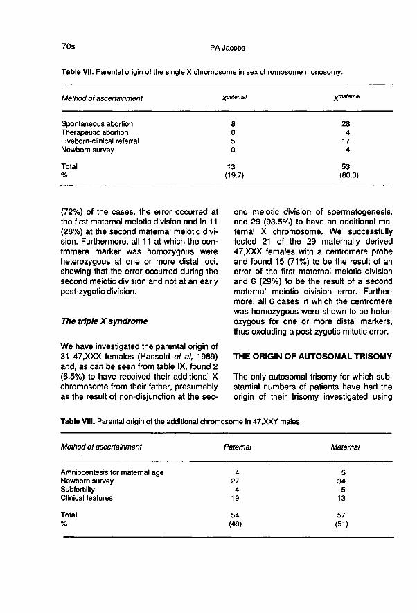
Using a number of X-linked RFLPS, wehave recently studied the parental origin of66 apparently non-mosaic 45.X individuals(Hassold et al, 1990). Unfortunately, as theresult of the error is not present, we canonly infer the parental origin of the missingchromosome and it is not possible to in-
vestigate the cell division at which the erroroccurred. As can be seen from table VII,we found 80% to have a maternal X and20% a paternal X irrespective of whetherthey were liveborn or spontaneously abort-ed. Thus, the error giving rise to the greatmajority of 45,X conceptuses occurs eitherduring spermatogenesis or subsequent tofertilization with the selective involvementof a paternal sex chromosome.
Kllnefelter’s syndrome
We have investigated the origin of the ad-ditional sex chromosome in 111 males witha 47,XXY chromosome complement (Har-vey et al, 1990). As the additional chromo-some is present and there is an X centro-mere-specific probe available, we wereable to determine not only the parent whocontributed the extra chromosome but alsothe cell division at which the error oc-
curred. As can be seen from table VIII, 54(49%) were paternal in origin, having bothan X and a Y chromosome from the fatheras a result of an error of the first paternalmeiotic division. The remaining 57 (51%)were maternal in origin. We successfullytested 39 of the 57 maternal cases with anX chromosome centromere-specific probe,pBamX9, in order to determine the cell di-vision at which the error occurred. In 28
(72%) of the cases, the error occurred atthe first maternal meiotic division and in 11 1
(28%) at the second maternal meiotic divi-sion. Furthermore, all 11 at which the cen-tromere marker was homozygous wereheterozygous at one or more distal loci,showing that the error occurred during thesecond meiotic division and not at an earlypost-zygotic division.
The triple X syndrome
We have investigated the parental origin of31 47,XXX females (Hassold et al, 1989)and, as can be seen from table IX, found 2(6.5%) to have received their additional Xchromosome from their father, presumablyas the result of non-disjunction at the sec-
ond meiotic division of spermatogenesis,and 29 (93.5%) to have an additional ma-ternal X chromosome. We successfullytested 21 of the 29 maternally derived47,XXX females with a centromere probeand found 15 (71%) to be the result of anerror of the first maternal meiotic divisionand 6 (29%) to be the result of a secondmaternal meiotic division error. Further-more, all 6 cases in which the centromerewas homozygous were shown to be heter-ozygous for one or more distal markers,thus excluding a post-zygotic mitotic error.
THE ORIGIN OF AUTOSOMAL TRISOMY
The only autosomal trisomy for which sub-stantial numbers of patients have had theorigin of their trisomy investigated using

molecular probes is trisomy 21 (Takaesuet al, 1990). As can be seen from table X,in 27 (18%) of the patients, the additionalchromosome was paternal and in 125
(82%) it was maternal. This figure agreeswell with the estimates of 20% paternaland 80% maternal based on cytogeneticheteromorphisms (Takaesu et al, 1990).Comparatively few trisomies for other auto-somes have been studied using molecularprobes but all showed the majority to bematernal in origin (table X). When the ob-servations on chromosome 21 are com-
pared with those on the other autosomes,there appears to be a larger proportion ofpaternally derived cases for chromosome21 than for the other autosomes, with thepossible exception of chromosome 13.
Clearly many more cases of trisomy must
be studied before we know whether or notthere are real differences in the proportionof paternally and maternally derived casesamong different chromosomes.
ESTIMATES OF THE NUMBEROF GAMETES WITH SPECIFICCHROMOSOME ABNORMALITIES
It is possible to predict the number of
sperm and eggs with a specific chromo-some abnormality from a knowledge of thefrequency of the abnormality and the pro-portion that are attributable to a paternal ora maternal error. However, in so doing, anumber of assumptions must be made. 1)All monosomies and trisomies result fromerrors at gametogenesis and no significant

proportion arises post-fertilization. Thisseems a reasonable assumption for bothsex and autosomal trisomies based on epi-demiological information, the absence of asubstantial proportion of mosaics and thefailure to find evidence of somatic originwith molecular probes. However, it seems
likely that most sex chromosome mono-somies arise by a mechanism differentfrom that causing trisomy. This suggestionis based on epidemiological evidence, thelarge number of sex chromosome mono-somies compared to sex chromosome tri-somies, and the very high proportion of
mosaics, at least among the liveborn. Themechanism resulting in 45,X conceptusesmay involve the production of abnormal
gametes, eg, anaphase lag at meiosis, orit may occur after fertilization, eg, ana-
phase lag at an early cell division of thezygote. Therefore, the frequency of sex
chromosomally abnormal gametes hasbeen calculated in two ways, both includ-
ing and excluding the data from 45,X con-ceptuses. 2) For every trisomic gametethere is a corresponding monosomic gam-ete. This seems a reasonable assumptionbased on the symmetry of the meiotic pro-
cess and on evidence from the mouse
(Gropp and Winkin, 1981). 3) There is nogametic selection prior to or at fertilization.This seems a reasonable assumptionbased on evidence from a very wide varie-
ty of organisms. 4) The calculated frequen-cies of trisomics among all clinically recog-nized pregnancies are an accurate
reflection of their rate at conception. Thisseems an unlikely assumption becausethere is every reason to suppose that
many autosomal trisomies are selected
against prior to their giving rise to a clini-cally recognized pregnancy. However, it is
impossible to obtain data on losses at thevery early stages of pregnancy. Thereforethe gametic frequencies calculated underassumption 4 must be serious underesti-mates for many chromosomes.
The frequency of 7 types of chromo-some abnormalities among all clinicallyrecognized pregnancies is given in tableXi. These 7 abnormalities were selectedeither because the parental origin hadbeen studied using molecular probes onmore than 10 cases or because the paren-tal origin could be deduced from the abnor-mality itself (XYY). The proportion of ab-

normal gametes giving rise to these 7 ab-normalities is also shown (table XI). As canbe seen, over 1.3% of sperm and 1.9% ofeggs must have a chromosome abnormali-
ty leading to these 7 types of abnormalconceptuses.When we consider individual auto-
somes, the frequency of nullisomic ga-metes must be at least the same as thatfor disomic gametes. This suggests that0.18% of sperm and 2.08% of eggs have a
missing or additional chromosome 16;0.02% of sperm and 0.34% of eggs have a
missing or additional chromosome 18 and0.16% of sperm and 0.74% of eggs have a
missing or additional chromosome 21. Thedata for the sex chromosomes are shownin table XII. The figures have been calcu-lated in two ways; firstly, on the assump-tion that the 3 types of sex chromosometrisomies are the results of errors of gamet-ogenesis that produce disomic and nulli-somic gametes with equal frequency andthat the majority of 45,X conceptuses re-sult from a post-fertilisation error and notan abnormal gamete and, secondly, on theassumption that all sex chromosome ab-normalities are the result of errors of ga-metogenesis but that the nullisomic ga-metes that give rise to the majority of 45,Xconceptuses have no disomic counter-
parts. On the first assumption 0.17% of
sperm and 0.17% of eggs have a missing
or additional sex chromosome, while onthe second assumption 1.2% of sperm and0.43% of eggs have a missing or additionalsex chromosome.
A comparison of these predicted mini-mal frequencies of abnormal gametes withthose actually observed for sperm, usingthe hamster oocyte test and in unfertilizedand fertilized eggs, will be of great interest.Such comparisons should enable us to de-termine whether the large number of 45,Xconceptions which have no paternal sexchromosome are the result of a gametic ora post-fertilization error and also enable usto estimate the number of undetected pre-implantation and early post-implantationlosses that are attributable to chromoso-
mally abnormal gametes.
ACKNOWLEDGMENTS
This work was supported by the WellcomeTrust.
REFERENCES
Carr DH (1963) Chromosome studies in abortus-es and stillborn infants. Lancet ii, 603
Gropp A, Winkin H (1981) Robertsonian translo-cations: cytology, meiosis, segregation pat-terns and biological consequences of hetero-

zygosity. Symp Zool Soc London 47, 141-181
Harvey J, Jacobs P, Hassold T, Pettay D (1990)The parental origin of 47,XXY males. In:March of Dimes, Birth Defects Foundation,Birth Defects, Original Article Series (Ham-merton JL, Evans JA, eds)
Hassold TJ, Takeasu N (1989) Analysis of non-disjunction in human trisomic spontaneousabortions. In: Molecular and CytogeneficStudies of Non-Disjunction (Hassold T, Ep-stein, eds) Alan R Liss, Inc, New York, 115-134
Hassold T, Jacobs PA, Leppert M, Sheldon M(1987) Cytogenetic and molecular studies oftrisomy 13. J Med Genet 24, 725-732
Hassold T, Arnovitz K, Jacobs PA, May K, Rob-inson D (1990) The parental origin of themissing or additional chromosomes in 45,Xand 47,XXX females. In: March of Dimes,Birth Defects Original Article Series (Hamer-ton JL, Evans JA, eds)
Hook EB, Hammerton JL (1977) The frequencyof chromosome abnormalities detected inconsecutive newborn studies. In: Population
Cytogenetics. Academic Press, New York,63-79
Hook EB, Healy N, Willey A (1989) How muchdifference does chromosome banding make?Ann Hum Genet 54, 237-242
Jacobs PA, Hassold TJ (1987) Chromosome ab-normalities: comparative epidemiology inabortions and livebirths. In: Proc 7th Int
Congr Hum Genet Berlin, 1986 (Vogel F,Sperling K, eds) Springer-Verlag, Berlin, 233-244
Kline J, Stein Z (1985) Very early pregnancy. In:Reproductive Toxicology, Raven Press, NewYork, 251-265
Kupke KG, Muller U (1989) Parental origin ofthe extra chromosome in trisomy 18. Am JHum Genet 45, 599-606
Short RV (1979) When a conception fails to be-come a pregnancy. In: Maternal Recognitionof Pregnancy, Exerpta Medica, Amsterdam,377-387
Takaesu N, Jacobs PA, Cockwell A, BlackstonRD, Freeman S, Nuccio J, Kurnit D, Uchida I,Hassold T (1989) Non-disjunction of chromo-some 21. Am J Med Genet (in press)