the role of fibroblast growth factors in tooth...
TRANSCRIPT
Review ArticleThe Role of Fibroblast Growth Factors in Tooth Development andIncisor Renewal
Wen Du ,1 Wei Du ,2 and Haiyang Yu 1
1State Key Laboratory of Oral Diseases, National Clinical Research Center for Oral Diseases, Department of Prosthodontics, WestChina Hospital of Stomatology, Sichuan University, Chengdu 610041, China2State Key Laboratory of Oral Diseases, National Clinical Research Center for Oral Diseases, Department of Endodontics, West ChinaHospital of Stomatology, Sichuan University, Chengdu 610041, China
Correspondence should be addressed to Haiyang Yu; [email protected]
Received 19 November 2017; Accepted 4 February 2018; Published 11 March 2018
Academic Editor: Bo Yu
Copyright © 2018Wen Du et al. This is an open access article distributed under the Creative Commons Attribution License, whichpermits unrestricted use, distribution, and reproduction in any medium, provided the original work is properly cited.
The mineralized tissue of the tooth is composed of enamel, dentin, cementum, and alveolar bone; enamel is a calcified tissue with noliving cells that originates from oral ectoderm, while the three other tissues derive from the cranial neural crest. The fibroblastgrowth factors (FGFs) are critical during the tooth development. Accumulating evidence has shown that the formation of dentaltissues, that is, enamel, dentin, and supporting alveolar bone, as well as the development and homeostasis of the stem cells inthe continuously growing mouse incisor is mediated by multiple FGF family members. This review discusses the role of FGFsignaling in these mineralized tissues, trying to separate its different functions and highlighting the crosstalk between FGFs andother signaling pathways.
1. Introduction
Organogenesis is a complex physiological process. An intri-cate array of signaling molecules such as FGFs, bone mor-phogenetic proteins (BMPs), Wnt, and Hedgehog (Hh)families are known to regulate the formation, differentiation,and maintenance of the tooth and alveolar bone during thedevelopment and throughout adulthood [1–4].
FGF signaling occupies a significant position in inducingthe proliferation and differentiation of multiple cell typesduring embryonic stages [5–10], as well as in regulatingthe development in different animals [11–14]. In addition,FGFs have been shown to regulate mouse tooth develop-ment [2, 15–17]. Nevertheless, a comprehensive descriptionabout the mechanism underlying FGFs that regulate differ-ent mineralized tissues of tooth during the embryonic stages,as well as incisor renewal in the adulthood, is still needed.Here, we summarize the roles of FGF signaling in mousetooth development and the ways FGFs control the stem cellsin incisor renewal, trying to separate its different functions
and highlighting the crosstalk between FGFs and other sig-naling pathways.
2. Development of Tooth and SupportingBone Structure
Most vertebrate groups have the ability to replace their teeth.Mammals have two sets of teeth: primary and adult teeth. Incontrast, mice contain one set with two different types:molars located at the proximal area and incisor located atthe distal area, which are separated by the toothless diastemaregion. Mouse incisors grow continuously throughout thelifetime in sharp contrast to the molars. It has been demon-strated that the presence of stem cells, which are located inthe proximal end of the incisor, gives rise to the differentiatedtooth cell types, thus promoting continuous growth of thistooth [18].
It has been widely held that tooth morphogenesis is char-acterized by the sequential interactions between the mesen-chymal cells derived from the cranial neural crest, and the
HindawiStem Cells InternationalVolume 2018, Article ID 7549160, 14 pageshttps://doi.org/10.1155/2018/7549160
stomadial epithelium [19, 20]. This process consists of severalphases, that is, bud, cap, and bell stages. In mice, the dentalmesenchyme is attributed to neural crest cells which arederived from the midbrain and hindbrain regions aroundembryonic day 8.5 (E8.5) [21–24]. The determination oftooth-forming sites during E10.5 [25–27] and the thickeningof the dental epithelium at E11.5 have been considered as thefirst signs of tooth development [28]. During the bud stage(E12.5–E13.5), in both incisor and molar, the thickened den-tal epithelium buds into the underlying mesenchyme, thusforming the epithelial tooth bud around the condensed mes-enchymal cells. At the subsequent cap stage (E14.5–E15.5),the epithelial component undergoes specific folding. A cen-tral event, during the transitional process between bud andcap stages, is the formation of the enamel knot (EK), a struc-ture composed of a group of nondividing cells. Moreover,several signaling molecules, such as Shh, FGF4, FGF9,BMP4, and BMP7, as well as Wnt10a/b, are restrictedlyexpressed in the enamel knot. Several studies have shownthat the EK, as the signaling center, has an important rolein tooth cusp patterning control [29, 30]. During the follow-ing bell stage, the ameloblasts and odontoblasts originatefrom the dental epithelium and mesenchyme, respectively[2]. At this stage, the secondary EKs (sEK) succeed the pri-mary EKs (pEK) in the molar. In addition, the condensedmesenchymal cells around the developing epithelial toothgerm at the bud stage go on to differentiate into a supportingalveolar bone that forms the sockets for the teeth at the bellstage [31–33].
With reference to its origin, it has been reported that thealveolar bone is formed by intramembranous ossification[32, 33]. Intramembranous ossification starts with the mes-enchymal cells which are derived from embryonic lineagescorrespondingly, which then migrate towards the locationsof the future bones. Here, they form high cellular densitycondensations that outline the size and shape of the futurebones. The mesenchymal cells subsequently differentiateinto osteoblasts, thus forming bone directly within the con-densations [3].
3. Stem Cells in Incisor Renewaland Osteogenesis
As it was previously mentioned, the adult mouse incisors cangrow unceasingly throughout their lifetime, and this growthis counterbalanced by continuous abrasion. Essential to thisphenomenon is the presence of active somatic stem cellswhich reside at the proximal end of the incisor. As a result,extensive studies have uncovered that the epithelial and mes-enchymal stem cells of the incisor give rise to ameloblasts andodontoblasts, which are in turn responsible for producingnew tissue which replaces worn enamel and dentin [1].
The epithelial stem cells reside in a niche called the cervi-cal loop. From contemporary understanding of ameloblastdevelopment and maturation, these stem cells are located inthe outer enamel epithelium (OEE) and the stellate reticulum(SR) of the labial cervical loop. These stem cells give rise tothe transit-amplifying (TA) cells, which are divided for sev-eral generations and then differentiate into preameloblasts.
In turn, these cells give rise to mature ameloblasts that arecharacterized by three component stages: presecretory, secre-tory, and maturation zones [34]. In contrast, compared to theepithelial counterparts, the stem cells which are derived fromthe mesenchyme and reside in the dental pulp are relativelypoorly characterized [1].
In addition to incisor renewal, stem cells also show pow-erful osteogenic potential due to their ability to differentiateinto osteoblasts. For instance, the condensation of mesenchy-mal stem cells (MSCs) from the neural crest or mesoderm hasshown to stimulate the beginning of mammalian skeletaldevelopment [4]. The alveolar bone tissue regenerates duringthe process of bone repair and synostosis after implantation,exodontia, and orthodontic treatment, indicating the impor-tance of stem cells in bone repair and regeneration. Numer-ous techniques have been used to stimulate stem cell-drivenosteogenesis [35], including direct implantation of undiffer-entiated cells, or after in vitro differentiation, as well as stim-ulation of native stem cell differentiation through cytokineintroduction. Adult bone marrow-derived mesenchymalstem cells are potentially useful for craniofacial mineralizedtissue engineering [36]. It has been shown that comparedwith conventional guided bone regeneration, implanted tis-sue repair cells induce regeneration of alveolar bone anddecrease the need for secondary bone grafting [37].Adipose-derived stem cells (ADSCs), like bone marrow stemcells (BMSCs) that are derived from the mesenchyme andprovide a supportive stroma for cell differentiation, may beextensively used in osteogenesis. Yet, larger quantities ofADSCs may be harvested with less pain as opposed toBMSCs [38]. In the clinical setting, further investigationsof optimization for stem cell harvesting as well as scaffold-based delivery are required given the challenges in stem celltransplantation [36].
4. FGFs and the Receptors
The mouse FGF family comprises 22 members and could bedivided into seven subfamilies: FGF1 (FGF1 and FGF2),FGF4 (FGF4–6), FGF7 (FGF3, FGF7, FGF10, and FGF22),FGF8 (FGF8, FGF17, and FGF18), FGF9 (FGF9, FGF16,and FGF20), FGF11 (FGF11–14), and FGF15 subfamilies(FGF15, FGF21, and FGF23) [39, 40]. FGF11 subfamilies(FGF11–14), also known as iFGFs, lack signal peptides andthus work as intracellular proteins. FGF15 subfamilies, con-sisting of FGF15, FGF21, and FGF23, are also known ashormone-like subfamilies (hFGFs) [41]. It is widely believedthat iFGFs and hFGFs act in an FGFR-independent manner[42]. Other FGFs, which are also defined as canonical sub-families, mediate their biological responses as extracellularproteins by binding to and activating cell surface tyrosinekinase FGF receptors (FGFRs) [39, 43]. FGFRs have beenidentified as four related transmembrane proteins compris-ing of a single transmembrane domain, an extracellularligand-binding domain, and an intracellular tyrosine kinasedomain [44].
Fgfr1–3 undergo alternative mRNA splicing events andthereby generate alternative versions of the immunoglobulin-like domain III (IIIb or IIIc) [45]. This process increases the
2 Stem Cells International
ligand-binding properties via regulation in a tissue-dependent manner [46–48]. The IIIb splice variant expres-sion is predominantly detected in epithelial lineages and isresponsible for transducing signals initiated by FGFsdetected in the mesenchyme. Furthermore, the IIIc splicevariant is restrictedly expressed in mesenchymal lineagesand it transduces signaling from epithelial FGFs [49–53].By contrast, Fgfr4 is not alternatively spliced [54].
Triggered by the dimerization of receptors, the transpho-sphorylation and activation of FGFRs initiate signaling viamultiple downstream intracellular pathways [55]. By bindingto various arrays of adaptor proteins such as SHP2 andgrowth factor receptor-bound protein 2 (GRB2) [56–59],the activated receptor’s cytosolic domain in turn mediatesRas signals to activate the downstream signaling cascades,such as PI3K/AKT and MAPK pathways [60].
While FGF signaling, encompassing FGF and FGFRs,occupies a critical position in regulating diverse cellularfunctions, it could be regulated by various upstream regula-tors. The most well-investigated regulator group are theSprouty genes, which encode antagonists of FGF signalingby binding with GRB2 thus preventing Ras activation [61].Other signaling pathways, for example, the Wnt pathway,have been recently identified as a positive regulator of FGFsignaling [62].
5. Expression Patterns of FGFs duringTooth Development
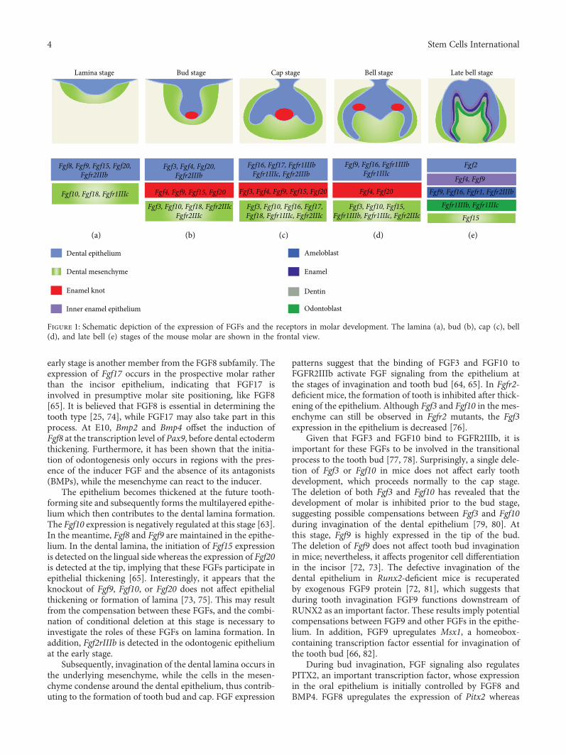
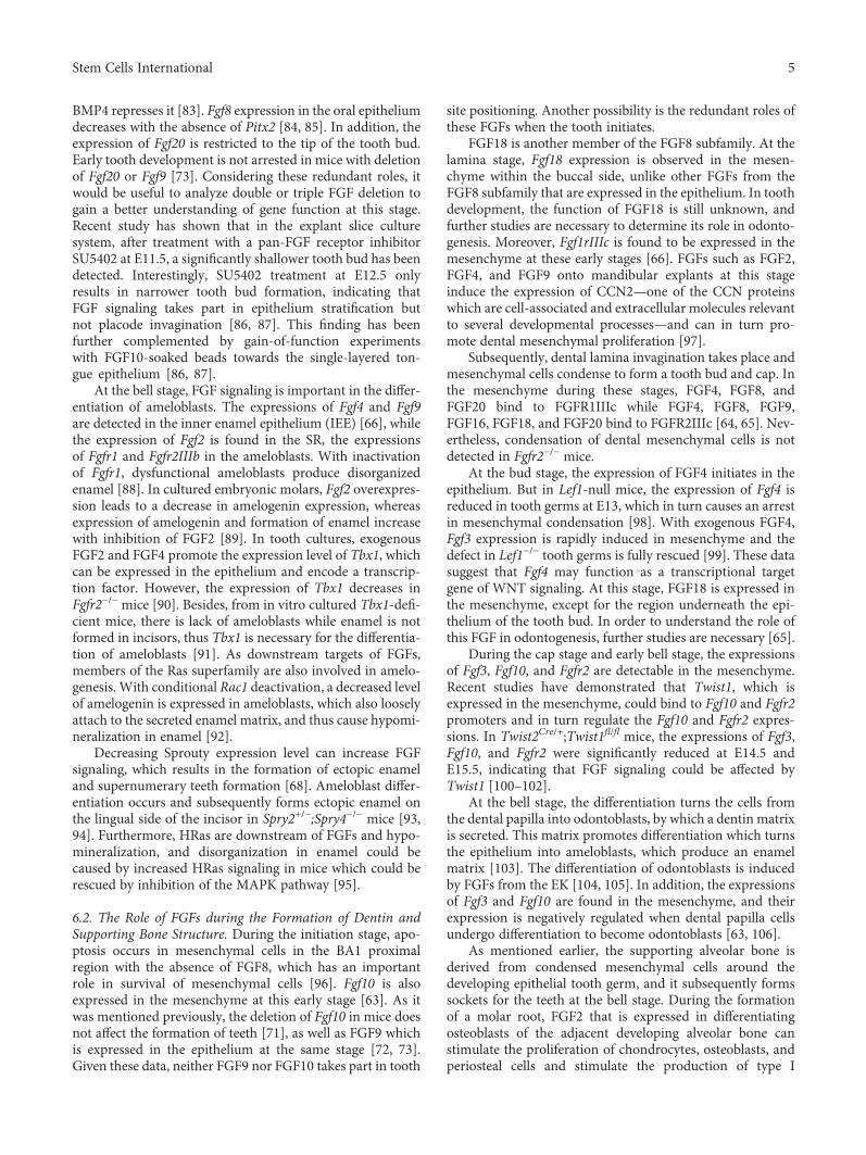
FGFs are expressed in the dental epithelium throughouttooth development (Figure 1). During the initiation stage ofodontogenesis, the expressions of Fgf8, Fgf9, Fgf10, Fgf17,and Fgfr2IIIb are detected in the prospective tooth regionaround E10.5 to E11.5 [63–66]. In the same region, followingthe formation of the dental lamina, Fgf8, Fgf9, Fgf15, andFgf20 are expressed, while the expression of Fgf10 in the epi-thelium is decreased [63]. As the epithelial bud is formedunceasingly in the dental lamina, the Fgf9 and Fgf20 expres-sions persist while Fgf3 and Fgf4 are initiated [65, 66]. Fgf3,Fgf4, Fgf9, Fgf15, and Fgf20 are expressed in the pEK afterits formation, while the expressions of Fgfr1IIIb, Fgfr1IIIc,and Fgfr2IIIb are found in the dental epithelium. Fgf16 andFgf17 are expressed in the cervical loop epithelium [65]. Inthe sEK at the bell stage, the Fgf4 and Fgf20 expressions arerestricted in the forming cusps. The expressions of Fgf9,Fgf16, Fgfr1IIIb, and Fgfr1IIIc are detected in the differentiat-ing ameloblasts. At the same time, the expressions of Fgf1,Fgf9, Fgf16, and Fgf17 can be found in the cervical loop epi-thelium of the incisor [65, 66].
During tooth development, the expressions of FGFs arealso detected in the mesenchyme (Figure 1). Fgfr1IIIc andFgf10 expressions are detected in the prospective tooth regionduring the early stage [63, 66]. During the thickening of theprospective tooth region epithelium which then forms thedental lamina, the expressions of Fgf10 and Fgf18 are foundin the mesenchyme [63, 65]. After the formation of the epi-thelial bud, the expressions of Fgf10 and Fgf18, as well as thatof Fgf3, are found; besides, Fgfr2IIIc expression appears [65].After pEK formation, Fgf3, Fgf10, and Fgf18 are found in the
mesenchyme [65]. The expressions of Fgf16 and Fgf17 aredetected in the cervical loop mesenchyme while Fgfr1IIIcand Fgfr2IIIc are expressed in the mesenchyme of the buc-cal side [63, 65, 66]. At the late bell stage, Fgf3 is expressedin the dental papilla, while Fgf10 is expressed in the differ-entiating odontoblasts. In addition, Fgf15 is restricted tothe mesenchyme while the expressions of Fgfr1IIIb andFgfr1IIIc are located in odontoblasts [63, 65]. Moreover,Fgf3, Fgf7, Fgf10, Fgf16, Fgf18, and Fgf21 are also detectedin the incisor [65].
The mesenchymal-derived alveolar bone is histologicallydetectable after E13.0, and its early formation occurs byE14.0. After E15.0, the development of the alveolar bone iswell progressed. Comparative PCR array analysis has shownan increased statistical significance (14-fold) in the Fgf3expression levels between E13.0 and E15.0 [67]. In addition,Fgf7 transcripts have been detected in the developing bonesurrounding the tooth germ [63].
During tooth development, Sprouty (Spry) genes, as FGFantagonists, are also expressed in different tissues [68]. Dur-ing the cap stage, the expression of Spry1 appears in diastemabuds and is highly expressed in the tooth germs of the firstmolar (M1), whereas Spry2 is strongly expressed in the epi-thelium of both M1 tooth germ and diastema. Spry4 isuniquely expressed in the mesenchyme in tooth germs ofM1 and in the diastema. Nevertheless, Spry3 is not detectedwithin the tooth germ.
6. The Role of FGFs during Tooth Development
6.1. The Role of FGFs during the Formation of Enamel. Toothformation begins with the first signals from the future toothepithelium at E9.5 [69]. In the area where a prospective toothforms, the oral ectoderm thickens; the epithelial Fgf8, Fgf9,and Fgf17 expressions suggest that these FGFs may take partin the initiation of tooth development [65, 66]. An early studyhas shown that FGF8 can induce the expression of Pax9 inmice, which reveals the prospective odontogenesis locations,and is essential beyond the bud stage of tooth development[25]. In the first branchial arch (BA1) with ectodermNestin-Cre, conditional Fgf8 knockout leads to a decreasein Pax9 expression in the expected molar region, and the for-mation of molar is stopped. The deletion of Fgf8 does notaffect Pax9 expression within the presumptive incisor region,and thus the incisor is formed in a normal manner. Therecent study has indicated that Fgf8-expressing cells labeledduring the initiation stage of molars can furnish the epithelialcells and collectively migrate towards the dental lamina sitewhich is important for prospective molar positioning [70].In addition, the conditional deletion of Fgf8 by E11.5 leadsto an arrest in the formation of the dental lamina, and it alsoaffects further development of the dental primordium andleads to a shorter invaginated structure [70]. At this earlystage, Fgf10, a member from another FGF subfamily, isexpressed in the epithelium [63]. Teeth develop in Fgf10-defi-cient mice, although a defect of the stem cell compartment inthe incisor cervical loop has been observed [71], and deletionof Fgf9 which is also expressed at the early stage does notaffect tooth formation either [72, 73]. Fgf17 expressed at the
3Stem Cells International
early stage is another member from the FGF8 subfamily. Theexpression of Fgf17 occurs in the prospective molar ratherthan the incisor epithelium, indicating that FGF17 isinvolved in presumptive molar site positioning, like FGF8[65]. It is believed that FGF8 is essential in determining thetooth type [25, 74], while FGF17 may also take part in thisprocess. At E10, Bmp2 and Bmp4 offset the induction ofFgf8 at the transcription level of Pax9, before dental ectodermthickening. Furthermore, it has been shown that the initia-tion of odontogenesis only occurs in regions with the pres-ence of the inducer FGF and the absence of its antagonists(BMPs), while the mesenchyme can react to the inducer.
The epithelium becomes thickened at the future tooth-forming site and subsequently forms the multilayered epithe-lium which then contributes to the dental lamina formation.The Fgf10 expression is negatively regulated at this stage [63].In the meantime, Fgf8 and Fgf9 are maintained in the epithe-lium. In the dental lamina, the initiation of Fgf15 expressionis detected on the lingual side whereas the expression of Fgf20is detected at the tip, implying that these FGFs participate inepithelial thickening [65]. Interestingly, it appears that theknockout of Fgf9, Fgf10, or Fgf20 does not affect epithelialthickening or formation of lamina [73, 75]. This may resultfrom the compensation between these FGFs, and the combi-nation of conditional deletion at this stage is necessary toinvestigate the roles of these FGFs on lamina formation. Inaddition, Fgf2rIIIb is detected in the odontogenic epitheliumat the early stage.
Subsequently, invagination of the dental lamina occurs inthe underlying mesenchyme, while the cells in the mesen-chyme condense around the dental epithelium, thus contrib-uting to the formation of tooth bud and cap. FGF expression
patterns suggest that the binding of FGF3 and FGF10 toFGFR2IIIb activate FGF signaling from the epithelium atthe stages of invagination and tooth bud [64, 65]. In Fgfr2-deficient mice, the formation of tooth is inhibited after thick-ening of the epithelium. Although Fgf3 and Fgf10 in the mes-enchyme can still be observed in Fgfr2 mutants, the Fgf3expression in the epithelium is decreased [76].
Given that FGF3 and FGF10 bind to FGFR2IIIb, it isimportant for these FGFs to be involved in the transitionalprocess to the tooth bud [77, 78]. Surprisingly, a single dele-tion of Fgf3 or Fgf10 in mice does not affect early toothdevelopment, which proceeds normally to the cap stage.The deletion of both Fgf3 and Fgf10 has revealed that thedevelopment of molar is inhibited prior to the bud stage,suggesting possible compensations between Fgf3 and Fgf10during invagination of the dental epithelium [79, 80]. Atthis stage, Fgf9 is highly expressed in the tip of the bud.The deletion of Fgf9 does not affect tooth bud invaginationin mice; nevertheless, it affects progenitor cell differentiationin the incisor [72, 73]. The defective invagination of thedental epithelium in Runx2-deficient mice is recuperatedby exogenous FGF9 protein [72, 81], which suggests thatduring tooth invagination FGF9 functions downstream ofRUNX2 as an important factor. These results imply potentialcompensations between FGF9 and other FGFs in the epithe-lium. In addition, FGF9 upregulates Msx1, a homeobox-containing transcription factor essential for invagination ofthe tooth bud [66, 82].
During bud invagination, FGF signaling also regulatesPITX2, an important transcription factor, whose expressionin the oral epithelium is initially controlled by FGF8 andBMP4. FGF8 upregulates the expression of Pitx2 whereas
Fgf8, Fgf9, Fgf15, Fgf20,Fgfr2IIIb
Fgf10, Fgf18, Fgfr1IIIc
Fgf3, Fgf4, Fgf20,Fgfr2IIIb
Fgf3, Fgf10, Fgf18, Fgfr2IIIcFgfr2IIIc
Fgf16, Fgf17, Fgfr1IIIbFgfr1IIIc, Fgfr2IIIb
Fgf3, Fgf4, Fgf9, Fgf15, Fgf20
Fgf3, Fgf10, Fgf16, Fgf17,Fgf18, Fgfr1IIIc, Fgfr2IIIc
Fgf3, Fgf10, Fgf15,Fgfr1IIIb, Fgfr1IIIc, Fgfr2IIIc
Fgf4, Fgf9
Fgf4, Fgf9, Fgf15, Fgf20 Fgf9, Fgf16, Fgfr1, Fgfr2IIIb
Fgf2
Fgf4, Fgf20
Fgfr1IIIb, Fgfr1IIIc
Fgf15
Fgf9, Fgf16, Fgfr1IIIbFgfr1IIIc
Lamina stage Late bell stageBell stageCap stageBud stage
Dental epithelium
Dental mesenchyme
Enamel knot
Inner enamel epithelium
Ameloblast
Enamel
Dentin
Odontoblast
(a) (b) (c) (d) (e)
Figure 1: Schematic depiction of the expression of FGFs and the receptors in molar development. The lamina (a), bud (b), cap (c), bell(d), and late bell (e) stages of the mouse molar are shown in the frontal view.
4 Stem Cells International
BMP4 represses it [83]. Fgf8 expression in the oral epitheliumdecreases with the absence of Pitx2 [84, 85]. In addition, theexpression of Fgf20 is restricted to the tip of the tooth bud.Early tooth development is not arrested in mice with deletionof Fgf20 or Fgf9 [73]. Considering these redundant roles, itwould be useful to analyze double or triple FGF deletion togain a better understanding of gene function at this stage.Recent study has shown that in the explant slice culturesystem, after treatment with a pan-FGF receptor inhibitorSU5402 at E11.5, a significantly shallower tooth bud has beendetected. Interestingly, SU5402 treatment at E12.5 onlyresults in narrower tooth bud formation, indicating thatFGF signaling takes part in epithelium stratification butnot placode invagination [86, 87]. This finding has beenfurther complemented by gain-of-function experimentswith FGF10-soaked beads towards the single-layered ton-gue epithelium [86, 87].
At the bell stage, FGF signaling is important in the differ-entiation of ameloblasts. The expressions of Fgf4 and Fgf9are detected in the inner enamel epithelium (IEE) [66], whilethe expression of Fgf2 is found in the SR, the expressionsof Fgfr1 and Fgfr2IIIb in the ameloblasts. With inactivationof Fgfr1, dysfunctional ameloblasts produce disorganizedenamel [88]. In cultured embryonic molars, Fgf2 overexpres-sion leads to a decrease in amelogenin expression, whereasexpression of amelogenin and formation of enamel increasewith inhibition of FGF2 [89]. In tooth cultures, exogenousFGF2 and FGF4 promote the expression level of Tbx1, whichcan be expressed in the epithelium and encode a transcrip-tion factor. However, the expression of Tbx1 decreases inFgfr2−/− mice [90]. Besides, from in vitro cultured Tbx1-defi-cient mice, there is lack of ameloblasts while enamel is notformed in incisors, thus Tbx1 is necessary for the differentia-tion of ameloblasts [91]. As downstream targets of FGFs,members of the Ras superfamily are also involved in amelo-genesis. With conditional Rac1 deactivation, a decreased levelof amelogenin is expressed in ameloblasts, which also looselyattach to the secreted enamel matrix, and thus cause hypomi-neralization in enamel [92].
Decreasing Sprouty expression level can increase FGFsignaling, which results in the formation of ectopic enameland supernumerary teeth formation [68]. Ameloblast differ-entiation occurs and subsequently forms ectopic enamel onthe lingual side of the incisor in Spry2+/−;Spry4−/− mice [93,94]. Furthermore, HRas are downstream of FGFs and hypo-mineralization, and disorganization in enamel could becaused by increased HRas signaling in mice which could berescued by inhibition of the MAPK pathway [95].
6.2. The Role of FGFs during the Formation of Dentin andSupporting Bone Structure. During the initiation stage, apo-ptosis occurs in mesenchymal cells in the BA1 proximalregion with the absence of FGF8, which has an importantrole in survival of mesenchymal cells [96]. Fgf10 is alsoexpressed in the mesenchyme at this early stage [63]. As itwas mentioned previously, the deletion of Fgf10 in mice doesnot affect the formation of teeth [71], as well as FGF9 whichis expressed in the epithelium at the same stage [72, 73].Given these data, neither FGF9 nor FGF10 takes part in tooth
site positioning. Another possibility is the redundant roles ofthese FGFs when the tooth initiates.
FGF18 is another member of the FGF8 subfamily. At thelamina stage, Fgf18 expression is observed in the mesen-chyme within the buccal side, unlike other FGFs from theFGF8 subfamily that are expressed in the epithelium. In toothdevelopment, the function of FGF18 is still unknown, andfurther studies are necessary to determine its role in odonto-genesis. Moreover, Fgf1rIIIc is found to be expressed in themesenchyme at these early stages [66]. FGFs such as FGF2,FGF4, and FGF9 onto mandibular explants at this stageinduce the expression of CCN2—one of the CCN proteinswhich are cell-associated and extracellular molecules relevantto several developmental processes—and can in turn pro-mote dental mesenchymal proliferation [97].
Subsequently, dental lamina invagination takes place andmesenchymal cells condense to form a tooth bud and cap. Inthe mesenchyme during these stages, FGF4, FGF8, andFGF20 bind to FGFR1IIIc while FGF4, FGF8, FGF9,FGF16, FGF18, and FGF20 bind to FGFR2IIIc [64, 65]. Nev-ertheless, condensation of dental mesenchymal cells is notdetected in Fgfr2−/− mice.
At the bud stage, the expression of FGF4 initiates in theepithelium. But in Lef1-null mice, the expression of Fgf4 isreduced in tooth germs at E13, which in turn causes an arrestin mesenchymal condensation [98]. With exogenous FGF4,Fgf3 expression is rapidly induced in mesenchyme and thedefect in Lef1−/− tooth germs is fully rescued [99]. These datasuggest that Fgf4 may function as a transcriptional targetgene of WNT signaling. At this stage, FGF18 is expressed inthe mesenchyme, except for the region underneath the epi-thelium of the tooth bud. In order to understand the role ofthis FGF in odontogenesis, further studies are necessary [65].
During the cap stage and early bell stage, the expressionsof Fgf3, Fgf10, and Fgfr2 are detectable in the mesenchyme.Recent studies have demonstrated that Twist1, which isexpressed in the mesenchyme, could bind to Fgf10 and Fgfr2promoters and in turn regulate the Fgf10 and Fgfr2 expres-sions. In Twist2Cre/+;Twist1fl/fl mice, the expressions of Fgf3,Fgf10, and Fgfr2 were significantly reduced at E14.5 andE15.5, indicating that FGF signaling could be affected byTwist1 [100–102].
At the bell stage, the differentiation turns the cells fromthe dental papilla into odontoblasts, by which a dentin matrixis secreted. This matrix promotes differentiation which turnsthe epithelium into ameloblasts, which produce an enamelmatrix [103]. The differentiation of odontoblasts is inducedby FGFs from the EK [104, 105]. In addition, the expressionsof Fgf3 and Fgf10 are found in the mesenchyme, and theirexpression is negatively regulated when dental papilla cellsundergo differentiation to become odontoblasts [63, 106].
As mentioned earlier, the supporting alveolar bone isderived from condensed mesenchymal cells around thedeveloping epithelial tooth germ, and it subsequently formssockets for the teeth at the bell stage. During the formationof a molar root, FGF2 that is expressed in differentiatingosteoblasts of the adjacent developing alveolar bone canstimulate the proliferation of chondrocytes, osteoblasts, andperiosteal cells and stimulate the production of type I
5Stem Cells International
collagen [107]. FGF7, detected in the developing bonesurrounding the molar tooth germ and the mesenchymeadjacent to the incisor cervical loop, is involved in the forma-tion of alveolar bone [63]. Furthermore, the addition of FGF4or FGF8 beads into mouse dental mesenchymal cells canpromote their osteogenic differentiation and the expressionof CBFA1, which belongs to the CBFA family and functionsas an important regulator for differentiating osteoblasts invertebrata [81]. Given the strong expression of CBFA1 inosteoblasts in tooth alveolar bone at the late bell stage, sig-naling of FGF4 and FGF8 from the epithelium may also havean important role during alveolar bone formation. It has alsobeen reported that increased β-catenin signaling is related tothe fate of dental mesenchymal cells, while FGF3 can sustainthe odontogenic fate of incisor mesenchymal cells by down-regulating intracellular β-catenin signaling [108]. Therefore,the lack of FGF3 could induce the potency of mesenchymalcells to differentiate into osteoblasts which are responsiblefor the formation of the supporting bone structure. Sincethe role of FGFs in supporting alveolar bone remains largelyunexplored, further investigations are still needed.
6.3. The Role of FGFs in Tooth Size, Shape, Number, andArrangement. The signaling center pEK, which regulatesthe size and shape of the tooth, consists of nonproliferativecells [109]. Different signaling molecules and their antago-nists, including FGFs, Shh, Sprouty genes, BMPs, severalWNTs, and follistatin, are expressed in pEK [110]. pEKcells cannot respond to FGFs since there are no FGF recep-tors expressed in these cells [66]. The nonproliferative cellsin the pEK and the surrounding extensive proliferationcells may explain the epithelial folding and the transitionprocess between the tooth bud and cap stages [15, 109].Afterwards, the pEK induces the sEK in multicuspid teeth.The spatial arrangement of sEK has also been shown tocontain a network of activators and inhibitors [111, 112].The location and shape of the cusps are determined bythe proliferation and differentiation of the epithelial cellswhich are regulated by the sEK; thus, the shape of the toothcrown is determined.
In molars, pEK size can affect the shape of the invagi-nated epithelium. Tooth size and cusp number decrease ifthe size of the pEK is too small, since a small size can affectthe dental epithelium folding as well as the cervical loopand sEK formation. Ectodysplasin (Eda) and Traf6 are twomembers of the TNF-α family involved in tooth developmentregulation. A small size of the pEK will be present in micewithout either of those proteins, and it will then result inreduced tooth size and cusp number [113, 114]. The arrange-ment of sEK will be changed in case signaling from the pEK iscompromised by changing its size or shape; thus, defects ofcusp will occur. Furthermore, molar shape and cusp patternswill be altered under modulation in the levels of gene expres-sion in BMP, SHH, and WNT signaling [62, 115–119].
In the mesenchyme, the expression of Fgf3 is maintainedby FGF4 and FGF9, which are detected to be highly expressedin the pEK and sEK [63, 66]. FGF4 from the EK promotes theproliferation and has a role in the development of tooth cusps[30, 109]. Besides, FGF4 can also prevent cell apoptosis in the
dental epithelium and mesenchyme [120, 121]. Nevertheless,inactivation of neither Fgf4 nor Fgf9 can affect tooth shape ornumber [72, 73]. Moreover, epiprofin, a transcription factorfrom the Sp family, can promote dental epithelial FGF9which could elicit proliferation of dental mesenchymal cellsthrough FGFR1c; this is essential for the tooth morphogene-sis with the correct shapes and proper sizes [122].
FGF20 is another member of the FGF9 family, and itsexpression is found in the anterior bud of the lamina andthe EK, along with the expressions of Fgf3, Fgf4, Fgf9, andFgf15 [65, 66, 123]. During tooth development, FGF20 func-tions as a downstream target of EDA: in Eda mutant mice,the Fgf20 expression was reduced in molars, while it wasincreased in Eda-overexpressing (K14-Eda) mice [73]. Inaddition, Fgf20 knockout mice exhibited molar teeth withreduced size and a mild change in the anterior cusp, whilethe overall pattern of the cusp was normal in Fgf20 mutants.Therefore, FGF20 has shown to have a crucial role in finetuning of the pattern of the anterior cusp and functions as aregulator of tooth size. Double knockout of Fgf9 and Fgf20has shown strong additive effects by strikingly shorteningthe length of EK in comparison with the length of either sin-gle deletion mutant, which implies the redundancy betweenthese two FGF ligands [73].
In themesenchyme, FGFs have been shown to be involvedin tooth shaping. Like Fgf20-deficient mice, Fgf3−/−;Fgf10+/−
mice exhibit small molars [73, 80], and the Eda−/− molarphenotype can be partially offset by FGF10 in vitro [113].Consequently, decrease in FGF signaling in either epithe-lium or mesenchyme can lead to similar effects duringtooth formation.
Tooth number and arrangement are also found to betightly regulated by FGF signaling within the dentition.Supernumerary teeth, which are mainly positioned at theprospective site of the premolar, have been found in severalmutant mice. K14-Eda has been discovered as the first trans-genic mouse line with ectopic teeth [124]. The followingstudies have reported that in this genetic background, theformation frequency of an extra tooth increased with lackof Fgf20, while single deletion of Fgf20 could hardly promotethe formation of an extra molar [73]. Supernumerary incisorsand teeth anterior to the first molar have also been discoveredin mice with deletion of Sprouty genes [68, 125]. To sum up,these findings indicate that FGFs function as stimulators,while Sprouty genes function as endogenous antagonists ofFGF signaling in the development of the tooth.
7. The Role of FGFs in Incisor StemCell Renewal
It is well known that continuous growth of rodent incisor iscounterbalanced by wear, which is promoted by the lack ofenamel on the lingual side of the tooth surface. The absenceof lingual ameloblasts results in the lack of enamel on thatside [126]. Asymmetric wear maintains the length of incisorand leads to a sharp tip. The cervical loop includes variouscell types: IEE cells, OEE cells, SR cells, TA cells, and stratumintermedium (SI) cells. In addition, an extra group of cells
6 Stem Cells International
has been found between the SR and OEE [127]; however,their exact function still remains unknown.
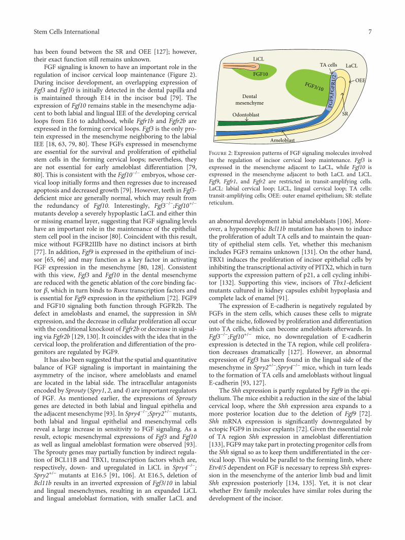
FGF signaling is known to have an important role in theregulation of incisor cervical loop maintenance (Figure 2).During incisor development, an overlapping expression ofFgf3 and Fgf10 is initially detected in the dental papilla andis maintained through E14 in the incisor bud [79]. Theexpression of Fgf10 remains stable in the mesenchyme adja-cent to both labial and lingual IEE of the developing cervicalloops from E16 to adulthood, while Fgfr1b and Fgfr2b areexpressed in the forming cervical loops. Fgf3 is the only pro-tein expressed in the mesenchyme neighboring to the labialIEE [18, 63, 79, 80]. These FGFs expressed in mesenchymeare essential for the survival and proliferation of epithelialstem cells in the forming cervical loops; nevertheless, theyare not essential for early ameloblast differentiation [79,80]. This is consistent with the Fgf10−/− embryos, whose cer-vical loop initially forms and then regresses due to increasedapoptosis and decreased growth [79]. However, teeth in Fgf3-deficient mice are generally normal, which may result fromthe redundancy of Fgf10. Interestingly, Fgf3−/−;Fgf10+/−
mutants develop a severely hypoplastic LaCL and either thinor missing enamel layer, suggesting that FGF signaling levelshave an important role in the maintenance of the epithelialstem cell pool in the incisor [80]. Coincident with this result,mice without FGFR2IIIb have no distinct incisors at birth[77]. In addition, Fgf9 is expressed in the epithelium of inci-sor [65, 66] and may function as a key factor in activatingFGF expression in the mesenchyme [80, 128]. Consistentwith this view, Fgf3 and Fgf10 in the dental mesenchymeare reduced with the genetic ablation of the core binding fac-tor β, which in turn binds to Runx transcription factors andis essential for Fgf9 expression in the epithelium [72]. FGF9and FGF10 signaling both function through FGFR2b. Thedefect in ameloblasts and enamel, the suppression in Shhexpression, and the decrease in cellular proliferation all occurwith the conditional knockout of Fgfr2b or decrease in signal-ing via Fgfr2b [129, 130]. It coincides with the idea that in thecervical loop, the proliferation and differentiation of the pro-genitors are regulated by FGF9.
It has also been suggested that the spatial and quantitativebalance of FGF signaling is important in maintaining theasymmetry of the incisor, where ameloblasts and enamelare located in the labial side. The intracellular antagonistsencoded by Sprouty (Spry1, 2, and 4) are important regulatorsof FGF. As mentioned earlier, the expressions of Sproutygenes are detected in both labial and lingual epithelia andthe adjacent mesenchyme [93]. In Spry4−/−;Spry2+/−mutants,both labial and lingual epithelial and mesenchymal cellsreveal a large increase in sensitivity to FGF signaling. As aresult, ectopic mesenchymal expressions of Fgf3 and Fgf10as well as lingual ameloblast formation were observed [93].The Sprouty genes may partially function by indirect regula-tion of BCL11B and TBX1, transcription factors which are,respectively, down- and upregulated in LiCL in Spry4−/−;Spry2+/− mutants at E16.5 [91, 106]. At E16.5, deletion ofBcl11b results in an inverted expression of Fgf3/10 in labialand lingual mesenchymes, resulting in an expanded LiCLand lingual ameloblast formation, with smaller LaCL and
an abnormal development in labial ameloblasts [106]. More-over, a hypomorphic Bcl11b mutation has shown to inducethe proliferation of adult TA cells and to maintain the quan-tity of epithelial stem cells. Yet, whether this mechanismincludes FGF3 remains unknown [131]. On the other hand,TBX1 induces the proliferation of incisor epithelial cells byinhibiting the transcriptional activity of PITX2, which in turnsupports the expression pattern of p21, a cell cycling inhibi-tor [132]. Supporting this view, incisors of Tbx1-deficientmutants cultured in kidney capsules exhibit hypoplasia andcomplete lack of enamel [91].
The expression of E-cadherin is negatively regulated byFGFs in the stem cells, which causes these cells to migrateout of the niche, followed by proliferation and differentiationinto TA cells, which can become ameloblasts afterwards. InFgf3−/−;Fgf10+/− mice, no downregulation of E-cadherinexpression is detected in the TA region, while cell prolifera-tion decreases dramatically [127]. However, an abnormalexpression of Fgf3 has been found in the lingual side of themesenchyme in Spry2+/−;Spry4−/− mice, which in turn leadsto the formation of TA cells and ameloblasts without lingualE-cadherin [93, 127].
The Shh expression is partly regulated by Fgf9 in the epi-thelium. The mice exhibit a reduction in the size of the labialcervical loop, where the Shh expression area expands to amore posterior location due to the deletion of Fgf9 [72].Shh mRNA expression is significantly downregulated byectopic FGF9 in incisor explants [72]. Given the essential roleof TA region Shh expression in ameloblast differentiation[133], FGF9 may take part in protecting progenitor cells fromthe Shh signal so as to keep them undifferentiated in the cer-vical loop. This would be parallel to the forming limb, whereEtv4/5 dependent on FGF is necessary to repress Shh expres-sion in the mesenchyme of the anterior limb bud and limitShh expression posteriorly [134, 135]. Yet, it is not clearwhether Etv family molecules have similar roles during thedevelopment of the incisor.
LiCLLaCL
Dentalmesenchyme FG
F9,F
GFR
1/2
FGF3/10
FGF10
Odontoblast
Ameloblast
TA cells
OEE
SR
Figure 2: Expression patterns of FGF signaling molecules involvedin the regulation of incisor cervical loop maintenance. Fgf3 isexpressed in the mesenchyme adjacent to LaCL, while Fgf10 isexpressed in the mesenchyme adjacent to both LaCL and LiCL.Fgf9, Fgfr1, and Fgfr2 are restricted in transit-amplifying cells.LaCL: labial cervical loop; LiCL, lingual cervical loop; TA cells:transit-amplifying cells; OEE: outer enamel epithelium; SR: stellatereticulum.
7Stem Cells International
BMP4 and activin, two proteins from the TGFβ family,modulate the activity of FGF and the regulation of theasymmetry of the incisor during incisor development. Thesymmetrical expression of BMP4 occurs throughout the mes-enchyme and suppresses the expression of Fgf3 indirectly inthe lingual mesenchyme. The expression of activin is morerobust in the labial mesenchyme, and the bead implantationstudy in incisor explants at E16 indicates that activin offsetsthe effect of BMP4 [80]. This can maintain the expressionof Fgf3 on the labial side of the mesenchyme and in turnincrease the proliferation of stem cells. In addition, the activ-ity of residual activin on the lingual side is counteracted byfollistatin that was detected in the lingual epithelium andfunctions to preserve the effect of BMP4 on repressing theFgf3 expression in the lingual mesenchyme. Consequently,embryos without the Fst gene which encodes follistatin haveshown to exhibit ectopic expression of Fgf3 in the lingualmesenchyme; these results in the expanded LiCL and lingualameloblasts as well as enamel formation [80]. On the con-trary, Fst misexpression in the epithelium leads to a reduc-tion in the expression of Fgf3 and subsequently causesreduced proliferation and the size of LaCL [80]. BMP4 canalso increase the differentiation ability of ameloblasts in themore distal side of the labial epithelium, while in the lingualepithelium this process is repressed by follistatin expressedlocally to maintain the asymmetry of the incisor [136]. Coin-cident with the view that BMP4 acts in two regions of theincisor during its development, misexpression of noggin(the inhibitor of BMP) leads to incisor hyperplasia becausein the cervical loop the proliferation of the population of pro-genitor cells is promoted. However, as ameloblast differenti-ation normally promoted by BMP signaling is inhibited, theincisors do not form enamel in the mutant [137]. Further-more, mesenchymal TGFβ receptor type I (Alk5/Tgfbr1)can modulate the proper initiation of tooth and the epithe-lium development of the incisor [138, 139]. MesenchymalFgf3 and Fgf10 expressions were downregulated when Alk5was knocked out specifically in the mesenchyme, causingfewer label-retaining cells and decreased proliferation in thecervical loop. Exogenous FGF10 proteins could rescue thisphenotype in incisor explant culture [138]. The mesenchy-mal expression of Fgf is partially activated via transcriptionfactors MSX1 and PAX9, which can initiate Fgf3 and Fgf10by E12.5 and in turn contribute to subsequent incisor devel-opment [128, 139, 140]. Moreover, with epithelial deletion ofIsl1, FGF signaling is upregulated and is associated with bothlingual cervical loop-generated ectopic enamel and labial sidepremature enamel formation [141]. FGF signaling anddownstream signal transduction pathways are also sup-pressed in Ring1a−/−;Ring1bcko/cko incisors [142].
It has also been reported that FGF signaling is requiredfor stem cell self-renewal and can prevent differentiation ofdental epithelial stem cells (DESCs) in the cervical loop andin the DESC spheres. The inhibition of the FGF signalingpathway can decrease proliferation and increase apoptosisof the cells in the DESC spheres. On the other hand, inhibit-ing FGFR or its downstream targets can decrease Lgr5-expressing cells in the cervical loop and induce cell differen-tiation in both cervical loop and the DESC spheres [143]. In
addition, FGF signaling may also be required for YAP-induced proliferation in T-A cells [144].
8. The Importance of FGF Signaling in HumanTooth Development
It has been shown that in clinics, FGFs are required forhuman tooth development. Its dysregulation seriously affectstooth development in humans, leading to enamel defects andtooth agenesis. Lacrimo-auriculo-dento-digital (LADD;Online Mendelian Inheritance in Man (OMIM) databaseno. 149730) syndrome, a congenital autosomal dominantdisorder, results from the heterozygous missense mutationsin FGF10, FGFR2, and FGFR3. LADD is characterized byaplasia, hypoplasia/atresia of salivary/lacrimal glands, earswith cup shape, and hearing loss [145–148], as well as variousdental phenotypes, including hypodontia, teeth with pegshape, and hypoplastic enamel [149]. In addition, compoundheterozygous or homozygous FGF3mutations cause congen-ital deafness with labyrinthine aplasia, microtia, and micro-dontia (LAMM; OMIM no. 610706) syndrome which isalso characterized by malformed external ear, malformed/missing inner ear, and peg-shaped teeth with reduced size[150–152].
Mutations in FGFRs can also cause several syndromessuch as Apert and Crouzon syndromes. Among them, theApert syndrome (OMIM no. 101200) derives from gain offunction in FGFR2 mutations and is characterized by hypo-plasia of midface, craniosynostosis, and syndactyly of thehands and feet [153]. The mutations in FGFR2 can causeCrouzon syndrome (OMIM no. 123500) characterized bycraniosynostosis, leading to hypertelorism, prognathism ofmandible, hypoplastic maxillary, and short upper lip [154].Patients with Apert and Crouzon syndromes usually exhibithypodontia, mostly of the third molar, second incisor inmaxillary, and second premolar in mandible [155, 156].
It has also been reported that the application of FGF2 canpromote the regeneration of periodontal tissues [157, 158].In this study, a clinical trial was performed in 253 adult peri-odontitis patients. A modified Widman periodontal surgerywas carried out, and during the surgery, a 200μL investiga-tional formulation containing FGF2 in different concentra-tions was applied to 2- or 3-walled vertical bone defects.The application of FGF2 showed a significant effect overthe placebo-control group (p < 0 01) for the bone fill percent-age after 36 weeks of administration. The results demonstratethat topical FGF2 application can treat the bone defectcaused by periodontitis and it can be efficacious in humanperiodontal tissue regeneration [158]. In addition, FGF2can also promote the neovascularization of human dentalpulps which is severed [159]. Human molars without carieswere used for preparation of tooth slices which were thentreated with 0–50ng/mL recombinant human FGF2 for aweek in vitro. The result showed that the density of microves-sel in dental pulps was enhanced with FGF2 treatment com-pared with untreated controls, indicating that topicalapplication of FGF2 in advance of replantation might be effi-cacious in the treatment for avulsed teeth [159]. Anotherstudy isolated and characterized stem cells from inflamed
8 Stem Cells International
pulp tissue of human functional deciduous teeth (iSHFD) inorder to investigate the role of FGF2 on the potential ofregeneration of these cells [160]. Application of FGF2 toiSHFD during their expansion improved the colony-forming efficiency of the cells and increased their potentialof migration and proliferation, but decreased their potentialof differentiation in vitro. This provides a good stem cellsource for future applications in clinics and a new way touse inflamed tissues which has to be discarded before.
Given the results of these studies, the application of FGFscan be a potential treatment for human dental diseases, evenfor those defects in tooth development as well as for thesyndromes caused by mutations in FGFs and FGFRs. Thedelivery of FGFs to the primary nidus still needs to beimproved, and further clinical trials are also required.
9. Conclusion
FGF signaling has been the focus of intense interest over thepast years, and thus, it has been investigated both in vitro andin vivo, by using different cell and genetic mouse models. TheFGF expression has an important role in different stages oftooth development, including tooth initiation and mineral-ized tissue formation. Uniquely in rodents, FGFs are essentialto maintaining the stem cell niche fueling the unceasinglygrowing incisor throughout their lifetime. The tooth offersan attractive model to further dissect the regulation andtransduction of FGFs in developmental as well as stem cellbiology. Despite the understanding of the role of FGF signal-ing, many questions remain unexplored. Thus, it is necessaryto further investigate more molecular mechanisms whichregulate FGFs and examine their other pathways. In addition,like the irreplaceable function of FGFs in regeneration andtissue homeostasis in the mouse model, FGFs have also beenfound to be involved in these processes in humans. By con-trolling the activity of FGFs, it could be possible to obtainnovel methods to treat human diseases. Studies on the under-lying mechanism of FGF regulation in teeth may potentiallyextend the current knowledge of other organ systems andmay also offer insights into progression of diseases, present-ing new therapeutic approaches.
Conflicts of Interest
The authors confirm that this article content has no conflictsof interest.
Acknowledgments
This work was supported by a grant from the NationalKey Research and Development Program of China (no.2016YFC1102704) to Haiyang Yu.
References
[1] J. Kuang-Hsien Hu, V. Mushegyan, and O. D. Klein, “On thecutting edge of organ renewal: identification, regulation, andevolution of incisor stem cells,”Genesis, vol. 52, no. 2, pp. 79–92, 2014.
[2] Y. D. Zhang, Z. Chen, Y. Q. Song, C. Liu, and Y. P. Chen,“Making a tooth: growth factors, transcription factors,and stem cells,” Cell Research, vol. 15, no. 5, pp. 301–316, 2005.
[3] A. D. Berendsen and B. R. Olsen, “Bone development,” Bone,vol. 80, pp. 14–18, 2015.
[4] M. Wu, G. Chen, and Y. P. Li, “TGF-β and BMP signaling inosteoblast, skeletal development, and bone formation,homeostasis and disease,” Bone Research, vol. 4, no. 1, article16009, 2016.
[5] P. H. Crossley, G. Minowada, C. A. MacArthur, and G. R.Martin, “Roles for FGF8 in the induction, initiation, andmaintenance of chick limb development,” Cell, vol. 84,no. 1, pp. 127–136, 1996.
[6] B. Christen and J. M. W. Slack, “FGF-8 is associated withanteroposterior patterning and limb regeneration in Xeno-pus,” Developmental Biology, vol. 192, no. 2, pp. 455–466,1997.
[7] A. Vogel, C. Rodriguez, and J. C. Izpisua-Belmonte, “Involve-ment of FGF-8 in initiation, outgrowth and patterning of thevertebrate limb,”Development, vol. 122, no. 6, pp. 1737–1750,1996.
[8] B. T. Phillips, K. Bolding, and B. B. Riley, “Zebrafish fgf3 andfgf8 encode redundant functions required for otic placodeinduction,” Developmental Biology, vol. 235, no. 2, pp. 351–365, 2001.
[9] F. V. Mariani, C. P. Ahn, and G. R. Martin, “Genetic evidencethat FGFs have an instructive role in limb proximal-distalpatterning,” Nature, vol. 453, no. 7193, pp. 401–405, 2008.
[10] K. Ono, T. Kita, S. Sato et al., “FGFR1-Frs2/3 signalling main-tains sensory progenitors during inner ear hair cell forma-tion,” PLoS Genetics, vol. 10, no. 1, article e1004118, 2014.
[11] P. H. Crossley, S. Martinez, and G. R. Martin, “Midbraindevelopment induced by FGF8 in the chick embryo,” Nature,vol. 380, no. 6569, pp. 66–68, 1996.
[12] J.M.W. Slack, B.G.Darlington, J. K.Heath, and S. F. Godsave,“Mesoderm induction in early Xenopus embryos by heparin-binding growth factors,” Nature, vol. 326, no. 6109,pp. 197–200, 1987.
[13] D. Sutherland, C. Samakovlis, and M. A. Krasnow, “branch-less encodes a Drosophila FGF homolog that controls trachealcell migration and the pattern of branching,” Cell, vol. 87,no. 6, pp. 1091–1101, 1996.
[14] B. Feldman, W. Poueymirou, V. E. Papaioannou, T. M.DeChiara, and M. Goldfarb, “Requirement of FGF-4 forpostimplantation mouse development,” Science, vol. 267,no. 5195, pp. 246–249, 1995.
[15] J. Jernvall and I. Thesleff, “Reiterative signaling and pattern-ing during mammalian tooth morphogenesis,” Mechanismsof Development, vol. 92, no. 1, pp. 19–29, 2000.
[16] C. Y. Li, J. Prochazka, A. F. Goodwin, and O. D. Klein, “Fibro-blast growth factor signaling in mammalian tooth develop-ment,” Odontology, vol. 102, no. 1, pp. 1–13, 2014.
[17] I. Thesleff and P. Sharpe, “Signalling networks regulatingdental development,” Mechanisms of Development, vol. 67,no. 2, pp. 111–123, 1997.
[18] H. Harada, P. Kettunen, H. S. Jung, T. Mustonen, Y. A.Wang, and I. Thesleff, “Localization of putative stem cells indental epithelium and their association with Notch andFGF signaling,” The Journal of Cell Biology, vol. 147, no. 1,pp. 105–120, 1999.
9Stem Cells International
[19] I. Thesleff and P. Nieminen, “Tooth morphogenesis and celldifferentiation,” Current Opinion in Cell Biology, vol. 8,no. 6, pp. 844–850, 1996.
[20] A. Balic and I. Thesleff, “Chapter seven – tissue interactionsregulating tooth development and renewal,” Current Topicsin Developmental Biology, vol. 115, pp. 157–186, 2015.
[21] H. Imai, N. Osumi-Yamashita, Y. Ninomiya, and K. Eto,“Contribution of early-emigrating midbrain crest cells tothe dental mesenchyme of mandibular molar teeth in ratembryos,” Developmental Biology, vol. 176, no. 2, pp. 151–165, 1996.
[22] G. Kontges and A. Lumsden, “Rhombencephalic neural crestsegmentation is preserved throughout craniofacial ontog-eny,” Development, vol. 122, no. 10, pp. 3229–3242, 1996.
[23] Y. Chai, X. Jiang, Y. Ito et al., “Fate of the mammalian cranialneural crest during tooth and mandibular morphogenesis,”Development, vol. 127, no. 8, pp. 1671–1679, 2000.
[24] Y. Zhang, S. Wang, Y. Song, J. Han, Y. Chai, and Y. P. Chen,“Timing of odontogenic neural crest cell migration andtooth-forming capability in mice,” Developmental Dynamics,vol. 226, no. 4, pp. 713–718, 2003.
[25] A. Neubuser, H. Peters, R. Balling, and G. R. Martin, “Antag-onistic interactions between FGF and BMP signaling path-ways: a mechanism for positioning the sites of toothformation,” Cell, vol. 90, no. 2, pp. 247–255, 1997.
[26] A. S. Tucker, K. L. Matthews, and P. T. Sharpe, “Transforma-tion of tooth type induced by inhibition of BMP signaling,”Science, vol. 282, no. 5391, pp. 1136–1138, 1998.
[27] H. Peters and R. Balling, “Teeth: where and how to makethem,” Trends in Genetics, vol. 15, no. 2, pp. 59–65, 1999.
[28] A. H. Jheon, K. Seidel, B. Biehs, and O. D. Klein, “From mol-ecules to mastication: the development and evolution ofteeth,” Wiley Interdisciplinary Reviews: Developmental Biol-ogy, vol. 2, no. 2, pp. 165–182, 2013.
[29] I. Thesleff and M. Mikkola, “The role of growth factors intooth development,” International Review of Cytology,vol. 217, pp. 93–135, 2002.
[30] W. Du, J. K. H. Hu, W. Du, and O. D. Klein, “Lineage tracingof epithelial cells in developing teeth reveals two strategies forbuilding signaling centers,” The Journal of Biological Chemis-try, vol. 292, no. 36, pp. 15062–15069, 2017.
[31] R. M. Palmer and A. G. S. Lumsden, “Development of peri-odontal ligament and alveolar bone in homografted recombi-nations of enamel organs and papillary, pulpal and follicularmesenchyme in the mouse,” Archives of Oral Biology, vol. 32,no. 4, pp. 281–289, 1987.
[32] L. Diep, E. Matalova, T. A. Mitsiadis, and A. S. Tucker,“Contribution of the tooth bud mesenchyme to alveolarbone,” Journal of Experimental Zoology. Part B, Molecularand Developmental Evolution, vol. 312B, no. 5, pp. 510–517, 2009.
[33] T. G. Diekwisch, “Pathways and fate of migratory cells duringlate tooth organogenesis,” Connective Tissue Research,vol. 43, no. 2-3, pp. 245–256, 2002.
[34] H. Warshawsky and C. E. Smith, “Morphological classifica-tion of rat incisor ameloblasts,” The Anatomical Record,vol. 179, no. 4, pp. 423–445, 1974.
[35] B. A. Fishero, N. Kohli, A. Das, J. J. Christophel, and Q. Cui,“Current concepts of bone tissue engineering for craniofacialbone defect repair,” Cranial Maxillofac Trauma Reconstruc-tion, vol. 8, no. 1, pp. 23–30, 2015.
[36] K. D. Fong, R. P. Nacamuli, H. M. Song, S. M. Warren, H. P.Lorenz, and M. T. Longaker, “New strategies for craniofacialrepair and replacement: a brief review,” The Journal of Cra-niofacial Surgery, vol. 14, no. 3, pp. 333–339, 2003.
[37] D. Kaigler, G. Pagni, C. H. Park et al., “Stem cell therapy forcraniofacial bone regeneration: a randomized, controlled fea-sibility trial,” Cell Transplantation, vol. 22, no. 5, pp. 767–777,2013.
[38] P. Yang, X. Huang, C. Wang, X. Dang, and K. Wang, “Repairof bone defects using a new biomimetic construction fabri-cated by adipose-derived stem cells, collagen I, and porousbeta-tricalcium phosphate scaffolds,” Experimental Biologyand Medicine, vol. 238, no. 12, pp. 1331–1343, 2013.
[39] D. M. Ornitz and N. Itoh, “Fibroblast growth factors,”Genome Biology, vol. 2, no. 3, article REVIEWS3005.1, 2001.
[40] L. Maddaluno, C. Urwyler, and S.Werner, “Fibroblast growthfactors: key players in regeneration and tissue repair,” Devel-opment, vol. 144, no. 22, pp. 4047–4060, 2017.
[41] D. M. Ornitz and N. Itoh, “The fibroblast growth factor sig-naling pathway,” Wiley Interdisciplinary Reviews: Develop-mental Biology, vol. 4, no. 3, pp. 215–266, 2015.
[42] M. Goldfarb, “Fibroblast growth factor homologous factors:evolution, structure, and function,” Cytokine & Growth Fac-tor Reviews, vol. 16, no. 2, pp. 215–220, 2005.
[43] N. Itoh and D. M. Ornitz, “Evolution of the Fgf and Fgfr genefamilies,” Trends in Genetics, vol. 20, no. 11, pp. 563–569,2004.
[44] J. Z. Hou, M. K. Kan, K. McKeehan, G. McBride, P. Adams,and W. McKeehan, “Fibroblast growth factor receptors fromliver vary in three structural domains,” Science, vol. 251,no. 4994, pp. 665–668, 1991.
[45] X. Zhang, O. A. Ibrahimi, S. K. Olsen, H. Umemori,M. Mohammadi, and D. M. Ornitz, “Receptor specificity ofthe fibroblast growth factor family. The complete mamma-lian FGF family,” The Journal of Biological Chemistry,vol. 281, no. 23, pp. 15694–15700, 2006.
[46] S. Werner, D. S. Duan, C. de Vries, K. G. Peters, D. E.Johnson, and L. T. Williams, “Differential splicing in theextracellular region of fibroblast growth factor receptor 1generates receptor variants with different ligand-bindingspecificities,” Molecular and Cellular Biology, vol. 12, no. 1,pp. 82–88, 1992.
[47] A. T. Chellaiah, D. G. McEwen, S. Werner, J. Xu, and D. M.Ornitz, “Fibroblast growth factor receptor (FGFR) 3. Alterna-tive splicing in immunoglobulin-like domain III creates areceptor highly specific for acidic FGF/FGF-1,” The Journalof Biological Chemistry, vol. 269, no. 15, pp. 11620–11627,1994.
[48] D. E. Johnson, J. Lu, H. Chen, S. Werner, and L. T. Williams,“The human fibroblast growth factor receptor genes: a com-mon structural arrangement underlies the mechanisms forgenerating receptor forms that differ in their third immuno-globulin domain,” Molecular and Cellular Biology, vol. 11,no. 9, pp. 4627–4634, 1991.
[49] A. Avivi, A. Yayon, and D. Givol, “A novel form of FGFreceptor-3 using an alternative exon in the immunoglobulindomain III,” FEBS Letters, vol. 330, no. 3, pp. 249–252, 1993.
[50] A. Orr-Urtreger, M. T. Bedford, T. Burakova et al., “Develop-mental localization of the splicing alternatives of fibroblastgrowth factor receptor-2 (FGFR2),” Developmental Biology,vol. 158, no. 2, pp. 475–486, 1993.
10 Stem Cells International
[51] E. T. Alarid, J. S. Rubin, P. Young et al., “Keratinocyte growthfactor functions in epithelial induction during seminal vesicledevelopment,” Proceedings of the National Academy ofSciences of the United States of America, vol. 91, no. 3,pp. 1074–1078, 1994.
[52] G. Yan, Y. Fukabori, G. McBride, S. Nikolaropolous, andW. L. McKeehan, “Exon switching and activation of stromaland embryonic fibroblast growth factor (FGF)-FGF receptorgenes in prostate epithelial cells accompany stromal indepen-dence and malignancy,” Molecular and Cellular Biology,vol. 13, no. 8, pp. 4513–4522, 1993.
[53] E. Gilbert, F. Del Gatto, P. Champion-Arnaud, M. C. Gesnel,and R. Breathnach, “Control of BEK and K-SAM splice sitesin alternative splicing of the fibroblast growth factor receptor2 pre-mRNA,” Molecular and Cellular Biology, vol. 13, no. 9,pp. 5461–5468, 1993.
[54] S. Vainikka, J. Partanen, P. Bellosta et al., “Fibroblast growthfactor receptor-4 shows novel features in genomic structure,ligand binding and signal transduction,” The EMBO Journal,vol. 11, no. 12, pp. 4273–4280, 1992.
[55] V. P. Eswarakumar, I. Lax, and J. Schlessinger, “Cellularsignaling by fibroblast growth factor receptors,” Cytokine &Growth Factor Reviews, vol. 16, no. 2, pp. 139–149, 2005.
[56] S. G. Clark, M. J. Stern, and H. R. Horvitz, “C. elegans cell-signalling gene sem-5 encodes a protein with SH2 and SH3domains,” Nature, vol. 356, no. 6367, pp. 340–344, 1992.
[57] W. Li, R. Nishimura, A. Kashishian et al., “A new function fora phosphotyrosine phosphatase: linking GRB2-Sos to areceptor tyrosine kinase,” Molecular and Cellular Biology,vol. 14, no. 1, pp. 509–517, 1994.
[58] T. M. Saxton, M. Henkemeyer, S. Gasca et al., “Abnormalmesoderm patterning in mouse embryos mutant for theSH2 tyrosine phosphatase Shp-2,” The EMBO Journal,vol. 16, no. 9, pp. 2352–2364, 1997.
[59] Y. R. Hadari, H. Kouhara, I. Lax, and J. Schlessinger, “Bindingof Shp2 tyrosine phosphatase to FRS2 is essential for fibro-blast growth factor-induced PC12 cell differentiation,”Molecular and Cellular Biology, vol. 18, no. 7, pp. 3966–3973, 1998.
[60] R. T. Bottcher and C. Niehrs, “Fibroblast growth factorsignaling during early vertebrate development,” EndocrineReviews, vol. 26, no. 1, pp. 63–77, 2005.
[61] H. Hanafusa, S. Torii, T. Yasunaga, and E. Nishida, “Sprouty1and Sprouty2 provide a control mechanism for the Ras/MAPK signalling pathway,” Nature Cell Biology, vol. 4,no. 11, pp. 850–858, 2002.
[62] M. Aurrekoetxea, I. Irastorza, P. García-Gallastegui et al.,“Wnt/β-catenin regulates the activity of Epiprofin/Sp6,SHH, FGF, and BMP to coordinate the stages of odontogen-esis,” Frontiers in Cell and Development Biology, vol. 4, p. 25,2016.
[63] P. Kettunen, J. Laurikkala, P. Itäranta, S. Vainio, N. Itoh, andI. Thesleff, “Associations of FGF-3 and FGF-10 with signalingnetworks regulating tooth morphogenesis,” DevelopmentalDynamics, vol. 219, no. 3, pp. 322–332, 2000.
[64] P. Kettunen, I. Karavanova, and I. Thesleff, “Responsivenessof developing dental tissues to fibroblast growth factors:expression of splicing alternatives of FGFR1, -2, -3, and ofFGFR4; and stimulation of cell proliferation by FGF-2, -4,-8, and -9,” Developmental Genetics, vol. 22, no. 4, pp. 374–385, 1998.
[65] T. Porntaveetus, Y. Otsuka-Tanaka, M. A. Basson, A. M.Moon, P. T. Sharpe, and A. Ohazama, “Expression of fibro-blast growth factors (Fgfs) in murine tooth development,”Journal of Anatomy, vol. 218, no. 5, pp. 534–543, 2011.
[66] P. Kettunen and I. Thesleff, “Expression and function ofFGFs-4, -8, and -9 suggest functional redundancy and repet-itive use as epithelial signals during tooth morphogenesis,”Developmental Dynamics, vol. 211, no. 3, pp. 256–268, 1998.
[67] M. Minarikova, V. Oralova, B. Vesela, R. J. Radlanski, andE. Matalova, “Osteogenic profile of mesenchymal cell popula-tions contributing to alveolar bone formation,” Cells, Tissues,Organs, vol. 200, no. 5, pp. 339–348, 2015.
[68] O. D. Klein, G. Minowada, R. Peterkova et al., “Sprouty genescontrol diastema tooth development via bidirectional antago-nism of epithelial-mesenchymal FGF signaling,” Develop-mental Cell, vol. 11, no. 2, pp. 181–190, 2006.
[69] C. A. Ferguson, A. S. Tucker, and P. T. Sharpe, “Temporospa-tial cell interactions regulating mandibular and maxillaryarch patterning,” Development, vol. 127, no. 2, pp. 403–412,2000.
[70] J. Prochazka, M. Prochazkova, W. Du et al., “Migration offounder epithelial cells drives proper molar tooth positioningand morphogenesis,” Developmental Cell, vol. 35, no. 6,pp. 713–724, 2015.
[71] T. Yokohama-Tamaki, H. Ohshima, N. Fujiwara et al.,“Cessation of Fgf10 signaling, resulting in a defective dentalepithelial stem cell compartment, leads to the transition fromcrown to root formation,” Development, vol. 133, no. 7,pp. 1359–1366, 2006.
[72] H. Kurosaka, M. N. Islam, K. Kuremoto et al., “Core bindingfactor beta functions in the maintenance of stem cells andorchestrates continuous proliferation and differentiation inmouse incisors,” Stem Cells, vol. 29, no. 11, pp. 1792–1803,2011.
[73] O. Haara, E. Harjunmaa, P. H. Lindfors et al., “Ectodysplasinregulates activator-inhibitor balance in murine tooth devel-opment through Fgf20 signaling,” Development, vol. 139,no. 17, pp. 3189–3199, 2012.
[74] A. S. Tucker, G. Yamada, M. Grigoriou, V. Pachnis, andP. T. Sharpe, “Fgf-8 determines rostral-caudal polarity inthe first branchial arch,” Development, vol. 126, no. 1,pp. 51–61, 1999.
[75] H. Ohuchi, Y. Hori, M. Yamasaki et al., “FGF10 acts as amajor ligand for FGF receptor 2 IIIb in mouse multi-organdevelopment,” Biochemical and Biophysical Research Com-munications, vol. 277, no. 3, pp. 643–649, 2000.
[76] P. Kettunen, B. Spencer-Dene, T. Furmanek et al., “Fgfr2bmediated epithelial-mesenchymal interactions coordinatetooth morphogenesis and dental trigeminal axon patterning,”Mechanisms of Development, vol. 124, no. 11-12, pp. 868–883, 2007.
[77] L. De Moerlooze, B. Spencer-Dene, J. M. Revest,M. Hajihosseini, I. Rosewell, and C. Dickson, “An importantrole for the IIIb isoform of fibroblast growth factor receptor 2(FGFR2) in mesenchymal-epithelial signalling during mouseorganogenesis,” Development, vol. 127, no. 3, pp. 483–492,2000.
[78] R. Hosokawa, X. Deng, K. Takamori et al., “Epithelial-specificrequirement of FGFR2 signaling during tooth and palatedevelopment,” Journal of Experimental Zoology. Part B,Molecular and Developmental Evolution, vol. 312B, no. 4,pp. 343–350, 2009.
11Stem Cells International
[79] H. Harada, T. Toyono, K. Toyoshima et al., “FGF10 main-tains stem cell compartment in developing mouse incisors,”Development, vol. 129, no. 6, pp. 1533–1541, 2002.
[80] X. P. Wang, M. Suomalainen, S. Felszeghy et al., “An inte-grated gene regulatory network controls stem cell prolifera-tion in teeth,” PLoS Biology, vol. 5, no. 6, article e159, 2007.
[81] R. N. D'Souza, T. Aberg, J. Gaikwad et al., “Cbfa1 is requiredfor epithelial-mesenchymal interactions regulating toothdevelopment in mice,” Development, vol. 126, no. 13,pp. 2911–2920, 1999.
[82] I. Satokata and R. Maas, “Msx1 deficient mice exhibit cleftpalate and abnormalities of craniofacial and tooth develop-ment,” Nature Genetics, vol. 6, no. 4, pp. 348–356, 1994.
[83] T. R. St Amand, Y. Zhang, E. V. Semina et al., “Antagonisticsignals between BMP4 and FGF8 define the expression ofPitx1 and Pitx2 in mouse tooth-forming anlage,” Develop-mental Biology, vol. 217, no. 2, pp. 323–332, 2000.
[84] M. F. Lu, C. Pressman, R. Dyer, R. L. Johnson, and J. F.Martin, “Function of Rieger syndrome gene in left-rightasymmetry and craniofacial development,” Nature, vol. 401,no. 6750, pp. 276–278, 1999.
[85] C. R. Lin, C. Kioussi, S. O'Connell et al., “Pitx2 regulates lungasymmetry, cardiac positioning and pituitary and tooth mor-phogenesis,” Nature, vol. 401, no. 6750, pp. 279–282, 1999.
[86] J. Li, L. Chatzeli, E. Panousopoulou, A. S. Tucker, and J. B. A.Green, “Epithelial stratification and placode invagination areseparable functions in early morphogenesis of the molartooth,” Development, vol. 143, no. 4, pp. 670–681, 2016.
[87] R. Kim, J. B. A. Green, and O. D. Klein, “From snapshots tomovies: understanding early tooth development in fourdimensions,” Developmental Dynamics, vol. 246, no. 6,pp. 442–450, 2017.
[88] K. Takamori, R. Hosokawa, X. Xu, X. Deng, P. Bringas Jr, andY. Chai, “Epithelial fibroblast growth factor receptor 1 regu-lates enamel formation,” Journal of Dental Research, vol. 87,no. 3, pp. 238–243, 2008.
[89] T. Tsuboi, S. Mizutani, M. Nakano, K. Hirukawa, andA. Togari, “Fgf-2 regulates enamel and dentine formation inmouse tooth germ,” Calcified Tissue International, vol. 73,no. 5, pp. 496–501, 2003.
[90] T. A. Mitsiadis, A. S. Tucker, C. De Bari, M. T. Cobourne, andD. P. C. Rice, “A regulatory relationship between Tbx1 andFGF signaling during tooth morphogenesis and ameloblastlineage determination,” Developmental Biology, vol. 320,no. 1, pp. 39–48, 2008.
[91] J. Catón, H. U. Luder, M. Zoupa et al., “Enamel-free teeth:Tbx1 deletion affects amelogenesis in rodent incisors,” Devel-opmental Biology, vol. 328, no. 2, pp. 493–505, 2009.
[92] Z. Huang, J. Kim, R. S. Lacruz et al., “Epithelial-specificknockout of the Rac1 gene leads to enamel defects,” EuropeanJournal of Oral Sciences, vol. 119, Supplement 1, pp. 168–176,2011.
[93] O. D. Klein, D. B. Lyons, G. Balooch et al., “An FGF signalingloop sustains the generation of differentiated progeny fromstem cells in mouse incisors,” Development, vol. 135, no. 2,pp. 377–385, 2008.
[94] T. Boran, R. Peterkova, H. Lesot, D. B. Lyons, M. Peterka, andO. D. Klein, “Temporal analysis of ectopic enamel productionin incisors from sprouty mutant mice,” Journal of Experimen-tal Zoology. Part B, Molecular and Developmental Evolution,vol. 312B, no. 5, pp. 473–485, 2009.
[95] A. F. Goodwin, W. E. Tidyman, A. H. Jheon et al., “AbnormalRas signaling in Costello syndrome (CS) negatively regulatesenamel formation,”HumanMolecular Genetics, vol. 23, no. 3,pp. 682–692, 2014.
[96] A. Trumpp, M. J. Depew, J. L. R. Rubenstein, J. M. Bishop,and G. R. Martin, “Cre-mediated gene inactivation demon-strates that FGF8 is required for cell survival and patterningof the first branchial arch,” Genes & Development, vol. 13,no. 23, pp. 3136–3148, 1999.
[97] M. Kanyama, T. Shimo, H. Sugito et al., “Regulation of CCN2gene expression and possible roles in developing toothgerms,” Archives of Oral Biology, vol. 58, no. 11, pp. 1659–1666, 2013.
[98] C. van Genderen, R. M. Okamura, I. Farinas et al., “Develop-ment of several organs that require inductive epithelial-mesenchymal interactions is impaired in LEF-1-deficientmice,” Genes & Development, vol. 8, no. 22, pp. 2691–2703,1994.
[99] K. Kratochwil, J. Galceran, S. Tontsch, W. Roth, andR. Grosschedl, “FGF4, a direct target of LEF1 andWnt signal-ing, can rescue the arrest of tooth organogenesis in Lef1-/-
mice,” Genes & Development, vol. 16, no. 24, pp. 3173–3185, 2002.
[100] T. Meng, Y. Huang, S. Wang et al., “Twist1 is essential fortooth morphogenesis and odontoblast differentiation,” TheJournal of Biological Chemistry, vol. 290, no. 49, pp. 29593–29602, 2015.
[101] Y. Lu, Y. Li, A. C. Cavender, S. Wang, A. Mansukhani, andR. N. D'Souza, “Molecular studies on the roles of Runx2and Twist1 in regulating FGF signaling,” DevelopmentalDynamics, vol. 241, no. 11, pp. 1708–1715, 2012.
[102] H. Guenou, K. Kaabeche, S. L. Mee, and P. J. Marie, “A rolefor fibroblast growth factor receptor-2 in the altered osteo-blast phenotype induced by Twist haploinsufficiency in theSaethre-Chotzen syndrome,” Human Molecular Genetics,vol. 14, no. 11, pp. 1429–1439, 2005.
[103] I. Thesleff and K. Hurmerinta, “Tissue interactions in toothdevelopment,” Differentiation, vol. 18, no. 1-3, pp. 75–88,1981.
[104] I. Thesleff, S. Keranen, and J. Jernvall, “Enamel knots as sig-naling centers linking tooth morphogenesis and odontoblastdifferentiation,” Advances in Dental Research, vol. 15, no. 1,pp. 14–18, 2001.
[105] K. Tompkins, “Molecular mechanisms of cytodifferentiationin mammalian tooth development,” Connective TissueResearch, vol. 47, no. 3, pp. 111–118, 2009.
[106] K. Kyrylkova, S. Kyryachenko, B. Biehs, O. Klein, C. Kioussi,and M. Leid, “BCL11B regulates epithelial proliferation andasymmetric development of the mouse mandibular incisor,”PLoS One, vol. 7, no. 5, article e37670, 2012.
[107] A. K. Madan and B. Kramer, “Immunolocalization of fibro-blast growth factor-2 (FGF-2) in the developing root and sup-porting structures of the murine tooth,” Journal of MolecularHistology, vol. 36, no. 3, pp. 171–178, 2005.
[108] C. Liu, S. Gu, C. Sun et al., “FGF signaling sustains the odon-togenic fate of dental mesenchyme by suppressing β-cateninsignaling,” Development, vol. 140, no. 21, pp. 4375–4385,2013.
[109] J. Jernvall, P. Kettunen, I. Karavanova, L. B. Martin, andI. Thesleff, “Evidence for the role of the enamel knot as a con-trol center in mammalian tooth cusp formation: non-
12 Stem Cells International
dividing cells express growth stimulating Fgf-4 gene,” TheInternational Journal of Developmental Biology, vol. 38,no. 3, pp. 463–469, 1994.
[110] M. Tummers and I. Thesleff, “The importance of signal path-way modulation in all aspects of tooth development,” Journalof Experimental Zoology. Part B, Molecular and Developmen-tal Evolution, vol. 312B, no. 4, pp. 309–319, 2009.
[111] I. Salazar-Ciudad and J. Jernvall, “A gene network modelaccounting for development and evolution of mammalianteeth,” Proceedings of the National Academy of Sciences ofthe United States of America, vol. 99, no. 12, pp. 8116–8120,2002.
[112] I. Salazar-Ciudad and J. Jernvall, “A computational model ofteeth and the developmental origins of morphological varia-tion,” Nature, vol. 464, no. 7288, pp. 583–586, 2010.
[113] J. Pispa, H. S. Jung, J. Jernvall et al., “Cusp patterning defect inTabby mouse teeth and its partial rescue by FGF,” Develop-mental Biology, vol. 216, no. 2, pp. 521–534, 1999.
[114] A. Ohazama, J. M. Courtney, A. S. Tucker et al., “Traf6 isessential for murine tooth cusp morphogenesis,” Develop-mental Dynamics, vol. 229, no. 1, pp. 131–135, 2004.
[115] H. R. Dassule, P. Lewis, M. Bei, R. Maas, and A. P. McMahon,“Sonic hedgehog regulates growth and morphogenesis ofthe tooth,” Development, vol. 127, no. 22, pp. 4775–4785,2000.
[116] E. Harjunmaa, A. Kallonen, M. Voutilainen, K. Hämäläinen,M. L. Mikkola, and J. Jernvall, “On the difficulty of increasingdental complexity,” Nature, vol. 483, no. 7389, pp. 324–327,2012.
[117] E. Jarvinen, I. Salazar-Ciudad, W. Birchmeier, M. M. Taketo,J. Jernvall, and I. Thesleff, “Continuous tooth generation inmouse is induced by activated epithelial Wnt/β-catenin sig-naling,” Proceedings of the National Academy of Sciences ofthe United States of America, vol. 103, no. 49, pp. 18627–18632, 2006.
[118] F. Liu, E. Y. Chu, B. Watt et al., “Wnt/β-catenin signalingdirects multiple stages of tooth morphogenesis,” Develop-mental Biology, vol. 313, no. 1, pp. 210–224, 2008.
[119] X. P. Wang, M. Suomalainen, C. J. Jorgez et al., “Modula-tion of activin/bone morphogenetic protein signaling byfollistatin is required for the morphogenesis of mousemolar teeth,” Developmental Dynamics, vol. 231, no. 1,pp. 98–108, 2004.
[120] A. Vaahtokari, T. Aberg, and I. Thesleff, “Apoptosis in thedeveloping tooth: association with an embryonic signalingcenter and suppression by EGF and FGF-4,” Development,vol. 122, no. 1, pp. 121–129, 1996.
[121] J. Jernvall, T. Aberg, P. Kettunen, S. Keranen, and I. Thesleff,“The life history of an embryonic signaling center: BMP-4induces p21 and is associated with apoptosis in the mousetooth enamel knot,” Development, vol. 125, no. 2, pp. 161–169, 1998.
[122] T. Nakamura, L. Jimenez-Rojo, E. Koyama et al., “Epiprofinregulates enamel formation and tooth morphogenesis bycontrolling epithelial-mesenchymal interactions during toothdevelopment,” Journal of Bone and Mineral Research, vol. 32,no. 3, pp. 601–610, 2017.
[123] T. Aberg, X. P. Wang, J. H. Kim et al., “Runx2 mediates FGFsignaling from epithelium to mesenchyme during tooth mor-phogenesis,” Developmental Biology, vol. 270, no. 1, pp. 76–93, 2004.
[124] T. Mustonen, J. Pispa, M. L. Mikkola et al., “Stimulation ofectodermal organ development by Ectodysplasin-A1,” Devel-opmental Biology, vol. 259, no. 1, pp. 123–136, 2003.
[125] C. Charles, M. Hovorakova, Y. Ahn et al., “Regulation oftooth number by fine-tuning levels of receptor-tyrosinekinase signaling,” Development, vol. 138, no. 18, pp. 4063–4073, 2011.
[126] C. E. Smith and H. Warshawsky, “Histological and threedimensional organization of the odontogenic organ in thelower incisor of 100 gram rats,” The American Journal ofAnatomy, vol. 142, no. 4, pp. 403–429, 1975.
[127] C. Y. Li, W. Cha, H. U. Luder et al., “E-cadherin regulates thebehavior and fate of epithelial stem cells and their progeny inthe mouse incisor,” Developmental Biology, vol. 366, no. 2,pp. 357–366, 2012.
[128] M. Bei and R. Maas, “FGFs and BMP4 induce both Msx1-independent and Msx1-dependent signaling pathways inearly tooth development,” Development, vol. 125, no. 21,pp. 4325–4333, 1998.
[129] S. Parsa, K. Kuremoto, K. Seidel et al., “Signaling by FGFR2bcontrols the regenerative capacity of adult mouse incisors,”Development, vol. 137, no. 22, pp. 3743–3752, 2010.
[130] Y. Lin, Y. S. L. Cheng, C. Qin, C. Lin, R. D'Souza, andF. Wang, “FGFR2 in the dental epithelium is essential fordevelopment and maintenance of the maxillary cervical loop,a stem cell niche in mouse incisors,” Developmental Dynam-ics, vol. 238, no. 2, pp. 324–330, 2009.
[131] Y. Katsuragi, J. Anraku, M. Nakatomi et al., “Bcl11b tran-scription factor plays a role in the maintenance of theameloblast-progenitors in mouse adult maxillary incisors,”Mechanisms of Development, vol. 130, no. 9-10, pp. 482–492, 2013.
[132] H. Cao, S. Florez, M. Amen et al., “Tbx1 regulates progenitorcell proliferation in the dental epithelium by modulatingPitx2 activation of p21,” Developmental Biology, vol. 347,no. 2, pp. 289–300, 2010.
[133] K. Seidel, C. P. Ahn, D. Lyons et al., “Hedgehog signalingregulates the generation of ameloblast progenitors in thecontinuously growing mouse incisor,” Development, vol. 137,no. 22, pp. 3753–3761, 2010.
[134] J. Mao, E. McGlinn, P. Huang, C. J. Tabin, and A. P.McMahon, “Fgf-dependent Etv4/5 activity is required forposterior restriction of Sonic Hedgehog and promoting out-growth of the vertebrate limb,” Developmental Cell, vol. 16,no. 4, pp. 600–606, 2009.
[135] Z. Zhang, J. M. Verheyden, J. A. Hassell, and X. Sun, “FGF-regulated Etv genes are essential for repressing Shh expres-sion in mouse limb buds,” Developmental Cell, vol. 16,no. 4, pp. 607–613, 2009.
[136] X. P. Wang, M. Suomalainen, C. J. Jorgez, M. M. Matzuk,S. Werner, and I. Thesleff, “Follistatin regulates enamel pat-terning in mouse incisors by asymmetrically inhibitingBMP signaling and ameloblast differentiation,” Developmen-tal Cell, vol. 7, no. 5, pp. 719–730, 2004.
[137] M. V. Plikus, M. Zeichner-David, J. A. Mayer et al., “Mor-phoregulation of teeth: modulating the number, size, shapeand differentiation by tuning Bmp activity,” Evolution &Development, vol. 7, no. 5, pp. 440–457, 2005.
[138] H. Zhao, S. Li, D. Han, V. Kaartinen, and Y. Chai, “Alk5-mediated transforming growth factor β signaling actsupstream of fibroblast growth factor 10 to regulate the
13Stem Cells International
proliferation and maintenance of dental epithelial stem cells,”Molecular and Cellular Biology, vol. 31, no. 10, pp. 2079–2089, 2011.
[139] H. Zhao, K. Oka, P. Bringas, V. Kaartinen, and Y. Chai,“TGF-β type I receptor Alk5 regulates tooth initiation andmandible patterning in a type II receptor-independent man-ner,” Developmental Biology, vol. 320, no. 1, pp. 19–29, 2008.
[140] M. Nakatomi, X. P. Wang, D. Key et al., “Genetic interactionsbetween Pax9 andMsx1 regulate lip development and severalstages of tooth morphogenesis,” Developmental Biology,vol. 340, no. 2, pp. 438–449, 2010.
[141] A. Naveau, B. Zhang, B. Meng et al., “Isl1 controls patterningand mineralization of enamel in the continuously renewingmouse incisor,” Journal of Bone and Mineral Research,vol. 32, no. 11, pp. 2219–2231, 2017.
[142] P. Lapthanasupkul, J. Feng, A. Mantesso et al., “Ring1a/bpolycomb proteins regulate the mesenchymal stem cell nichein continuously growing incisors,” Developmental Biology,vol. 367, no. 2, pp. 140–153, 2012.
[143] J. Y. F. Chang, C. Wang, J. Liu et al., “Fibroblast growth factorsignaling is essential for self-renewal of dental epithelial stemcells,” The Journal of Biological Chemistry, vol. 288, no. 40,pp. 28952–28961, 2013.
[144] J. K. H. Hu, W. Du, S. J. Shelton, M. C. Oldham, C. M.DiPersio, and O. D. Klein, “An FAK-YAP-mTOR signalingAxis regulates stem cell-based tissue renewal in mice,” CellStem Cell, vol. 21, no. 1, pp. 91–106.e6, 2017.
[145] E. Rohmann, H. G. Brunner, H. Kayserili et al., “Mutations indifferent components of FGF signaling in LADD syndrome,”Nature Genetics, vol. 38, no. 4, pp. 414–417, 2006.
[146] J. M. Milunsky, G. Zhao, T. A. Maher, R. Colby, and D. B.Everman, “LADD syndrome is caused by FGF10 mutations,”Clinical Genetics, vol. 69, no. 4, pp. 349–354, 2006.
[147] I. Shams, E. Rohmann, V. P. Eswarakumar et al., “Lacrimo-auriculo-dento-digital syndrome is caused by reduced activ-ity of the fibroblast growth factor 10 (FGF10)-FGF receptor2 signaling pathway,” Molecular and Cellular Biology,vol. 27, no. 19, pp. 6903–6912, 2007.
[148] S. Bagheri-Fam, M. Ono, L. Li et al., “FGFR2 mutation in46,XY sex reversal with craniosynostosis,” Human MolecularGenetics, vol. 24, no. 23, pp. 6699–6710, 2015.
[149] Y. Guven, R. O. Rosti, E. B. Tuna, H. Kayserili, andO. Aktoren, “Orodental findings of a family with lacrimo-auriculo-dento digital (LADD) syndrome,” Oral Surgery,Oral Medicine, Oral Pathology, Oral Radiology, and End-odontics, vol. 106, no. 6, pp. e33–e44, 2008.
[150] M. Tekin, B. O. Hişmi, S. Fitoz et al., “Homozygous muta-tions in fibroblast growth factor 3 are associated with a newform of syndromic deafness characterized by inner ear agen-esis, microtia, andmicrodontia,”American Journal of HumanGenetics, vol. 80, no. 2, pp. 338–344, 2007.
[151] M. Tekin, H. Oztürkmen Akay, S. Fitoz et al., “HomozygousFGF3 mutations result in congenital deafness with inner earagenesis, microtia, and microdontia,” Clinical Genetics,vol. 73, no. 6, pp. 554–565, 2008.
[152] A. Sensi, S. Ceruti, P. Trevisi et al., “LAMM syndrome withmiddle ear dysplasia associated with compound heterozygosityfor FGF3 mutations,” American Journal of Medical Genetics.Part A, vol. 155A, no. 5, pp. 1096–1101, 2011.
[153] A. O. M. Wilkie, S. F. Slaney, M. Oldridge et al., “Apert syn-drome results from localized mutations of FGFR2 and is
allelic with Crouzon syndrome,” Nature Genetics, vol. 9,no. 2, pp. 165–172, 1995.
[154] W. Reardon, R. M. Winter, P. Rutland, L. J. Pulleyn, B. M.Jones, and S. Malcolm, “Mutations in the fibroblast growthfactor receptor 2 gene cause Crouzon syndrome,” NatureGenetics, vol. 8, no. 1, pp. 98–103, 1994.
[155] D. Stavropoulos, T. Bartzela, E. Bronkhorst, B. Mohlin, andC. Hagberg, “Dental agenesis patterns of permanent teeth inApert syndrome,” European Journal of Oral Sciences,vol. 119, no. 3, pp. 198–203, 2011.
[156] J. H. Reitsma, E. M. Ongkosuwito, A. J. van Wijk, andB. Prahl-Andersen, “Patterns of tooth agenesis in patientswith crouzon or apert syndrome,” The Cleft Palate-Craniofacial Journal, vol. 51, no. 2, pp. 178–183, 2014.
[157] X. E. Dereka, C. E. Markopoulou, and I. A. Vrotsos, “Role ofgrowth factors on periodontal repair,” Growth Factors,vol. 24, no. 4, pp. 260–267, 2006.
[158] M. Kitamura, M. Akamatsu, M. Machigashira et al., “FGF-2stimulates periodontal regeneration: results of a multi-center randomized clinical trial,” Journal of Dental Research,vol. 90, no. 1, pp. 35–40, 2010.
[159] E. M.Mullane, Z. Dong, C. M. Sedgley et al., “Effects of VEGFand FGF2 on the revascularization of severed human dentalpulps,” Journal of Dental Research, vol. 87, no. 12,pp. 1144–1148, 2008.
[160] J. Kim, J. C. Park, S. H. Kim et al., “Treatment of FGF-2 onstem cells from inflamed dental pulp tissue from humandeciduous teeth,” Oral Diseases, vol. 20, no. 2, pp. 191–204,2014.
14 Stem Cells International

Hindawiwww.hindawi.com
International Journal of
Volume 2018
Zoology
Hindawiwww.hindawi.com Volume 2018
Anatomy Research International
PeptidesInternational Journal of
Hindawiwww.hindawi.com Volume 2018
Hindawiwww.hindawi.com Volume 2018
Journal of Parasitology Research
GenomicsInternational Journal of
Hindawiwww.hindawi.com Volume 2018
Hindawi Publishing Corporation http://www.hindawi.com Volume 2013Hindawiwww.hindawi.com
The Scientific World Journal
Volume 2018
Hindawiwww.hindawi.com Volume 2018
BioinformaticsAdvances in
Marine BiologyJournal of
Hindawiwww.hindawi.com Volume 2018
Hindawiwww.hindawi.com Volume 2018
Neuroscience Journal
Hindawiwww.hindawi.com Volume 2018
BioMed Research International
Cell BiologyInternational Journal of
Hindawiwww.hindawi.com Volume 2018
Hindawiwww.hindawi.com Volume 2018
Biochemistry Research International
ArchaeaHindawiwww.hindawi.com Volume 2018
Hindawiwww.hindawi.com Volume 2018
Genetics Research International
Hindawiwww.hindawi.com Volume 2018
Advances in
Virolog y Stem Cells International
Hindawiwww.hindawi.com Volume 2018
Hindawiwww.hindawi.com Volume 2018
Enzyme Research
Hindawiwww.hindawi.com Volume 2018
International Journal of
MicrobiologyHindawiwww.hindawi.com
Nucleic AcidsJournal of
Volume 2018
Submit your manuscripts atwww.hindawi.com