the role of gapdh in maintaining the functional state of
TRANSCRIPT
Université de Montréal
The Role of GAPDH in maintaining the functional state of the
DNA repair enzyme APE1
par
Emily Ayoub
Biochimie
Faculté de Médecine
Mémoire présenté à la Faculté de Médecine
en vue de l’obtention du grade de M.Sc. en Biochimie
Août 2010
© Emily Ayoub, 2010
Université de Montréal
Faculté des études supérieures et postdoctorales
Ce mémoire intitulé :
The Role of GAPDH in maintaining the functional state of the
DNA repair enzyme APE1
Présenté par :
Emily Ayoub
a été évalué par un jury composé des personnes suivantes :
Président-raporteur : Martine Raymond
Directeur de recherche : Pascal Chartrand
Co-directeur de recherche : Dindial Ramotar
Membre du jury : El Bachir Affar
i
Résumé
Les sites apuriniques/apyrimidiniques (AP) sont des sites de l’ADN hautement mutagène.
Les dommages au niveau de ces sites peuvent survenir spontanément ou être induits par
une variété d’agents. Chez l’humain, les sites AP sont réparés principalement par APE1,
une enzyme de réparation de l’ADN qui fait partie de la voie de réparation par excision
de base (BER). APE1 est une enzyme multifonctionnelle; c’est une AP endonucléase, 3’-
diestérase et un facteur redox impliqué dans l’activation des facteurs de transcription.
Récemment, il a été démontré qu’APE1 interagit avec l’enzyme glycolytique GAPDH.
Cette interaction induit l’activation d’APE1 par réduction. En outre, la délétion du gène
GAPDH sensibilise les cellules aux agents endommageant l’ADN, induit une
augmentation de formation spontanée des sites AP et réduit la prolifération cellulaire. A
partir de toutes ces données, il était donc intéressant d’étudier l’effet de la délétion de
GAPDH sur la progression du cycle cellulaire, sur la distribution cellulaire d’APE1 et
d’identifier la cystéine(s) d’APE1 cible(s) de la réduction par GAPDH. Nos travaux de
recherche ont montré que la déficience en GAPDH cause un arrêt du cycle cellulaire en
phase G1. Cet arrêt est probablement dû à l’accumulation des dommages engendrant un
retard au cours duquel la cellule pourra réparer son ADN. De plus, nous avons observé
des foci nucléaires dans les cellules déficientes en GAPDH qui peuvent représenter des
agrégats d’APE1 sous sa forme oxydée ou bien des focis de la protéine inactive au niveau
des lésions d’ADN. Nous avons utilisé la mutagénèse dirigée pour créer des mutants (Cys
en Ala) des sept cystéines d’APE1 qui ont été cloné dans un vecteur d’expression dans
les cellules de mammifères. Nous émettons l’hypothèse qu’au moins un mutant ou plus
va être résistant à l’inactivation par oxydation puisque l’alanine ne peut pas s’engager
dans la formation des ponts disulfures. Par conséquent, on anticipe que l’expression de ce
mutant dans les cellules déficientes en GAPDH pourrait restaurer une distribution
cellulaire normale de APE1, libérerait les cellules de l’arrêt en phase G1 et diminuerait la
sensibilité aux agents endommageant l’ADN. En conclusion, il semble que GAPDH, en
préservant l’activité d’APE1, joue un nouveau rôle pour maintenir l’intégrité génomique
des cellules aussi bien dans les conditions normales qu’en réponse au stress oxydatif.
Mots clés : APE1, GAPDH, délétion de GAPDH, dommage à l’ADN, cysteine, site AP.
ii
Abstract
Apurinic/apyrimidinic (AP) sites are highly mutagenic DNA lesions occurring either
spontaneously or by the action of DNA damaging agents. In human cells, AP sites are
processed by the major DNA repair enzyme APE1 through the base excision repair
(BER) pathway. APE1 is a multifunctional protein that has AP endonuclease/3’-
diesterase activities in addition to its role as a redox factor in activating many
transcription factor. Recently, it has been shown that APE1 interacts with the glycolytic
enzyme glyceraldehyde-3-phosphate dehydrogenase (GAPDH), an interaction that results
in the activation of APE1 by reduction. Interestingly, depletion of GAPDH sensitized the
cells to DNA damaging agents and induced an increase in spontaneous AP sites
frequency. Moreover, cells knocked-down for GAPDH showed defects in proliferation.
Here we set up to investigate the effects of GAPDH knockdown on cell cycle
progression, APE1 subcellular localization and to identify the cysteine residue(s) of
APE1, target(s) of GAPDH reduction. Our studies showed that GAPDH deficient cells
arrested in G1 phase of the cell cycle. The defect in cell cycle progression is most
probably due to accumulation of DNA damage which activates checkpoints leading to a
delay in the cell cycle to allow DNA repair. Furthermore, in GAPDH deficient cells,
APE1 formed nuclear foci-like structures that could represent aggregates of the oxidized
form of APE1 or inactive APE1 foci on DNA lesions. Using site-directed mutagenesis,
we created seven APE1 cysteine to alanine mutants which were cloned into a mammalian
expression vector. We expect that at least one of these mutants is likely to resist the
inactivation by oxidation as it cannot engage in disulfide bridge formation. Therefore, the
expression of this mutant(s) in GAPDH knockdown cells is expected to restore a normal
APE1 cellular distribution, rescue the cell cycle defects, and render the cells less sensitive
to DNA damaging agents. In conclusion, our results show a new role of GAPDH in
maintaining genomic stability under oxidative stress by maintaining APE1 in its
functional state.
Key words: APE1, GAPDH, GAPDH knockdown, DNA damage, cysteine, AP sites.
iii
Table of contents
Résumé………………………………………………………… i
Abstract………………………………………………………... ii
Table of contents......................................................................... iii
List of figures.............................................................................. viii
List of tables................................................................................ ix
List of Abbreviations................................................................... x
Acknowledgment........................................................................ xiii
Dedication................................................................................... xiv
Chapter 1: Introduction........................................................... 1
1. General Overview................................................................. 1
2. DNA damage…………………………………………….... 2
2.1. Sources of DNA damage.............................................. 2
2.1.1. Endogenous origin................................................... 2
2.1.2. Exogenous sources................................................... 3
2.2. Types of DNA damage................................................. 4
2.2.1. Spontaneous base loss and AP sites......................... 4
2.2.2. Base modification..................................................... 5
2.2.2.1. Base oxidation.................................................. 5
2.2.2.2. Base alkylation................................................. 6
iv
2.2.2.3. Base deamination............................................. 7
2.2.3. Bulky lesions and cross links................................... 8
2.2.4. Sugar-phosphate backbone breakage........................ 9
2.2.4.1. Single strand breaks.......................................... 9
2.2.4.2. Double strand breaks......................................... 9
3. DNA damage response…………………………………….. 10
4. DNA repair: a crucial line of defence.................................... 12
4.1. DNA repair pathway...................................................... 12
4.1.1. Base excision repair.................................................. 12
4.1.1.1. Damage recognition.......................................... 15
4.1.1.2. AP sites and blocking groups processing.......... 15
4.1.1.3. BER sub-pathways: the short and
long patch.......................................................... 16
4.1.1.3.1. Short Patch BER....................................... 16
4.1.1.3.2. Long Patch BER........................................ 17
4.1.1.4. BER and cancer................................................. 17
5. AP Endonuclease/3’-Diesterase…………………………... 18
5.1. Two Distinct families of AP endonucleases/3’
Diesterase...................................................................... 19
6. APE1................................................................................... 21
6.1. APE1: a DNA repair enzyme………………………… 22
6.2. APE1: a Redox enzyme……………………………… 25
6.3. Importance of APE1 cellular functions……………… 27
6.4. Regulation of APE1 Activity………………………… 28
v
6.4.1. Gene expression regulation of APE1……………… 29
6.4.2. Post-translational modifications…………………… 29
6.4.2.1. APE1 structure……………………………….. 29
6.4.2.2. Phosphorylation……………………………… 32
6.4.2.3. Acetylation…………………………………… 32
6.4.2.4. Ubiquitination………………………………… 33
6.4.2.5. Nitrosylation………………………………….. 33
6.4.2.6. Redox regulation……………………………... 33
7. GAPDH: a multifunctional protein………………………... 34
8. Research project…………………………………………… 36
8.1. Background…………………………………………… 36
8.2. Hypothesis……………………………………………. 38
8.3. Objectives…………………………………………….. 38
Chapter 2: Materials and Methods…………………………. 41
1. Expression and Purification of His-APE1 variants…. 41
2. Expression and purification of GST-GAPDH………. 42
3. Silver staining……………………………………….. 42
4. Western Blot Analysis……………………………… 43
5. Site-Directed Mutagenesis…………………………… 44
6. Bacterial Transformation……………………………. 46
7. Cloning of APE1 variants in PEYFP-N1…………….. 48
vi
8. Cloning of native Apn1 and its inactive mutant
E158G in pEYFP-N1……………………………………. 50
9. HCT116 Cell culture……………………………………. 51
10. GAPDH knockdown by siRNA………………………….. 51
11. Protein extraction from HCT116………………………… 52
12. Western blot to verify knock down of GAPDH………… 52
13. HCT116 plasmid DNA transfection…………………….. 53
14. Establishing Stable clones expressing APE1
variants, Apn1 and Apn1-E158G……………………….. 53
15. Cell Cycle analysis………………………………………. 54
16. Indirect immunofluorescence…………………………..... 55
Chapter 3: Results……………………………………………. 57
1. GAPDH knockdown in HCT116 causes G1 arrest………. 57
2. SiRNA mediated GAPDH silencing triggers
the formation of APE1 aggregates……………………… 61
3. Identification of the cysteine residue(s) in APE1
that are reduced by GAPDH……………………………. 62
4. In vitro reduction of APE1 by GAPDH............................ 67
5. Cloning of APE1 variants in pEYFP-N1 vector
for expression in mammalian cells………………………. 71
6. Establishing Stable clones expressing APE1
cysteine variants…………………………………………. 72
vii
Chapter 4: Discussion……………………………………….. 73
1. GAPDH deficiency causes G1 arrest……………………. 74
2. Use of Apn1 for complementation assays………………. 75
3. Apn1 expression in HCT116 cells is lethal……………… 75
4. Apn1 Stable clones………………………………………. 76
5. APE1 foci-like structure could be aggregates
of oxidized species………………………………............ 78
6. Purified Cysteine mutants aggregate……………………. 79
7. Expression of APE1 mutants in AP endonuclease
deficient bacteria……………………………………….. 80
8. Conclusion and future work……………………………. 81
8.1. Study the AP endonuclease activity
of APE1 variants…………………………………… 82
8.2. Expression of APE1 cysteine mutants in
GAPDH knockdown cells…………………………… 83
8.3. Expression of APE1 cysteine mutants in
AP endonuclease deficient yeast strain…………….. 83
8.4. GAPDH-induced APE1 conformational change…… 84
8.5. Study the effect of GAPDH on APE1
redox function……………………………………….. 84
9. Perspectives………………………………....................... 85
References…………………………………………………….. 87
viii
List of Figures
Figure 1. Abasic site…………………………………………........................ 5
Figure 2. Examples of oxidized bases………………………………………. 6
Figure 3. Examples of alkylated bases………………………………………. 7
Figure 4. Main DNA lesions induced by UV-light......................................... 8
Figure 5. Cell cycle checkpoint pathways...................................................... 11
Figure 6. The main damage-specific DNA repair pathways in human cells… 13
Figure 7. Overview of the base excision repair pathway……………………. 14
Figure 8. APE1 active site interactions with the AP site…………………….. 31
Figure 9. AP site cleavage mechanisms by APE1 catalytic
site residues……………………………………………………… 31
Figure 10. GAPDH structure……………………………………………….. 36
Figure 11. Verification of GAPDH knockdown in HCT116……………….. 57
Figure 12. GAPDH knockdown causes a G1 arrest………………………… 60
Figure 13. APE1 nuclear aggregates in GAPDH knockdown cells…….. ….. 62
Figure 14. His-APE1 cloning sites in pET-14b……………………….... ….. 63
Figure 15. APE1 linear map………………………………………………… 64
Figure 16. Positions of Cysteine residues within APE1 positioned
relative to the active site H309……………………………….. …. 64
Figure 17. Purification of His-APE1, native and cysteine mutants……….. 66
Figure 18. GST-GAPDH purification…………………………………….. 67
Figure 19. In vitro reduction of His-APE1 by GST-GAPDH…………. 69
Figure 20. GAPDH reduction of APE1 is a fast reaction……………… 70
Figure 21. pEYFP-APE1 construct for expression in HCT116……….. 71
ix
List of tables
Table 1. Site-directed mutagenic primers………………………………….47
Table 2. Effects of the created mutations on APE1 redox and DNA repair
functions…………………………………………………………65
x
List of Abreviations
3MeA 3-methyladenine 4NQO 4-Nitroquinoline 1-oxide 7MeG 7-methylguanine 8oxoG 8-oxo-7,8-dihydrodeoxyguanosine A Adenine AAG Alkyladenine-DNA glycosylase AD Alzheimer’s disease AID Activation-Induced cytidine Deaminase AP site Apurinic-apyrimidinic site AP-1 activator protein-1 APE1 AP endonuclease 1 APOBEC1 Apolipopro tein B mRNA Editing Catalytic subunit1Asp aspartic acid AT ataxia telangiectasia ATM Ataxia telangiectasia mutated ATR ataxia telangiectasia and Rad3-related BER Base excision repair C.elegans Caenorhabditis elegans CPDs cyclobutane pyrimidine dimmers CS Cockayne syndrome Cys Cysteine DDR DNA damage response DHT Dihydrothymidine DNA deoxyribonucleic acid DNase I Deoxyribonuclease I dRP Deoxyribosephosphate DSB double strand breaks dsDNA double-stranded DNA DTT Dithiothreitol E.Coli Escherichia coli EDTA ethylenediaminetetraacetic acid Endo IV Endonuclease IV ER endoplasmic reticulum EXO III Exonuclease III EYFP enhanced yellow fluorescent protein G Guanine
xi
GAPDH glyceraldehyde-3-phosphate dehydrogenase GST Glutathione S-transferase
H2O2 Hydrogen Peroxide HD Huntington’s disease HDACs histone deacetylases HIF-1α hypoxia inducible factor-1α His Histidine HR Homologous recombination IPTG Isopropyl β-D-1-thiogalactopyranoside IR ionizing radiation kb Kilo bases kDa Kilo Dalton LB Luria Broth LP Long patch MDM2 mouse double minute 2 MMR Mismatch repair MMS methyl methanesulfonate MTS mitochondrial targeting sequence MW molecular weight NAC N-acetyl cysteine NCA Lucanthone and 7-Nitroindole-2-carboxylic acid nCARE negative calcium regulatory element NER Nucleotide Excision Repair NF-κB nuclear factor-κB NHEJ Non-homologous end joining NIR Nucleotide Incision Repair NLS Nuclear localization signal OGG1 8-oxoguanine glycosylase PAGE Polyacrylamide gel electrophoresis PARP1 poly(ADP-ribose) polymerase 1 PCNA proliferating cell nuclear antigen PCR Polymerase Chain Reaction PD Parkinson’s disease PTEN Phosphatase and tensin homolog RNA Ribonucleic Acid RNS reactive nitrogen species ROS reactive oxygen species rpm Revolutions Per Minute SAM S-adenosylmethionine SDS Sodium Dodecyl Sulfate siRNA small interference RNA
xii
SP Short patch SSB Single strand breaks ssDNA single-stranded DNA TRX Thioredoxin UDG Uracil-DNA glycosylase UV UltraViolet V(D)J Variable Diversity Joining XP Xeroderma Pigmentosum
xiii
Acknowledgment The writing of this thesis has been one of the most challenging academic realizations I’ve ever had to do. Without the support, patience and guidance of the following people, this work would have not been possible, I am grateful to:
Dr Dindial Ramotar, my research supervisor who gave me the opportunity to be part of his lab, who guided me throughout the one year and half that I spent working on the research project, and who made the stressful times easier by his enthusiasm and energy. Now I’ve come to believe in the equation written on his office door: work=fun!
Dr El Bachir Affar, my academic “Godfather”, mostly for his guidance and suggestions that contributed a lot to my project. He was always there when I needed advice and encouragement. I am also grateful that he accepted to be part of the jury even though he is so occupied during the period of the submission and review of my thesis.
My labmates: Siham Berra, Rim Marrakchi, Karima El Fadili, Nathalie Jouvet, Jim Daley for providing a warm and cheerful environment. BIG thanks to Nath and Karima for their help in some procedures. A special thanks to Jim, our bright post-doc, for giving me his helpful comments on this thesis, I’m sure he’s going to be a great scientist.
Our lab technicians, Rad and xiaoming. Rad for her amazing spirit, her humour, patience, generosity and her sound mother advice. Xiaoming, for his constant happy mood, for cheering me up when an experiment failed me, for praising my work even when I screwed up.
Dr Martine Raymond, Dr Pascal Chartrand, Dr El Bachir Affar, members of my thesis jury for their efforts, time and comments. A special thanks For Dr Chartrand for being my supervisor at the Biochemistry department.
My big family: cousins, uncles and aunts for supporting me in all aspects. I am so grateful to my grandma, Emily, who always thought of me as a successful intelligent person, someone that I am trying to be, for her love and prayers and her patience in raising me as a second mother.
The Canadian International development agency and the “Programme canadien de bourses de la francophonie” for the merit scholarship, their financial and moral support over the past two years especially Mme Jeanne Gallagher for her loving and caring spirit.
Chadi, my labmate, my friend, and my other half, for being the man he is, for always being there for me for his patience during my thesis writing, for believing in me and making me believe in myself, for lifting my spirit when I felt so down, for his constant love and care, for encouraging me when I was about to give up, for him I owe the most...
My dear parents and sisters to whom this thesis is dedicated, for standing beside me in my success and failures, for giving me more than everything to survive a tough life in Lebanon, for making me dream, for giving me wings and make me reach the unreachable, for their endless sacrifices, for their trust, for everything I am... for just being the ideal warm family anyone dream to have...

xiv
To Mom, Dad, Tina and Perla...
For the good times and the time yet to come ...

15
Chapter 1: Introduction
1. General Overview
140 years after its discovery, deoxyribonucleic acid (DNA), the genetic material and the
blueprint of the cell, is known to be responsible for holding the information needed to
constitute all the components of a living cell, hence of a whole organism. Today, we are
still studying its structure, its transmission from one generation to the other, its
interaction with its environment and its regulation by different known and unknown
factors in a cell’s micro- and macro-environment.
The cell faces many challanges throughout its life, leaving it with different kinds of
damaging agents that cause damage to proteins (enzymes, structural proteins, etc...), to
DNA and to other important components. The cellular response to damage depends on
the cell type and the nature of damage. It either resolves the problem or bypasses it,
leaving in the latter case a potential permanent damage [1]. As one would expect, damage
to DNA is the most harmful damage that a cell might undergo due to many facts. First,
DNA replication will allow this damage, or “mutation” in other terms, to be transmitted
to daughter cells; second, DNA transcription will give a modified protein product that
might be non-functional. In fact, damage to DNA is considered to be the early step to
carcinogenesis, mutagenesis, and aging [2, 3]. Consequently, the cell has evolved DNA
repair pathways to overcome these challenges, as one of its surviving tools.
2
2. DNA Damage
Genome integrity is continuously challenged by endogenous and exogenous agents,
which might generate different unwanted insults. Identifying the sources and nature of
these insults is of crucial importance for the cell to determine the damage-specific
strategy in order to remove a given lesion.
2.1 Sources of DNA damage
2.1.1 Endogenous origin
While the cell struggles to protect its genome from different damaging agents, it
produces, ironically, its own “homemade” DNA damaging products, mainly reactive
oxygen species (ROS) and reactive nitrogen species (RNS).
ROS are oxygen-derived free radicals that have one or more unpaired electrons.
Superoxide (O2−), Hydrogen peroxide (H2O2) (non radical), hydroxyl radical (OH·)
among others belong to ROS family. On the other hand, RNS are nitrogenous products of
nitric oxide synthases, ranging from Nitric Oxide to Nitrates [4]. ROS and RNS are
produced by cellular metabolisms as by-products of the mitochondrial electron transport
reactions and some metal-catalyzed reactions. They are also produced by immune system
cells like neutrophils and macrophages during inflammation. Moreover, their formation
can be induced by external factors like ultra-violet (UV) light, gamma radiation and X-
rays. ROS can exist as pollutants in the atmosphere as well [5]. Once in the cell, these
species may damage proteins, lipids and nucleic acids [6]. The harmful effects of ROS
3
can be balanced by an antioxidant system which includes enzymatic (e.g. superoxide
dismutase SOD) and non-enzymatic antioxidants (e.g. vitamin E). Oxidative stress occurs
when the balance between antioxidant and ROS production favours the latter. In DNA,
ROS primarily induces oxidized bases and single-strand breaks (See section 2.2 for
details). Paradoxically, at low concentrations, ROS and RNS can benefit the cell by
increasing defence against infectious agents [6].
In addition to ROS, some naturally occurring phenomena, such as meiosis, V(D)J and
class switch recombination in immunoglobulin rearrangements may also cause damage to
DNA [7].
2.1.2 Exogenous Sources
Besides endogenous sources of DNA damage, the cell is subject to many exogenous
factors that are either naturally occurring in a given cellular environment, like viral
infection, UV-light from the sun and ionizing radiations (e.g. gamma radiation, X-rays...),
or that are human-made like many mutagenic/carcinogenic substances (e.g. tobacco,
DNA intercalating agents like ethidium bromide, etc...). These factors might act directly
on DNA or indirectly by inducing an increase in ROS levels which, subsequently will
damage the DNA. Chemotherapeutic drugs targeting the DNA are another example of
man-made damaging agents [6]. They are divided into many categories: alkylating
agents, antimetabolites, anthracyclines, plant alkaloids and topoisomerase inhibitors. All
of these drugs lead to accumulated damage on the DNA which might interfere with
cancer cell division and survival. Many secondary effects were associated with this type
4
of cancer treatment and the main reason is the untargeted mode of action of many of
these drugs and their impact on healthy cells [8].
2.2 Types of DNA damage
DNA damage refers to any structural alteration of the DNA that will interfere with its
normal conformation and composition leading to errors in transcription and replication.
The types of DNA lesions vary depending on the origin of the damage and the targeted
part of the DNA. DNA damaging agents can affect the nitrogenous bases as well as the
sugar backbone. Base damage includes: oxidation, alkylation and deamination. Bases can
also be spontaneously lost by hydrolysis. The DNA backbone can be broken on one or
both strands leading to single strand breaks (SSBs) and double strand breaks (DSBs) [9].
2.2.1 Spontaneous base loss and AP sites
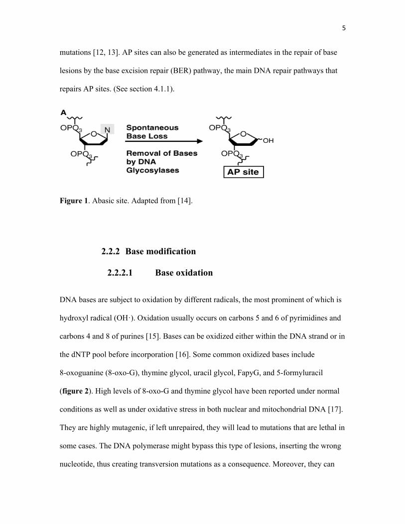
The N-glycosidic bonds that hold the nitrogenous bases to the sugar-phosphate backbone
are weak points in the structure of DNA. Hydrolysis of this bond by a water molecule
leads to the loss of the base and creating an apurinic-apyrimidinic (AP) site, also known
as abasic site (figure1) [10]. The double ring structure of purines, adenine (A) and
guanine (G), makes them more susceptible for hydrolysis than pyrimidines [10, 11]. AP
sites are highly mutagenic lesions that interfere with DNA transcription and replication
by blocking DNA and RNA polymerases. Some DNA polymerases bypass AP sites,
incorporating an untemplated nucleotide into the newly synthesized strand, introducing
5
mutations [12, 13]. AP sites can also be generated as intermediates in the repair of base
lesions by the base excision repair (BER) pathway, the main DNA repair pathways that
repairs AP sites. (See section 4.1.1).
Figure 1. Abasic site. Adapted from [14].
2.2.2 Base modification
2.2.2.1 Base oxidation
DNA bases are subject to oxidation by different radicals, the most prominent of which is
hydroxyl radical (OH·). Oxidation usually occurs on carbons 5 and 6 of pyrimidines and
carbons 4 and 8 of purines [15]. Bases can be oxidized either within the DNA strand or in
the dNTP pool before incorporation [16]. Some common oxidized bases include
8-oxoguanine (8-oxo-G), thymine glycol, uracil glycol, FapyG, and 5-formyluracil
(figure 2). High levels of 8-oxo-G and thymine glycol have been reported under normal
conditions as well as under oxidative stress in both nuclear and mitochondrial DNA [17].
They are highly mutagenic, if left unrepaired, they will lead to mutations that are lethal in
some cases. The DNA polymerase might bypass this type of lesions, inserting the wrong
nucleotide, thus creating transversion mutations as a consequence. Moreover, they can
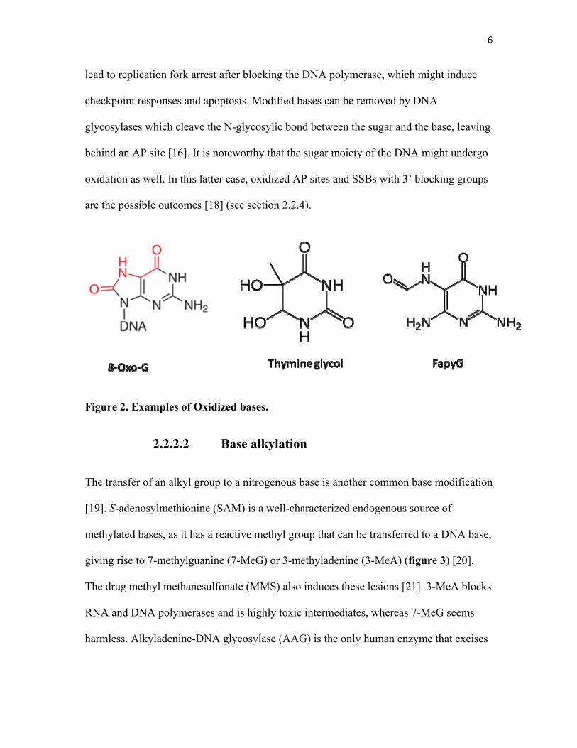
6
lead to replication fork arrest after blocking the DNA polymerase, which might induce
checkpoint responses and apoptosis. Modified bases can be removed by DNA
glycosylases which cleave the N-glycosylic bond between the sugar and the base, leaving
behind an AP site [16]. It is noteworthy that the sugar moiety of the DNA might undergo
oxidation as well. In this latter case, oxidized AP sites and SSBs with 3’ blocking groups
are the possible outcomes [18] (see section 2.2.4).
Figure 2. Examples of Oxidized bases.
2.2.2.2 Base alkylation
The transfer of an alkyl group to a nitrogenous base is another common base modification
[19]. S-adenosylmethionine (SAM) is a well-characterized endogenous source of
methylated bases, as it has a reactive methyl group that can be transferred to a DNA base,
giving rise to 7-methylguanine (7-MeG) or 3-methyladenine (3-MeA) (figure 3) [20].
The drug methyl methanesulfonate (MMS) also induces these lesions [21]. 3-MeA blocks
RNA and DNA polymerases and is highly toxic intermediates, whereas 7-MeG seems
harmless. Alkyladenine-DNA glycosylase (AAG) is the only human enzyme that excises
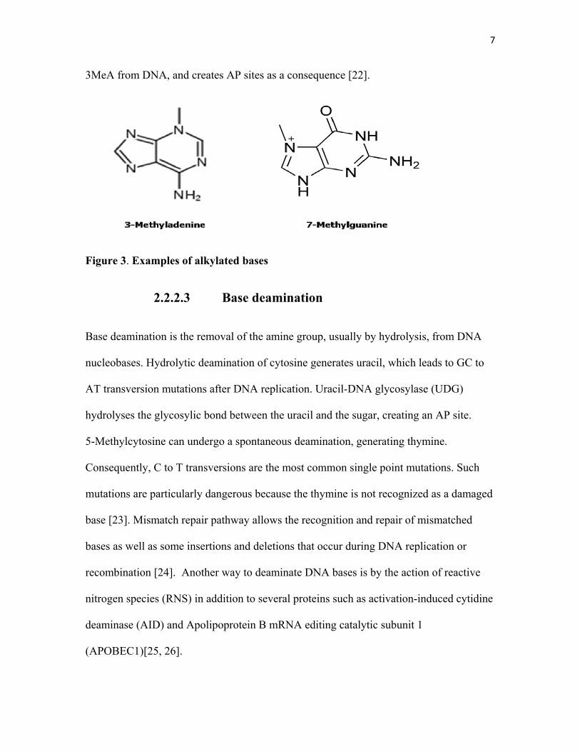
7
3MeA from DNA, and creates AP sites as a consequence [22].
Figure 3. Examples of alkylated bases
2.2.2.3 Base deamination
Base deamination is the removal of the amine group, usually by hydrolysis, from DNA
nucleobases. Hydrolytic deamination of cytosine generates uracil, which leads to GC to
AT transversion mutations after DNA replication. Uracil-DNA glycosylase (UDG)
hydrolyses the glycosylic bond between the uracil and the sugar, creating an AP site.
5-Methylcytosine can undergo a spontaneous deamination, generating thymine.
Consequently, C to T transversions are the most common single point mutations. Such
mutations are particularly dangerous because the thymine is not recognized as a damaged
base [23]. Mismatch repair pathway allows the recognition and repair of mismatched
bases as well as some insertions and deletions that occur during DNA replication or
recombination [24]. Another way to deaminate DNA bases is by the action of reactive
nitrogen species (RNS) in addition to several proteins such as activation-induced cytidine
deaminase (AID) and Apolipoprotein B mRNA editing catalytic subunit 1
(APOBEC1)[25, 26].
8
2.2.3 Bulky lesions and cross links
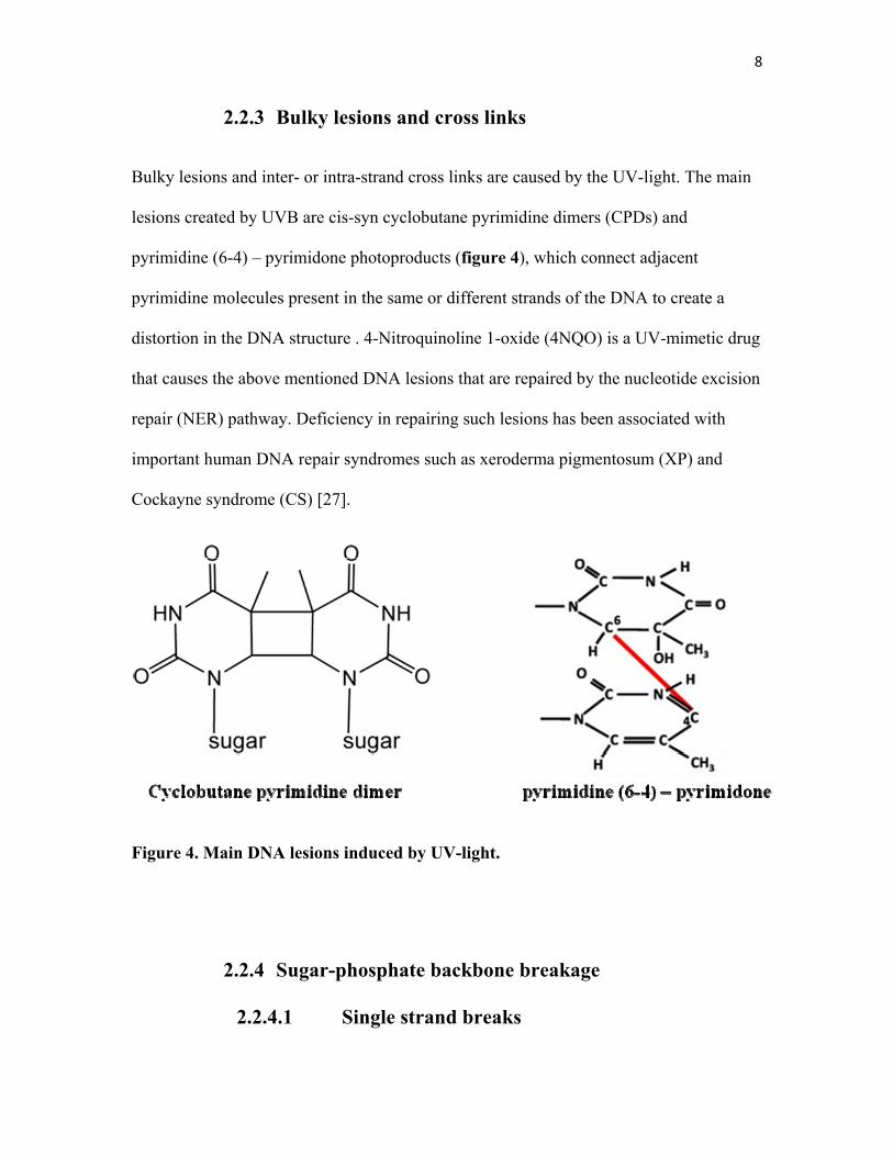
Bulky lesions and inter- or intra-strand cross links are caused by the UV-light. The main
lesions created by UVB are cis-syn cyclobutane pyrimidine dimers (CPDs) and
pyrimidine (6-4) – pyrimidone photoproducts (figure 4), which connect adjacent
pyrimidine molecules present in the same or different strands of the DNA to create a
distortion in the DNA structure . 4-Nitroquinoline 1-oxide (4NQO) is a UV-mimetic drug
that causes the above mentioned DNA lesions that are repaired by the nucleotide excision
repair (NER) pathway. Deficiency in repairing such lesions has been associated with
important human DNA repair syndromes such as xeroderma pigmentosum (XP) and
Cockayne syndrome (CS) [27].
Figure 4. Main DNA lesions induced by UV-light.
2.2.4 Sugar-phosphate backbone breakage
2.2.4.1 Single strand breaks
9
SSBs are discontinuities in one strand of the DNA duplex. They occur as a result of
spontaneous hydrolysis of the sugar-phosphate backbone, as a consequence of ionizing
radiation (IR), or elevated ROS levels. SSBs can form directly by the removal of the
oxidized sugar or indirectly during the base-excision repair (BER) of damaged bases. In
the latter case, a DNA glycosylase cleaves off the damaged base to create an AP sites
which is then cleaved by an AP endonuclease or lyase to create a SSB [28-30]. In
addition to the sources mentioned above, some chemotherapeutic drugs directly induce
SSBs. For example, bleomycin creates SSBs with 5’-phosphate and 3’-phosphoglycolate
or 3’-phosphate termini [31].
If not repaired rapidly or appropriately, SSBs create a serious threat to chromosomal
stability and hence cell survival. Consequently, cells have evolved rapid and efficient
mechanisms for their repair during later stages of the BER pathway for instance [32].
(See section 4.1.1 for more details).
2.2.4.2 Double strand breaks
DSBs are the most dangerous DNA lesions because they can lead to large-scale genomic
rearrangement and chromosomal fusions (translocations, deletions). A lot of naturally
occurring phenomena prompt the formation of DSBs like: meiosis, mating type switching
(in yeast), V(D)J and immunoglobulin class switch recombination. External factors like
IR, drugs like bleomycin and chemicals also contribute to the formation of DNA DSBs.
They are repaired mainly by two pathways: Homologous recombination (HR) and Non-
homologous end joining (NHEJ) [7, 33-35].
10
3. DNA Damage response
Cells have developed elaborate mechanisms to sense and respond to DNA damage. These
mechanisms are collectively called DNA damage response (DDR). DDR is today known
to be a signal transduction pathway as it appeared to be a kinase cascade activated by
DNA damage and replication stress. The main kinases of this pathways are ataxia
telangiectasia and Rad3-related (ATR) and Ataxia telangiectasia mutated (ATM), a
defect in which was associated with ataxia telangiectasia (AT) disease[36]. Although it
was thought for years that the purpose of this cascade is to regulate the cell cycle
transition, in the last decade it was shown that the DDR is versatile and that cell cycle
control, at checkpoints level, is only one of many outcomes controlled by this sensory
network whose central goal is the repair of DNA [37].
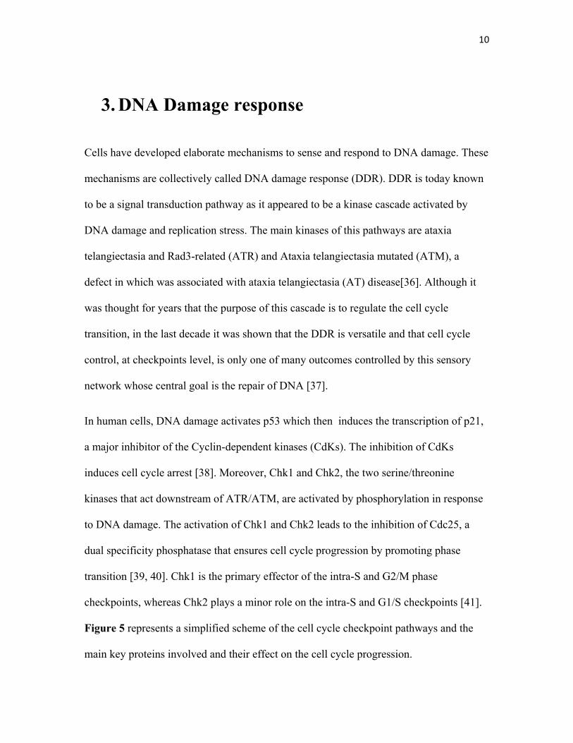
In human cells, DNA damage activates p53 which then induces the transcription of p21,
a major inhibitor of the Cyclin-dependent kinases (CdKs). The inhibition of CdKs
induces cell cycle arrest [38]. Moreover, Chk1 and Chk2, the two serine/threonine
kinases that act downstream of ATR/ATM, are activated by phosphorylation in response
to DNA damage. The activation of Chk1 and Chk2 leads to the inhibition of Cdc25, a
dual specificity phosphatase that ensures cell cycle progression by promoting phase
transition [39, 40]. Chk1 is the primary effector of the intra-S and G2/M phase
checkpoints, whereas Chk2 plays a minor role on the intra-S and G1/S checkpoints [41].
Figure 5 represents a simplified scheme of the cell cycle checkpoint pathways and the
main key proteins involved and their effect on the cell cycle progression.
11
Figure 5. Cell cycle checkpoint pathways induced in response to DNA damage. In red: Tumor suppressors ; in green: proto-oncogenes. Adapted from [42].
12
4. DNA repair: a crucial line of defence
4.1 DNA repair pathways
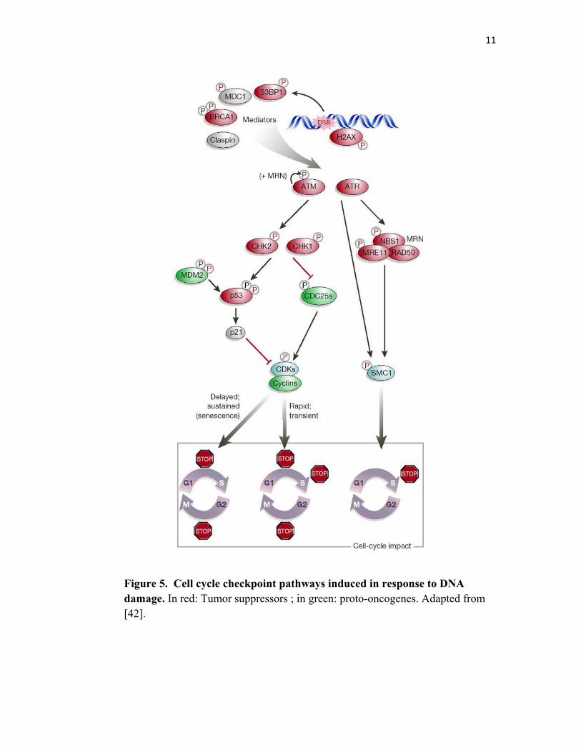
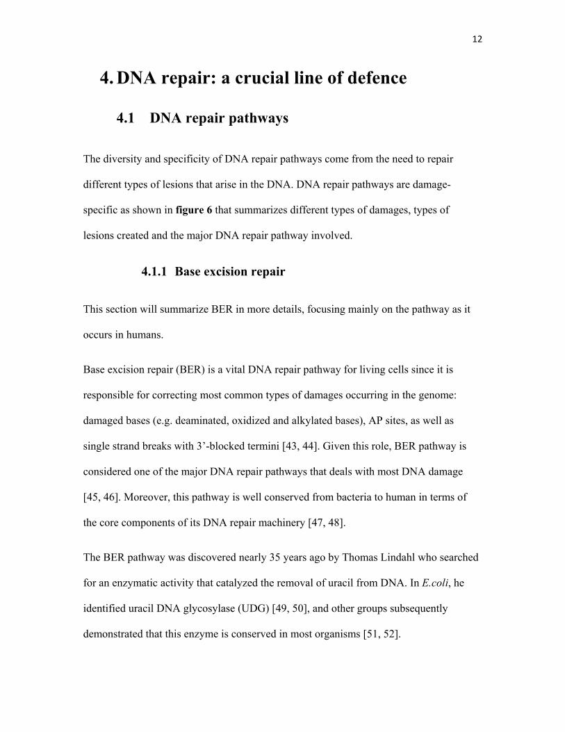
The diversity and specificity of DNA repair pathways come from the need to repair
different types of lesions that arise in the DNA. DNA repair pathways are damage-
specific as shown in figure 6 that summarizes different types of damages, types of
lesions created and the major DNA repair pathway involved.
4.1.1 Base excision repair
This section will summarize BER in more details, focusing mainly on the pathway as it
occurs in humans.
Base excision repair (BER) is a vital DNA repair pathway for living cells since it is
responsible for correcting most common types of damages occurring in the genome:
damaged bases (e.g. deaminated, oxidized and alkylated bases), AP sites, as well as
single strand breaks with 3’-blocked termini [43, 44]. Given this role, BER pathway is
considered one of the major DNA repair pathways that deals with most DNA damage
[45, 46]. Moreover, this pathway is well conserved from bacteria to human in terms of
the core components of its DNA repair machinery [47, 48].
The BER pathway was discovered nearly 35 years ago by Thomas Lindahl who searched
for an enzymatic activity that catalyzed the removal of uracil from DNA. In E.coli, he
identified uracil DNA glycosylase (UDG) [49, 50], and other groups subsequently
demonstrated that this enzyme is conserved in most organisms [51, 52].
13
Figure 6. The main damage-specific DNA repair pathways in human cells. DNA-damaging agents
(top), examples of DNA lesions (middle), and the Corresponding DNA repair pathways (bottom). The
essential genes involved in each DNA repair pathway are shown below the corresponding titles. HR,
homologous recombination; NHEJ non-homologous end-joining. Adapted from [53], [54] and [55].
Subsequent work over the next two decades identified more glycosylases specific for
other damaged bases as well as AP endonucleases [56, 57].
14
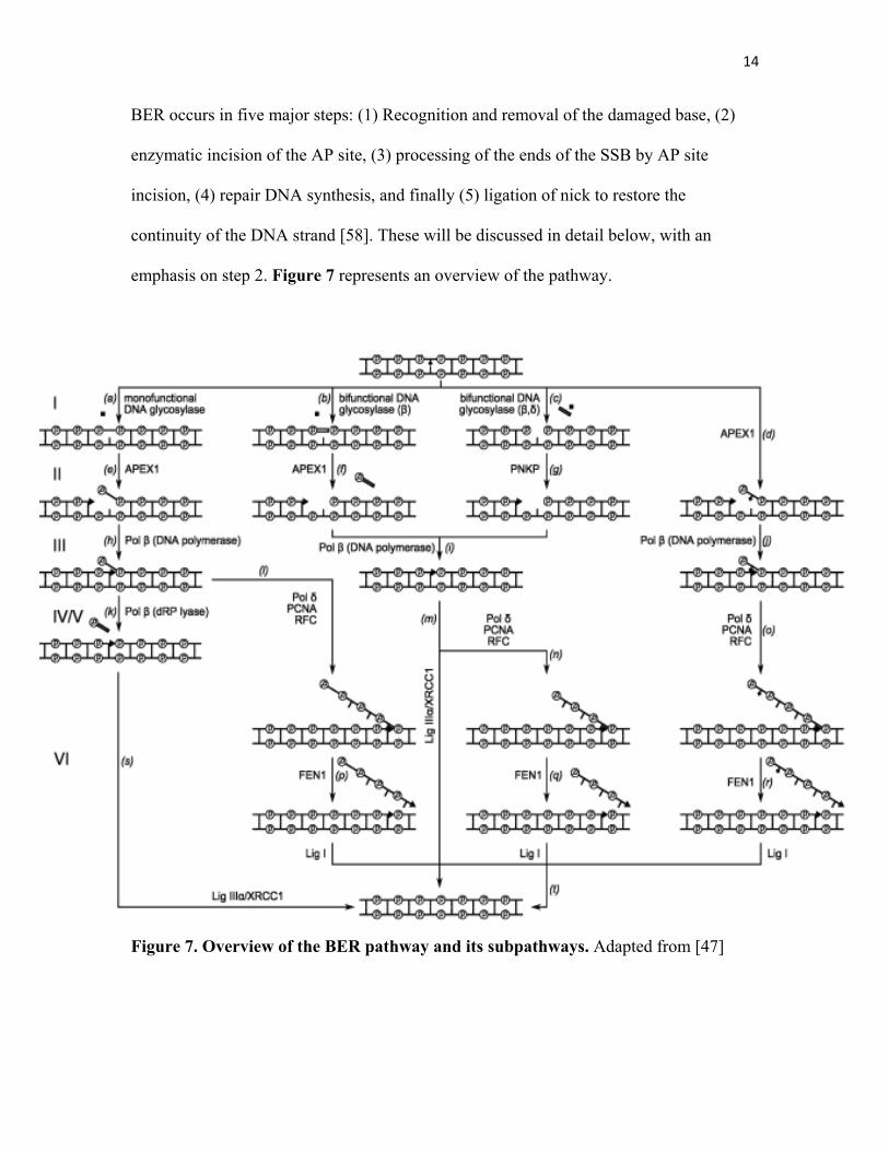
BER occurs in five major steps: (1) Recognition and removal of the damaged base, (2)
enzymatic incision of the AP site, (3) processing of the ends of the SSB by AP site
incision, (4) repair DNA synthesis, and finally (5) ligation of nick to restore the
continuity of the DNA strand [58]. These will be discussed in detail below, with an
emphasis on step 2. Figure 7 represents an overview of the pathway.
Figure 7. Overview of the BER pathway and its subpathways. Adapted from [47]
15
4.1.1.1 Damage Recognition
The first step in BER is the recognition of the modified base by a DNA glycosylase.
Glycosylases cleave the N-glycosylic bond linking the damaged base to its corresponding
deoxyribose sugar. DNA glycosylases are classified as either mono or bi-functional.
While both classes of glycosylases hydrolyze the N-glycosylic bond, bifunctional
glycosylases, such as 8-oxoguanine glycosylase (OGG1) and NEIL1/2/3, are endowed
with an additional AP lyase activity. These enzymes can further process the AP site
through β or β-δ-elimination to generate SSB with 3’-polyunsaturated aldehyde or 3’-
phosphate termini, respectively [59-61]. Although DNA glycosylases are substrate
specific, the recognition of different modified bases by the same glycosylase, or the same
damaged base by multiple glycosylases still occur, thus providing redundancy [62].
4.1.1.2 AP sites and blocking groups processing
The removal of the modified base by the DNA glycosylase generates the highly
mutagenic AP site intermediate. These lesions are substrates of the catalytic activity of
AP endonuclease family, AP lyases and delta lyases. While delta lyases create an incision
at AP sites and process 3’-aldehyde to create 3’-phosphate termini, AP endonuclease
enzymes incise the phosphodiester backbone at the 5’side of AP sites to generate 3’-
hydroxyl and 5’-deoxyribosephosphate (dRP) termini [61]. These unconventional termini
have to be restored to 3’-OH and 5’-P through deoxyribose-phosphatase diesterase
16
(dRPase) activity of Pol β (5’-dRP), 3’ diesterase activity of the Apurinic/apyrimidinic
endonuclease 1 APE1 (3’-PUA) and the phosphatase activity of PNKP in order to allow
further repair [14, 58].
However, between the AP endonucleases and the AP lyases cleavage of AP sites, the AP
endonuclease incision is the predominant pathway [61]. The importance of AP
endonucleases functions over the AP lyases has also been supported by findings
showing that endonuclease deficient yeast strains are more sensitive to MMS, but not AP
lyase deficient yeast strains [63, 64]. AP endonuclease families will be discussed in
further details in sections 5.
4.1.1.3 BER sub-pathways: the short and long patch
4.1.1.3.1 Short Patch BER
In SP-BER, DNA polymerase β (POL β) performs a one-nucleotide gap-filling reaction
and removes the 5’dRP via its lyase activity. This is then followed by sealing of the
remaining nick by the XRCC1–ligase3 complex. XRCC1 is a scaffold protein that
interacts with several of the BER core components and may therefore be important in
protein exchange [65].
17
4.1.1.3.2 Long Patch BER
In long-patch BER (LP-BER), APE1 catalyzes the formation of a nick 5’ to the AP site.
This action recruits POL β or DNA polymerase δ (POL δ), PCNA, flap structure-specific
endonuclease 1 (FEN1), and DNA ligase 1 (LIG1). In a PCNA-dependent manner POL β
performs displacing synthesis and polymerizes a tract of DNA from ~2-10 bases, which
produces a flapped substrate that is refractory to ligation. FEN1 cleaves the flap at the
ssDNA-dsDNA junction, creating a ligatable nick, which is then sealed by DNA ligase I
[66].
It was concluded that the short-patch BER (SP-BER) repair is probably the dominant
subpathway of BER. [53]. However, it was shown that many factors such as cell state
(cell cycle phase, differentiation stage), BER protein–protein interactions and the types of
lesions are all involved in the selection between SP- and LP-BER [67].
4.1.1.4 BER and cancer
Cancer is, without any doubt, the disease of the past century. Since AP sites are
considered the most occurring type of lesions arising in the DNA, they are major
contributors to DNA mutations. Accumulated mutations often lead to subsequent
activation of proto-oncogenes and inactivation of tumour-suppressor genes, the hallmarks
of cancer. Therefore, BER implication as a cancer-prevention pathway is essential.
Indeed, numerous links have been identified between oncogenesis and acquired or
inherited alterations in the genome “guardians”, highlighting the key role of DNA repair
18
systems in tumour prevention. In the last decade, evidence of association of BER
deficiency with cancer has started to emerge. For instance, studies have demonstrated a
link between defects in the adenine–DNA glycosylase MYH and adenomatous colorectal
polyposis and colorectal cancer risk [68]. Reduced activity of OGG1 was associated with
elevated lung cancer [69].
Moreover, BER enzymes are attractive targets for inhibitor-based cancer therapy. Many
chemotherapeutic drugs act by damaging the DNA, leading to cell death. Therefore,
selective inhibition of repair in cancer cells could sensitize them to the drug [70].
5. AP Endonuclease/3’ -Diesterase
AP sites are very common because, besides being formed spontaneously, they are
intermediates in repair of most types of base damage. AP sites are repaired primarily by
AP lyases and AP endonucleases. AP endonuclease family members cleave the sugar-
phosphate bond 5′ of an AP site to produce a 3′ hydroxyl group and a 5′-deoxyribose
phosphate [71, 72]. The first evidence of the presence of an enzyme that is capable of
processing AP sites emerged from studies in E.coli. These studies showed that an enzyme
termed exonuclease III, first described as a 3’ to 5’ exonuclease with a 3’-phosphatase
activity, also possesses an AP endonuclease activity [73, 74]. To date, two families of AP
endonucleases / 3’-diesterases, named Endonuclease IV (Endo IV) and Exonuclease III
(Exo III) after the ancestral bacterial enzymes, have been well characterized [61]. In
addition to AP endonucleases, AP lyases are also capable of processing AP sites. In fact,
the E. coli endonuclease III is an AP lyase that cleaves on the 3′ side of the AP site
19
through a β-elimination reaction to produce a SSB with 3’ α,β-unsaturated aldehyde,
processed by AP endonucleases [75, 76].
5.1 Two Distinct families of AP endonucleases/3’
Diesterase
Despite a certain degree of redundancy between the Exo III and Endo IV AP
endonucleases families, some differences in expression and substrate specificity do exist.
However, the key feature used to differentiate between the two families is their
magnesium dependency. While members of the Endo IV family are Mg2+ independent,
Exo III family members require magnesium for their DNA repair activity [61].
Interestingly, these two proteins use completely different structural folds to accomplish
basically the same set of activities [77].
In E.coli, Exo III is encoded by the xth gene and is the major AP endonuclease
accounting for around 90% of the total AP endonuclease activity in extracts. Exo III
possesses four different catalytic activities. First, Exo III is an apurinic/apyrimidinic
endonuclease activity. Second, it has a 3’ to 5’ exonuclease with an activity specific for
dsDNA allowing it to degrade blunt ends and 5’ overhangs. Third, it can also act as a 3’ -
phosphodiesterase that removes groups including, phosphates and phosphoglycolate in
addition to the aldehydes left by AP lyases [78-80] to allow the DNA polymerase to
prime DNA synthesis from a 3’ -OH [71]. Fourth, it has an RNase H activity to degrade
RNA in DNA-RNA hybrids [57, 81, 82].
20
While Exo III family members are present in all kingdoms of life, Endo IV homolog exist
in prokaryotes and lower eukaryotes, but not in plants, mammals and many other
vertebrates. Like Exo III family members, the Endo IV enzymes possess 3’-diesterase,
3’ -exonuclease and RNase H activities [61].
In E.coli Endo IV is encoded by the nfo gene. It accounts for only 10% of the basal AP
endonuclease activity but is induced by oxidative stress [57, 82]. In contrast, Apn1, the
S. cerevisiae Endo IV homolog, is the major AP endonuclease in yeast and shares 41%
sequence identity with the bacterial enzyme [83]. Yeast apn1Δ cells show less than 1% of
AP endonuclease/3’ diesterase activities of wild-type, a 60 fold increase in ATGC
transversion mutations, and hypersensitivity to alkylating agents and chemical
oxidants[84, 85]. Apn2, the E.coli Exo III homolog in yeast was discovered as the
second and the minor AP endonuclease in yeast after Apn1 [86]. As in E.coli, yeast Apn2
was shown to possess AP endonuclease, 3’ diesterase and 3’ to 5’ exonuclease activities
that require magnesium [63]. The transcription of Apn2 gene was found to be induced up
to six-fold by treatment with MMS [87] and its 3’-diesterase and 3’→5’ exonuclease
activities stimulated by proliferating cell nuclear antigen (PCNA) [88].
In human cells, AP endonuclease 1 (APE1) and AP endonuclease 2 (APE2) are the
human homologues of the Exo III family. APE1 is the major human AP endonuclease/3’
diesterase and since the research project concentrates on the DNA repair capacity of
APE1, this enzyme will be discussed in more details in section 6 below. This enzyme has
previously been known as APEX in mice [89], “HAP1” for Human AP endonuclease 1
[90], and redox factor-1 (Ref-1) for its role as a redox regulator [91], but will be referred
to as APE1 here.
21
Based on genomic databases from various organisms, APE2 was identified as a nuclear
and mitochondrial AP endonuclease in human cells[92]. APE2 was found to possess a
very weak AP endonuclease activity and an even weaker (7-fold lower) 3’ diesterase
activity [93]. Moreover, APE2, unlike APE1, could not complement yeast AP
endonuclease mutants [94]. While APE1 has been reported to have no functional
mitochondrial targeting sequence (MTS), it was shown that APE2 possesses a MTS and
has a potential role in the repair of mitochondrial DNA lesions [92]. Moreover, it was
reported that APE2 has a long C-terminal domain that contains a PCNA binding motif,
similar to the one present in S. cerevisiae Apn2, suggesting a possible role in LB-BER
[92]. However, the presence of APE2 did not rescue the lethal phenotypes of the
knockout and the knockdown of APE1, which rule out possible significant roles for
APE2 in DNA repair. The in vivo role of APE2 remains unclear; it might be a
pseudogene or an evolutionary relic.
Many attempts failed to identify the Endo IV homolog in human. The highest organism
where both AP endonucleases were identified and characterized is Caenorhabditis
elegans (C.elegans) [95, 96]. In addition, Exo III and Endo IV were both identified but
not yet characterized in the frog Xenopus tropicalis and the fish Danio rerio [97].
6. APE1
APE1 was first characterized and purified from HeLa cells in 1981 by the group of
Stuart Linn [98]. Later, cDNAs encoding several mammalian AP endonucleases (human,
22
murine and bovine) were published [99-102]. The APE1 protein is closely related to other
mammalian AP endonucleases (91-93% identity) and more distantly to E.coli Exo III
(28% identity). Like Exo III, APE1 has a Mg2+- dependent AP endonuclease activity in
vitro [98, 103].
6.1 APE1: a DNA repair enzyme
As described earlier, APE1 is considered of major importance in protecting the cell from
the effects of cytotoxic and mutagenic unrepaired AP sites.
The expression of the APE1 protein in human cells is activated by DNA damaging agents
and ROS, in order to process the resulting AP sites during the BER [104]. The second
major role of APE1 in the BER is its 3’ diesterase activity. Although the diesterase
activity of APE1 is about 200-fold lower than its AP endonuclease activity, this function
allows it to remove a variety of 3’ blocking groups including 3’phosphate and 3’
phosphoglycolate occurring at single-strand breaks created by oxidative agents, ionizing
radiation or by a chemotherapeutic agent like Bleomycin which creates 3’
phosphoglycolate [105, 106].
APE1 is one of the major enzymes orchestrating the mammalian BER. In addition to its
AP endonuclease/3’ –diesterase activity, APE1 may have also an implication in the
selection between the SP- and LP-BER subpathways. It was proposed that APE1 is active
in both LP- and SP-BER and that it interacts with several proteins in these two
subpathways including OGG1, XRCC1, PCNA, FEN1, and DNA polymerase β. Hence,
APE1, owing to its interactions with other BER proteins and to its structural mechanism
23
of recognition of the AP site, it serves as an assembly and coordination factor for
subsequent proteins after its initial cleavage of the DNA [107, 108].
In the first step of BER, after the DNA glycosylase removes the damaged base, it remains
bound and presents the AP site to APE1. Due to the extensive surface of APE1-DNA
interaction that covers both DNA strands and the DNA kinking, APE1 displaces the
glycosylase allowing it to release the damaged base. After cleaving the AP site, the
nicked DNA is exposed to DNA polymerase and/or XRCC1. This product-substrate
exchange might be partially mediated by APE1 N-terminal and the DNA polymerase
dRP-diesterase domain. Following repair, the DNA can no longer accommodate the
bending and the component enzymes likely dissociate [109]. It appears that APE1
interactions with other BER proteins show a high level of DNA repair organization for
maximal efficiency in a relatively large mammalian genome.
Although they belong to different AP endonucleases families and to two different
species, it is noteworthy that the human APE1 shares many common features with the
yeast Apn1. First, each one of them is the major AP endonuclease/3’ diesterase enzyme
in its corresponding cellular context. Second, they are both capable of incising the
phosphodiester bond at the 5’ of an AP site and of removing similar 3’ blocking groups,
e.g. 3’ phosphate, 3’phosphoglycolate that are associated with Ionizing radiation (IR) and
ROS [71, 110, 111]. However, in contrast to what might be predicted based of the
biochemical repair activities of APE1 and Apn1, APE1 knockout mouse blastocysts and
antisense-expressing human cells are hypersensitive to γ rays and oxidizing agents,
whereas Apn1 yeast mutants are at best mildly sensitive to γ rays and oxidizing agents
like (e.g. H2O2) [105]. These results suggest that there might be other species-specific
24
factors implicated in alternative repair mechanisms that need to be taken into
consideration when assessing the right model (yeast or mammalian cells) to study DNA
damage response to certain genotoxic agents.
Besides its activity in the BER pathway, APE1 was shown to be the damage-specific
endonuclease in the nucleotide incision repair (NIR) pathway, a known feature in Endo-
IV family of endonucleases [112]. NIR pathway was proposed as an alternative pathway
to classic BER. In this pathway, APE1 nicks oxidatively damaged DNA (e.g. 5,6-
dihydro-2’-deoxyuridine, 5,6-dihydrothymidine (DHT), 5-hydroxy-2’-deoxyuridine,
alpha-2’-deoxyadenosine and alpha thymidine adducts) in a DNA glycosylase-
independent manner, generating 3’-OH and 5’ phosphate. This mechanistic feature is
distinct from DNA glycosylase-mediated BER and has the advantage of avoiding the
genotoxic intermediates (i.e. AP sites) generated in the BER pathway [113].
A recent study showed that APE1 appears to suppress the activation of poly(ADP-ribose)
polymerase 1 (PARP1) during oxidative damage repair which promotes cell survival
[114].
Furthermore, some groups showed that APE1 also possesses 3’ to 5’ exonuclease activity
which plays a role in the excision of deoxyribonucleoside analogs from the DNA [115,
116]. If this is the case, the inhibition of this activity could have implications in treating
some cancers where nucleoside analogs such as gemcitabine are used. Conflicting data
are present in the literature regarding this latter activity of APE1. Some groups showed
that this activity is structure specific and presented a preference for 3’-mismatched
nucleotides [115, 117, 118], while others showed that it presented no exonuclease activity
against blunt ended dsDNA [93, 119]. Interestingly, the yeast Apn1 is endowed with a
25
well-characterized 3’ to 5’ exonuclease activity that is capable of removing incorporated
8-oxo-G from the DNA, a function that possibly APE1 might have [120].
Given the many DNA repair functions of APE1, it is clear that this enzyme is
fundamental to the maintenance of the genome and survival of cells. Hence the
observation that even in the absence of exogenous DNA damage, siRNA directed against
APE1 results in a decrease in proliferation, an increase in AP sites and increased levels of
apoptosis [121].
6.2 APE1: a Redox enzyme
In addition to its role in DNA repair, APE1 has also been implicated in transcription
regulation by reducing and thus activating many transcription factors. APE1 was first
shown to reduce the conserved cysteine residues in Fos and Jun. Fos and Jun form a
heterodimeric complex that regulates gene transcription by binding to the activator
protein-1 (AP-1) DNA sequence motif. APE1 activation of Fos and Jun stimulates AP-1
DNA-binding activity [91].
Later reports have revealed a similar role of APE1 in activating p53, Fos, Jun, nuclear
factor-κB (NF-κB), PAX (paired box-containing family of genes), hypoxia inducible
factor-1α (HIF-1α), HIF-1-like factor and others [105]. The biological implication of
these transcription factors activation is no fully clear yet, but what is known so far that
APE1 redox function facilitates the DNA binding of some of these transcription factors
or change the transcriptional activity of others [105]. Interestingly, it was demonstrated
26
that p53 binding to DNA and the subsequent transcriptional activation of p21, BAX or
cyclin G is dependent on APE1. Moreover, APE1 protein was reported to stimulate p53
activity by both redox-dependent and -independent means [122].
APE1 has also been implicated in a number of other redox activations, one of which is
the activation of bioreductive drugs requiring reduction for their activity [123]. Hence
APE1 acts as a redox factor that senses the redox state of the cell.
APE1 redox activity requires the N-terminal region of the protein as the truncation of the
62 N-terminal residues renders the enzyme redox deficient (Figure 15 and 16) [124,
125]. It is noteworthy that human APE1 has 61 N-terminal additional amino acids that
are not present in the E.coli Exo III. It was proposed that APE1 redox activity involves a
cysteine residue(s) [124] that according to LJ Walker et al. was Cys 65 that forms a
disulfide bridges with Cys 93 [125]. However, several reported crystal structures of
APE1 showed three different observations contradicting the latter finding. First, no
disulfide bridges were found in the crystal structures. Second, Cys 65 is buried inside the
protein and is not accessible by the target transcription factors reduced by APE1 [77, 126,
127]. Third, all the reported APE1 crystallographic lattices are similar, suggesting the
absence of conformational changes that could reposition Cys 65 [77]. Moreover, mice in
which Cys 64 (equivalent to human Cys 65) is mutated to Ala show no developmental
defects [128]. Thus, the mechanism by which Cys 65 acts in APE1 reduction of
transcription factor needs to be further investigated.
27
6.3 Importance of APE1 cellular functions
Given the mild phenotype seen in other AP endonuclease-deficient organisms, lethality of
APE1 -/- mice and knockout cells was a big surprise.
Unlike bacteria and yeast, which tolerate the complete loss of AP endonuclease activity
with little consequence unless challenged with DNA damaging agents, APE1 knockout in
mice is lethal at early embryonic stages, E3.5–E9.5 [129, 130].
However, heterozygous mutant mice developed into adulthood without any apparent
abnormalities [129]. Although it was not clear at the time which function(s) of APE1 is
contributing to the lethality phenotype seen in mice, a recent study using a specific
inhibitor of APE1 redox function demonstrated that APE1 may play a role in normal
embryonic hematopoiesis and that its redox function, but not the DNA repair
endonuclease activity, is critical in normal embryonic hematopoietic development [131].
On the contrary, evidence for importance of DNA repair function was presented by
another group who showed that epiblast cells have fragmented nuclei and undergo
apoptosis and that explanted homozygous APE1-null blastocysts displayed increased
sensitivity to γ-irradiation [130].
Further research showed that loss of APE1 is lethal at the level of the cell. Human cells in
which APE1 is deleted by a microinjection of a Cre expression plasmid, undergo
apoptosis within 48 hours [132].
Since APE1 deletion is lethal, many groups manipulated APE1 expression levels by
either downregulation or overexpression. RNA interference (RNAi) knockdown resulted
28
in decreased cell proliferation and apoptosis which was correlated with an accumulation
of AP sites [121]. Moreover, lower levels of APE1 render mammalian cells
hypersensitive to MMS and H2O2 [133]. Importantly, the defects seen in knockdown cells
were rescued by the expression of the yeast Apn1. Thus, the AP endonuclease activity of
APE1 was shown to be essential for cell viability.
To date, many types of inhibitors of APE1 activities have been discovered. Lucanthone
and 7-Nitroindole-2-carboxylic acid (NCA) were shown to be direct inhibitors of APE1
DNA repair activity [70, 134]. Recently, a novel quinone derivative that inhibits APE1
redox activity was identified, namely E3330 ([(2E)-3-[5-(2,3dimethoxy-6-methyl-1,4-
benzoquinolyl)]-2-nonyl-2-propenoic acid]) [135].
In this context, a beneficial effect of the reduction of APE1 levels is to sensitize cells to
chemotherapeutic agents such as bleomycin, carmustine, gemcitabine [136-138].
On the other hand, APE1 overexpression in human cells enhances tumour resistance to
DNA damaging drugs and radiotherapy, as expected, a phenotype seen in many cancer
cells [139].
6.4 Regulation of APE1 Activity
APE1 is regulated at two different levels: transcriptional and post-translational.
Regulation of the activity of an essential enzyme like APE1 is of great importance for the
cell to respond properly to DNA damage and to other diverse stimuli where it is involved.
29
6.4.1 Gene expression regulation
APE1 transcription is known to be induced by ROS. This regulation was proposed to
happen in two steps: first APE1 translocates from the cytoplasm to the nucleus. Second,
de novo protein synthesis takes place via transcriptional activation at the promoter level
[104, 140-142]. Interestingly, APE1 was shown to autoregulate its own transcription by
binding to its own promoter followed by inhibition of transcription at the level of its
negative calcium regulatory element (nCARE) [143].
6.4.2 Post-translational modifications
APE1 is an abundant protein (~104–105 copies/cell) within eukaryotic cells and with a
relatively long half-life of about 8 hours [144]. Therefore, the needed regulation of this
multifunctional enzyme must lie in the many post-translational modifications that this
enzyme undergoes.
6.4.2.1 APE1 structure
The N-terminal region contains the nuclear localization signal (NLS) and also appears to
regulate the DNA binding activity of many transcription factors in vitro. Particularly, the
residues 43-62 are necessary for APE1 redox activity [91, 122, 124, 125].
The active site residues of APE1 have been determined by site-directed mutagenesis and
by analysis of sequence conservation between APE1 and Exo III [72, 145, 146]. There is
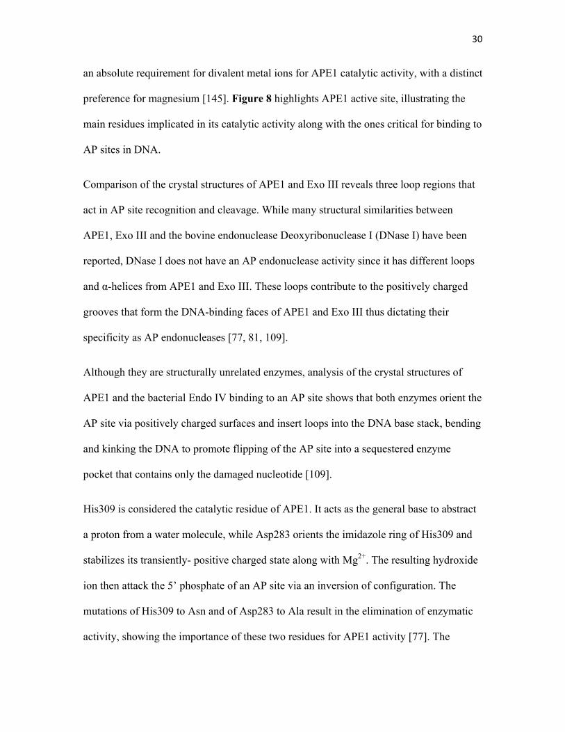
30
an absolute requirement for divalent metal ions for APE1 catalytic activity, with a distinct
preference for magnesium [145]. Figure 8 highlights APE1 active site, illustrating the
main residues implicated in its catalytic activity along with the ones critical for binding to
AP sites in DNA.
Comparison of the crystal structures of APE1 and Exo III reveals three loop regions that
act in AP site recognition and cleavage. While many structural similarities between
APE1, Exo III and the bovine endonuclease Deoxyribonuclease I (DNase I) have been
reported, DNase I does not have an AP endonuclease activity since it has different loops
and α-helices from APE1 and Exo III. These loops contribute to the positively charged
grooves that form the DNA-binding faces of APE1 and Exo III thus dictating their
specificity as AP endonucleases [77, 81, 109].
Although they are structurally unrelated enzymes, analysis of the crystal structures of
APE1 and the bacterial Endo IV binding to an AP site shows that both enzymes orient the
AP site via positively charged surfaces and insert loops into the DNA base stack, bending
and kinking the DNA to promote flipping of the AP site into a sequestered enzyme
pocket that contains only the damaged nucleotide [109].
His309 is considered the catalytic residue of APE1. It acts as the general base to abstract
a proton from a water molecule, while Asp283 orients the imidazole ring of His309 and
stabilizes its transiently- positive charged state along with Mg2+. The resulting hydroxide
ion then attack the 5’ phosphate of an AP site via an inversion of configuration. The
mutations of His309 to Asn and of Asp283 to Ala result in the elimination of enzymatic
activity, showing the importance of these two residues for APE1 activity [77]. The
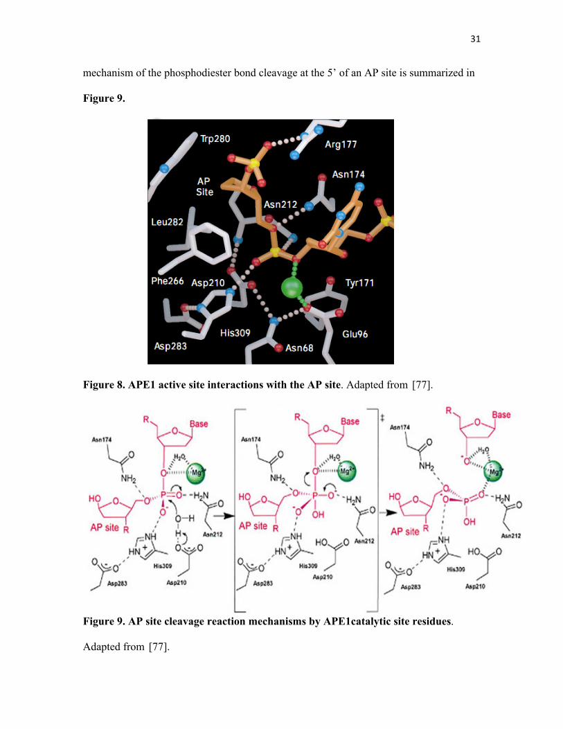
31
mechanism of the phosphodiester bond cleavage at the 5’ of an AP site is summarized in
Figure 9.
Figure 8. APE1 active site interactions with the AP site. Adapted from [77].
Figure 9. AP site cleavage reaction mechanisms by APE1catalytic site residues.
Adapted from [77].
32
6.4.2.2 Phosphorylation
It was demonstrated that APE1 is a substrate for phosphorylation by different kinases
such as serine/threonine casein Kinases (CK) I and II and protein kinase C. However,
phosphorylation uniquely by CK II was shown to inhibit the AP endonuclease activity of
APE1 on AP sites [147]. Conversely, two years after this discovery another group
showed that this phosphorylation by CK II does not have any impact on the DNA repair
capacity of APE1, instead it enhance the redox function of APE1 shown by the increase
of AP-1 binding to DNA [148].
6.4.2.3 Acetylation
p300 is a transcriptional co-activator that was shown to acetylate APE1 at Lys6 and Lys7
in vitro and in vivo in a Ca2+-dependent manner. This modification enhances APE1
binding to nCARE which was shown to downregulate the parathyroid hormone (PTH)
expression [149]. This acetylation is reversed by class I histone deacetylases (HDACs).
Moreover, it was demonstrated that APE1 acetylation is needed for the activation of the
Phosphatase and tensin homolog (PTEN) gene [150].
33
6.4.2.4 Ubiquitination
A recent discovery identified APE1 as a novel target of ubiquitination. This modification
occurs on Lys residues of the N-terminus of the protein and involves the E3 ubiquitin
ligase mouse double minute 2 (MDM2), in a p53-dependent manner. The ubiquitinated
APE1 was shown to be predominantly present in the cytoplasm compared to nuclear
localization of the unmodified protein. Although it was proposed that cytoplasmic
localization of the ubiquitinated APE1 might be an indication of either its degradation or
an alteration of its function, the effect of ubiquitination on APE1 is still not clear [151].
6.4.2.5 Nitrosylation
Oxidative stress was shown to have an effect on APE1 activity. Interestingly, nitrosative
stress was also shown to modify this enzyme. The nitrosation of APE1 on Cys93 and
Cys310 induces the translocation of APE1 to the cytoplasm, a phenomenon reversed by
treatment with reducing agents [152]. The biological implication of this modification
needs more investigation.
6.4.2.6 Redox regulation
In addition to the post-translational modifications listed above, APE1 diverse activities
were found to be regulated by the reduction and oxidation. Thioredoxin (TRX) is a
pleiotropic cellular factor that has thiol-mediated redox activity implicated in many
cellular processes, including the reduction and activation of APE1 redox function [153].
34
The reduction of oxidized APE1 by TRX leads to intensified APE1-mediated p53
activation and enhanced AP-1 DNA binding [154]. Although the target residues of the
reduction on APE1 are not known, they may include Cys65 and Cys93 since these sites
are thought to be redox sensitive [125]. Moreover, it was shown that the redox state of
APE1 affects its AP endonuclease activity in vitro and that this modification involves a
specific cysteine residue of this enzyme [155].
Recently in our laboratory, we showed that APE1 interacts directly with the glycolytic
enzyme glyceraldehyde-3-phosphate dehydrogenase (GAPDH), which converts the
oxidized form of APE1 to the reduced active form, thus maintaining its AP endonuclease
activity to repair AP sites [156]. Investigating the implications and aspects of this novel
finding will be the object of my research project.
7. GAPDH: a multifunctional protein
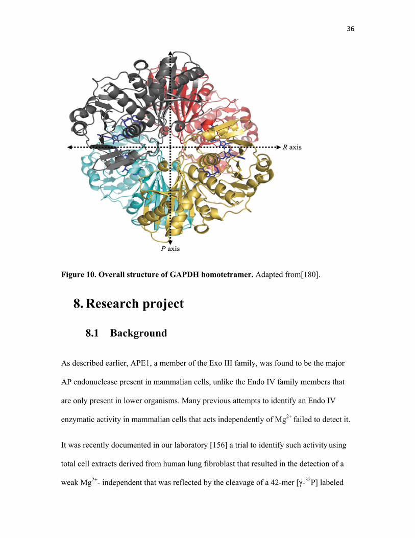
GAPDH is a tetrameric enzyme (Figure 10) that uses the oxidized form of nicotinamide
adenine dinucleotide (NAD+) to converts glyceraldehyde-3-phosphate to 1,3-
bisphosphoglycerate, releasing NADH [157]. GAPDH is regarded as a housekeeping
gene whose transcript level remains constant under most experimental conditions, and it
has been frequently used as an internal control in studying the regulation of gene
expression. Mounting evidence, however, has started to support the multi-functionality of
this enzyme, identifying roles in a variety of cellular processes, independent from its
glycolytic role.
35
GAPDH was shown to have phosphotransferase/kinase activity that serves to
phosphorylate other proteins [158-162]. Interestingly, GAPDH was shown to have
autophosphorylation activity as well [163]. Other new functions includes regulation of
the cytoskeleton [164], membrane fusion and transport [165, 166], transport of nuclear
RNA [167], glutamate accumulation into presynaptic vesicles [168], activation of
transcription in neuronal cells [169], and protection of telomeres against chemotherapy-
induced degradation [170]. Moreover, GAPDH exhibited a DNA repair activity by acting
as a Uracil DNA glycocylase (UDG) which removes mis-incorporated uracil from the
DNA [171, 172].
Notably, GAPDH was demonstrated to play a crucial role in neuronal apoptotic cell death
that has pathophysiological implications in many neurodegenerative diseases such as
Alzheimer’s disease (AD) [173], Huntington’s disease (HD) [174] and Parkinson’s
disease (PD) [175].
Under oxidative stress, GAPDH undergoes oxidation and manifests a reduction in its
enzymatic activity [176]. This oxidation also enhances the binding of GAPDH to RNA
and DNA [177]. Treatment with H2O2 induces the translocation of GAPDH from the
cytoplasm to the nucleus in mammalian cells [178]. Moreover, a recent study showed that
nitric oxide (NO) induces GAPDH nuclear localization followed by its acetylation by
p300/CBP that will result in the transcriptional activation of many genes such as p53
[179]. Considering all of these alterations of function upon oxidative conditions, GAPDH
seems to be playing an important role in cellular response to oxidative stress.
36
Figure 10. Overall structure of GAPDH homotetramer. Adapted from[180].
8. Research project
8.1 Background
As described earlier, APE1, a member of the Exo III family, was found to be the major
AP endonuclease present in mammalian cells, unlike the Endo IV family members that
are only present in lower organisms. Many previous attempts to identify an Endo IV
enzymatic activity in mammalian cells that acts independently of Mg2+ failed to detect it.
It was recently documented in our laboratory [156] a trial to identify such activity using
total cell extracts derived from human lung fibroblast that resulted in the detection of a
weak Mg2+- independent that was reflected by the cleavage of a 42-mer [γ-32P] labeled
37
synthetic oligonucleotide substrate containing a single AP site. Subsequent purification
steps followed by mass spectrometry analysis of the protein fraction having this activity
revealed the presence of GAPDH protein. Purified recombinant GAPDH lacks an AP
endonuclease activity, the only explanation to the observed activity is that GAPDH was
co-purified with another protein possessing this activity, namely APE1. Since APE1 has
an Mg2+-dependent activity, a possible explanation is the EDTA concentration in the
buffer used was not enough to chelate all the Mg2+ from the extract in the reaction,
leaving some APE1 enzymes active, contributing to the detected AP endonuclease
activity.
Further experiments showed that GAPDH interacts with APE1 in vivo and in vitro. This
interaction causes the conversion of the oxidized and AP endonuclease deficient APE1 to
a reduced and active protein capable of cleaving AP sites. This reducing potential of
GAPDH was shown to be dependent on Cys 152 residue of its active site. Other reports
showed that APE1 exists as heterogeneous molecules that are either reduced or oxidized
[153]. Moreover, it was reported that APE1 can be oxidized by H2O2 to the inactive form,
a phenomenon partially reversed by the treatment with dithiothreitol (DTT) [155].
Interestingly, siRNA knockdown of GAPDH in HCT116 cells caused sensitivity to MMS
and bleomycin, two drugs known to create DNA lesions that are repairable by APE1, but
not to UV irradiations which produces bulky lesions. A similar phenotype was observed
when using siRNA directed against APE1 [138]. Furthermore, total extracts of these cells
showed a reduced AP endonuclease activity. They also exhibited an increase in
spontaneous AP sites frequency in their genomic DNA.
38
8.2 Hypothesis
Since GAPDH interaction with APE1 activates the AP endonuclease activity of this
enzyme in vitro, and since the deficiency in GAPDH sensitized cells to DNA damaging
drugs, induced an increase in AP sites formation and reduced the AP endonuclease
capacity, we hypothesize that deficiency in GAPDH affects cell cycle progression and
APE1 cellular distribution.
Moreover, since GAPDH activates APE1 by reduction, we postulate that this reduction
occurs at the level of disulfide bridge(s) formed between APE1 cysteine residues.
8.3 Objectives
Objective 1: To determine whether activation of APE1 by
GAPDH is required for efficient cell cycle progression
It is critical for dividing cells to ensure their DNA integrity in order to avoid any
inherited mutations in daughter cells. DNA lesions activate checkpoint pathways that
regulate specific DNA repair mechanisms during different phases of the cell cycle.
Checkpoint-arrested cells resume their cell cycle progression once the damage has been
repaired, whereas cells with unrepaired DNA lesions undergo permanent cell cycle arrest
or apoptosis.
Since GAPDH Knockdown cells showed a defect in proliferation and since this
knockdown affected the DNA repair capacity of the cells, i.e. AP endonuclease activity,
39
we studied the cell cycle progression of GAPDH knocked down cells and we found that
GAPDH knockdown cells had a delay in progressing through the S phase as compared to
control cells.
Objective 2: To determine whether depletion of GAPDH affects
the subcellular localization of APE1
Although APE1 is mainly a nuclear protein, it has been reported that its cellular
distribution varies from normal cells to cancer cells, as it was shown that APE1 localizes
to the cytoplasm in cancer cells as in the case of lung [181], ovarian [182], thyroid[183],
and breast cancer[184]. This localization has been linked to higher aggressiveness of the
tumor. It was also shown to be present in the endoplasmic reticulum (ER) and the
mitochondria [185, 186].
Moreover, upon oxidative stress some DNA repair enzymes like OGG1 and APE1
localize to nuclear speckles, structures that are present between chromatin domains
enriched with RNA processing and transcription proteins [187].
For these reasons, we investigated the cellular distribution of the presumably oxidized
and inactive APE1 in GAPDH knock down cells. We found that APE1 forms foci-like
structures in GAPDH deficient cells.
40
Objective 3: To identify the redox-sensitive cysteine(s) of APE1
that is reduced by GAPDH
APE1 has seven cysteine residues at positions 65, 93, 99, 138, 208, 296 and 310, some of
which were shown to be important for its redox activity (Cys 65). Since GAPDH was
shown to reduce APE1, we expect that this reduction occurs at the level of one or more of
these cysteines, causing a reduction of a disulfide bridge(s) inducing a conformational
change in APE1 structure which allows it to be active and capable of cleaving AP sites.
To identify the cysteine residue (s) involved we mutated each of the 7 cysteine to alanine,
which cannot form disulfide bridges and assayed for the activity of these mutants in vitro
and in the cells. We specifically asked whether one or more cysteine mutant would
maintain its AP endonuclease activity under oxidative conditions in the absence of
GAPDH and show no enhancement in AP endonuclease activity or redox state when
GAPDH is added. Hence the expression of the candidate mutant(s) should rescue the
defect in proliferation seen in GAPDH knock down cells.
Chapter 2: Materials and Methods
1. Expression and Purification of His-APE1
variants
His-APE1 was cloned in pET14-b vector under a T7 promoter and expressed in BL21
(DE3) pLysS strain and purified using Talon Metal Affinity column (Clonetech)
according to manufacturer instructions with some modifications. Briefly, BL21 pLysS
E.coli cells expressing His-APE1 (wild type or cysteine or active site mutants) were
streaked out on Luria Broth (LB) agar plates containing Ampicillin (100 μg/mL), a
single colony was inoculated in 20 mL of LB containing Ampicillin (100 μg/mL) for
overnight incubation at 37˚C with shaking (180 rpm). Next morning, 10mL of the
overnight culture was subcultured in 1L of LB and grown up to O.D= 0.5. IPTG (0.4mM)
was added to the culture and left for two hours at 37˚C with shaking. Cells were
harvested by centrifugation at 6000 rpm for 15 minutes at 4˚C. Cell pellet was
resuspended in the washing buffer (50 mM sodium phosphate pH 7.0, 300mM NaCl)
containing protease inhibitors (Invitrogen) and is subjected to sonication for short bursts
on ice at 50% amplitude (Branson Digital Sonifier ®) to lyse the cells. Cell lysate was
then centrifuged at 13,000 rpm for 20 minutes at 4˚C. The purification steps were done
at room temperature. The supernatant was loaded onto a column made of 1 ml of Talon
metal affinity resin (Clonetech) and the flow through was collected and passed again on
the column to maximize the amount of binding proteins. The column was washed with
42
the washing buffer containing protease inhibitors. His-tagged bound proteins were eluted
by elution buffer (50mM sodium phosphate pH 7.0, 300nM NaCl, 150mM Imidazole),
and collected as fractions of 500 μL each. Protein concentration and purity were
estimated by Bradford assay and SDS-PAGE subsequently. The purified proteins were
dialyzed against 1L of the storage buffer (20mM sodium phosphate pH 7.0, 100mM
NaCl, 1mM EDTA, 10% Glycerol) at 4˚C.
2. Expression and purification of GST-GAPDH
GST-GAPDH was cloned in pGEX-4T-1 (Amersham Biosciences) and expressed in
DH5α. Cells were cultured and proteins were extracted as previously described in section
1 of Materials and Methods except for protein extracts that were loaded on glutathione-
Sepharose 4B mini columns (GE Healthcare). The column was washed with the wash
buffer (50 mM Tris-HCl pH 7.0, 300mM NaCl). And the proteins were eluted with
50mM Tris-HCl pH 9.0, 20mM reduced glutathione.
3. Silver staining
Immediately after electrophoresis, the SDS-PAGE was fixed in a solution of 50%
Methanol, 10% acetic acid for 30 minutes. The gel was then washed three times in 15%
methanol for 10 minutes each with gentle shaking. The gel was then transferred to 7.5%
Glutaraldehyde solution and incubated at 37˚C for 30 minutes, with gentle shaking.
43
Another washing step with 15% methanol and distilled water subsequently for 15 minutes
each, for three times. The Gel was then stained with a silver nitrate (AgNO3) for 10
minutes with gentle shaking. To remove excess silver nitrate, the gel is washed with
water until the color of the solution becomes clear. To develop the gel, developing
solution (0.005% citric acid, 0.2% formaldehyde) was added to the gel with gentle
shaking, the staining should take place in the first 10 to 20 minutes of the incubation. A
stop solution (5% acetic acid) was added to stop further staining.
4. Western Blot Analysis
Western blot analysis was performed according to standard protocols. For purified His-
APE1 and GST-GAPDH in vitro assays, purified proteins concentrations were
determined using the Bradford analysis. Proteins were loaded onto a 10% SDS
polyacrylamide gel, which was run at 100 V. The proteins were then transferred to a
nitrocellulose membrane at 100 V for 1 hour. The membrane was blocked with 5% of
skim milk in TBSET (6.05 g Tris-Base, 43.83 g NaCl, 1.85 g of EDTA and 500 μl tween
20 in 500 ml of distilled water and then probed overnight at 4°C with mouse monoclonal
anti-His (Santa Cruz biotechnology) antibody at a concentration of 1/2500 in 5% milk.
Three successive washes (10 min each) with TBSET were then performed to wash all
excess of the primary antibody. After the last wash, the membrane was incubated for 1
hour with the anti-mouse polyclonal antibody and was followed by 3 washes of 10 min
each with TBSET. Finally the membrane was incubated for 1 min in chemiluminescence
44
reagent and developed using a FujiFilm Intelligent Dark Box and images were acquired
with a LAS-3000 camera and the Image Reader LAS-3000 Lite software.
5. Site-Directed Mutagenesis
Site-directed mutagenesis (QuikChange® lightning kit, Stratagene) was used according to
the manufacturer’s instructions to mutate APE1 seven cysteines and the active site
residues to alanine using native His-APE1 in pET-14b as the template. The primers
corresponding to every mutant are listed in table II. Briefly, prior to PCR, sample
reactions were prepared as follows:
• 100 ng of the dsDNA template containing plasmid
• 125 ng of each of the mutagenic primers, forward and reverse
• dNTP mix
• reaction buffers
• 1 μl of QuickChange® Lightning enzyme
Each reaction is then cycled using the following parameters:
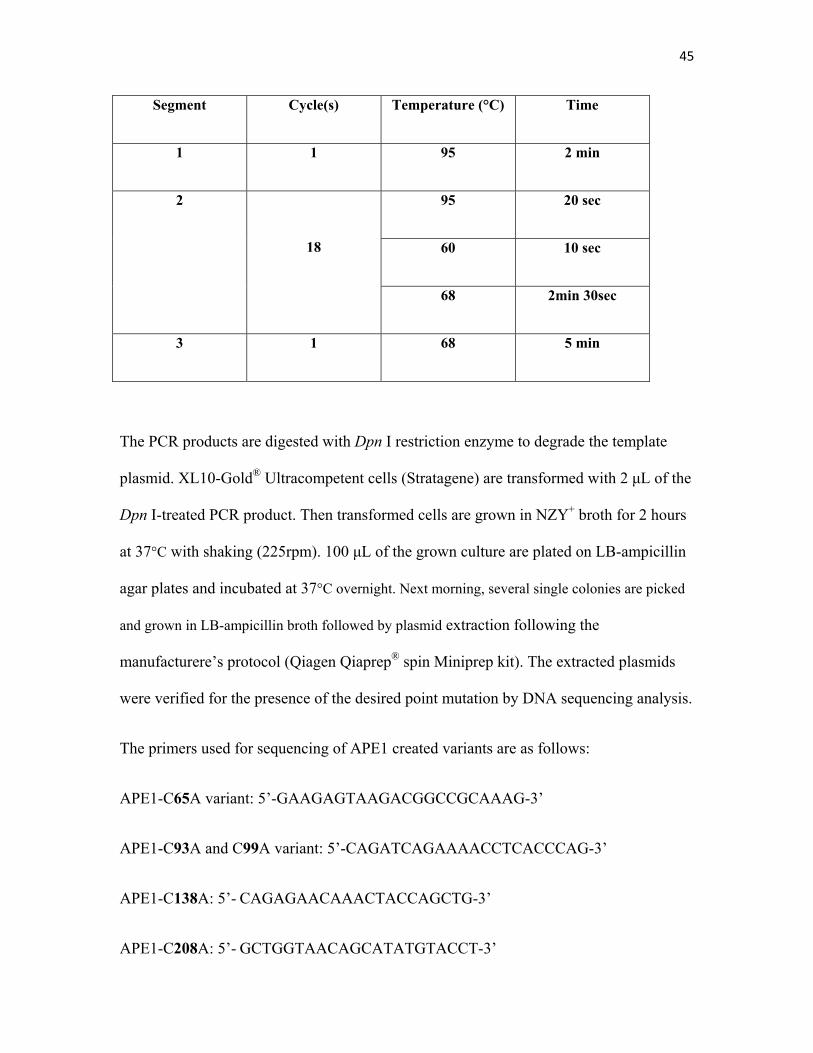
45
Segment Cycle(s) Temperature (°C) Time
1 1 95 2 min
2
18
95 20 sec
60 10 sec
68 2min 30sec
3 1 68 5 min
The PCR products are digested with Dpn I restriction enzyme to degrade the template
plasmid. XL10-Gold® Ultracompetent cells (Stratagene) are transformed with 2 μL of the
Dpn I-treated PCR product. Then transformed cells are grown in NZY+ broth for 2 hours
at 37°C with shaking (225rpm). 100 μL of the grown culture are plated on LB-ampicillin
agar plates and incubated at 37°C overnight. Next morning, several single colonies are picked
and grown in LB-ampicillin broth followed by plasmid extraction following the
manufacturere’s protocol (Qiagen Qiaprep® spin Miniprep kit). The extracted plasmids
were verified for the presence of the desired point mutation by DNA sequencing analysis.
The primers used for sequencing of APE1 created variants are as follows:
APE1-C65A variant: 5’-GAAGAGTAAGACGGCCGCAAAG-3’
APE1-C93A and C99A variant: 5’-CAGATCAGAAAACCTCACCCAG-3’
APE1-C138A: 5’- CAGAGAACAAACTACCAGCTG-3’
APE1-C208A: 5’- GCTGGTAACAGCATATGTACCT-3’
46
APE1-C296A and C310A:5’- CACCTCTACCCCAACACAC-3’
A control reaction provided by the manufacturer was used to verify the efficiency of
mutant plasmid generation.
6. Bacterial Transformation
For plasmid amplification and cloning purposes we prepared bacterial competent cells as
follows: 1 mL of an overnight culture was subcultured in 100 mL for 4 hours at 37°C.
The obtained bacterial pellet was incubated with 100 mM Calcium Chloride (CaCl2) on
ice for at least one hour. To 50 μL of prepared competent bacteria, 1 μg of the desired
plasmid DNA was added and left to incubate on ice for 30 minutes. Then the bacteria-
DNA mixture was incubated at 42°C for 90 seconds for heat shock. 1mL of LB broth was
added to the transformation reaction and left to incubate at 37°C with shaking for 2-4
hours. The transformed bacteria were then plated on selective LB agar plates (containing
either ampicillin (100 μg/ml) for pET-14b vector, or Kanamycin (50 μg/ml) for pEYFP-
N1 vector).
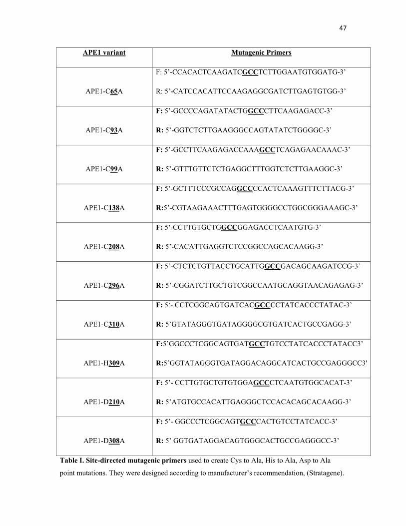
47
APE1 variant Mutagenic Primers
APE1-C65A
F: 5’-CCACACTCAAGATCGCCTCTTGGAATGTGGATG-3’
R: 5’-CATCCACATTCCAAGAGGCGATCTTGAGTGTGG-3’
APE1-C93A
F: 5’-GCCCCAGATATACTGGCCCTTCAAGAGACC-3’
R: 5’-GGTCTCTTGAAGGGCCAGTATATCTGGGGC-3’
APE1-C99A
F: 5’-GCCTTCAAGAGACCAAAGCCTCAGAGAACAAAC-3’
R: 5’-GTTTGTTCTCTGAGGCTTTGGTCTCTTGAAGGC-3’
APE1-C138A
F: 5’-GCTTTCCCGCCAGGCCCCACTCAAAGTTTCTTACG-3’
R:5’-CGTAAGAAACTTTGAGTGGGGCCTGGCGGGAAAGC-3’
APE1-C208A
F: 5’-CCTTGTGCTGGCCGGAGACCTCAATGTG-3’
R: 5’-CACATTGAGGTCTCCGGCCAGCACAAGG-3’
APE1-C296A
F: 5’-CTCTCTGTTACCTGCATTGGCCGACAGCAAGATCCG-3’
R: 5’-CGGATCTTGCTGTCGGCCAATGCAGGTAACAGAGAG-3’
APE1-C310A
F: 5’- CCTCGGCAGTGATCACGCCCCTATCACCCTATAC-3’
R: 5’GTATAGGGTGATAGGGGCGTGATCACTGCCGAGG-3’
APE1-H309A
F:5’GGCCCTCGGCAGTGATGCCTGTCCTATCACCCTATACC3’
R:5’GGTATAGGGTGATAGGACAGGCATCACTGCCGAGGGCC3'
APE1-D210A
F: 5’- CCTTGTGCTGTGTGGAGCCCTCAATGTGGCACAT-3’
R: 5’ATGTGCCACATTGAGGGCTCCACACAGCACAAGG-3’
APE1-D308A
F: 5’- GGCCCTCGGCAGTGCCCACTGTCCTATCACC-3’
R: 5’ GGTGATAGGACAGTGGGCACTGCCGAGGGCC-3’
Table I. Site-directed mutagenic primers used to create Cys to Ala, His to Ala, Asp to Ala
point mutations. They were designed according to manufacturer’s recommendation, (Stratagene).
48
7. Cloning of APE1 variants in pEYFP-N1
APE1 variants were cloned in pEYFP-N1 vector that has and enhanced yellow
fluorescent protein (EYFP) for expression in mammalian cells. Briefly, APE1 wild type
and variants were amplified by PCR from the pET-14b construct. Primers were designed
in a way to introduce APE1 between Hind III and BamH I in fusion with the EYFP gene
by removing APE1 stop codon from the reverse PCR primer. A Kozak sequence
(underlined) was added upstream of the start codon in the forward primer to enhance the
translation of the resulting mRNA. Two additional nucleotides were added to the reverse
primer downstream of BamH I restriction site, to put the insert in frame with the EYFP
gene.
Oligo sequence for APE1 variants cloning in pEYFP-N1:
F: 5’- ATACGATAGC-AAGCTT- GCCACCATGCCGAAGCGTGGGAAAAA-3’
R: 5’-AGGCTGAGAT-GGATCCAG–CAGTGCTAGGTATAGGGTGATAGG- 3’
PCR was conducted in the presence of Pfu DNA polymerase (Feldan-Bio). PCR
parameters used are the following:
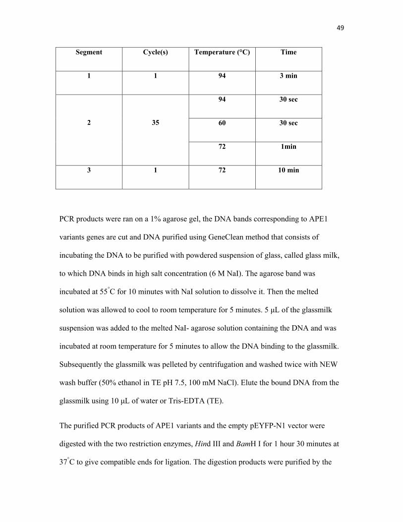
49
Segment Cycle(s) Temperature (°C) Time
1 1 94 3 min
2
35
94 30 sec
60 30 sec
72 1min
3 1 72 10 min
PCR products were ran on a 1% agarose gel, the DNA bands corresponding to APE1
variants genes are cut and DNA purified using GeneClean method that consists of
incubating the DNA to be purified with powdered suspension of glass, called glass milk,
to which DNA binds in high salt concentration (6 M NaI). The agarose band was
incubated at 55°C for 10 minutes with NaI solution to dissolve it. Then the melted
solution was allowed to cool to room temperature for 5 minutes. 5 μL of the glassmilk
suspension was added to the melted NaI- agarose solution containing the DNA and was
incubated at room temperature for 5 minutes to allow the DNA binding to the glassmilk.
Subsequently the glassmilk was pelleted by centrifugation and washed twice with NEW
wash buffer (50% ethanol in TE pH 7.5, 100 mM NaCl). Elute the bound DNA from the
glassmilk using 10 μL of water or Tris-EDTA (TE).
The purified PCR products of APE1 variants and the empty pEYFP-N1 vector were
digested with the two restriction enzymes, Hind III and BamH I for 1 hour 30 minutes at
37°C to give compatible ends for ligation. The digestion products were purified by the
50
previously described GeneClean method. Ligation mixtures containing the digested PCR
products and empty vector were incubated with T4 DNA ligase (New England BioLabs)
overnight at 16°C (20 μL reaction).
Transformation of DH5α competent cells with 4 μL of the ligation reaction was
conducted the next morning, using the previously described transformation protocol.
Transformed bacteria were plated on LB-kanamycin agar plates for selection of the
positive clones.
Single colonies were grown in LB-kanamycin broth and plasmid extraction was done
according to the manufacturer instructions (Qiagen). Double digestion using Hind III and
BamH I was done to verify the success of the cloning. The digestion product was
assessed for the presence of the insert (APE1 variants) by electrophoresis on an agarose
gel.
8. Cloning of native Apn1 and its inactive mutant
E158G in pEYFP-N1
Apn1 was amplified from p3xFLAGCMV10-Apn1 (a gift from Dr Bruce Demple,
Harvard Medical School) as described in section 7. Apn1-E158A was amplified from
pYES 2.0 vector. Primers used for cloning in pEYFP-N1 vector are listed below. It was
cloned in fusion with EYFP gene. The cloning procedure including the PCR, the
digestion, ligation and verification of the cloning were done as described in section 7 for
APE1 variants cloning.
51
Primers used for Apn1 and its variant Apn1-E158G cloning in pEYFP-N1:
F: 5’-AATCGATGCT-AAGCTT-GCCACCATGCCTTCGACACCTAGC-3’
R: 5’-AGGCTGAGAT-GGATCCAG- TTCTTTCTTAGTCTTCCTCTTCTT-3’
9. HCT116 Cell culture
Human colon carcinoma HCT116 cell line was kindly provided by Dr.Elliot Drobetsky
(University of Montreal). They were culture in Dulbecco's Modified Eagle Medium
(DMEM) (Wisent Inc.) complemented with 10% of fetal bovine serum (FBS) (Wisent
Inc.) and 0.1mg/ml penicillin, and 0.1 mg/ml streptomycin. Cells were incubated at 37°C
and 5% CO2. The media was changed every 48 hours.
10. GAPDH Knockdown by siRNA
Human Silencer GAPDH siRNA (Ambion) that targets the 5' medial region of GAPDH
mRNA sequence was used to knock down the expression of GAPDH in HCT116 cells.
The cells were cultured in Petri plates and transfected with 75 nM of GAPDH siRNA
using Lipofectamine 2000 (Invitrogen) in Opti-MEM (Invitrogen) according to the
manufacturer’s protocol. Negative control (75 nM) that has no homology to any known
gene was also included in the transfection. The plates were incubated at 37°C in 5% CO2
52
for 4 h, after which the complete growth medium was added, and the cells were proceed
for subsequent assays.
11. Protein extraction form HCT116
HCT116 adherent cells are collected from the Petri plates using a cell scraper and not
trypsin, to avoid any possible protein degradation. Cell were washed with cold PBS and
only the pellet was frozen at -80°C if the protein extraction was not done on the same
day. For protein extraction, 200 μL of RIPA buffer (NaCl, EDTA, Tris Base, 1%
Deoxycholic acid, 1% Nonidet p-40 in millipore water) supplemented with protease
inhibitors and then added to the pelleted cells and incubated on ice for 1 hour. The lysed
cells are centrifuged at 12,000 rpm at 4°C for 30 minutes. The supernatant was then
stored at -80°C until used. Protein concentration was estimated using Bradford method.
12. Western blot to verify Knock down of GAPDH
For the verification of GAPDH Knock down in HCT116 cells, total cell extracts were
quantified using Bradford protein estimation method, and equal amounts were loaded on
a 10% SDS-PAGE and proceeded for western blot analysis as described in section 4
except for the use of the monoclonal mouse anti-GAPDH followed by probing with anti-
mouse antibody. The membrane was probed with anti-tubulin antibody as a loading
control.
53
13. HCT116 plasmid DNA transfection
Transfection of HCT116 with 1-2 μg of pEYFP-N1 plasmid carrying APE1 different
variants was done as previously described in section 10 using lipofectamine 2000 and
Opti-MEM media. The expression of APE1-EYFP proteins was detected after 4 hours of
the transfection under a fluorescent microscope.
14. Establishing Stable Clones expressing APE1
variants, Apn1 and Apn1-E158G
HCT116 were transfected as described in section 10. 48 hours after transfection, G418
(Neomycin) (Wisent Inc.) was added to the media of the transfected cells at a
concentration of 150 μg/mL for selection of the positive clones expressing the APE1-
EYFP or Apn1-EYFP that has a neomycin resistant gene as a marker for selection. A
serial selection of positive cells was done by increasing concentrations of G418 (150,
200,250, 350, and 400 μg/mL) over three weeks, the approximate time needed for single
colonies of transfected cells expressing differential amounts of the protein of interest.
Cell sorting (FACS Aria III, BD) was used to pick single cells that were then cultured in
200 μL of media containing 200 μg/mL of G418, and then subcultured in 1ml, 2ml, 5ml
and 10 mL successively. G418 at 200 μg/mL was used for maintenance of the stably
54
transfected cells in culture. The efficiency of the transfection was assessed under the
microscope and by western blot analysis.
15. Cell Cycle analysis
For cell cycle analysis of GAPDH knocked down cells, control siRNA cells, APE1-
EYFP,Apn1- EYFP expression cells, cells were transfected 36 hours prior to nocodazole
(200 ng/mL) (Sigma) treatment to arrest cells in G2/M phase. The collection of the
sample starts after nocodazole treatment by the addition of trypsin (Wisent). Collected
cells were then washed with PBS/1% EDTA to prevent clumping of the cells. Cells are
stored in 75% Ethanol in PBS in FACs tubes at -20°C until the FACs analysis was done.
During cell collection for cell cycle analysis a fraction of the collected cells was kept at -
80°C for western blot analysis. Samples were collected as follows:
-Sample 0: cells not treated with nocodazole after 36 hours of transfection.
-Sample 12 hours, 24 hours and 36 hours: cells collected after 12, 24 and 36 hours of
nocodazole treatment.
Before flow cytometry analysis, cell suspensions were centrifuged and washed with PBS
to remove ethanol. Then they were resuspended in 500 μL of PBS with 0.05 μg/μL of
propidium iodide to stain the DNA. The collected samples are subject to FACs analysis
on the FACScan machine (Becton Dickinson) according the standard protocols. Data
were analyzed using Cell Quest Pro allias software.
55
16. Indirect immunofluorescence
For monitoring APE1 localization in GAPDH knock down HCT116, 750,000 cells were
plated. 24 hours cell were transfected with the corresponding siRNA as section 10. 24
hours after transfection, 125,000 cells were transferred to a sterilized cover slip in a 6-
well plate. Cells were left for 48 hours to allow them to adhere to the glass surface of the
coverslip.
Fixation and permeabilization: remove the media from the 6-well plates and wash the
cells with 2mL of PBS. Cells were then incubated with 1 mL of 3% Paraform aldehyde
(PFA) (Sigma) for 20 minutes. The PFA was removed and the cells are incubated with
2 mL of permeabilization buffer (PBS, 0.5% NP-40, 1mM Sodium azide) for 30 minutes.
The cells are then washed with washing buffer (PBS, 0.1% NP-40, 1mM Sodium azide)
and the blocking solution (10% FBS) for 1 hour.
Primary antibody: The primary antibody mouse anti-APE1 (cedarlane) 1:150 dilution
was prepared in blocking solution. The coverslips holding the cells were then removed
from the 6-well plate and 50 μL of primary antibody was added onto the cells and
covered with a parafilm of the size of the coverslip. The primary antibody was left to
incubate for 3 hours at room temperature.
The cells were then washed with washing buffer for 3 times/15 minutes each.
56
Secondary antibody: The secondary antibody, goat anti-mouse coupled to AlexaFluor
(AlexaFluor 488, Invitrogen) was used at 1:500 dilution and was added to the cells as
described for the primary antibody.
The cells were then washed with washing buffer for 3 times/15 minutes each. Then the
cells were washed twice with PBS.
Mounting: the coverslips were rinsed with milliQ water, and dried gently using a paper
towel. 15 μL of the mounting media (Vector laboratories, Inc. Burlingame, CA)
containing DAPI was put on the slide and the coverslip was placed on top of it, with the
cells facing down. The slides were then ready for microscopy. The slides were
subsequently examined under a Leica, DMRE, Wetzlar fluorescence microscope
supplemented with a camera. Exposure times varied from 300-400 ms. Slides were stored
in the dark.
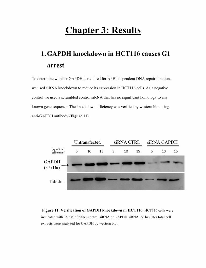
Chapter 3: Results
1. GAPDH knockdown in HCT116 causes G1
arrest
To determine whether GAPDH is required for APE1-dependent DNA repair function,
we used siRNA knockdown to reduce its expression in HCT116 cells. As a negative
control we used a scrambled control siRNA that has no significant homology to any
known gene sequence. The knockdown efficiency was verified by western blot using
anti-GAPDH antibody (Figure 11).
Figure 11. Verification of GAPDH knockdown in HCT116. HCT116 cells were
incubated with 75 nM of either control siRNA or GAPDH siRNA, 36 hrs later total cell
extracts were analyzed for GAPDH by western blot.
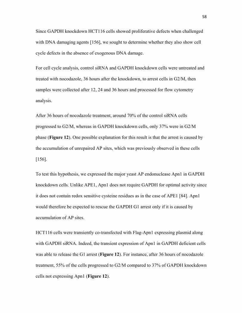
58
Since GAPDH knockdown HCT116 cells showed proliferative defects when challenged
with DNA damaging agents [156], we sought to determine whether they also show cell
cycle defects in the absence of exogenous DNA damage.
For cell cycle analysis, control siRNA and GAPDH knockdown cells were untreated and
treated with nocodazole, 36 hours after the knockdown, to arrest cells in G2/M, then
samples were collected after 12, 24 and 36 hours and processed for flow cytometry
analysis.
After 36 hours of nocodazole treatment, around 70% of the control siRNA cells
progressed to G2/M, whereas in GAPDH knockdown cells, only 37% were in G2/M
phase (Figure 12). One possible explanation for this result is that the arrest is caused by
the accumulation of unrepaired AP sites, which was previously observed in these cells
[156].
To test this hypothesis, we expressed the major yeast AP endonuclease Apn1 in GAPDH
knockdown cells. Unlike APE1, Apn1 does not require GAPDH for optimal activity since
it does not contain redox sensitive cysteine residues as in the case of APE1 [84]. Apn1
would therefore be expected to rescue the GAPDH G1 arrest only if it is caused by
accumulation of AP sites.
HCT116 cells were transiently co-transfected with Flag-Apn1 expressing plasmid along
with GAPDH siRNA. Indeed, the transient expression of Apn1 in GAPDH deficient cells
was able to release the G1 arrest (Figure 12). For instance, after 36 hours of nocodazole
treatment, 55% of the cells progressed to G2/M compared to 37% of GAPDH knockdown
cells not expressing Apn1 (Figure 12).
59
These results suggest that accumulated DNA damage underlies the observed cell cycle
progression defects. It is most probably due to defective APE1 proteins in the absence of
adequate amount of GAPDH for their activation.
To validate the complementation effect of Apn1, we next attempted to establish stable
HCT116 clones expressing it. The selection was done using G418 over 3 weeks. Then
single colonies were picked and subcultured. Protein extracts of different colonies were
subject to western blot analysis using a Flag antibody. Unfortunately, we were unable to
detect Apn1 expression. Another attempt to create Apn1-expressing stable clones was
done with a different construct that expresses Apn1-EYFP as shown in Figure 21 for
APE1 variants. A fluorescent tag would make the selection process easier by allowing
monitoring of expression using microscopy and giving the advantage of the use of cell
sorting as another mean of selection. Similarly, we created a construct that expresses
APN1-E158G-YFP, the AP endonuclease-deficient mutant of Apn1[188]. G418 was used
for the selection as described in section 14 of chapter 2. The expression of the YFP-
tagged Apn1 and Apn1-E158G in these stably transfected cell lines was monitored under
a fluorescent microscope (data not shown). The expression of both proteins was seen as
short as 6 hours after transfection by microscopy. Unfortunately, 48 hours later the
number of cells showing fluorescent proteins start to decrease from the cell culture. Four
days later, no cells were showing any expression of the EYFP-tagged proteins as
compared to the empty vector that showed a very good expression of the EYFP. One
possible explanation is that, as a foreign protein, Apn1 might need some posttranslational
modification by yeast proteins (e.g. chaperones, etc…) that are not present in human cells
for its proper folding.
60
Figure 12. GAPDH knock down causes a G1 arrest. GAPDH knockdown cells transfected with
Apn1 showed normal cell cycle progression, a result obtained in 3 independent experiments.
Interestingly, we observed that cells start to die few hours after transfection with Apn1
plasmid, which could explain the gradual decrease in the number of cells expressing Apn1 and
hence the failure in establishing stable clones.
61
2. siRNA mediated GAPDH silencing triggers
the formation of APE1 aggregates
Upon oxidative stress, some BER enzymes including OGG1 and APE1 localize to
nuclear speckles, structures that are present between chromatin domains enriched with
RNA processing and transcription proteins [187]. To verify whether GAPDH cellular
level affects APE1 localization, we carried out an indirect-immunofluorescence
experiment using APE1 antibody in GAPDH knockdown and control siRNA cells. While
normal nuclear distribution was observed in control siRNA cells, in GAPDH knockdown
cells we detected foci in the nucleus of the majority of the cellular population (Figure
13). The nature of the detected structures needs further investigations; we speculate that
they could be stalled DNA repair foci of inactive APE1 on DNA lesions or it could just
be an aggregation due to misfolding of the oxidized proteins in the absence of enough
GAPDH to reduce them.
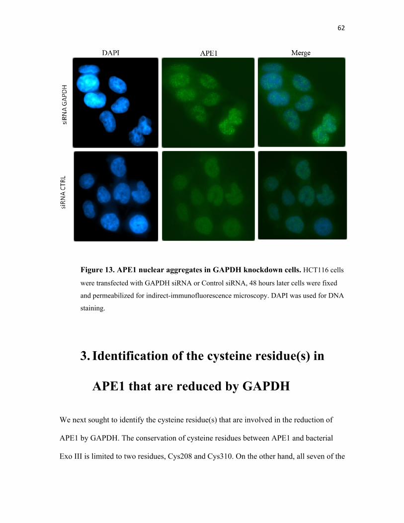
62
Figure 13. APE1 nuclear aggregates in GAPDH knockdown cells. HCT116 cells
were transfected with GAPDH siRNA or Control siRNA, 48 hours later cells were fixed
and permeabilized for indirect-immunofluorescence microscopy. DAPI was used for DNA
staining.
3. Identification of the cysteine residue(s) in
APE1 that are reduced by GAPDH
We next sought to identify the cysteine residue(s) that are involved in the reduction of
APE1 by GAPDH. The conservation of cysteine residues between APE1 and bacterial
Exo III is limited to two residues, Cys208 and Cys310. On the other hand, all seven of the
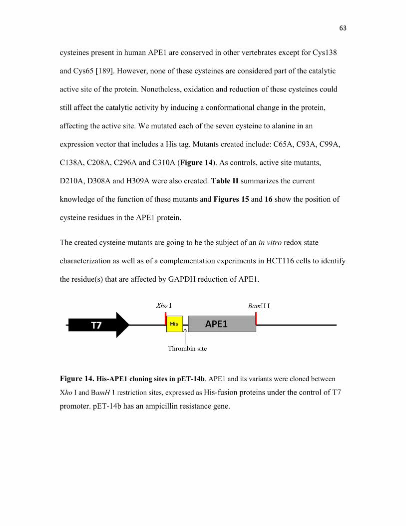
63
cysteines present in human APE1 are conserved in other vertebrates except for Cys138
and Cys65 [189]. However, none of these cysteines are considered part of the catalytic
active site of the protein. Nonetheless, oxidation and reduction of these cysteines could
still affect the catalytic activity by inducing a conformational change in the protein,
affecting the active site. We mutated each of the seven cysteine to alanine in an
expression vector that includes a His tag. Mutants created include: C65A, C93A, C99A,
C138A, C208A, C296A and C310A (Figure 14). As controls, active site mutants,
D210A, D308A and H309A were also created. Table II summarizes the current
knowledge of the function of these mutants and Figures 15 and 16 show the position of
cysteine residues in the APE1 protein.
The created cysteine mutants are going to be the subject of an in vitro redox state
characterization as well as of a complementation experiments in HCT116 cells to identify
the residue(s) that are affected by GAPDH reduction of APE1.
Figure 14. His-APE1 cloning sites in pET-14b. APE1 and its variants were cloned between
Xho I and BamH 1 restriction sites, expressed as His-fusion proteins under the control of T7
promoter. pET-14b has an ampicillin resistance gene.
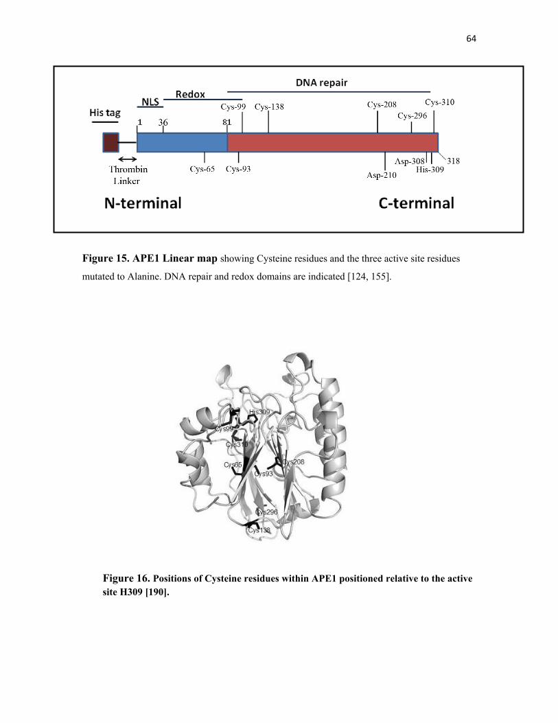
64
Figure 15. APE1 Linear map showing Cysteine residues and the three active site residues
mutated to Alanine. DNA repair and redox domains are indicated [124, 155].
Figure 16. Positions of Cysteine residues within APE1 positioned relative to the active site H309 [190].
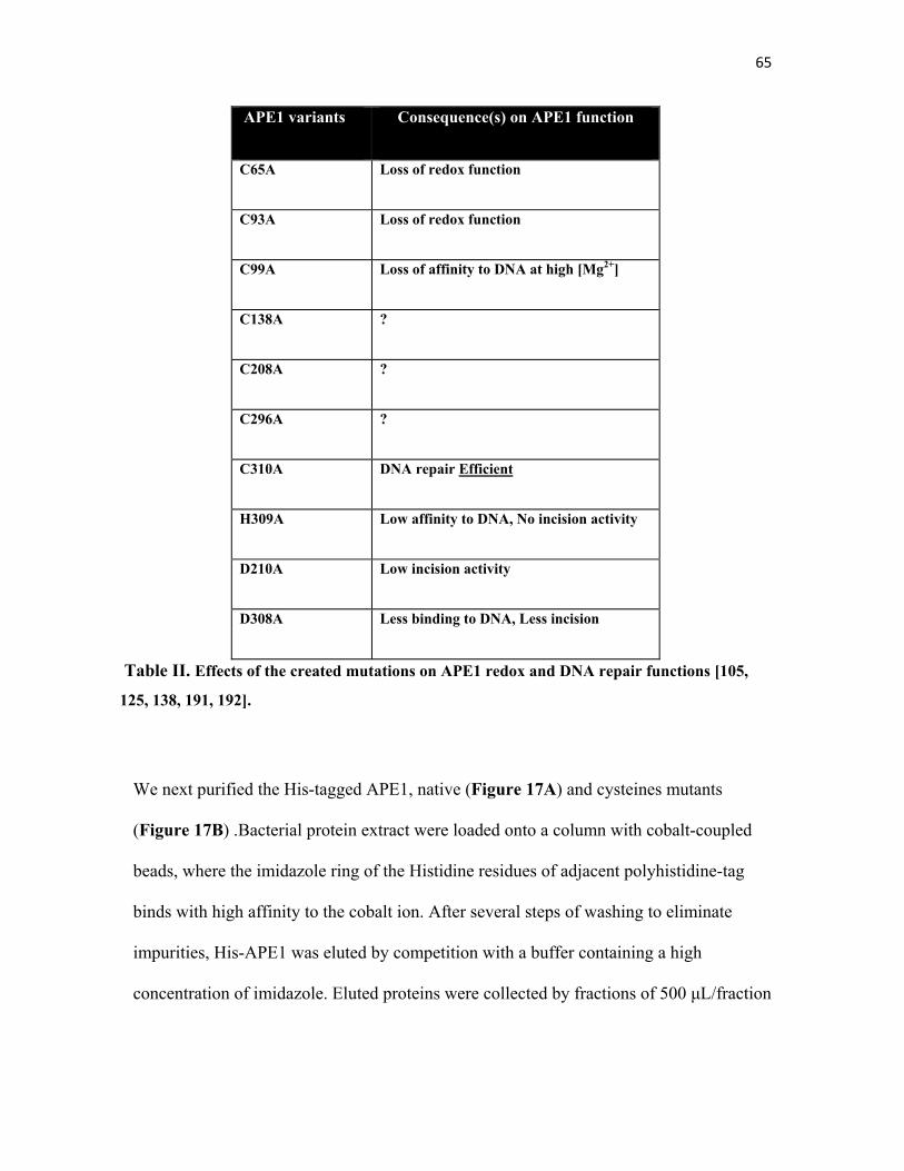
65
APE1 variants Consequence(s) on APE1 function
C65A Loss of redox function
C93A Loss of redox function
C99A Loss of affinity to DNA at high [Mg2+]
C138A ?
C208A ?
C296A ?
C310A DNA repair Efficient
H309A Low affinity to DNA, No incision activity
D210A Low incision activity
D308A Less binding to DNA, Less incision
Table II. Effects of the created mutations on APE1 redox and DNA repair functions [105,
125, 138, 191, 192].
We next purified the His-tagged APE1, native (Figure 17A) and cysteines mutants
(Figure 17B) .Bacterial protein extract were loaded onto a column with cobalt-coupled
beads, where the imidazole ring of the Histidine residues of adjacent polyhistidine-tag
binds with high affinity to the cobalt ion. After several steps of washing to eliminate
impurities, His-APE1 was eluted by competition with a buffer containing a high
concentration of imidazole. Eluted proteins were collected by fractions of 500 μL/fraction
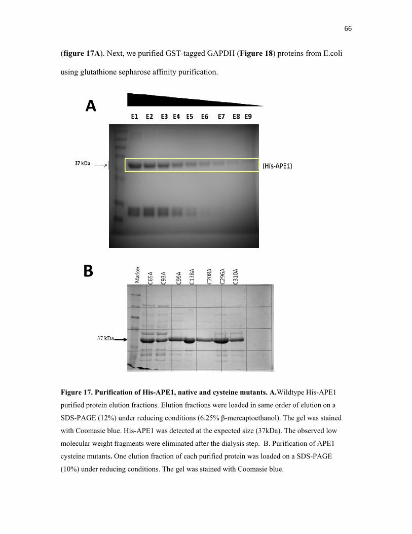
66
(figure 17A). Next, we purified GST-tagged GAPDH (Figure 18) proteins from E.coli
using glutathione sepharose affinity purification.
Figure 17. Purification of His-APE1, native and cysteine mutants. A.Wildtype His-APE1
purified protein elution fractions. Elution fractions were loaded in same order of elution on a
SDS-PAGE (12%) under reducing conditions (6.25% β-mercaptoethanol). The gel was stained
with Coomasie blue. His-APE1 was detected at the expected size (37kDa). The observed low
molecular weight fragments were eliminated after the dialysis step. B. Purification of APE1
cysteine mutants. One elution fraction of each purified protein was loaded on a SDS-PAGE
(10%) under reducing conditions. The gel was stained with Coomasie blue.
A
B
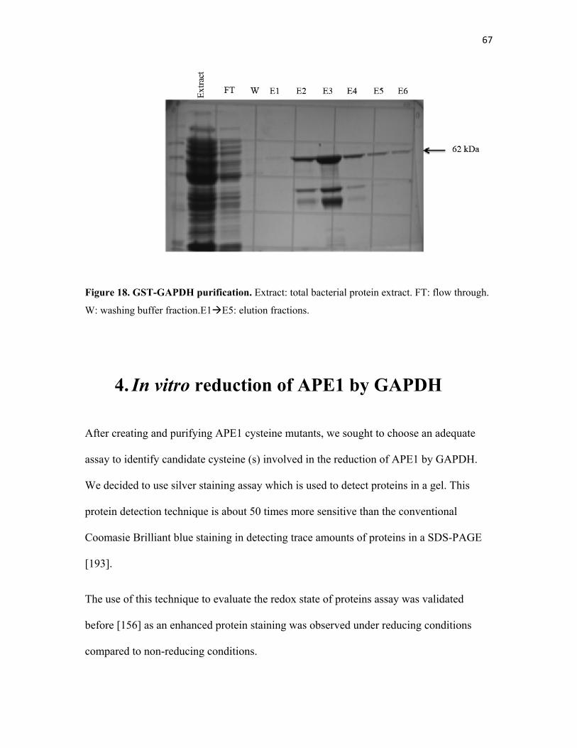
67
Figure 18. GST-GAPDH purification. Extract: total bacterial protein extract. FT: flow through.
W: washing buffer fraction.E1E5: elution fractions.
4. In vitro reduction of APE1 by GAPDH
After creating and purifying APE1 cysteine mutants, we sought to choose an adequate
assay to identify candidate cysteine (s) involved in the reduction of APE1 by GAPDH.
We decided to use silver staining assay which is used to detect proteins in a gel. This
protein detection technique is about 50 times more sensitive than the conventional
Coomasie Brilliant blue staining in detecting trace amounts of proteins in a SDS-PAGE
[193].
The use of this technique to evaluate the redox state of proteins assay was validated
before [156] as an enhanced protein staining was observed under reducing conditions
compared to non-reducing conditions.
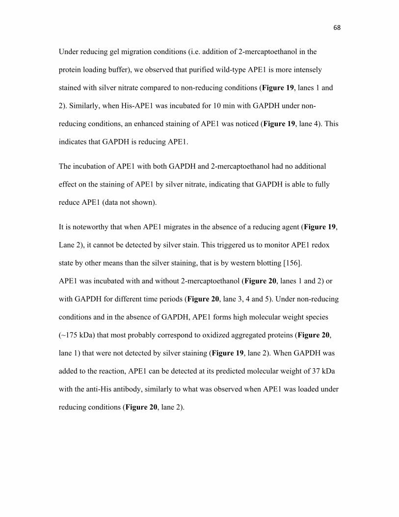
68
Under reducing gel migration conditions (i.e. addition of 2-mercaptoethanol in the
protein loading buffer), we observed that purified wild-type APE1 is more intensely
stained with silver nitrate compared to non-reducing conditions (Figure 19, lanes 1 and
2). Similarly, when His-APE1 was incubated for 10 min with GAPDH under non-
reducing conditions, an enhanced staining of APE1 was noticed (Figure 19, lane 4). This
indicates that GAPDH is reducing APE1.
The incubation of APE1 with both GAPDH and 2-mercaptoethanol had no additional
effect on the staining of APE1 by silver nitrate, indicating that GAPDH is able to fully
reduce APE1 (data not shown).
It is noteworthy that when APE1 migrates in the absence of a reducing agent (Figure 19,
Lane 2), it cannot be detected by silver stain. This triggered us to monitor APE1 redox
state by other means than the silver staining, that is by western blotting [156].
APE1 was incubated with and without 2-mercaptoethanol (Figure 20, lanes 1 and 2) or
with GAPDH for different time periods (Figure 20, lane 3, 4 and 5). Under non-reducing
conditions and in the absence of GAPDH, APE1 forms high molecular weight species
(~175 kDa) that most probably correspond to oxidized aggregated proteins (Figure 20,
lane 1) that were not detected by silver staining (Figure 19, lane 2). When GAPDH was
added to the reaction, APE1 can be detected at its predicted molecular weight of 37 kDa
with the anti-His antibody, similarly to what was observed when APE1 was loaded under
reducing conditions (Figure 20, lane 2).
69
Figure 19. In vitro reduction of His-APE1 by GST-GAPDH. Purified His-APE1 and GST-
GAPDH were incubated for 10 min at 37°C; the reaction was stopped on ice and by the addition
of the protein loading buffer. Samples were run on a SDS-PAGE (12%) and the gel was stained
with silver nitrate as described in chapter 2, section 3. The lower band in lanes 4 and 5 is an
impurity (GST as seen by western blot using anti-GST).
We next attempted to test the purified APE1 cysteine mutants under the same assay
conditions. We anticipate that GAPDH might cause a conformational change in APE1
structure by reducing an intramolecular disulfide bridge(s) between cysteine residues that
forms as a result of oxidation of the protein. Therefore, APE1 cysteine mutants will be
assayed for their redox state to identify which mutant(s) will have enhanced silver
staining. This mutant is expected to be more reduced, independently of GAPDH
compared to the native APE1, as a Cys to Ala mutation will simulate the reduced state of
the substituted cysteine since Alanine cannot be engaged in disulfide bridge formation
upon oxidation. This experiment is still in progress.
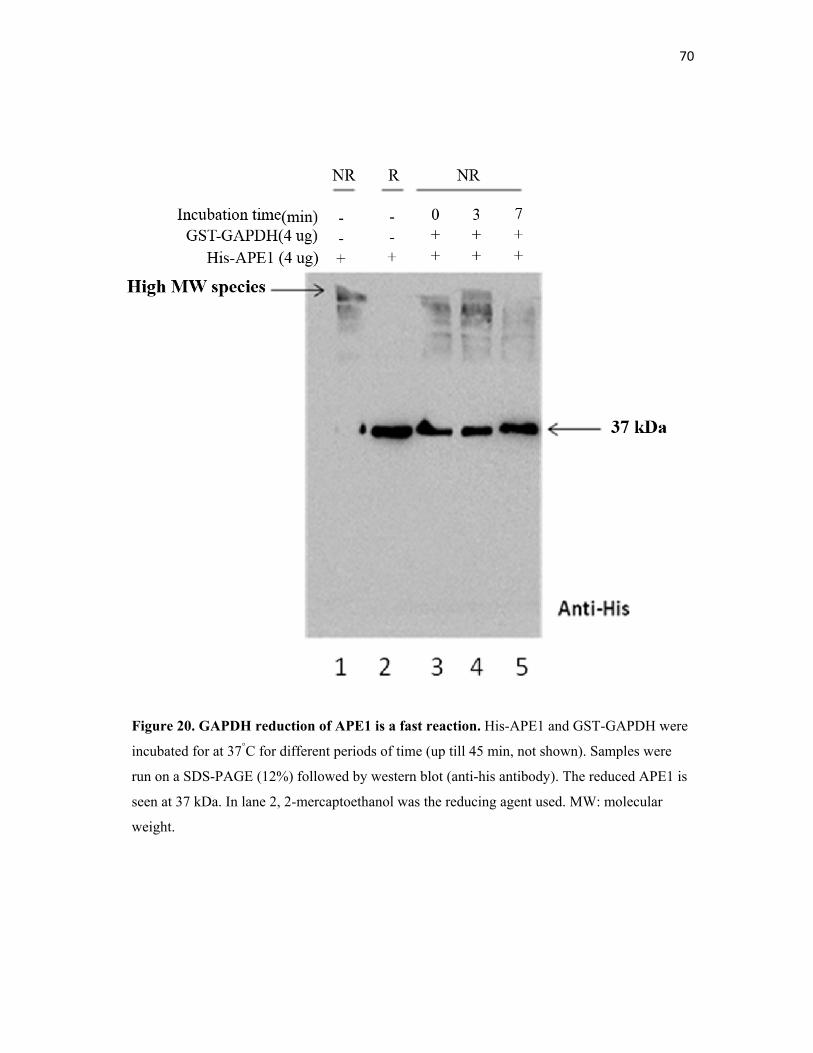
70
Figure 20. GAPDH reduction of APE1 is a fast reaction. His-APE1 and GST-GAPDH were
incubated for at 37°C for different periods of time (up till 45 min, not shown). Samples were
run on a SDS-PAGE (12%) followed by western blot (anti-his antibody). The reduced APE1 is
seen at 37 kDa. In lane 2, 2-mercaptoethanol was the reducing agent used. MW: molecular
weight.
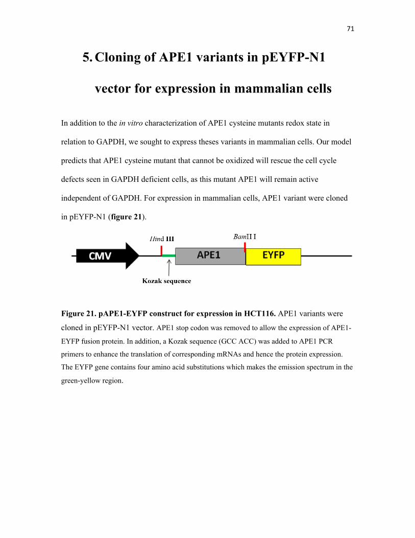
71
5. Cloning of APE1 variants in pEYFP-N1
vector for expression in mammalian cells
In addition to the in vitro characterization of APE1 cysteine mutants redox state in
relation to GAPDH, we sought to express theses variants in mammalian cells. Our model
predicts that APE1 cysteine mutant that cannot be oxidized will rescue the cell cycle
defects seen in GAPDH deficient cells, as this mutant APE1 will remain active
independent of GAPDH. For expression in mammalian cells, APE1 variant were cloned
in pEYFP-N1 (figure 21).
Figure 21. pAPE1-EYFP construct for expression in HCT116. APE1 variants were
cloned in pEYFP-N1 vector. APE1 stop codon was removed to allow the expression of APE1-
EYFP fusion protein. In addition, a Kozak sequence (GCC ACC) was added to APE1 PCR
primers to enhance the translation of corresponding mRNAs and hence the protein expression.
The EYFP gene contains four amino acid substitutions which makes the emission spectrum in the
green-yellow region.
72
6. Establishing stable clones expressing APE1
cysteine variants
HCT116 were transfected with APE1-EYFP expression constructs and subjected to G418
serial selection to generate stable clones. The expression of APE1 variants in the
established stable clones is monitored by fluorescent microscopy and by western blotting
(in progress). All of the APE1-EYFP variants were localized in the nucleus (data now
shown).
Chapter 4: Discussion
The diverse functions of APE1 as a DNA repair enzyme and a redox factor render it an
essential gene for cell survival. Studying the consequences of APE1 activation by
GAPDH, another essential gene, is of great importance to unravel a new posttranslational
modification affecting APE1 activity as well as the indirect involvement of GAPDH in
maintaining the genomic stability.
In the present study we investigated the consequences of GAPDH knockdown on the cell
cycle progression and we reported that cells in which GAPDH is knocked-down arrest in
G1 phase. APE1 subcellular localization study showed that APE1 forms foci-like
structures in the nucleus of cells knocked down for GAPDH.
Since GAPDH activation of APE1 is achieved by reduction, we sought to search for the
cysteine residue(s) that are possibly targeted by GAPDH. APE1 cysteine mutants were
created by site-directed mutagenesis and purified using affinity chromatography. In vitro
attempts to identify the redox-sensitive cysteine (s) target(s) of GAPDH reduction did not
succeed so far due to the quality of the purified proteins. A construct for expression of
EYFP-tagged APE1 variants, including cysteine and active site mutants, in mammalian
cells was successfully established and the expression was seen under the microscope.
74
1. GAPDH deficiency causes G1 arrest
GAPDH is an essential gene, hence a knockdown instead of a knockout approach was
used to evaluate GAPDH role in activating APE1 in mammalian cells. siRNA
knockdown of GAPDH caused a defect in cell cycle progression, which was not
surprising since a decrease in cell proliferation was also seen in GAPDH knockdown
cells (Ramotar Lab, unpublished data). Moreover, GAPDH deficiency caused an increase
in the formation of spontaneous AP sites and showed a decrease in the AP endonuclease
activity of the cells [156]. These data suggest that the delay seen in the cell cycle is most
probably due to an accumulation of AP sites which is known to activate checkpoints,
delaying cell cycle progression to allow repair [194]. Checkpoints signalling pathways
activate damage-specific DNA repair mechanisms, in this case the BER, before the
transition from G1 to S phase and prior to DNA synthesis [195], as BER pathway is
mostly active in G1 phase where oxidized bases and strand breaks are repaired [194].
The observed G1 arrest of GAPDH depleted cells might be occurring via the classical
activation of p53 by phosphorylation on Ser-15, which was reported by another group
that also showed that siRNA-mediated GAPDH knockdown accumulate in G1 phase
[196]. It would be also interesting to examine checkpoints activation in knockdown cells
by monitoring Chk1 and Chk2 phosphorylation state by western blot analysis.
75
2. Use of Apn1 for complementation assays
Given that APE1 is the major human AP endonuclease responsible for processing AP
sites, the accumulation of oxidized inactive species of this protein in GAPDH knockdown
cells explains the deficiency in AP endonuclease activity observed in these cells [156],
which might also validate the observed cell cycle defects. Complementation strategy was
used to verify that the lack of APE1 activity is the reason behind the G1 arrest.
Overexpression of wild type APE1 in GAPDH depleted cells can be confusing since the
ratio of the endogenous to the introduced APE1 in addition to the redox state of the total
APE1 population were a concern. Consequently, the expression of the yeast Apn1 that is
not redox sensitive was used to verify the implication of APE1 activity in the defective
cell cycle, as this enzyme shares many functional features with APE1, most importantly
the repair of AP sites. A direct measurement of AP sites frequency in GAPDH
knockdown cells transfected with or without Apn1 plasmid can be done to further verify
Apn1 rescuing effects. In fact, complementation experiments in which Apn1 was
expressed in mammalian cells and compensated for a deficiency in APE1 activity were
reported [121].
3. Apn1 expression in HCT116 cells is lethal
We noticed that stable and transient transfection of HCT116 cells with Flag- and EYFP-
tagged Apn1 was causing many cells to die about 24 hours following transfection. The
observed cell death, that probably explains the expression problems seen in Apn1
76
transfected cells, prompted us to think that Apn1 might be toxic for the cells. Importantly,
Apn1 expression under the constitutive CMV promoter in GAPDH knockdown cells,
where higher AP site frequency was observed, caused even an increased lethality. In fact,
it was shown that the overexpression of Apn1 in a cells with high frequency of AP sites
enhances cells sensitivity to MMS [197]. It seems that cells are better able to tolerate
intact AP sites than the strand breaks generated by Apn1 incision.
Hence, during cell cycle analysis, the rescuing phenotype attributed by Apn1 in GAPDH
knockdown cells might be due to surviving untransfected cells.
Interestingly, the expression of the enzymatically inactive variant of Apn1, E158G, did
not increase cell death in GAPDH knockdown cells as compared to wild type Apn1.
Therefore, we plan to design a survival assay (e.g. the clonogenic assay) where we
monitor the effects of Apn1 and its inactive variant on the survival of GAPDH
knockdown cells.
4. Apn1 stable clones
Considering the difficulties faced in transiently expressing Apn1 in HCT116, we tried to
establish stable HCT116 cell lines expressing Flag-Apn1 similarly to what was done
before [121]. After three weeks of selection with G418 and after isolating and growing 20
separate single colonies, western blot analysis with anti-Flag antibody failed to show any
expression of Apn1. Another attempt to detect Apn1 expression using a serum containing
Apn1 antibody prepared in the laboratory was unsuccessful.
77
Surprisingly, Fung and Demple (2005) [121] successfully established stable clones
expressing Flag-Apn1 in HCT116 and that was proved by western blot analysis.
Although we were using the same protocols and the same cell line, we were unable to
reproduce their work.
Establishing stable clones using Apn1-EYFP and its variant Apn1- E158G-EYFP
constructs was not very successful either. We detected a low level of expression
corresponding to a low number of cells showing fluorescent proteins. Four days after
starting the treatment with G418, and a total of six days after the transfection, almost
none of the cells expressed the fluorescent Apn1 and its variant, most probably due to cell
death. The pEYFP-N1 vector that we are using has an origin of replication that is induced
by T antigen, hence expressing Apn1 in HEK 293T cells that have SV40 T antigen might
be an alternative.
It is noteworthy that using the same construct and the same cell line, the expression of
APE1- EYFP variants was successful and stable clones were established. As a foreign
protein, Apn1 proteins might be unstable in mammalian cells context, which could lead
to their degradation due to the absence of potentially needed yeast proteins for its proper
folding. Moreover, the presence of a tag might also be interfering with the protein folding
and stability. RT-PCR of RNA extracts from Apn1-transfected cells will provide an
evidence of whether the expression problem is at the protein level or at the transcriptional
level.
78
5. APE1 foci-like structure could be aggregates
of oxidized species
Upon oxidative stress, as a DNA repair enzyme and a transcription regulator, APE1
translocates to the nucleus [104]. In fact it is considered mainly a nuclear protein,
although many reports showed its cytoplasmic localization that was correlated to
tumoregenesis [198]. It was shown to localize to the endoplasmic reticulum (ER) where it
forms a complex with other proteins and might get cleaved at its Lys 31 giving rise to
NΔ33APE1 form that was shown to localize to the mitochondria [185, 186].
It was not surprising that APE1 was localized to the nucleus in our indirect-
immunofluorescence experiments, but what was striking is the foci-like structures seen
only in GAPDH knockdown cells and not in the control cells. They could be either
nuclear foci composed of recruited and inactive APE1 that retains the ability to be
recruited to DNA damage sites or they simply represent aggregates of oxidized forms of
APE1 in the absence of enough GAPDH to reduce the proteins.
A key experiment where we test if the observed APE1 structures co-localizes with
XRCC1, a BER enzyme that usually co-localize with APE1 on DNA lesions [199], might
favor the nuclear foci hypothesis. Additionally, the expression of Apn1 in these cells is
expected to abolish the formation of these foci by competing with the inactive APE1 for
recruitment and binding to DNA lesions. Alternatively, if what we observed was just
protein aggregates, treatment of GAPDH knocked down cells with reducing agents such
as the antioxidant N-acetyl cysteine (NAC) should abolish APE1 nuclear “aggregates” by
conferring a reduced cellular environment that can compensate for the lack of GAPDH.
79
Furthermore, monitoring APE1 subcellular localization in each phase of the cell cycle of
GAPDH knockdown cells will provide us with the status of APE1 in G1 where the cells
are arresting: are these nuclear foci present mainly in G1? If these foci are present evenly
in all cell cycle phases, they are most probably a result of aggregation of oxidized
proteins.
6. Purified cysteine mutants aggregate
There are many possible reasons behind the problems encountered with the purified
APE1 mutants. First, the purified proteins were aggregating, as we saw the non-
homogeneity of the protein-containing solution by the naked eye. Attempts to change
storage conditions (buffer, temperature) and to remove the observed white aggregates
from the solution by high speed centrifugation, failed to resolve the quantification
problem. The addition of DTT might have solved the problem but since we are interested
in the redox state of these proteins, the addition of a strong reducing agent will interfere
with our assays.
Second, purified proteins were extracted from 1L of bacterial culture, which yielded
enormous amounts of proteins as visualized by SDS-PAGE. The high concentration of
purified proteins can cause aggregation. We tried to dilute the protein suspensions but the
aggregation problem was not solved. Starting with a smaller bacterial culture could be a
better idea than diluting what is already aggregated.
80
Oxidized proteins tend to aggregate in solution due to many reasons but mainly because
of the formation of inter-and intra-molecular disulfide bridges. However, it is surprising
that cysteine mutants would aggregate taking into consideration that a lack in a cysteine
residue presumably will result in less disulfide bridge formation, hence less aggregation.
Other groups have purified these variant with no mention of such problems [191].
However, to make cysteine mutants they used Cys to Ser mutation and they also used
DTT in their purification buffers.
7. Expression of APE1 mutants in AP
endonuclease deficient bacteria
To test the functional state of APE1 cysteine mutants in vivo, we tried to express them in
a bacterial strain, BW528 [Δ(xth-pnc), nfo1::kan], lacking both Endo IV and Exo III AP
endonucleases and look for complementation when challenged with DNA damaging
drugs. Interestingly, the expression of wild type APE1 as well as its cysteine mutants in
this strain was lethal compared to the empty vector. Lethality was more striking in wild
type APE1, C65A, C93A and C99A transformed bacteria (data not shown). Considering
that it lacks AP endonuclease activity, this strain is expected to accumulate a high level of
unrepaired AP sites. If they are functional, the expression of APE1 and its variants in this
strain will possibly catalyze the incision of these abundant AP sites, leading to DNA
fragmentation which possibly explains the lethality, an observation reported in yeast
where the overexpression of Apn1 is lethal [197]. Another observation in this context was
in C.elegans where the knockdown of APN-1 suppressed lethality in strain where
81
depletion of dUTP nucleotidohydrolase (DUT-1) results in an increase of mis-
incorporated uracil in the DNA, which will be removed by uracil-DNA glycosylase
(UNG-1) leaving a lot of AP sites behind [95]. It is noteworthy that the transformation of
BL21 E.coli strain that does not lack Exo III and Endo IV was successful.
8. Conclusion and future work
GAPDH was shown once again to be more than just a glycolytic enzyme. We revealed a
novel role for this enzyme in DNA repair by maintaining APE1 activity. Depletion in
GAPDH caused an increase in the mutagenic AP site lesions, arrested the cells in G1
phase, halted cell proliferation and caused APE1 to form nuclear foci-like structures that
needs to be studied further. Therefore, by activating APE1, GAPDH is helping in
maintaining the genomic integrity of the cells and promoting their survival under normal
conditions, during oxidative stress and when challenged with DNA damaging agents.
Moreover, GAPDH reduction of APE1 emphasizes the importance of APE1
posttranslational modifications in defining its functional pattern as a multifunctional
protein. But more questions related to APE1-GAPDH interaction need to be addressed:
which APE1 cysteine residue(s) is the target of reduction by GAPDH? Does this
reduction reaction trigger a conformational change in APE1 structure rendering it active?
In addition to its DNA repair function, does this interaction affect the redox function of
APE1?
82
8.1 Study the AP endonuclease activity of APE1
variants
Although there are many reports where the activity of these variants was tested (see table
I of chapter 3), some of these reports used Cys to Ser instead of Cys to Ala substitution
and used different conditions under which the assays were performed. Therefore, in the
process of identifying the cysteine residue(s) of APE1 that are reduced by GAPDH, it is
of major importance to study the AP endonuclease activity of these mutants after
oxidizing them with H2O2 using the AP endonuclease assay. This assay allows the
detection of AP endonuclease activity using a 42 mer radiolabled oligonucleotide
containing a centered AP site as described previously [96]. This substrate is then
incubated with the enzyme that, when active, will cleave the AP site, leading to the
formation of a 20 mer radiolabled fragment that migrates faster than the intact substrate
on a 10% polyacrylamide/7 M urea gels. The mutant(s) that retain their AP endonuclease
activity will be expected to play a complementing role in AP endonuclease deficient
yeast cells for instance and in GAPDH deficient cells. This mutant will be a possible
candidate of GAPDH reduction. On the other hand, the mutant(s) lacking the AP
endonuclease activity might be crucial for APE1 catalytic activity and will need further
investigation since no cysteine residue has been reported to participate in APE1 catalytic
reaction. As controls, we are going to use APE1 active site mutants, H309A, D210A and
D308 in addition to the wild type APE1 and Apn1. We are mostly interested in the
C310A variant since it was shown that the oxidation of this protein by H2O2 did not affect
its AP endonuclease activity [155].
83
8.2 Expression of APE1 cysteine mutants in GAPDH
knockdown cells
The HCT116 stable clones expressing wild type APE1 and its cysteine mutants will be
knocked down for GAPDH and will be assayed for cell cycle analysis, subcellular
localization studies, and measurement of AP sites frequency and AP endonuclease
activity of total cell extracts. We expect that at least one cysteine mutant will be active in
the cells similarly to a wild type but that is not susceptible for redox inactivation. This
variant is expected to release cells from the G1 arrest seen in GAPDH depleted cells. In
addition, whether APE1 foci-like structures seen in GAPDH knocked down cells are
protein aggregates or nuclear foci on DNA lesions, this variant is not expected to form
such structures as we expect it to be reduced and active independent of GAPDH. We also
anticipate that GAPDH knockdown cell expressing this variant will have less
spontaneous AP sites and an enhanced AP endonuclease activity.
8.3 Expression of APE1 cysteine mutants in AP
endonuclease deficient yeast strain
The AP endonuclease deficient S. cerevisiae YW778 strain in which APN1 and APN2
were deleted will be used as a host for APE1 cysteine mutants complementation studies.
To clone these mutants in a yeast expression vector we are planning to use gap-repair
84
cloning. Next, the transformed YW778 strain will be subjected to survival assays using
many DNA damaging agents such as MMS, H2O2 and bleomycin. UVC will be used as a
negative control.
8.4 GAPDH-induced APE1 Conformational change
Most posttranslational modifications affect the activity of their targets proteins by acting
on their conformation, such as hiding or exposing the active site, an interaction domain, a
localization signal, etc… In proteins, thiol groups (-SH) present on cysteine residues are
susceptible to oxidation that will engage two cysteines to form a disulfide bridge (R-S-S-
R), a phenomenon known to be reversible by the addition of an adequate reducing agent.
These intra-molecular changes are expected to cause conformational changes of the
protein; especially if thiol groups are abundant as in the case of APE1 that has seven
cysteine residues. We believe that this is the case of APE1 reduction by GAPDH. To test
this hypothesis, we plan to use circular dichroism (CD) analysis to study purified
APE1structural changes after its interaction with GAPDH, in addition to the proper
controls, in the far UV range.
8.5 Study the effect of GAPDH on APE1 redox function
So far we know that GAPDH reduction of oxidized APE1 activates its DNA repair
function [156], but no characterization of its redox function in relation to this reduction
has been done yet. Thioredoxin was shown to reduce and enhance APE1 redox function
85
which in turn enhances the AP-1 DNA binding and p53 activation [153, 154]. To
examine whether GAPDH is involved in the regulation of APE1 redox function, activity
of the major transcription factors regulated by APE1 (AP-1, p53, Jun, Fos…) in GAPDH
knockdown cells should be tested. For instance, to check AP-1 activity we plan to check
the activity of the human collagenase I promoter in GAPDH knockdown cells, which
contains an AP-1-binding site, as described previously[153]. It remains possible that
thioredoxin and GAPDH may perform distinct redox functions on oxidized APE1.
However, since Apn1 does not have a redox function, the effect of its expression in
GAPDH knockdown cells is of great importance in differentiating between the redox and
repair functions of APE1 that could be regulated by GAPDH.
9. Perspectives
Unrepaired AP sites lead to an increase in cytotoxicity, cancer or apoptosis [12]. As the
major DNA repair enzyme that processes AP sites, APE1 plays an essential role in
maintaining genomic integrity and promoting cell survival, hence the importance of the
regulation of its activities in normal as well as in cancer cells. In our laboratory, we
showed that GAPDH deficiency sensitizes the cells to bleomycin which induces DNA
strand breaks with blocked 3’-termini processed by APE1. Consequently targeting its
DNA repair function through GAPDH could be a novel mechanism for APE1
involvement in chemotherapeutic treatments. By preventing structural inactivation of
APE1, we believe that GAPDH is acting to maintain the genetic stability which might
explain GAPDH involvement in the pathology of many oxidative stress related

86
neurodegenerative diseases where an accumulation of mutations leading to apoptotic cell
death was shown [200].
87
References
1. Kultz, D., Molecular and evolutionary basis of the cellular stress response. Annu Rev Physiol, 2005. 67: p. 225-57.
2. Marnett, L.J., Oxyradicals and DNA damage. Carcinogenesis, 2000. 21(3): p. 361-70. 3. Cooke, M.S., et al., Oxidative DNA damage: mechanisms, mutation, and disease. Faseb
J, 2003. 17(10): p. 1195-214. 4. Valko, M., et al., Free radicals, metals and antioxidants in oxidative stress-induced
cancer. Chem Biol Interact, 2006. 160(1): p. 1-40. 5. Cadenas, E., Biochemistry of oxygen toxicity. Annu Rev Biochem, 1989. 58: p. 79-110. 6. Poli, G., et al., Oxidative stress and cell signalling. Curr Med Chem, 2004. 11(9): p.
1163-82. 7. Dudley, D.D., et al., Mechanism and control of V(D)J recombination versus class switch
recombination: similarities and differences. Adv Immunol, 2005. 86: p. 43-112. 8. Takimoto CH, C.E., Principles of Oncologic Pharmacotherapy. 11 ed. Cancer
Management: A Multidisciplinary Approach. 2008. 9. Gates, K.S., An overview of chemical processes that damage cellular DNA:
spontaneous hydrolysis, alkylation, and reactions with radicals. Chem Res Toxicol, 2009. 22(11): p. 1747-60.
10. Lindahl, T. and A. Andersson, Rate of chain breakage at apurinic sites in double-stranded deoxyribonucleic acid. Biochemistry, 1972. 11(19): p. 3618-23.
11. Lindahl, T. and B. Nyberg, Rate of depurination of native deoxyribonucleic acid. Biochemistry, 1972. 11(19): p. 3610-8.
12. Loeb, L.A. and B.D. Preston, Mutagenesis by apurinic/apyrimidinic sites. Annu Rev Genet, 1986. 20: p. 201-30.
13. Goodman, M.F., et al., Nucleotide insertion and primer extension at abasic template sites in different sequence contexts. Ann N Y Acad Sci, 1994. 726: p. 132-42; discussion 142-3.
14. Sung, J.S. and B. Demple, Roles of base excision repair subpathways in correcting oxidized abasic sites in DNA. Febs J, 2006. 273(8): p. 1620-9.
15. Aust, A.E. and J.F. Eveleigh, Mechanisms of DNA oxidation. Proc Soc Exp Biol Med, 1999. 222(3): p. 246-52.
16. Nakabeppu, Y., et al., Biological significance of the defense mechanisms against oxidative damage in nucleic acids caused by reactive oxygen species: from mitochondria to nuclei. Ann N Y Acad Sci, 2004. 1011: p. 101-11.
17. Ames, B.N., Endogenous oxidative DNA damage, aging, and cancer. Free Radic Res Commun, 1989. 7(3-6): p. 121-8.
18. Imlay, J.A. and S. Linn, DNA damage and oxygen radical toxicity. Science, 1988. 240(4857): p. 1302-9.
19. Xiao, W. and L. Samson, In vivo evidence for endogenous DNA alkylation damage as a source of spontaneous mutation in eukaryotic cells. Proc Natl Acad Sci U S A, 1993. 90(6): p. 2117-21.
88
20. Sedgwick, B., Nitrosated peptides and polyamines as endogenous mutagens in O6-alkylguanine-DNA alkyltransferase deficient cells. Carcinogenesis, 1997. 18(8): p. 1561-7.
21. Lundin, C., et al., Methyl methanesulfonate (MMS) produces heat-labile DNA damage but no detectable in vivo DNA double-strand breaks. Nucleic Acids Res, 2005. 33(12): p. 3799-811.
22. Sedgwick, B., Repairing DNA-methylation damage. Nat Rev Mol Cell Biol, 2004. 5(2): p. 148-57.
23. Barnes, D.E. and T. Lindahl, Repair and genetic consequences of endogenous DNA base damage in mammalian cells. Annu Rev Genet, 2004. 38: p. 445-76.
24. Iyer, R.R., et al., DNA mismatch repair: functions and mechanisms. Chem Rev, 2006. 106(2): p. 302-23.
25. Lim, K.S., et al., Potential artifacts in the measurement of DNA deamination. Free Radic Biol Med, 2006. 40(11): p. 1939-48.
26. Coker, H.A. and S.K. Petersen-Mahrt, The nuclear DNA deaminase AID functions distributively whereas cytoplasmic APOBEC3G has a processive mode of action. DNA Repair (Amst), 2007. 6(2): p. 235-43.
27. Leibeling, D., P. Laspe, and S. Emmert, Nucleotide excision repair and cancer. J Mol Histol, 2006. 37(5-7): p. 225-38.
28. Demple, B. and M.S. DeMott, Dynamics and diversions in base excision DNA repair of oxidized abasic lesions. Oncogene, 2002. 21(58): p. 8926-34.
29. Hegde, M.L., T.K. Hazra, and S. Mitra, Early steps in the DNA base excision/single-strand interruption repair pathway in mammalian cells. Cell Res, 2008. 18(1): p. 27-47.
30. Pogozelski, W.K. and T.D. Tullius, Oxidative Strand Scission of Nucleic Acids: Routes Initiated by Hydrogen Abstraction from the Sugar Moiety. Chem Rev, 1998. 98(3): p. 1089-1108.
31. Chen, J. and J. Stubbe, Bleomycins: towards better therapeutics. Nat Rev Cancer, 2005. 5(2): p. 102-12.
32. Caldecott, K.W., Single-strand break repair and genetic disease. Nat Rev Genet, 2008. 9(8): p. 619-31.
33. Shrivastav, M., L.P. De Haro, and J.A. Nickoloff, Regulation of DNA double-strand break repair pathway choice. Cell Res, 2008. 18(1): p. 134-47.
34. Sutherland, B.M., et al., Clustered DNA damages induced in isolated DNA and in human cells by low doses of ionizing radiation. Proc Natl Acad Sci U S A, 2000. 97(1): p. 103-8.
35. Sutherland, B.M., et al., Clustered DNA damages induced by x rays in human cells. Radiat Res, 2002. 157(6): p. 611-6.
36. McKinnon, P.J., ATM and ataxia telangiectasia. EMBO Rep, 2004. 5(8): p. 772-6. 37. Harper, J.W. and S.J. Elledge, The DNA damage response: ten years after. Mol Cell,
2007. 28(5): p. 739-45. 38. Weinert, T., DNA damage checkpoints update: getting molecular. Curr Opin Genet
Dev, 1998. 8(2): p. 185-93. 39. Nurse, P., Checkpoint pathways come of age. Cell, 1997. 91(7): p. 865-7. 40. Morgan, D.O., Principles of CDK regulation. Nature, 1995. 374(6518): p. 131-4. 41. Bartek, J. and J. Lukas, Chk1 and Chk2 kinases in checkpoint control and cancer. Cancer
Cell, 2003. 3(5): p. 421-9. 42. Kastan, M.B. and J. Bartek, Cell-cycle checkpoints and cancer. Nature, 2004. 432(7015):
p. 316-23.
89
43. McCullough, A.K., M.L. Dodson, and R.S. Lloyd, Initiation of base excision repair: glycosylase mechanisms and structures. Annu Rev Biochem, 1999. 68: p. 255-85.
44. Dianov, G.L. and J.L. Parsons, Co-ordination of DNA single strand break repair. DNA Repair (Amst), 2007. 6(4): p. 454-60.
45. Lindahl, T. and R.D. Wood, Quality control by DNA repair. Science, 1999. 286(5446): p. 1897-905.
46. Wood, R.D., DNA repair in eukaryotes. Annu Rev Biochem, 1996. 65: p. 135-67. 47. Zharkov, D.O., Base excision DNA repair. Cell Mol Life Sci, 2008. 65(10): p. 1544-65. 48. Hitomi, K., S. Iwai, and J.A. Tainer, The intricate structural chemistry of base excision
repair machinery: implications for DNA damage recognition, removal, and repair. DNA Repair (Amst), 2007. 6(4): p. 410-28.
49. Lindahl, T., An N-glycosidase from Escherichia coli that releases free uracil from DNA containing deaminated cytosine residues. Proc Natl Acad Sci U S A, 1974. 71(9): p. 3649-53.
50. Lindahl, T., Uracil-DNA glycosylase from Escherichia coli. Methods Enzymol, 1980. 65(1): p. 284-90.
51. Friedberg, E.C., A.K. Ganesan, and K. Minton, N-Glycosidase activity in extracts of Bacillus subtilis and its inhibition after infection with bacteriophage PBS2. J Virol, 1975. 16(2): p. 315-21.
52. Olsen, L.C., et al., Molecular cloning of human uracil-DNA glycosylase, a highly conserved DNA repair enzyme. Embo J, 1989. 8(10): p. 3121-5.
53. Hoeijmakers, J.H., Genome maintenance mechanisms for preventing cancer. Nature, 2001. 411(6835): p. 366-74.
54. Khanna, K.K. and S.P. Jackson, DNA double-strand breaks: signaling, repair and the cancer connection. Nat Genet, 2001. 27(3): p. 247-54.
55. Svejstrup, J.Q., Mechanisms of transcription-coupled DNA repair. Nat Rev Mol Cell Biol, 2002. 3(1): p. 21-9.
56. Weiss, B., Endonuclease II of Escherichia coli is exonuclease III. J Biol Chem, 1976. 251(7): p. 1896-901.
57. Ljungquist, S., A new endonuclease from Escherichia coli acting at apurinic sites in DNA. J Biol Chem, 1977. 252(9): p. 2808-14.
58. Baute, J. and A. Depicker, Base excision repair and its role in maintaining genome stability. Crit Rev Biochem Mol Biol, 2008. 43(4): p. 239-76.
59. Krokan, H.E., R. Standal, and G. Slupphaug, DNA glycosylases in the base excision repair of DNA. Biochem J, 1997. 325 ( Pt 1): p. 1-16.
60. Yamamoto, R., et al., A novel monofunctional DNA glycosylase activity against thymine glycol in mouse cell nuclei. J Radiat Res (Tokyo), 2008. 49(3): p. 249-59.
61. Ramotar, D., The apurinic-apyrimidinic endonuclease IV family of DNA repair enzymes. Biochem Cell Biol, 1997. 75(4): p. 327-36.
62. Scharer, O.D. and J. Jiricny, Recent progress in the biology, chemistry and structural biology of DNA glycosylases. Bioessays, 2001. 23(3): p. 270-81.
63. Unk, I., et al., Apurinic endonuclease activity of yeast Apn2 protein. J Biol Chem, 2000. 275(29): p. 22427-34.
64. Gellon, L., et al., Intrinsic 5'-deoxyribose-5-phosphate lyase activity in Saccharomyces cerevisiae Trf4 protein with a possible role in base excision DNA repair. DNA Repair (Amst), 2008. 7(2): p. 187-98.
65. Robertson, A.B., et al., DNA repair in mammalian cells: Base excision repair: the long and short of it. Cell Mol Life Sci, 2009. 66(6): p. 981-93.
90
66. Klungland, A. and T. Lindahl, Second pathway for completion of human DNA base excision-repair: reconstitution with purified proteins and requirement for DNase IV (FEN1). Embo J, 1997. 16(11): p. 3341-8.
67. Fortini, P. and E. Dogliotti, Base damage and single-strand break repair: mechanisms and functional significance of short- and long-patch repair subpathways. DNA Repair (Amst), 2007. 6(4): p. 398-409.
68. Jones, S., et al., Biallelic germline mutations in MYH predispose to multiple colorectal adenoma and somatic G:C-->T:A mutations. Hum Mol Genet, 2002. 11(23): p. 2961-7.
69. Chevillard, S., et al., Mutations in OGG1, a gene involved in the repair of oxidative DNA damage, are found in human lung and kidney tumours. Oncogene, 1998. 16(23): p. 3083-6.
70. Fishel, M.L. and M.R. Kelley, The DNA base excision repair protein Ape1/Ref-1 as a therapeutic and chemopreventive target. Mol Aspects Med, 2007. 28(3-4): p. 375-95.
71. Demple, B. and L. Harrison, Repair of oxidative damage to DNA: enzymology and biology. Annu Rev Biochem, 1994. 63: p. 915-48.
72. Barzilay, G. and I.D. Hickson, Structure and function of apurinic/apyrimidinic endonucleases. Bioessays, 1995. 17(8): p. 713-9.
73. Friedberg, E.C. and D.A. Goldthwait, Endonuclease II of E. coli. I. Isolation and purification. Proc Natl Acad Sci U S A, 1969. 62(3): p. 934-40.
74. Friedberg, E.C., S.M. Hadi, and D.A. Goldthwait, Endonuclease II of Escherichia coli. II. Enzyme properties and studies on the degradation of alkylated and native deoxyribonucleic acid. J Biol Chem, 1969. 244(21): p. 5879-89.
75. Cunningham, R.P. and B. Weiss, Endonuclease III (nth) mutants of Escherichia coli. Proc Natl Acad Sci U S A, 1985. 82(2): p. 474-8.
76. Kim, J. and S. Linn, The mechanisms of action of E. coli endonuclease III and T4 UV endonuclease (endonuclease V) at AP sites. Nucleic Acids Res, 1988. 16(3): p. 1135-41.
77. Mol, C.D., et al., DNA-bound structures and mutants reveal abasic DNA binding by APE1 and DNA repair coordination [corrected]. Nature, 2000. 403(6768): p. 451-6.
78. Levin, J.D., A.W. Johnson, and B. Demple, Homogeneous Escherichia coli endonuclease IV. Characterization of an enzyme that recognizes oxidative damage in DNA. J Biol Chem, 1988. 263(17): p. 8066-71.
79. Johnson, A.W. and B. Demple, Yeast DNA diesterase for 3'-fragments of deoxyribose: purification and physical properties of a repair enzyme for oxidative DNA damage. J Biol Chem, 1988. 263(34): p. 18009-16.
80. Sander, M. and S.M. Huang, Characterization of the nuclease activity of Drosophila Rrp1 on phosphoglycolate- and phosphate-modified DNA 3'-termini. Biochemistry, 1995. 34(4): p. 1267-74.
81. Mol, C.D., et al., Structure and function of the multifunctional DNA-repair enzyme exonuclease III. Nature, 1995. 374(6520): p. 381-6.
82. Ljungquist, S., T. Lindahl, and P. Howard-Flanders, Methyl methane sulfonate-sensitive mutant of Escherichia coli deficient in an endonuclease specific for apurinic sites in deoxyribonucleic acid. J Bacteriol, 1976. 126(2): p. 646-53.
83. Popoff, S.C., et al., Yeast structural gene (APN1) for the major apurinic endonuclease: homology to Escherichia coli endonuclease IV. Proc Natl Acad Sci U S A, 1990. 87(11): p. 4193-7.
84. Ramotar, D., et al., Cellular role of yeast Apn1 apurinic endonuclease/3'-diesterase: repair of oxidative and alkylation DNA damage and control of spontaneous mutation. Mol Cell Biol, 1991. 11(9): p. 4537-44.
91
85. Kunz, B.A., et al., Specificity of the mutator caused by deletion of the yeast structural gene (APN1) for the major apurinic endonuclease. Proc Natl Acad Sci U S A, 1994. 91(17): p. 8165-9.
86. Johnson, R.E., et al., Identification of APN2, the Saccharomyces cerevisiae homolog of the major human AP endonuclease HAP1, and its role in the repair of abasic sites. Genes Dev, 1998. 12(19): p. 3137-43.
87. Bennett, R.A., The Saccharomyces cerevisiae ETH1 gene, an inducible homolog of exonuclease III that provides resistance to DNA-damaging agents and limits spontaneous mutagenesis. Mol Cell Biol, 1999. 19(3): p. 1800-9.
88. Unk, I., et al., Stimulation of 3'-->5' exonuclease and 3'-phosphodiesterase activities of yeast apn2 by proliferating cell nuclear antigen. Mol Cell Biol, 2002. 22(18): p. 6480-6.
89. Seki, S., et al., A mouse DNA repair enzyme (APEX nuclease) having exonuclease and apurinic/apyrimidinic endonuclease activities: purification and characterization. Biochim Biophys Acta, 1991. 1079(1): p. 57-64.
90. Robson, C.N., et al., Structure of the human DNA repair gene HAP1 and its localisation to chromosome 14q 11.2-12. Nucleic Acids Res, 1992. 20(17): p. 4417-21.
91. Xanthoudakis, S. and T. Curran, Identification and characterization of Ref-1, a nuclear protein that facilitates AP-1 DNA-binding activity. Embo J, 1992. 11(2): p. 653-65.
92. Tsuchimoto, D., et al., Human APE2 protein is mostly localized in the nuclei and to some extent in the mitochondria, while nuclear APE2 is partly associated with proliferating cell nuclear antigen. Nucleic Acids Res, 2001. 29(11): p. 2349-60.
93. Chen, D.S., T. Herman, and B. Demple, Two distinct human DNA diesterases that hydrolyze 3'-blocking deoxyribose fragments from oxidized DNA. Nucleic Acids Res, 1991. 19(21): p. 5907-14.
94. Ribar, B., T. Izumi, and S. Mitra, The major role of human AP-endonuclease homolog Apn2 in repair of abasic sites in Schizosaccharomyces pombe. Nucleic Acids Res, 2004. 32(1): p. 115-26.
95. Zakaria, C., et al., Caenorhabditis elegans APN-1 plays a vital role in maintaining genome stability. DNA Repair (Amst). 9(2): p. 169-76.
96. Shatilla, A., et al., Characterization of Caenorhabditis elegans exonuclease-3 and evidence that a Mg2+-dependent variant exhibits a distinct mode of action on damaged DNA. Biochemistry, 2005. 44(38): p. 12835-48.
97. Daley, J.M., C. Zakaria, and D. Ramotar, The endonuclease IV family of apurinic/apyrimidinic endonucleases. Mutat Res.
98. Kane, C.M. and S. Linn, Purification and characterization of an apurinic/apyrimidinic endonuclease from HeLa cells. J Biol Chem, 1981. 256(7): p. 3405-14.
99. Demple, B., T. Herman, and D.S. Chen, Cloning and expression of APE, the cDNA encoding the major human apurinic endonuclease: definition of a family of DNA repair enzymes. Proc Natl Acad Sci U S A, 1991. 88(24): p. 11450-4.
100. Robson, C.N. and I.D. Hickson, Isolation of cDNA clones encoding a human apurinic/apyrimidinic endonuclease that corrects DNA repair and mutagenesis defects in E. coli xth (exonuclease III) mutants. Nucleic Acids Res, 1991. 19(20): p. 5519-23.
101. Robson, C.N., et al., Isolation of cDNA clones encoding an enzyme from bovine cells that repairs oxidative DNA damage in vitro: homology with bacterial repair enzymes. Nucleic Acids Res, 1991. 19(5): p. 1087-92.
102. Seki, S., et al., cDNA and deduced amino acid sequence of a mouse DNA repair enzyme (APEX nuclease) with significant homology to Escherichia coli exonuclease III. J Biol Chem, 1991. 266(31): p. 20797-802.
92
103. Wilson, D.M., 3rd and D. Barsky, The major human abasic endonuclease: formation, consequences and repair of abasic lesions in DNA. Mutat Res, 2001. 485(4): p. 283-307.
104. Ramana, C.V., et al., Activation of apurinic/apyrimidinic endonuclease in human cells by reactive oxygen species and its correlation with their adaptive response to genotoxicity of free radicals. Proc Natl Acad Sci U S A, 1998. 95(9): p. 5061-6.
105. Evans, A.R., M. Limp-Foster, and M.R. Kelley, Going APE over ref-1. Mutat Res, 2000. 461(2): p. 83-108.
106. Parsons, J.L., Dianova, II, and G.L. Dianov, APE1 is the major 3'-phosphoglycolate activity in human cell extracts. Nucleic Acids Res, 2004. 32(12): p. 3531-6.
107. Tom, S., et al., Regulatory roles of p21 and apurinic/apyrimidinic endonuclease 1 in base excision repair. J Biol Chem, 2001. 276(52): p. 48781-9.
108. Fan, J. and D.M. Wilson, 3rd, Protein-protein interactions and posttranslational modifications in mammalian base excision repair. Free Radic Biol Med, 2005. 38(9): p. 1121-38.
109. Mol, C.D., D.J. Hosfield, and J.A. Tainer, Abasic site recognition by two apurinic/apyrimidinic endonuclease families in DNA base excision repair: the 3' ends justify the means. Mutat Res, 2000. 460(3-4): p. 211-29.
110. Doetsch, P.W. and R.P. Cunningham, The enzymology of apurinic/apyrimidinic endonucleases. Mutat Res, 1990. 236(2-3): p. 173-201.
111. Mitra, S., et al., Complexities of DNA base excision repair in mammalian cells. Mol Cells, 1997. 7(3): p. 305-12.
112. Gros, L., et al., The major human AP endonuclease (Ape1) is involved in the nucleotide incision repair pathway. Nucleic Acids Res, 2004. 32(1): p. 73-81.
113. Ischenko, A.A. and M.K. Saparbaev, Alternative nucleotide incision repair pathway for oxidative DNA damage. Nature, 2002. 415(6868): p. 183-7.
114. Peddi, S.R., et al., The human apurinic/apyrimidinic endonuclease-1 suppresses activation of poly(adp-ribose) polymerase-1 induced by DNA single strand breaks. Toxicology, 2006. 224(1-2): p. 44-55.
115. Chou, K.M. and Y.C. Cheng, An exonucleolytic activity of human apurinic/apyrimidinic endonuclease on 3' mispaired DNA. Nature, 2002. 415(6872): p. 655-9.
116. Chou, K.M., M. Kukhanova, and Y.C. Cheng, A novel action of human apurinic/apyrimidinic endonuclease: excision of L-configuration deoxyribonucleoside analogs from the 3' termini of DNA. J Biol Chem, 2000. 275(40): p. 31009-15.
117. Hadi, M.Z., et al., Determinants in nuclease specificity of Ape1 and Ape2, human homologues of Escherichia coli exonuclease III. J Mol Biol, 2002. 316(3): p. 853-66.
118. Jiricny, J., An APE that proofreads. Nature, 2002. 415(6872): p. 593-4. 119. Barzilay, G., et al., Site-directed mutagenesis of the human DNA repair enzyme HAP1:
identification of residues important for AP endonuclease and RNase H activity. Nucleic Acids Res, 1995. 23(9): p. 1544-50.
120. Ishchenko, A.A., et al., The 3'->5' exonuclease of Apn1 provides an alternative pathway to repair 7,8-dihydro-8-oxodeoxyguanosine in Saccharomyces cerevisiae. Mol Cell Biol, 2005. 25(15): p. 6380-90.
121. Fung, H. and B. Demple, A vital role for Ape1/Ref1 protein in repairing spontaneous DNA damage in human cells. Mol Cell, 2005. 17(3): p. 463-70.
122. Jayaraman, L., et al., Identification of redox/repair protein Ref-1 as a potent activator of p53. Genes Dev, 1997. 11(5): p. 558-70.
123. Prieto-Alamo, M.J. and F. Laval, Overexpression of the human HAP1 protein sensitizes cells to the lethal effect of bioreductive drugs. Carcinogenesis, 1999. 20(3): p. 415-9.
93
124. Xanthoudakis, S., G.G. Miao, and T. Curran, The redox and DNA-repair activities of Ref-1 are encoded by nonoverlapping domains. Proc Natl Acad Sci U S A, 1994. 91(1): p. 23-7.
125. Walker, L.J., et al., Identification of residues in the human DNA repair enzyme HAP1 (Ref-1) that are essential for redox regulation of Jun DNA binding. Mol Cell Biol, 1993. 13(9): p. 5370-6.
126. Gorman, M.A., et al., The crystal structure of the human DNA repair endonuclease HAP1 suggests the recognition of extra-helical deoxyribose at DNA abasic sites. Embo J, 1997. 16(21): p. 6548-58.
127. Beernink, P.T., et al., Two divalent metal ions in the active site of a new crystal form of human apurinic/apyrimidinic endonuclease, Ape1: implications for the catalytic mechanism. J Mol Biol, 2001. 307(4): p. 1023-34.
128. Ordway, J.M., D. Eberhart, and T. Curran, Cysteine 64 of Ref-1 is not essential for redox regulation of AP-1 DNA binding. Mol Cell Biol, 2003. 23(12): p. 4257-66.
129. Xanthoudakis, S., et al., The redox/DNA repair protein, Ref-1, is essential for early embryonic development in mice. Proc Natl Acad Sci U S A, 1996. 93(17): p. 8919-23.
130. Ludwig, D.L., et al., A murine AP-endonuclease gene-targeted deficiency with post-implantation embryonic progression and ionizing radiation sensitivity. Mutat Res, 1998. 409(1): p. 17-29.
131. Zou, G.M., et al., Ape1 regulates hematopoietic differentiation of embryonic stem cells through its redox functional domain. Blood, 2007. 109(5): p. 1917-22.
132. Izumi, T., et al., Two essential but distinct functions of the mammalian abasic endonuclease. Proc Natl Acad Sci U S A, 2005. 102(16): p. 5739-43.
133. Bobola, M.S., et al., Apurinic/apyrimidinic endonuclease activity is associated with response to radiation and chemotherapy in medulloblastoma and primitive neuroectodermal tumors. Clin Cancer Res, 2005. 11(20): p. 7405-14.
134. Madhusudan, S. and M.R. Middleton, The emerging role of DNA repair proteins as predictive, prognostic and therapeutic targets in cancer. Cancer Treat Rev, 2005. 31(8): p. 603-17.
135. Hiramoto, M., et al., Nuclear targeted suppression of NF-kappa B activity by the novel quinone derivative E3330. J Immunol, 1998. 160(2): p. 810-9.
136. Lau, J.P., et al., Effects of gemcitabine on APE/ref-1 endonuclease activity in pancreatic cancer cells, and the therapeutic potential of antisense oligonucleotides. Br J Cancer, 2004. 91(6): p. 1166-73.
137. Ono, Y., et al., Stable expression in rat glioma cells of sense and antisense nucleic acids to a human multifunctional DNA repair enzyme, APEX nuclease. Mutat Res, 1994. 315(1): p. 55-63.
138. Walker, L.J., et al., A role for the human DNA repair enzyme HAP1 in cellular protection against DNA damaging agents and hypoxic stress. Nucleic Acids Res, 1994. 22(23): p. 4884-9.
139. Robertson, K.A., et al., Altered expression of Ape1/ref-1 in germ cell tumors and overexpression in NT2 cells confers resistance to bleomycin and radiation. Cancer Res, 2001. 61(5): p. 2220-5.
140. Tell, G., et al., An 'environment to nucleus' signaling system operates in B lymphocytes: redox status modulates BSAP/Pax-5 activation through Ref-1 nuclear translocation. Nucleic Acids Res, 2000. 28(5): p. 1099-105.
94
141. Gillardon, F., B. Bottiger, and K.A. Hossmann, Expression of nuclear redox factor ref-1 in the rat hippocampus following global ischemia induced by cardiac arrest. Brain Res Mol Brain Res, 1997. 52(2): p. 194-200.
142. Grosch, S. and B. Kaina, Transcriptional activation of apurinic/apyrimidinic endonuclease (Ape, Ref-1) by oxidative stress requires CREB. Biochem Biophys Res Commun, 1999. 261(3): p. 859-63.
143. Okazaki, T., et al., A redox factor protein, ref1, is involved in negative gene regulation by extracellular calcium. J Biol Chem, 1994. 269(45): p. 27855-62.
144. Tell, G., et al., The many functions of APE1/Ref-1: not only a DNA repair enzyme. Antioxid Redox Signal, 2009. 11(3): p. 601-20.
145. Barzilay, G., et al., Identification of critical active-site residues in the multifunctional human DNA repair enzyme HAP1. Nat Struct Biol, 1995. 2(7): p. 561-8.
146. Rothwell, D.G. and I.D. Hickson, Asparagine 212 is essential for abasic site recognition by the human DNA repair endonuclease HAP1. Nucleic Acids Res, 1996. 24(21): p. 4217-21.
147. Yacoub, A., M.R. Kelley, and W.A. Deutsch, The DNA repair activity of human redox/repair protein APE/Ref-1 is inactivated by phosphorylation. Cancer Res, 1997. 57(24): p. 5457-9.
148. Fritz, G. and B. Kaina, Phosphorylation of the DNA repair protein APE/REF-1 by CKII affects redox regulation of AP-1. Oncogene, 1999. 18(4): p. 1033-40.
149. Bhakat, K.K., et al., Role of acetylated human AP-endonuclease (APE1/Ref-1) in regulation of the parathyroid hormone gene. Embo J, 2003. 22(23): p. 6299-309.
150. Fantini, D., et al., APE1/Ref-1 regulates PTEN expression mediated by Egr-1. Free Radic Res, 2008. 42(1): p. 20-9.
151. Busso, C.S., T. Iwakuma, and T. Izumi, Ubiquitination of mammalian AP endonuclease (APE1) regulated by the p53-MDM2 signaling pathway. Oncogene, 2009. 28(13): p. 1616-25.
152. Qu, J., et al., Nitric oxide controls nuclear export of APE1/Ref-1 through S-nitrosation of cysteines 93 and 310. Nucleic Acids Res, 2007. 35(8): p. 2522-32.
153. Hirota, K., et al., AP-1 transcriptional activity is regulated by a direct association between thioredoxin and Ref-1. Proc Natl Acad Sci U S A, 1997. 94(8): p. 3633-8.
154. Ueno, M., et al., Thioredoxin-dependent redox regulation of p53-mediated p21 activation. J Biol Chem, 1999. 274(50): p. 35809-15.
155. Kelley, M.R. and S.H. Parsons, Redox regulation of the DNA repair function of the human AP endonuclease Ape1/ref-1. Antioxid Redox Signal, 2001. 3(4): p. 671-83.
156. Azam, S., et al., Human glyceraldehyde-3-phosphate dehydrogenase plays a direct role in reactivating oxidized forms of the DNA repair enzyme APE1. J Biol Chem, 2008. 283(45): p. 30632-41.
157. Tatton, W.G., et al., Glyceraldehyde-3-phosphate dehydrogenase in neurodegeneration and apoptosis signaling. J Neural Transm Suppl, 2000(60): p. 77-100.
158. Ashmarina, L.I., et al., Phosphorylation of D-glyceraldehyde-3-phosphate dehydrogenase by Ca2+/calmodulin-dependent protein kinase II. FEBS Lett, 1988. 231(2): p. 413-6.
159. Duclos-Vallee, J.C., et al., Phosphorylation of the hepatitis B virus core protein by glyceraldehyde-3-phosphate dehydrogenase protein kinase activity. J Gen Virol, 1998. 79 ( Pt 7): p. 1665-70.
95
160. Reiss, N., et al., Phosphatidylserine directs differential phosphorylation of actin and glyceraldehyde-3-phosphate dehydrogenase by protein kinase C: possible implications for regulation of actin polymerization. Biochem Mol Biol Int, 1996. 40(6): p. 1191-200.
161. Reiss, N., et al., Interaction of purified protein kinase C with key proteins of energy metabolism and cellular motility. Biochem Mol Biol Int, 1996. 38(4): p. 711-9.
162. Reiss, N., H. Kanety, and J. Schlessinger, Five enzymes of the glycolytic pathway serve as substrates for purified epidermal-growth-factor-receptor kinase. Biochem J, 1986. 239(3): p. 691-7.
163. Kawamoto, R.M. and A.H. Caswell, Autophosphorylation of glyceraldehydephosphate dehydrogenase and phosphorylation of protein from skeletal muscle microsomes. Biochemistry, 1986. 25(3): p. 657-61.
164. Huitorel, P. and D. Pantaloni, Bundling of microtubules by glyceraldehyde-3-phosphate dehydrogenase and its modulation by ATP. Eur J Biochem, 1985. 150(2): p. 265-9.
165. Glaser, P.E. and R.W. Gross, Rapid plasmenylethanolamine-selective fusion of membrane bilayers catalyzed by an isoform of glyceraldehyde-3-phosphate dehydrogenase: discrimination between glycolytic and fusogenic roles of individual isoforms. Biochemistry, 1995. 34(38): p. 12193-203.
166. Robbins, A.R., R.D. Ward, and C. Oliver, A mutation in glyceraldehyde 3-phosphate dehydrogenase alters endocytosis in CHO cells. J Cell Biol, 1995. 130(5): p. 1093-104.
167. Singh, R. and M.R. Green, Sequence-specific binding of transfer RNA by glyceraldehyde-3-phosphate dehydrogenase. Science, 1993. 259(5093): p. 365-8.
168. Ikemoto, A., D.G. Bole, and T. Ueda, Glycolysis and glutamate accumulation into synaptic vesicles. Role of glyceraldehyde phosphate dehydrogenase and 3-phosphoglycerate kinase. J Biol Chem, 2003. 278(8): p. 5929-40.
169. Morgenegg, G., et al., Glyceraldehyde-3-phosphate dehydrogenase is a nonhistone protein and a possible activator of transcription in neurons. J Neurochem, 1986. 47(1): p. 54-62.
170. Demarse, N.A., et al., Direct binding of glyceraldehyde 3-phosphate dehydrogenase to telomeric DNA protects telomeres against chemotherapy-induced rapid degradation. J Mol Biol, 2009. 394(4): p. 789-803.
171. Baxi, M.D. and J.K. Vishwanatha, Uracil DNA-glycosylase/glyceraldehyde-3-phosphate dehydrogenase is an Ap4A binding protein. Biochemistry, 1995. 34(30): p. 9700-7.
172. Meyer-Siegler, K., et al., A human nuclear uracil DNA glycosylase is the 37-kDa subunit of glyceraldehyde-3-phosphate dehydrogenase. Proc Natl Acad Sci U S A, 1991. 88(19): p. 8460-4.
173. Schulze, H., et al., Rat brain glyceraldehyde-3-phosphate dehydrogenase interacts with the recombinant cytoplasmic domain of Alzheimer's beta-amyloid precursor protein. J Neurochem, 1993. 60(5): p. 1915-22.
174. Burke, J.R., et al., Huntingtin and DRPLA proteins selectively interact with the enzyme GAPDH. Nat Med, 1996. 2(3): p. 347-50.
175. Kragten, E., et al., Glyceraldehyde-3-phosphate dehydrogenase, the putative target of the antiapoptotic compounds CGP 3466 and R-(-)-deprenyl. J Biol Chem, 1998. 273(10): p. 5821-8.
176. Brodie, A.E. and D.J. Reed, Cellular recovery of glyceraldehyde-3-phosphate dehydrogenase activity and thiol status after exposure to hydroperoxides. Arch Biochem Biophys, 1990. 276(1): p. 212-8.
96
177. Arutyunova, E.I., et al., Oxidation of glyceraldehyde-3-phosphate dehydrogenase enhances its binding to nucleic acids. Biochem Biophys Res Commun, 2003. 307(3): p. 547-52.
178. Dastoor, Z. and J.L. Dreyer, Potential role of nuclear translocation of glyceraldehyde-3-phosphate dehydrogenase in apoptosis and oxidative stress. J Cell Sci, 2001. 114(Pt 9): p. 1643-53.
179. Sen, N., et al., Nitric oxide-induced nuclear GAPDH activates p300/CBP and mediates apoptosis. Nat Cell Biol, 2008. 10(7): p. 866-73.
180. Ismail, S.A. and H.W. Park, Structural analysis of human liver glyceraldehyde-3-phosphate dehydrogenase. Acta Crystallogr D Biol Crystallogr, 2005. 61(Pt 11): p. 1508-13.
181. Puglisi, F., et al., Prognostic significance of Ape1/ref-1 subcellular localization in non-small cell lung carcinomas. Anticancer Res, 2001. 21(6A): p. 4041-9.
182. Moore, D.H., et al., Alterations in the expression of the DNA repair/redox enzyme APE/ref-1 in epithelial ovarian cancers. Clin Cancer Res, 2000. 6(2): p. 602-9.
183. Russo, D., et al., ApeI/Ref-I expression and cellular localization in human thyroid carcinoma cell lines. J Endocrinol Invest, 2001. 24(3): p. RC10-2.
184. Puglisi, F., et al., Prognostic role of Ape/Ref-1 subcellular expression in stage I-III breast carcinomas. Oncol Rep, 2002. 9(1): p. 11-7.
185. Chattopadhyay, R., et al., Identification and characterization of mitochondrial abasic (AP)-endonuclease in mammalian cells. Nucleic Acids Res, 2006. 34(7): p. 2067-76.
186. Jackson, E.B., et al., Analysis of nuclear transport signals in the human apurinic/apyrimidinic endonuclease (APE1/Ref1). Nucleic Acids Res, 2005. 33(10): p. 3303-12.
187. Campalans, A., et al., UVA irradiation induces relocalisation of the DNA repair protein hOGG1 to nuclear speckles. J Cell Sci, 2007. 120(Pt 1): p. 23-32.
188. Jilani, A., et al., Characterization of two independent amino acid substitutions that disrupt the DNA repair functions of the yeast Apn1. Biochemistry, 2003. 42(21): p. 6436-45.
189. Georgiadis, M.M., et al., Evolution of the redox function in mammalian apurinic/apyrimidinic endonuclease. Mutat Res, 2008. 643(1-2): p. 54-63.
190. Luo, M., et al., Redox regulation of DNA repair: implications for human health and cancer therapeutic development. Antioxid Redox Signal. 12(11): p. 1247-69.
191. Mantha, A.K., et al., Unusual role of a cysteine residue in substrate binding and activity of human AP-endonuclease 1. J Mol Biol, 2008. 379(1): p. 28-37.
192. Xi, T., I.M. Jones, and H.W. Mohrenweiser, Many amino acid substitution variants identified in DNA repair genes during human population screenings are predicted to impact protein function. Genomics, 2004. 83(6): p. 970-9.
193. Switzer, R.C., 3rd, C.R. Merril, and S. Shifrin, A highly sensitive silver stain for detecting proteins and peptides in polyacrylamide gels. Anal Biochem, 1979. 98(1): p. 231-7.
194. Branzei, D. and M. Foiani, Regulation of DNA repair throughout the cell cycle. Nat Rev Mol Cell Biol, 2008. 9(4): p. 297-308.
195. Sancar, A., et al., Molecular mechanisms of mammalian DNA repair and the DNA damage checkpoints. Annu Rev Biochem, 2004. 73: p. 39-85.
196. Phadke, M.S., et al., Glyceraldehyde 3-phosphate dehydrogenase depletion induces cell cycle arrest and resistance to antimetabolites in human carcinoma cell lines. J Pharmacol Exp Ther, 2009. 331(1): p. 77-86.
97
197. Wu, X. and Z. Wang, Relationships between yeast Rad27 and Apn1 in response to apurinic/apyrimidinic (AP) sites in DNA. Nucleic Acids Res, 1999. 27(4): p. 956-62.
198. Tell, G., et al., The intracellular localization of APE1/Ref-1: more than a passive phenomenon? Antioxid Redox Signal, 2005. 7(3-4): p. 367-84.
199. Campalans, A., et al., XRCC1 interactions with multiple DNA glycosylases: a model for its recruitment to base excision repair. DNA Repair (Amst), 2005. 4(7): p. 826-35.
200. Obulesu, M. and D.M. Rao, DNA damage and impairment of DNA repair in Alzheimer's disease. Int J Neurosci. 120(6): p. 397-403.