the role of n-cadherin and s1p/s1p1 in pancreas ... · 4 issn 1652-8220 isbn 978-91-86443-82-5 lund...
TRANSCRIPT

LUND UNIVERSITY
PO Box 117221 00 Lund+46 46-222 00 00
The role of N-cadherin and S1P/S1P1 in pancreas development
Johansson, Jenny
2010
Link to publication
Citation for published version (APA):Johansson, J. (2010). The role of N-cadherin and S1P/S1P1 in pancreas development. Lund University: Facultyof Medicine.
General rightsUnless other specific re-use rights are stated the following general rights apply:Copyright and moral rights for the publications made accessible in the public portal are retained by the authorsand/or other copyright owners and it is a condition of accessing publications that users recognise and abide by thelegal requirements associated with these rights. • Users may download and print one copy of any publication from the public portal for the purpose of private studyor research. • You may not further distribute the material or use it for any profit-making activity or commercial gain • You may freely distribute the URL identifying the publication in the public portal
Read more about Creative commons licenses: https://creativecommons.org/licenses/Take down policyIf you believe that this document breaches copyright please contact us providing details, and we will removeaccess to the work immediately and investigate your claim.

1
The role of N-cadherin and S1P/S1P1 in pancreas development
Jenny Johansson
Stem Cell and Pancreas Developmental Biology
Department of Laboratory Medicine Lund
Lund Strategic Research Center for Stem Cell Biology and Cell Therapy
Faculty of Medicine, Lund University
Sweden
With the approval of the Lund University Faculty of Medicine, this thesis will be defended on June 10th 2010 at 13.15 in the Segerfalk lecture hall,
BMC A10, Lund
Supervisor:
Henrik Semb
Faculty Opponent:
Mikael Nilsson, Ph.D.
Department of Medical Chemistry and Cell Biology
Göteborg University, Sweden

2

3
The role of N-cadherin and S1P/S1P1 in pancreas development
Jenny Johansson
Stem Cell and Pancreas Developmental Biology
Department of Laboratory Medicine Lund
Lund Strategic Research Center for Stem Cell Biology and Cell Therapy
Faculty of Medicine, Lund University
Sweden

4
ISSN 1652-8220
ISBN 978-91-86443-82-5
Lund University, Faculty of Medicine Doctoral Disserta on Series 2010:66
©2010 Jenny Johansson
Printed by Media-Tryck, Lund, Sweden

5
Till minne av farmor, Elin Johansson

6
TABLE OF CONTENTS
LIST OF PAPERS..........................................................10
ABBREVIATIONS.........................................................11
ABSTRACT..................................................................14
POPULÄRVETENSKAPLIG SAMMANFATTNING............15
INTRODUCTION..........................................................17
CELL ADHESION AND CADHERINS...............................17
CELL ADHESION............................................................................17
CELL ADHESION MOLECULES (CAMs)...........................................17
THE CADHERIN SUPERFAMILY.......................................................18
CADHERINS IN CELLULAR JUNCTIONS..........................................19
N-CADHERIN................................................................................19
EPITHELIAL ORGAN DEVELOPMENT............................21
MORPHOGENESIS......................................................22
CELLULAR POLARIZATION.............................................................22
CELL MIGRATION..........................................................................23
CELL SORTING...............................................................................24
CELL SURVIVAL.............................................................................24
DIFFERENTIATION......................................................25
PANCREAS DEVELOPMENT.........................................26

7
ANATOMY AND FUNCTION........................................................26
INITIATION OF THE PANCREATIC PROGRAM..............................27
PANCREATIC MORPHOGENESIS.................................................28
SPECIFICATION AND DIFFERENTIATION OF THE PANCREAS.......29
EXOCRINE LINEAGE....................................................................29
DUCT LINEAGE...........................................................................29
ENDOCRINE LINEAGE................................................................30
CELL ADHESION MOLECULES IN PANCREAS DEVELOPMENT.....30
BLOODVESSELS IN PANCREAS DEVELOPMENT...........................31
SPHINGOSINE-1-PHOSPHATE....................................32
SPHINGOSINE-1-PHOSPHATE RECEPTOR-1................32
INSULIN CELL MATURATION
AND GLUCOSE HOMEOSTASIS..................................33
DIABETES.................................................................35
TECHNICAL CONSIDERATIONS..................................36
THE GENE-TRAP SYSTEM...........................................................36
THE Cre/loxP-SYSTEM................................................................36
RECOMBINATION VARIABILITY´S IN Cre TRANSGENES..............36
AIM OF THE THESIS..................................................37
PAPERS IN SUMMARY...............................................38
PAPER I......................................................................................38

8
INTRODUCTION............................................................................38
RESULTS........................................................................................38
Cardiac-rescued N-cadherin-defi cient mice develop a dorsal pancreas due to an intact circulatory system
S1P rescues the forma on of the dorsal pancreas in N-cadherin-defi cient pancrea c explants
S1P receptors in pancreas
S1P regulates dorsal pancrea c mesenchymal cell prolifera on
SUMMARY....................................................................................39
PAPER II........................................................................................40
INTRODUCTION............................................................................40
RESULTS........................................................................................40
Growth and branching morphogenesis of the pancrea c epithelium is compromised in S1P1-defi cient embryos
S1P1 is required for prolifera on of Pdx1+ pancrea c progenitors
S1P1-defi ciency results in defec ve pancrea c morphogenesis in vitro
Blood vessel abla on does not mimic the S1P1 phenotype
SUMMARY....................................................................................41
PAPER III.......................................................................................42
INTRODUCTION............................................................................42
RESULTS........................................................................................42
N-cadherin expression during pancreas development
Pdx1-Cre-mediated abla on of N-cadherin

9
Pancrea c morphogenesis and endocrine specifi ca on is not aff ected in condi onal N-cadherin-knockout mice
N-cadherin controls insulin granule turnover
N-cadherin regulates insulin secre on
SUMMARY.................................................................................44
CONCLUDING REMARKS..........................................44
PAPER I......................................................................................44
PAPER II.....................................................................................44
PAPER III....................................................................................45
FUTURE PERSPECTIVES............................................45
PAPER I AND II...........................................................................45
PAPER III....................................................................................46
ACKNOWLEDGEMENT..............................................47
REFERENCES............................................................50
PAPER I-III................................................................61

10
LIST OF PAPERS
This thesis was based on the following papers, which will be referred to
by their roman numerals (I-III):
I. Vascular func on and sphingosine-1-phosphate regulate
development of the dorsal pancrea c mesenchyme.
Edsbagge J*, Johansson JK*, Esni F, Luo Y, Radice GL, Semb H.
Development. 2005 Mar:132(5):1085-92.
*Contributed equally to this work
II. S1P1 signaling control endoderm development.
Fredrik Wol agen Sand, Chris na Lorén*, Jenny K. Johansson*, Josefi na
Edsbagge, Anders Ståhlberg, Judith Magenheim, Ohad Ilovich, Eyal
Mishani, Yuval Dor, Richard L. Proia, Ulf Ahlgren, and Henrik Semb.
Manuscript, *Contributed equally to this work
III. N-cadherin is dispensable for pancreas development but required
for β-cell granule turnover.
Jenny K Johansson, Ulrikke Voss, Gokul Kesavan, Igor Kostetskii, Nils
Wierup, Glenn L Radice, Henrik Semb.
Accepted in Genesis April 2010

11
ABBREVIATIONSADP Adenosine di phosphate
Arx Aristaless-related homeobox gene
ATP Adenosine tri phosphate
Β-Geo β-galactosidase-NeoRfusion
BMP Bone morphogene c protein
BrdU Bromodeoxyuridine
CAM Cell adhesion molecule
cAMP Cyclic adenosine monophosphate
Cdc Cell division cycle
cKO Condi onal knockout
Cpa1 Carboxypep dase A1
Cre Cycliza on recombina on
DE-cadherin Drosophila E-cadherin
Dlg Disc large
DNA Deoxyribonucleic acid
E Embryonic day
EC Extracellular cadherins
E-cadherin Epithelial cadherin
ECM Extra cellular matrix
EGF Epidermal growth factor
EMT Epithelial to mesenchymal transi on
EP EPLIN
FGF Fibroblast growth factor

12
GLP1 Glucagon-like-pep de 1
GLUT2 Glucose transporter 2
HAV His dine, Alanine, Valine
HDL High density lipoprotein
hESC Human embryonic stem cells
Hlxb9 Homeo box HB9
Hnf Hepatocyte nuclear factor
Ig Immunoglobin
Ihh Indian hedgehog
Isl1 Islet 1
Lgl Lethal giant larvae
loxP Locus of X-over P1
N-cadherin Neural cadherin
Ngn3 Neurogenin-3
OPT Op cal Projec on Tomography
Par Par oning defec ve
PALS1 Protein linked to protein associated with Lin-7
PATJ Pals-associated ght junc on protein
Pax Paired box gene
P-cadherin Placental cadherin
PDGF Platelet-derived growth factor
Pdx1 Pancrea c and duodenal homeobox 1
P 1a Pancreas specifi c transcrip on factor 1a
Rac1 RAS-related C3 botulinum substrate 1
R-cadherin Re nal cadherin
RER Rough endoplasmic re culum

13
Rho-GTPase Ras homology family of GTPase
RRP Readily releasable pool
RT-PCR Reverse transcrip on polymerase chain reac on
Shh Sonic hedgehog
S1P Sphingosine-1-phosphate
S1P1-5 Sphingosine-1-phosphate receptor 1-5
Sox2 (Sex determing region Y)-box 2
Sox9 (Sex determing region Y)-box 9
Stat3 Signal transducer and ac vator of transcrip on-3
TGF Transforming growth factor
TGN Trans-golgi network
VE-cadherin Vascular endothelial cadherin
VEGF Vascular endothelial growth factor
14
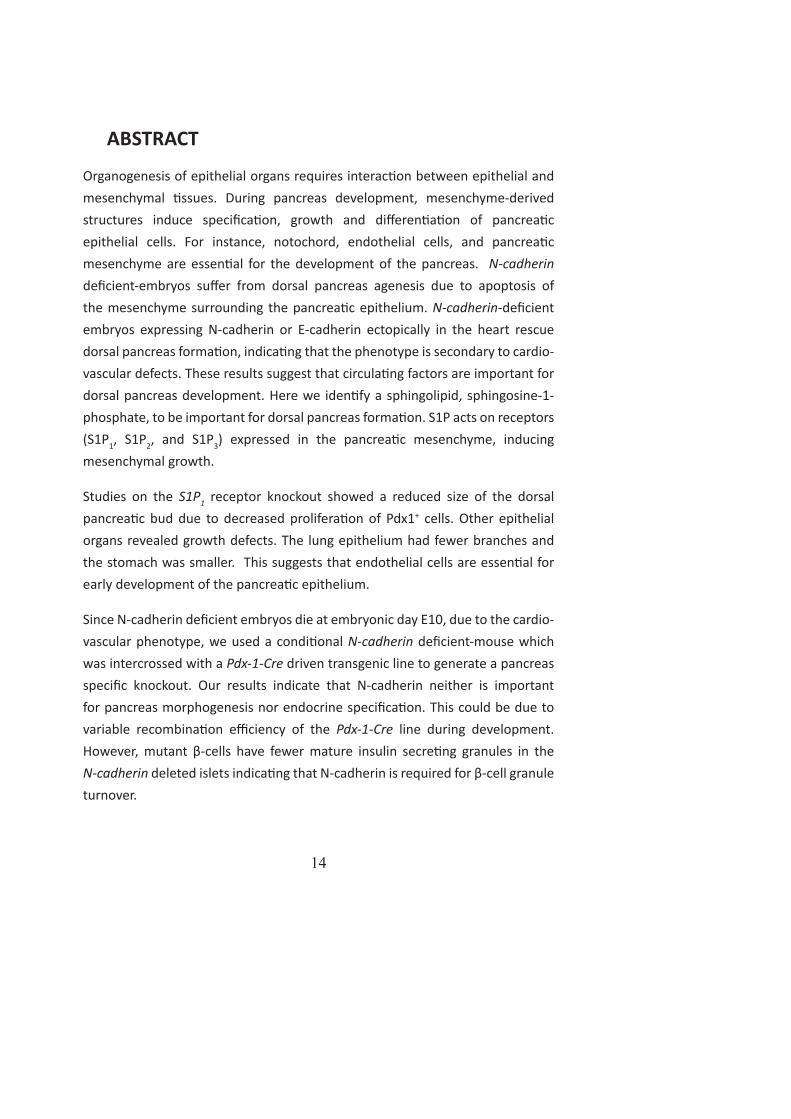
ABSTRACT
Organogenesis of epithelial organs requires interac on between epithelial and mesenchymal ssues. During pancreas development, mesenchyme-derived structures induce specifi ca on, growth and diff eren a on of pancrea c epithelial cells. For instance, notochord, endothelial cells, and pancrea c mesenchyme are essen al for the development of the pancreas. N-cadherin defi cient-embryos suff er from dorsal pancreas agenesis due to apoptosis of the mesenchyme surrounding the pancrea c epithelium. N-cadherin-defi cient embryos expressing N-cadherin or E-cadherin ectopically in the heart rescue dorsal pancreas forma on, indica ng that the phenotype is secondary to cardio-vascular defects. These results suggest that circula ng factors are important for dorsal pancreas development. Here we iden fy a sphingolipid, sphingosine-1-phosphate, to be important for dorsal pancreas forma on. S1P acts on receptors (S1P1, S1P2, and S1P3) expressed in the pancrea c mesenchyme, inducing mesenchymal growth.
Studies on the S1P1 receptor knockout showed a reduced size of the dorsal pancrea c bud due to decreased prolifera on of Pdx1+ cells. Other epithelial organs revealed growth defects. The lung epithelium had fewer branches and the stomach was smaller. This suggests that endothelial cells are essen al for early development of the pancrea c epithelium.
Since N-cadherin defi cient embryos die at embryonic day E10, due to the cardio-vascular phenotype, we used a condi onal N-cadherin defi cient-mouse which was intercrossed with a Pdx-1-Cre driven transgenic line to generate a pancreas specifi c knockout. Our results indicate that N-cadherin neither is important for pancreas morphogenesis nor endocrine specifi ca on. This could be due to variable recombina on effi ciency of the Pdx-1-Cre line during development. However, mutant β-cells have fewer mature insulin secre ng granules in the N-cadherin deleted islets indica ng that N-cadherin is required for β-cell granule turnover.

15
POPULÄRVETENSKAPLIG SAMMANFATTNING
β-celler producerar insulin som frisä s när blodsockret höjs, t.ex. e er en mål d. Pa enter med diabetes saknar de insulinproducerande β-cellerna eller har nedsa känslighet för insulin. För de pa enter som saknar insulin kan det vara svårt a reglera blodsockernivåerna i blodet trots dagliga injek oner med insulin, varför transplanterbara insulinproducerande β-celler skulle kunna vara av stor ny a för dessa pa enter. Hi lls har försök gjorts med a injicera hormonproducerande celler från donatorer ll pa enter. Dessa pa enter har klarat sig utan insulin i några år men har sedan behövt återgå ll insulininjek oner. E annat problem med denna behandling är bristen på donatorer. För a komma runt de a görs försök med a framställa insulinproducerande β-celler från humana embryonala stamceller, dvs de stamceller som ger upphov ll e människo-foster. För a driva de humana embryonala stamcellerna ll a bli insulinproducerande β-celler behövs forskning om hur dessa celler bildas normalt i bukspo körtelns utveckling.
Tidigt i utvecklingen bildar bukspo körteln två utbuktningar, så kallade buddar, från tarmen. Dessa två buddar växer sedan samman och bildar bukspo körteln som består av exokrin och endokrin vävnad samt kanaler. Den exokrina vävnaden bildar de enzymer som används vid matspjälkningen. Dessa enzymer transporteras från de exokrina cellerna via kanalerna ll tarmen. Den endokrina vävnaden bildar hormonproducerande celler. Dessa celler utsöndrar hormoner ll blodet, t.ex. insulin.
I den här avhandlingen har vi fokuserat på blodkärl och proteinet N-cadherins inverkan på bukspo körtelns utveckling. I det första arbetet har vi använt en musmodell där endast en av de två buddarna bildas. Denna musmodell saknar N-cadherin i embryots alla celler. De a leder ll a embryot dör digt under utveckling på grund av avsaknad av blodcirkula on. Forskare har visat a signalsubstanser ifrån blodcirkula onen är vik gt för bukspo körtelns utveckling. Här har vi visat a plasma ifrån blodet är kapabel ll a bilda den budd som saknas i musmodellen. Vi har även iden fi erat en lipid, sphingosine-

16
1-phosphate (S1P), som är nödvändig för bildandet av budden.
I det andra arbetet har vi använt oss av en genmodifi erad mus som saknar receptorn S1P1 och har en drama sk kärldefekt. I denna modell bildas båda buddarna men de är mindre än buddarna hos kontrollmössen, även storleken på mage, lever och lungor är mindre.
Sy et med det tredje arbetet var a studera hur N-cadherin reglerar bukspo körtelns utveckling. För a studera hur N-cadherin reglerar bukspo körtelns utveckling använde vi oss av en musmodell som saknar N-cadherin endast i bukspo körteln. Resultaten från denna studie i embryon visade a N-cadherin inte har en funk on i utvecklingen av bukspo körteln eller i bildandet av de hormonproducerande cellerna. N-cadherin har dock visat sig ha en betydelse för utsöndrandet av insulin i vuxna möss. De a beror på a mindre insulin bildas.
Sammanfa ningsvis visar våra resultat a bukspo körteln behöver signaler ifrån blodet, i de a fall sphingosine-1-phosphate, för a ini era bildandet av bukspo körteln, samt a N-cadherin är vik gt för insulinfrisä ningen.

17
INTRODUCTION
CELL ADHESION AND CADHERINS
CELL ADHESION
Cell adhesion mediates adhesive contacts between cells or helps cells to adhere to the extra cellular matrix (ECM) (Figure 1). Cell adhesion molecules (CAMs) prevent the ssue from dissocia ng. This was fi rst studied in the 1950’s, when Townes and Holfreter dissociated amphibian embryos into single cells and mixed them. Strikingly, upon reaggrega on the cells sorted out to form their par cular germ-layer [1]. From this experiment the researchers formed the idea of cell adhesion molecules expressed on the cell-surface. Today cell adhesion is implicated in signal transduc on, morphogenesis, and in diseases such as cancer.
Figure 1. Adherens junc on. Cadherins form homophilic interac ons with their extracellular part. The intracellular part binds to a catenin complex. The catenin complex binds to EPLIN. EPLIN is required for linkage between cadherins and F-ac n, and for maintaining the circumferen al ac n belt. Abbreva ons: B, β-Catenin; a, α-Catenin; EP, EPLIN. The picture is modifi ed from Nishimura et al 2009.
CELL ADHESION MOLECULES (CAMs)
CAMs are important during both the development of new ssue and the controlled growth and turnover of adult ssue. CAMs are cell surface glycoproteins that display two diff erent kinds of adhesive mechanisms, Ca2+-dependent and non-Ca2+-dependent [2]. Based on their structures, CAMs are classifi ed into

18
four major families; cadherins, selec ns, integrins and the immunoglobin (Ig) superfamily [3]. Cadherins and selec ns belong to the Ca2+-dependent group, while integrins and immunoglobins belong to Ca2+-independent group. Cadherins and immunoglobins promote cell-cell adhesion, whereas integrins mediate cell-matrix interac ons [4, 5]. Selec ns are involved in infl ammatory responses.
THE CADHERIN SUPERFAMILY
The cadherin family of proteins can be divided into several groups. These subgroups include classical cadherins type I and type II, fat-like cadherins, seven-pass transmembrane cadherins, DCad102F-like cadherins, Desmosomal cadherins, and Protocadherins [6]. The classifi ca on of cadherins into diff erent groups is based on the number of the extracellular cadherins repeats (EC). Each repeat consists of specifi c residues that bind calcium. Calcium provides structural rigidity of the extracellular domain, and is essen al for the cadherins’ adhesive func on and protec on against protease diges on (Figure 2) [7].
Classical cadherins are divided into type I and II based on the presence or absence of HAV (his dine, alanine, valine) tri-pep de within the most extracellular N-terminal repeat EC1. The EC1 domain promotes homophilic adhesive interac ons of cadherins on opposing cells. The classical cadherins have fi ve EC domains, a single-pass transmembrane part, and an intracellular part binding to the catenin complex [8].
Figure 2. Structure of classical cadherins. Classical cadherins have an extracellular part containing fi ve extracellular domains, a single transmembrane part, and an intracellular part. The intracellular part binds to the catenin complex and mediates the binding

19
between cadherins and F-ac n. Abbrevia ons: N, N-terminal; EC, extracellular domain; B, β-Catenin; a, α-Catenin; EP, EPLIN. Picture modifi ed from Ivanov et al 2001.
CADHERINS IN CELLULAR JUNCTIONS
Cells are connected with mul ple types of junc ons; ght junc ons, adherens junc ons, gap junc ons, and desmosomes. The cell-cell junc ons in epithelial cells contain ght and adherens junc ons as well as desmosomes. These three junc onal components are clustered together at the apical-lateral cell-cell contacts, forming the apical “junc onal complex” [9]. The adherens junc ons and desmosomes located in the junc onal complex are termed zonal adherens (Figure 1).
The adhesive func on of cadherins requires catenins (-, β-, γ-catenin, and p120) that bind the cadherins to the ac n fi bers. The cytoplasmic part of classical cadherins binds to p120 [10] and β-catenin. P120 regulates cytoskeleton dynamics through direct modula on of the Rho GTPases [11] by inhibi ng RhoA and ac va ng Rac1 and Cdc42 [12-14]. β-catenin binds directly to -catenin, which in turn, binds and bundles ac n fi laments and interact with other ac n partners. Studies performed by Drees and colleagues and Yamada and colleagues showed that the cadherin-β-catenin--catenin complex associa ng with F-ac n can not bind directly to F-ac n. Only free -catenin could link directly to F-ac n. Later it has been shown that a molecule called EPLIN mediate the linkage between the cadherin-β-catenin--catenin complex and the F-ac n [15, 16] (Figure1).
N-CADHERIN
N-cadherin was fi rst iden fi ed as a cell adhesion molecule expressed in neural ssue [17], but was later found to be expressed in various non-neural ssues,
such as the cardiac muscle [18], tes s [19], kidney [20], and liver [21].
During embryogenesis, the expression pa ern of N-cadherin changes as the cells undergo diff erent morphological events such as gastrula on, neurola on, cardiogenesis, and somitogenesis [22, 23]. During gastrula on, when the cells delaminate from the primi ve streak to form mesoderm, they switch from E-cadherin to N-cadherin expression. During neurola on the neural plate switches from E-cadherin to N-cadherin expression. A er the neural tube closure, E-cadherin is lost and N-cadherin proteins are the major cadherin.

20
However, N-cadherin is not expressed in the neural crest cells where the neural tube closes [24]. In heart development, the mesoderm is divided into soma c mesoderm (dorsal) and the splanchnic mesoderm (ventral). N-cadherin expression is downregulated in the soma c mesoderm, but con nues to be expressed in the splanchnic mesoderm [25]. When the somites are forming, N-cadherin is redistributed to the luminal side of the epithelium.
N-cadherin mutants die at E10. They express several developmental abnormali es such as malformed somites and yolk sac, undulated neural tube, dorsal pancrea c bud agenesis and severe cardiovascular defect, which is the cause of death [26, 27]. Due to the fact that N-cadherin is widely expressed in the embryo it is diffi cult to ascertain which phenotypes those are primary or secondary to the muta on. Therefore, the cardiac-mediated adhesion was restored by expressing either N-cadherin or E-cadherin in the heart of mutant embryos. These embryos survived one to two days longer than mutant N-cadherin embryos. N-cadherin and E-cadherin can restore cardiovascular phenotype and N-cadherin mediated cell-cell adhesion is required for cell survival [28].
As shown in rescue experiments, cadherins can compensate for each other. For instance, cadherin-11 is expressed in somites and may subs tute for the loss of N-cadherin. There is also data showing that cadherin-11 can subs tute for N-cadherin in skeletal muscle diff eren a on [26]. In embryonic stem cells E-cadherin and N-cadherin have been shown to compensate for each other [29].
N-cadherin is involved in many processes such as cell sor ng, cell migra on, and cell aggrega on. In endothelial cells, N-cadherin regulates prolifera on and mo lity by controlling VE-cadherin expression at the cell membrane [30]. In the cerebral cortex N-cadherin has been shown to be important for normal architecture of the neuroepithelial or radial glial cells and abla on of N-cadherin randomizes the internal structure of the cortex [31]. In endothelial cells, N-cadherin is required for pericyte recruitment [32].

21
EPITHELIAL ORGAN DEVELOPMENT
Endoderm has two func ons; 1) to induce the forma on of several mesodermal organs (notochord, heart, and blood vessels), and 2) to construct the lining of the diges ve tube and respiratory tube. The diges ve tube forms the pharynx, esophagus, stomach, small intes ne and colon, while the respiratory tube forms the lungs (Figure 3). Caudal to the stomach liver, gallbladder, and pancreas are formed. The diges ve and respiratory tubes are surrounded by mesenchyme that induces diff erent endodermal structures along the anterior-posterior axis. Ini ally the expression of specifi c transcrip on factors is established to defi ne the areas along the endodermal tube. These areas will become specifi c organs (e.g. Pdx1 expression) that defi ne the future pancrea c and duodenal region. The endodermal epithelium then diff eren ates into the organ specifi c cell types. Concomitantly with specifi ca on and diff eren a on the ssues undergo morphogenesis. During this process the cells create cell-cell contacts and polarity, the ssues migrate and branches, and epithelial-mesenchymal interac ons occurs. Coordina on between these mul ple processes results in the development of epithelial organs [33].
Figure 3. Anterior/posterior pa erning of the diges ve tract. Anterior foregut gives rise to esophagus, trachea, lung, and forestomach expressing Sox2. Posterior foregut gives rise to antral stomach, dorsal bud, ventral bud and liver expressing Pdx1. Midgut and hindgut develop small and large intes ne expressing Cdx2 and Cdx1. Abbreva ons: lu, lung; eso, esophagus; tra, trachea; fs, forestomach; as, antral stomach; dp, dorsal pancreas; liv, liver; vp, ventral pancreas; si, small intes ne; li, large intes ne. Picture
modifi ed from Guney et al 2009.

22
MORPHOGENESIS
Morphogenesis establishes appropriate environment for cells to interact and respond to signals. During epithelial organ development several cellular processes such as ini a on and expansion of cell polariza on, coordinated migra on of cells towards par cular chemo a ractants, segrega on of cells into specifi c groups, and cell survival occur. These morphogene c processes can be triggered by signals like fi broblast growth factors (FGFs) secreted from the mesenchyme or by signaling pathways such as notch. For example, FGF has been shown to be important for lung development in mouse, while Notch has been
proven to be required for tooth morphogenesis [34, 35].
CELLULAR POLARIZATION
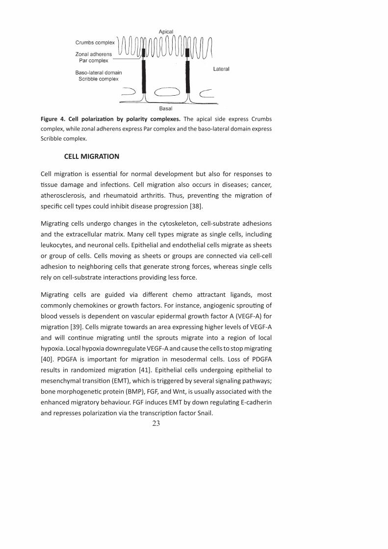
Epithelial cells have three dis nct surfaces, the apical surface facing the lumen, the lateral surface facing neighboring cells and the basal surface facing the ECM. Epithelial cells are polarized and their cell polarity is regulated by intrinsic mechanisms, such as coordinated targe ng of vesicles with apical and basolateral proteins to dis nct surfaces. There are three groups of proteins responsible for forming and maintaining baso-apical polarity: (1) the Par complex (Par proteins and atypical protein kinase C); (2) the Scribble (Scrib) complex (Scrib, Disc large (Dlg), and Lethal giant larvae (Lgl)); (3) the Crumbs (Crb) complex (Crb, PALS1, and PATJ) [36] (Figure 4). Extrinsic factors regulate polarity by forming junc onal complexes to create barriers between apical-basal surfaces and cell contacts to the ECM. This infl uences the vesicular transport as well as transcrip on of genes necessary for establishing polarity.
Adherens junc on proteins like cadherins are shown to play an important role during cell polariza on. Disrup on of adherens junc ons can alter cell polariza on. This was inves gated by McNeill and colleagues by transfec ng non-polar L-cells with E-cadherin. The cells expressing E-cadherin redistributed Na+, K+ ATPase from a diff use pa ern to the regions of cell-cell contacts. This indicates a direct role of cell adhesion molecules as inducers of cell polarity [37].
23
Figure 4. Cell polariza on by polarity complexes. The apical side express Crumbs complex, while zonal adherens express Par complex and the baso-lateral domain express Scribble complex.
CELL MIGRATION
Cell migra on is essen al for normal development but also for responses to ssue damage and infec ons. Cell migra on also occurs in diseases; cancer,
atherosclerosis, and rheumatoid arthri s. Thus, preven ng the migra on of specifi c cell types could inhibit disease progression [38].
Migra ng cells undergo changes in the cytoskeleton, cell-substrate adhesions and the extracellular matrix. Many cell types migrate as single cells, including leukocytes, and neuronal cells. Epithelial and endothelial cells migrate as sheets or group of cells. Cells moving as sheets or groups are connected via cell-cell adhesion to neighboring cells that generate strong forces, whereas single cells rely on cell-substrate interac ons providing less force.
Migra ng cells are guided via diff erent chemo a ractant ligands, most commonly chemokines or growth factors. For instance, angiogenic sprou ng of blood vessels is dependent on vascular epidermal growth factor A (VEGF-A) for migra on [39]. Cells migrate towards an area expressing higher levels of VEGF-A and will con nue migra ng un l the sprouts migrate into a region of local hypoxia. Local hypoxia downregulate VEGF-A and cause the cells to stop migra ng [40]. PDGFA is important for migra on in mesodermal cells. Loss of PDGFA results in randomized migra on [41]. Epithelial cells undergoing epithelial to mesenchymal transi on (EMT), which is triggered by several signaling pathways; bone morphogene c protein (BMP), FGF, and Wnt, is usually associated with the enhanced migratory behaviour. FGF induces EMT by down regula ng E-cadherin and represses polariza on via the transcrip on factor Snail.

24
Cadherins are not only important for EMT, but also to promote cell-cell adhesion. When cells migrate cadherin-mediated cell-cell interac ons have to break and reform. DE-cadherin has been shown to be important in border cell migra on in Drosophila. Cadherin 11 regulates fi llipodia and lamellipodia forma on via small RhoGTPases [42]. Lamellipodia forma on is dependent on fi bronec n-integrin interac ons. The connec on between the ECM and the integrins is also essen al for cell-adhesion, development of directed protrusions, and cell polarity-processes highly relevant to migratory cells.
CELL SORTING
Cell sor ng is a process where diff eren ated cells separate from one another and form clusters that cons tute organized ssues. This process is mediated by classical cadherins. Cadherins are expressed to a variable degree in dis nct ssues during development. For example, E-cadherin is expressed in all epithelial
cells, N-cadherin in neural ssue and muscles, R-cadherin in forebrain and bone, and VE-cadherin in endothelial cells and so on. The specifi c expression pa ern of cadherins combined with homophilic adhesive proper es, may facilitate sor ng of specifi c cell types in ssues. Experiments performed by mixing cadherin-nega ve L-cells transfected with E-cadherin and L-cells transfected with P-cadherin, showed that the cells were able to sort out and cluster [43]. Even mixtures of cells expressing the same cadherin but with diff erent levels of adhesion sorted from one another [44].
CELL SURVIVAL
Cellular interac ons with neighboring cells induce a variety of signaling events, including survival and diff eren a on. Studies in diff erent gene c models where cadherins have been deleted reveal an essen al role for these molecules in cell survival. For instance, studies on R-cadherin, N-cadherin, and E-cadherin show that cadherins are important for cell survival.
Abla on of R-cadherin in the ureteric bud epithelium show an altered morphology and branching behavior, including increased apoptosis [45]. It is not known if the decreased cell survival is directly due to the dele on of R-cadherin or if it is secondary due to the changes in morphology of the epithelium.

25
N-cadherin defi cient embryos exhibit a decrease in cell survival. The defi cient embryos have increased apoptosis within the collapsing neural folds and somites [26]. Since N-cadherin defi cient embryos have a cardio-vascular phenotype, the researchers performed a rescue experiment expressing N-cadherin or E-cadherin in the heart. The embryos survived longer but were s ll smaller and exhibited increased apoptosis, demonstra ng that N-cadherin is directly involved in cell survival [28, 46].
E-cadherin is also known to mediate cell survival. Arulanandam and colleagues have shown that E-cadherin promotes cell survival via Rac1 and Cdc42 by ac va ng the signal transducer and ac vator of transcrip on-3 (Stat3). Stat3 increases both the expression levels and ac vates Rac1 and Cdc42. Morever, inhibitors of Rac1 and Cdc42 block E-cadherin-mediated Stat3 ac va on, indica ng that Rac1 and Cdc42, are the mediators of the cadherin signal to Stat3.
ES-cells lacking E-cadherin undergo a drama c induc on of apoptosis. The survival signals may be mediated by an increase of Rac1 and Cdc42 ac vity, leading to Stat3 s mula on. This could explain why Stat3 inhibi on in cells expressing cell-cell adhesion can induce apoptosis [47].
DIFFERENTIATION
The genera on of cellular diversity is called diff eren a on. Diff eren a on depends on many signaling pathways, e.g. Wnt, Hedgehog, and Notch. These signaling pathways ac vate various transcrip on factors, which in turn ini ate cell diff eren a on, for example, suppression of Wnt signaling in the anterior endoderm is required for both liver and pancreas development [48]. The mesodermal ssues surrounding the posterior foregut provide secreted signals that promote liver and pancreas. In the ventral posterior foregut, BMP secreted by the septum transversum mesenchyme and FGF1 and FGF2 produced by the cardiac mesoderm promote liver development while concomitantly suppressing the pancrea c diff eren a on program [46, 49-54].
Ini ally, transcrip on factors pa ern the endoderm along the anterior to posterior axis. For example, Sox2 is expressed in the anterior domain of the endoderm that will give rise to the esophagus and stomach; Pdx1 expression is found in the antral stomach, presump ve pancreas, common bile duct, and rostral duodenum [55]; Cdx2 is expressed in the en re postgastric epithelium

26
in the regions which will form intes ne [56] (Figure 3). Ini ally, the presump ve pancrea c domain is marked by overlapping expression of Pdx1, P 1a, and Hlxb9/Hb9 [55, 57-59].
PANCREAS DEVELOPMENT
ANATOMY AND FUNCTION
The pancreas is an endoderm derived organ consis ng of two major cell types: exocrine and endocrine cells (Figure 5). The exocrine part of the pancreas consists of acinar cells and ductal cells forming a highly branched duct system. The acinar cells produce diges ve enzymes, such as proteases, lipases, and nucleases. These enzymes are secreted into the ductal system and transported to the intes ne by the ampulla of Vater. The enzymes produced by the exocrine ssue are necessary for diges ng food.
Approximately 98% of the pancreas consists of exocrine ssue; the other 2% are endocrine ssue [54]. The endocrine cells form clusters called islets of Langerhans. The islets are divided into fi ve cell types; -, β-, ε-, -, and pancrea c-polypep de cells. These fi ve cell types produce the hormones; glucagon, insulin, ghrelin, somatosta n, and pancrea c polypep de respec vely. The core of the islets contains mainly β-cells; which are surrounded by -, ε-, -, and PP-cells. Insulin-producing β-cells make up for ~60-80% of the islet, while 15-20% are glucagon-producing -cells; 5-10% are somatosta n-producing -cells, <2% are pancrea c-polypep de-producing cells, and <1% is ghrelin-producing ε-cells [60].
Endocrine cells are important for controlling blood glucose homeostasis. A er food intake, blood sugar levels increases, and insulin is released from β-cells. This is the signal for liver, muscle, and fat to store glucose and thereby control glucose homeostasis. In contrast, glucagon secre on is s mulated at low blood-sugar levels. The release of glucagon primes β-cells to secrete more insulin when the glucose levels rises. Somatosta n and pancrea c polypep de apply inhibitory eff ects on both endocrine and exocrine secre on [60].

27
Figure 5. Anatomy of mouse pancreas. Pancreas develops posterior to the stomach and is connected to the gut via Ampulla of water. The pancreas consists of exocrine and endocrine ssues. The exocrine part is composed of acinar cells and ducts. The endocrine part forms islets of Langerhans.
INITIATION OF THE PANCREATIC PROGRAM
The pancreas develops from the posterior foregut endoderm expressing Pdx1/Ipf1. At E9.5 in the mouse, the dorsal and ventral buds of the pancreas begin to evaginate. All cell lineages in the pancreas originate from Pdx1+ cells [61]. Dele on of Pdx1 results in arrested pancreas development at E10.5. Before the dorsal pancreas starts budding the endoderm is in close proximity to the notochord (Figure 6). Factors from the notochord, such as ac vin-βB and FGF2, prevent the expression of the hedgehog genes. If the hedgehog genes, sonic hedgehog (Shh) and Indian hedgehog (Ihh) are not suppressed, the intes nal diff eren a on program will be ini ated at the expense of dorsal pancreas development [62]. The ventral pancreas is not in contact with the notochord indica ng that another pathway inhibits hedgehog genes in the ventral pancreas [63]. Pa erning of the ventral bud relies on signals from the overlying cardiac mesenchyme and the lateral plate mesoderm.
When the dorsal pancreas starts to protrude, the notochord is no longer in close proximity to the dorsal pancreas. Instead the dorsal aortas fuse and become in direct contact with the dorsal pancrea c endoderm. Signals from the dorsal aorta have been shown to be important for diff eren a on and growth of the

28
pancrea c endoderm [64], partly by inducing expression of P 1a [65]. P 1a is expressed in the pancreas from E9.5 and P 1a+ cells give rise to all endoderm derived cell lineages of the pancreas. P 1a knockouts fail to form a ventral pancreas and forma on of the dorsal pancreas does not proceed beyond the ini al budding.
Figure 6. Early pancreas development. The developing gut endoderm is in contact with the notochord at E8.5. One day later the dorsal aortas have fused and are in close vicinity to the dorsal pancrea c endoderm. At E10 the mesenchyme is surrounding the dorsal pancrea c bud. Abbrevia ons: da, dorsal aorta; m, mesenchyme; dp, dorsal pancreas; vp, ventral pancreas. Picture modifi ed from Slack, J 1995.
PANCREATIC MORPHOGENESIS
At E10 the pancrea c epithelium is surrounded by mesenchymal cells that control its growth and diff eren a on [66, 67]. Several mesenchymal factors are essen al for proper pancreas growth. The transcrip on factor Isl1 is expressed throughout the dorsal mesenchyme during bud forma on. In Isl1 mutants, the mesenchyme is absent, and there is an associated failure of exocrine cell diff eren a on in the dorsal but not ventral pancreas [68]. There is also a complete loss of diff eren ated islet cells. By adding wild type mesenchyme to pancreas Isl1-/- epithelium, the exocrine phenotype could be rescued. This shows that Isl1 in the mesenchyme is important for dorsal exocrine pancreas and that Isl1 in the endoderm is required for the genera on of all endocrine cells.

29
SPECIFICATION AND DIFFERENTIATION OF THE PANCREAS
Genera on of a ductal tree reprints prolifera on of the pancrea c epithelium and mesenchyme, branching morphogenesis, and fusion of the dorsal and ventral buds at E12.5. This branched epithelial organ contains the precursor cells for islets, acini, and ducts [69]. The p cells in the ductal tree are marked by expression of Craboxypep dase A1 (Cpa1), Pdx1, P 1a, and c-Myc [70]. At early stages, these cells contribute to all lineages by ac ng as mul potent progenitors. Later in development cells from the stalk in the ductal tree will give rise to endocrine- or ductal-cells, while the p cells diff eren ate into acinar cells. The peak in exocrine and endocrine diff eren a on, begins at E13.5 in mouse, and is referred to as the secondary transi on.
EXOCRINE LINEAGE
Diff eren a on of the epithelium into exocrine ssue is mesenchyme dependent. Cultures with pancreas epithelium lacking mesenchyme promote endocrine cell diff eren a on resul ng in impaired exocrine diff eren a on [71-73]. Mouse pancrea c epithelium exposed to the ligands (ac vin and transforming growth factor-β1 (TGF-β1) for TGF-β signaling promotes development of endocrine cells, par cular β-cells and PP-cells [74], and disrupts epithelial branching and acinar forma on [75]. Furthermore, an antagonist of TGF-β signaling (follista n) promotes exocrine diff eren a on [73].
DUCT LINEAGE
By E12.5 the specifi ca on of diff erent lineages (exocrine, ductal, and endocrine) is ini ated. The factors regula ng ductal diff eren a on are not well understood [76]. However, several gene dele ons have branching phenotypes. Inac va on of FGF10 or FGFR2IIIb leads to a reduced number of branches and decreased prolifera on [77, 78]. The epidermal growth factor (EGF) family is also important for growth and branching [79]. Abla on of hepatocyte nuclear factor-6 (Hnf-6) demonstrate enlarged lumina and mul ple cysts within the ducts as well as impaired forma on of primary cilia and misloca on of the adherens junc on proteins, including β-catenin [80]. Sox9 mutants also reveal cys c ducts and reduced exocrine diff eren a on [81]. Small Rho GTPases such as Cdc42 have also been demonstrated to be required for pancrea c duct diff eren a on. Abla on of Cdc42 increased acinar diff eren a on at the expense of endocrine and duct lineage commitment [82].
30
ENDOCRINE LINEAGES
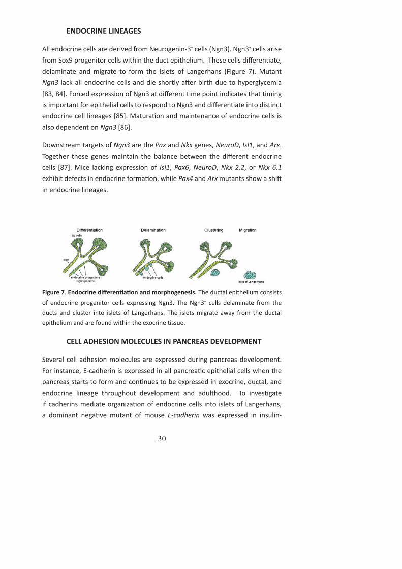
All endocrine cells are derived from Neurogenin-3+ cells (Ngn3). Ngn3+ cells arise from Sox9 progenitor cells within the duct epithelium. These cells diff eren ate, delaminate and migrate to form the islets of Langerhans (Figure 7). Mutant Ngn3 lack all endocrine cells and die shortly a er birth due to hyperglycemia [83, 84]. Forced expression of Ngn3 at diff erent me point indicates that ming is important for epithelial cells to respond to Ngn3 and diff eren ate into dis nct endocrine cell lineages [85]. Matura on and maintenance of endocrine cells is also dependent on Ngn3 [86].
Downstream targets of Ngn3 are the Pax and Nkx genes, NeuroD, Isl1, and Arx. Together these genes maintain the balance between the diff erent endocrine cells [87]. Mice lacking expression of Isl1, Pax6, NeuroD, Nkx 2.2, or Nkx 6.1 exhibit defects in endocrine forma on, while Pax4 and Arx mutants show a shi in endocrine lineages.
Figure 7. Endocrine diff eren a on and morphogenesis. The ductal epithelium consists of endocrine progenitor cells expressing Ngn3. The Ngn3+ cells delaminate from the ducts and cluster into islets of Langerhans. The islets migrate away from the ductal epithelium and are found within the exocrine ssue.
CELL ADHESION MOLECULES IN PANCREAS DEVELOPMENT
Several cell adhesion molecules are expressed during pancreas development. For instance, E-cadherin is expressed in all pancrea c epithelial cells when the pancreas starts to form and con nues to be expressed in exocrine, ductal, and endocrine lineage throughout development and adulthood. To inves gate if cadherins mediate organiza on of endocrine cells into islets of Langerhans, a dominant nega ve mutant of mouse E-cadherin was expressed in insulin-

31
producing cells, resul ng in displacement of both E-cadherin and N-cadherin in the β-cells. As a result, the architecture of the islets was disorganized. β-cells were found as individual cells within the ssue, whereas -cells aggregated into islets [88]. R-cadherin is also expressed in the majority of the cells in the pancrea c buds. In adults, R-cadherin is primarily expressed in the ductal system of the pancreas, on the apical side of the exocrine ssue, in intraductal endocrine cells, but low or no expression in endocrine cells [89, 90]. The R-cadherin mutant mice had no pancrea c phenotype (Dahl and Semb unpublished data). N-cadherin has been shown to be expressed in both dorsal and ventral bud at embryonic day E9.5 and being specifi cally expressed in the islets of adult pancreas [27, 91]. Abla on of N-cadherin resulted in a lack of the dorsal bud [27].
Another cell-cell adhesion molecule expressed in the pancreas is N-CAM. It is expressed both in pancrea c mesenchyme and epithelium. The expression becomes restricted to endocrine cells, and peripheral nerve endings and ganglia. N-CAM mutants develop a normal pancreas, consis ng of all pancrea c cell types and islets of normal size and number, sca ered within the exocrine ssue. However, lack of NCAM results in disrupted organiza on and morphology of the islets. -cells were distributed centrally in the islets and not in the periphery. This suggests that N-CAM regulates cell type segrega on within islets. Islet cell polarity was also aff ected in N-CAM mutant mice. E-cadherin and N-cadherin were distributed subcellularly, whereas R-cadherin distribu on was unaff ected [92].
BLOOD VESSELS IN PANCREAS DEVELOPMENT
During ini a on of pancreas organogenesis the buds are in close vicinity to large vessels, the aorta dorsally and the vitelline veins ventrally. Several experiments have shown that endothelial signaling is important for pancreas development. Cleaver and colleagues removed the endothelial cell precursors of the dorsal aorta from frog embryos. Thus endocrine gene expression was inhibited, while the development of liver and neural tube proceeded normally [64, 93]. The result suggests that endothelial cells are required for endocrine development.
Recombina on of prepa erned dorsal endoderm with dorsal aorta, notochord, and neural tube indicated that dorsal endoderm together with dorsal aorta can only ini ate Pdx1 expression and insulin expression. Dorsal endoderm with
32
notochord and neural tube did not induce insulin. However, the notochord did induce Pdx1. This experiment showed that endothelial cells supply signals suffi cient for insulin expression [64].
The third experiment showing that endothelial cells are important for insulin expression, was to analyze the role of VEGF-A. Overexpression of VEGF-A in the Pdx1-expressing domain resulted in increased vasculariza on. These endothelial cells instructed foregut cells to diff eren ate into pancrea c cells.
The results presented above all indicate that endothelial cells promote pancreas development. In vitro experiment on Flk1-/- embryos (VEGFR2) lacking endothelial cells induced Pdx1, indica ng that aorta or endothelial cells are not required for ini al pancreas development. The expression of P 1a was lost in the in the Flk1-
/- embryos, indica ng that endothelial cells are important for the appearance of the dorsal bud and to maintain Pdx1 expression [65].
SPHINGOSINE-1-PHOSPHATE
Sphingosine-1-phosphate (S1P) is a metabolite formed by the phosphoryla on of sphingosine by sphingosine kinase [94, 95]. S1P can be recycled back to sphingosine by S1P phosphatase or be degraded by an S1P lyase [96, 97, 98]. S1P is produced and secreted by red blood cells, platelets, monocytes, mast cells, and possibly by endothelial cells [99-101]. Since S1P is highly soluble in water it is bound to high density lipoprotein (HDL) or to albumin in plasma [102].
S1P triggers several ac vi es in cells including prolifera on, migra on, cytoskeletal changes, adhesion molecule expression, and an -apopto c eff ects [103-107]. Many of these ac vi es are produced via the interac on of S1P with G protein-coupled receptors [108].
SPHINGOSINE-1-PHOSPHATE RECEPTOR-1
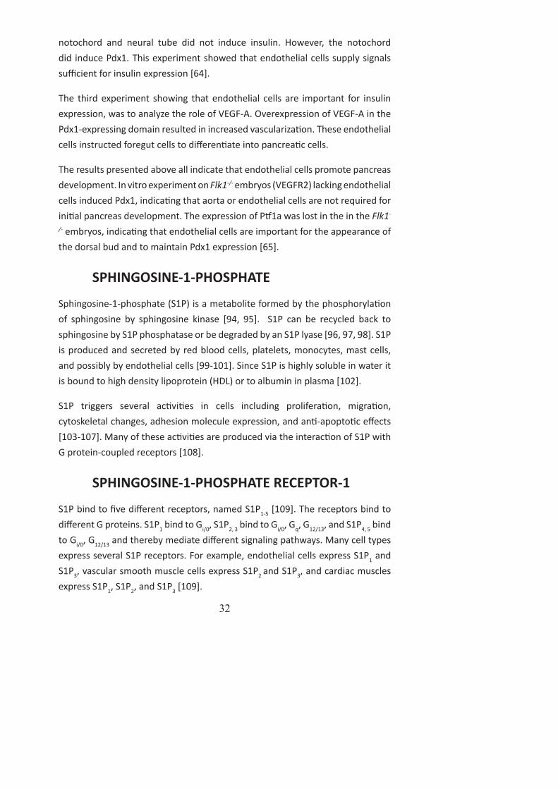
S1P bind to fi ve diff erent receptors, named S1P1-5 [109]. The receptors bind to diff erent G proteins. S1P1 bind to Gi/0, S1P2, 3 bind to Gi/0, Gq, G12/13, and S1P4, 5 bind to Gi/0, G12/13 and thereby mediate diff erent signaling pathways. Many cell types express several S1P receptors. For example, endothelial cells express S1P1 and S1P3, vascular smooth muscle cells express S1P2 and S1P3, and cardiac muscles express S1P1, S1P2, and S1P3 [109].

33
S1P1 is highly expressed in the cardiovascular system during mouse development [110] and in the adult mouse; in humans S1P1 is expressed at signifi cant levels in brain, heart, lung, spleen, liver and low expression was detected in kidney, muscle and thymus [111-113]. Mice lacking S1P1 die at E12.5-14.5 due to vascular matura on defects. These embryos develop a normal blood vessel network at E12.5, indica ng that S1P1 was not essen al for endothelial cell diff eren a on, prolifera on, migra on and tube forma on during vasculogenesis or for sprou ng and branching of vessels during angiogenesis [110]. Instead, the associa on of vascular smooth muscle cells and pericytes was defected. The dorsal aorta was not covered by vascular smooth muscle cells. Cells were lining the ventral part of the aorta and not the dorsal. The defects in vascular smooth muscle cell coverage extended to arteries and capillaries in the brain and also to small vessels in the limbs. The lack of vascular smooth muscle cells resulted in a weakened vasculature with disrupted and leaky vessels. The defect seen in the S1P1 mutant might be caused by impaired vascular smooth muscle cell and pericyte diff eren a on, prolifera on and/or migra on. For example, fi broblast cells from the S1P1 mutant failed to migrate towards S1P in vitro [114].
INSULIN CELL MATURATION AND GLUCOSE HOMEOSTASIS
A er diges on, a variety of nutri onal factors in circula on, including amino acids, fa y acids and glucose ini ate release of insulin. A serial of signaling cascades responsible for the suppression of hepa c glucose output, increased synthesis of glycogen and triglycerides, and s mula on of peripheral ssue uptake of glucose begin. In addi on, hormones from the gut (incre ns) are secreted, which increases insulin secre on from the pancreas in a glucose-dependent manner [115].
Insulin is synthesized as preproinsulin on the rough endoplasmic re culum (RER). It is converted into proinsulin and transferred to the Trans-Golgi Network (TGN), where it is packed into immature secretory granules. These vesicles fuse directly to the plasma membrane or enter the recycling endosome system. The immature granules undergo several matura on processes, such as acidifi ca on, conversion of proinsulin into insulin and C-pep de, and loss of the coat protein clathrin [116, 117].

34
There are two pools of insulin secretory granules, the readily releasable pool (RRP) that is responsible for the ini al phase of insulin secre on and a second reserve pool that is responsible for a more prolonged insulin secre on phase [118-121]. The RRP is pre-docked to the membrane and is released within 1-5 min. During this fi rst phase approximately 15 granules per minute are released. The second phase con nues for 5-60 min and releases 5 granules per minute [119]. A mouse β-cell contains more than 10 000 secretory granules [122]. Only 1-5% of the secretory granules belong to the RRP.
High glucose in the blood is sensed by glucose transporter 2 (GLUT2) in the plasma membrane. Via glycolysis the ra o of ATP/ADP increases and closes the ATP-sensi ve K+ channels. This leads to a depolariza on that ac vates Ca2+ channels resul ng in extracellular Ca2+ infl ux and fusion of insulin granules with the plasma membrane. At the same me GLP-1 binds to its receptor and ac vates cAMP that binds to EPAC. EPAC increases the numbers of insulin granules in the RRP at the plasma membrane (Figure 8). Together these processes results in insulin secre on [119].
Figure 8. Insulin signaling. Glucose increase ATP/ADP ra o leading to closing of K+ channels. Depolariza on of the membrane, increased concentra on of Ca2+ within the cell, and fusion of insulin granules with the plasma membrane increase cAMP. cAMP binds to EPAC to increase Ca2+ and thereby increasing the insulin granules within the readily releasable pool. Light blue dots represent insulin granules. Picture modifi ed from Hou et al 2009.

35
DIABETES
The most common forms of diabetes are type 1 and 2, however there are several diff erent types of diabetes. Type 1 diabetes is an autoimmune disease resul ng in destruc on of β-cells by the immune system. Type 2 diabetes is caused by insulin resistance. The pancreas in type 2 diabe c pa ent has an increased β-cell mass to compensate for the elevated need for insulin. Eventually this compensatory mechanism fails and the pa ent develops diabetes.
Diabetes is a disease that is associated with major complica ons, including re nopathy, neuropathy, kidney failure and cardiovascular defects. The diabe c re nopathy causes blindness, while diabe c neuropathy causes symptoms of ngling, pain, numbness, or weakness in the feet and hands. Kidney failure
as well as cardiovascular defects is also common causes of death in diabetes pa ents (World Health Organiza on; www.who.int).
The most common treatment for pa ents with type 1 diabetes is daily injec ons of recombinant insulin. This is a fairly effi cient treatment but excursions in blood glucose levels may s ll be diffi cult to control. Thus alterna ve treatment op ons are currently explored. Type 1 diabe c pa ents have been injected with donor islets. This procedure was able to restore euglycemia [123]. However, two major obstacles are associated with this procedure: only 10% of the pa ents were insulin free a er 5 years of transplanta on, and secondly there are not enough donors.
Thus alterna ve sources for β-cells are needed. One possibility would be to diff eren ate human Embryonic Stem Cells (hESC) into transplantable β-cells. hESC have been proven to be able to diff eren ate into insulin producing β-cells [124] and in vitro studies have also succeeded in diff eren a ng hESC into insulin producing β-cells [125-127]. Effi cient and reproducible diff eren a on protocols will be needed to develop fully func onal, transplantable β-cells.

36
TECHNICAL CONSIDERATIONS
THE GENE-TRAP SYSTEM
Gene-trap mutagenesis is a technique that randomly generates loss-of-func on muta ons and reports the expression of many mouse genes. A Gene-trap vector contains a splice acceptor site immediately upstream of a promoterless reporter. Since the Gene-trap has no promoter of its own, it is driven by an endogenous promoter close to the inser on site. The Gene-trap also contains a β-galactosidase-NeoRfusion (β-Geo) casse e in order to work as a reporter. A disadvantage using a Gene-trap vector is that, because the inser on occurs in an intron, alterna ve splicing can some mes take place. This might lead to lower levels of the wild-type transcripts, resul ng in hypomorphic alleles [128].
THE Cre/loxP-SYSTEM
Tradi onal knockout mice may be hard to inves gate since many mutants are lethal during embryogenesis. However, even if the mutants do survive un l adulthood it might be hard to interpret the data. Since an observed phenotype might be due to a primary defect in another ssue. A solu on to this problem is to use the Cre-loxP recombina on system. The Cre-loxP system allows dele on of a gene and its expression in a par cular ssue or cell type. Cre is a recombinase that recognizes 34 base-pair loxP-sequences and catalyses the recombina on between two loxP sites by excising the intervening DNA [129]. A sequence that is fl anked with two loxP sites is referred to as a fl oxed sequence. The fl oxed sequence will get excised from the genome in the presence of a Cre-recombinase.
RECOMBINATION VARIABILITY’S IN Cre TRANSGENES
A known problem with Cre transgenic mouse strains is the variability of recombina on. This could be due to several factors, including integra on site. Integra on of a transgene into a site with an open chroma n structure is expected to increase availability for recombina on events. Increased variability of transgenic expression is observed with increased number of backcrosses [130]. Diff erent backgrounds of the responder mice may also aff ect variability in recombina on effi ciency.

37
AIM OF THE THESIS
The overall objec ve of this thesis was to understand the roles of S1P/S1P1 and N-cadherin in pancreas development
The specifi c aims were:
Paper I. To address whether the pancrea c phenotype in N-cadherin defi cient mice is due to a cell-autonomous func on of N-cadherin within the mesenchyme or if it is secondary to other defects, e.g. cardiac and/or vascular func on.
Paper II. To study the func onal role of S1P signaling from endothelial cells in pancreas development using S1P1 defi cient embryos.
Paper III. To inves gate the consequences of N-cadherin abla on in the developing pancreas, specifi cally in morphogenesis and endocrine cell diff eren a on.
38
PAPERS IN SUMMARY
PAPER I
INTRODUCTION
N-cadherin is required for the forma on of the dorsal pancreas, by media ng dorsal pancrea c mesenchymal cell survival [27]. N-cadherin defi cient mice die of cell adhesion defects in the heart at E9.5. These mice also exhibit defects in the development of the nervous system, somites and yolk sac. Importantly, when cadherin func on was restored in the heart of N-cadherin mutant mice, the embryos survived un l E10.5-11 due to rescued heart and vascular func on. The aim of this inves ga on was to address whether the pancrea c phenotype in N-cadherin-defi cient mice refl ects a cell-autonomous func on of N-cadherin within the pancrea c mesenchyme or if it is secondary to other defects, e.g. cardiac and/or vascular func on.
RESULTS
Cardiac-rescued N-cadherin-defi cient mice develop a dorsal pancreas due to an intact circulatory system
Cardiac rescued N-cadherin-knockout mice were analyzed to assess if the lack of the dorsal pancreas in N-cadherin-defi cient mice is due to a cell-autonomous func on within the mesenchyme or if it secondarily caused by other defects (e.g. cardiac and/or vascular func on). The most drama c consequence of expressing N-cadherin within the heart in the N-cadherin-mutant mice was the restora on of an intact circulatory system. As a consequence of a func oning circulatory system the dorsal pancreas was formed. The lack of mesenchyme recruitment in the N-cadherin-defi cient mice was not observed in the rescued N-cadherin-knockout mice. This indicates that N-cadherin does not play a cell-autonomous role in mesenchymal cell survival. To iden fy how the cardiac/vascular func on aff ects pancreas development, explants from N-cadherin-defi cient embryos were incubated with beads soaked in plasma. The dorsal pancreas was rescued. Endoderm with and without mesenchyme was also cultured with plasma to address whether plasma acts on mesenchyme or epithelium. Our results show that plasma acts primarily on mesenchyme.

39
S1P rescues the forma on of the dorsal pancreas in N-cadherin-defi cient pancrea c explants.
The dorsal aorta is in close vicinity to the pancrea c endoderm and the vascular smooth muscle cells of the dorsal aorta are derived from mesenchymal cells. It has been demonstrated that vascular matura on is defi cient in mice lacking the S1P receptor S1P1 due to defi cient vascular smooth muscle cell recruitment. S1P is a lipid present in the circula on. To test whether the plasma-induced rescue of the dorsal pancreas could be mediated by S1P, we incubated explants with beads soaked in S1P. The explants developed a dorsal pancreas and S1P’s eff ect was specifi cally seen on the mesenchyme.
S1P receptors in pancreas
S1P binds to receptors S1P1-5 with high affi nity. RT-PCR and in situ hybridiza on of these receptors show that S1P1 is expressed in endothelial cells and S1P2-3 are preferen ally expressed in the mesenchyme, but that S1P4-5 cannot be detected. S1P receptors are G-protein coupled and to block their ac vity we cultured explants with pertussis toxin that inac vates Gi. Pertussis toxin blocked the S1P-mediated rescue of the dorsal pancreas as well as early pancreas development in wt explants.
S1P regulates dorsal pancrea c mesenchymal cell prolifera on
The mechanism by which S1P rescued the dorsal pancreas development may involve a direct eff ect on pancrea c mesenchyme. Mesenchymal development involves both cell migra on and prolifera on, and to inves gate if S1P act on any of these processes, we cultured primary mesenchymal cells. BrdU-labeling experiments demonstrated that S1P increase the number of prolifera ng cells. Importantly, pertussis toxin inhibits this eff ect.
SUMMARY
We showed that restoring cardiac and circulatory func on in N-cadherin-defi cient mice by cardiac-specifi c expression of N-cadherin, rescues forma on of the dorsal pancreas. This indicates that the phenotype observed is secondary to defects related to cardiac/vascular func on. Based on this observa on, we demonstrated that plasma and S1P-mediated G-protein-coupled signaling rescues forma on of the dorsal pancreas in vitro.

40
PAPER II
INTRODUCTION
During early pancrea c development major blood vessels are in close contact with the pancrea c endoderm. The dorsal bud forms close to the dorsal aorta, while the ventral buds emerge close to the vitelline veins. Endothelial signals have been shown to be important for ini a on of the dorsal bud. However, endothelial signals are not required for ini a on of the ventral bud.
Later in development the blood vessels and the pancrea c endoderm get separated by invading mesenchyme. Blood vessels are embedded within the mesenchyme and endoderm-endothelial interac ons con nue. Previously, we have shown that a func onal vascular system is required for ini a ng dorsal pancreas forma on. Signaling cues from the circula on, in par cular the bioac ve sphingolipid metabolite S1P is essen al. S1P1 defi cient embryos were analyzed to inves gate the role of S1P signaling in endothelial cells during pancreas development.
RESULTS
Growth and branching morphogenesis of the pancrea c epithelium is compromised in S1P1-defi cient embryos
To measure the eff ects of specifi c abla on of S1P1 in the pancreas, the pancrea c volume was scanned with Op cal Projec on Tomography (OPT). Analysis showed that the pancrea c buds from S1P1-defi cient embryos did not protrude as far as pancrea c buds from wild type embryos. The dorsal bud was signifi cantly smaller in the S1P1-defi cient embryos. The ventral bud was also smaller but not signifi cantly smaller. There were no diff erences in the structure of the tubular network in the distal parts of the dorsal and ventral buds. However, the proximal parts were less branched in the S1P1-defi cient embryos.
Other endoderm-derived organs, such as liver, ventricle, and lung, revealed a more general eff ect of S1P dele on on endodermal development, for example the lung had fewer branches, and liver and ventricle were smaller.

41
S1P1 is required for prolifera on of Pdx1+ pancrea c progenitors
Possible explana ons for smaller dorsal and ventral buds in the S1P1-defi cient embryos are less prolifera ng cells and/or more apoptosis. Wild type and mutant embryos were BrdU-pulse labelled and stained for Pdx1 and BrdU. There were signifi cantly fewer prolifera ng Pdx1+ cells in the S1P1-defi cient embryos compared to li ermate controls. To inves gate if there were more apopto c cells, we stained for Caspase 3. No signifi cant change was observed. We also looked at endocrine diff eren a on but there were no signifi cant change between S1P1-defi cient and control embryos. Thus, S1P1 is required for prolifera on of mul potent Pdx1+ pancrea c progenitor cells, but not for endocrine cell specifi ca on.
S1P1 defi ciency results in defec ve pancrea c morphogenesis in vitro
To exclude that the phenotype observed is caused by the cardiovascular defect, in vitro explants studies were performed. All control explants developed normally, while the S1P1-defi cient explants showed a rather diverse phenotype. The explants development spanned from severe growth defects to showing only size reduc on and less branching.
Blood vessel abla on does not mimic the S1P1 phenotype
Our results indicate that endothelial cells, via S1P1, provide induc ve cues that are necessary for pancrea c endoderm development. To test this, we ablated all blood vessels using quinolin-urea. A er two days of incuba on, star ng at E11.5, there were no aff ect on the pancrea c endoderm, sugges ng that expansion of progenitor cells beyond E11.5 does not require endothelial cells.
SUMMARY
Developing organism are all dependent on blood vessels providing oxygen and nutrients as well as induc ve cues that control cell prolifera on and cell specifi ca on. It is known that endothelial cells control dorsal pancreas outgrowth and early endocrine cell specifi ca on. However, it is not understood how endothelial cells control these processes. Here, we iden fy S1P1 as a new signaling pathway that is necessary for development of foregut derived organs, such as lung, liver, stomach and pancreas. Abla on of S1P1 results in reduced size of the dorsal pancrea c bud due to decreased prolifera on of Pdx1+ cells.

42
PAPER III
INTRODUCTION
Previously we demonstrated that N-cadherin is expressed in the pancrea c epithelium at E9.5, but later becomes restricted to endocrine aggregates in mice. Furthermore, in the absence of N-cadherin the dorsal pancrea c bud fails to form. This is a secondary phenotype due to cardiac failure. The early lethality of N-cadherin-defi cient embryos excludes a complete analysis of N-cadherin func on in pancreas development. We generated a ssue specifi c knockout of N-cadherin in the early pancrea c epithelium to study the role of N-cadherin during pancreas forma on and func on.
RESULTS
N-cadherin expression during pancreas development
At E10.5-13.5, N-cadherin is expressed throughout the Pdx1+ pancrea c epithelium. Later, at E14.5 N-cadherin is expressed at low levels in Sox9+ cells, acinar cells and at high levels in mature endocrine cells. At E15.5, N-cadherin expression becomes restricted to a subpopula on of Sox9+ cells, and never in Ngn3+ cells. In addi on Isl1+ endocrine progenitors (hormone nega ve) exhibit a mosaic expression of N-cadherin and all hormone producing cells express N-cadherin. At this point acinar cells do not express N-cadherin. Throughout development N-cadherin is also expressed in neurons and blood vessels. From E18.5 and onwards N-cadherin is restricted to hormone producing cells, neurons and blood vessels.
Pdx1Cre-mediated abla on of N-cadherin
To study the func on of N-cadherin during pancreas development N-cadherin was condi onally deleted in Pdx1+ cells (cKO). To ensure that N-cadherin was ablated, N-cadherin expression was examined by immunofl uorescence, and immunoblo ng. At E13.5, the effi cacy of N-cadherin abla on varied from <5% to almost complete abla on of N-cadherin. At E15.5, the expression varied between li ermates, but N-cadherin expression was consistently maintained in more than 50% of the cells. From E18.5 and onwards, N-cadherin was no longer detectable in the pancreas of cKO individuals.

43
Pancrea c morphogenesis and endocrine specifi ca on is not aff ected in condi onal N-cadherin-knockout mice.
To determine if N-cadherin is important for cell lineage specifi ca on, expression of specifi c markers for acinar, ductal, and endocrine cells was analyzed. No developmental defects were observed within the exocrine and endocrine compartment. To inves gate if N-cadherin is important for ini a on and/or maintenance of islet cell polarity, the distribu on of characteris c epithelial junc onal, apical and lateral markers was analyzed. However, the normal alloca on of these cell polarity markers indicates that islet cell contacts and polarity is not altered. To understand if microtubule dynamics are altered in islets, α- and β-tubulin were analyzed. There was no diff erence observed between control and cKO. To inves gate the role of N-cadherin in endocrine cell specifi ca on and islets forma on we measured insulin area versus E-cadherin area. This experiment did not reveal any diff erence between control and cKO, sugges ng that N-cadherin is not required for β-cell specifi ca on. To study if other hormone-producing cells were aff ected, the ra o of glucagon+, PP+, Somatosta n+ cells versus insulin+ cells, respec vely, were es mated in adult mice. The ra o was not altered, sugges ng that N-cadherin appears to be dispensable for endocrine development.
N-cadherin controls insulin granule turnover
Transmission electron microscopy studies of islets showed a signifi cant overall reduc on (27%) of insulin secre ng granules in mutant islets. 16% of the β-cells contained very few insulin granules which are a 67% reduc on. Even if the β-cells with very few insulin granules were not included in the analysis, the diff erence was sta s cal signifi cant. In contrast there is no diff erence in immature granules in the cKO, sugges ng that the decrease in mature granules is not due to a change in biogenesis of granules.
N-cadherin regulates insulin secre on
Insulin secre on was studied in response to low and high concentra on of glucose. In response to low glucose insulin secre on was signifi cantly reduced in cKO. At high glucose, insulin secre on was also reduced but the change was not signifi cant.

44
SUMMARY
By abla ng N-cadherin specifi cally during pancreas development we show that N-cadherin is not essen al for pancreas organogenesis. However, the late onset and high variability of recombina on, suggest that a poten al requirement of N-cadherin during pancreas development prior to E15.5 cannot be ruled out. N-cadherin-defi cient endocrine cells aggregate into islets with normal morphology, indica ng that N-cadherin is not required for islets morphogenesis. However, ultrastructural analysis of adult islets revealed a reduc on in number of mature insulin secretory granules in N-cadherin-defi cient β-cells. In conclusion our fi ndings suggest that N-cadherin is dispensable to pancreas morphogenesis and cell fate specifi ca on, but is required for insulin secretory granules turnover and insulin secre on.
CONCLUDING REMARKS
PAPER I
N-cadherin does not act cell-autonomously on the mesenchyme surrounding the dorsal pancreas. When expressing cadherins in the heart the mesenchyme was recruited and the dorsal pancreas started to form. This data show that the agenesis seen in N-cadherin defi cient mice is secondarily due to cardiac-vascular defects. The rescue experiment led us to believe that circula on is important for dorsal pancreas development. Therefore we soaked beads in plasma from E15.5 embryos and cultured explants from N-cadherin-defi cient mice for two days. The addi on of plasma was able to rescue the dorsal pancreas. S1P, a lipid produced and secreted by red blood cells, platelets, monocytes, mast cells, and possibly by endothelial cells, was also able to rescue dorsal pancreas forma on. S1P secreted from the dorsal aorta binds to its receptors in the mesenchyme (S1P1-3) and induce mesenchymal prolifera on, which induce dorsal pancreas forma on.
PAPER II
S1P1 knockout have defects in endodermal organ forma on, such as pancreas, stomach, liver and lung. In the pancreas this is due to less prolifera on of the endoderm and not by increased cell death. To evaluate if the phenotype is due to cardiovascular defects, blood vessels were ablated from E11.5 wild type explants. The S1P1 phenotype was not observed, sugges ng that S1P1 only

45
has an eff ect early in development and not during secondary transi on. In conclusion we iden fi ed a pathway, by which endothelial cells, via S1P1, control early pancreas development.
PAPER III
Abla on of N-cadherin specifi cally during pancreas development demonstrates that N-cadherin is not essen al for pancreas organogenesis. However, due to the late onset and high variability of recombina on, a role of N-cadherin during early pancrea c development can not be ruled out.
Concomitant with islet forma on the effi ciency of recombina on was high. Normal architecture was seen in the N-cadherin defi cient islets indica ng that N-cadherin is not required for islet morphogenesis. However, ultrastructural analysis of adult islets revealed a reduc on in the number of mature insulin secretory granules in N-cadherin-defi cient β -cells. Since the number of immature insulin secretory granules was unaff ected we conclude that N-cadherin is not required for granule biogenesis. Consistent with the low number of mature insulin secretory granules, N-cadherin-defi cient islets displayed less insulin secre on when s mulated with low glucose. We speculate that this is due to fewer granules docked to the membrane and/or fewer granules in the ready releasable pool. Our fi ndings suggest that N-cadherin is dispensable for pancreas morphogenesis and cell fate specifi ca on, but that N-cadherin is required for insulin secretory granules turnover and insulin secre on.
FUTURE PERSPECTIVES
PAPER I AND II
Understanding how S1P regulates growth in endoderm derived organs would be of great importance. Most likely the mechanism involves endothelial cells that secrete molecule/molecules that is/are important for endodermal growth.
It could also be of interest to iden fy other compounds from plasma that can induce dorsal pancreas development. Preliminary results show that lysophospha dic acid (LPA) is one poten al compound.

46
PAPER III
To further analyze if N-cadherin plays a role in pancreas development, it is of importance to use a Pdx1-Cre transgenic mouse line with high recombina on effi ciency or other Cre-lines with an early onset of expression in the pancreas, such as P 1a. Thereby, the role of N-cadherin in morphogenesis can be studied in greater detail.
To analyze how N-cadherin is required for insulin granule turnover, diff erent insulin secre ng pathways need to be studied. It would be important to clarify if fewer granules are docked to the membrane or if there are fewer granules in the ready releasable pool. TEM studies could be used to address this issue. It could poten ally be of interest to see if any of the components in the insulin secre ng pathway is defected in the cKO. Insulin secre on studies on islet s mulated with diff erent components involved in insulin signaling pathway would answer this. To analyze if there is less insulin content, perfusion studies on islets will be performed. By adding high percentage of triton, the membrane breaks and all insulin granules are secreted. Preliminary data show that the cKO has lower insulin content.
47
ACKNOWLEDGEMENT
Vem hade kunnat tro a det skulle ta så här lång d a doktorera, inte jag iallafall. Även om den här resan har varit väldigt lång har jag få uppleva många fantas ska saker och lärt mig mycket på vägen. Jag vill härmed passa på a tacka de människor som på e eller annat sä bidragit ll min avhandling:
Min handledare Henrik Semb. För a du har givit mig friheten a driva mina projekt självständigt, på go och ont. De a har gjort a jag utvecklas både som forskare och människa. Jag vill även tacka för a du har skapat en bra miljö a forska i, samt a du har det goda omdömet a rekrytera intressanta medarbetare som var och en på si sä förgyller vardagen.
I would also like to express my gra tude to Glenn Radice for all help with the N-cadherin projects. It has been much appreciated.
Jag skulle också vilja tacka alla mina nuvarande kollegor. Gokul for being a great companion. I have really enjoyed working with you. Fredrik för a du är en god människa med e stort hjärta. Tänk om du hade lagt all din energi på forskning istället för skitsnack, då hade du varit professor vid det här laget. Jackie för din omtanke och för din vilja a all d hjälpa ll om du kan. Hävdar for arande a det vore bä re om du tänkte innan du pratar. Marie för a du är så lä a ha a göra med och för a du all d ställer upp när jag behöver hjälp. An och Karro för a ni underlä ar livet på labbet. Utan er hade det inte gå . Ingar för a du sköter om beställningar och gör det bästa fi kat. Zarah för du är en god medarbetare med den rä a takten. Elvira för a du ger labbet en helt ny dimension. Siqin för många intressanta diskussioner som o a får mig a skra a. Pia för dina nya ini a v på labbet och för a du verkar vara genuint intresserad av forskning. Yvonne för a du är down to earth. Uppska as i denna dramadro ningarnas högborg. Novo-tjejerna (Jenny, Nina och Katja) för a ni kryddar ll livet på labbet lite extra. Till de nya rekryterna Hanna, Maria och Karen vill jag bara säga: Livet på labbet kan liknas vid e dagis för vuxna. Roligt, frustrerande, men sam digt utvecklande.
Jag vill även tacka våra inneboende i labbet. Isabella för all hjälp med allt. Jag uppska ar det verkligen. Magda för a du precis som jag tycker om ordning och reda samt e välstädat labb. Jesper för du är easy going.
48
Har man varit i e labb så länge som jag har, har man också många före de a kollegor. Jag vill tacka alla som passerat dörrarna på BMC i Lund. Thomas det har varit fantas skt roligt a ha ha dig som kollega under nästan hela resan. Du stod vid di ord länge men sköt ut dig i sista stund. Vet få människor som kan vara så entusias ska som du. Karin min gamla student och sedermera arbetskamrat för di alldeles egna sä a se på saker och ng. Mar na för a du är den bästa rumskamrat man kan ha. Camilla för all hjälp genom åren med mössen. Anders för a du introducerade QPCRen i våra liv samt försökte få oss a tänka outside the box. Sune för den idéspruta du är. Maria H för a du höll koll på allt på jobbet. Vet få människor som besi er den förmågan. Ingrid för a du all d bidrog med god stämning, inte minst på Ålagillet.
Jag vill även tacka mina gamla kollegor från den på MedKem i Göteborg. Fina för e fantas skt samarbete. Din sprudlande energi smi ar av sig och gör a allt blir mycket roligare. Gabriella för din omtanke och fantas ska diskussioner om allt. Vet ingen som kan älta e problem så som du. Jocke för lång och trogen tjänst. Di engagemang i saker du tror på är enormt. Xiaojie for being the strongest woman on earth. I admire you! Gunilla för a du är den bästa BMA man kan önska sig. Hade gärna velat ha med dig ner ll Lund. Nico för a du är en oerhört fascinerande person. Mikael du bidrog kanske inte så mycket ll forskningen men desto mer ll en helt fantas skt galen atmosfär a jobba i. Anna för a du är den mest ambi ösa person jag någonsin träff at.
Jag vill också passa på a tacka alla andra i min omgivning för en trevlig arbetsmiljö. Grupperna som var på MedKem när det begav sig. Medlemmarna av CBz’s grupp, Per Lindahls grupp, Sven Enerbäcks grupp, Pekna och Pekny-gruppen. Ni gjorde MedKem ll den bästa av arbetsplatser. I Lund vill jag passa på a tacka alla på BMC B10, och övriga grupper på BMC såsom Bo Ahrens grupp för all hjälp under åren, Nils Wierups grupp för hjälp med insulinsekre onsstudier samt Udo Häckers grupp, Stefan Baumgartner och Edgar Peras grupp för gemensamma journal clubs.
Till alla mina vänner (ingen nämnd, ingen glömd) vill jag bara säga a ni är de coolaste, smartaste och roligaste vännerna man kan ha. Jag är oerhört tacksam för a jag for arande får ha er som vänner, fast jag är så kiss kass värdelös på a höra av mig. Tänker på er o a!

49
Jag vill även tacka min familj. Till mormor Emma Ma lda för a du försöker förstå vad jag gör med de stackars mössen. Till min syster Mia för a du är den bästa syster man kan ha. Min svåger Håkan tackar jag för all uppmuntran under vägen. Min systerson Plu en för a du llför så mycket glädje ll familjen och för a du är nästan lika envis som jag. Till min mamma och pappa för all kärlek, stöd och uppmuntran som ni ge mig under årens lopp. Jag är oerhört glad för a ni har tro på mig och lå t mig göra som jag vill. Sist men inte minst det största av tack ll min älskade Ma as. Tack för a du har stö at mig under upp- och nedgångar under de här åren. Du gör livet så mycket roligare och jag är väldigt tacksam a få vara med dig.

50
REFERENCES1. Townes, P.L.a.H., J., Directed movements and selective adhesion of
embryonic amphibian cells. J. Exp. Zool., 1955. 128: p. 53-120.
2. Takeichi, M., Functional correlation between cell adhesive properties and some cell surface proteins. J Cell Biol, 1977. 75(2 Pt 1): p. 464-74.
3. Kemler, R., Classical cadherins. Semin Cell Biol, 1992. 3(3): p. 149-55.
4. Tuckwell, D.S. and M.J. Humphries, Molecular and cellular biology of integrins. Crit Rev Oncol Hematol, 1993. 15(2): p. 149-71.
5. DeSimone, D.W., Adhesion and matrix in vertebrate development. Curr Opin Cell Biol, 1994. 6(5): p. 747-51.
6. Tepass, U., et al., Cadherins in embryonic and neural morphogenesis. Nat Rev Mol Cell Biol, 2000. 1(2): p. 91-100.
7. Ivanov, D.B., M.P. Philippova, and V.A. Tkachuk, Structure and functions of classical cadherins. Biochemistry (Mosc), 2001. 66(10): p. 1174-86.
8. Hatta, K., et al., Cloning and expression of cDNA encoding a neural calcium-dependent cell adhesion molecule: its identity in the cadherin gene family. J Cell Biol, 1988. 106(3): p. 873-81.
9. Farquhar, M.G. and G.E. Palade, Junctional complexes in various epithelia. J Cell Biol, 1963. 17: p. 375-412.
10. Thoreson, M.A., et al., Selective uncoupling of p120(ctn) from E-cadherin disrupts strong adhesion. J Cell Biol, 2000. 148(1): p. 189-202.
11. Anastasiadis, P.Z., p120-ctn: A nexus for contextual signaling via Rho GTPases. Biochim Biophys Acta, 2007. 1773(1): p. 34-46.
12. Anastasiadis, P.Z., et al., Inhibition of RhoA by p120 catenin. Nat Cell Biol, 2000. 2(9): p. 637-44.
13. Grosheva, I., et al., p120 catenin affects cell motility via modulation of activity of Rho-family GTPases: a link between cell-cell contact formation and regulation of cell locomotion. J Cell Sci, 2001. 114(Pt 4): p. 695-707.
14. Noren, N.K., et al., p120 catenin regulates the actin cytoskeleton via Rho family GTPases. J Cell Biol, 2000. 150(3): p. 567-80.

51
15. Abe, K. and M. Takeichi, EPLIN mediates linkage of the cadherin catenin complex to F-actin and stabilizes the circumferential actin belt. Proc Natl Acad Sci U S A, 2008. 105(1): p. 13-9.
16. Maul, R.S., et al., EPLIN regulates actin dynamics by cross-linking and stabilizing ilaments. J Cell Biol, 2003. 160(3): p. 399-407.
17. Grunwald, G.B., R.S. Pratt, and J. Lilien, Enzymic dissection of embryonic cell adhesive mechanisms. III. Immunological identi ication of a component of the calcium-dependent adhesive system of embryonic chick neural retina cells. J Cell Sci, 1982. 55: p. 69-83.
18. Volk, T. and B. Geiger, A 135-kd membrane protein of intercellular adherens junctions. EMBO J, 1984. 3(10): p. 2249-60.
19. Andersson, A.M., K. Edvardsen, and N.E. Skakkebaek, Expression and localization of N- and E-cadherin in the human testis and epididymis. Int J Androl, 1994. 17(4): p. 174-80.
20. Nouwen, E.J., et al., Stage- and segment-speci ic expression of cell-adhesion molecules N-CAM, A-CAM, and L-CAM in the kidney. Kidney Int, 1993. 44(1): p. 147-58.
21. Nuruki, K., et al., E-cadherin but not N-cadherin expression is correlated with the intracellular distribution of catenins in human hepatocellular carcinomas. Oncol Rep, 1998. 5(5): p. 1109-14.
22. Hatta, K. and M. Takeichi, Expression of N-cadherin adhesion molecules associated with early morphogenetic events in chick development. Nature, 1986. 320(6061): p. 447-9.
23. Hatta, K., et al., Spatial and temporal expression pattern of N-cadherin cell adhesion molecules correlated with morphogenetic processes of chicken embryos. Dev Biol, 1987. 120(1): p. 215-27.
24. Nakagawa, S. and M. Takeichi, Neural crest cell-cell adhesion controlled by sequential and subpopulation-speci ic expression of novel cadherins. Development, 1995. 121(5): p. 1321-32.
25. Linask, K.K., N-cadherin localization in early heart development and polar expression of Na+,K(+)-ATPase, and integrin during pericardial coelom formation and epithelialization of the differentiating myocardium. Dev Biol, 1992. 151(1): p. 213-24.
26. Radice, G.L., et al., Developmental defects in mouse embryos lacking N-cadherin. Dev Biol, 1997. 181(1): p. 64-78.
27. Esni, F., et al., Dorsal pancreas agenesis in N-cadherin- de icient mice. Dev Biol, 2001. 238(1): p. 202-12.

52
28. Luo, Y., et al., Rescuing the N-cadherin knockout by cardiac-speci ic expression of N- or E-cadherin. Development, 2001. 128(4): p. 459-69.
29. Larue, L., et al., A role for cadherins in tissue formation. Development, 1996. 122(10): p. 3185-94.
30. Luo, Y. and G.L. Radice, N-cadherin acts upstream of VE-cadherin in controlling vascular morphogenesis. J Cell Biol, 2005. 169(1): p. 29-34.
31. Kadowaki, M., et al., N-cadherin mediates cortical organization in the mouse brain. Dev Biol, 2007. 304(1): p. 22-33.
32. Tillet, E., et al., N-cadherin de iciency impairs pericyte recruitment, and not endothelial differentiation or sprouting, in embryonic stem cell-derived angiogenesis. Exp Cell Res, 2005. 310(2): p. 392-400.
33. Gilbert, S.F., Developmental Biology, Seventh Edition 2003.
34. Metzger, R.J. and M.A. Krasnow, Genetic control of branching morphogenesis. Science, 1999. 284(5420): p. 1635-9.
35. Mitsiadis, T.A., L. Regaudiat, and T. Gridley, Role of the Notch signalling pathway in tooth morphogenesis. Arch Oral Biol, 2005. 50(2): p. 137-40.
36. Assemat, E., et al., Polarity complex proteins. Biochim Biophys Acta, 2008. 1778(3): p. 614-30.
37. McNeill, H., et al., Novel function of the cell adhesion molecule uvomorulin as an inducer of cell surface polarity. Cell, 1990. 62(2): p. 309-16.
38. Ridley, A.J., Rho GTPases and cell migration. J Cell Sci, 2001. 114(Pt 15): p. 2713-22.
39. Gerhardt, H., et al., VEGF guides angiogenic sprouting utilizing endothelial tip cell ilopodia. J Cell Biol, 2003. 161(6): p. 1163-77.
40. Stone, J., et al., Development of retinal vasculature is mediated by hypoxia-induced vascular endothelial growth factor (VEGF) expression by neuroglia. J Neurosci, 1995. 15(7 Pt 1): p. 4738-47.
41. Nagel, M., et al., Guidance of mesoderm cell migration in the Xenopus gastrula requires PDGF signaling. Development, 2004. 131(11): p. 2727-36.
42. Jaffe, A.B. and A. Hall, Rho GTPases: biochemistry and biology. Annu Rev Cell Dev Biol, 2005. 21: p. 247-69.

53
43. Nose, A., A. Nagafuchi, and M. Takeichi, Expressed recombinant cadherins mediate cell sorting in model systems. Cell, 1988. 54(7): p. 993-1001.
44. Steinberg, M.S. and M. Takeichi, Experimental speci ication of cell sorting, tissue spreading, and speci ic spatial patterning by quantitative differences in cadherin expression. Proc Natl Acad Sci U S A, 1994. 91(1): p. 206-9.
45. Dahl, U., et al., Genetic dissection of cadherin function during nephrogenesis. Mol Cell Biol, 2002. 22(5): p. 1474-87.
46. Semb, H., Cadherins in Development. HEP, 2004. 165: p. 53-68.
47. Arulanandam, R., et al., Activated Rac1 requires gp130 for Stat3 activation, cell proliferation and migration. Exp Cell Res. 316(5): p. 875-86.
48. McLin, V.A., S.A. Rankin, and A.M. Zorn, Repression of Wnt/beta-catenin signaling in the anterior endoderm is essential for liver and pancreas development. Development, 2007. 134(12): p. 2207-17.
49. Deutsch, G., et al., A bipotential precursor population for pancreas and liver within the embryonic endoderm. Development, 2001. 128(6): p. 871-81.
50. Rossi, J.M., et al., Distinct mesodermal signals, including BMPs from the septum transversum mesenchyme, are required in combination for hepatogenesis from the endoderm. Genes Dev, 2001. 15(15): p. 1998-2009.
51. Zhang, W., et al., Regulation of Hex gene expression and initial stages of avian hepatogenesis by Bmp and Fgf signaling. Dev Biol, 2004. 268(2): p. 312-26.
52. Shin, D., et al., Bmp and Fgf signaling are essential for liver speci ication in zebra ish. Development, 2007. 134(11): p. 2041-50.
53. Chung, W.S., C.H. Shin, and D.Y. Stainier, Bmp2 signaling regulates the hepatic versus pancreatic fate decision. Dev Cell, 2008. 15(5): p. 738-48.
54. Guney, M.A. and M. Gannon, Pancreas cell fate. Birth Defects Res C Embryo Today, 2009. 87(3): p. 232-48.
55. Of ield, M.F., et al., PDX-1 is required for pancreatic outgrowth and differentiation of the rostral duodenum. Development, 1996. 122(3): p. 983-95.

54
56. Silberg, D.G., et al., Cdx1 and cdx2 expression during intestinal development. Gastroenterology, 2000. 119(4): p. 961-71.
57. Harrison, K.A., et al., Pancreas dorsal lobe agenesis and abnormal islets of Langerhans in Hlxb9-de icient mice. Nat Genet, 1999. 23(1): p. 71-5.
58. Li, H., et al., Selective agenesis of the dorsal pancreas in mice lacking homeobox gene Hlxb9. Nat Genet, 1999. 23(1): p. 67-70.
59. Kawaguchi, Y., et al., The role of the transcriptional regulator Ptf1a in converting intestinal to pancreatic progenitors. Nat Genet, 2002. 32(1): p. 128-34.
60. Edlund, H., Pancreatic organogenesis--developmental mechanisms and implications for therapy. Nat Rev Genet, 2002. 3(7): p. 524-32.
61. Ohlsson, H., K. Karlsson, and T. Edlund, IPF1, a homeodomain-containing transactivator of the insulin gene. EMBO J, 1993. 12(11): p. 4251-9.
62. Apelqvist, A., U. Ahlgren, and H. Edlund, Sonic hedgehog directs specialised mesoderm differentiation in the intestine and pancreas. Curr Biol, 1997. 7(10): p. 801-4.
63. Kim, S.K., M. Hebrok, and D.A. Melton, Notochord to endoderm signaling is required for pancreas development. Development, 1997. 124(21): p. 4243-52.
64. Lammert, E., O. Cleaver, and D. Melton, Induction of pancreatic differentiation by signals from blood vessels. Science, 2001. 294(5542): p. 564-7.
65. Yoshitomi, H. and K.S. Zaret, Endothelial cell interactions initiate dorsal pancreas development by selectively inducing the transcription factor Ptf1a. Development, 2004. 131(4): p. 807-17.
66. Golosow, N. and C. Grobstein, Epitheliomesenchymal interaction in pancreatic morphogenesis. Dev Biol, 1962. 4: p. 242-55.
67. Wessells, N.K., Differentiation of epidermis and epidermal derivatives. N Engl J Med, 1967. 277(1): p. 21-33.
68. Ahlgren, U., et al., Independent requirement for ISL1 in formation of pancreatic mesenchyme and islet cells. Nature, 1997. 385(6613): p. 257-60.
69. Kim, S.K. and R.J. MacDonald, Signaling and transcriptional control of pancreatic organogenesis. Curr Opin Genet Dev, 2002. 12(5): p. 540-7.

55
70. Zhou, Q., et al., A multipotent progenitor domain guides pancreatic organogenesis. Dev Cell, 2007. 13(1): p. 103-14.
71. Rutter, W.J., et al., An analysis of pancreatic development: role of mesenchymal factor and other extracellular factors. Symp Soc Dev Biol, 1978(35): p. 205-27.
72. Gittes, G.K., et al., Lineage-speci ic morphogenesis in the developing pancreas: role of mesenchymal factors. Development, 1996. 122(2): p. 439-47.
73. Miralles, F., et al., TGF-beta plays a key role in morphogenesis of the pancreatic islets of Langerhans by controlling the activity of the matrix metalloproteinase MMP-2. J Cell Biol, 1998. 143(3): p. 827-36.
74. Sanvito, F., et al., TGF-beta 1 in luences the relative development of the exocrine and endocrine pancreas in vitro. Development, 1994. 120(12): p. 3451-62.
75. Ritvos, O., et al., Activin disrupts epithelial branching morphogenesis in developing glandular organs of the mouse. Mech Dev, 1995. 50(2-3): p. 229-45.
76. Grapin-Botton, A., Ductal cells of the pancreas. Int J Biochem Cell Biol, 2005. 37(3): p. 504-10.
77. Bhushan, A., et al., Fgf10 is essential for maintaining the proliferative capacity of epithelial progenitor cells during early pancreatic organogenesis. Development, 2001. 128(24): p. 5109-17.
78. Pulkkinen, M.A., et al., The IIIb isoform of ibroblast growth factor receptor 2 is required for proper growth and branching of pancreatic ductal epithelium but not for differentiation of exocrine or endocrine cells. Mech Dev, 2003. 120(2): p. 167-75.
79. Cras-Meneur, C., et al., Epidermal growth factor increases undifferentiated pancreatic embryonic cells in vitro: a balance between proliferation and differentiation. Diabetes, 2001. 50(7): p. 1571-9.
80. Pierreux, C.E., et al., The transcription factor hepatocyte nuclear factor-6 controls the development of pancreatic ducts in the mouse. Gastroenterology, 2006. 130(2): p. 532-41.
81. Seymour, P.A., et al., SOX9 is required for maintenance of the pancreatic progenitor cell pool. Proc Natl Acad Sci U S A, 2007. 104(6): p. 1865-70.
82. Kesavan, G., et al., Cdc42-mediated tubulogenesis controls cell speci ication. Cell, 2009. 139(4): p. 791-801.

56
83. Gradwohl, G., et al., neurogenin3 is required for the development of the four endocrine cell lineages of the pancreas. Proc Natl Acad Sci U S A, 2000. 97(4): p. 1607-11.
84. Gu, G., J. Dubauskaite, and D.A. Melton, Direct evidence for the pancreatic lineage: NGN3+ cells are islet progenitors and are distinct from duct progenitors. Development, 2002. 129(10): p. 2447-57.
85. Johansson, K.A., et al., Temporal control of neurogenin3 activity in pancreas progenitors reveals competence windows for the generation of different endocrine cell types. Dev Cell, 2007. 12(3): p. 457-65.
86. Wang, S., et al., Sustained Neurog3 expression in hormone-expressing islet cells is required for endocrine maturation and function. Proc Natl Acad Sci U S A, 2009. 106(24): p. 9715-20.
87. Wilson, M.E., D. Scheel, and M.S. German, Gene expression cascades in pancreatic development. Mech Dev, 2003. 120(1): p. 65-80.
88. Dahl, U., A. Sjodin, and H. Semb, Cadherins regulate aggregation of pancreatic beta-cells in vivo. Development, 1996. 122(9): p. 2895-902.
89. Hutton, J.C., et al., Molecular cloning of mouse pancreatic islet R-cadherin: differential expression in endocrine and exocrine tissue. Mol Endocrinol, 1993. 7(9): p. 1151-60.
90. Sjodin, A., U. Dahl, and H. Semb, Mouse R-cadherin: expression during the organogenesis of pancreas and gastrointestinal tract. Exp Cell Res, 1995. 221(2): p. 413-25.
91. Moller, C.J., et al., Differential expression of neural cell adhesion molecule and cadherins in pancreatic islets, glucagonomas, and insulinomas. Mol Endocrinol, 1992. 6(8): p. 1332-42.
92. Esni, F., et al., Neural cell adhesion molecule (N-CAM) is required for cell type segregation and normal ultrastructure in pancreatic islets. J Cell Biol, 1999. 144(2): p. 325-37.
93. Cleaver, O. and P.A. Krieg, VEGF mediates angioblast migration during development of the dorsal aorta in Xenopus. Development, 1998. 125(19): p. 3905-14.
94. Olivera, A. and S. Spiegel, Sphingosine kinase. Assay and product analysis. Methods Mol Biol, 1998. 105: p. 233-42.
95. Kohama, T., et al., Molecular cloning and functional characterization of murine sphingosine kinase. J Biol Chem, 1998. 273(37): p. 23722-8.

57
96. van Veldhoven, P.P. and G.P. Mannaerts, Sphingosine-phosphate lyase. Adv Lipid Res, 1993. 26: p. 69-98.
97. Saba, J.D., et al., The BST1 gene of Saccharomyces cerevisiae is the sphingosine-1-phosphate lyase. J Biol Chem, 1997. 272(42): p. 26087-90.
98. Zhou, J. and J.D. Saba, Identi ication of the irst mammalian sphingosine phosphate lyase gene and its functional expression in yeast. Biochem Biophys Res Commun, 1998. 242(3): p. 502-7.
99. Yatomi, Y., et al., Quantitative measurement of sphingosine 1-phosphate in biological samples by acylation with radioactive acetic anhydride. Anal Biochem, 1995. 230(2): p. 315-20.
100. Venkataraman, K., et al., Vascular endothelium as a contributor of plasma sphingosine 1-phosphate. Circ Res, 2008. 102(6): p. 669-76.
101. Pappu, R., et al., Promotion of lymphocyte egress into blood and lymph by distinct sources of sphingosine-1-phosphate. Science, 2007. 316(5822): p. 295-8.
102. Murata, N., et al., Interaction of sphingosine 1-phosphate with plasma components, including lipoproteins, regulates the lipid receptor-mediated actions. Biochem J, 2000. 352 Pt 3: p. 809-15.
103. Goetzl, E.J. and S. An, A subfamily of G protein-coupled cellular receptors for lysophospholipids and lysosphingolipids. Adv Exp Med Biol, 1999. 469: p. 259-64.
104. Goetzl, E.J., Y. Kong, and B. Mei, Lysophosphatidic acid and sphingosine 1-phosphate protection of T cells from apoptosis in association with suppression of Bax. J Immunol, 1999. 162(4): p. 2049-56.
105. Edsall, L.C., et al., Sphingosine kinase expression regulates apoptosis and caspase activation in PC12 cells. J Neurochem, 2001. 76(5): p. 1573-84.
106. Hobson, J.P., et al., Role of the sphingosine-1-phosphate receptor EDG-1 in PDGF-induced cell motility. Science, 2001. 291(5509): p. 1800-3.
107. Paik, J.H., et al., Sphingosine 1-phosphate-induced endothelial cell migration requires the expression of EDG-1 and EDG-3 receptors and Rho-dependent activation of alpha vbeta3- and beta1-containing integrins. J Biol Chem, 2001. 276(15): p. 11830-7.
108. Proia, R.R., et al., The effect of endothelial cell overexpression of plasminogen activator inhibitor-1 on smooth muscle cell migration. J Vasc Surg, 2002. 36(1): p. 164-71.

58
109. Rosen, H., et al., Sphingosine 1-phosphate receptor signaling. Annu Rev Biochem, 2009. 78: p. 743-68.
110. Liu, Y., et al., Edg-1, the G protein-coupled receptor for sphingosine-1-phosphate, is essential for vascular maturation. J Clin Invest, 2000. 106(8): p. 951-61.
111. Liu, C.H. and T. Hla, The mouse gene for the inducible G-protein-coupled receptor edg-1. Genomics, 1997. 43(1): p. 15-24.
112. Zhang, G., et al., Comparative analysis of three murine G-protein coupled receptors activated by sphingosine-1-phosphate. Gene, 1999. 227(1): p. 89-99.
113. An, S., E.J. Goetzl, and H. Lee, Signaling mechanisms and molecular characteristics of G protein-coupled receptors for lysophosphatidic acid and sphingosine 1-phosphate. J Cell Biochem Suppl, 1998. 30-31: p. 147-57.
114. Allende, M.L. and R.L. Proia, Sphingosine-1-phosphate receptors and the development of the vascular system. Biochim Biophys Acta, 2002. 1582(1-3): p. 222-7.
115. Hou, J.C., L. Min, and J.E. Pessin, Insulin granule biogenesis, traf icking and exocytosis. Vitam Horm, 2009. 80: p. 473-506.
116. Orci, L., Macro- and micro-domains in the endocrine pancreas. Diabetes, 1982. 31(6 Pt 1): p. 538-65.
117. Orci, L., et al., Clathrin-immunoreactive sites in the Golgi apparatus are concentrated at the trans pole in polypeptide hormone-secreting cells. Proc Natl Acad Sci U S A, 1985. 82(16): p. 5385-9.
118. Bratanova-Tochkova, T.K., et al., Triggering and augmentation mechanisms, granule pools, and biphasic insulin secretion. Diabetes, 2002. 51 Suppl 1: p. S83-90.
119. Rorsman, P. and E. Renstrom, Insulin granule dynamics in pancreatic beta cells. Diabetologia, 2003. 46(8): p. 1029-45.
120. Rutter, G.A., Nutrient-secretion coupling in the pancreatic islet beta-cell: recent advances. Mol Aspects Med, 2001. 22(6): p. 247-84.
121. Straub, S.G. and G.W. Sharp, Glucose-stimulated signaling pathways in biphasic insulin secretion. Diabetes Metab Res Rev, 2002. 18(6): p. 451-63.
122. Olofsson, C.S., et al., Fast insulin secretion re lects exocytosis of docked granules in mouse pancreatic B-cells. P lugers Arch, 2002. 444(1-2): p. 43-51.
59
123. Shapiro, A.M. and J.R. Lakey, Future trends in islet cell transplantation. Diabetes Technol Ther, 2000. 2(3): p. 449-52.
124. Brolen, G.K., et al., Signals from the embryonic mouse pancreas induce differentiation of human embryonic stem cells into insulin-producing beta-cell-like cells. Diabetes, 2005. 54(10): p. 2867-74.
125. Kroon, E., et al., Pancreatic endoderm derived from human embryonic stem cells generates glucose-responsive insulin-secreting cells in vivo. Nat Biotechnol, 2008. 26(4): p. 443-52.
126. D’Amour, K.A., et al., Ef icient differentiation of human embryonic stem cells to de initive endoderm. Nat Biotechnol, 2005. 23(12): p. 1534-41.
127. D’Amour, K.A., et al., Production of pancreatic hormone-expressing endocrine cells from human embryonic stem cells. Nat Biotechnol, 2006. 24(11): p. 1392-401.
128. Stanford, W.L., J.B. Cohn, and S.P. Cordes, Gene-trap mutagenesis: past, present and beyond. Nat Rev Genet, 2001. 2(10): p. 756-68.
129. Sauer, B. and N. Henderson, The cyclization of linear DNA in Escherichia coli by site-speci ic recombination. Gene, 1988. 70(2): p. 331-41.
130. Hara-Kaonga, B., et al., Variable recombination ef iciency in responder transgenes activated by Cre recombinase in the vasculature. Transgenic Res, 2006. 15(1): p. 101-6.

60