the role of puf3p and puf4p in the regulation of mrna
TRANSCRIPT

University of Missouri, St. LouisIRL @ UMSL
Dissertations UMSL Graduate Works
9-17-2010
The Role of Puf3p and Puf4p in the Regulation ofmRNA Decay in Yeast Saccharomyces cerevisiaeFlorencia Andrea Lopez LebanUniversity of Missouri-St. Louis, [email protected]
Follow this and additional works at: https://irl.umsl.edu/dissertation
Part of the Biology Commons
This Dissertation is brought to you for free and open access by the UMSL Graduate Works at IRL @ UMSL. It has been accepted for inclusion inDissertations by an authorized administrator of IRL @ UMSL. For more information, please contact [email protected].
Recommended CitationLopez Leban, Florencia Andrea, "The Role of Puf3p and Puf4p in the Regulation of mRNA Decay in Yeast Saccharomyces cerevisiae"(2010). Dissertations. 462.https://irl.umsl.edu/dissertation/462

THE ROLE OF Puf3p AND Puf4p IN THE REGULATION
OF mRNA DECAY IN YEAST Saccharomyces cerevisiae
by
FLORENCIA A. LOPEZ LEBAN M.V.D., Universidad de Buenos Aires – Buenos Aires, Argentina, 1992
M.S., Bacteriology, Universidad Nacional de La Plata – Buenos Aires, Argentina, 1995
A DISSERTATION
Submitted to the Graduate School of the
UNIVERSITY OF MISSOURI- ST. LOUIS
In Partial Fulfillment of the Requirements for the Degree
DOCTOR OF PHILOSOPHY
in
BIOLOGY with an emphasis in cellular and molecular biology
November, 2008
Advisory Committee
Wendy Olivas, Ph.D. Advisor/Chair
Teresa Thiel, Ph.D. Marc Spingola, Ph.D.
Cynthia Dupureur, Ph.D.
© Copyright, Florencia A. López Leban, 2009

López Leban, 2009, UMSL, p.
ii
ABSTRACT
Proper regulation of gene expression at a cellular level is required in all organisms
for their successful adaptation and survival to physiological or environmental changes. In
eukaryotes, a convenient way of regulating gene expression is achieved by post-
transcriptionally adjusting the decay rates of different mRNAs. The Puf family of
proteins in yeast belong to a widespread group of eukaryotic RNA-binding proteins that
regulate the lifespans of target mRNAs by sequence specifically binding to 3’
untranslated regions (UTRs) and modulating their decay rates. For example, the yeast
Puf3 protein binds the COX17 3’UTR, stimulating its deadenylation and subsequent
decay. However, the specific mechanism by which Puf3p regulates these decay
processes was not known.
In this research, insight was gained on Puf3 protein interactions and its
mechanism of action for COX17 mRNA regulation. Through biochemical and genetic
approaches, several decay factors involved in decapping and deadenylation events were
identified to bind Puf3p via protein-protein interactions. Specifically, a four amino acid
loop structure on the outer surface of Puf3p (R7A loop) was found to be the interaction
point to which Pop2p directly (and Dhh1p indirectly through Pop2p) binds the Puf3RD.
Other decay factors were found to bind Puf3RD independent of the R7A loop and Pop2p.
Puf3p activity was also analyzed under different environmental conditions. While Puf3p
was inactivated by ethanol, galactose and raffinose, Puf3 protein levels were not
decreased. Instead, the different carbon sources are likely triggering a post-translational

López Leban, 2009, UMSL, p.
iii
modification, such as a change in the phosphorylation state of the protein that would
account for its change in activity. Finally, four new Puf4p mRNA targets (Rrs1,
YJL122W, Ebp2 and Pus7) were experimentally determined. All of these Puf4p target
RNAs were also regulated by Puf5p, suggesting that combinatorial regulation of RNAs
by Puf4p and Puf5p is a common mechanism.
In conclusion, the results from this research provide insight into the mechanism of
Puf protein action and contribute to the understanding of Puf3p interactions that function
to regulate mRNA decay in yeast. In addition, this research provides evidence that
physiological conditions play a key role in post-translational regulation of Puf3p activity
and therefore mRNA decay. Given the structural and functional similarities between Puf
proteins, these results will significantly increase our understanding of the role of Puf
proteins in yeast and other eukaryotes.

López Leban, 2009, UMSL, p.
iv
DEDICATION
This work is dedicated to Elaine and Frank Moss for
their love and unconditional support.

López Leban, 2009, UMSL, p.
v
ACKNOWLEDGEMENTS
First of all, I would like to extend my everlasting gratitude to my advisor Dr.
Wendy M. Olivas for her constant support, encouragement and guidance. I would also
like to thank the other members of my dissertation committee: Dr. Teresa Thiel, Dr. Marc
Spingola and Dr. Cynthia Dupureur, for their assistance in the progression towards my
degree.
I would like to thank my lab-mates: Sean Houshmandi, Melanie Miller, Randi
Ulbricht, John Jackson, Jeff Griesemer, Jeanne Pitts, Susana Pulido-Fernandez and
Anthony Fischer for their participation in some of the projects and for their input and
discussions of new ideas throughout my research. I would also like to acknowledge the
following individuals: Dr. MacDiarmid and Dr. Spingola, for advising and helping me in
my research; Ms. Maryann Hempen, for her wonderful secretarial abilities and always
being extremely helpful and efficient; the Spingola Lab, Thiel Lab, Dupureur Lab,
Kellogg Lab and MacDiarmid Lab for their additional support, and of course the UMSL
Biology Department.
I would also like to thank a very special group of people who, while not directly
involved in my research, has given me immense moral support that lifted my spirits in
times of despair and stress. Many of them are now my close friends; thank you for the
memorable moments shared, I will keep them in my heart forever. I would like to thank
“my new younger sister” Roxana, together with Renata and Sara with whom I have

López Leban, 2009, UMSL, p.
vi
enjoyed wonderful meals and so many good shared moments. Daisuke and Kasumi,
thank you so much for your presence and support always at the right time. Melanie,
Rafeiza and Greg thank you for sharing your joy and support on a daily basis. Consuelo
and Ricardo, thank you for your presence in my life, you made a big difference. Sean,
thank you for all your support, advice and good company, I enjoyed working with you
very much. Thanks also to Don and Rusty for your friendship and company, and the Tai-
Chi group led by Linda, for helping me achieve a healthier and more harmonious life.
I thank God for this unique opportunity in my life of getting this degree in a
special place like St. Louis through the participation of wonderful human beings He put
on my way. I want to thank my parents and brother for having shaped the person I am
through the love and care they gave me. But mainly I have to give thanks to three people
without whom this could never have been possible. One of them is Jorge my husband,
who initially encouraged me on my studies and has always been very patient and
supportive, and lately for the constant moral support of Elaine and Frank Moss who
became “my St. Louis parents”, allowing me not only to share their house but also their
lives. The three of you helped me grow in knowledge and in spirit to become a better
human being, and for this I will be eternally grateful.

López Leban, 2009, UMSL, P. 1
TABLE OF CONTENTS
TABLE OF CONTENTS .................................................................................... 1 LIST OF FIGURES AND TABLES ........................................................................ 3 CHAPTERS
I. Introduction ................................................................................................ 5 - Regulation of gene expression 6 - mRNA life cycle in eukaryotes 9 - Decay pathways for mRNAs in eukaryotes 9 - RNA-binding proteins: the Puf family of proteins 15 General properties and characteristics 15 Puf-mediated decay in yeast 19 Pufs and their interactions with the decay machinery 21 The effect of environmental conditions on Puf3p activity 25 - Dissertation Overview 27 - References 29 II. General Experimental Methodology ....................................................... 35 - Comparing mRNA decay rates 36 Transcriptional shut-off analysis 36 - Analysis of protein levels 38 - Identifying specific protein-protein interactions 38 Directed yeast two-hybrid assay 38 Modified yeast three-hybrid assay 40 Co-immunoprecipitation Analysis 40 - Epifluorescence microscopy 44 - References 46 III. How does Puf3p regulate mRNA decay? …………….......................... 47 - Introduction 48 - Experimental Procedures 52 - Results 56 - Discussion 68 - References 72

López Leban, 2009, UMSL, P. 2
IV. Puf3p activity is affected by the type of carbon source present in the media …................................................ 74 - Introduction 75 - Experimental Procedures 78 - Results 82 - Discussion 94 - References 97
V. Experimental identification of bona fide Puf4p mRNA targets in yeast and analysis of Puf4p role in mRNA decay ……...................... 98 - Introduction 99 - Experimental Procedures 101 - Results 103 - Discussion 108
- References 109 VI. Summary and Future Directions..............................................................111 - References 119

López Leban, 2009, UMSL, P. 3
LIST OF FIGURES AND TABLES CHAPTER I .............................................................................................................. 5
- Figure 1: Schematic of mRNA life cycle 10 - Figure 2: Main mRNA decay pathway in yeast 13 - Figure 3: Pathways of mRNA decay 14 - Figure 4: Human Pum-HD crystal structure 18 - Figure 5: Yeast Puf proteins 18 - Figure 6: Co-immunoprecipitation of decay factors 24 - Figure 7: Predicted 3-D Puf3RD structure 24 - Figure 8: Puf3p activity is altered in different carbon sources 26 CHAPTER II ............................................................................................................ 35 - Figure 1: Transcriptional shut-off analysis 37 - Figure 2: Northern blots transcriptional shut-off analysis 37 - Figure 3: Directed yeast two-hybrid assay 40 - Figure 4: Modified yeast-hybrid assay 40 - Figure 5: Co-immunoprecipitation assay 43 - Figure 6: GFP tagging 45 CHAPTER III ........................................................................................................... 47 - Table 1: Strains used 55 - Table 2: Genes cloned for prey protein expression 55 - Figure 1: COX 17 mRNA decay in different deletion strains 58
- Figure 2: COX 17 mRNA decay is NMD independent 58 - Figure 3: Puf3p baits (directed yeast two-hybrid assay) 60
- Figure 4: Modified yeast three-hybrid assay 60 - Figure 5: Puf3RD Co-Ips with decay factors 63 - Figure 6 A: Puf3RD interactions are RNA independent 66 - Figure 6 B: Puf3RD &R7A Co-Ips 66 - Figure 7: Pop2p required for Dhh1p binding 67 - Figure 8: Model comparing Puf3p vs. Puf5p w/decay factors 71 CHAPTER IV ........................................................................................................... 74 - Table 1: Strains used 81 - Table 2: Phosphorylation mutant strains 81
- Figure 1: Shut-off experiments under different carbon sources 85 - Figure 2: Puf3p and PUF3 mRNA levels in different conditions 86
- Figure 3: Puf3p is phosphorylated 86 - Figure 4: Decay rates of the phosphorylation mutants 90 - Figure 5: Puf3-GFP in different carbon sources 93
- Figure 6: Puf3p recovered with different protocols 93

López Leban, 2009, UMSL, P. 4
CHAPTER V ............................................................................................................ 98 - Table 1 103 - Figure 1: Northern blots of steady-state experiments 104 - Figure 2: Transcriptional shut-off experiments with GAL promoter 107

López Leban, 2009, UMSL, P. 5
Chapter I
Introduction

López Leban, 2009, UMSL, P. 6
Regulation of gene expression
In prokaryotes as well as in unicellular eukaryotes as yeast, gene regulation
allows cells to respond economically and appropriately to sudden changes of
environmental conditions and efficiently adapt to the unique features of each
environment (Gasch et al, 2000). For example, in natural conditions yeast typically shift
from fermentation (anaerobic way of processing fermentable sugars with ethanol
production) to respiration (aerobic metabolism using respiratory chain in mitochondria)
when the media is depleted of glucose and ethanol becomes the main carbon source
(DeRisi et al., 1997). This change in metabolism is known as the diauxic shift and
requires an adequate and coordinate regulation of the expression of the yeast genome.
With the help of high throughput assays such as DNA microarrays, gene expression can
be explored on a genomic scale. A wide variety of signature patterns can be identified
under different conditions tested in which diverse sets of genes are being up or down-
regulated coordinately. In the particular case of the diauxic shift, genes involved in
mitochondrial biogenesis such as cytochrome c-related genes and those required for the
TCA/glyoxylate cycle and carbohydrate storage are all induced under glucose depletion.
This up-regulation of mRNAs and their proteins involved in mitochondrial functionality
metabolically makes sense because their activity should be increased under circumstances
in which the cell needs to use its mitochondria to obtain energy. On the other hand, genes
involved in protein synthesis, ribosomal and tRNA biogenesis, and translation elongation
and initiation factors, all show a decrease in their expression patterns under these
conditions (DeRisi et al., 1997). The down-regulation of all these genes involved in
anabolic processes when switching from fermentation to respiration also makes much

López Leban, 2009, UMSL, P. 7
sense metabolically speaking, since environmental growth conditions are unfavorable and
thus the cell responds in an energy conserving way. In this metabolic reprogramming that
occurs after the diauxic shift, the coordinate response of large groups of functionally
related genes shows the way yeast successfully adapts to environmental changes to
achieve survival.
While the main concern in unicellular organisms is their adaptation to growth
conditions, in higher eukaryotes multicellularity comes associated with cell
differentiation and specialization and the requirement to regulate gene expression in each
type of cell. Cells differentiate in tissues by activating different subsets of genes. Due to
the existence of numerous different tissues in multicellular organisms, and the need to
adapt to the environment for survival in the unicellular organisms, regulation of gene
expression becomes very complex and to achieve the goal, this regulation occurs at
different levels.
Gene expression covers the whole process from transcription through protein
synthesis and is ultimately measured by the amount of protein present in the cell able to
perform the function specified by that particular gene. The modulation of gene expression
includes not only the rate of transcription of the gene and translation of the mRNA, but
also the rates of RNA processing and export from the nucleus to the cytoplasm. In
addition, the rates of protein and mRNA decay contribute to the amount of protein
present in the cell. All of these processes are highly regulated. In fact, it has been recently
accepted that the customized modification of the messenger ribonucleoprotein particle
(mRNP) composition contributes significantly to the different gene expression profiles
that are achieved by regulation at a post-transcriptional level. mRNPs are composed of

López Leban, 2009, UMSL, P. 8
mRNAs associated with RNA-binding proteins and/or small non-coding RNAs such as
microRNAs and siRNAs (Moore, 2005). The “RNA operon theory”describes an efficient
and flexible way of post-transcriptionally regulating gene expression through coordinated
regulation of multiple RNAs at the level of mRNA stability and translation. This
regulation occurs through the combinatorial interaction of trans-acting factors targeting
multiple mRNA regulatory elements known as USERs (untranslated sequence elements
for regulation). In this way, mRNAs that encode functionally related proteins are
coordinately regulated during cell growth and differentiation as post-transcriptional RNA
operons, orchestrated by the RNP composition (Keene, 2007).
Problems in the proper regulation of gene expression may lead to the development of
certain types of cancers in mammals. This can be due to the pathological stabilization of
mRNAs that encode for growth factors, cyclins and proto-oncogenes. In normal resting
mammalian cells, the turnover of these mRNAs is achieved by the presence of A-U-rich
elements (AREs) in their 3’ untranslated regions (UTRs) that allow a rapid recruitment of
the decay machinery through RNA-binding zinc finger proteins such as tristetraprolin
(TTP). It has been experimentally shown that the presence of AREs as antioncogenic
targets can affect mRNA turnover, and tumor suppression can be achieved (Stoecklin et
al., 2003).
Several other 3’ UTR elements exist that regulate mRNA turnover – Puf binding
elements are one of them. Taking advantage of the conservation across eukaryotes of
several of these elements and proteins, allow us to use yeast as a simple eukaryotic model
system to understand their mechanism of action.

López Leban, 2009, UMSL, P. 9
mRNA life cycle in eukaryotes
In eukaryotic cells, pre-mRNAs are transcribed in the nucleus where they are
capped at the 5’ end, polyadenylated at the 3’ end, spliced if necessary, and exported to
the cytoplasm. Here the mRNAs are translated into proteins in the ribosomes; the longer
these mRNAs persist in the cytoplasm, the more they will be translated and thus the
larger the amount of protein produced (Fig.1). These mRNAs will finally be degraded,
with the length of their lifespans being determined by their translational efficiency and by
specific sequences present in their 3’ untranslated regions (UTRs). These 3’UTR control
elements regulate mRNA translation and decay of a wide variety of transcripts, with
lifespans varying by more than two orders of magnitude (Cabrera et al., 1984).
Eukaryotic mRNA turnover is physiologically important for the cell not only for
regulating gene expression, but also for providing quality control of mRNA biogenesis
and antiviral defenses.
Decay pathways for mRNAs in eukaryotes
In eukaryotic cells there are multiple pathways by which polyadenylated mRNAs can be
degraded. In yeast, the main degradation pathway begins with the deadenylation of the
poly(A) tail at the 3’ end, followed by decapping and then 5’ to 3’ digestion of the
transcript (Beelman et al, 1996; Decker & Parker, 1993; Muhlrad et al, 1994) (Fig.2).

López Leban, 2009, UMSL, P. 10
Nuclear Synthesisand Processing
m7G AAA
Export
m7G AAA
TranslationDecay
Nuclear Synthesisand Processing
m7G AAA
Export
m7G AAA
TranslationDecay
Fig.1 Schematic of a eukaryotic mRNA life cycle. mRNAs are synthesized in the nucleus where they are processed, undergoing splicing, the addition of a 5’ cap structure and a 3’ polyA tail. These now mature mRNAs are next exported to the cytoplasm where they go through several rounds of translation into proteins in the ribosomes and are finally degraded by the decay machinery of the cell (Figure adapted from Pat Hilleren).

López Leban, 2009, UMSL, P. 11
Deadenylation is performed by two mRNA deadenylase complexes: PAN (composed of
PAN2p and PAN3p) is involved in the initial shortening of the poly(A) tail, while the
Ccr4-Pop2-Not complex deadenylates the remainder of the poly(A) tail. This latter multi-
protein complex is built around a core of seven proteins: Ccr4p and Pop2p (both
containing a nuclease domain and therefore potentially responsible as deadenylators) and
five Not proteins (Not 1 – 5). Published data suggests that Ccr4 provides most of the
nuclease activity in yeast (Chen et al, 2002; Tucker et al, 2002). The loss of the poly(A)
tail and its associated poly(A) binding (PAB) proteins disrupts the interaction of Pabp
with eIF-4F at the 5’ end of the mRNA, thus enabling the linearization of the transcript
and exposure of the cap to the Dcp1/Dcp2 decapping holoenzyme, with Dcp2p as the
catalytic subunit. The MutT motif located within of Dcp2 is found in a class of
phosphatases and is necessary and sufficient for Dcp2 to perform its decapping function
(Dunkley & Parker, 1999). For efficient decapping in vivo, other proteins that interact
with Dcp1/Dcp2 are also required. The heteroheptameric ring composed of Lsm1-7p is
thought to rearrange, facilitating the mRNP assembly and activation of the decapping
machinery (He & Parker, 2000). Pat1p is required for efficiency of both decapping and
formation of processing bodies (P bodies) in vivo. P bodies are subcytoplasmic
compartments that contain deadenylated non-translating mRNAs and are sites of mRNA
degradation (Sheth and Parker, 2003). Another protein, Dhh1p, which is a member of the
ATP-dependent DEAD/H box helicase family, is also required for efficient decapping in
vivo, as well as interactions with the deadenylation complex ((Hata et al., 2001; Fischer
and Weis, 2002). Dhh1p physically interacts in an RNA independent manner with Pop2p,
Dcp1p, Lsm1p and Pat1p (Coller et al, 2001). In addition, a protein group known as

López Leban, 2009, UMSL, P. 12
enhancer of decapping proteins formed by Edc1p and Edc2p assist decapping (Dunckley
et al., 2001). Finally, once the cap structure is removed, the transcript is degraded by
Xrn1p exonuclease in a 5’ to 3’ manner (Coller and Parker, 2004) (Fig.2). There are still
many other proteins suggested to be involved in mRNA turnover, but their functions
remain unclear.
An alternative decay pathway is 3’ to 5’ degradation after deadenylation (Fig. 3).
This degradation is catalyzed by the cytoplasmic exosome, a large complex of 3’ to 5’
exonucleases. While this pathway appears to be slower than the decapping and 5’ to 3’
decay steps for most yeast mRNAs, 3’ to 5’ decay occurs on all mRNAs when decapping
is blocked, and may be the primary pathway for some mRNAs. In mammalian cells, the
3’ to 5’ decay pathway may be more predominant, and the residual cap structure resulting
from such decay is then hydrolyzed by the scavenger decapping enzyme DcpS (Decker
and Parker, 2002).
In eukaryotic cells, the same degradation machinery is used to process aberrant
mRNA transcripts (Fig. 3). Through the nonsense-mediated decay (NMD) pathway,
transcripts containing premature translational stop codons are detected and rapidly
degraded either in a deadenylation-independent decapping fashion performed by Dcp2
(Dunkley & Parker, 1999) or by an accelerated deadenylation and 3’ to 5’ exonucleolytic
digestion. mRNAs that do not possess a stop codon are degraded in a process known as
nonstop decay (NSD) through a rapid 3’ to 5’ degradation by the cytoplasmic exosome
(Parker and Song, 2004).

López Leban, 2009, UMSL, P. 13
m7Gpp
AAAAAA
Dcp1p/ Dcp2p
p m7Gpp
m7Gpp
AAAA A
Xrn1p
5’- 3’ Exonuclease
m7Gp
Deadenylation
Decapping
PAN 2 / 3&
Ccr4-Pop2-Not complexes
Pab
Pab
5’ to 3’ digestion
Lsm1-7
Dhh1p
Dhh1p
Edc1-2p
Pat1p
Fig.2 Main mRNA degradation pathway in yeast. The first step is deadenylation, which is performed by two deadenylase complexes: PAN and Ccr4-Pop2-Not. With the loss of the poly(A) tail, the poly A binding proteins (Pab) fall off the RNA, losing cap and tail interaction and thus allowing linearization of the transcript. The second event, decapping, now takes place mainly performed by Dcp1/Dcp2, but aided by several other proteins: Lsm1-7 (hetero-heptameric ring), Dhh1, Pat1 and enhancers of decapping Edc1-2p. The last step involves the exonucleolytic degradation in a 5’ to 3’ direction by Xrn1p.

López Leban, 2009, UMSL, P. 14
Fig.3 Pathways of mRNA decay (adapted from Parker and Song, 2004). The main degradation pathway in yeast begins with deadenylation, followed by decapping and finishes with 5’ to 3’ exonucleolysis. An alternative pathway that is more prominent in mammalians starts with deadenylation and is followed by 3’ to 5’ exonucleolysis by the exosome; DcpS (scavenger decapping enzyme) degrades the residual cap structure. These two first pathways are deadenylation-dependent, while the three remaining are independent of the deadenylation step. The lack of a stop codon triggers fast exosome degradation through non-stop decay (NSD). The detection of a premature stop codon in a transcript signals to the nonsense-mediated decay (NMD) pathway, responding with the rapid decapping of the aberrant mRNA species that has not been deadenylated. Another alternative begins with an endonucleolytic cleavage.
AUG UAA AAAAAA70
Deadenylation
AUG UAA
p AUG UAA A oligo
AUG UAA A oligo
p
Decapping
Exonucloelyticdecay
5’ 3’
NMD deadenylation independent decapping
AUG m³Gppp
Premature NSD
Exonucloelytic decay
3’ 5’
Endonuclease cleavage
m7Gpppm7Gppp m7Gpp
m7Gpp
m7Gppp
AAAAAA70
AAAAAA70
AAAAAA70
p

López Leban, 2009, UMSL, P. 15
RNA-binding proteins: the Puf family of proteins
General properties and characteristics
There are several different types of RNA-binding proteins that specifically interact
with sequences in 3’UTRs to regulate the decay of the transcripts to which they bind. The
Puf family of RNA-binding proteins is one such family that is widely conserved
throughout the eukaryotic kingdom. The Puf family is characterized by eight imperfect
repeats present in the RNA binding region known as the Pumilio homology domain
(PUM-HD). Each repeat consists of 36 amino acids arranged in three alpha-helices, with
a “core consensus” of aromatic and basic residues (Zhang et al, 1997; Zamore et al,
1999). Wang et al. have solved the crystal structure of the PUM-HD from human
Pumilio1 bound to an mRNA target (Fig.4). They show a crescent shape of this domain
in which the concave surface binds to the RNA while the outer convex surface interacts
with other proteins.
Puf proteins have been studied in different organisms such as human, mouse, frog,
fly, worm and yeast. Pumilio in Drosophila (Dm-PUM) and FBF in C. elegans were the
founding members of the family. All Puf proteins studied to date bind regulatory
elements in the 3’UTRs of their target mRNAs. By doing so, they post-transcriptionally
control expression by stimulating decay and repressing translation. In Drosophila and C.
elegans, Pufs target mRNAs that encode key regulators of development. In both cases,
Dm-PUM and FBF require interactions with other proteins to regulate the expression of
their target mRNAs. Pumilio together with Nanos and Brat generate a gradient of
hunchback mRNA expression necessary for normal development of posterior embryonic
patterning in flies (Wharton & Struhl, 1991; Murata & Wharton, 1995). Similarly, FBF in

López Leban, 2009, UMSL, P. 16
C. elegans physically interacts with NOS-3 and participates in the sperm-oocyte switch
by forming a regulatory complex that controls fem-3 mRNA (Kraemer, et. al. 1999). Not
only are Drosophila and C. elegans Pufs involved in cell development and differentiation,
but human and Xenopus Pufs also regulate germ cell development and oocyte maturation
(Moore, 2003; Nakahata, 2003). By repressing translation of cyclin B mRNA in
Drosophila and gld-1 mRNA in C. elegans, Pufs achieve regulation of germline stem cell
development and maintenance (Forbes and Lehmann, 1998; Crittenden, 2002). In another
area Dm-Pum affects neurons by regulating proper neuronal excitability, dendrite
morphogenesis, long-term memory formation, and synaptic growth and plasticity
(Schweers, 2002; Mee, 2004; Ye, 2004; Dubnau, 2003; Menon, 2004). Human Pum-2 is
also present in neurons and has been shown to participate in dendritic stress granule
formation (Vessey, 2006). More recent results show that while both human Pufs (PUM1
and PUM2), have a 91% amino acid identity in their homology domain and share
association with functionally related groups of mRNAs, they also uniquely target
particular set of transcripts. For example, PUM1 targets angiogenesis-related transcripts,
while PUM2 targets transcripts linked to Parkinson’s disease (Galgano et al, 2008).
Predicted miRNA binding sites seem to be significantly enriched in 3’UTRs of PUM1
and PUM2 experimentally-determined targets. These findings suggest possible functional
interactions between human Pufs and the miRNA regulatory system as a way of
combinatorial mRNA regulation. This hints towards a higher, more precise way of
regulation that is achieved through network crosstalk between different post-
transcriptional regulatory systems.

López Leban, 2009, UMSL, P. 17
In Saccharomyces cerevisiae, six Pufs have been identified (Fig.5). Puf proteins 1-
5 appear to primarily regulate the decay of target mRNAs, while the known mRNA target
of Puf6p is regulated at the level of translation. Puf3p controls mitochondrial function
and metabolism (Glerum et al, 1996; Olivas and Parker, 2001), Pufs 4, 5 and 6 regulate
different aspects of mating type switching (Sil and Herskowitz, 1996; Tadauchi et al,
2001; Gu et al, 2004; Goldstrohm et al, 2006), while 1, 4 and 5 regulate mRNA targets
involved in translation efficiency (TIF1) and sugar metabolism (HXK1) (Ulbricht and
Olivas, 2008)

López Leban, 2009, UMSL, P. 18
Fig.4 Human PUM-HD crystal structure bound to RNA. The concave inner surface of the protein binds the RNA, while the convex outer surface interacts with other proteins (adapted from Wang et.al. Cell, 2002).
PUF6 YDR496C
PUF# ORF# GENE PUF1 PUF2 PUF3 PUF4 PUF5
YJR091C YPR042C YLL013C
YGL014W YGL178W MPT5
JSN1 PUF PUF PUF PUF PUF
XXXX
XXXX
Zn
Zn
Fig.5 Yeast Puf proteins. Alignment and sequence elements of the five yeast Puf proteins. Each Puf repeat region is denoted as eight black (or red) vertical rectangles. Puf1p and Puf2p also contain putative RRM RNA-binding domains (blue boxes), while Puf3p and Puf4p contain putative zinc finger domains (box labeled Zn). A C-terminal sequence region related in Puf2p and Puf5p is denoted by boxes labeled XXXX (adapated from Olivas et al., 2000). Puf6p consists of 656 amino acids and contains seven Puf repeats between amino acids 171 and 419. The D/E region is rich in aspartic and glutamic acids. A nuclear localization signal (NLS) is also present (adapted from Gu et al., 2004)

López Leban, 2009, UMSL, P. 19
Puf-mediated decay in yeast
COX17 mRNA encodes a protein that transports copper into the mitochondria
for cytochrome oxidase assembly, and this mRNA was experimentally shown to be a
target of Puf3p-mediated decay. Puf3p specifically promotes COX17 mRNA decay by
binding two conserved UGUANAUA sequences present in the 3’UTR of COX17 mRNA,
thereby promoting the mRNA’s rapid deadenylation and decay (Olivas and Parker 2000;
Jackson et al, 2004). Transcriptional shut off experiments were carried out with WT and
puf3Δ strains, which have a temperature sensitive RNA polymerase II, in order to
compare COX17 mRNA half-lives in both strains. In these strains, transcription can be
arrested by switching from a permissive (24°C) to a non-permissive (37°C) temperature.
Several samples of the culture are collected at different experimental time points after
transcription is stopped, and the RNA from each sample is extracted and visualized by
Northern blot analysis to monitor how quickly an mRNA decays over time. The results
showed more than a 5-fold difference in the half-life of COX17 mRNA from the WT
strain (3 minutes) versus the puf3Δ strain (17 minutes), indicating that Puf3p promotes
COX17 mRNA decay.
Recently in our lab several new Puf3p mRNA targets have been
experimentally confirmed, including TUF1 and CYT2 (Melanie Miller, personal
communication). These targets had previously been identified as possible Puf3p
candidates through co-immunoprecipitation experiments and microarray analyses. These
types of high throughput experiments identified several hundreds of functionally related
mRNAs that were pulled down with each one of the five TAP-tagged Pufs. Results from
these experiments showed that Puf3p preferentially bound mRNAs encoding
mitochondrial proteins, while Puf4p associated mainly with mRNAs encoding nucleolar

López Leban, 2009, UMSL, P. 20
ribosomal RNA-processing factors. Puf1p and Puf2p were mainly pulled down together
with mRNAs that encoded membrane associated proteins and Puf5p preferentially bound
mRNAs encoding chromatin modifiers (Gerber et al, 2004). It has also been recently
shown that Puf3p co-immunoprecipitates with the mitochore (required for cytoskeletal
interactions) and the Arp2/3 complex (required for mitochondrial movement towards the
bud using actin cables) (Garcia-Rodrigez, L. J., 2007).
Candidate Puf target RNAs were also identified by an alternative computational
method. The Bussemaker lab developed a high throughput algorithm termed Matrix
REDUCE (for Regulatory Element Detection Using Correlation with Expression), which
was applied to a set of ~ 700 microarray experiments. This algorithm identifies mRNAs
whose levels are coordinately regulated and contain similar 3’UTR sequence elements.
As a result, several candidate mRNA 3’UTR cis-regulatory elements were identified,
including predicted binding sites for Puf3p and Puf4p (Foat et al, 2005).
Though HO mRNA had originally been identified as a Puf5p target (Tadauchi et
al., 2001)), it has been recently shown that both Puf4p and Puf5p stimulate its decay by
enhancing its deadenylation. HO mRNA is greatly stabilized in a puf4-puf5 double
deletion strain compared to WT or puf5∆ (Goldstrohm et al., 2006). In our lab two other
mRNAs, TIF1 and HXK1, have been established as Puf-mediated decay targets with
multiple Puf proteins involved in their regulation. TIF1 mRNA is regulated both by
Puf1p and Puf5p while HXK1 mRNA requires Puf1p, Puf4p and Puf5p for its full decay
stimulation. The absence of any of the Pufs is sufficient for obtaining partial decay
phenotypes. This suggests a coordinated regulation rather than a redundant performance
(Ulbricht and Olivas, 2008).

López Leban, 2009, UMSL, P. 21
Pufs and their interaction with the decay machinery
We have shown that the repeat domain of Puf3p is sufficient both for binding and
regulating COX17 mRNA decay (Jackson, et. al., 2004). We therefore speculated that
Puf3p may interact directly or indirectly with the decay machinery (such as deadenylation
and decapping enzymes) through its repeat domain. Co-immunoprecipitation experiments
were performed by Sean Houshmandi in the Olivas lab to determine whether proteins that
play a role as decay factors were interacting with Puf3p, specifically with its repeat
domain. He tested the deadenylation factors Ccr4p and Pop2p, and the decapping factors
Dcp1p, Lsm1p and Dhh1p. For these experiments he endogenously myc-tagged each one
of these factors in a puf3∆ strain, then each individually myc-tagged strain was
transformed with a plasmid expressing FLAG-tagged Puf3RD. The assay involved the
immunoprecipitation of FLAG-Puf3RD onto an anti-FLAG resin, along with any other
proteins that might be interacting with Puf3RD. After several washes, the eluates from
the resin were electrophoresed on an SDS-PAGE gel and blotted. The western blot
obtained was hybridized with anti-myc antibodies to visualize any myc-tagged decay
factors that had co-immunoprecipitated with Puf3RD. The results showed a positive
interaction between Puf3RD and all five proteins tested, suggesting that the repeat
domain may regulate COX17 mRNA decay by binding to the decay machinery (Fig.6)
(Sean Houshmandi, unpublished data).
Studies were done to further understand the key amino acids within the repeat
domain that are involved in the specific COX17 mRNA binding and decay regulation. In
vitro binding and in vivo functionality studies of several Puf3RD mutant proteins were
performed. A Puf3RDp structure was created using Swiss-Model and aligned with Hs-

López Leban, 2009, UMSL, P. 22
Pum and Dm-Pum of known structure (Houshmandi and Olivas, 2005). It is known that
NANOS and BRAT in Drosophila bind the outer surface loops of PumRD between
repeats 6, 7 and 8 (Edwards et al., 2001). Similar loop structures on the outer convex
surface of Puf3RDp were identified. Therefore, amino acids located in these loops were
predicted to be involved in protein interactions, so four different loop deletion mutants
were created and tested (R6A, R6B, R7A and R7B) (Fig.7, Houshmandi and Olivas,
2005). The R6B and R7B mutants were shown to inhibit proper RNA binding and decay,
and were thought to alter overall Puf3RD structure. The R6A mutation showed no effect
on COX17 binding or regulation. However, the R7A mutation did not inhibit binding, but
eliminated proper regulation. Specially, the Puf3RDp-R7A mutant in a puf3∆ strain was
unable to rescue rapid decay of the COX17 mRNA as did the WT Puf3RDp. This
suggests that the R7A loop region may be critical for interactions with proteins involved
in RNA decay. In my work, I analyze the effect of the R7A mutant on binding of Puf3p
to the myc-tagged decay factors. In addition, I use two other alternative methods, the
yeast-two hybrid assay and modified yeast three-hybrid approach, to further study
Puf3RD protein interactions.
Similar to the findings of Puf3RD interacting with the decay machinery, TAP-
tagged Puf5p was recently shown to co-immunoprecipitate with T7 epitope-tagged
deadenylation and decapping factors (Ccr4, Pop2, Dcp1 and Dhh1). In vitro experiments
showed that Puf5RD bound directly to Pop2p in an RNA independent manner
(Goldstrohm et al., 2006). Using our FLAG-tagged Puf3RD, my work has analyzed the
RNA dependence of its interactions, as well as the directness of its protein interactions.

López Leban, 2009, UMSL, P. 23
Puf5p in yeast stimulates HO mRNA deadenylation. For in vivo and in vitro
regulation both Pop2p and Ccr4 are required, even when the enzymatic activity is
performed by Ccr4. It is hypothesized that Pop2p plays a role in bridging Ccr4 as the
catalytic deadenylator to Puf5p and the HO mRNA in this event. Ccr4 by itself in a
Pop2∆ strain has a non-specific deadenylation activity, and the presence of Puf protein is
unable to enhance this activity. Thus, the presence of Pop2p is required to promote HO
mRNA deadenylation by Ccr4 in a Puf-mediated manner (Goldstrohm et al., 2006).

López Leban, 2009, UMSL, P. 24
Ccr4
Dcp1
Dhh1
Lsm
1
Pop2
200 KD
97 KD
68 KD
43 KD
29 KD
Puf3RD
p
23456 1
Ccr4
Dcp1
Dhh1
Lsm
1
Pop2
200 KD
97 KD
68 KD
43 KD
29 KD
Puf3RD
p
23456 1
Ccr4
Dcp1
Dhh1
Lsm
1
Pop2
200 KD
97 KD
68 KD
43 KD
29 KD
Puf3RD
p
23456 1
Fig.6 Puf3p interaction with decay factors as analyzed by co-immunoprecipitation experiments. Western blot showing the myc-tagged decay factors (Ccr4, Dcp1, Dhh1, Lsm1 and Pop2) that have been pulled down by interaction with FLAG-Puf3RD. The decay factors were detected using anti-myc antibodies. FLAG tagged Puf3RD was immunoprecipitated using anti-FLAG resin. (Sean Houshmandi, unpublished data).
Fig.7 Predicted 3-D structure of Puf3-RD showing the regions where deletions were made. R6A and R6B are colored
in yellow and purple respectively. R7A and R7B are shown in red and blue. These four mutations are localized on the outer surface of Puf3-RD. The R7A loop mutation was the only one that did not inhibit binding of the protein to RNA yet did affect its decay rate (Houshmandi et. al., 2005).

López Leban, 2009, UMSL, P. 25
The effect of environmental conditions on Puf3p activity
Not only can proteins that interact with Puf3p affect its ability to regulate COX17
mRNA, but environmental conditions can alter Puf3p activity as well. It was
experimentally verified that the ability of Puf3p to destabilize its target mRNA is
dependent on a fermentable carbon source as predicted by computational analyses of
microarray data. Through transcriptional shut-off experiments it was shown that a hybrid
transcript (MFA2/COX17) that contains the COX17 3’UTR, and is thus under Puf3p
regulation, has a half-life four-fold longer in ethanol compared to glucose conditions. In
contrast, the MFA2 transcript that is not Puf–regulated does not show any significant
difference in its decay between these conditions (Foat et al, 2005) (Fig.8). This indicates
that Puf3p is not functioning to enhance decay in ethanol conditions. In my work, I
further investigate the mechanisms by which Puf3p activity is altered by different
environmental conditions.

López Leban, 2009, UMSL, P. 26
Fig.8 Puf3p activity is altered in different carbon sources. Northern blot analyses of the decay of MFA2 mRNA and the hybrid MFA2/COX17 in both 2% glucose and 2% ethanol media are shown in WT and puf3∆ strains. t½ indicates the half-lives of the mRNAs after transcription is shut off at the 0 minute time point (Foat et. al, 2005).

López Leban, 2009, UMSL, P. 27
Dissertation overview
In Chapter II, the general experimental methodology used for this research is
presented. Chapter III focuses on the mechanism of action of Puf3p to mediate rapid
COX17 mRNA decay. Protein-protein interactions between Puf3RD and the decay
machinery are shown both through co-immunoprecipitation experiments, directed yeast
two-hybrid and modified yeast three-hybrid assays. These interactions with the
deadenylation and decapping factors are RNA independent and the R7A loop of the
Puf3RD is found to be required for binding of Pop2p and Dhh1p, but not the other decay
factors. COX17 mRNA decay rate is shown to be dependent on all the tested decay
factors including Dcp1p, Dhh1p and Lsm1p. Strains deleted of these decay factors show
a half-life similar to that of a puf3∆. It is also shown that COX17 mRNA decay, is
independent of the NMD (nonsense mediated decay) pathway.
In Chapter IV, Puf3p activity is analyzed by comparing the decay rate of COX17
mRNA from a WT strain grown under different environmental conditions. Amounts of
Puf3 protein and mRNA are compared under glucose, ethanol, galactose and raffinose
conditions. Both Puf3 protein and mRNA levels seem to be equal or even higher under
the latter three conditions in which the COX17 mRNA decay phenotype corresponds to
that of an inactive protein. This suggests that Puf3p activity is controlled post-
translationally. This hypothesis is addressed by investigating whether post-translational
modification of the protein through phosphorylation might be involved. Results indicated
Puf3RD can be phosphorylated. Using a comparative mutational analysis of particular
serines or tyrosines of the Puf3RD, different decay phenotypes are found. Using a C-
terminal GFP-tagged Puf3p fusion protein, experiments are conducted to visualize any

López Leban, 2009, UMSL, P. 28
difference in localization or aggregation of the protein under different environmental
conditions.
Chapter V discusses the identification of Puf4p mRNA targets and the
investigation of its activity under different conditions. Together, all of this information
will help us better understand the mechanism of action and the role of Puf proteins as
important regulators of mRNA metabolism.

López Leban, 2009, UMSL, P. 29
References
Cabrera, C., Lee, J, Ellison, J., Britten, R., Davidson, E. (1984) Regulation of cytoplasmic mRNA prevelance in sea urchin embryos: rates of appearance and turnover for specific sequences. J. Mol. Biol. 174: 85-111.
Coller, C., Morgan, T., Sheth, U., Valencia-Sanchez, M. and Parker, R. (2001) The DEAD box helicase, Dhh1p, functions in mRNA decapping and interacts with both the decapping and the deadenylase complexes. RNA 7: 1717-27. Chen, C., Gherzi, R., Ong, S, Chan, E. L., Raijmakers, R., Pruijn, G. J. M., Stoecklin, G.,Moroni, C., Mann, M. and Karin, M. (2001) AU binding proteins recruit the exosome to degrade ARE- containing mRNAs. Cell 107: 451-64. Coller, J. and Parker, R. (2004) Eukaryotic mRNA decapping. Annu. Rev. Biochem. 73: 861-90. Crittenden, S.l., Bernstein, D.S., Bachorik, J.l., Thompson, B.E., Gallegos, M., Petcherski, A.G., Moulder, G., Barstead, R., Wickens, M. and Kimble, J. (2002) A conserved RNA-binding protein controls germline stem cells in Caenorhabditis elegans. Nature 417: 660-3. Decker, C. J. and Parker, R. (1993) A turnover pathway for both stable and unstable mRNAs in yeast: evidence for a requirement for deadenylation. Genes & development 7: 1632-43. Decker, C. J. and Parker, R. (1995) Diversity of cytoplasmic functions for the 3’ untranslated region of eukaryotic transcripts. Current Opinion in Cell Biology 7: 386-92. Decker, C. and Parker, R. (2002) mRNA decay enzymes: Decappers conserved between yeast and mammals. PNAS 99: 12512-4. DeRisi, J. L., Iyer, V. R., Brown, P. O. (1997) Exploring the metabolic and genetic control of gene expression on a genomic scale. Science 278: 680-5.

López Leban, 2009, UMSL, P. 30
Dubnau, J., Chiang, A-S., Grady, L., Barditch, J., Gossweiler, McNeil, J., Smith, P., Buldoc, F., Scott, R., Certa, U., Broger, C. and Tully, T. (2003) The staufen / pumilio pathway is involved in Drosophila long- term memory. Current Biology 13: 286- 96. Dunckley, T. and Parker, R. (1999) The Dcp2 protein is required for mRNA decapping in Saccharomyces cerevisiae and contains a functional Mut T motif. EMBO J. 18: 5411-22. Dunkley, T., Tucker, M. and Parker, R. (2001) Two related proteins, Edcp1 and Edcp2, stimulate mRNA decappping in Saccharomyces cerevisiae. Genetics 157: 27-37. Edwards, T. A., Pyle, S. E., Wharton, R. P. and Aggarwal, A.K. (2001) Structure of Pumilio reveals similarity between RNA and peptide binding motifs. Cell 105: 281-9. Fischer, N. and Weis, K. (2002) The DEAD box protein stimulates the decapping enzyme Dcp1. EMBO J. 21: 2788-97. Foat, B.C., Houshmandi, S.S., Olivas, W. M. and Bussemaker, H. J. (2005) Profiling condition-specific, genome –wide regulation of mRNA stability in yeast. PNAS 102: 17675-80. Forbes, A. and Lehmann, R. (1998) Nanos and Pumilio have critical roles in the development and function of Drosophila germline stem cells. Development 125: 679-90. Galgano, A., Forrer, M., Jaskiewicz, L., Kanitz, A., Zavolan, M. and Gerber, A. (2008) Comparative analysis of mRNA targets for Human Puf-family proteins suggests extensive interaction with the miRNA regulatory system.PLoS one 3 (9): e3164. Garcia-Rodriguez, L. J. Card Gay, A. and Pon, L. A. (2007) Puf3p, a Pumilio family RNA binding protein, localizes to mitochondria and regulates mitochondrial biogenesis and motility in budding yeast. The Journal of cell biology 197-207. Gasch, A. P., Spellman, P. T., Kao, C. M., Carmel-Harel, O., Eisen M. B., Storz, G., Botstein, D. and Brown, P. O. (2000) Genomic expression programs in the response of yeast cells to environmental changes. Molec Biol.of the Cell 11: 4241-57.

López Leban, 2009, UMSL, P. 31
Gerber, A.P., Herschlag, D. and Brown, P.O. (2004) Extensive association of functionally and cytotopically related mRNAs with Puf family RNA-binding proteins in yeast. PLoS Biology 2: 342-54.
Glerum D., Shtanko, A., Tzagoloff, A. (1996) Characterization of COX17, a yeast gene involved in copper metabolism and assembly of cytochrome oxidase. J. Biol. Chem. 271: 14504-09. Goldstrohm, A. C., Hook, B.A., Seay, D. J. and Wickens, M. (2006) Puf proteins bind Pop2p to regulate messenger RNAs. Nature Struct. Mol. Biol. 13: 533-9. Gu, W., Deng, Y. Zenklusen, D., Singer, R.H. (2004) A new Yeast Puf family protein, Puf6, represses ASH1 mRNA translation and is required for its localization. Genes and Development 18: 1452-1465. Hata, H., Mitsui, H., Liu, H., Bai, Y., Denis, C., Shimizu, Y., Sakai, A. (1998) Dhh1p, a putative RNA delicase, associates with the general factors Pop2p and Ccr4p from Saccharomyces cerevisiae. Genetics 148: 571-579. He, W. and Parker, R. (2000) Functions of Lsm proteins in mRNA degradation and splicing. Current Opinion in Cell Biology 12: 346-50. Hook, B.A., Goldstrohm, A.C., Seay, D. J. and Wickens, M. (2007) Two yeast Puf proteins negatively regulate a single mRNA. J. Biol.. Chem. 282: 15430-8. Houshmandi, S.S. and Olivas, W.M. (2005) Yeast Puf3 mutants reveal the complexity of Puf-RNA binding and identify a loop required for regulation of mRNA decay. RNA 11: 1655-66. Jackson, J.S., Houshmandi, S.S., Lopez leban, F. and Olivas, W.M. (2004) Recruitment of puf3 protein to its mRNA target for regulation of mRNA decay in yeast. RNA 10: 1625-36. Keene, J. D. (2007) RNA regulons: coordination of post-transcriptional events. Nature Reviews/ Genetics 8: 533-43.

López Leban, 2009, UMSL, P. 32
Kraemer, Crittenden, Gallegos, Moulder, Barstead, Kimble and Wickens (1999) NANOS-3 and FBF protein physically interact to control the sperm-oocyte switch in C. elegans. Current biology 9: 1009-1018. Mee, C. J., Pym, E. C. G., Moffat, K.g. and Baines R. A. (2004) Regulation of neuronal excitability through Pumilio-dependent control of a sodium channel gene. The Journal of Neuroscience 24: 8695- 8703. Menon, K. P., Sanayal, S., Habara, Y., Sanchez, R. Wharton R. P., Ramaswami, M. and Zinn, K. (2004) The translational repressor Pumilio regulates presynaptic morphology and controls postsynaptic accumulation of translation factor eIF-4E. Neuron 44: 663-76. Moore, F. L., Jaruzelska, J., Fox, M.S., Urano, J., Firpo, M. T., Turek, P. J., Dorfman, D. M. and Reijo Pera, R. (2003) Human Pumilio-2 is expressed in embryonic stem cells and germ cells and interacts with DAZ (Deleted in Azoospermia) and DAZ-Like proteins. PNAS 100: 538- 43. Moore, M. J. (2005) From birth to death: the complex lives of eukaryotic mRNAs. Science Review 309: 1514- 18. Muhlrad, D., Decker, C.J. and Parker, R. (1994) Deadenylation of the unstable mRNA encoded by the yeast MFA2 gene leads to decapping followed by 5’->3’ digestion of the transcript. Genes & Development 8: 855-66. Muhlrad, D., Decker, C.J. and Parker, R. (1995) Turnover mechanism of the stable yeast PGK1 mRNA. Molecular and Cellular Biology 15: 2145-56. Murata, Y. and Wharton, R. (1995) Binding of Pumilio to maternal hunchback mRNA is required for posterior patterning in Drosophila embryos. Cell 80: 747-56. Olivas, W. and Parker, R. (2000) The Puf3 protein is a transcript-specific regulator of mRNA degradation in yeast. EMBO Journal 19: 6602-11. Parker, R. and Song, H. (2004) The enzymes and control of mRNA turnover. Nature struct & molec boil 11: 121-7.

López Leban, 2009, UMSL, P. 33
Schweers, B.A., Walters, k.J. and Stern, M. (2002) The Drosophila melanogaster translational repressor pumilio regulates neuronal excitability. Genetics 161: 1177-85. Sheth, U. and Parker, R. (2003) Decapping and decay of messenger RNA occur in cytoplasmic processing bodies. Science 300: 805-8. Sil, A. and Herskowitz, I. (1996) Identification of the asymmetrically localized determinany, Ash1p, required for lineage-specific transcription of the yeast HO gene. Cell 84: 711-722. Stoecklin G., Gross, B., Ming, X-F. and Moroni, C. (2003) A novel mechanism of tumor supressionby destabilizing AU-rich growth factor mRNA. Oncogene 22: 3554- 61. Tadauchi, T., Matsumoto, K., Herskowitz, I., Irie, K. (2001) Post-transcriptional regulation through the HO3’-UTR by Mpt5, a yeast homolog of Pumilio and FBF. The EMBO Journal 20: 552-61. Ulbricht, R. J. and Olivas, W.M. (2008) Puf1p acts in combination with other yeast Puf proteins to control mRNA stability. RNA 14: 246-62. Vessey, J. P., Vaccani, A., Xie, Y., Dahm, R., Karra, D., Kiebler, M. A. and Macchi, P. (2006) Dendritic localization of the translational repressor Pumilio 2 and its contribution to dendritic stress granules. The Journal of Neuroscience 26: 6496- 6508. Wharton, R. and Stuhl, G. (1991) RNA regulatory elements mediate control of Drosophila body pattern by the posterior morphogen nanos. Cell 67: 955-67. Wickens, M., Bernstein, D. S., Kimble, J. and Parker, R. (2002) A Puf family portrait: 3’UTR regulation as a way of life. Trends in Genetics 18: 150-7. Ye, B., Petritsch, C., Clark, I.E., Gavis, E. R., Jan, L. Y. and Jan, Y. N. (2004) Nanos and Pumilio are essential for dendrite morphogenesis in Drosophila peripheral neurons. Current Biology 14: 314- 21.

López Leban, 2009, UMSL, P. 34
Zamore, P.D., Williamson, J.R. and Lehmann, R. (1997) The pumilio protein binds RNA through a conserved domain that defines a new class of RNA-binding proteins. RNA 3: 1421-33. Zhang, B., Gallegos, M., Puoti, A., Durkin, E., Fields, S., Kinmble, J. and Wickens, M. (1997) A conserved RNA-binding protein that regulates sexual fates in the C. elegans hermaphrodite germ line. Nature 390: 477-84.

López Leban, 2009, UMSL, P. 35
Chapter II
General experimental methodology

López Leban, 2009, UMSL, P. 36
Comparing mRNA decay rates
Transcriptional shut-off analysis
The transcriptional shut-off experiment is one of the main tools used to analyze
and compare mRNA decay rates between different yeast strains. The strains used for
these experiments carry a mutation in RNA polymerase II (rpb1-1) which makes the
polymerase functional at 24˚C, but inactive at 37˚C. Taking advantage of this mutation,
transcription of mRNAs can be stopped by switching the yeast culture from a permissive
temperature of 24˚C to the non-permissive 37˚C. Specifically, yeast cultures are grown to
mid-log phase (OD 0.4) at 24˚C, then the cells are harvested by centrifugation and
resuspended in 37˚C media. An aliquot of the culture is taken at this time (the 0 minute
time point), representing the steady-state RNA pool at the time of transcriptional arrest.
Culture aliquots are then collected at increasing time points after transcription is arrested
(Fig.1). RNA is extracted from cells and visualized by northern blot analysis (Olivas and
Parker, 2000). Equal amounts of total RNA are loaded on 1% agarose gels,
electrophoresed and transferred to a nylon membrane to which the RNA is cross-linked
by UV-light exposure. 7S is used as a loading control and both the RNA of interest and
the 7S are detected using radio-labeled probes with P32. An example of this technique is
shown in Figure 2, where the COX17 mRNA is analyzed in a WT strain versus a puf3∆
strain. The half-life of an mRNA is determined as the time it takes for half of the steady–
state pool of mRNA at time zero to decay away.

López Leban, 2009, UMSL, P. 37
Cox17mRNA transcribed normally
Transcriptional Shut-off due to a (ts)-mutation in RNA Polymerase II
mRNA isolated at increasing time points after shut-off
24oC 37oC 37oC
Fig.1 Diagram showing the different steps for performing a transcriptional shut-off experiment. The strain that is used has an RNA Polymerase II that is temperature sensitive, meaning that it is only functional at 24˚C. When the yeast cells are shifted to 37˚C, transcription is arrested (pol II is non-functional) and from that point onwards no more mRNA will be produced. Decay rates of mRNAs can be compared in different strains by gathering samples of the culture at increasing time points and visualizing RNA on a northern.
WT
puf3 Δ
Τ 1/20 2 4 6 8 10 15 20 30 45
3
0 2 4 6 8 10 15 20 30 45
17
Production blocked
Fig.2 Northern blot analysis of transcriptional shut-off experiments in WT and puf3∆ strains. Puf3p promotes rapid decay of COX17 mRNA. T ½ indicates the half-life of the mRNA in each strain (Olivas and Parker, 2000).

López Leban, 2009, UMSL, P. 38
Analysis of protein levels
For measuring the amount of Puf3 protein present in yeast cultures growing in
different carbon sources, proteins were extracted using the protein boil prep method or
the YPER method. The main difference between the two methods is that while the YPER
method only extracts the soluble portion of the proteins, the protein boil prep accounts for
the extraction of both soluble and insoluble proteins. In the latter method, pelleted cells
from a 10 ml yeast culture are resuspended in 100 μl of sample buffer in a 1.5 ml tube.
Glass beads are added, and cells are disrupted by three consecutive cycles of vortexing
and boiling at one minute each. A needle hole is made in the bottom of the tube, the
sample is centrifuged at 4000 rpm for 2 minutes and the clear supernatant (protein
extract) is collected.
In the YPER method, the cell pellet obtained form the culture is resuspended in a
buffer that consists of the YPER solution (Pierce) with the addition of 10% DTT and a
mini-complete protease inhibitor cocktail tablet for every 10 ml of solution. Then the
sample is rocked at room temperature for 1 hour to disrupt the cells. The samples are
centrifuged and the clear supernatant (protein extract) is collected.
Identifying specific protein-protein interactions
Directed Yeast two-hybrid assay
This is one of the assays used to determine protein-protein interactions with
Puf3p. “The two-hybrid system utilizes two plasmid-borne gene fusions that are co-
transformed into a host yeast strain containing inducible reporter genes” (James et al.,
López Leban, 2009, UMSL, P. 39
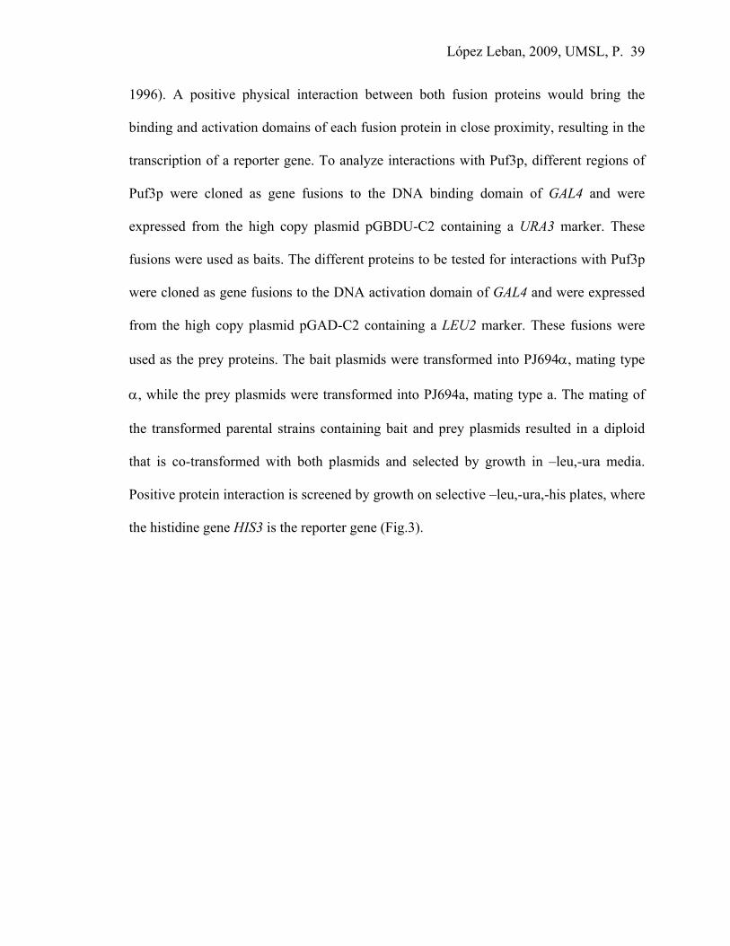
1996). A positive physical interaction between both fusion proteins would bring the
binding and activation domains of each fusion protein in close proximity, resulting in the
transcription of a reporter gene. To analyze interactions with Puf3p, different regions of
Puf3p were cloned as gene fusions to the DNA binding domain of GAL4 and were
expressed from the high copy plasmid pGBDU-C2 containing a URA3 marker. These
fusions were used as baits. The different proteins to be tested for interactions with Puf3p
were cloned as gene fusions to the DNA activation domain of GAL4 and were expressed
from the high copy plasmid pGAD-C2 containing a LEU2 marker. These fusions were
used as the prey proteins. The bait plasmids were transformed into PJ694α, mating type
α, while the prey plasmids were transformed into PJ694a, mating type a. The mating of
the transformed parental strains containing bait and prey plasmids resulted in a diploid
that is co-transformed with both plasmids and selected by growth in –leu,-ura media.
Positive protein interaction is screened by growth on selective –leu,-ura,-his plates, where
the histidine gene HIS3 is the reporter gene (Fig.3).

López Leban, 2009, UMSL, P. 40
Fig.4 The modified yeast three hybrid assay requires the presence of the target RNA in the nucleus where transcription occurs. This will allow any conformational change induced by RNA binding to occur in the bait protein in order for it to interact with the prey. A positive interaction is detected by the expression of the reporter gene.
GAL4 Promoter
GAL4 (BD)
X
GAL4 (AD) Y
Transcription
GAL4 (BD) GAL4 (AD)
Bait fusion constructs URA marker
Prey fusion constructs LEU marker
Puf3p regions Decay factors & other proteins X Y
Reporter gene HIS3
GAL4 Promoter Reporter gene
X
GAL4 (AD) Y
Transcription GAL4 (BD)
Cox17 3’ UTR
HIS3
NLS
Fig.3 The directed yeast two-hybrid technique detects protein-protein interactions by measuring transcription of a reporter gene. If protein X and protein Y interact, then their DNA-binding domain (BD) and activation domain (AD) will combine to form a functional transcriptional activator (TA). The TA will then promote transcription of the reporter gene, in this case HIS3, allowing yeast to grow on –his plates.

López Leban, 2009, UMSL, P. 41
Modified yeast- three hybrid assay
Some RNA-binding proteins must bind to their RNA target before they are able
to stably interact with other proteins. To analyze this kind of protein-protein interaction, a
modified yeast three-hybrid assay is used. The assay utilizes the same concept as the
directed yeast-two hybrid, but with the addition of the expression of a target RNA
containing a NLS (nuclear localization signal) that will allow it to enter and interact with
the bait/prey proteins in the nucleus. In this assay, the pIIIMS2-2 plasmid (TRP1 marker)
expressing COX17 3’UTR was co-transformed into strains containing the Puf3-Repeat
Domain fusion bait construct. The diploids obtained from the mating of this strain with
strains containing the prey plasmids were selected on –trp,-ura,-leu plates. To test for
protein-protein interactions in the presence of the COX17 3’UTR, yeast colonies were
selected on –trp,-ura,-leu,-his plates, with HIS3 as the reporter gene (Fig.4).
Co-immunoprecipitation assays
An alternative procedure used to analyze protein-protein interactions was the co-
immunoprecipitation assay. All strains used for this assay were puf3∆ strains transformed
with a plasmid expressing FLAG-tagged Puf3p. Five different yeast strains were used in
which the genes coding for decay factors CCR4, DCP1, DHH1, LSM1 or POP2 were
individually myc-tagged by homologous recombination (Sean Houshmandi, unpublished
data). The strains used in these co-immunoprecipitation experiments are shown on table 1
p.55 Ch. III. Yeast cells pelleted from 400 ml cultures were resuspended in 800 μl of IP
buffer with protease inhibitor and 10% glycerol. Glass beads were added to these pellets
and five consecutive cycles of vortex – ice of 2 minutes each allowed cell lysis. Protein

López Leban, 2009, UMSL, P. 42
extracts were quantitated using the colorimetric assay to measure equal amounts of crude
protein extracts at the beginning of the assay. The assay was performed by incubating the
different protein extracts obtained from each one of the strains for 45 minutes with the
anti-FLAG resin and immunoprecipitating FLAG-Puf3RD onto the resin, along with any
other proteins that might be interacting with Puf3RD. After several washes the eluates
from the resin were electrophoresed on an SDS-PAGE gel and blotted. The western blot
obtained was hybridized with specific anti-myc antibodies then HRP-tagged anti-mouse
secondary antibodies to visualize any myc-tagged decay factors that had co-
immunoprecipitated with Puf3p (Fig. 5).

López Leban, 2009, UMSL, P. 43
FLAG-Puf3RD
puf3∆ strain
Decay factor
Myc-tagged
Beads covered with Anti-FLAG
antibodies Incubation Yeast protein extract
Elution Washes
Proteins of the extract that do not
interact with Puf3RD
Decay factor
Myc-tagged FLAG- Puf3RD
SDS-PAGE electrophoresis & western blot detection with antibodies
Fig.5 Diagram showing the main steps of the co-immunoprrecipitation experiment. Protein extracts obtained from yeast cultures expressing both FLAG-Puf3RD and a myc-tagged decay factor are incubated with anti-FLAG resin. The anti-FLAG antibodies will pull down FLAG-tagged Puf3RD and any interacting myc-tagged decay factor. After three consecutive washes of the beads, the only remaining proteins should be the ones interacting with Puf3RD. The eluted samples are run on anSDS-PAGE and electroblotted on a nitrocellulose membrane.

López Leban, 2009, UMSL, P. 44
Epi-fluorescence microscopy
Fluorescence is based on the property fluorophores have in absorbing short
wavelength light and producing a longer wavelength light which is emitted and detected.
Epi-fluorescence is a type of excitation-emission configuration, in which both the
illumination and emission light travel through the objective and reaches the specimen
from above. In our experiments we used Green Fluorescent Protein (GFP) as the
fluorophore molecule that was fused to the protein of interest (Puf3p) to be able to detect
differences in appearance or localization of Puf3p-GFP when grown in minimal media
using different carbon sources.
A mercury or xenon lamp produces excitation light with several wavelengths. An
excitation filter removes the unwanted frequencies from the light emitted except those
that will be used to cause the sample to fluoresce. A dichroic beam splitter (mirror)
reflects blue light (~ 395nm for GFP) down through the objective lens and onto the
specimen. GFP molecules in the specimen (yeast cells) emit fluorescence (~509nm),
which passes up, through the objective lens, through the dichroic mirror to be detected by
eye or a camera. The barrier filter just above the dichroic mirror selects a particular
wavelength range so that background noise is eliminated. Figure 6 is an example for
visualizing GFP expression of Dcp2-GFP tagged protein that is known to localize to P-
bodies versus a negative control strain.

López Leban, 2009, UMSL, P. 45
Negative
control
Dcp2-GFP
positive
control
Negative
control
Dcp2-GFP
positive
control
Fig.6 GFP tagging. The image on the left corresponds to a yeast strain used as a negative control that lacks a GFP protein. The image on the right shows a Dcp2-GFP tagged strain; Dcp2 localizes to discrete cytoplasmic foci known as p-bodies.

López Leban, 2009, UMSL, P. 46
References
Decker, C., Parker, R. (1993) A turnover pathway for both stable and unstable mRNA in yeast: Evidence for requirement for deadenylation. Genes & Dev. 7: 1632-1643.
Felici, F., Cesareni, G., Hughes, J. (1989) The most abundant small cytoplasmic RNA of Sacharomyces cerevisiae has an important function required for normal cell growth. Mol. Cell. Biol. 9: 3260- 68.
James, P., Halladay, J. and Craig E. A. (1996) Genomic libraries and host strains designed for highly efficient two-hybrid selection in yeast. Genetics 144: 1425-1436.
Olivas, W., and Parker, R. (2000) The Puf3 protein is a transcript-specific regulator of mRNA degradation in yeast. The EMBO Journal 19: 6602-11.

López Leban, 2009, UMSL, P. 47
Chapter III
How does Puf3p regulate decay?

López Leban, 2009, UMSL, P. 48
Introduction
One way of regulating gene expression in the cell is by affecting mRNA turnover.
In yeast, the main mRNA decay pathway involves deadenylation-dependent decapping
followed by 5’ to 3’ exonucleolytic degradation by Xrn1p. The deadenylation of an
mRNA not only includes the poly(A) tail removal by Ccr4-Pop2, the main deadenylator
in yeast, but also implies a reorganization of the mRNP ribonucleoprotein, which will
mean a transition from a translating mRNA to a transcript targeted for degradation
(Schwartz and Parker, 1999; Tharun et al, 2000). This transition involves the loss of
Pab1p associated with the poly(A) tail and the loss of translation initiation factors eIF4E
and eIF4G associated with the cap structure. These changes allow the mRNA to associate
with Dhh1p and the Lsm1-7p complex, which enhance the interaction of the mRNA with
the Dcp1/Dcp2 decapping enzyme complex (Tharun and Parker, 2001). Physiologically,
mRNAs can thus be described as belonging to one of two sets of functionally different
groups in the cell, one group associated with polysomes involved in translation and the
other as a non-translating pool sequestered in discrete cytoplasmic foci where the
mRNAs are substrates for decapping and subsequent decay. Sheth and Parker were the
first to identify these foci in yeast, which they called processing bodies or p-bodies
(Sheth and Parker, 2003). In these sites, decapping and 5’ to 3’ exonucleolysis occur,
being therefore sites of congregation of Dhh1p, Dcp1/2p, Xrn1p, Lsm1p and Pat1p with a
pool of deadenylated, non-translating mRNAs that are no longer bound to translation
initiation factors eIF4E and eIF4G.
Dhh1p, which is required for efficient decapping of mRNAs after deadenylation,
belongs to a highly conserved subfamily of DEAD box helicases. Dhh1p has been shown

López Leban, 2009, UMSL, P. 49
to physically interact in an RNA- independent manner with Pop2p, Dcp1p, Lsm1p, Pat1p
and Xrn1p (Coller et al., 2001). Though Dhh1p is not essential for yeast viability, it has
been described as a regulator of mRNA decapping (Fischer et al., 2002) and is present in
p-bodies. Yeast strains lacking Dhh1p tend to accumulate transcripts with an intact cap
structure, but that have lost the poly(A) tail. The Dhh1p homolog in Xenopus, Xp54, is
also a DEAD-box helicase described as an integral component of mRNP particles of
Xenopus oocytes (Weston et al, 2006). Since Dhh1p co-localizes to p-bodies, there could
be mechanistic similarities between mRNA turnover in the deadenylation-dependent
decay pathway in yeast and maternal mRNA storage in higher eukaryotes. These
helicases are considered key regulators of post-transcriptional gene expression since they
modulate mRNA metabolism by remodeling mRNPs for entry into translation, storage or
decay pathways (Weston et al., 2006).
In this chapter, I dissect the molecular mechanism of Puf3p-mediated COX17
mRNA decay. Utilizing genetic and biochemical approaches, I propose that Puf3p plays a
central role in the recruitment of both deadenylators and decapping factors to promote
mRNA degradation by directing protein-protein interactions between the Puf3RD and the
decay machinery.
Goldstrohm et al have recently shown that Puf4p and Puf5p regulate HO mRNA
decay by enhancing its deadenylation rate (Goldstrohm et al., 2006). This mRNA
encodes a DNA endonuclease required for mating type switching in yeast. The authors
showed that TAP-tagged Puf5p co-immunoprecipitated with T7 epitope tagged decay
factors: Ccr4, Pop2, Dcp1 and Dhh1. The Puf5p-decay factor interactions were all RNA
independent. They also demonstrated that only Pop2 can bind Puf5p in vitro, and found

López Leban, 2009, UMSL, P. 50
the interactions to be evolutionarily conserved by testing Pufs and Pop2p from different
species. They proposed that Pop2p is the bridging molecule between Puf5p and all the
other decay factors. The presence of Pop2p is required to promote HO mRNA
deadenylation by Ccr4 in a Puf-dependent mechanism. HO mRNA deadenylation occurs
at a slow basal rate in a puf4-5∆ strain since the Ccr4-Pop2-Not complex is not
efficiently recruited to its 3’ UTR (Hook et al., 2006). In addition, the absence of Ccr4p,
Puf5p, but not Puf4p, was still able to repress mRNA expression into protein by an
unknown mechanism independent of deadenylation but dependent on Pop2p.
In our lab, the Puf3p repeat domain alone has been shown to be sufficient and
essential for both in vitro binding to the COX17 3’UTR and in vivo stimulation of
COX17 mRNA decay (Jackson et al, 2004). The Puf3RDp-R7A mutant was created by
deletion of the R7A outer surface loop of the Puf3RDp. This mutant was shown to be
incapable of mediating rapid COX17 mRNA decay, but was still capable of mRNA
binding. This region was thus predicted to be involved in protein-protein interactions
important for signaling to the decay machinery (Houshmandi and Olivas, 2005). The
focus of this chapter is to study the role Puf3RDp plays in recruiting the decay machinery
to promote COX17 mRNA decay. Through in vivo co-immunoprecipitation experiments
(co-IPs), Puf3RDp was found to interact in an RNA-independent manner with Ccr4p,
Dcp1p, Dhh1p, Lsm1p and Pop2p. All of these proteins were found to be actively
involved in the deadenylation and decapping of COX17 mRNA. The R7A loop was also
identified as the region of the repeat domain involved in the binding of Pop2p and
Dhh1p. Furthermore, co-IPs were performed in pop2Δ strains, demonstrating that Pop2

López Leban, 2009, UMSL, P. 51
bridges Dhh1 binding to the Puf3RD through the R7A loop, but Pop2p is not required for
Dcp1p binding to Puf3RD.
Puf3-mediated COX17 mRNA decay involves rapid deadenylation and decapping
rates (Olivas and Parker, 2000). In the yeast cell, the decay machinery is shared by
different decay pathways, including the Nonsense Mediated Decay (NMD) pathway,
which is in charge of the degradation of aberrant mRNAs as well as many normal
mRNAs with short lifespans. To determine whether the NMD pathway was involved in
the rapid COX17 mRNA decay, transcriptional shut-off assays were used to analyze
COX17 decay in a upf1Δ strain, which is NMD deficient. However, COX17 mRNA decay
was found to be NMD independent. Thus, the data of this chapter suggests that Puf3p
mediates rapid mRNA decay by recruiting the standard mRNA decay factors to the
Puf3p-bound mRNA.
Together this research implies that Puf3RD regulates COX17 mRNA decay by
binding its 3’UTR and recruiting Ccr4 and Pop2 (deadenylators), as well as Dhh1, Lsm1
and Dcp1 (decapping factors). All these interactions between Puf3RD and the decay
machinery are RNA independent, being therefore direct protein-protein interactions. In
contrast with Puf5p regulation of HO mRNA decay, in which Pop2 is thought to bridge
the interactions between Puf5p and all the remaining decay factors, this does not seem to
be the case for Puf3p. Instead, our data suggests a different model for Puf3p regulation,
whereby the R7A loop region is required for Pop2 binding and indirectly for Dhh1
binding, while Dcp1 and Ccr4 appear to bind Puf3p independently of the R7A loop and
Pop2p. While all Puf proteins appear to be engaged in the decay of their mRNA targets,

López Leban, 2009, UMSL, P. 52
each one has its particular way of interacting with the decay machinery and achieving this
regulation.
Experimental Procedures
Directed yeast two-hybrid assay
Full length Puf3p, the C terminal half of Puf3p (containing the repeat
domain) and different portions of the N-terminal half of Puf3p were used as baits in the
yeast two-hybrid assay (see Fig.3 for a schematic of the baits used). These different
Puf3p fragments were each expressed as a gene fusion to the DNA binding domain of
GAL4 from the high copy plasmid pGBDU-C2 containing a URA3 marker. The different
prey proteins were each expressed as a gene fusion to the DNA activation domain of
GAL4 from the high copy plasmid pGAD-C2 containing a LEU2 marker (see table 1 for
the genes cloned for prey protein expression). The mating of the parental strains
transformed with either the bait or the prey plasmid resulted in a diploid that contained
both plasmids and was selected by growth in –leu,-ura media. The parental strains were
PJ694α for the baits and PJ694a for the preys. Positive protein interaction was screened
by growth on selective –leu,-ura,-his plates, where the histidine gene HIS3 was the
reporter gene under the control of the GAL promoter. HIS3 is a leaky gene, so 0.5mM 3-
AT (3-amino-1,2,4-triazole) was added to the selective plates to prevent growth of false
positives. 3-AT competitively inhibits imidazole glycerol phosphate dehydratase, a His
biosynthetic enzyme (Hilton et al., 1965), and therefore can limit histidine biosynthesis
and growth. The HIS3 gene encodes the enzyme activity inhibited by 3-AT. In the

López Leban, 2009, UMSL, P. 53
directed yeast two-hybrid assays that use a HIS3 as a reporter gene, the high expression
that arises from a successful two-hybrid interaction can overcome the growth-inhibitory
effect of 3-AT in the medium. When 3-AT is added to yeast media (0.5-10 mM), it will
limit histidine biosynthesis and is used in two-hybrid screens to "fine tune" leaky
expression of the HIS3 reporter gene. Therefore, the use of 3-AT and the HIS3 reporter
enables positive selection for successful two-hybrid interactions.
Modified yeast three-hybrid assay
In the modified yeast three-hybrid assay, interactions between the Puf3
protein and a prey protein that occur in the presence of the Puf3p target mRNA were
tested by expressing the COX17 3’ UTR fused to the MS2 RNA for nuclear localization,
with the bait and prey proteins. The Puf3-RD fusion bait expressed from pGBDU-C2
(URA3 marker) was co-transformed into the PJ694α strain together with the pIIIMS2-2
(TRP1 marker) plasmid expressing COX17 3’UTR RNA tagged with the MS2 sequence
containing a nuclear localization signal (NLS). This bait strain was mated with PJ694a
strains individually transformed with plasmids expressing 53 different candidate prey
proteins. Each prey protein was expressed as a gene fusion to the DNA activation domain
of GAL4 from pGAD-C2 (LEU2 marker). The diploids obtained from the mating of the
transformed PJ694α and PJ694a strains were selected on –trp,-ura,-leu plates. To test for
protein-protein interactions in the presence of COX17 3’UTR RNA, yeast colony growth
was screened on –trp,-ura,-leu,-his plates with the addition of 0.5mM ATZ, where HIS3
is the reporter gene under the control of the GAL promoter.

López Leban, 2009, UMSL, P. 54
Co-immunoprecipitation experiments
The myc-tagged strains lacking endogenous Puf3p were transformed with
the FLAG- Puf3RD expressing construct and 400 ml cultures were grown to an OD of
0.4. Protein extracts were prepared from each strain by applying consecutive cycles of
vortex ice, to break open the cells and obtain the soluble cytoplasmic extract. These
samples are incubated with an anti-FLAG resin that will trap the FLAG-tagged Puf3RD
proteins along with any other protein that might be interacting with it. After a series of
washes, the eluates from the resin are electrophoresed on an SDS-PAGE gel and blotted.
The western blot obtained is hybridized with specific anti-myc antibodies to bind any co-
immunoprecipitated decay factors. Since the primary anti-myc antibody was developed in
mouse, a secondary anti-mouse antibody developed in goat that is HRP tagged allowed
the detection of these proteins on the western.
Transcriptional shut-off analysis
Transcriptional shut off experiments were carried out to analyze COX17
mRNA half lives in a WT strain versus puf3∆, dcp1Δ, dhh1Δ and lsm1Δ strains. All
strains were transformed with pST30/COX17pG in which COX17 is expressed under the
GAL UAS. Cultures were grown at 30°C in 2% galactose overnight to activate the control
of the expression and then switched to 4% glucose to shut transcription off. Culture
samples were gathered at specific time points once transcription is arrested. Then RNA is
extracted from these cells, electrophoresed on 1% agarose gels and visualized and
compared by northern blot analysis.

López Leban, 2009, UMSL, P. 55
Deletion Strain Genotype Source puf3Δ yWO 186 MAT a, trp1, ura3-52, leu2-3,112, his4-539,cup1:LEU2/PM, puf3::NEO Olivas
&Parker(2000) puf3Δ yWO 187 MAT a, ura3-52, leu2-3,112, his4-539,cup1:LEU2/PM, puf3::NEO,Ccr4-myc::TRP1 This study
puf3Δ yWO 188 MAT a, ura3-52, leu2-3,112, his4-539,cup1:LEU2/PM, puf3::NEO,Dcp1-myc::TRP1 This study puf3Δ yWO 189 MAT a, ura3-52, leu2-3,112, his4-539,cup1:LEU2/PM, puf3::NEO,Dhh1-myc::TRP1 This study puf3Δ yWO 190 MAT a, ura3-52, leu2-3,112, his4-539,cup1:LEU2/PM, puf3::NEO,Lsm1-myc::TRP1 This study puf3Δ yWO 191 MAT a, ura3-52, leu2-3,112, his4-539,cup1:LEU2/PM, puf3::NEO,Pop2-myc::TRP1 This study W T MAT a, ura3-52, his3-11,15, trp1-1, leu2-3,112 Invitrogen puf3Δ MAT a, ura352, his3-11,15, trp1-1, leu2-3,112,puf3::KanMX Invitrogen
dhh1Δ yWO 207 MAT a, ura352, his3-11,15, trp1-1, leu2-3,112, dhh1::KanMX Invitrogen
dcpΔ MAT a, ura352, his3-11,15, trp1-1, leu2-3,11, dcp1::KanMX Invitrogen
lsm1Δ MAT a, ura352, his3-11,15, trp1-1, leu2-3,112, lsm1::KanMX Invitrogen
W T yWO 225 MAT a, ura3-52, his3-11,15, trp1-1, leu2-3,112 Audrey Atkin upf1Δ yWO 226 MAT a, his3-11,15, trp1-1, leu2-3,112, upf1::URA3 Audrey Atkin pop2∆ yWO 223 MAT a, ura3-52, leu2-3,112, his4-539,cup1:LEU2/PM, puf3::NEO, Dcp1-myc::TRP1,
pop2::NAT This study
pop2∆ yWO 224 MAT a, ura3-52, leu2-3,112, his4-539,cup1:LEU2/PM, puf3::NEO, Dhh1-myc::TRP1, pop2::NAT
This study
TABLE 1. Strains used in this study.
TABLE 2. Genes cloned in the pOAD for prey protein expression for yeast two and three- hybrid assays.
AIR1 CDC39 LSM2 NOT3 PUF4 TUB3 AIR2 CDC50 LSM5 NRG2 PUF5 YHR035W ARP2 CRZ1 LSM7 PAB1 REX1 YPR013C CAF4 DCP1 MIG1 PAT1 REX2 YER130C CAF17 DHH1 MOT2 PBP1 RPA14 YKL1 CAF40 EDC1 MSN2 POP2 RPB4 ALPHA-1 CAF130 EDC3 MSN4 PRP4 SIG1 YHR121W CCR4 ELA1 NAM7 PSU1 SMB1 CDC28 HRR25 NHD2 PUF1 SPB8 CDC36 LSM1 NIL2 PUF3 SPP381

López Leban, 2009, UMSL, P. 56
Results
Rapid COX17 mRNA decay is dependent on Dhh1p, Dcp1p and Lsm1p decay
factors.
COX17 mRNA has been used as a model to study Puf3p-mediated decay in yeast.
It was previously shown through pulse chase experiments that the rapid deadenylation of
COX17 mRNA, which is stimulated by Puf3RD, requires the Pop2p and Ccr4p
deadenylation factors (Tucker et al., 2002). It was likely that COX17 decay was also
dependent on the standard decapping complex, Dcp1p/Dcp2p, as well as the enhancers of
decapping, Dhh1p and the Lsm1-7p complex. However, it was possible that Puf3p-
mediated decay utilized some other mechanism of decay stimulation that bypassed the
need for one or all of these factors. We therefore investigated whether COX17 mRNA
decay was slowed in strains deleted of DCP1, DHH1 or LSM1. To measure mRNA
decay, transcriptional shut-off assays were performed. Five different strains (WT, puf3∆,
dcp1∆, dhh1∆ and lsm1∆) were transformed with pWO 153 that has COX17pG under a
GAL promoter, so that COX17 with a 3’ UTR poly(G) tag is expressed in galactose and
its transcription can be shut-off when switched to glucose. The results in Fig.1
demonstrate that all three of these decay factors are required for rapid COX17 mRNA
decay. The individual deletion of either DHH1 or LSM1 results in a two fold increase in
the COX17 mRNA half-life, similar to the puf3∆ strain. The DCP1 deletion results in a
more drastic effect, causing an 8 fold increase in the half-life of COX17 mRNA. Clearly,
Puf3p cannot bypass the need for these factors in promoting rapid decay of COX17
mRNA. Instead, Puf3p likely recruits or stimulates the activity of these factors on the
mRNA.

López Leban, 2009, UMSL, P. 57
COX17 mRNA decay is NMD-independent.
In cells there exists a mechanism of mRNA surveillance known as non-sense
mediated decay (NMD). In Saccharomyces cerevisiae, NMD is responsible for detecting
mRNAs with premature stop codons as well as some wild-type mRNAs and targeting
them for accelerated decay (Lelivelt and Coulbertson, 1999). NMD uses the standard
degradation machinery of the cell but with the addition of Upf1p, Upf2p and Upf3p. The
deletion of any of these three UPF genes results in a similar phenotype of knocking out
the NMD pathway (Atkin et al., 1997). Thus, in any of these NMD mutants, mRNAs that
are usually degraded by the NMD pathway will accumulate to higher levels compared to
a WT strain.
In order to determine whether COX17 mRNA decay depended on NMD or
followed the typical steps of deadenylation, decapping and 5’ to 3’ degradation,
transcriptional shut-off experiments were performed. In this case, transcription of COX17
was repressed by switching temperature from 24°C to 37°C in WT and upf1∆ strains
since both of them have a temperature sensitive mutation in RNA polymerase II (rpb1-1).
The results in Fig.2 show that the half-life of COX17 mRNA is similar in both strains,
implying that the NMD pathway is not involved in the rapid COX17 mRNA decay.
Rather, Puf3p is stimulating COX17 mRNA decay through the classical deadenylation,
decapping and 5’ to 3’ decay as occurs in most yeast transcripts (Fig.2).

López Leban, 2009, UMSL, P. 58
WT
puf3Δ
dcp1Δ
dhh1Δ
0 2 4 6 8 10 15 20 30 40
lsm1Δ
Min
WT
upf1∆
0 2 4 6 8 10 15 20 30 40 Min T ½
4.5
4.5
Fig.2 COX!7 mRNA decay is NMD independent. Nortrhern blots of transcriptional shut-off experiments for WT and upf1∆ strains. At the top, time points are shown in minutes after transcription was repressed and samples taken. On the right, the half-life of COX17 mRNA decay is shown in minutes for each of the strains.
Fig.1 Puf3p-mediated rapid decay of COX17 mRNA requires Dhh1p, Dcp1p and Lsm1p. At the top, time in minutes is shown after transcription was shut off and samples of the culture were taken for the analysis. On the right, half-life in minutes of COX17 mRNA decay is shown in different deletion strains compared to WT and puf3∆ strains.
40
10
Half-life 5 12 13
López Leban, 2009, UMSL, P. 59
Puf3p interacts with decay factors involved both in deadenylation and decapping.
Genetic approach for detecting protein-protein interactions using directed yeast
two-hybrid and modified yeast three-hybrid assays.
Puf proteins from Drosophila and C. elegans must interact with other regulatory
proteins, such as Nanos and Brat, to mediate repression of their target mRNAs. Though
there are no Nanos or Brat homologs in yeast, it is possible that Puf3p might also be
interacting with other regulatory proteins to mediate decay of COX17 mRNA.
Alternatively, Puf3p may directly interact with mRNA decay factors, such as those
involved in deadenylation (Ccr4, Pop2) and/or decapping (Dcp1/Dcp2, Dhh1, Lsm1-7
and Pat1). To identify proteins that interact with Puf3p, a yeast two-hybrid library screen
was first performed by Andrew Van Brunt in the Olivas lab (See Materials and Methods
for overview of the yeast two-hybrid assay). In this assay, the Puf3RDp was used as bait
against a genomic library of prey proteins, however no significant interactions were
detected (unpublished data). I then tested through a directed yeast two-hybrid assay the
interaction of Puf3p with various known mRNA decay factors and several other proteins
that through systematic analyses had been identified as potential Puf interacting proteins.
The results from the directed yeast two-hybrid assay were not very revealing (Fig.3). The
Puf3RDp did not show detectable interactions with any of the prey proteins, while the
full-length Puf3p interacted with all the proteins, including the negative controls. I
determined that the first 65 amino acids of the protein were responsible for this non-
specific binding, since this 65 amino acid region interacted with all proteins, while a
construct lacking this region could not bind to any of the tested prey proteins.

López Leban, 2009, UMSL, P. 60
Fig.3 Schematics of Puf3p baits. Full-length Puf3p and different regions of the protein were used as baits in the directed-yeast two hybrid assay.
Fig.4 Modified yeast three-hybrid assay. Left plate shows four positive interactions between Puf3RD and Lsm2, Pop2, Dcp1 and Ccr4. Lsm5 and Not3 show weak interactions. On the right plate, negative controls do not grow, showing no interaction.
Bait Puf3p constructs tested
65 357 532 880
N terminal domain C terminal domain
Full length Puf3p (880 Aac)
1 – 65 aa sticky region
Both had positive interactions with all proteins tested including negative controls
NT2
Puf3RD
NT 1
93% Puf3p
None showed any interactions with all proteins tested including negative controls
Mer I Snu17
Snu56 Prp4Not3
Lsm5 Lsm2
Pop2 Dcp1
Ccr4

López Leban, 2009, UMSL, P. 61
It is important to note that protein interaction in the yeast-two hybrid assay must
occur in the cell’s nucleus, which is not where Puf proteins normally function to bind and
regulate mRNA targets. Therefore, it was possible that the absence of an mRNA target in
the nuclear two-hybrid assay was detrimental to the ability of the proteins to interact. In
fact, studies have shown that the Drosophila Puf protein, Pumilio, requires binding to its
mRNA target prior to binding to the Nanos and Brat proteins (Wharton et. al., 1998).
Therefore, to test whether Puf3RDp requires binding to its mRNA target in order to be
competent for protein interaction, a modified yeast three-hybrid assay expressing a
nuclear localized COX17 3’ UTR RNA was performed as described in Materials and
Methods. The results obtained showed interactions (albeit weak) between Puf3RDp and
several of the prey proteins tested. Since protein interaction is assayed by the ability to
activate expression of the HIS3 reporter gene, positive interactions were observed as
growth on selective plates lacking histidine as compared to no growth of the negative
controls. Addition of 0.5 mM amino tetrazolium was used to inhibit leaky expression of
the HIS3 reporter gene. Positive interactions with Puf3RDp included proteins involved in
mRNA degradation: Pop2, Dcp1, Ccr4, Lsm2, Lsm7 and Caf4. These proteins belong to
the deadenylation and decapping machinery in yeast. Other proteins also identified
through the directed yeast three-hybrid assay that are not related to mRNA decay were:
Tub3, YHR035W, HRR25, α1, Smb1, Rpb4, YHR121W, Ela1 and Mig1. The rest of the
39 proteins tested did not show any interaction with Puf3RDp (Fig. 4).

López Leban, 2009, UMSL, P. 62
Co-immunoprecipitation experiments
Since the yeast-three hybrid assay showed interactions with deadenylating
and decapping factors, but also with other proteins not related to the decay machinery, we
wanted to use another experimental approach to test these interactions between the
Puf3RD and the decay factors. Therefore, co-immunoprecipitation experiments were
developed in which Puf3RD was FLAG-tagged and expressed from the pG-1 vector,
while the decay factors were endogenously myc-tagged at their C-terminal ends by
homologous recombination. The overall scheme is described in a diagram shown in Fig.5
of Chapter II- General Experimental Methodology. In brief, protein extracts are applied
to anti-FLAG resin that will trap the FLAG-tagged Puf3RD proteins along with any other
protein that might be interacting with the Puf3RD. After a series of washes, the eluates
from the resin are electrophoresed on an SDS-PAGE gel and blotted. The western blot
obtained is hybridized with specific anti-myc antibodies to visualize any co-
immunoprecipitated decay factors. Sean Houshmandi in the lab originally demonstrated
that the five myc-tagged decay factors (Ccr4p, Dcp1p, Dhh1p, Lsm1p and Pop2p) all co-
immunoprecipitated with Puf3RD (Fig.5). Similar results were obtained by another
research group in which the Ccr4p, Dcp1p, Dhh1p and Pop2p decay factors epitope-
tagged with T7 were pulled down by TAP-tagged Puf5 (Goldstrohm et al, 2006).
Together, these results suggest a conserved mechanism of yeast Puf proteins binding
directly with mRNA decay machinery, and these interactions likely act to recruit the
machinery to the target mRNA for promotion of decay.

López Leban, 2009, UMSL, P. 63
Fig.5 Puf3RD co-immunoprecipitates with decay factors. These decay factors (Ccr4, Dcp1, Dhh1, Lsm1 & Pop2) that are endogenously myc-tagged co-immunoprecipitate with FLAG-Puf3RD and are detected with anti-myc antibodies (Houshmandi, S. unpublished data).

López Leban, 2009, UMSL, P. 64
Puf3p interactions with decay factors are RNA independent and Puf3p binds Dhh1p
and Pop2p through the 7A loop region of the repeat domain (RD).
After having detected the interactions of Puf3RD with the various decay
factors through co-immunoprecipitation assays, we wanted to further evaluate the nature
of the interactions. First, we wanted to determine whether the Puf3p/decay factor
interactions were really due to protein-protein interactions, or whether the co-
immunoprecipitation results were simply due to proteins binding to different points along
a single mRNA molecule. To differentiate between these possibilities, protein extracts
were incubated with RNase A before performing the co-immunoprecipitation. The results
show no difference in the binding of Puf3RD to any of the decay factors in the presence
or absence of RNase A (Fig.6A). This data suggests that interactions between Puf3p and
the decay factors are mediated through protein interactions.
Next, previous work in the lab indicated that a deletion of four amino acids
on the outer surface of Puf3RD, the R7A mutant, failed to promote rapid decay of
COX17, while another deletion mutant of an outer surface loop of Puf3RD, the R6A
mutant, had no effect on COX17 decay (Houshmandi and Olivas, 2005). We wanted to
determine whether the failure of the R7A mutant to promote rapid decay was due to an
inability to interact with one or more decay factors. Therefore the myc-tagged strains
lacking endogenous Puf3p were transformed with each of the FLAG-tagged Puf3p
constructs: WT Puf3RD, Puf3R6A mutant and Puf3R7A mutant.
Co-immunoprecipitation experiments with the R6A mutant demonstrated
that it is able to bind Ccr4p, Dcp1p, Dhh1p, Lsm1p and Pop2p, thus behaving in a similar
way as WT Puf3RD (results not shown). In contrast, the R7A mutant disrupted

López Leban, 2009, UMSL, P. 65
interactions with two of the decay factors (Pop2p and Dhh1p), while interactions with
Dcp1p, Lsm1p and Ccr4p were maintained (Fig.6B). This indicates that the R7A loop is
the specific region of Puf3RD to which Pop2p and Dhh1p bind. It is possible that each
one of these proteins binds directly to the R7A loop. However, other work with Puf5p
indicates that Pop2p acts as a bridge between Puf5p and all other decay factors
(Goldstrohm et al., 2007). This suggests that Pop2p may be acting as a bridge between
the Puf3RD-R7A loop and Dhh1p. To test this hypothesis, POP2 was deleted from the
myc-tagged DHH1 and DCP1 strains, then the strains were transformed with vector alone
or with the Puf3RD expressing construct. The results of Co-Ip experiments with these
pop2∆ strains clearly show that Dhh1p binding to Puf3RD is disrupted in a pop2∆ strain,
while Dcp1p is still able to bind (Fig.7). These experiments provide evidence that Dhh1p,
but not other decay machinery is recruited via Pop2p to Puf3p. It might be that different
Pufs interact with decay factors in a customized mechanism for achieving unique modes
of decay of their target mRNAs.

López Leban, 2009, UMSL, P. 66
Ccr4
Dcp1
Lsm1
Dhh1
Pop2
Vector WT R7A Vector WT R7A
Fig.6B Extracts and Co-Ip for Puf3RD and Puf3R7A. On the left, western blots show protein extracts that were loaded onto the resin. On the right, the results after performing the Co-Ip are shown comparing the pull-down with WTRD and that by the mutant RD that contains the 7A loop deletion. Myc-tagged decay factors (Ccr4, Dcp1, Dhh1, Lsm1 and Pop2) were detected with anti-myc antibodies. Red boxes denote interactions inhibited by the 7A mutation.
Fig.6A Puf3RD interactions with decay factors are RNA independent. Western blot of Co-Ip experiments with and without RNAse treatment. Myc-tagged decay factors (Ccr4, Dcp1, Dhh1, Lsm1 and Pop2) were detected with anti-myc antibodies.

López Leban, 2009, UMSL, P. 67
V WT WT V Pop2 WT pop2∆
Input
Output
Input
Output
Dcp1 Myc-tagged
Dhh1 Myc-tagged
Lanes 1 2 3 4
Fig.7 Pop2p is required for Dhh1p binding. Western blots of Co-Ip experiments comparing a WT strain versus a pop2∆.strain with Dcp1 or Dhh1 myc-tagged decay factors. Anti-myc antibodies were used for detecting the decay factors. Input denotes extracts that were loaded onto the resin, and output denotes the elutions from the co-immunoprecipitations. Lanes 1 and 4 correspond to the results obtained with the strains transformed with vector alone, while 2 and 3 show the results with the strains expressing Puf3WTRD.

López Leban, 2009, UMSL, P. 68
Discussion
Puf 3p mechanism of action
COX17 mRNA decay shows a prolonged half-life in a puf3Δ strain compared to a WT
strain. It has been shown that the deadenylation rate of COX17 in the puf3Δ strain is
slower and at least partially accounts for the mRNA stabilization (Olivas and Parker
2000). A similar phenotype is observed in strains lacking either CCR4 or POP2, which
are the main yeast deadenylators (Tucker et al, 2002). The decapping step of COX17
mRNA also appears to be slowed by Puf3p (Olivas and Parker, 2000). In this research we
show that deletions of DCP1, DHH1 and LSM1, which encode proteins involved in
decapping, all promote COX17 mRNA stabilization. Deletion of DCP1, which encodes
part of the holoenzyme responsible for decapping, showed the most drastic effect,
prolonging the COX17 mRNA half-life 8-fold. Thus, the promotion of rapid COX17
mRNA decay by Puf3p requires the action of each of these deadenylation and decapping
factors. In addition, we show that this decay is NMD independent since the impairment of
the NMD pathway in a upf1∆ strain does not affect rapid COX17 mRNA decay.
Through co-immunoprecipitation experiments, we demonstrated that Puf3p
directly interacts with the Ccr4p, Pop2p, Dcp1p, Dhh1p and Lsm1p deadenylation and
decapping factors. Previous work had shown that the four amino acid deletion of the
Puf3RD-R7A mutant disrupted Puf3p’s ability to promote mRNA deadenylation
(Houshmandi and Olivas, 2005). Here we demonstrate that this mutant prevents the
binding of Pop2p and Dhh1p to the Puf3RD, yet maintains binding to Dcp1p, Lsm1p and
Ccr4p. To analyze the arrangement of Pop2p and Dhh1p binding to the R7A outer
surface loop, Co-Ip experiments were performed in the absence of Pop2p. These results

López Leban, 2009, UMSL, P. 69
revealed that Pop2p is required to bridge Dhh1p binding to Puf3p, while Pop2p likely
binds directly to the R7A loop region. In contrast, Dcp1p was shown to independently
bind Puf3RD without the presence of Pop2p. This result confirms that Dcp1p binds
Puf3RD in a different region other than the R7A loop, and the interaction is not bridged
by Pop2p (Fig.8). Previous work has shown that Pop2p is required for rapid
deadenylation of COX17 (Tucker et al., 2002). So it makes sense that the R7A mutant
that cannot bind Pop2p would also be defective in rapid COX17 deadenylation. Thus,
even if Ccr4p can independently bind to the Puf3RD-R7A mutant, this interaction
apparently cannot override the need for Pop2p to stimulate Ccr4p catalytic activity for
deadenylation. However, it is unclear whether the R7A mutant can still promote mRNA
decapping through Dcp1p binding, or if the absence of Dhh1p binding to this mutant
prevents such mRNA stimulation as well. Together, our results of Puf3RD interactions
with the decay machinery provide a similar yet distinct model as compared to the model
of Puf5p interactions (Fig.8) in which Pop2p is the only factor to directly bind Puf5p and
thus bridges interactions with all the rest of the decay machinery (Ccr4, Dhh1 and Dcp1)
(Goldstrohm et al., 2007).
Future work will be necessary to determine the unique arrangement of
interactions for other Puf proteins and any conserved bridging role of Pop2p. The Puf3p
protein interaction results from the yeast two-hybrid and three hybrid assays suggest that
the presence of the target RNA is required in order for the Puf3RD to interact with the
decay factors. Yet the Co-Ip results show that RNAse treatment does not affect Puf3RD’s
protein interactions. This treatment assures that both proteins being pulled down are
physically interacting and not just being tethered to different parts of a single RNA

López Leban, 2009, UMSL, P. 70
molecule. It might still be that the Puf3RD is required to first bind the RNA target in
order to recruit the decay machinery, as has previously been shown for the Drosophila
Pum to first bind hunchback mRNA before recruiting Nanos (Sonada et al., 1999; Sonada
et al., 2001). Once the Puf protein binds the RNA target, there might be conformational
changes in the Puf that will then favor the interactions of its outer surface with other
proteins. So in the case of Puf3p, even after RNAse treatment, the small portion of RNA
to which the Puf3RD may be attached would be protected from RNAse degradation and
could still be allowing a conformational change in the Puf3RD to enable it to directly
interact with decay factors. It would be very interesting in future work to test binding-
incompetent mutants such as the Puf3RD-R1 mutant (Houshmandi and Olivas, 2005) in
Co-Ip experiments to determine if such a mutant can still interact with the decay
machinery, or if protein binding indeed requires prior mRNA interaction.

López Leban, 2009, UMSL, P. 71
Fig.8 Models comparing Puf3p vs. Puf5p interactions with decay factors. Pop2p binds the Puf3RD at the R7A outer surface loop. Either by the R7A loop deletion or the deletion of Pop2p, Dhh1p binding is prevented. However, Dcp1p binding remains unaffected, being independent of both the R7A loop and Pop2p. Puf5p binds all the shown decay factors through Pop2p.
Puf3p Model
Puf5p
P5E
Pop2
Dhh1
WT Pop2 strain
Dcp1
Not1-5p
Ccr4
Puf3WTRD P3E
Dhh1p
WT Pop2 strain
WT Puf3RD
Dcp1
m7Gppp
Pab1 P3E
Puf3WTRD
AAAAAAAAA
Puf3WTRD P3E
Puf3R7A P3E WT Pop2
strain R7A mutant
pop2∆ strain WT Puf3RD
Ccr4
Dcp1 Dhh1p
Dhh1p
Dcp1 Pop2
Ccr4
Ccr4
Pop2
Puf5p Model

López Leban, 2009, UMSL, P. 72
References Atkin, A. L., Schenkman , L. R., Eastham, M., Dahlseid, J. N., Lelivelt M. J. and Culbertson, M. R. (1997) Relationship between Yeast Polyribosomes and Upf Proteins Required for Nonsense mRNA Decay. JBC 272: 22163–72. Coller, C., Morgan, T., Sheth, U., Valencia-Sanchez, M. and Parker, R. (2001) The DEAD box helicase, Dhh1p, functions in mRNA decapping and interacts with both the decapping and the deadenylase complexes. RNA 7: 1717-27. Fischer, N. and Weis, K. (2002) The DEAD box protein stimulates the decapping enzyme Dcp1. EMBO J. 21: 2788-97 Foat, B.C., Houshmandi, S.S., Olivas, W. M. and Bussemaker, H. J. (2005) Profiling condition-specific, genome –wide regulation of mRNA stability in yeast. PNAS 102: 17675-80. Goldstrohm, A. C., Hook, B.A., Seay, D. J. and Wickens, M. (2006) Puf proteins bind Pop2p to regulate messenger RNAs. Nature Struct. Mol. Biol. 13: 533-9. Hilton, J.l., Kearney, P.C. and Annes, B.N. (1965) Mode of action of the herbicide, 3-amino-1,2,4-triazole(amitrole): inhibition of an enzyme of histidine biosynthesis. Arch Biochem Biophys 112 (3): 544-7 Hook, B.A., Goldstrohm, A.C., Seay, D. J. and Wickens, M. (2007) Two yeast Puf proteins negatively regulate a single mRNA. J. Biol.. Chem. 282: 15430-8. Houshmandi, S.S. and Olivas, W.M. (2005) Yeast Puf3 mutants reveal the complexity of Puf-RNA binding and identify a loop required for regulation of mRNA decay. RNA 11: 1655-66. Jackson, J.S., Houshmandi, S.S., Lopez leban, F. and Olivas, W.M. (2004) Recruitment of puf3 protein to its mRNA target for regulation of mRNA decay in yeast. RNA 10: 1625-36. Olivas, W. and Parker, R. (2000) The Puf3 protein is a transcript-specific regulator of mRNA degradation in yeast. EMBO Journal 19: 6602-11.

López Leban, 2009, UMSL, P. 73
Tharun, S., Mayes, A. E., Lennertz, P., Beggs J. D. and Parker, R. (2000) Yeast Sm-like proteins function in mRNA decapping and decay. Nature 404: 515-8. Sheth, U. and Parker, R. (2003) Decapping and decay of messenger RNA occur in cytoplasmic processing bodies. Science 300: 805-8. Sonoda, J. and Wharton, R. (2001) Drosophila Brain Tumor is a translational repressor. Genes & Development, 15: 762-73. Sonoda, J. and Wharton, R. (1999) Recruitment of Nanos to hunchback mRNA by Pumilio. Genes& Development, 13: 2704-12. Tucker, M., Staples, R., Valencia-Sanchez, M., Muhlrad, D., Parker, R. (2002) Ccr4p is the catalytic subunit of a Ccr4/Pop2/Notp mRNA deadenylase complex in Saccharomyces cerevisiae. EMBO J. 21: 1427-36. Wharton, R., Sonoda, J., Lee, T., Patterson, M., Murata, Y. (1998) The Pumilio RNA-binding domain is also a translational regulator. Molecular Cell 1: 863-72.

López Leban, 2009, UMSL, P. 74
Chapter IV
Puf3p activity is affected by the type of carbon
source present in the media

López Leban, 2009, UMSL, P. 75
Introduction
The regulation of mRNA stability by cis-regulatory elements (CREs) is a dynamic
process and is affected by several environmental stimuli. DNA microarrays in
Saccharomyces cerevisiae have allowed the analysis of high throughput genomic data in
which different expression profiles can be observed and compared under different
conditions. It is known that in the adaptation of the cell to environments that have poor
conditions for growth and survival, the environmental stress response program (ESR) is
triggered (Gasch et al., 2000). This is a survival skill of the cell in which a broad
spectrum of genes are turned on to respond in a positive way, adapting the cell’s
metabolism to the new physiological requirements. Mechanisms of DNA repair, protein
folding, defense against reactive oxygen species, energy generation, and storage are
increased functions while cell growth and protein synthesis are arrested (Gasch et al.,
2000). Although ESR is a stereotypical response in which the cell will automatically turn
on and off particular sets of genes, other environmental stimuli will specifically target
other genes, rendering a unique gene expression pattern customized for each specific
condition (DeRisi et al., 1997; Brown et al., 2000). These signature patterns for
individual environmental conditions give the possibility of grouping genes according to
whether they are up- or down-regulated under a specific condition, and the patterns
display at a genomic network level the response to environmental changes through
coordinated changes in gene expression profiles. These data can be useful for acquiring
information about which genes respond coordinately with a group of genes, versus those
that belong to a different group that responds in an opposite manner. Gaining knowledge

López Leban, 2009, UMSL, P. 76
on the coordinated regulation of these gene networks will allow us to better manipulate
gene expression to trigger the desired response at a cellular level.
In an attempt to study and classify gene expression in gene clusters and to better
analyze the information obtained by high throughput data, the Matrix REDUCE
algorithm (Regulatory Element Detection Using Correlation with Expression) was
created by Foat et al. This algorithm was applied to 200 bp sequences downstream of
every ORF of the yeast genome and to the microarray expression profiles of these genes
tested under 700 different conditions. As a result, groups of different genes containing the
same CRE were detected as coordinately regulated in a similar fashion under different
environmental conditions (Foat et al., 2005). Predictions were made for the functional
role of the P3E (Puf3 element) present in most of the mitochondrial-related transcripts.
According to the results obtained through the algorithm, these transcripts would be down-
regulated under growth conditions where a fermentable carbon source is present and thus
mitochondria would not be necessary, while the transcripts would be up-regulated under
a non-fermentable carbon source when mitochondria are required for respiration. These
predictions were experimentally confirmed in the Olivas lab. The COX17 mRNA half-
life was four fold higher in ethanol, in which mitochondria need to be functional to obtain
energy through the respiratory chain, compared to glucose where fermentation takes
place (Foat et al., 2005)).
In this work I was seeking to understand the mechanisms involved in the
regulation of Puf3p activity by different environmental conditions. For this purpose, I
measured the amount of Puf3 mRNA and protein present in the different conditions.
Finding that protein and mRNA quantities in both ethanol and glucose were similar (or

López Leban, 2009, UMSL, P. 77
even higher in ethanol versus glucose), led to further investigations searching for post-
translational modifications in Puf3p. In fact, it has been shown in Dictyostelium that Puf
activity is regulated through a change in its phosphorylation state (Mendes Souza et al.,
1999). Puf6p in yeast, which is involved in ASH1 mRNA translational repression, was
also recently shown to be regulated by the kinase CK2, which releases Puf6p repressive
activity by phosphorylation (Deng et al., 2008). I discovered that Puf3p is phosphorylated
when recovered from yeast cultures grown under different carbon sources by using
phospho-specific antibodies. Specific conserved amino acids of the Puf3RD that were
predicted to be phosphorylated were mutated to alanines, and these nutants provided
evidence that phosphorylation might be affecting Puf3p activity. Future work to
understand the role of post-translational modifications could include: mass-spectrometry
study of tryptic digests of protein purified from different conditions; analysis of Puf3p
activity in different kinase mutants; and testing the rescue of the phosphorylation mutants
by substituting the alanines with aspartic acid to test whether this mimic of the
phosphorylated state restores Puf3p activity.
Other possible post-translational mechanisms of regulating Puf3p activity by the
available carbon source included differences in localization or differences in solubility.
We observed Puf3-GFP localization under different carbon sources and found that non-
functional Puf3p (in ethanol, galactose and raffinose) appears with a more punctate
pattern in the cytoplasm rather than a homogeneouos one. We were also able to show that
much of the non-functional Puf3p was in an insoluble state by comparing protein extracts
obtained using different protein extraction protocols.

López Leban, 2009, UMSL, P. 78
Experimental Procedures
Transcriptional shut-off analysis
Transcriptional shut-off experiments were used to analyze and compare mRNA
decay rates between different yeast strains. The strains used for these experiments carry a
mutation in RNA polymerase II (rpb1-1), which makes the polymerase functional at
24˚C, but inactive at 37˚C. This allows mRNA transcription to be arrested by switching
from permissive to non-permissive temperature. The entire procedure is explained under
general methodology. In these experiments, two rpb1-1 strains were used. yWO 7 is a
WT Puf3p strain in which COX17 mRNA’s half-life was measured from cultures grown
in different carbon sources. yWO 51 is a puf3Δ strain used as a control .
Analysis of the amount of Puf3p in different conditions
For measuring the amount of Puf3 protein present in yeast cultures growing in
different carbon sources, proteins were extracted using the protein boil prep method or
the YPER method. The main difference between the two methods is that while the YPER
method only extracts the soluble portion of the proteins, the protein boil prep accounts for
the extraction of both soluble and insoluble proteins. In the latter method, pelleted cells
from a 10 ml yeast culture are resuspended in 100 μl of sample buffer in a 1.5 ml tube.
Glass beads are added, and cells are disrupted by three consecutive cycles of vortexing
and boiling at one minute each. A needle hole is made in the bottom of the tube, the
sample is centrifuged at 4000 rpm for 2 minutes and the clear supernatant (protein
extract) is collected.

López Leban, 2009, UMSL, P. 79
In the YPER method, the cell pellet obtained form the culture is resuspended in a
buffer that consists of the YPER solution (Pierce) with the addition of 10% DTT and a
mini-complete protease inhibitor cocktail tablet for every 10 ml of solution. Then the
sample is rocked at room temperature for 1 hour to disrupt the cells. The samples are
centrifuged and the clear supernatant (protein extract) is collected.
Phosphorylation experiments
Eluates obtained from the Co-Ip experiments that contained a higher
concentration of Puf3RD were used as substrates for detecting the phosphorylation state
of the protein. Lambda phosphatase was used to treat these elution samples obtained from
cultures grown in different carbon sources. Samples were incubated with or without
phosphatase for 20 minutes at 30°C, then were separated via SDS-PAGE. Westerns were
developed using anti-Serine or anti-Tyrosine phospho-specific antibodies (Qiagen).
Site-directed mutagenesis
In vitro site-directed mutagenesis was performed to mutate individual amino
acids present in the Puf3RD in pWO 14 using the QuickChange XL site-directed
mutagenesis kit (Stratagene). Two Serine residues located in repeat 1 (R-1A and R-1B)
were mutated to Ala using two set of primers oWOs 351 and 352 for R-1A and oWOs
353 and 354 for R-1B. A Tyrosine residue in repeat 3 (R-3) was mutated to Ala using
primers oWOs 355 and 356, and another Ser in repeat 8 (R-8) was changed using oWOs
357 and 358. Different combinations of mutagenesis were performed to obtain double,

López Leban, 2009, UMSL, P. 80
triple and quadruple mutants. All resulting mutants were confirmed by sequencing
(Table 2).
GFP tagged proteins
Green fluorescent protein (GFP) fusion proteins of full-length yeast Puf3p and
Dcp2p created in the Parker Lab were used to study Puf3p-GFP localization and
appearance under different carbon sources. These fusion proteins are labeled at the C-
terminal and are functional. Puf3-GFP was shown to be uniformly distributed throughout
the cytoplasm while Dcp2-GFP was concentrated in discrete cytoplasmic foci when
visualized from cultures grown in glucose (Sheth and Parker, 2003). In our work, yWO
5, which is not GFP-tagged, was used as a negative control strain. Puf3-GFP was tested
under different carbon sources and Dcp2-GFP was used as a positive control.
50 ml yeast cultures were grown in minimal media with the addition of a unique
carbon source (glucose, ethanol, galactose or raffinose) at 30°C and harvested at an OD
of 0.4. The total volume was split into two 50 ml falcon tubes and centrifuged. One
pellet was resuspended in 3 ml minimal media while the other was resuspended in water,
and both were incubated at room temperature for 5 minutes. Aliquots from each sample
(2 ul) were applied to clean slides previously covered with concanavaline A + lysine
(fixer), then squeezed below the cover-slip to obtain a thin layer of yeast cells for
microscopic observation.

López Leban, 2009, UMSL, P. 81
Table 1. Strains used in these studies.Deletion Strain Genotype Source WT yWO 7 MAT x, ura3, leu2, rpb1-1 Caponigro et al.
1993 Puf3∆ yWO 51 MAT a, trp1-1, ura3(-1 or-52), leu2-3,112, his4-539,rpb1-1, COX17::TRP,
PUF3::NEO Olivas and Parker 2000
Puf3-GFP yWO 185 MAT a, trp1-1, ura3-52, leu2-3,112, his4-539, cup1::LEU2/PGK1pG/MFA2pG, PUF3-GFP (NEO)
Sheth and Parker 2003
Dcp2-GFP yWO 186 MAT a, trp1-1, ura3-52, leu2-3,112, his4-539, cup1::LEU2/PGK1pG/MFA2pG, DCP2-GFP (NEO)
Sheth and Parker 2003
WT yWO 5 MAT a, ura, his4, trp, leu, CUP1::LEU2/PM Olivas and Parker 2000
Table 2. Puf3RDp phosphorylation site mutants Protein Mutation Yeast plasmid
Puf3RDp Wild type pWO 14 Puf3RDp/ R1A S553A pWO 144
Puf3RDp/R1B S563A pWO 145 Puf3RDp/R3 Y634A pWO 146
Puf3RDp/R8 S835A pWO 147 Puf3RDp/R1B-3 S563A and Y634A pWO 148
Puf3RDp/R3-8 Y634A and S835A pWO 149 Puf3RDp/R1A-3-8 S553A, Y634A and S835A pWO 150
Puf3RDp/R1B-3-8 S563A, Y634A and S835A pWO 151 Puf3RDp/R1A-1B-3-8 S553A, S563A, Y634A and S835A pWO 152

López Leban, 2009, UMSL, P. 82
Results
Puf3p activity depends on the carbon source available in the media
It had already been experimentally shown in our lab that Puf3p is inactive when
yeast is grown in ethanol as a carbon source. This experimental result matched the
computational model prediction made by the matrix REDUCE algorithm (Foat et. al.,
2005). This algorithm also made other predictions for Puf3p activity under several more
conditions which had not been experimentally tested. Theoretical modeling provides a
fast mechanism for obtaining global genome expression results, in this case predicting the
response of an organism to changes in the environment through coordinated regulation of
its gene expression. However, these predictions, though useful, can sometimes be
mistaken since not all the variables that exist in a living organism can be taken into
account when developing an algorithm.
In this research I have further experimentally tested the computational prediction
results obtained through the matrix REDUCE algorithm. Using transcriptional shut-off
experiments performed under different environmental conditions, I show that Puf3p
activity can be altered by several conditions. Confirming the model’s predictions, Puf3p
is similarly active under glucose, fructose or sucrose conditions (fermentable sugars),
thus mediating rapid decay of the target COX17 mRNA, which is seen as a half-life of 3-
6 minutes (Fig. 1). In contrast, COX17 mRNA half-life is extended to 10-13 minutes in
ethanol, galactose, raffinose, acetate and glycerol, indicative of inactive Puf3p (Fig.1).
These results show that the regulation of gene expression by Puf3p is greatly affected by
the carbon source present in the environment. It is to be expected that unicellular
microorganisms such as yeast need this type of regulation in response to environmental

López Leban, 2009, UMSL, P. 83
changes, since the adaptation to the medium will assure their survival. Results from
mRNA decay experiments in which yeast were switched from glucose to galactose, or
from galactose to glucose for two minutes, showed that rapid inactivation or activation of
Puf3p was achieved (Melanie Miller personal communication), implying that Puf3p
activity is altered post-translationally.
The amount of Puf3p and PUF3 mRNA present in ethanol, galactose or raffinose
conditions is similar to that in glucose.
The proper regulation of gene expression is necessary at a molecular level in the
cell to achieve the adequate response to adapt to the environment and survive. In
multicellular organisms this regulation allows not only for cell development and
differentiation into a variety of tissues, but ultimately allows the expression of life itself.
Understanding the basic mechanisms of gene regulation is of key importance for future
development of stem cell research, gene therapy and cancer treatment, which has already
brought a new perspective for more effective and targeted treatment for a wide variety of
human diseases.
To better understand one aspect of gene regulation in yeast, we wished to find the
cause of the inactivation of Puf3p depending on which carbon source is available. Based
on the hypothesis that Puf3p may be non-functional in ethanol conditions due to less
protein being expressed under such conditions, I first wanted to measure and compare the
amount of PUF3 mRNA and of Puf3 protein between the different conditions.
Steady state experiments were performed in which 100 ml yeast cultures in YEP
media with different carbon sources (glucose, ethanol, galactose and raffinose) were
López Leban, 2009, UMSL, P. 84
grown to an OD of 0.4 and harvested. 70 ml of each culture was used for mRNA
extraction, and the remaining 30 ml was used for making protein extracts. The analysis of
PUF3 mRNA by northern blots and Puf3 protein by western blots indicated that both the
amount of PUF3 mRNA and Puf3p measured in ethanol, galactose or raffinose is equal
or greater than that measured in glucose (Fig.2). These findings suggest that the extended
COX17 mRNA half-life in these carbon sources compared to glucose is likely due to a
post-translational alteration of Puf3p activity.

López Leban, 2009, UMSL, P. 85
Glucose
Ethanol
Acetate
Fructose
Galactose
Raffinose
Sucrose
Glycerol
0 2 4 6 8 10 15 20 30 40 Min Half-life
3 6 6 12 13 12 10 10
Fig.1 Analysis of Puf3p activity under different conditions as measured by COX17 mRNA decay. The destabilizing activity of Puf3RD is dependent on the available carbon source. At the top, time in minutes is shown after transcription was shut off and samples of the culture were taken for the analysis. On the right, half-life in minutes of COX17 mRNA decay is shown in different carbon sources in the WT strain.

López Leban, 2009, UMSL, P. 86
Western
Glu Eth Gal Raff
Anti-Puf3RD
Puf3 mRNA Northern
EtBr Loading control
Fig.2 Puf3p and PUF3 mRNA levels in different conditions. In ethanol (Eth), galactose (Gal) and raffinose (Raff) conditions, the levels of both Puf3p and PUF3 mRNA are similar or even higher than in glucose (Glu) conditions. Puf3p was detected with anti-Puf3RD antibodies. PUF3 mRNA was detected with radiolabeled Puf3 oligo probe. Ethidium bromide (EtBr) staining of ribosomal RNA shows amount of RNA loaded onto Northern.
Anti- P Tyr
Anti -P Ser
Phospho-specific antibodies
Phosphatase + -
Fig.3 Puf3p is phosphorylated. Western blots of FLAG-tag purified Puf3RDp treated with and without phosphatase. Phosphorylated amino acids were detected using anti-phospho serine or anti-phosphotyrosine as primary antibodies. Shown is protein purified from glucose cultures.

López Leban, 2009, UMSL, P. 87
Puf3p is phosphorylated.
In Dictyostelium, Puf A binds the 3’ end of PKA-C mRNA, thereby regulating
PKA-C translation. Under starvation conditons, Dictyostelium responds by repressing Puf
A via phosphorylation by kinase Yak A. This relieves the negative control Puf A exerts
on PKA-C, which is required for initiation of development (Mendes Souza et al., 1999). It
might be that in a similar way as the Dictyostelium Puf, yeast Puf3p activity is regulated
by its phosphorylation state. We first wanted to determine whether phosphorylation was
one of the post-translational modifications occurring on Puf3p when purified from yeast
cultures grown under different carbon sources. We wanted to see if there was a
correlation between Puf3p activity and a possible difference in its phosphorylation state.
We obtained protein extracts from yeast cultures, one grown in glucose and another in
galactose as carbon sources. To enrich the extracts for Puf3RD, equal amounts of extract
were used to perform immunoprecipitations in which FLAG-tagged Puf3RD was
captured and eluted, then treated with or without phosphatase. The westerns of these
treated samples were developed using phospho-specific antibodies (anti-P-Ser and anti-P-
Tyr). Both Ser and Tyr residues were found to be phosphorylated in Puf3p recovered
from cultures grown with either glucose or galactose as carbon sources (Fig.3 and data
not shown). This data establishes that Puf3p is phosphorylated. However, because
multiple sites on the protein may be phosphorylated, this result does not distinguish
whether there is a particular site that is differentially phosphorylated under the different
conditions. Future mass spectrometry work analyzing tryptic digests of Puf3p purified
from different carbon sources might shed light on any differential phosphorylation event.

López Leban, 2009, UMSL, P. 88
Different phosphorylation mutants of Puf3RD show different COX17 mRNA decay
phenotypes.
Using the phospho-specific antibodies, I was unable to detect a particular
difference in phosphorylation state. Thus, I took an alternative approach to try and
identify any critical amino acid residues in the RD that might alter Puf3RD activity when
phosphorylated. We took advantage of a bioinformatic program (NetPhos 2.0) that
utilizes the protein’s primary structure to predict Ser, Thr or Tyr residues most likely to
be phosphorylated according to their surrounding sequence. Three Ser and one Tyr
residue within the Puf RD were selected for subsequent mutation into Ala through site-
directed mutagenesis. Two of the Ser were located in repeat 1 and the mutants were
identified as R1A and R1B. The third mutation was made in Tyr of the third repeat, and
the last one corresponded to a Ser of repeat 8; each was named as R3 and R8,
respectively. Different combinations of these single mutations were made, resulting in
double, triple and quadruple mutants. Each of the plasmids expressing the Puf3RD with
one or more mutations was transformed into a puf3∆ strain, and transcriptional shut off
experiments were performed with each of the resulting strains (Fig.4).
Results show that by mutating different conserved amino acid residues present in
the Puf3RD that could be phosphorylated, several COX17 mRNA decay phenotypes can
be detected. With the experiments done so far, the most significant differences were seen
in the R1A, R3 and R8 individual mutations, as well as the respective triple mutant in
affecting Puf3RD activity. It is possible that the substitution of Ser or Tyr for Ala is not
altering the phosphorylation of the Puf3p protein, but may simply be causing
conformational changes in the protein that affect its ability to bind to the RNA or the

López Leban, 2009, UMSL, P. 89
decay factors. Future work should analyze these amino acids by mutating them to Asp in
order to mimic phosphorylation. If this mutation restores Puf3p activity versus the Ala
mutant, then this would indicate that it is the phosphorylation state of the amino acid that
is altering activity and not just a structural defect.
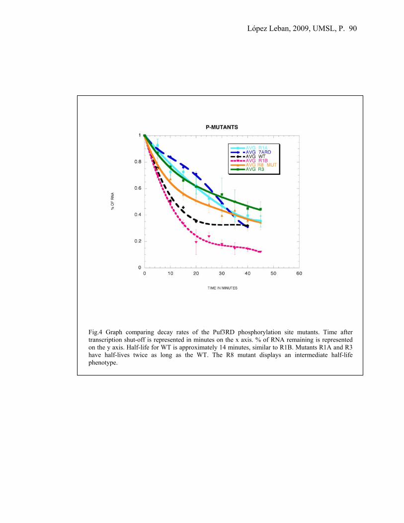
López Leban, 2009, UMSL, P. 90
Fig.4 Graph comparing decay rates of the Puf3RD phosphorylation site mutants. Time after transcription shut-off is represented in minutes on the x axis. % of RNA remaining is represented on the y axis. Half-life for WT is approximately 14 minutes, similar to R1B. Mutants R1A and R3 have half-lives twice as long as the WT. The R8 mutant displays an intermediate half-life phenotype.

López Leban, 2009, UMSL, P. 91
Puf3-GFP images under different environmental conditions may show a difference
in protein aggregation.
P-bodies have been defined as sites of mRNA storage, decapping and decay. In
order to visualize these decay factors in the cell, several GFP fusion constructs of
different yeast mRNA decay factors were observed in vivo. Puf3-GFP was shown to be
evenly distributed throughout the cytoplasm, while Dcp2-GFP was found in P-bodies. All
fusion constructs have been shown to be functional and were made with full-length
proteins GFP labeled at the C terminal (Sheth and Parker, 2003).
Another hypothesis to explain the change in the activity of Puf3p under different
conditions may be that alteration of protein localization prevents it from performing its
function. I used the same GFP tagged strains as described above to evaluate any
difference in localization of Puf3-GFP in yeast grown under different carbon sources.
Dcp2-GFP was used as a positive control, and yWO5, a WT yeast strain that is not GFP
tagged, as a negative control. Though the imaging of Puf3-GFP does not show a radical
difference in protein localization when comparing ethanol, galactose, and raffinose
conditions versus glucose, the images do show a more clumpy phenotype under the first
three conditions. This punctate pattern may be reflecting aggregated protein versus
homogeneously distributed protein. However, this punctuate pattern is clearly not the
same type as the P-body loci seen with Dcp2-GFP (Fig.5). These results are supported by
experiments in which the amount of Puf protein isolated was measured under different
conditions. Using the protein boil prep method, in which both soluble and insoluble
protein is recovered, the westerns show a greater amount of Puf3 protein present in
conditions other than glucose. On the other hand, when using the YPER method, in

López Leban, 2009, UMSL, P. 92
which most of the protein recovered is in a soluble state, the greater amount of Puf3
protein is recovered in glucose compared with the other conditions (Fig.6). This means
that even though there might be more Puf3 protein in ethanol, galactose and raffinose
compared to glucose, most of this protein may be in an insoluble, non-functional state. To
conclude, non-functional protein can be visualized and compared to the functional
version obtained in glucose media by using a hybrid Puf3p with a GFP tag, which allows
the observation of a clumpy versus a homogeneous cytoplasmic distribution correlated to
media and protein activity.

López Leban, 2009, UMSL, P. 93
Fig.5 Images of Puf3-GFP in different carbon sources (central column). Images on the left column correspond to a negative control strain tested in glucose, galactose, ethanol and raffinose N/D. Images on the right column correspond to Dcp2-GFP, which was used as a positive control and localizes in very distinctive spots known as P-bodies. The central column shows the distribution and appearance of Puf3-GFP under the conditions tested.
Soluble protein extraction
Glu Eth
Total protein extraction
Glu Eth
YPER Protein boil prep
Fig.6 Puf3p recovered using different extraction protocols. Western blots developed with anti Puf3RD antibodies of protein extracts recovered from cultures grown in glucose and in ethanol. Extractions were performed using protein boil prep protocol on the left and YPER protocol on the right.

López Leban, 2009, UMSL, P. 94
Discussion
Previous research in our lab has shown Puf3RD’s activity being dependent on the
type of carbon source available. It was shown to be inactive in ethanol and active in
glucose, with activity measured by comparing COX17 mRNA half-life (a Puf3-mediated
decay target) in the different conditions. This half-life was shown to be 2.5 minutes in
glucose and 10 minutes in ethanol (Foat, et al, 2005). So far we have detected similar
levels of both Puf3 protein and mRNA under ethanol, galactose and raffinose conditions
in which the protein was in an inactive state compared to glucose. This information,
together with the observation of very rapid Puf3p activity changes in response to different
carbon sources in the media, led us to consider one of the most common post-
translational modifications, phosphorylation, as a way of quickly responding to these
environmental changes. In fact, I demonstrated that Puf3RD is phosphorylated by
performing a phosphatase treatment of purified Puf3p and using phospho-specific
antibodies to detect phosphorylated serines or tyrosines. Further site–directed
mutagenesis analysis, in which four of the most conserved potential phosphorylation
residues within the Puf3RD were mutated to alanines, allowed us to evaluate their
functional role in Puf3p activity through their ability to rescue a puf3Δ strain. Several
decay phenotypes were detected, but mutations in R1A, R3 and R8 seem to be very
important in promoting Puf3RD activity. These point mutations in the serines or tyrosine
might alternately be responsible for a general conformational change affecting RNA
binding or the recruitment of the decay factors. Further studies should be performed with
the R1A-R3-R8 triple P-mutant since preliminary data suggests that these three mutations
together might be decreasing Puf3RD activity to a greater extent. Substitution of these

López Leban, 2009, UMSL, P. 95
specific alanines to aspartic acid to mimic a phosphorylated state should show a re-gain
in function if it is indeed phosphorylation that controls activity. To further confirm any
possible role of phosphorylation on Puf3p activity, a set of kinases involved in nutrient
response will be tested for their role in Puf3p activity through transcriptional shut-off
experiments using different kinase knockout strains under different carbon sources and
detecting any differences in the COX17 mRNA decay rate.
In this work we also addressed the possibility of associating an inactive state of
Puf3p with it becoming inaccessibly localized or aggregated. Visualizing Puf3-GFP
protein from yeast cultures grown in glucose demonstrated a cytoplasmic and
homogeneous distribution in the yeast cells, while a difference in distribution was
detected under galactose, raffinose and ethanol conditions that coincided with inactive
Puf3p, which all demonstrated a more punctate appearance. These differences were
shown to correlate with differences in solubility. Two protein extraction protocols
showed that even when the amount of Puf3p present in ethanol, galactose and raffinose is
greater than in glucose, most of it is in an insoluble, non-functional state. On the contrary,
most of the protein recovered from yeast cultures grown in glucose was soluble and
functional.
As it has been previously shown in Dictyostelium in which activation of PufA was
achieved by a different phosphorylation state of the protein to allow developmental
changes (Mendes Souza et al., 1999). This research further confirms that this type of
post-translational regulation may also occur in Puf3p in yeast. By better understanding
the mechanisms by which yeast and other lower eukaryotes are able to regulate gene
expression in their response to changing environmental conditions, a stronger basis will

López Leban, 2009, UMSL, P. 96
be established that might be applicable to the regulation of Puf activity in higher
eukaryotes.

López Leban, 2009, UMSL, P. 97
References
Decker, C. J. and Parker, R. (1995) Diversity of cytoplasmic functions for the 3’ untranslated region of eukaryotic transcripts. Current Opinion in Cell Biology 7: 386-92. Decker, C. and Parker, R. (2002) mRNA decay enzymes: Decappers conserved between yeast and mammals. PNAS 99: 12512-4. DeRisi, J. L., Iyer, V. R., Brown, P. O. (1997) Exploring the metabolic and genetic control of gene expression on a genomic scale. Science 278: 680-5. Dunckley, T. and Parker, R. (1999) The Dcp2 protein is required for mRNA decapping in Saccharomyces cerevisiae and contains a functional Mut T motif. EMBO J. 18: 5411-22. Dunkley, T., Tucker, M. and Parker, R. (2001) Two related proteins, Edcp1 and Edcp2, stimulate mRNA decappping in Saccharomyces cerevisiae. Genetics 157: 27-37. Gasch, A. P., Spellman, P. T., Kao, C. M., Carmel-Harel, O., Eisen M. B., Storz, G., Botstein, D. and Brown, P. O. (2000) Genomic expression programs in the response of yeast cells to environmental changes. Molec Biol.of the Cell 11: 4241-57. Gerber, A.P., Herschlag, D. and Brown, P.O. (2004) Extensive association of functionally and cytotopically related mRNAs with Puf family RNA-binding proteins in yeast. PLoS Biology 2: 342-54. Goldstrohm, A. C., Hook, B.A., Seay, D. J. and Wickens, M. (2006) Puf proteins bind Pop2p to regulate messenger RNAs. Nature Struct. Mol. Biol. 13: 533-9. Jackson, J.S., Houshmandi, S.S., Lopez leban, F. and Olivas, W.M. (2004) Recruitment of puf3 protein to its mRNA target for regulation of mRNA decay in yeast. RNA 10: 1625-36. Muhlrad, D., Decker, C.J. and Parker, R. (1995) Turnover mechanism of the stable yeast PGK1 mRNA. Molecular and Cellular Biology 15: 2145-56.

López Leban, 2009, UMSL, P. 98
Chapter V
Experimental identification of bona fide Puf4p
mRNA targets in yeast and analysis of Puf4p role
in mRNA decay

López Leban, 2009, UMSL, P. 99
Introduction
Genome-wide RNA-protein interaction analyses have shown the presence of
conserved sequence elements in the 3’UTRs of distinct groups of mRNAs that help
provide a coordinated control of gene expression in response to different physiological
and environmental conditions. One such post-transcriptional regulation through RNA-
binding proteins in yeast is the Puf family of proteins. There might be a general
mechanism for controlling in a coordinated way localization, translation and decay of a
particular set of mRNAs as an integral part of the global gene expression program.
Through DNA microarrays, conserved sequence elements in the 3’UTRs of mRNAs that
were pulled down were identified that are needed for Puf3p, Puf4p and Puf5p binding
(Gerber et al, 2004). This UGUANAUA sequence was also experimentally confirmed to
be required for Puf3p binding in our lab; in fact, COX17 3’ UTR contains two distinct
binding sites for recruiting Puf3p and stimulating its decay (Jackson et al. 2004). Results
obtained from this genome-wide analysis of the mRNAs pull-down by individually
tagged Pufs showed a preferential association of Puf1p and Puf2p with mRNAs encoding
membrane associated proteins, Puf3p with cytoplasmic mRNAs encoding mitochondrial
proteins, Puf5p with mRNAs encoding chromatin modifiers and components of the
spindle pole body and Puf4p with mRNAs encoding nucleolar ribosomal RNA-
processing factors (Gerber et al. 2004). Puf4 protein has also been shown to somehow be
involved in localization of Sir proteins to the nucleolus, and thus, in the regulation of
aging in yeast (Kennedy et al., 1997), its mode of function is yet unknown.
Puf4p in yeast consists of 888 amino acids, is approximately 97 KDa and as a
characteristic feature of the Puf family it contains the Pumilio homology domain,

López Leban, 2009, UMSL, P. 100
comprised of eight ɑ-helical repeats each of which recognizes one RNA base. Recently
Puf4p crystal structure has been solved showing an altered RNA binding specificity that
allows the protein to accommodate an extra nucleotide by allowing the rotation of one of
the bases away from the RNA binding surface (Miller et al, 2008). At the beginning of
this study, there were no experimentally determined Puf4p mRNA targets known. Taking
advantage of previous high throughput studies, I wanted to experimentally test mRNA
candidates that were ranked as top Puf4p targets by both co-
immunoprecipitation/microarray studies (Gerber et al., 2004) and computational
predictions using Matrix REDUCE (Foat et al., 2005). The information obtained from
these types of studies is very useful to help narrow down the selection of possible
candidates for targeted decay by a particular yeast Puf. However, experiments in our lab
have shown that not all the hundreds of mRNAs that are predicted to be targets of a
specific Puf turn out to be real Puf-mediated decay targets. It could be that in the
complexity of the cell, factors other than Pufs might be influencing the expression of
these Puf predicted targets under these conditions, or that these targets are indirectly
being affected by Pufs.
In this research I experimentally identified targets of Puf4p-mediated decay and
analyzed the mechanism of Puf4p action in yeast. This is especially interesting given that
expression profiles of predicted Puf4p targets are so different than for targets of the well-
studied Puf3p based on the inferred activities of Puf3p and Puf4p under different
environmental conditions from the Matrix REDUCE results (Foat et.al, 2005). I first
compared the steady state levels of each mRNA candidate from a wild type strain and a
puf4∆ strain. Having identified differentially expressed candidates, transcriptional shut

López Leban, 2009, UMSL, P. 101
off experiments were performed to see whether there were differences in the decay rate of
the transcripts in the presence or absence of Puf4p. These targets were found to be co-
regulated by both Puf4p and Puf5p.
Experimental Procedures
Steady state experiments
Seven mRNA candidates were initially chosen based on two lists of possible
Puf4p target mRNAs. One list from the Bussemaker lab was computationally selected
using microarray data and the 200bp sequence downstream of every yeast ORF as input,
while the other list from the Brown lab was obtained from co-immunoprecipitation of
mRNAs with Puf proteins. Complementary DNA probes were designed for each one of
these mRNAs and were proven to be unique using the PATmatch search from the
Saccharomyces Genome Database. The mRNAs tested included, PWP1, HPT1, ECM1,
YJL122W, PUS7, EBP2 and RRS1.
To perform these steady state experiments, wild type (yWO 7) and puf4∆ (yWO
105) strains were each grown in either rich YEPD (dextrose) or YEPE (ethanol) media at
24˚C to an OD of 0.4, at which time they were harvested. The cells were then processed
for RNA extraction, 40μg of each RNA sample was separated on 1% agarose gels, and
Northern blots were prepared and probed for each candidate mRNA. Blots were also
probed for 7S RNA to control for loading.

López Leban, 2009, UMSL, P. 102
Transcriptional shut-off experiments
In order to determine if the steady-state differences were due to the Puf4p
affecting the decay rate of these mRNAs, a set of transcriptional shut-off experiments
were carried out to monitor the decay rate of a pool of steady-state mRNA following
repression of their transcription. These experiments were originally attempted in strains
containing the temperature sensitive RNA polymerase II, where transcription is repressed
by a shift of the culture media to 37˚C.
An alternative method of performing transcriptional shut-offs is to clone each
gene of interest into a yeast vector under the control of a repressible promoter. For
example, a GAL promoter can be induced by the addition of 2% galactose and repressed
by switching to a media containing 4% glucose. The TET promoter can be repressed by
the addition of doxycycline (2µg/ml) to the culture media (Belli et al, 1998). Both
promoters were tested since there is some evidence from microarrays that Puf4p may be
inactivated by galactose, while the TET promoter construct is more difficult to clone and
ensure expression. In the case of YJL122W and PUS7, which are both non-essential
genes, the endogenous genes were knocked out of the wild type and puf4∆ strains so that
only the plasmid derived mRNA would be visualized on Northern blots. To make the
knock-outs, the TRP1 gene was PCR amplified from pRS200 as a template using primers
that were designed to be complementary to either side of the TRP1 gene at their 3’ ends,
but contained ~ 40 nt of secquence complementary to either side of the target gene to be
knocked out. oWOs 369 and 370 were complementary to YJL122W, and oWOs 371 and
372 were complementary to PUS7. The PCR products obtained with the corresponding
flanking homologous regions were used to knock out YJL122W or PUS7 in each of the

López Leban, 2009, UMSL, P. 103
strains yWO 5 (WT) and yWO 22 (puf4∆). However, for the essential genes EBP2 and
RRS1, unique sequence tags have to be inserted into the plasmid genes so that probes
complementary to these sequences will allow visualization of only the plasmid derived
mRNAs on Northerns. Both RRS1 genes flanked by BamH I and Hind III one with a
GAL10 promoter and the other with a TET promoter were inserted in pGEM3Z and they
were cut at the Acc I site for annealing an oWO for tagging purposes.
Table 1 Strains used in these studies
Deletion Strain Genotype Source W T yWO 5 MAT a, trp1, ura3-52, leu2, his4-539,cup1:LEU2/PM Olivas
&Parker(2000) W T yWO 7 MAT x, ura3-52, leu2, rpb1-1 Olivas
&Parker(2000) Puf4Δ yWO 22 MAT ɑ, ura3-52, leu2-3,112, trp1, lys2, cup1:LEU2/PM, YGLO14w::LYS2 Olivas
&Parker(2000) Puf4Δ yWO 105 MAT ɑ, ura3,his4,rpb1-1, YGLO14w::LYS2 Ulbricht & Olivas
(2008)
Results
Steady state experiments
An initial analysis of the seven selected mRNA candidates indicated that only
four (YJL122W, PUS7, EBP2 and RRS1) showed a 2 to 3 fold difference in levels
between the WT and puf4∆ strain when grown in glucose (Fig.1). There were no
significant differences in mRNA levels between both strains grown in ethanol. Since
mRNA levels were higher in the WT versus puf4∆ grown in glucose, these results
suggest a stabilizing activity of Puf4p on its targets. Moreover, the equally low mRNA
levels in both WT and puf4∆ strains grown in ethanol suggest a loss of stabilizing activity
of Puf4p when grown in a non-fermentable carbon source. These results also showed that
not every candidate mRNA is a true target of Puf4p.

López Leban, 2009, UMSL, P. 104
PUS7
EBP2
G EG E G EG
RRS1
YJL122W
E WT puf4∆ WT puf4∆
1
1
1
1 0.3
0.30.4
0.4
7S
EtBr
PUS7
EBP2
G EG E G EG
RRS1
YJL122W
E WT puf4∆ WT puf4∆
1
1
1
1 0.3
0.30.4
0.4
7S
EtBr
PUS7
EBP2
G EG E G EG
RRS1
YJL122W
E
PUS7
EBP2
G EG E G EG E G EG E G EG
RRS1
YJL122W
E WT puf4∆ WT puf4∆
1
1
1
1 0.3
0.30.4
0.4
7S
EtBr
Fig.1 Northern blots of steady state experiments. mRNA levels compared between WT and puf4∆ strains of yeast cultures grown in media with glucose (G) or ethanol (E) as carbon sources. PUS7 and EBP2 are shown on the left, RRS1 and YJL122W are shown on the right. 7S and ethidium bromide staining of ribosomal RNA are used as loading controls. The numbers on top of the corresponding lanes show RNA quantification after normalization to 7S.

López Leban, 2009, UMSL, P. 105
Transcriptional shut-off experiments.
The four mRNAs YJL122W, PUS7, EBP2 and RRS1 that showed a difference in
steady-state mRNA levels in WT versus puf4∆ strains all encode proteins involved in
ribosomal RNA biogenesis. EBP2 and RRS1 are essential genes in the cell, while
YJL122W and PUS7 are non-essential. Rrs1p (Regulator of Ribosome Synthesis) is an
essential protein that binds ribosomal protein L11 and is required for 25S rRNA
maturation and 60S ribosomal subunit assembly and export from the nucleolus to the
cytoplasm (Miyoshi, K. 2002; Mizuta, K., 2004). RRS1 was originally found as the gene
responsible for signaling transcriptional repression for rRNA and ribosomal proteins
because of a blockage in the secretory pathway (Horigome et al. 2008). It has been shown
in mouse model of Huntington’s disease (HD) in which the expression of mutant 111-
glutamine murine huntingtin protein leads to a dominant phenotype, that is correlated
with an elevated RRS1 mRNA expression that can be used to detect very early onset of
HD (Fossale et al. 2002). Ebp2p is also an essential protein that might be Rrs1p target for
performing 25 S rRNA maturation and 60 S subunit assembly (Tsuyii et al. 2000).
Ebp2p, the yeast homolog of human Epstein-Barr virus nuclear antigen 1- binding protein
was shown to interact Rps16p (ribosomal protein S16) and Utp11p (the 40 S ribosomal
subunit assembly factor) (Shirai et al. 2004). Ebp2p similarly to Rrs1p has been found to
be implicated in the secretory response (Horigome et al. 2008). PUS7 is a non-essential
gene that encodes for a pseudouridine –synthase Pus7p that in yeast acts catalyzing U to
ψ conversion both in U2 snRNA and in pre-tRNA (Behm- Ansmant et al. 2003).
In order to determine if the steady-state differences are due to the Puf4p affecting
the decay rate of these mRNAs, a set of transcriptional shut-off experiments were carried

López Leban, 2009, UMSL, P. 106
out to monitor the decay rate of a pool of steady-state mRNA following repression of
their transcription. First a set of experiments was done in strains containing the
temperature sensitive RNA polymerase II, where transcription is repressed by increasing
the temperature to 37˚C. However, all mRNA targets tested decayed fairly rapidly in both
wild-type and puf4∆ strains. We believe that this result was due to the Puf4p being
inactivated by the heat shock. Microarray analysis showed that candidate Puf4 target
mRNA levels are low in cultures grown at high temperature, which suggests an
inactivation of Puf4p stabilizing activity (Foat et.al. 2005).
Next, two alternative ways of performing transcriptional shut-offs were done in
which each gene of interest was cloned into a yeast vector under either a GAL or TET
repressable promoter. YJL122W and PUS7, which are both non-essential genes, were
knocked out of the wild type and puf4∆ strains so that only the plasmid derived mRNA
would be expressed. Since EBP2 and RRS1 are essential genes, unique sequence tags
were inserted in the cloned genes so that specific complementary probes would allow
visualization of the plasmid derived mRNAs. The results in figure 2 show the decay rates
of the plasmid-derived YJL122W under a GAL promoter in WT and puf4∆ strains as an
example. The half-lives of the four different mRNAs tested (YJL122W, EBP2, RRS1 and
PUS7) when comparing WT to puf4∆ were very similar for each mRNA in both strains.
Further research performed by two undergraduates under my supervision (Jeanne Pitts
and Susana Pulido-Fernandez), showed that these mRNA targets were being co-regulated
by both Pu4p and Puf5p. Preliminary results indicate that Puf4p activity might be
inhibited by galactose and ethanol, while Puf5p retains its activity for decay stimulation
under these conditions.

López Leban, 2009, UMSL, P. 107
Fig. 2 Decay rates of YJL122W under GAL promoter. Time in minutes is represented in the x axis and % of RNA is represented on the y axis. WT and puf4∆ strains are represented.

López Leban, 2009, UMSL, P. 108
Discussion
During the process of this research, evidence of redundant regulation by Puf4p
and Puf5p was shown for HO mRNA (Goldstrohm et al., 2007). The fact of having found
in our lab two new Puf targets, TIF1 and HXK1, which are both regulated by Puf1p and
Puf5p with the addition of Puf4p for HXK1 also showed the possibility of a coordinated
regulation, since all these Pufs are needed for full decay stimulation of their target mRNA
(Ulbricht and Olivas, 2008).
So far, the steady state analyses using strains grown both at 24ºC and at 30ºC
indicate that the identified Puf4p targets are also regulated by Puf5p. Though both Pufs
seem to be active and redundantly regulating their targets in glucose, their activities
appear to be differently affected by the presence of other carbon sources. These results
together with the recent finding of Puf4p’s altered RNA-binding specificity that allows it
to bind either a eight or nine nucleotide sequence, supports the idea of redundant
regulation. On the other hand, the fact of Pufs showing different activity profiles under
different carbon sources aligns with the idea of a combinatorial way of regulation.
Probably both types of regulation provide the cell with a robust and also flexible
mechanism to better respond to changing environmental conditions.
Future work will need to determine whether the combination of Puf4p and Puf5p
is really altering target mRNA levels via changes in RNA decay rates. A broader
spectrum of strains including WT, puf4∆, puf5∆, puf4&5∆ and all 5pufs∆, should be used
for performing transcriptional shut-off experiments to evaluate under different carbon
sources, the extent of each one of the Puf’s regulation in a particular environment.

López Leban, 2009, UMSL, P. 109
References Behm-Ansmant, I., Urban, A., Ma, X., Yu, Y., Motorin, Y. and Branlant, C. (2003) The Saccharomyces cerevisiae U2 snRNA:pseudouridine-synthase Pus7p is a novel multisite–multisubstrate RNA:Ψ-synthase also acting on tRNAs. RNA 9: 1371-82. Belli, G., Aldea, M. and Herrero, E. (1998) Functional analysis of yeast essential genes using a promoter substitution cassette and the tetracycline-regulatable dual expression system. Yeast 14: 1127-38. Foat, B.C., Houshmandi, S.S., Olivas, W. M. and Bussemaker, H. J. (2005) Profiling condition-specific, genome –wide regulation of mRNA stability in yeast. PNAS 102: 17675-80. Fossale, E., Wheeler, V. C., Vrbanac, V., Lebel, L. A., Teed, A., Mysore, J. S., Gusella, J. F., MacDonald, M. E. and Persichetti, F. (2002) Identification of presymptomatic molecular phenotype in Hdh CAG knock-in mice. Human Molecular Genetics 11: 2233-41. Gerber, A.P., Herschlag, D. and Brown, P.O. (2004) Extensive association of functionally and cytotopically related mRNAs with Puf family RNA-binding proteins in yeast. PLoS Biology 2: 342-54. Horigome, C., Okada, T., Matsuki, K. and Mizuta, K (2008) A ribosome assembly factor Ebp2p, the yeast homolog of EBNA1-Binding Protein 2, is involved in the secretory response. Biosci. Biotechnol. Biochem. 72: 1080-6. Kennedy, B., Gotta, K., Sinclair, D., Mills, K., McNabb, D., Murthy, M., Pak, S., Laroche, T., Gasser, S., Guarente, L. (1997) Redistribution of silencing proteins from telomeres to the nucleolus is associated with extension of life span in S. cerevisiae. Cell 89: 381-91. Miller, M. T., Higgin, J. J. and Tanaka Hall, T. M. (2008). Basis of altered RNA-binding specificity by PUF proteins revealed by crystal structure of yeast Puf4p. Nature Structural and Molecular Biology 15: 397-402.
López Leban, 2009, UMSL, P. 110
Miyoshi, K., Tsujii, R, Yoshida, H., Maki, Y., Wada, A., Matsui, Y., Toh-e, A. and Mizuta, K. (2002) Normal assembly of 60 S ribosomal subunits is required for signaling in response to a secretory defect in Saccharomyces cerevisiae. JBC 277: 18334-9. Mizuta, K.(2004) Rrs1p, a ribosomal protein L11- binding protein, is required for nuclear export of the 60 S pre-ribosomal subunit in Saccharomyces cerevisiae. FEBS Letters 565: 106- 110. Shirai, C., Takai, T., Nariai, M., Horigome, C. and Mizuta K. (2004) Ebp2, the yeast homolog of Epstein-Barr virus nuclear antigen 1-binding protein 2, interacts with factors of both the 60S and 40S ribosomal subunit assembly. JBC 279: 25353-8. Ulbricht, R. J. and Olivas, W.M. (2008) Puf1p acts in combination with other yeast Puf proteins to control mRNA stability. RNA 14: 246-62.

López Leban, 2009, UMSL, P. 111
Chapter VI
Summary and future directions

López Leban, 2009, UMSL, P. 112
Organisms have to adapt to changing environmental conditions in order to
survive. A precise and efficient regulation of gene expression is required to obtain the
optimal response at the lowest cost of energy. The Puf protein family of RNA binding
proteins has been identified as an important and widespread group that regulates post-
transcriptional steps of the gene expression pathway. Regulation is achieved by altering
the decay and translation rates of specific target mRNAs. In Drosophila, Pumilio has not
only been shown to be critical for embryo patterning by establishing a gradient of
hunchback mRNA through translational repression, but has also been shown to regulate
neuronal excitability by affecting the expression of a Na+ channel gene (Schweers et al,
2002; Mee, 2004). Together the Puf protein family is involved in mRNA expression
regulation for mediating several developmental decisions such as cell differentiation and
stem cell maintenance, as well as involved in neuronal plasticity accounting for long-
lasting structural and functional modifications in the synapsing neurons that are required
for the acquisition of long-term memory. However, the mechanism of Puf protein action
in these processes is largely unknown. In yeast, Puf proteins were found associated with
hundreds of different mRNA targets, though the functional significance of most of these
interactions is unclear (Gerber et al., 2004). Of the six Pufs identified in Saccharomyces
cerevisiae, Puf3p has been the best characterized to date. Puf3p regulates COX17 mRNA
decay by specifically binding two conserved UGUANAUA sequences present in its 3’
UTR and thereby stimulating both deadenylation and decapping. It has also been shown
that the Puf3p repeat domain alone is sufficient not only for in vitro binding to the
3’UTR, but also for in vivo stimulation of COX17 mRNA decay (Jackson et al, RNA,
2004).

López Leban, 2009, UMSL, P. 113
In this research, insight was gained on Puf3p interactions with other proteins that
contribute to its mechanism of action for COX17 mRNA regulation. Similar to the Puf5p
mechanism of action for achieving HO mRNA regulation, Puf3p binds through protein
interactions with several members of the decay machinery including, Pop2p, Ccr4p,
Lsm1p, Dcp1p and Dhh1p. I found the R7A outer surface loop to be the precise region to
which Pop2p directly and Dhh1p indirectly through Pop2p binds the Puf3RD, while the
other decay factors bind to Puf3RD independently of this loop region and Pop2p. This
differs from the model of Puf5p interactions in which Pop2p is thought to bridge between
Puf5p and all the deadenylating and decapping factors, and no specific binding site has
yet been described (Goldstrohm et al, 2007). The interactions between Puf3RD and the
decay machinery were verified through both modified yeast three-hybrid assays and co-
immunoprecipitation experiments. I also confirmed that at least in the case of Puf3-
mediated decay of COX17 mRNA, the process is independent of the nonsense-mediated
decay pathway (NMD).
Puf3p activity was next analyzed under different environmental conditions to
better understand how its activity responds to the carbon source available. Research in
our lab shows Puf3p inactivity under ethanol, galactose and raffinose conditions versus
glucose, yielding extended mRNA half-lifes of Puf3p targets under these inactivating
conditions. The high levels of both Puf3 protein and mRNA found under ethanol,
galactose and raffinose in which the protein was in an inactive state compared to glucose,
led us to consider post-translational modifications of Puf3p as a way of quickly
responding to the environmental changes. In fact, other work from the Olivas lab has
observed that Puf3p activity can be rapidly turned on and off by simply changing the

López Leban, 2009, UMSL, P. 114
carbon source in the culture media (Miller, unpublished data). Puf3 protein was found to
be consistently phosphorylated under different carbon sources. This could be one of the
most common post-translational modifications to regulate its activity. Both
phosphorylated Ser and Tyr residues were detected using phospho-specific antibodies,
but our assays were unable to determine whether specific sites were being differentially
phosphorylated under different conditions. Further work will involve using mass
spectrometry for detecting any specific modification sites on tryptic fragments of the
Puf3RD purified from different conditions. It will also be interesting to test interactions
between Puf3RD and all the decay factors under different carbon sources to determine
whether the same protein interaction network is maintained under conditions when the
protein is active versus when it is inactive. Another experiment that could be used for
detecting differences in Puf3p protein interactions is a translational repression assay
procedure (TRAP). For this assay, a GFP reporter construct is designed such that it has a
binding site for the protein being tested at its 5’ UTR. If the protein is able to bind, then
ribosome recruitment is inhibited and there will be no GFP translated. The difference in
binding capability due to the carbon source will be reflected in the amount of GFP
measured by flow cytometry.
To further analyze the effect of phosphorylation on Puf3p activity, potential
phosphorylation sites were identified through bioinformatic analyses, and four of the
most conserved sites within the Puf3RD were mutated to alanines to test their function in
Puf3p activity. Each mutant demonstrated a less than 2-fold effect in extending the
COX17 mRNA half-life, suggesting that phosphorylation of these sites is likely not the
key trigger for activating Puf3p activity. Instead, mutation of these sites may be causing

López Leban, 2009, UMSL, P. 115
structural changes to the protein that cause a slight decrease in protein efficiency. It is
possible that phosphorylation acts to inhibit Puf3p activity, so further studies could
mutate these sites to aspartic acid to mimic a phosphorylated state and determine if this
inhibits activity. To further analyze any possible role of phosphorylation on Puf3p
activity, yeast strains deleted of different kinases known to be involved in nutrient
response could be screened for any altered COX17 mRNA decay rate in glucose versus
ethanol conditions to test a role in Puf3p activation or inhibition.
This work also addressed the possibility that the localization or aggregation state
of Puf3p may be involved in the inactivation of Puf3p. Puf3-GFP localization under
different carbon source conditions appears to be cytoplasmic. However, the protein
seems to have a more punctate pattern under inactivating conditions (galactose, raffinose
and ethanol). This might indicate a more aggregated or insoluble state of the protein in
these conditions compared to glucose. In support of this hypothesis, more soluble Puf3
protein is extracted from cells grown in glucose versus ethanol using the YPER
extraction protocol that preferentially obtains soluble protein, while more Puf3p protein is
actually produced in ethanol versus glucose conditions as obtained from the boil prep
extraction procedure that collects both soluble and insoluble protein. This suggests that
even when the amount of Puf3p present in ethanol is greater than in glucose, much of it
may be in an insoluble, non-functional state.
Another aspect of this research focused on experimentally confirming new Puf4p
target mRNAs and evaluating whether Puf4p activity might also be affected by
environmental conditions. Computational predictions by the Matrix REDUCE algorithm
showed a complete opposite expression pattern for transcripts containing Puf3p and

López Leban, 2009, UMSL, P. 116
Puf4p binding elements. Under conditions with fermentable carbon sources when Puf3p
regulated transcripts are down-regulated, Puf4p target transcripts are up-regulated. These
expression pattern predictions make sense metabolically. Puf3p target transcripts code for
proteins involved in mitochondrial function, so these proteins are only needed under non-
fermentable carbon sources such as ethanol. Thus, the Puf3p target transcripts should be
down-regulated in glucose when mitochondria are not needed. In contrast, many
predicted Puf4p target transcripts code for proteins involved in ribosomal biogenesis. In
glucose conditions when the growth rate is fast, there is a need for high numbers of
ribosomes for protein production. Thus, the Puf4p target transcripts should be up-
regulated in such conditions.
Four Puf4p mRNA targets (out of seven) were experimentally validated using
analysis of steady-state RNA levels from different conditions. These targets were:
YJL122W, RRS1, PUS7 and EBP2. However, transcriptional shut-off experiments to
measure mRNA decay rates of these targets revealed no significant differences between
wild-type and puf4Δ strains. It is possible that these mRNA targets are not regulated by
Puf4p at the level of mRNA decay, or that the experimental treatments used to shut off
transcription altered Puf4 activity. However, the Olivas lab and others have shown that
many Puf target mRNAs are regulated by combinations of Pufs, including HXK1 and HO
mRNAs both regulated by Puf4p and Puf5p (Ulbricht and Olivas 2008, Goldstrohm et al.
2006; Hook et al. 2007). I therefore supervised studies performed by two undergraduates
(Jeanne Pitts and Susana Pulido Fernandez) that provided evidence from steady-state
RNA analyses that all of the identified Puf4p target RNAs are also regulated by Puf5p,
and these two Pufs may be acting redundantly under glucose conditions. One or both Puf

López Leban, 2009, UMSL, P. 117
protein activities may then be altered under different carbon source conditions to cause
changes in RNA levels. Future work will need to determine whether the combination of
Puf4p and Puf5p is really altering target mRNA levels via changes in RNA decay rates,
and how different carbon sources may affect Puf4p and Puf5p activity.
Up to date we know that Puf proteins belong to a family of RNA-binding proteins
that are widely conserved across eukaryotes. We can assign them a central role in
regulating mRNA stability of several targets that contain specific control elements in
their 3’ UTRs. Through this regulation, two main roles are fulfilled: one involved in cell
development and differentiation, and the other related to neuronal morphology and
physiology. Although we and others have shown the importance and sufficiency of the
Puf repeat domain for binding and stimulating mRNA decay, we should also keep in
sight that there may be some important yet currently unknown role for the rest of the Puf
protein outside the repeat domain.
Puf proteins in humans have been shown to be expressed in the brain and form
complexes with different proteins, one of them being CPEB (Cytoplasmic
Polyadenylation Element Binding protein), which controls translation of synaptic
mRNAs. Human Pum 2 has specifically been shown to be present in dendritic stress
granules (SG) in mammalian hippocampal neurons. The induction of these granules
depends on both the RNA-binding domain and the glutamine-rich N terminus of Pum 2,
which makes it prone to aggregation. Both Pumilio and Staufen, another protein also
present in SGs, are involved in the process of acquiring long term memory (Dubnau,
2003). CPEB’s presence in hippocampal cells is important for regulating local protein
synthesis essential for late phase of long term potentiation. The conversion of CPEB to a

López Leban, 2009, UMSL, P. 118
prion-like state in stimulated synapses contributes to the long-term maintenance of a self-
sustaining synapse-specific change associated with memory storage. Since this prion-like
state is self-perpetuating, it no longer requires continued signaling either by kinases or
phosphatases. We have shown in yeast that different environmental conditions are
responsible for an active or inactive Puf protein. Similarly, particular local conditions in
the mammalian brain that occur to induce a change to the aggregated prion-like state for
acquisition of long term memory may also be regulating Puf activity. We have seen how
certain carbon sources in yeast induce a punctate, possibly aggregated form of Puf3p. It
is possible that the N-terminal portion of the protein that is glutamine-rich might make it
prone to aggregation. A speculative hypothesis suggests that aggregation of Puf proteins
may in one case cause a loss of function (COX17 mRNA regulation) and on the other
hand may create a gain of function in the neuron for memory storage.
In conclusion, Puf proteins are involved in several important processes in
eukaryotic cells. Given their role in stem cell maintenance and cell differentiation, Puf
protein activity may be an important factor to address in the growing field of stem cell
therapy research. Knowledge of Puf protein activity in neural function may also be
important in the development of new therapies for neurodegenerative diseases such as
Alzheimers, Huntingtons, Parkinsons and several of the prion-related illneses that cause
neuronal damage and loss of memory. The research that we have performed to
understand the detailed mechanisms of Puf protein function in yeast cells should provide
critical pieces of the puzzle of Puf protein regulation in higher eukaryotes.

López Leban, 2009, UMSL, P. 119
References
Dubnau, J., Chiang, A-S., Grady, L., Barditch, J., Gossweiler, McNeil, J., Smith, P., Buldoc, F., Scott, R., Certa, U., Broger, C. and Tully, T. (2003) The staufen / pumilio pathway is involved in Drosophila long- term memory. Current Biology 13: 286- 96. Gerber, A.P., Herschlag, D. and Brown, P.O. (2004) Extensive association of functionally and cytotopically related mRNAs with Puf family RNA-binding proteins in yeast. PLoS Biology 2: 342-54. Goldstrohm, A. C., Hook, B.A., Seay, D. J. and Wickens, M. (2006) Puf proteins bind Pop2p to regulate messenger RNAs. Nature Struct. Mol. Biol. 13: 533-9. Hook, B.A., Goldstrohm, A.C., Seay, D. J. and Wickens, M. (2007) Two yeast Puf proteins negatively regulate a single mRNA. J. Biol.. Chem. 282: 15430-8. Jackson, J.S., Houshmandi, S.S., Lopez leban, F. and Olivas, W.M. (2004) Recruitment of puf3 protein to its mRNA target for regulation of mRNA decay in yeast. RNA 10: 1625-36. Mee, C. J., Pym, E. C. G., Moffat, K.g. and Baines R. A. (2004) Regulation of neuronal excitability through Pumilio-dependent control of a sodium channel gene. The Journal of Neuroscience 24: 8695- 8703. Schweers, B.A., Walters, k.J. and Stern, M. (2002) The Drosophila melanogaster translational repressor pumilio regulates neuronal excitability. Genetics 161: 1177-85. Ulbricht, R. J. and Olivas, W.M. (2008) Puf1p acts in combination with other yeast Puf proteins to control mRNA stability. RNA 14: 246-62.