the rubella virus e2 and e1 spike glycoproteins are targeted to
TRANSCRIPT

The Rubella Virus E2 and E1 Spike Glycoproteins Are Targeted to the Golgi Complex Tom C. H o b m a n , L u a n n Woodward , and Mar i lyn Gist Farquhar
Division of Cellular and Molecular Medicine and the Center for Molecular Genetics, University of California, San Diego, La JoUa, California 92093-0651
Abstract. Rubella virus (RV) has been reported to bud from intracellular membranes in certain cell types. In this study the intracellular site of targeting of RV envelope E2 and E1 glycoproteins has been inves- tigated in three different cell types (CHO, BHK-21 and Vero cells) transfected with a cDNA encoding the two glycoproteins. By indirect immunofluorescence, E2 and E1 were localized to the Golgi region of all three cell types, and their distribution was disrupted by treatment with BFA or nocodazole. Immunogold label- ing demonstrated that E2 and E1 were localized to Golgi cisternae and indicated that the glycoproteins were distributed across the Golgi stack. Analysis of immunoprecipitates obtained from stably transfected
CHO cells revealed that E2 and E1 become endo H resistant and undergo sialylation without being trans- ported to the cell surface. Transport of RV glycopro- teins to the Golgi complex was relatively slow (tta = 60-90 min). Coprecipitation experiments indicated that E2 and E1 form a heterodimer in the RER. E1 was found to fold much more slowly than E2, suggest- ing that the delay in transport of the heterodimer to the Golgi may be due to the slow maturation of E1 in the ER. These results indicate that RV glycoproteins behave as integral membrane proteins of the Golgi complex and thus provide a useful model to study tar- geting and turnover of type I membrane proteins in this organdie.
T hE study of enveloped viruses has been highly instru- mental in understanding the pathways and mecha- nisms of intracellular transport in eukaryotic cells.
While most viruses mature at the plasma membrane, a limited number of viruses acquire their envelopes by bud- ding into intracellular compartments. Viral assembly may take place on the inner nuclear membrane (herpesvirus), ER membranes (coronavirus, flavivirus, and rotavirus), inter- mediate compartment (coronavirus), and Golgi complex (coronavirus, bunyavirus, and pox virus) (43). The site of budding of a particular virus is likely determined by the tar- geting of one or more viral glycoproteins to the budding site. For this reason, viral glycoproteins have been useful tools for the study of protein targeting.
Rubella virus (RV) t is a Togavirus which has been re- ported to bud from intracellular membranes in some cell types (4, 51). RV contains two envelope glycoproteins, E2 and El, both of which are type I membrane proteins, and a capsid protein which associates with the positive-strand ge- nomic RNA in the virion interior (40). The subgenomic RNA which encodes these proteins (41) has been cloned and sequenced (10, 13, 14). Expression of the cDNAs encoding
1. Abbreviations used in this paper: BFA, brefeldin A; DFBS, dialyzed FBS; NEM, N-ethylmaleimide; RV, rubella virus.
E2 and E1 together or E1 alone in transfected COS cells re- suited in their accumulation in a juxtanuclear location sug- gestive of the Golgi (18). We have recently demonstrated that E1 expressed in the absence of E2 accumulates in a post-ER, pre-Golgi compartment composed of a network of tubular smooth membranes (20). In this paper, we have studied the fate of E2 and E1 and have found that when expressed to- gether they are targeted to the Golgi cisternae in several cell types. Results obtained with agents that perturb the organiza- tion of the Golgi, e.g., nocodazole and brefeldin A (BFA), indicate that E2 and E1 behave as integral membrane pro- teins of the Golgi. The results suggest that E2 and E1 are transported as a complex and retained in the Golgi appara- tus. Thus, one or both of these membrane proteins must con- tain a Golgi retention signal.
Materials and Methods
Materials Reagents and supplies were obtained from the following sources: BFA was from Epieentre Technologies (Madison, WI). Protein A- and G-Sepharose were purchased from Pharmacia (Alameda, CA). Geneficin (G418) was from Gibco Laboratories (Grand Island, NY). Fibronectin, SDS, nocoda- zole, BSA, and dialyzed fetal FBS (DFBS) were purchased from Sigma Chemical Co. (St. Louis, MO). [35S]cysteine (1,000 Ci/mM) was from ICN Biomedicals Inc. (Irvine, CA). Amplify, 14C-labeled protein stan-
© The Rockefeller University Press, 0021-9525/93/04/269/13 $2.00 The Journal of Cell Biology, Volume 121, Number 2, April 1993 269-281 269
on April 4, 2019jcb.rupress.org Downloaded from http://doi.org/10.1083/jcb.121.2.269Published Online: 15 April, 1993 | Supp Info:

dards, and goat anti-rabbit IgG-gold conjugate (5 nm) were purchased from Amersham Corp. (Arlington Heights, IL). Endoglycosidases D (endo D) and H (endo H), and neuraminidase (Arthrobacter ureafaciens) were pur- chased from Boehringer Mannheim Biochemical (Indianapolis, IN). TRITC-conjngated goat anti-mouse IgG and FITC-conjngated donkey anti- rabbit IgG were purchased from Jackson Immuno Research Laboratories (West Grove, PA). FITC-conjugated goat anti-human IgG was purchased from Zymed Labs, Inc. (San Francisco, CA). Sulfo-NHS-biotin and strep- tavidin agarose were obtained from Pierce Chemical Co. (Rockford, IL). The expression vector Moloney leukemia virus (MLV)-neo was a gift from Dr. Alien Wells (University of Alabama, Birmingham, AL).
Antibodies
Human anti-rubella virus serum was obtained from Dr. Aubrey Tingle (University of British Columbia, Vancouver, B.C.). Mouse monoclonais against E2 and El (ascites) were kindly provided by Dr. John Safford (Ab- bott Laboratories, North Chicago, IL). Polyclonal rabbit anti-RV serum was a gift from Dr. Christian Oker-Blom (Abo Akademi, Turku, Finland). Rabbit anti-lgp 120 serum was provided by Dr. W'dliam Dunn (University of Florida, Gainesville, FL). Rabbit antibodies to the rat cation-indepen- dent maunose-6-phosphate receptor were characterized previously (9). Rabbit polyspecific antiserum to ER membrane proteins was from Dr. D. Louvard (Pasteur Institute, France) (31). mAb to a conserved epitope on the endodomain of human transferrin receptor was a gift from Dr. Ian Trow- bridge (Saik Institute, La Jolla, CA). Rabbit antiserum to ct-mannosidase II (man II) was prepared with antigen provided by Dr. Kelley Moremen (University of Georgia, Athens, GA) using antigen prepared as described (39). Mouse mAb BWSG5 to the luminal domain of VSV G protein was a gift from Dr. W'dliam Baich (Scripps Research Institute, La Jolla, CA). Mouse monoclonal antibody (X22) to clathrin heavy chain was obtained from Dr. Francis Brodsky (University of California, San Francisco, CA) and rabbit anti-/~-COP (anti-EAGE) serum was a gift from Dr. Thomas Kries (European Molecular Biology Laboratory, Heidelberg, Germany) (12).
Cell Culture and Transfections CHOE2E1 are stably transfected CHO cells that express both E2 and E1 glycoproteins (20). CHOE2 cells were established by cotransfecting CHODG44 cells with pCMVSE2 (18) and pFR400 as described (20). The RV genes are under the transcriptional control of the human cytomegaiovi- rus immediate early promoter in the expression vector pCMV5 (1, 18). Cells were grown in MEM a supplemented with 10 % DFBS, 100 U/rnl pen- icillin, 100 ~g/ml streptomycin and 2 mM glntamine at 37°C in a 5% CO2 incubator. Vero cells and BHK-21 cells were grown in MEM supplemented with 10% FBS, antibiotics and glntamine. Stable BHK-21 lines expressing RV E2 and El were obtained by transfecting (using Lipofectin) the cells with a eDNA encoding RV E2 and E1 (18) subcloned downstream from the MLV long terminal repeat in the vector MLV-neo. Transfectants were selected by culturing in 700 ttg/ml O418 for 12-14 d after which colonies were picked and expanded. Clones were screened by indirect immunofluorescence using human anti-RV serum. FOr transient transfection of Vero and BHK-21 cells, experiments were performed 2 d after transfection (Lipofectin) with pCM- V5E2EI (18).
Immunofluorescence Microscopy Cells were plated onto fibronectin-coated (10/~g/ml) chamber slides or 12 mm glass coverslips at a density of 2 x 10⁴ coils/era 2. After 2 d, cells were washed four times in PBS containing 0.5 mM Mg 2+ and then either fixed and permeabilized with 100% methanol at -20°C (6 rain), or fixed with 2% paraformaldehyde in PBS (15-20 rain) and permeabilized with 0.1% Triton X-100. They were then washed three times with PBS followed by in- cubation with PBS/I% BSA to block nonspecific binding. Primary and sec- ondary antibodies were diluted in PBS/1% BSA, and incubations were for I h at room temperature. Cells were mounted in 90% glycerol/PBS contain- ing 1 m~/mi paraphenylinediamine, examined and photographed with a Zeiss Axiophot. Where indicated, cells were incubated with media contain- ing 2/tg/ml BFA, 25/tg/ml nocodazole, or 250/~M cyclohexamide for the indicated time periods before fixation.
Metabolic Labeling and Radioimmunoprecipitation Subconfluent monolayers in 35-ram dishes were washed (two times) in PBS, incubated in cysteine-free MEM/10% DFBS for 30 rain at 37°C, and pulse
labeled with 200-1,000 t~Ci/rnl [35S]cysteine for the indicated time periods. Post pulse, the cells were incubated with complete media contain- ing 50x excess unlabeled cysteine. Cells were washed (three times) with cold PBS and lyscd by incubation in 150 mM NaCl, 50 mM Tris-HCl (pH 8.0), I% NP40, 0.5% sodium deoxychoiate, 0.1% SDS containing protease inhibitors (l ~g/ml chymostatin, leupeptin, antipain and pepstatin A) on ice for 15 rain. The lysates were centrifilged at top speed for 3 rain in a microfuge, and the supcrnatants were incubated with primary antibodies at 4°C for at least 3 h before addition of protein A or G-agarose beads. Im- munoprecipitates were washed (three times) with RIPA buffer (50 mM Tris- HCI, pH 7.5; 150 mM NaCl; I% NP40; 0.5% sodium dcoxycholate; 0.1% SDS) and once with water, and subjected to SDS-PAGE and fluorography as described (20).
When coprecipitation orE2 and El was being examined, cells wcrc solu- bilized in 50 mM Tris-HCl (pH 7.4), 150 mM NaCl, 2 mM EDTA and I% NP40, and immunoprecipitates were washed twice in NET buffer (50 mM Tris-HCl pH 7.4, 150 mM NaCI, 0.1% NP40, 0.25% gelatin) and once with I0 mM Tris-HCl (pH 7.4) and 0.1% NP40.
To examine folding of El and E2, CHOE2EI cells were pulse labeled for 2.5 rain with [35S]cysteine (I,000 pCi/ml) and chased with 100x ex- cess cysteine. Before lysis, cells were washed with PBS containing 20 mM N-ethylmaleimidc (NEM) to alkylate free sulfnydryl groups (8, 35). Immu- noprecipitates were prepared using rabbit anti-RV serum or human anti-RV as above and subjected to SDS-PAGE under reducing or nonreducing condi- tions. In nonreduced samples, ~mcrcaptocthanol was omitted from the SDS-gcl sample buffer.
Cell Surface Immunoprecipitation Cells were pulse labeled with [35S]cysteine and chased with excess unla- beled cysteine for the indicated time periods. Afterwards the cells were washed with ice-cold PBS, and cell surface proteins were biotinylated by incubation with Sulfo-NHS-biotin (1 mg/ml in PBS) for 30 min at 4°C with occasional rocking (29). Biotinylation reactions were terminated by quenching with PBS containing 50 mM lysine, and RV glycoproteins were immunoprecipitated from cell lysates as described above. Immune com- plexes were dissociated by successive boiling in 2 x 100/LI aiiquots of 200 mM Tris-HCl (pH 8.8); I% SDS; 5 mM EGTA. Biotinyiated RV proteins were recovered by incubating one half of the eluate with 50 ~1 of a 50% slurry of streptavidin agarose at 4°C for 30 min. The beads were washed as above and the biotinylated RV antigens were eluted in SDS gel sample buffer and subjected to SDS-PAGE and fluorography. The fraction of RV glycoproteins present in intracellular and cell surface pools was estimated by liquid scintillation counting of aliquots of the immunoprecipitates.
Endo H, Endo D and Neuraminidase Digestions For endo H digestions, immunoprecipitates were released from the protein A-Scpharosc beads by heating in 0.5% SDS, 100 mM sodium citrate (pH 5.5) at 100°C for 5 rain. The eluates were diluted with an equal volume of 100 mM sodium citrate buffer (pH 5.5) and incubated with or without 2.5 mU endo H for at least 8 h at 37°C (17). For endo D digestions, eluates were adjusted to 50 mM sodium citrate (pH 6.5), 10 mM EDTA, 2% Triton X-100, 0.25% SDS and incubated with 2.5 mU endo D for at least 8 h at 37°C as described (20). Neuraminidase digestions were performed by resnspending immune complexes (still attached to protein A-Sepharose) in 50 mM sodium acetate (pH 5.4) containing protease inhibitors (see above) followed by incubation at 37°C for 4 h with or without neuraminidase (1 U/ml) from A. ureafaciens. Immune complexes were then dissociated in sample buffer and separated by SDS-PAGE.
SDS-PAGE and Autoradiography Proteins were separated on 10% polyacrylamide gels (26). Gels were fixed in isopropanol:water:acetic acid (25:65:10) and soaked in Amplify (Amer- sham Corp.) before drying and exposure to Kodak XAR film at -80°C.
Electron Microscopy and Immunogold Labeling For routine morphology studies, cells were fixed (1 h) with 2 % glutaraide- hyde, 3% paraformaldehyde in 100 mM cacodylate-HCl buffer, pH 7.2, scraped from the culture dish, and pelleted in a microfuge. The cell pellets were then postfixed (1 h) in 2% OsO4 in the same buffer, stained in block (2 h) with 2 % uranyl acetate (pH 6.0), dehydrated in graded ethanols and embedded in Epon.
The Journal of Cell Biology, Volume 121, 1993 270

For immunogold labeling, cells were fixed with 3 % paraformaldehyde, 0.05 % glutaraldehyde in 100 mM cacodylate-HCl-buffer, pH 7.4 (1 h), after which they were scraped from the culture dish, pelleted in a microfuge and cryopmtected by infiltration with 2.3 M sucrose in 0.1 M phosphate buffer, pH 7.4, containing 20% polyvinylvyrrolidone, and then mounted on alumi- num nails and frozen in liquid N2 (49). Ultrathin cryosections prepared from the pellets were collected on carbordformvar coated nickel grids and incubated for I h in mouse monoclonal anti-El or E2 ascites (diluted 1/300 in 10% FCS/PBS), followed by rabbit anti-mouse IgG (1 h) and goat anti- rabbit IgG-gold (5 nm) conjugate (diluted 1/50) for 1 h. Grids were stained in 2 % neutral uranyl acetate (20 rain) and absorption stained with 0.2 % ura- nyl acetate (10 rain) in 0.2% methylcellulose, and 2% carbowax.
Infection of CHOE2EI Cells with VSV ts045
CHOE2EI cells grown in 35 mm dishes were infected 2-3 d after seeding with VSV ts045 (provided by Dr. William Balch, Scripps Research Insti- tute). Cells were washed twice with serum-free MEM ot containing one sixth the normal amount of sodium bicarbonate plus I0 mM Hepes (pH 7.2) before addition of virus. Virus was allowed to bind to cells for 60 rain at 32°C. The virus-containing media was removed and replaced with the same media containing 10% FBS, and the cells were incubated at 32°C for 4 h. They were then incubated in cysteine-free media for 30 rain, labeled for 10 min at 37°C with [35S]cysteine, chased for 0 or 60 rain at 32°C followed by treatment with sulfo-NHS-biotin as above.
Results
Rubella Virus Glycoproteins Are Localized to the Col# Complex RV has been reported to bud from either intracellular mem- branes (BHK-21 cells) or the plasma membrane (Vero cells) (4). The site of virus assembly is likely determined by the targeting of the E2-E1 spike complex. To determine if dif- ferential targeting occurred, pCMVSE2E1 (18) was used to transfect these two cell lines. In stably transfected BHK-21 cells, both E2 and E1 colocalized with the resident Golgi gly- coprotein cx-mannosidase II (man II) by indirect immuno- fluorescence (Fig. I, A-D). Similar results were obtained in transiently transfected cells (not shown). Next we examined transiently transfected Vero cells and found, surprisingly, that E2 and E1 glycoproteins also appear to be retained in the Golgi complex (Fig. 1 E). It should be noted, however, that we used a cDNA from a different strain of RV than that used in the previous study (4). Our results suggest that RV E2 and E1 are targeted to the Golgi of both BI-IK-21 and Vero cells.
Figure 1. RV E2 and E1 glyco- proteins are targeted to the Golgi complex of both BHK- 21 and Vero cells. Cells trans- fected with pCMVSE2E1 were grown on coverslips, fixed and permeabilized with methanol at -20°C, and double-labeled with mouse monoclonal to ei- ther E1 or E2 (left) and rabbit anti-man II (right) to visualize the Golgi complex followed by TRITC-goat anti-mouse IgG and FITC-donkey anti-rabbit IgG. (A-D) Stably transfected BHK-21 were stained for ei- ther E1 (A) or E2 (C) and man II (B and D). Staining for El and E2 coincides with that for the Golgi marker man II. (E) Xlero cells were transiently
• ansfected and stained for E2 and El. The viral glycopro- teins are concentrated in the Golgi region where they over- lap with man II. Bar, 5 #m.
Hobman et al. Golgi-specific Viral Glycoproteins 271

The Journal of Cell Biology, Volume 121, 1993 272

Figure 3. Golgi distribution of RV E2 and E1 glycoproteins is disrupted by BFA and nocoda- zole. (A and B) CHOE2E1 cells grown on coverslips were incubated with BFA (2 #g/ml) for 60 rain and processed for double staining with either E1 (left) or man II (right) by in- direct immunofluorescence. Staining for both RV glycopro- reins and man H is dispersed into a diffuse vesicular pattern. (C-F) Cells incubated with BFA for 60 rain, after which BFA was removed and replaced with medium containing 500 #M cycloheximide for 20 (C, D) or 40 (E, F) min. Staining for man II has relocaiized to the Golgi region as has most of the El. Some vesicles con- taining only E1 are dispersed throughout the cytoplasm. (G-H) CHOE2E1 cells were incubated with nocodazole (25/~g/ml) for 60 rain at 37°C before processing for double immunofluorescence. Staining for both E1 and man II is dis- persed after this treatment. Identical results were observed when cells were stained for E2 (not shown). Bar, 5 #m.
Figure 2. RV E2 and E1 are retained in the Golgi complex of CHO cells. Stably transfected CHO cells (CHOE2E1) expressing E2 and E1 were grown on coverslips and processed for indirect immunofluorescence as described for Fig. 1. (A and B) Cells doubly stained for E1 and man ft. (C and D) Cells doubly stained for E2 and man II. (E and F) Cells were treated with 500/~M cycloheximide for 3 h before fixation (CHX 3 h) and doubly stained for E1 and man H. (G and H) Cells similarly treated and doubly stained for E2 and man II. In all cases staining for RV glycoproteins coincides with that of man II. Bar, 5 ~,m.
Hobman et al. Golgi-specific Viral Glycoproteins 273

Figure 4. RV glycoproteins are distributed across the Golgi stack. Ultrathin cryosections were incubated with either mouse anti-E1 (A) or anti-E2 (B), followed by rabbit anti-mouse IgG (used as a bridging antibody) and goat anti-rabbit IgG coupled to 5 nm gold. Both E1 and E2 are detected across the Golgi stack (Gc) and within associated vesicles, but they are not detected in rough ER (er) membranes. Gold particles are more concentrated on one side of the stack (arrowheads) than the other, n = nucleus. Bar, 0.5 #m.
The Journal of Cell Biology, Volume 121, 1993 274
Although BHK-21 cells were our initial choice for study- ing transport of E2 and E1 to the Golgi, biochemical analysis of the RV glycoproteins by radioimmunoprecipitation from transfected BHK-21 cells indicated relatively low levels of expression in this cell line. To facilitate ultrastructural and biochemical analysis of these glycoproteins, we used stably transfected CHO cells expressing RV E2 and E1 (CHOE2E1) because moderate levels of expression are easily achieved in CHO cells (20). CHOE2E1 cells contained E2 and E1 pri- marily in the Golgi region as indicated by colocalization with man II (Fig. 2, A-D). No increase in cell surface expression of E2 and E1 was seen after treating CHOE2E1 with cyclo- heximide for 3 h (Fig. 2, E-H), suggesting that the bulk of these proteins is stably retained in the Golgi.
Distribution of E2 and E1 Glycoproteins Is Disrupted by Agents which Perturb the Golgi
BFA and nocodazole cause rapid, reversible disassembly of the Golgi in many cell types (23, 45). To determine their effects on the distribution of RV glycoproteins, CHOE2E1 cells were treated with nocodazole (25 /~g/ml) or BFA (2 #g/rra).
BFA treatment led to dispersal of both man II and E1 to a diffuse localization (Fig. 3, A and B). Similar results were obtained when cells had been treated with cycloheximide for 3 h before BFA treatment (not shown). Nocodazole treatment resulted in dispersal of the Golgi into vesicular structures, most of which contained both RV glycoproteins and man II (Fig. 3, G and H). Identical results were obtained using an anti-E2 monoclonal antibody. These results indicate that E2 and E1 behave as integral membrane proteins of the Golgi complex.
To determine the fate of RV glycoproteins during reassem- bly of the Golgi, BFA-containing media were removed and
replaced with media containing cycloheximide. 20 rain after BFA washout, E1 was found mainly in the Golgi region where it largely colocalized with man II (Fig. 3, C and D). Interestingly, between 40 (Fig. 3, E and F) and 60 re.in after BFA removal, E1 persisted in vesicular structures found in peripheral regions that did not contain man II (Fig. 3, E and F). These structures were no longer seen at 2 h after BFA washout (not shown). Similar results were obtained with E2. Increased levels of E2 and E1 were also seen at the cell sur- face 60 min after BFA washout (not shown). The nature of these E2El-containing structures remains unclear; they did not colocalize with markers for lysosomes (lgp 120), or early (transferfin receptor) or late (mannose-6-phosphate recep- tor) endosomes, and they did not stain for/3-COP or clathrin suggesting that they are not pre- or post-Golgi transport vesi- cles (not shown). Furthermore, they are not of RER origin because they were not stained by a polyspecific antiserum that recognizes four ER membrane proteins (not shown) (31). The results of the BFA washout experiments suggest that although most of the RV glycoproteins may reside in the same intraGolgi location as man II, a fraction of the E2 and E1 pool may reside in a different region of the Golgi.
Rubella Virus E2 and E1 Are Distributed across the Golgi Stack
To determine precisely where in the Golgi complex E2 and E1 are localized, E2 and E1 were localized by immunogold labeling of ultrathin cryosections. The RV glycoproteins were detected in all cisternae throughout the Golgi stack as well as within vesicles of uniform size in the vicinity of this organelle (Fig. 4, A and B). Very little labeling of the rough ER was detected. In favorable sections, a gradient of gold particles was seen across the stack with labeling on one side greater than that on the other. Similar results were obtained
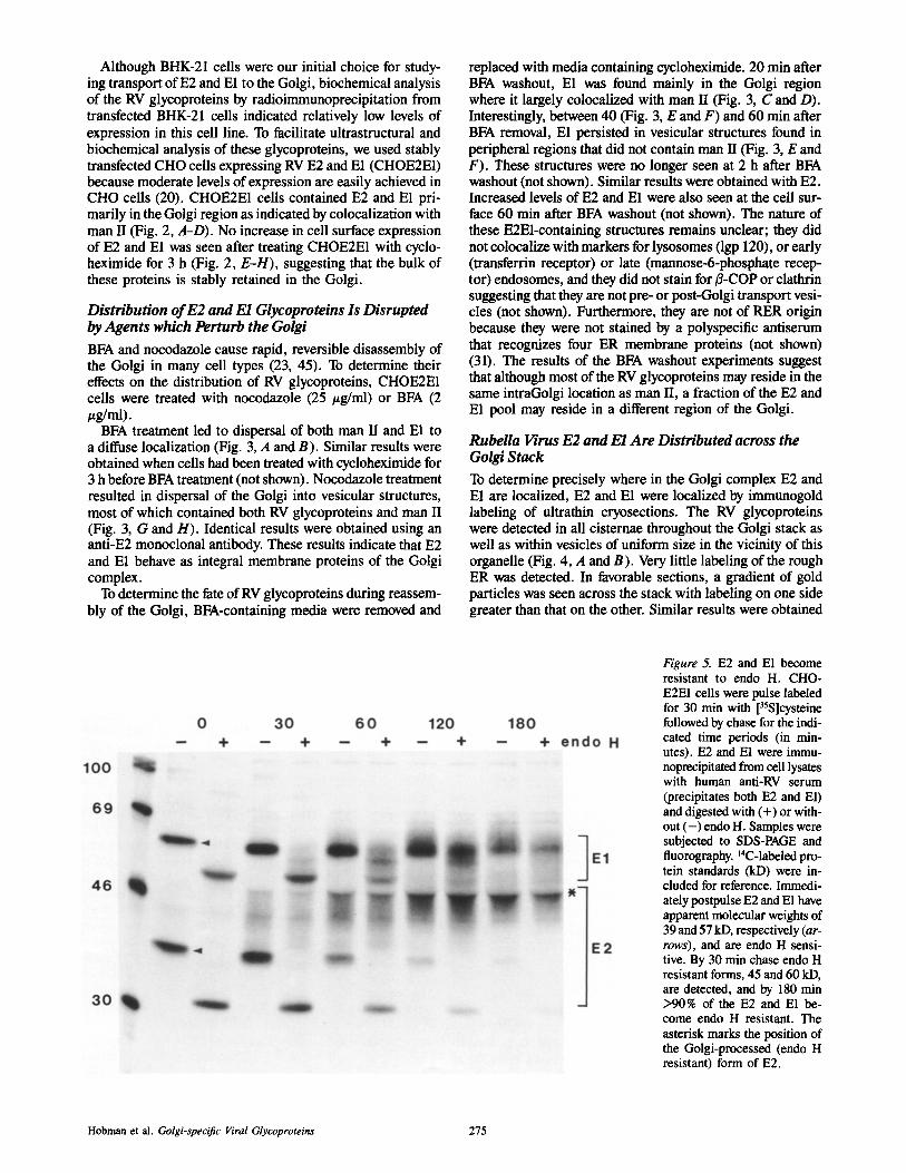
Figure 5. E2 and E1 become resistant to endo H. CHO- E2E1 cells were pulse labeled for 30 min with [35S]cysteine followed by chase for the indi- cated time periods (in min- utes). E2 and E1 were immu- noprecipitated from cell lysates with human anti-RV serum (precipitates both E2 and El) and digested with (+) or with- out ( - ) endo H. Samples were subjected to SDS-PAGE and fluorography. 14C-labeled pro- tein standards (kD) were in- cluded for reference. Immedi- ately postpulse E2 and E1 have apparent molecular weights of 39 and 57 kD, respectively (ar- rows), and are endo H sensi- tive. By 30 min chase endo H resistant forms, 45 and 60 kD, are detected, and by 180 min >90% of the E2 and E1 be- come endo H resistant. The asterisk marks the position of the Golgi-processed (endo H resistant) form of E2.
Hobman et al. Golgi-specific Viral Glycoproteins 275

when cycloheximide was added to the culture media. It was not possible to determine which side was more heavily la- beled due to the lack of markers indicating the cis/trans polarity. These results demonstrate that E2 and E1 accumu- late in bona fide Golgi elements.
Rubella Virus Glycoproteins Become Endo H Resistant and Acquire SiaUc Acid
E2 containsthree N-linked (10, 44) as well as O-linked gly- cans (32), whereas E1 contains only three N-linked glycans (19, 32). To obtain information on the extent of processing of El and E2 by Golgi enzymes, CHOE2E1 cells were pulse labeled with [35S]cysteine and chased for various time periods before lysis and immunoprecipitation with human anti-RV sera that recognize both E2 and El. Immunoprecipi- tates were divided into two and incubated with or without endo H before separation by SDS-PAGE and autoradiogra- phy (Fig. 5). The apparent molecular weights of E2 and E1 immediately after the labeling period are 39 and 57 kD, respectively (18). Their sensitivity to endo H digestion demonstrated that they correspond to high-mannose, ER forms of the glycoproteins. Chase with media containing ex- cess unlabeled cysteine resulted in acquisition of endo H re- sistance with a t~ of I-1.5 h indicating transport of E2 and E1 to the middle Golgi. Processing of E2 and E1 was detect- able at 0.5 h chase and by 3 h chase >90% of E1 and E2 were resistant to endo H. The Golgi processed, endo H-resistant forms of E2 and E1 were 42--47 and 57-66 kD, respectively (Fig. 5). The large shift in size of processed E2 seen by SDS- PAGE is at least in part due to the presence of O-linked oligosaecharides (32). The fact that Golgi processing of N-linked glycans on E2 and E1 proceeded at similar rates suggests that these proteins may be transported to the Golgi as a complex. Endo D sensitive forms of E2 and E1 were not detected at any time point (not shown) suggesting that trans- port is not delayed in the cis Golgi.
Immunoprecipitates were also digested with a neuramini- dase (A. ureafaciens) which has a broad-range specificity and cleaves sialic acid from both N- and O-linked glycans (30). The endo H resistant forms of both E2 and E1 became partially sensitive to neuraminidase in a time-dependent manner (Fig. 6) indicating that at least a portion of these gly- coproteins were modified by sialyltransferases. Neuramini- dase digestion affected only the endo H-resistant forms (>60 min chase) of RV glycoproteins indicating that some but not all of their electrophoretic heterogeneity was due to sialic acid. The neuraminidase-sensitivity of E2 was more difficult to discern (Fig. 6) due to the heterogeneous nature of neuraminidase-digested E2. It has been recently reported that sialylation of O-linked oligosaccharides may occur in the medial Golgi (30), whereas sialylation of N-linked gly- cans occurs in the TGN. The finding that El, which contains only N-linked glycans, was partially sensitive to neuramini- dase suggests that a significant proportion of these glycopro- teins reach trans Golgi eisternae.
Biosynthetic labeling experiments were also carried out on CHOE2 cells expressing only E2. In these cells, very little processing of N-linked glycans on E2 was observed after a 3 h chase (not shown), and by indirect immunofluorescence the majority of the glycoprotein appeared to be retained in the RER. However, some antigen was detected in the Golgi
Figure 6. E2 and E1 undergo sialylation. CHOE2E1 cells were pulse-labeled and processed for irnmunoprecipitation as in Fig. 5. E2 and E1 immunoprecipitates were digested with (+) or without ( - ) neurarninidase from A. ureafaciens. Neuraminidase-sensitive forms of E2 and E1 are first clearly evident at 120 min. Neurarnini- dase sensitivity of E2 is indicated by the decrease in intensity of the 47-kD species (asterisk) after treatment, and neuraminidase sensitivity of El is indicated by a reduction in the size of El.
based on colocalization with man II, and extremely low lev- els of E2 could be detected on the cell surface of nonpermea- bilized cells (not shown). We conclude that the majority of E2 expressed in the absence of E1 remains in the ER, but a minor fraction is transported to the Golgi and cell surface.
The Majority of the Rubella Virus Spike Complex Is Retained in the Golgi
The results obtained by indirect immunofluorescence and immunogold labeling of CHOE2E1 cells suggested that the bulk of the E2 and E1 remains in the Golgi. To confirm by an alternative approach that the majority of these proteins do not reach the plasma membrane, we utilized a commercially available reagent (sulfo-NHS-biotin) that has been used to selectively biotinylate primary amines of cell surface pro- teins (27). The biotinylated proteins subsequently can be iso- lated using streptavidin-agarose and thus, easily distin- guished from the intracellular pool. When radiolabeled CHOE2E1 cells were treated with sulfo-NHS-biotin to bi- otinylate cell surface proteins, <10% of either glycoprotein was detected at the cell surface after 180-min chase even though they had clearly entered the Golgi as evidenced by their increased molecular weight (Fig. 7). CHOE1 cells which contain large amounts of E1 arrested in a pre-Golgi compartment (20) were used as a negative control. E1 was not detected at the surface of CHOE1 cells using the biotiny- lation protocol (Fig. 7). As a positive control, when CHOE2E1 cells were infected with VSV ts045, G protein was transported to the cell surface at 32°C where it could be biotinylated and subsequently isolated using streptavidin- agarose (data not shown). From these results we conclude that (a) the majority of E2 and E1 do not reach the plasma membrane in CHOE2E1 cells, and (b) although transport of E2 and E1 is arrested in the Golgi complex, their accumula-
The Journal of Cell Biology, Volume 121, 1993 276

Figure 7. The majority of the RV spike complex is not trans- ported to the cell surface. CHOE2E1 (E2E1) and CHO- E1 (El) cells were radiolabeled for 30 min and chased for the indicated times (in minutes). Cell surface proteins were biotinylated with sulfo-NHS- biotin. RV proteins were im- munoprecipitated from cell lysates using human anti-RV serum and divided into two ali- quots. Biotinylated cell sur- face proteins were recovered from one half using streptavi- din agarose, and the remaining half constituted the intracellu- lar sample. Samples were then subjected to SDS-PAGE and fluorography. The protein marked by the asterisk is be- lieved to be a degradation prod- uct of El. Very little (<10%) of the E2 or E1 reaches the cell surface in cells expressing both proteins. As a negative control, E1 is not detected at the cell surface in cells express- ing E1 alone. (Note: because so little of the RV glycopro- teins was recovered in the cell surface fractions, the entire cell surface samples were loaded onto gels whereas only one half of the intracellular fractions were loaded).
tion does not prevent the egress of other glycoproteins from this organelle.
Folding of E2 and El Occur at Different Rates
The data presented here and in previously published work (5, 6, 18) are consistent with the assumption that E2 and E1 form a heterodimer in the ER that is transported as such to the Golgi complex. Furthermore, the fact that the tla for acquisition ofendo H resistance is 1-1.5 h indicates that trans- port to the medial/trans Golgi is slow. The simplest explana- tion for this delay in exiting the ER is that considerable time is required for folding and acquisition of correct tertiary and quaternary structures by RV glycoproteins in the ER. The folding of glycoproteins and formation of intramolecular disulfide bonds in the ER can be monitored by SDS-PAGE of the proteins under nonreducing conditions. Proteins that have not yet completed the formation of their intrachain disulfide bonds typically migrate with a slower elec- trophoretic mobility, presumably because they are in an ex- tended conformation (8, 35). To compare the folding rates of E2 and El, CHOE2E1 cells were pulse labeled for 2.5 min, chased for various times with excess unlabeled cys- teine, and treated with NEM before lysis (8, 35). Im- munoprecipitates were subjected to SDS-PAGE under non-
reducing (Fig. 8 A) and reducing (Fig. 8 B) conditions. Immediately after the pulse, one prominent El species and two faint bands were evident when samples were separated under nonreducing conditions. However, very little E2 was recognized by the polyclonal anti-RV serum at this time point (Fig. 8 A). After a 2 min chase, a faint homogeneous E2 band was detected, whereas E1 still migrated as at least two dis- tinct species in the absence of reducing agent. In contrast, under reducing conditions E1 did not display electrophoretic heterogeneity (Fig. 8 B). With increasing chase time, the mobility of nonreduced E2 did not change, whereas the mo- bility of El increased (Fig. 8 A). The two slower migrating forms of E1 present at early time points likely represent forms of the protein that have not attained their full comple- ment ofintrachain disulfide bonds. After 60-min chase, most of the E1 was present as the faster migrating compact form, Using a human anti-RV serum that recognizes only mature forms of E1 and E2, we also found that maximal expression of epitopes on E2 occurred between 5 and 15 min whereas E1 required up to 60 min (not shown). These results suggest that formation of intramolecular disulfide bonds and folding of E1 (which has 24 cysteine residues), occurs much more slowly than E2 which has only 13 cysteine residues (10).
The quaternary structure of the RV spike complex was examined via coprecipitation experiments. Radiolabeled
Hobman et al. Golgi-specific Viral Glycoproteins 277

Figure 8. Disulfide bond formation occurs more rapidly in E2 than in [/1. CHOE2E1 cells grown in 35-ram dishes were pulse labeled with [35S]cysteine (1,000/~Ci/ml) for 2.5 rain at 37°C. Prewarmed chase medium (2.5 ml) containing 100X excess unlabeled cysteine was added after the pulse, and the cells were incubated for the indicated time periods (in minutes). Cells were washed in ice cold PBS containing 20 mM NEM before lysis and radioimmunoprecipitation with rabbit anti-RV serum. Immunopreeipitates were separated under nonreducing (A) and reducing (B) conditions by SDS-PAGE. Disulfide formation in E2 appears to be completed by 5 min whereas folding of E1 requires up to 60 min.
CHOE2E1 cells were solubilized under mild conditions (1% NP-40 in isotonic Tris-saline) to preserve protein-protein in- teractions between E2 and El. A mAb to E2 was used to pre- pare immunoprecipitates from CHOE2E1 cells at various chase times after a short (5 rain) pulse. Immediately after the pulse, a faint E2 band was seen in addition to a protein of ,'~95 kD which likely represents the uncleaved E2E1 precur-
sor polyprotein (Fig. 9). E1 coprecipitated with E2 as early as 5 min postpulse, and the amount of E1 increased up to 30-60 min (Fig. 9, right). In contrast, the amount of E2 precipitated reached a maximum between 5 and 15 rain (Fig. 9). Half of the cell lysates were also immunoprecipitated with a polyclonal anti-RV serum to demonstrate that E1 was present in the samples at all the time points (Fig. 9, left). The
Figure 9. E2 and E1 form a heterodimeric complex in the ER. CHOE2E1 cells were pulse-labeled for 5 min with [35S]cysteine (1,000 /~Ci/ml) followed by incubation with chase medium for the indi- cated time periods (in min- utes). Cells were solubilized in buffer containing 1% NP-40 and immunoprecipitated with mouse anti-E2 (right) or hu- man anti-RV serum (left) fol- lowed by SDS-PAGE and fluo- rography. E1 is coprecipitated by mouse monoclonal anti-E2 (c~E2) beginning at 5 rain post- pulse (right) indicating that E2 and El form a heterodimer in the ER. Results obtained with polyclonal anti-RV serum (polyclonal c~RV) indicate that the amount of El does not in- crease during the chase period (after 5 mill), whereas the amount of El that coprecipi-
tates with E2 increases up to 30-60 rain. The large protein marked by an asterisk likely represents the 95 kD E2-EI polyprotein precursor before cleavage by signal peptidase. The intensity of the E2 (39 kD) and El (57 kD) bands decreases after 60 rain due to processing by Golgi enzymes and conversion to the larger endo H-resistant species.
The Journal of Cell Biology, Volume 121, 1993 278

fact that E2 and E1 coprecipitate at very early (5 rain) time points, i.e., before acquisition of endo H resistance (see Fig. 5), indicates that heterodimer formation precedes exit from the ER. Because transport of RV glycoproteins to the Golgi (based on acquisition of endo H resistance [Fig. 5]) appears to occur at a similar rate, our results suggest that the folding of E1 may be the rate limiting step in heterodimer formation and subsequent transport to the Golgi.
Discussion
In this paper we have undertaken a comprehensive study of the assembly and transport of the RV E2 and E1 glycopro- teins in transfected cells and have shown that when these two proteins are expressed together they are targeted to the Golgi and behave essentially as integral Golgi membrane proteins. This conclusion is based on the findings that: (a) E2 and E1 colocalized with the Golgi marker man 1/by immunofluores- cence in a number of cell types; (b) their Golgi localization was sensitive to agents that disrupt the Golgi such as nocoda- zole and BFA; (c) they were localized to the stacked Golgi cisternae by immunogold labeling; and (d) they became largely resistant to endo H and were at least partially sialylated but were not detected (<10%) on the plasma mem- brane, indicating that for the most part they were retained in the Golgi, We found that the time required for E2 and E1 to mature, leave the RER and become endo H resistant was relatively long (tu2 = 1-1.5 h). This presumably was due to the time required for folding of these proteins and maturation of the heterodimeric complex. Biosynthetic labeling experi- ments indicated that E1 glycoprotein folded much more slowly than E2. Coincidentally, processing of RV glycopro- teins by Golgi enzymes (detected after 30-rain chase) was not observed until heterodimer formation (as detected by coprecipitation of E1 with anti-E2 monoclonal) reached a maximum (30-60 min). These results suggest that the matu- ration of E1 is the limiting factor in transport of the RV spike complex to the Golgi.
IntraGolgi Location orE2 and E1
At the light microscopic level, the location of E2 and E1 was indistinguishable from the medial Golgi marker man II ex- cept that occasionally the RV glycoproteins were detected in peripheral vesicles which did not contain man H. The fact that the majority of the E2 and E1 glycoprotein became resis- tant to endo H and partially sialylated suggests that these gly- coproteins were transported to the medial and trans regions of the Golgi complex where the enzymes responsible for trimming (man II) and addition of sialic acid (sialyltransfer- ases) to N-linked oligosaccharides are assumed to reside. Unlike glycoproteins which traverse the Golgi completely, only a fraction of E2 and E1 appear to be modified by the addition of sialic acid. The fact that after BFA washout, in- creased levels of E2 and E1 were observed on the cell surface of nonpermeabilized cells could be explained if a fraction of the E2 and E1 complex reaches the "I'GN which has been reported to fuse with the endosomal network after BFA treat- ment (28, 52). Thus, the structures observed during BFA washout that did not contain man II may represent trans Golgi/endosomal elements which recycle to the cell surface. Moreover, in favorable sections, immunogold labeling re- vealed a polarized distribution or gradient of E2 and E1
across the Golgi cisternae in CHOE2E1 cells. Although it is difficult to determine the cis-trans polarity of the Golgi in CHO cells by morphology alone, the fact that the majority of the E2 and E1 glycoproteins become endo H resistant and at least some of the N-linked glycans on E1 are sialylated support the conclusion that these proteins have a predomi- nantly medial/trans Golgi localization.
Structure and Assembly of the E2/E1 Heterodimer
Generally, only properly assembled oligomeric complexes are able to be efficiently transported from the ER (21, 47) whereas unassembled subunits are frequently retained and ultimately degraded (7, 24). In the case ofRV glycoproteins, expression of E1 in the absence of E2 results in transport from the RER to a tubular pre-Golgi compartment which is physi- cally connected to the RER (20). When E2 was expressed alone in CHO cells, the majority of the protein was retained in the ER and to a lesser extent in the Golgi. Only trace amounts of E2 were detected on the cell surface by indirect immunofluorescence. These observations suggest that E2 and E1 must be expressed together to exit the ER and to be efficiently transported to the Golgi. Indeed, coprecipitation experiments indicated that E2 and E1 assemble into a hetero- dimeric complex in the ER. Because E2 and E1 associate shortly after synthesis (within 5 min) it is possible that the glycoproteins attain their tertiary structures by using each other as a scaffold. The rate limiting step in heterodimer maturation is likely the folding rate of E1 which is much slower than that of E2. The situation appears to be similar in the case of the Uukuniemi virus G1 and G2 spike glyco- proteins which also form a heterodimer in the ER and are targeted to the Golgi (42).
We were not able to detect oligomer formation compara- ble to that seen with other viral proteins (15, 50) in sucrose gradient velocity experiments; however, it is possible that any higher order structures (e.g., trimers) formed may dis- sociate during centrifugation (16).
Other Golgi-specific Glycoproteins
Other viral glycoproteins that localize to the Golgi include the coronavirus E1 glycoproteins (avian infectious bronchitis virus [37] and mouse hepatitis virus [2, 30, 48]) which have three membrane-spanning domains, and bunyavirus gtyco- protein G1/G2 spike complexes which have only one membrane-spanning domain (11, 22, 46). Bunyavirus glyco- proteins, like RV glycoproteins, are type I membrane pro- teins, whereas the integral Golgi membrane proteins that have been cloned and sequenced are type II membrane pro- teins (34). The only exception to date is TGN38 which is found in the TGN (33). When the avian and mouse coronavi- rus E1 glycoproteins are expressed using vaccinia virus recombinants, they have different intraGolgi sites of resi- dence, i.e., they are localized to the cis Golgi (36) and TGN (30), respectively. In the case of the avian E1 glycoprotein, retention signals responsible for targeting to the Golgi have been localized to its first membrane spanning domain (37), whereas in the mouse hepatitis virus E1 glycoprotein, the cy- toplasmic tail appears to be important but not sufficient for Golgi retention (3). Therefore, it is possible that the mecha- nism of retention of the two coronavirus E1 glycoproteins may be different. Golgi retention signals for type II Golgi
Hobman et al. Golgi-specific Viral Glycoproteins 279

membrane proteins are invariably located in the signal/ anchor domains and flanking sequences (34).
RV is perhaps most similar to bunyaviruses in that a het- erodimer is formed between two type I membrane proteins which are then retained in the Golgi as a complex. Interest- ingly, both RV and bunyavirus glycoproteins are unusually rich in cysteine residues (10, 11, 13, 14, 22, 46). The in- traGolgi location of the various bunyavirus glycoproteins ex- pressed from cDNAs has not yet been determined by immu- noelectron microscopy. We have detected trace amounts of E2 and E1 glycoproteins on the cell surface of transfected cells with significantly more being detected on the surface of transiently transfected COS cells (18) than in stably trans- fected CHO cells even though the ratio of intracellular to cell surface label was the same (i.e., >90% of labeled RV glyco- proteins retained intracellularly after 3-h chase). Typically, low levels of bunyavirus glycoproteins are also found on the surface of infected cells or cells expressing G1 and G2 from cDNA (25, 38).
RV Glycoproteins as Models for Golgi Membrane Proteins CHOE2E1 cells should prove to be very useful for further study of the synthesis, targeting and turnover of Golgi mem- brane proteins. Expression of high levels of RV glycopro- teins does not significantly impair the growth of these cells and did not cause any obvious cellular abnormalities (i.e., autophagic vacuoles, vesiculated Golgi) in CHO cells. Thus, for all intents and purposes, RV E2 and E1 behave as endoge- nous Golgi membrane proteins in CHO cells and should prove to be ideal for analysis of Golgi targeting signals. Ac- cording to prevailing concepts, for proteins to be retained in the Golgi they must contain a Golgi retention signal (34). It follows that E2 and/or E1 must contain such a retention sig- nal for Golgi targeting to occur. We are now attempting to localize the Golgi retention signal(s) on the RV glycoproteins by constructing chimeras of E2 and E1 with proteins that are normally transported to the cell surface.
Finally, the availability of a biochemicaily and morpholog- ically well defined cell line expressing high levels of E2 and E1 together with their relatively slow rate of transport from the ER to the Golgi make CHOE2E1 cells an excellent alter- native to cells infected with VSV G as a model system for further biochemical studies of ER to intermediate compart- ment and Golgi transport.
This research was supported by National Institutes of Health grant DK17780 (to M.G. Farquhar) and a postdoctoral fellowship from the Med- ical Research Council of Canada (to T. C. Hobman).
Received for publication 16 November 1992 and in revised form 28 Janu- ary 1993.
References
1. Andersson, S., D. L. Davis, H. Dahlback, H. Jornvall, and D. W. Russell. 1989. Cloning, structure, and expression of the mitochondrial cyto- chrome P-.450 stero126-hydroxylase, a bile acid biosynthetic enzyme. J. Biol. Chem. 264:8222-8229.
2. Armstrong, J., M. McCrae, and A. Colman. 1987. Expression of corona- virus E1 and rotavirus VPI0 membrane proteins from synthetic RNA. J. Cell. Biochem. 35:129-136.
3. Armstrong, J., and S. Patel. 1991. The Golgi sorting domain of coronavirus El protein. J. Cell Sci. 98:567-575.
4. Bardeltti, G., I. Tek'toff, and D. Gantheron. 1979. Rubella virus maturation and production in two host cell systems. Intervirology. 11:97-103.
5. Baron, M. D., T. Ehel, and M. Suomalaluen. 1992. Intracellular transport of rubella virus structural proteins expressed from cloned eDNA. J. Gen. Virol. 73:1073-1086.
6. Baron, M. D., and K. For~ell. 1991. Oligumerisation oftbe structural pro- teins of rubella virus. Virology. 185:811-819.
7. Bonifacino, J. S., and J. Lippincott-Schwartz. 1991. Degradation of pro- reins within the endoplasmic reticulum. Curr. Opin. Cell Biol. 3:592- 600.
8. Braakman, I., H. Hoover-Litty, K. R. Wagner, and A. Helenius. 1991. Folding of influenza hemagglutinin in the endopiasmic reticulum. J. Cell Biol. 114:401-411.
9. Brown, W. J., J. Goodhouse, and M. G. Farquhar. 1986. Mannose-6-phos- phate receptors (215 kD) cycle between the Golgi complex and endo- somes in Clone 9 hepatocytes. J. Cell Biol. 103:1235-1247.
10. Clarke, D. M., T. W. Loo, I. Hui, P. Chong, and S. Gillam. 1987. Nucleo- tide sequence and in vitro expression of rubella virus 24S subgenomic mRNA encoding the structural proteins El, E2 and C. Nucl. Acids ICes. 15:3041-3057.
11. Collett, M. S., A. F. Purchio, K. Keegan, S. Frazier, W. Hays, D. K. An- derson, M. D. Parker, C. Schmaljohn, J. Schmidt, and J. Dalrymple. 1985. Complete nueleotide sequence of the M RNA segment of Rift Valley fever virus. Virology. 144:228-245.
12. Duden, R., G. Griffiths, R. Frank, P. Argos, and T. E, Kreis. 1991. /~-COP, a 110 kD protein associated with non-clathrin-coated vesicles and the Golgi complex, shows homology to ~-adaptin. Cell. 64:649-665.
13. Frey, T. K., and I. D. Mart. 1988, Sequence of the region coding for virion proteins C and E2 and the carboxy terminus of the non-structural proteins of rubella virus: comparison to alphaviruses. Gene. 62:85-99.
14. Frey, T. K., I. D. Marr, M. L. Hemphill, and G. Dominguez. 1986. Molec- ular cloning and sequencing of the region of the rubella virus genome cod- ing for glycoprotein El. Virology. 154:228-232.
15. Fuller, S. D. 1987. The T = 4 envelope of Sindbis virus is organized by interactions with a complementary T --- 3 capsid. Cell. 48:923-934.
16. Gething, M. J., K. McCammon, and J. Sambrook. 1989. Protein folding and intraceIlular transport: Evaluation of conformational changes in na- scent exocytic proteins. Methods Cell Biol. 32:185-206.
17. Hobman, T. C., and S. Gillam. 1989. In vitro and in vivo expression of rubella virus E2 glycoprotein: The signal peptide is located in the C-ter- minal region of capsid protein. Virology. 173:241-250.
18. Hobman, T. C., M. L. Lundstrom, and S. Gillam. 1990. Processing and transport of rubella virus structural proteins in COS cells. Virology. 178: 122-133.
19. Hobman, T. C., Z. Qiu, H. Chaye, and S. Gillam. 1991. Analysis of rubella virus E1 glycosylation mutants expressed in COS cells. Virology. 181:768-772.
20. Hobman, T. C., L. Woodward, and M. G. Farquhar. 1992. The rubella virus E1 glycoprotein is arrested in a novel pest-ER, pre-Goigi compart- ment. J. Cell Biol. 118:795-811.
21. Hurtley, S., and A. Helenius. 1989. Protein oligomerization in the endo- plasmic reticulum. Annu. Rev. Cell. Biol. 5:277-307.
22. lhara, T., J. Smith, J. M. Dalrymple, and D. H. L. Bishop. 1985. Complete sequences of the glycoproteins and M RNA of Punta Toro phlebovirus compared to those of Rift Valley fever virus. Virology. 144:246-259.
23. Klausner, R. D., J. G. Donaldson, and J. Lippincott-Schwartz. 1992. Brefeldin A: insights in the control of membrane traffic and orgauelle structure. J. Cell Biol. 116:1071-1080.
24. Klausner, R. D., and R. Sitia. 1990. Protein degradation in the endoplasmic reticulum. Cell. 62:611-614.
25. Kuismanen, E., K. Hedman, J. Saraste, and R. F. Pettersson. 1982. Uu- kuniemi virus maturation: Accumulation of virus particles and viral anti- gens in the Golgi complex. Mol. Cell. Biol. 2:1444-1458.
26. Laemmli, U. K. 1970. Cleavage of structural proteins during the assembly of the head of bacteriophage T4. Nature (Lond.). 227:680-685.
27. Le Bivic, A., F. X. Real, and E. Rodriguez-Boulan. 1989. Vectorial target- ing of apical and basolateral plasma membrane proteins in a human ade- nocarcinoma epithelial cell line. Proc. Nail Acad. ScL USA. 86:9313- 9317.
28. Lippincott-Schwartz, J., L. Yuan, C. Tipper, M. Amherdt, L. Orci, and R. D. Klansner. 1991. Brefeldin A's effects on endosomes, lysosomes, and the TGN suggest a general mechanism for regulating organelle struc- ture and membrane traffic. Cell. 67:601-616.
29. Lobigs, M., J. M. Wahlberg, and H. Garoff. 1990. Spike protein oligumer- ization control of Semliki forest virus fusion. J. tirol. 64:5214-5218.
30. Locker, J. K., G. Griffiths, M. C. Horzink, and P. J. M. Rottier. 1992. O-glycosyiation of the coronavirus M protein. J. Biol. Chem. 267: 14094-14101.
31. Louvard, D., H. Reggio, and G. Warren. 1982. Antibodies to the Golgi complex and the rough endoplasmic reticulum. J. Cell Biol. 92:92-107.
32. Lundstrom, M. L., C. A. Mauracher, and A. J. Tingle. 1991. Characteriza- tion of carbohydrates linked to rubella virus glycoprotein E2. J. Gen. Virol. 72:843-850.
33. Luzio, J. P., B. Brake, G. Banting, K. E. Howell, P. Braghetta, and K. K. Stanley. 1990. Identification, sequencing and expression of an integral membrane protein of the trans-Golgi network (TGN38). Biochem. J. 270: 97-102.
The Journal of Cell Biology, Volume 121, 1993 280

34. Machamer, C. E. 1991. Golgi retention signals: do membranes hold the key.'? Trends Cell Biol. 1:141-144.
35. Machamer, C, E., R. W. Doms, D. G. Bole, A. Helenius, and J. K. Rose. 1990. Heavy chain binding protein recognizes incompletely disulfide- bonded forms of vesicular stomatitis virus G protein. J. Biol. Chem. 265:6875-6883.
36. Machamer, C. E., S, A. Mentone, J, K. Rose, and M. G. Farquhar. 1990. The El glycoprotein of an avian coronavirus is targeted to the cis Golgi complex. Proc. Natl. Acad. Sci. USA. 87:6944-6948.
37. Machamer, C. E., and J. K. Rose. 1987. A specific transmembrane domain ofa coronavirus El glycoprotein is required for its retention in the Golgi region. J. Cell Biol. 105:1205-1214.
38. Matsuoka, Y., T. lhara, D. H. L. Bishop, and R. W. Compans. 1988, lntra- cellular accumulation of Punta Toro virus glycoproteins expressed from cloned cDNA. Virology. 167:251-260.
39. Moremen, K. W., O. Touster, and P. W. Robbins. 1991. Novel purification of the catalytic domain of Golgi-ot-mannosidase II. J. Biol. Chem. 266:16876-16885.
40. Oker-Biom, C., N. Kalkkinen, L. Kaarianen, and R. F. Pettersson. 1983. Rubella virus contains one capsid protein and three envelope glycopro- teins, El, E2a and E2b. J. Virol. 46:964-973.
41. Oker-Blom, C., I. Ulmanen, L. Kaarianan, and R. F, Pettersson. 1984. Rubella virus 40S RNA specifies a 24S subgenomic RNA that codes for a precursor to structural proteins. J. Virol. 49:403-408.
42. Persson, R., and R. F. Pettersson. 1991. Formation and intracellular trans- port of a heterodimeric viral spike proteins complex. J. Cell Biol. 112:257-266.
43. Pettersson, R. F. 1991. Protein localization and virus assembly at intracel- lular membranes. Curr. Top. Microbiol. Immunol. 170:67-106.
44. Qiu, Z., T. C, Hobman, H. L. McDonald, N. O. Seto, andS. Gillam. 1992. Role of N-linked oligosaccharides in processing and intracellular trans- port of E2 glycoprotein of rubella virus. J. Virol. 66:3514-3521.
45. Rogalaski, A. A., J. E. Bergman, and S. J. Singer. 1984. Effect of microtu- bale assembly status on the intraceilular processing and surface expres- sion of an integral protein of the plasma membrane. J. Cell Biol. 99:1101-1109.
46. Ronnholm, R., and R. F. Pettersson. 1987. Complete nucleotide sequence of the M RNA segment of Uukuniemi virus encoding the membrane gly- coproteins G1 and G2. Virology. 160:191-202.
47. Rose, J. K., and R. W. Doms. 1988. Regulation of protein export from the endoplasmic reticulum. Annu. Rev. Cell. Biol. 4:257-288.
48. Rottier, P. J. M., and J. K. Rose. 1987. Coronavirus E1 glycoprotein ex- pressed from cloned eDNA localizes to the Golgi region. J. Virol. 61:2042-2045.
49. Tokuyasu, K. T. 1986. Application ofcryoultramicrotomy to immunocyto- chemistry. J. Microsc. (OxjO. 143:139-149.
50. Vogel, R. H., S. W. Provencber, C.-H. von Bonsdorff, M. Adrian, and J. Dubochet. 1986. Envelope structure of Semliki Forest virus recon- structed from cryo-electron micrographs. Nature (Lond.). 336:36-42.
51. von Bonsdortf, C., and A. Vaheri. 1969. Growth of rubella virus in BHK- 21 cells: Electron microscopy of morphogenesis. J. Gen. ViroL 5:47-51.
52. Wood, S. A., J. E. Park, and W. J. Brown. 1991. Brefeldin A causes a microtubule-mediated fusion of the trans-Golgi network and early endo- somes. Cell. 67:591-600.
Hobman et al. Golgi-specific Viral Glycoproteins 281