the safety and side effects of monoclonal...
TRANSCRIPT

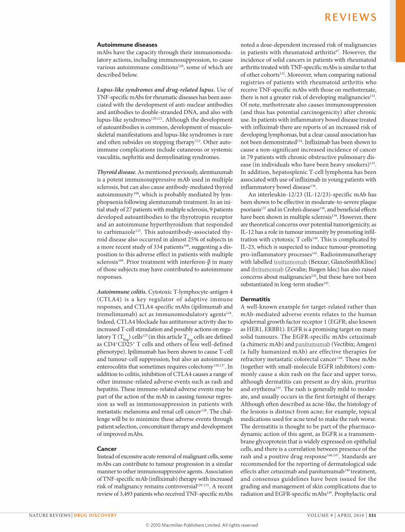
In 1975, Köhler and Milstein published their seminal manuscript on hybridoma technology enabling the production of mouse monoclonal antibodies (mAbs)1,2. Since then, technical advances have allowed the transi-tion from mouse, via chimeric and humanized, to fully human mAbs3,4, with a reduction in potentially immu-nogenic mouse components (FIG. 1a). This has led to mAbs having marked successes in the clinic5,6 (TABLE 1). Indeed, the US Food and Drug Administration has now approved more than 20 mAbs, and more than 150 other mAbs are currently in clinical trials7.
Among the advantages of protein therapeutics such as mAbs over conventional low-molecular-mass drugs are their high specificities, which facilitates precise action, and their long half-lives, which allows infrequent dos-ing8. Furthermore, molecular engineering technologies have enabled the structure of mAbs to be fine-tuned for specific therapeutic actions and to minimize immuno-genicity9–12, thus improving their risk–benefit ratio. This is reflected in mAbs having approval rates of around 20% compared with 5% for new chemical entities5,7. However, in addition to a range of adverse events that may be gen-erally associated with therapeutic mAbs, there are also adverse effects that are related to the specific target or mechanism of action13.
A review of safety-related regulatory actions performed for biologics approved between January 1995 and June 2007 (REF. 14) demonstrated that safety problems often
relate to immunomodulation and infection. Moreover, those biologics that were first-in-class to obtain approval have greater regulatory actions. European registers of biologics have proved to be useful new tools for pharma-covigilance15. In the case of mAbs directed against tumour necrosis factor (TNF), registers have been initiated by aca-demics associated with national rheumatology societies and been sponsored by the pharmaceutical industry.
Antibodies operate through various mechanisms16 (FIG. 1b). When the Fab part of an antibody binds to the antigen it blocks its interaction with a ligand. Signalling occurs when the binding of the antibody to a recep-tor delivers an agonist signal. These functions can be independent of the Fc part of the molecule (although inter actions of the Fc portion with other molecules can enhance these mechanisms). In addition, the antibody can exert actions through its Fc region: these include antibody-dependent cell-mediated cytotoxicity, comple-ment-dependent cytotoxicity and antibody-dependent cellular phagocytosis. Furthermore, the constant heavy-chain domain regions (CH2 and CH3) of Fc on immuno-globulin G (IgG) interact with the neonatal Fc receptor (FcR) to influence transport of IgG across cellular barriers and regulate the circulating levels of the antibody thus, extending its half-life17. Recruitment of these effectors is dependent on the isotype of the antibody, and its ability to recruit complement or effector cells. IgG1 is the most commonly used subclass of Ig to trigger cell death.
*Imperial Clinical Respiratory Research Unit (ICRRU), Mint Wing First Floor, St Mary’s Hospital, Paddington, London W2 1NY, UK. ‡National Heart and Lung Institute, Imperial College, Dovehouse Street, London SW3 6LY, UK. §F. Hoffmann-La Roche, Grenzacherstrasse 124, CH-4070 Basel, Switzerland. ||Immunobiology Section, Department of Medicine, Imperial College London, Hammersmith Hospital, London W12 ONN, UK.Correspondence to T.T.H.e-mail: [email protected]:10.1038/nrd3003 Published online 22 March 2010
The safety and side effects of monoclonal antibodiesTrevor T. Hansel*‡, Harald Kropshofer§, Thomas Singer§, Jane A. Mitchell‡ and Andrew J. T. George||
Abstract | Monoclonal antibodies (mAbs) are now established as targeted therapies for malignancies, transplant rejection, autoimmune and infectious diseases, as well as a range of new indications. However, administration of mAbs carries the risk of immune reactions such as acute anaphylaxis, serum sickness and the generation of antibodies. In addition, there are numerous adverse effects of mAbs that are related to their specific targets, including infections and cancer, autoimmune disease, and organ-specific adverse events such as cardiotoxicity. In March 2006, a life-threatening cytokine release syndrome occurred during a first-in-human study with TGN1412 (a CD28-specific superagonist mAb), resulting in a range of recommendations to improve the safety of initial human clinical studies with mAbs. Here, we review some of the adverse effects encountered with mAb therapies, and discuss advances in preclinical testing and antibody technology aimed at minimizing the risk of these events.
R E V I E W S
NATURE REvIEWS | Drug Discovery volUME 9 | ApRIl 2010 | 325
© 20 Macmillan Publishers Limited. All rights reserved10

Hinge
VH
Frameworkregions
a b
CDRs
Ligand
Receptor
TGN1412
Antigenbinding IgG4
IgG3IgG4 IgG1
IgG3IgM
IgG1IgG3
Antagonism Signalling CDC ADCC
CH1
CH2
Fc bindingTypes of mAbs
Murine Entirely murine amino acids ‘o’ = mousee.g. muromonab
Chimeric Human constant (C)+ murine variable (V) regions
‘xi’ = chimerice.g. rituximab
Humanized Murine complementaritydetermining regions (CDRs)
‘zu’ = humanizede.g. alemtuzumab
Human Entirely human amino acids ‘u’ = humane.g. adalimumab
CH3
Complement
InfliximabOmalizumabNatalizumabDaclizumab
Cell lysis
AlemtuzumabRituximab
Nature Reviews | Drug Discovery
AlemtuzumabRituximab
Cell lysis
VL
CL
T cell
Fcγreceptor
CD molecule
Fabregion
Fcregion
In cases where cytotoxicity is not wanted, IgG4 is com-monly used as its Fc region is relatively poor at inducing antibody-dependent cell-mediated cytotoxicity or com-plement-dependent cytotoxicity. It is also possible to modify the Fc region (for example, by removing carbo-hydrates) to further minimize recruitment of comple-ment or effector cells. omalizumab (Xolair; Genentech, Novartis) is a humanized IgE-specific mAb for severe allergic asthma that has been developed to target free IgE and membrane-bound IgE, but designed not to target IgE that is bound to IgE FcRs on mast cells, and thus not to trigger mast-cell degranulation18.
When developing therapeutic mAbs, the choice of IgG subclass is important, especially in oncology. In this case, IgG1 has the maximum potential for antibody-dependent cell-mediated cytotoxicity and is therefore ideal for eliminating cancer cells. By contrast, IgG3 is seldom used for therapeutic mAbs as the long hinge region is
prone to proteolysis and causes a decreased half-life19. Glycosylation of the Fc portion of IgG mAbs is essential to activate some effector functions, and cellular engi-neering can be used to generate selected glycoforms of antibodies20. Interestingly, IgG4 may have the potential to activate inflammatory reactions through FcRs21, and IgG4 can exhibit dynamic dissociation and exchange of the Fab arm22.
This Review discusses a range of adverse effects encountered with mAb therapy, some of which have been fatal, together with strategies to minimize these events23. We consider adverse events that have been documented for licensed mAbs (TABLE 1), as well as examples of side effects found during exploratory clin-ical studies with mAbs. of particular concern is that some of the severe adverse effects of biologics that were recently encountered were not anticipated from the currently available preclinical screening tools24,25 and
Figure 1 | Development of monoclonal antibodies: structure and function. a | Schematic structure of an immunoglobulin G (IgG) monoclonal antibody (mAb). There has been progressive development from murine mAbs, to chimeric mAbs (with murine variable (V) regions grafted onto human constant (C) regions), to humanized (which consist of a human Ig scaffold with only the complementarity-determining regions (CDRs) being of murine origin), to the recently generated fully human mAbs. The CDRs within the Fab region of a mAb bind to specific targets and cause antagonism or signalling. The Fc region of a mAb is composed of the hinge and constant heavy-chain domains (C
H2 and C
H3) and has
other functions, such as complement fixation or binding to Fc receptors. The nomenclature of mAbs reflects the type of mAb; for example, ‘xi’ in rituximab indicates that it is a chimeric mAb. b | Functions of mAbs, which include antagonism and signalling, are controlled by specific CDRs within the Fab region. Certain mAbs can specifically bind to either a ligand — for example, infliximab and omalizumab — or to a receptor — for example, natalizumab and daclizumab — and thereby prevent stimulation. By contrast, other mAbs can specifically induce signal transduction by binding to a receptor. TGN1412 is a CD28 superagonist (CD28SA), which means that ligation of the T-cell receptor is not required for T-cell activation. Functions of mAbs controlled by the Fc region include complement-dependent cytotoxicity (CDC), antibody-dependent cell-mediated cytotoxicity (ADCC) and antibody-dependent cellular phagocytosis (not shown). Certain mAbs can lyse cells (for example, T cells or B cells) through complement activation, whereas other mAbs can bind to Fc receptors and mediate cell lysis. Neonatal Fc receptor binding controls transport of IgG across cell barriers and influences the half-life of a mAb. C
L, constant light region; V
H, variable heavy region; V
L, variable light region. Panel b is
modified, with permission, from REF. 16 © (2008) Lancet Publishing Group.
R E V I E W S
326 | ApRIl 2010 | volUME 9 www.nature.com/reviews/drugdisc
© 20 Macmillan Publishers Limited. All rights reserved10

Serum sicknessA delayed reaction (generally over 4–10 days) to serum proteins or monoclonal antibodies, consisting of a hypersensitivity reaction with immune-complex generation and vascular damage in the skin, joints and kidneys.
Tumour lysis syndrome(TLS). A group of metabolic complications that can occur after treatment of cancer, usually lymphomas and leukaemias. It is generally caused by therapy that initiates the acute breakdown of cancer cells. The resultant biochemical abnormalities can cause kidney damage and acute renal failure.
Cytokine release syndrome(CRS). Also known as cytokine storm. An uncontrolled hypercytokinaemia that results in multiple organ damage and can be associated with monoclonal antibody therapy, infections and cytokine therapy.
AnaphylaxisA generally immediate and rapid loss of blood pressure (hypotension) due to a type 1 immunoglobulin E-mediated hypersensitivity reaction.
animal models26,27. With this in mind, we discuss adverse events, including exaggerated pharmacodynamic effects and mechanism-of-action-related effects, occurring with mAbs in clinical trials, and potential strategies to reduce the likelihood of such adverse events.
Immune reactionsmAbs are generally well tolerated in humans, despite containing elements that may be recognized by the recipient as foreign and can therefore cause activation of immune and innate reactions28. Acute reactions follow-ing infusion of mAbs can be caused by various mecha-nisms, including acute anaphylactic (IgE-mediated) and anaphyl actoid reactions against the mAb, serum sickness, tumour lysis syndrome (TlS) and cytokine release syndrome (CRS). The clinical manifestation can range from local skin reactions at the injection site, pyrexia and an influenza-like syndrome, to acute anaphylaxis and systemic inflammatory response syndrome, which could be fatal.
Infusion reactions commonly occur after initial dos-ing29–31, but these can be managed by recognition of risk factors, appropriate monitoring and prompt intervention32. First-dose infusion reactions to some mAbs may combine TlS, CRS and systemic inflammatory response syn-drome, as exemplified by rituximab (Rituxan/MabThera; Genentech, Biogen Idec) a chimeric CD20-specific mAb33. These initial reactions can be minimized by ensuring appropriate hydration and diuresis, premedication and cautious incremental increases in the rate of infusion.
Acute anaphylactic and anaphylactoid reactions are commonly described for certain mAbs such as the chi-meric epidermal growth factor receptor (EGFR)-specific mAb cetuximab (Erbitux; Bristol–Myers Squibb, ImClone Systems, Merck Serono), which has been attributed to the development of IgE antibodies against galactose-α-1,3-galactose34. omalizumab, as mentioned above, is directed against human IgE and is used in the treatment of severe allergic asthma35,36, but it has been found to cause anaphylaxis in approximately 0.1–0.2% of patients37–39 — this includes cases with delayed onset of symptoms40. The mechanisms underlying these acute reactions with omalizumab are still poorly understood.
A major restriction with mouse mAb therapy is the immunogenicity of the foreign protein, resulting in adverse effects and loss of efficacy41. Muromonab-CD3 (also known as orthoclone oKT3) is a mouse mAb against human CD3 that was used to suppress renal allo-graft rejection42, but it can cause CRS43. It can also cause an acute and sometimes severe influenza-like syndrome, which may be due in part to an interaction with human anti-mouse antibodies44–46. In patients with relapsed B-cell malignancies human anti-mouse antibodies to therapeu-tic mAbs can confer survival benefit47. With development of modern chimeric, humanized and fully human mAbs (FIG. 1a), it is still possible to generate human anti-human antibodies against the idiotype. Indeed, it has been noted that immunogenicity of a mAb is not simply a matter of the percentage homology with human antibody48, as alterations in particular amino acids at certain positions can also influence immunogenicity.
Natalizumab (Tysabri; Biogen Idec, Elan pharma-ceuticals) is a humanized mAb against the adhesion molecule α4 integrin, which, when used as a T-cell-directed therapy for multiple sclerosis, causes severe hypersensitivity reactions in up to 1% of subjects. It can also cause mild-to-moderate infusion reactions (such as urticaria or rash) in about 4% of patients49. These reac-tions generally occur in the first 2 hours after infusion, and are more common after the second or third infusion but usually less severe. Immunogenicity to natalizumab, with persistent neutralizing antibodies, is associated with both reduced efficacy and infusion reactions in patients with multiple sclerosis50.
Serum sickness is well described for antisera51, and both anaphylaxis and serum sickness can also be caused by mAb therapy; this has been noted especially for chimeric mAbs52.
There are now methods to minimize the immuno-genicity of mAbs53, as well as for the assessment of their immunogenicity54, with TNF-specific mAbs being an area of particular focus55. The European Medicines Agency (EMA) has issued guidelines for the assess-ment of immuno genicity of biologics56, and recently issued a concept paper on immunogenicity assessment of mAbs57.
TlS is a potentially life-threatening complication that can occur early with mAb therapy for neoplastic conditions, although this lysis is related to the desired effect of the agent58,59. The condition has been noted with rituximab for chronic lymphocytic leukaemia and different lymphomas60. Although guidelines have been issued for the management of paediatric and adult TlS58, these have attracted criticism for not being suffi-ciently evidence-based61. The initial focus should be on preventing TlS.
InfectionsInfectious diseases are a well-described side effect of certain mAbs, and they are a reflection of an acquired immunodeficiency, generally due to removal of the target ligand for that mAb. Indeed, particular types of infections illustrate the protective function of the tar-get ligand in the normal immune system, and provide insights into the function of this molecule to combat particular pathogens.
Reactivation of tuberculosis. Therapy directed against the pro-inflammatory cytokine TNFα has contributed greatly to the management of severe rheumatoid arthritis and other arthritides13,62–64. However, the tendency for reactivation of latent tuberculosis (presumably due to a key role for TNFα in immunity to Mycobacterium tuber-culosis) is a serious and limiting side effect65,66. In a meta-analysis, TNF-specific mAb therapy has been associated with an increased risk of serious infections and malig-nancies67. However, in a large cohort of elderly patients with rheumatoid arthritis there was no increase in seri-ous bacterial infections68.There is an increased risk of tuberculosis in patients with inflammatory bowel disease treated with TNF-specific mAbs69; although the chimeric mAb infliximab (Remicade; Centocor ortho Biotech)
R E V I E W S
NATURE REvIEWS | Drug Discovery volUME 9 | ApRIl 2010 | 327
© 20 Macmillan Publishers Limited. All rights reserved10

Table 1 | Side effects of licensed monoclonal antibodies
Target mAb Type FDA approval indications* selected side effects
Platelet glycoprotein IIb/IIIa
Abciximab (ReoPro; Centocor Ortho Biotech, Eli Lilly)
Chimeric antibody fragment: c7E3 Fab
1994 • Prevention of ischaemic cardiac complications of percutaneous coronary interventions and unstable angina
• Hypersensitivity and immunogenicity• Increased risk of bleeding • Thrombocytopaenia
Tumour necrosis factor-α
Adalimumab (Humira; Abbott)
Fully human 2002 • Rheumatoid arthritis• Ankylosing spondylitis• Psoriasis• Psoriatic arthritis• Crohn’s disease • Ulcerative colitis
• Infusion reactions and immunogenicity• Hypersensitivity reactions• Immunosuppression and infections
(tuberculosis)• Anaemia, leukopaenia and
thrombocytopaenia• Worsening heart failure• Malignancy, lymphoma and lympho-
proliferative disorders• Elevated liver transaminases• Increased nuclear-specific antibodies
Certolizumab (Cimzia; UCB)
Humanized pegylated
2008
Infliximab (Remicade; Centocor Ortho Biotech)
Chimeric 1998
CD52 on mature B, T and natural killer cells
Alemtuzumab (Campath; Genzyme)
Humanized 2001 • B cell chronic lymphocytic leukaemia
• Graft-versus-host disease
• Multiple myeloma • Multiple sclerosis • Vasculitis • Behçet’s disease
• Infusion reactions• Hypersensitivity and immunogenicity• CRS • Tumour lysis syndrome• Immunosuppression and opportunistic
infections• Cytopaenias: pancytopaenia, lymphopaenia
and thrombocytopaenia• Autoimmune haemolytic anaemia• Thyroid disorders• Cardiotoxicity
Interleukin-2 receptor-α on activated lymphocytes
Basiliximab (Simulect; Novartis)
Chimeric 1998 • Prophylaxis of renal transplant allograft rejection
• Severe acute hypersensitivity reactions• CRS and immunogenicity• Immunosuppression and infections• Local skin reactions• Warnings when combined with other
immunosuppressives
Daclizumab (Zenapax; Roche)
Humanized 1997 Discontinued in Europe
Vascular endothelial growth factor
Bevacizumab (Avastin; Genentech)
Humanized 2004 • Metastatic colorectal cancer
• Non-small-cell lung carcinoma
• Metastatic breast carcinoma
• Metastatic renal carcinoma
• Infusion reactions and immunogenicity• Local complications at tumour site • Arterial and venous thromboembolic
events • Haemorrhage • Severe hypertension• Cardiac failure• Reversible posterior leukoencephalopathy
syndrome • Slower wound healing and GI perforation
Ranibizumab (Lucentis; Genentech, Novartis)
Humanized (Fab fragment from bevacizumab)
2006 • Injected intravitreally for neovascular (wet) age-related macular degeneration
• Conjunctival haemorrhage• Intraocular inflammation• Increased intraocular pressure• Retinal detachment• Endophthalmitis
Complement C5 Eculizumab (Soliris; Alexion)
Humanized 2007 • Paroxysmal nocturnal haemoglobinuria
• Meningococcal and Neisseria infection• Intravascular haemolysis
CD11a Efalizumab (Raptiva; Genentech)
Humanized 2003 Recently discontinued
• No longer licensed for chronic plaque psoriasis
• First-dose reaction complex• Immunosuppression • Serious opportunistic infections• PML • Guillain–Barré syndrome, encephalitis,
meningitis• Immune haemolytic anaemia • Immune thrombocytopaenia
CD3 antigen on T cells
Muromonab- CD3 (Orthoclone OKT3; Ortho Biotech)
Mouse 1986 (no European Medicines Authority authorization)
• Acute resistant allograft rejection in renal, cardiac and hepatic transplant patients
• Severe acute infusion reactions• Immunosuppression and infections• Immunogenicity • Cardiovascular side effects• Hepatitis
R E V I E W S
328 | ApRIl 2010 | volUME 9 www.nature.com/reviews/drugdisc
© 20 Macmillan Publishers Limited. All rights reserved10

ThrombocytopaeniaA decrease in the number of circulatory platelets in the blood.
was generally well tolerated among patients with Crohn’s disease70. Several strategies can be used to minimize the risk of developing tuberculosis in patients receiving TNF-specific mAbs71, and screening can reduce, but not eliminate, the risk of reactivation69.
Progressive multifocal leukoencephalopathy. progressive multifocal leukoencephalopathy (pMl) is an often fatal, rapidly progressive demyelinating disease that is generally due to reactivation of latent infection in the central nervous system with the polyoma virus John Cunningham virus (JCv). Most healthy people are seropositive for JCv, and reactivation of JCv can occur after immunosuppression72,73. Reactivation has also been reported after using natalizu-mab to combat T-cell trafficking and adhesion in multiple sclerosis16,49,74,75. pMl occurring in patients with multiple sclerosis is remarkable as they are both demyelinating diseases, but of highly different origins and pathological features76.
In November 2004, natalizumab was approved by the US Food and Drug Administration for the treatment of relapsing-remitting multiple sclerosis, but it was sus-pended in February 2005 on the discovery of three cases of pMl: two cases in patients with multiple sclerosis77,78 and one in a patient with Crohn’s disease79. Natalizumab was reintroduced in July 2006 as second-line mono-therapy for multiple sclerosis with specific warnings and precautions49, including the ToUCH prescribing program to minimize risk of pMl. By mid-2009 there were a total of 14 cases of pMl in patients with multiple sclerosis treated with natalizumab76. Encouragingly, there are two reports suggesting that diagnosis and treatment by plasma exchange, with possible immuno-adsorption to remove natalizumab, is beneficial80,81. However, in both cases an immune–reconstitution inflammatory syndrome occurred.
Based on a detailed review of 3,147 patients taking part in clinical trials with natalizumab, it has been esti-mated that the risk of pMl corresponds to about 1 in 1,000 patients treated, occurring after a mean of about 18 months of natalizumab treatment82. Guidelines for patient selection and monitoring have been proposed to minimize the risk of pMl83, including clinical assess-ment, magnetic resonance imaging of the brain and cerebro spinal fluid analysis for JCv DNA84 (although this test can produce a negative result in early stages of the infection85). Asymptomatic reactivation of JCv has been described in 19 patients with multiple sclerosis treated with natalizumab, using quantitative pCR assays of JCv in blood and urine86,87. However, the predictive value of blood and urine markers of JCv infections needs to be further defined, as among healthy people up to 40% have JCv DNA in the urine and 1–3% have JCv viraemia at some point76. In pCR-negative patients with high clinical suspicion of pMl, a brain biopsy may be necessary to confirm the diagnosis88.
Interestingly, natalizumab mobilizes CD34+ haemato-poietic progenitor cells89,90 and these cells may be infected with JCv, contributing to the tendency for pMl. Understanding the molecular basis of predisposition for JCv infection, might help design more selective very-late
antigen-4 (vlA-4; also known as α4β1 integrin) inhibi-tors or partial vlA-4 inhibitors that retain activity against multiple sclerosis.
Rituximab is directed against B cells and used to treat non-Hodgkin’s lymphoma, but in 2006 the labelling was changed to reflect the danger of serious infections, including with JCv91. Recently, 57 cases of pMl have been described after rituximab therapy92.
So far, the humanized CD11a-specific mAb efalizu-mab (Raptiva; Genentech) has been associated with four confirmed cases of pMl when used to treat patients with chronic plaque psoriasis73,88. Suspension of marketing authorization has been recommended by the EMA, and there has been a phased voluntary withdrawal of efalizu-mab in the United States of America.
Platelet and thrombotic disordersDrug-induced immune thrombocytopaenia can be caused by many medications, including mAbs93. An acute, severe, self-limiting thrombocytopaenia can be caused by infliximab (TNFα-specific), efalizumab (CD11a-specific) and rituximab (CD20-specific); however the mechanisms of action remain obscure.
Abciximab (Reopro; Centocor ortho Biotech, Eli lilly) is an antiplatelet glycoprotein IIb/IIIa, chimeric Fab antibody fragment that has been extensively used to treat percutaneous coronary interventions, as it blocks interactions between platelets and fibrinogen94. Acute thrombocytopaenia develops after first infusion of abciximab in about 1% of patients. Acute thrombo-cytopaenia occurs in more than 10% of patients after a second infusion95–97. Thrombocytopaenia can also be delayed by 7 days, and be caused by antibodies against murine epitopes and abciximab-coated platelets98,99, and has caused fatalities100. Small-molecular-mass glycopro-tein IIb/IIIa antagonists are now increasingly being used, but they have similar safety concerns97,101.
Alemtuzumab (Campath; Genzyme) is a humanized mAb against CD52 that causes sustained depletion of CD52-expressing cells for more than a year102,103. Depleted cells include CD4+ and CD8+ T cells, natural killer cells and monocytes; circulating B cells are only transiently depleted. Alemtuzumab was originally used for graft-versus-host disease following bone-marrow transplanta-tion104,105 has also been used in the treatment of chronic lymphocytic leukaemia106 and during renal transplanta-tion107. More recently, alemtuzumab has been successfully used for autoimmune diseases, especially multiple sclero-sis108, and can be given as an annual pulsed intravenous therapy. However, the dramatic results found with alemtu-zumab in multiple sclerosis have occurred at the expense of serious side effects: thrombocytopaenia has occurred in around 3% of subjects receiving alemtuzumab for early multiple sclerosis108,109 and can be fatal110. The prolonged lymphopaenia frequently found with alemtuzumab might be mediated by its direct cytolytic effects, which are part of the mechanism of action of the mAb16,74. Alemtuzumab has also been shown to cause severe multi-lineage hae-matopoietic toxicity (involving lymphopaenia, neutro-paenia and thrombo cytopaenia) in 5 out of 11 patients with peripheral T-cell lymphoproliferative disorders111.
R E V I E W S
NATURE REvIEWS | Drug Discovery volUME 9 | ApRIl 2010 | 329
© 20 Macmillan Publishers Limited. All rights reserved10

CD40l-specific (CD154-specific) mAbs have been used to treat immune thrombocytopaenic purpura112 and systemic lupus erythematosus, and some of these mAbs have been linked with thrombocythaemia and thromboembolic complications in monkeys113–115. Thromboembolic complications encountered in human studies with certain mAbs against CD40l has halted further clinical assessment116. The mechanism of these
pro-aggregatory effects of CD40l-specific mAbs has been studied in porcine and human platelets116,117.
Bevacizumab (Avastin; Genentech) is a humanized mAb against vascular endothelial growth factor (vEGF) that has been associated with arterial (but not venous) thromboembolic events118. In addition, a meta-analysis study showed that it increased the incidence of venous thromboembolism119.
Table 1 (cont.) | Side effects of licensed monoclonal antibodies
Target mAb Type FDA approval indications* selected side effects
α4 integrin Natalizumab (Tysabri; Biogen-Idec, Elan Pharmaceuticals)
Humanized 2004 • Highly active relapsing-remitting multiple sclerosis
• Infusion and hypersensitivity reactions• Immunogenicity • PML (0.1%) with immunosuppressives • Hepatotoxicity
Immunoglobulin E (IgE)
Omalizumab (Xolair; Genentech, Novartis)
Humanized 2003 • Severe allergic asthma unresponsive to conventional therapy and with acute exacerbations
• Anaphylaxis (0.1%) • Injection site reactions • Immunogenicity• URTI• Churg–Strauss syndrome (rare)
Fusion protein on RSV
Palivizumab (Synagis; Medimmune)
Humanized 1998 • Prevention of RSV complications in high-risk infants
• Anaphylaxis and apnoea (rare)• Fever, injection site reactions
CD20 on B cells Rituximab (Rituxan/Mabthera; Genentech, Biogen Idec)
Chimeric 1997 • Follicular non-Hodgkin’s lymphoma
• CD20+ diffuse large B cell non-Hodgkin’s lymphoma
• Autoimmune haematological disorders
• Prominent acute infusion reactions • CRS• Tumour lysis syndrome • Transient hypotension• Immunogenicity• Serum sickness • Severe mucocutaneous reactions• Immunosuppression• Hepatitis B reactivation with fulminant
hepatitis• PML• Renal toxicity• Cardiac arrhythmias
EGFR Panitumumab (Vectibix; Amgen)
Fully human 2006 • Monotherapy for EGFR-positive metastatic colorectal carcinoma with non-mutated (wild-type) KRAS after failure of conventional chemotherapy
• Infusion reactions• Skin rashes in most patients (90%)• Diarrhoea (60%), nausea and vomiting • Hypomagnesaemia (2%)
Cetuximab (Erbitux; Bristol–Myers Squibb, ImClone Systems, Merck Serono)
Chimeric 2004 • EGFR-positive metastatic colorectal cancer
• Squamous cell carcinoma of head and neck
• Severe infusion reactions• IgE against oligosaccharide and HAMA• Urticaria and dermatological toxicity• Bronchospasm and pulmonary toxicity• Hypomagnesaemia
Trastuzumab (Herceptin; Genentech)
Humanized 1998 • ERBB2-positive breast carcinoma
• Hypersensitivity and infusion reactions• Cardiotoxicity with anthracyclines• Skin reactions• Pulmonary toxicity• Hypomagnesaemia
Interleukin-6 receptor
Tocilizumab (Actemra; Roche, Chugai)
Humanized 2009 • Unresponsive active rheumatoid arthritis
• Castleman’s disease
• Anaphylaxis and anaphylactoid reactions• UTRI• Headache• Serious infections• Abnormal liver function, neutropaenia and
lipid deregulation
CRS, cytokine release syndrome; EGFR, epidermal growth factor receptor; ERBB2, also known as HER2/neu; FDA, Food and Drug Administration; GI, gastrointestinal; HAMA, human anti-mouse antibodies; KRAS, v-Ki-ras2 Kirsten rat sarcoma viral oncogene homologue; PML, progressive multifocal leukoencephalopathy; RSV, respiratory syncytial virus; URTI, upper respiratory tract infection. *Some of these indications are not currently licensed.
R E V I E W S
330 | ApRIl 2010 | volUME 9 www.nature.com/reviews/drugdisc
© 20 Macmillan Publishers Limited. All rights reserved10
Autoimmune diseasesmAbs have the capacity through their immunomodu-latory actions, including immunosuppression, to cause various autoimmune conditions120, some of which are described below.
Lupus-like syndromes and drug-related lupus. Use of TNF-specific mAbs for rheumatic diseases has been asso-ciated with the development of anti-nuclear antibodies and antibodies to double-stranded DNA, and also with lupus-like syndromes120,121. Although the development of autoantibodies is common, develop ment of musculo-skeletal manifestations and lupus-like syndromes is rare and often subsides on stopping therapy122. other auto-immune complications include cutaneous or systemic vasculitis, nephritis and demye linating syndromes.
Thyroid disease. As mentioned previously, alemtuzumab is a potent immunosuppressive mAb used in multiple sclerosis, but can also cause antibody-mediated thyroid autoimmunity108, which is probably mediated by lym-phopaenia following alemtuzumab treatment. In an ini-tial study of 27 patients with multiple sclerosis, 9 patients developed autoantibodies to the thyrotropin receptor and an autoimmune hyperthyroidism that responded to carbimazole123. This autoantibody-associated thy-roid disease also occurred in almost 25% of subjects in a more recent study of 334 patients108, suggesting a dis-position to this adverse effect in patients with multiple sclerosis109. prior treatment with interferon-β in many of those subjects may have contributed to autoimmune responses.
Autoimmune colitis. Cytotoxic T-lymphocyte-antigen 4 (CTlA4) is a key regulator of adaptive immune responses, and CTlA4-specific mAbs (ipilimumab and tremelimumab) act as immunomodulatory agents124. Indeed, CTlA4 blockade has antitumour activity due to increased T-cell stimulation and possibly actions on regu-latory T (TReg) cells125 (in this article TReg cells are defined as CD4+CD25+ T cells and others of less well-defined phenotype). Ipilimumab has been shown to cause T-cell and tumour-cell suppression, but also an autoimmune enterocolitis that sometimes requires colectomy126,127. In addition to colitis, inhibition of CTlA4 causes a range of other immune-related adverse events such as rash and hepatitis. These immune-related adverse events may be part of the action of the mAb in causing tumour regres-sion as well as immunosuppression in patients with metastatic melanoma and renal cell cancer128. The chal-lenge will be to minimize these adverse events through patient selection, concomitant therapy and development of improved mAbs.
CancerInstead of excessive acute removal of malignant cells, some mAbs can contribute to tumour progression in a similar manner to other immunosuppressive agents. Association of TNF-specific mAb (infliximab) therapy with increased risk of malignancy remains controversial129–131. A recent review of 3,493 patients who received TNF-specific mAbs
noted a dose-dependent increased risk of malignancies in patients with rheumatoid arthritis67. However, the incidence of solid cancers in patients with rheumatoid arthritis treated with TNF-specific mAbs is similar to that of other cohorts132. Moreover, when comparing national registries of patients with rheumatoid arthritis who receive TNF-specific mAbs with those on methotrexate, there is not a greater risk of developing malignancies133. of note, methotrexate also causes immunosuppression (and thus has potential carcinogenicity) after chronic use. In patients with inflammatory bowel disease treated with infliximab there are reports of an increased risk of developing lymphomas, but a clear causal association has not been demonstrated134. Infliximab has been shown to cause a non-significant increased incidence of cancer in 79 patients with chronic obstructive pulmonary dis-ease (in individuals who have been heavy smokers)135. In addition, hepatosplenic T-cell lymphoma has been associated with use of infliximab in young patients with inflammatory bowel disease136.
An interleukin-12/23 (Il-12/23)-specific mAb has been shown to be effective in moderate-to-severe plaque psoriasis137 and in Crohn’s disease138, and beneficial effects have been shown in multiple sclerosis139. However, there are theoretical concerns over potential tumorigenicity, as Il-12 has a role in tumour immunity by promoting infil-tration with cytotoxic T cells140. This is complicated by Il-23, which is suspected to induce tumour-promoting pro-inflammatory processes141. Radioimmunotherapy with labelled tositumomab (Bexxar; GlaxoSmithKline) and ibritumomab (Zevalin; Biogen Idec) has also raised concerns about malignancies142, but these have not been substantiated in long-term studies143.
DermatitisA well-known example for target-related rather than mAb-mediated adverse events relates to the human epidermal growth factor receptor 1 (EGFR; also known as HER1, ERBB1). EGFR is a promising target on many solid tumours. The EGFR-specific mAbs cetuximab (a chimeric mAb) and panitumumab (vectibix; Amgen) (a fully humanized mAb) are effective therapies for refractory metastatic colorectal cancer144. These mAbs (together with small-molecule EGFR inhibitors) com-monly cause a skin rash on the face and upper torso, although dermatitis can present as dry skin, pruritus and erythema145. The rash is generally mild to moder-ate, and usually occurs in the first fortnight of therapy. Although often described as acne-like, the histology of the lesions is distinct from acne; for example, topical medications used for acne tend to make the rash worse. The dermatitis is thought to be part of the pharmaco-dynamic action of this agent, as EGFR is a transmem-brane glycoprotein that is widely expressed on epithelial cells, and there is a correlation between presence of the rash and a positive drug response146,147. Standards are recommended for the reporting of dermatological side effects after cetuximab and panitumumab148 treatment, and consensus guidelines have been issued for the grading and management of skin complications due to radiation and EGFR-specific mAbs149. prophylactic oral
R E V I E W S
NATURE REvIEWS | Drug Discovery volUME 9 | ApRIl 2010 | 331
© 20 Macmillan Publishers Limited. All rights reserved10

Nature Reviews | Drug Discovery
Trastuzumaba Breast cancer cell
ERBB2
Kinasedomain
ERBB3
NRG1
P P
P
p85GRB2
SOSRas
ERK
p110 p27
FOXO3A
PIP3
AKT
PI3K
P P P
PBAD
BCL-2 Cyt c Cyt c
BAXBCL-XL
Mitochondrion Caspaseactivation
Trastuzumabb Cardiomyocyte
ERBB2 ERBB4
NRG1
P P
P
p85GRB2
SOSRas
ERKP
Src
FAK
Contractility
p110
PIP3
AKT
PI3K
?
P PBAD
Cyt c Cyt c
BAXBCL-XLBCL-Xs
Caspase activation
minocycline has shown some efficacy in decreasing the severity of skin reactions in the first month of cetuximab therapy150.
CardiotoxicityTrastuzumab (Herceptin; Genentech) is a humanized mAb directed against human ERBB2 (also known as HER2/neu), and has been used successfully in women with ERBB2-positive metastatic breast cancer 151. However, an unexpected adverse event in women treated with trastuzumab in clinical trials was that of cardio-toxicity152,153. The antitumour and cytotoxic effects are linked through trastuzumab effects on mitochondrial outer membrane permeabilization (MoMp). B cell lymphoma 2 (BCl-2) is the prototype for a family of proteins that govern MoMp, with pro-apoptotic BCl-2-associated X protein (BAX) and BCl-2-associated ago-nist of cell death (BAD), and anti-apoptotic BCl-2 and BCl-Xl (also known as BCl2l1) (FIG. 2).
Cardiac dysfunction caused by trastuzumab is most commonly an asymptomatic decrease in left ventricular ejection fraction that tends to be reversible. However, if cardiac failure develops, this responds well to stand-ard medical management154. Cardiac dysfunction was observed in up to 4% of women treated with trastuzu-mab, with higher incidence in females taking additional anthracyclines155. Indeed, trastuzumab causes sensitiza-tion to anthracycline-induced cardiotoxic effects156: when trastuzumab was given alone for breast cancer, there were no cases of heart failure and no decreases in left ventricu-lar ejection fraction157. Cardiac dysfunction caused by trastuzumab seems to be target-related unless additional toxicity is related to signalling by trastuzumab.
The target for trastuzumab, ERBB2, is a membrane receptor tyrosine kinase with an extracellular ligand-binding domain and an intracellular kinase domain158,159. Mice with cardiac-specific deletion of ERBB2 develop age-related dilated cardiomyopathy, characterized by the presence of cardiac myocytes with increased numbers of mitochondria, vacuoles and sensitivity to anthra-cyclines160. Trastuzumab cardiotoxicity is an on-target effect due to blocking all downstream signalling from ERBB2, and causing MoMp, cytochrome c release and caspase activation, resulting in apoptosis of cardiac muscle cells with impaired contractility and ventricular function161.
Trastuzumab inhibits the actions of neuregulin 1 (NRG1) in cardiac myocytes by multiple mechanisms162, preventing NRG1’s potential role in the treatment of dis-orders of cardiac function163. In order to elucidate the mechanism of trastuzumab cardiac dysfunction, rodent and primate models have been developed154, and these may help to define effects on ERBB2-positive cancer cells without causing cardiotoxicity.
The cytokine stormvarious mAbs trigger the release of a range of cytokines, causing a cytokine storm or CRS164,165 (FIG. 3a). CRS is a prominent feature in the context of therapy with CD3-specific (muromonab)166, CD52-specific (alemtuzu-mab)167,168 and CD20-specific (rituximab) mAbs169. In
Figure 2 | Action of trastuzumab on breast cancer cells and on cardiomyocytes. a | Oncogenic signalling in a breast cancer cell can be mediated by members of the epidermal growth factor receptor (EGFR) family. Amplification of the gene encoding ERBB2 (also known as HER2/neu) tyrosine kinase is crucial for the progression of some forms of human breast cancer. ERBB2–ERBB3 kinase then activates the Ras–extracellular signal-regulated kinase (ERK) pathway and the phosphatidylinositol 3-kinase (PI3K)–AKT pathway. AKT has a central oncogenic role, partially through inhibiting B cell lymphoma 2 (BCL-2) and antagonist of cell death (BAD). Trastuzumab (Herceptin; Genentech) binds to the extracellular domain of ERBB2 and inhibits the proliferation and survival of ERBB2-dependent breast cancer cells. Trastuzumab also reverses inhibition of BAD, which leads to BCL-2-associated X protein (BAX) oligomerization at the mitochondrial membrane, release of cytochrome c (Cyt c), and caspase activation to cause apoptosis of tumour cells. In addition to inhibiting ERBB2 signalling, trastuzumab might also exert effects through antibody-dependent cell-mediated cytotoxicity (not shown). b | Signalling in cardiomyocytes through ERBB2–ERBB4 heterodimers is essential for cardiomyocyte proliferation during cardiac growth and development, and for contractile function in the adult. Although several of the same signalling pathways (such as Ras–ERK and PI3K–AKT) are activated in cardiomyocytes and in breast cancer cells, an increase in the ratio of BCL-Xs to BCL-X
L
induced by ERBB2-specific antibodies might trigger BAX oligomerization, mitochondrial membrane depolarization, ATP depletion and contractile dysfunction. In addition, antibody-dependent cell-mediated cytotoxicity might contribute to trastuzumab cardiotoxicity. Trastuzumab also blocks neuregulin 1 (NRG1)-mediated activation of Src and focal adhesion kinase (FAK), and this appears to worsen left ventricular dysfunction. GRB2, growth factor receptor-bound protein 2; PIP
3, phosphatidylinositol triphosphate. Adapted from REFS 152,159.
R E V I E W S
332 | ApRIl 2010 | volUME 9 www.nature.com/reviews/drugdisc
© 20 Macmillan Publishers Limited. All rights reserved10

Capillary leak syndromeA leakage of fluid from capillaries into interstitial fluid that results in hypotension, oedema and multiple organ failure due to limited perfusion.
2006, when the fully humanized mAb TGN1412 — a CD28 superagonist (CD28SA) — was first given to six healthy male volunteers it triggered an immediate and severe cytokine storm49,170,171.
The clinical, laboratory and immunological events following rapid intravenous infusion of TGN1412 were dramatic, and have been divided into four phases170. First, a systemic inflammatory response consisting of high levels of cytokines in the blood, and accompanied by headache, myalgias, nausea, diarrhoea, erythema, vasodi-lation and hypotension. Second, pulmonary infiltrates and lung injury, renal failure and disseminated intra-vascular coagulation. Third, severe blood lymphopaenia and monocytopaenia. Fourth, prolonged cardiovascular shock and acute respiratory distress syndrome.
Expert groups have highlighted the importance of considering the minimal anticipated biological effect level (MABEl) in deciding the initial dose of a biologic to be used in humans172–174. This MABEl approach selects the starting dose for a first-in-human study on the basis of the lowest dose that is found to be active in any in vitro potency assays. Based on the MABEl, the starting dose for TGN1412 should have been 20-times lower than that used in the phase I study. The MABEl approach also suggested a much lower dose than that derived from consideration of animal toxicology studies.
CD28SA mAbs cause activation of TReg cells in rats49,175, and have been used to treat experimental autoimmune disease176. In rats, lower concentrations of a CD28SA mAb induced nonspecific expansion of TReg cells without causing lymphocytosis175,177. In addi-tion, administration of a CD28SA mAb has recently been shown to cause a dramatic redistribution of T cells within 48 hours, with a later phase of TReg-cell activa-tion178. Selective stimulation of TReg cells is the ration-ale for use of CD28-specific mAbs for the treatment of human autoimmune diseases179.
From monkeys to humansFollowing the serious adverse events encountered in the TGN1412 first-in-human study, there has been a detailed scrutiny of the potential causal mechanism in humans180–184. The molecular details of why toxicity studies with TGN1412 involving cynomolgus monkeys (Macaca fascicularis) were poorly predictive of the clinical adverse effects in humans are important49,180,185 (FIG. 3b). one theory is that the three differences in the amino-acid sequence within the transmembrane portion of the monkey CD28 molecule could alter signalling fol-lowing TGN1412 binding186,187. Indeed, this is borne out by CD28SA causing a delayed but sustained calcium response in human but not cynomolgus T cells187.
Direct actions of TGN1412 on cells that express CD28 have the potential to cause a range of effects. This is because CD28 is present on almost all human CD4+ T cells, and roughly half of CD8+ T cells, on sub-sets of natural killer cells, on neutrophils, on apoptotic eosinophils, on mouse mast cells, and on certain B cells and plasma cells. Neutrophils may participate in the reaction to CD28SA mAbs and neutrophil activation may cause sialidase release188.
A new paradigm for T-cell activation involves con-sideration of T-cell receptor–CD28 microclusters within the immunological synapse189 (FIG. 3c). Indeed, during T-cell activation scattered microclusters consisting of five components aggregate to form a large highly ordered complex, the central supramolecular activation cluster. In this context the transmembrane amino-acid differences between monkey and human CD28 could affect the aggregation properties of this receptor within the T-cell membrane.
When the T cell becomes activated it is probable that leukocyte adhesion molecules such as CD11a/18 and CD11b/18 are rapidly upregulated. This phenomenon has already been demonstrated on peripheral blood lymphocytes following administration of a human CD3-specific mAb (muromonab-CD3) to patients190. Hence, administration of TGN1412 in humans, might lead to T-cell activation through the immunological synapse, which is associated with increased expression of T-cell adhesion molecules. There is the possibility that activated T cells bind to endothelial cells, causing local endothelial damage and a capillary leak syndrome. Indeed a T cell–endothelial complex may have increased the propensity of cytokine release, and be central to the pathogenesis of clinical events following infusion of TGN1412 in humans.
In addition, following interaction with T cells, actions of TGN1412 in humans may be partly mediated by the interaction of the Fc region of the mAb with FcRs on other cells179, involving a cross-linking of TGN1412 (REF. 187). Interestingly, humanized mAbs of the IgG4 isotype, such as TGN1412, are inefficient at binding to monkey FcRs27,191–193. Therefore, Fc interactions on the surface of the human FcR-positive cell could lead to more efficient cross-linking of the target molecule on a T cell. CD3-specific mAbs, such as muromonab-CD3, which have been engineered to have decreased FcR binding, have a reduced capacity to induce cytokine release166. likewise, cytokine release by natural killer cells in the presence of alemtuzumab is mediated through involve-ment of FcγRIII (CD16)165. In addition, in studies with an IgG4 version of the mAb alemtuzumab it was shown that IgG4 mAbs deplete target cells (T cells and B cells) in humans — albeit weaker than their IgG1 counter-parts — through FcR-mediated antibody-dependent cell-mediated cytotoxicity194. It is worth noting that in humans, polymorphisms involved in the Fc–FcR interac-tion may result in inter-individual variations in response to these antibodies.
Immunoregulation may be generally greater in ani-mals with regard to CD28SA, causing a cytokine storm to be more likely in humans. Monkey and human lym-phocytes have differences in the expression of sialic acid-binding Ig-like lectins (SIGlECs)193,195,196, which are known to be both positive and negative regulators of the immune system197. CD33-related SIGlECs, for example, show particular variation between different mammalian species. As a consequence, the threshold for cytokine release in human cells that lack SIGlECs may be significantly lower compared with cells from other species that express SIGlECs. In addition, a rapid
R E V I E W S
NATURE REvIEWS | Drug Discovery volUME 9 | ApRIl 2010 | 333
© 20 Macmillan Publishers Limited. All rights reserved10

Nature Reviews | Drug Discovery
CD52
T cell
b
c
a
CD3CD28
CD28 within IS
CD11bCD28
CD4Alemtuzumab Muromonab-CD3 TGN1412
CytokinesEndothelial cells
CD28SA(e.g. TGN1412)
T cell–endothelialcell adhesion
FcγR ICAM1
T cell
Multiple organ failure• Pulmonary infiltrates• Lung injury• Acute respiratory distress syndrome• Cardiovascular shock• Disseminated intravascular coagulant• Renal failure
Cytokine stormTNFα, IFNγIL-1β, IL-2, IL-4, IL-6, IL-8, IL-10, IL-12
Capillary leak syndromeEndothelial damage
Potential differences between humans and monkeys:• CD28 structure: difference in three
transmembrane residues. • CD28SA binding kinetics and calcium
response (sustained in humans). • Immunological synapse (IS) formation
involving CD28 cross-linking. • Greater T-cell adhesion to endothelial
cells through CD28SA/FcγR and CD11b/ICAM1 in humans
• Greater immunoregulation in animals (through SIGLECs, TRegs and cytokines).
Indu
ctio
n ph
ase
End-
orga
n da
mag
eIm
mun
o-pa
thog
enes
is
IS formation
Surface of T cell
TCR–CD28microcluster
c-SMAC
response by TReg cells may prevent the cytokine storm when mice are given CD28SA mAbs198, and animals may be more prone to produce anti-inflammatory cytokines. Transforming growth factor-β (TGFβ) may have a key role in protecting mice against a cytokine storm caused by CD3-specific mAbs199.
RegulationsThere are a range of guidance documents that support first-in-human clinical trials with mAbs200. As an imme-diate response to the TGN1412 disaster, the EMA issued a guideline to identify and decrease risk with new medici-nal products being studied in first-in-human clinical trials201. In addition, detailed regulatory guidance is avail-able on preclinical safety evaluation of pharmaceuticals202 and biologics203.
Microdosing is a method of studying drug action in humans with doses so low that they do not cause whole body effects, but have cellular responses204. A micro-dose study is performed early in drug development before the start of phase I clinical trials, and uses a dose at a small fraction of the predicted pharmacological dose. A position paper is available from the EMA on non-clinical safety studies to support clinical trials with a single microdose205.
Predicting the capacity to cause CRS. The development of preclinical tests to predict the capacity of biologics to cause CRS in humans is a major challenge26,27,182,206,207. We need to learn lessons from disasters such as the TGN1412 trial, and expand our thinking of current paradigms if we are to adequately test preclinical safety of biologics.
The cytokine storm was observed after intravenous administration of mAbs, and the serum cytokines found in vivo could be released and synthesized by cir-culating leukocytes. Therefore, in vitro tests have been established that rely on TGN1412 being incubated with human whole blood or cell populations such as periph-eral blood mononuclear cells208,209. Endothelial cells are another key source of pro-inflammatory cytokines, such as Il-6, and may be included as well. So far, a few protocols have been developed for presentation of TGN1412 to human peripheral blood mononuclear cells and whole blood before assessing cytokine release and lymphocyte activation97. When TGN1412 was air-dried onto a tissue-culture plate it caused the release of TNFα, Il-6 and Il-8 when cultured with diluted human blood209. Interestingly, there was negligible release of cytokines with aqueous unbound TGN1412. other methods of immobilizing TGN1412 also caused striking release of cytokines and profound lymphocyte prolifera-tion; most notably presentation of TGN1412 bound to endothelial cells. This suggests that under in vitro set-tings, TGN1412 needs to be bound to a solid surface before it is able to activate lymphocytes, but dry-coating may yield too many false positives165.
By contrast, alemtuzumab and muromonab-CD3 cause cytokine release in vitro and in vivo in aqueous solution without immobilization165,167, and it is note-worthy that alemtuzumab may operate through FcγRIII
Figure 3 | Monoclonal antibodies and the cytokine storm. a | Surface receptors on T cells can cause a cytokine storm when activated by therapeutic monoclonal antibodies (mAbs). Three mAbs that cause cytokine release on infusion in humans are alemtuzumab (Campath; Genzyme), muromonab-CD3 (Orthoclone OKT3) and TGN1412. Alemtuzumab recognizes the CD52 molecule on T cells and confers efficient complement-dependent lysis of lymphocytes. Muromonab targets CD3, a part of the T-cell receptor (TCR) complex. TGN1412 is an example of a CD28 superagonist (CD28SA); that is, a co-stimulator molecule contributing to activation of naive T cells. b | TGN1412 can directly cause some cytokine release, as CD28 is expressed on a variety of cells in the normal immune system. TGN1412 is more potent on human T cells than those from monkeys. This is possibly due to human CD28 having three different transmembrane amino acids, which could cause a sustained calcium response within human T cells. Cross-linking of human CD28 may contribute to the formation of an activated immunological synapse (IS) on the surface of T cells, and binding of CD28SA to Fcγ receptors (FcγRs) on endothelial cells and other leukocytes could cause further cytokine release. Activation of CD28 may also cause upregulation of adhesion molecules such as CD11b on the surface of T cells or other cells of the innate immune system, which can then bind to intracellular adhesion molecule 1 (ICAM1) on endothelial cells. T cell–endothelial complexes have the capacity to cause amplified cytokine production and local endothelial damage. Hence, the cytokine storm and neutrophil infiltration could mediate the capillary leak syndrome with resultant multiple organ failure. c | The IS forms in a dynamic process on the T-cell plasma membrane, in which the five components of the TCR–CD28 microcluster aggregate to form a central supramolecular activation cluster (c-SMAC). The latter consists of a core of TCR and CD3 molecules, surrounded by a ring of CD28 molecules with associated protein kinase Cθ, which causes sustained T-cell activation. Adapted from REF. 189.
R E V I E W S
334 | ApRIl 2010 | volUME 9 www.nature.com/reviews/drugdisc
© 20 Macmillan Publishers Limited. All rights reserved10
on natural killer cells168. So, there are multiple mecha-nisms to cause CRS, and each mAb will require individual assessment in a range of assays for the capacity to cause this cytokine release165.
To identify and validate relevant preclinical screens for CRS it would be useful if the scientific community had access to TGN1412 and related CD28-specific mAbs and immunostimulatory antibodies and cytokines. However, technical difficulties are being encountered because TGN1412-like mAbs of IgG4 isotype tend to dissociate into two halves following conventional purification steps.
predictive preclinical screening assays should fulfil four key remits for CRS. First, they should be performed on a range of human cell types (preferentially derived from the target population) that encompass potential mechanisms for CRS, including blood and tissue cells, but especially endothelial cells. Second, they should have relevant, validated and technically feasible read-outs. Third, to determine their predictive power and limitations, they should take into consideration a range of biologics and controls — TGN1412 is a necessary test reagent. Finally, they should have predictive capac-ity not only for CRS, but also for immune and tissue cell activation, Toll-like receptor activation, capillary leak, disseminated intravascular coagulation, cardio-vascular shock and systemic inflammatory response syndrome.
In addition to improved in vitro tissue-based screens, other essential approaches to consider when assessing the safety of biologics include testing the molecules in local circulation (for example, the nose or skin) in humans and in combinations of human and animal in vivo and in vitro models.
one approach that needs greater consideration is the use of microdosing studies204, with careful pharmaco-kinetic and pharmacodynamic evaluation in prelimi-nary human studies. provided that prior animal data are available with regard to target distribution and efficacy, this approach might include whole body as well as micro-scopic imaging to allow evaluation of the distribution of the molecule210,211, and tailored assays to determine any biological or clinical effects of the molecule. If the ini-tial doses chosen are very low, then such studies could be done relatively safely and might be more informative than primate or other animal investigations. They should also allow more rapid evaluation of molecules in humans, allowing efficient selection or rejection of candidate molecules to take forward for further evaluation.
Future directions and conclusionsFrom the outset, we need to recognize which types of risks apply to a particular mAb, and take steps to identify and minimize potential adverse effects. Infusion reactions can be minimized by sound preclinical and clinical practice, whereas predisposition to infection can be minimized by appropriate monitoring and selection of therapies. preclinically, the major need is for development and vali-dation of appropriate in vitro safety tests with biologics on human blood and tissues, and to have predictive tests for CRS on administration to humans. To ensure the safety of volunteers in clinical trials there is the need for communi-cation to be maintained between scientists and clinicians, pharmaceutical and biotechnology companies, and individuals involved in carrying out and regulating clini-cal studies. Together, these measures will help increase the safety of mAbs, which is vital for a greater use of mAb-based therapy in the treatment of human disease.
1. Köhler, G. & Milstein, C. Continuous cultures of fused cells secreting antibody of predefined specificity. Nature 256, 495–497 (1975).The original manuscript describing the breakthrough of hybridoma technology and the production of mAbs.
2. Strebhardt, K. & Ullrich, A. Paul Ehrlich’s magic bullet concept: 100 years of progress. Nature Rev. Cancer 8, 473–480 (2008).
3. Dubel, S. (ed.) Handbook of Therapeutic Antibodies. Volume I: Technologies, Volume II: Emerging Developments, Volume III: Approved Therapeutics (Wiley, Weinhem, 2007).A comprehensive three volume multiple-author text on therapeutic antibodies.
4. Lonberg, N. Human antibodies from transgenic animals. Nature Biotech. 23, 1117–1125 (2005).
5. Reichert, J. M., Rosensweig, C. J., Faden, L. B. & Dewitz, M. C. Monoclonal antibody successes in the clinic. Nature Biotech. 23, 1073–1078 (2005).
6. Waldmann, T. A. Immunotherapy: past, present and future. Nature Med. 9, 269–277 (2003).
7. Reichert, J. M. & Dewitz, M. C. Anti-infective monoclonal antibodies: perils and promise of development. Nature Rev. Drug Discov. 5, 191–195 (2006).
8. Leader, B., Baca, Q. J. & Golan, D. E. Protein therapeutics: a summary and pharmacological classification. Nature Rev. Drug Discov. 7, 21–39 (2008).
9. Waldmann, H. & Hale, G. CAMPATH: from concept to clinic. Philos. Trans. R. Soc. Lond. B Biol. Sci. 360, 1707–1711 (2005).
10. Nissim, A. & Chernajovsky, Y. Historical development of monoclonal antibody therapeutics. Handb. Exp. Pharmacol. 181, 3–18 (2008).
11. Presta, L. G. Molecular engineering and design of therapeutic antibodies. Curr. Opin. Immunol. 20, 460–470 (2008).
12. Hale, G. Therapeutic antibodies-delivering the promise? Adv. Drug Deliv. Rev 58, 633–639 (2006).
13. Tracey, D., Klareskog, L., Sasso, E. H., Salfeld, J. G. & Tak, P. P. Tumor necrosis factor antagonist mechanisms of action: a comprehensive review. Pharmacol. Ther. 117, 244–279 (2008).
14. Giezen, T. J. et al. Safety-related regulatory actions for biologicals approved in the United States and the European Union. JAMA 300, 1887–1896 (2008).An important review of regulatory actions regarding the safety of biologics.
15. Zink, A. et al. European biologicals registers: methodology, selected results and perspectives. Ann. Rheum. Dis. 68, 1240–1246 (2009).
16. Lutterotti, A. & Martin, R. Getting specific: monoclonal antibodies in multiple sclerosis. Lancet Neurol. 7, 538–547 (2008).
17. Yeung, Y. A. et al. Engineering human IgG1 affinity to human neonatal Fc receptor: impact of affinity improvement on pharmacokinetics in primates. J. Immunol. 182, 7663–7671 (2009).
18. Chang, T. W. Developing antibodies for targeting immunoglobulin and membrane-bound immunoglobulin E. Allergy Asthma Proc. 27, S7–S14 (2006).
19. Hassan, M. S., bedi-Valugerdi, M., Lefranc, G., Hammarstrom, L. & Smith, C. I. Biological half-life of normal and truncated human IgG3 in SCID mice. Eur. J. Immunol. 21, 1319–1322 (1991).
20. Jefferis, R. Recombinant antibody therapeutics: the impact of glycosylation on mechanisms of action. Trends Pharmacol. Sci. 30, 356–362 (2009).
21. Holland, M. et al. Anti-neutrophil cytoplasm antibody IgG subclasses in Wegener’s granulomatosis: a possible pathogenic role for the IgG4 subclass. Clin. Exp. Immunol. 138, 183–192 (2004).
22. van der Neut Kolfschoten, M. et al. Anti-inflammatory activity of human IgG4 antibodies by dynamic Fab arm exchange. Science 317, 1554–1557 (2007).
23. Tabrizi, M. A. & Roskos, L. K. Preclinical and clinical safety of monoclonal antibodies. Drug Discov. Today 12, 540–547 (2007).
24. Cavagnaro, J. A. (ed.) Preclinical Safety Evaluation of Biopharmaceuticals: A Science Based Approach to Facilitating Clinical Trials (Wiley, London, 2008).A recent book on preclinical safety testing of biopharmaceuticals.
25. Longstaff, C., Whitton, C. M., Stebbings, R. & Gray, E. How do we assure the quality of biological medicines? Drug Discov. Today 14, 50–55 (2009).
26. Loisel, S. et al. Relevance, advantages and limitations of animal models used in the development of monoclonal antibodies for cancer treatment. Crit. Rev. Oncol. Hematol. 62, 34–42 (2007).
27. Chapman, K., Pullen, N., Graham, M. & Ragan, I. Preclinical safety testing of monoclonal antibodies: the significance of species relevance. Nature Rev. Drug Discov. 6, 120–126 (2007).
28. Presta, L. G. Engineering of therapeutic antibodies to minimize immunogenicity and optimize function. Adv. Drug Deliv. Rev. 58, 640–656 (2006).
29. Chung, C. H. Managing premedications and the risk for reactions to infusional monoclonal antibody therapy. Oncologist 13, 725–732 (2008).
30. Klastersky, J. Adverse effects of the humanized antibodies used as cancer therapeutics. Curr. Opin. Oncol. 18, 316–320 (2006).
R E V I E W S
NATURE REvIEWS | Drug Discovery volUME 9 | ApRIl 2010 | 335
© 20 Macmillan Publishers Limited. All rights reserved10

31. Kang, S. P. & Saif, M. W. Infusion-related and hypersensitivity reactions of monoclonal antibodies used to treat colorectal cancer — identification, prevention, and management. J. Support. Oncol. 5, 451–457 (2007).
32. Lenz, H. J. Management and preparedness for infusion and hypersensitivity reactions. Oncologist 12, 601–609 (2007).
33. Coiffier, B. et al. CHOP chemotherapy plus rituximab compared with CHOP alone in elderly patients with diffuse large-B-cell lymphoma. N. Engl. J. Med. 346, 235–242 (2002).
34. Chung, C. H. et al. Cetuximab-induced anaphylaxis and IgE specific for galactose-alpha-1,3-galactose. N. Engl. J. Med. 358, 1109–1117 (2008).
35. Poole, J. A., Matangkasombut, P. & Rosenwasser, L. J. Targeting the IgE molecule in allergic and asthmatic diseases: review of the IgE molecule and clinical efficacy. J. Allergy Clin. Immunol. 115, S376–S385 (2005).
36. Gould, H. J. & Sutton, B. J. IgE in allergy and asthma today. Nature Rev. Immunol. 8, 205–217 (2008).
37. Cox, L. et al. American Academy of Allergy, Asthma & Immunology/American College of Allergy, Asthma and Immunology Joint Task Force Report on omalizumab-associated anaphylaxis. J. Allergy Clin. Immunol. 120, 1373–1377 (2007).
38. Corren, J. et al. Safety and tolerability of omalizumab. Clin. Exp. Allergy 39, 788–797 (2009).
39. Cox, L. S. How safe are the biologicals in treating asthma and rhinitis? Allergy Asthma Clin. Immunol. 5, 4 (2009).
40. Limb, S. L., Starke, P. R., Lee, C. E. & Chowdhury, B. A. Delayed onset and protracted progression of anaphylaxis after omalizumab administration in patients with asthma. J. Allergy Clin. Immunol. 120, 1378–1381 (2007).
41. Carter, P. Improving the efficacy of antibody-based cancer therapies. Naure Rev. Cancer 1, 118–129 (2001).
42. Loertscher, R. The utility of monoclonal antibody therapy in renal transplantation. Transplant. Proc. 34, 797–800 (2002).
43. Gaston, R. S. et al. OKT3 first-dose reaction: association with T cell subsets and cytokine release. Kidney Int. 39, 141–148 (1991).
44. Kuus-Reichel, K. et al. Will immunogenicity limit the use, efficacy, and future development of therapeutic monoclonal antibodies? Clin. Diagn. Lab. Immunol. 1, 365–372 (1994).
45. Mascelli, M. A. et al. Molecular, biologic, and pharmacokinetic properties of monoclonal antibodies: impact of these parameters on early clinical development. J. Clin. Pharmacol. 47, 553–565 (2007).
46. Carter, P. J. Potent antibody therapeutics by design. Nature Rev. Immunol. 6, 343–357 (2006).
47. Azinovic, I. et al. Survival benefit associated with human anti-mouse antibody (HAMA) in patients with B-cell malignancies. Cancer Immunol. Immunother. 55, 1451–1458 (2006).
48. Clark, M. Antibody humanization: a case of the ‘Emperor’s new clothes’? Immunol. Today 21, 397–402 (2000).
49. Ransohoff, R. M. Natalizumab for multiple sclerosis. N. Engl. J. Med. 356, 2622–2629 (2007).
50. Cohen, B. A., Oger, J., Gagnon, A. & Giovannoni, G. The implications of immunogenicity for protein-based multiple sclerosis therapies. J. Neurol. Sci. 275, 7–17 (2008).
51. Schellekens, H. Factors influencing the immunogenicity of therapeutic proteins. Nephrol. Dial. Transplant. 20 (Suppl. 6), vi3–vi9 (2005).
52. Todd, D. J. & Helfgott, S. M. Serum sickness following treatment with rituximab. J. Rheumatol. 34, 430–433 (2007).
53. Schellekens, H., Crommein, D. & Jiskoot, W. in Handbook of Therapeutic Antibodies Vol. 1 Ch. 11 (ed. Dubel, S.) (Wiley, Weinheim, 2007).
54. Shankar, G., Shores, E., Wagner, C. & Mire-Sluis, A. Scientific and regulatory considerations on the immunogenicity of biologics. Trends Biotechnol. 24, 274–280 (2006).
55. Aarden, L., Ruuls, S. R. & Wolbink, G. Immunogenicity of anti-tumor necrosis factor antibodies-toward improved methods of anti-antibody measurement. Curr. Opin. Immunol. 20, 431–435 (2008).
56. European Medicines Agency, Committee for Medicinal Products for Human Use (CHMP). Guideline on immunogenicity assessment of biotechnology-derived
therapeutic proteins. Doc. Ref. EMEA/CHMP/BMWP/14327/2006. EMA website [online], http://www.ema.europa.eu/pdfs/human/biosimilar/1432706enfin.pdf (2007).
57. European Medicines Agency, Committee for Medicinal Products for Human Use (CHMP). Concept paper on immunogenicity assessment of monoclonal antibodies intended for in vivo clinical use. Doc. Ref. EMEA/CHMP/BMWP/114720/2009. EMA website [online], http://www.ema.europa.eu/pdfs/human/biosimilar/11472009en.pdf (2009).Recent EMA guidelines on immunogenicity testing of mAbs.
58. Coiffier, B., Altman, A., Pui, C. H., Younes, A. & Cairo, M. S. Guidelines for the management of pediatric and adult tumor lysis syndrome: an evidence-based review. J. Clin. Oncol. 26, 2767–2778 (2008).
59. Tosi, P. et al. Consensus conference on the management of tumor lysis syndrome. Haematologica 93, 1877–1185 (2008).
60. Otrock, Z. K., Hatoum, H. A. & Salem, Z. M. Acute tumor lysis syndrome after rituximab administration in Burkitt’s lymphoma. Intern. Emerg. Med. 3, 161–163 (2008).
61. Feusner, J. H., Ritchey, A. K., Cohn, S. L. & Billett, A. L. Management of tumor lysis syndrome: need for evidence-based guidelines. J. Clin. Oncol. 26, 5657–5658 (2008).
62. Taylor, P. C. & Feldmann, M. Anti-TNF biologic agents: still the therapy of choice for rheumatoid arthritis. Nature Rev. Rheumatol. 5, 578–582 (2009).
63. Feldmann, M. & Maini, S. R. Role of cytokines in rheumatoid arthritis: an education in pathophysiology and therapeutics. Immunol. Rev. 223, 7–19 (2008).
64. Moss, M. L., Sklair-Tavron, L. & Nudelman, R. Drug insight: tumor necrosis factor-converting enzyme as a pharmaceutical target for rheumatoid arthritis. Nature Clin. Pract. Rheumatol. 4, 300–309 (2008).
65. Keane, J. TNF-blocking agents and tuberculosis: new drugs illuminate an old topic. Rheumatology 44, 714–720 (2005).
66. Askling, J. et al. Risk and case characteristics of tuberculosis in rheumatoid arthritis associated with tumor necrosis factor antagonists in Sweden. Arthritis Rheum. 52, 1986–1992 (2005).
67. Bongartz, T. et al. Anti-TNF antibody therapy in rheumatoid arthritis and the risk of serious infections and malignancies: systematic review and meta-analysis of rare harmful effects in randomized controlled trials. JAMA 295, 2275–2285 (2006).
68. Schneeweiss, S. et al. Anti-tumor necrosis factor alpha therapy and the risk of serious bacterial infections in elderly patients with rheumatoid arthritis. Arthritis Rheum. 56, 1754–1764 (2007).
69. Theis, V. S. & Rhodes, J. M. Review article: minimizing tuberculosis during anti-tumour necrosis factor-alpha treatment of inflammatory bowel disease. Aliment. Pharmacol. Ther. 27, 19–30 (2008).
70. Colombel, J. F. et al. The safety profile of infliximab in patients with Crohn’s disease: the Mayo clinic experience in 500 patients. Gastroenterology 126, 19–31 (2004).
71. British Thoracic Society Standards of Care Committee. BTS recommendations for assessing risk and for managing Mycobacterium tuberculosis infection and disease in patients due to start anti-TNF-α treatment. Thorax 60, 800–805 (2005).
72. Major, E. O. Progressive multifocal leukoencephalopathy in patients on immunomodulatory therapies. Annu. Rev. Med. 61, 35–47 (2010).
73. Carson, K. R. et al. Monoclonal antibody-associated progressive multifocal leucoencephalopathy in patients treated with rituximab, natalizumab, and efalizumab: a Review from the Research on Adverse Drug Events and Reports (RADAR) project. Lancet Oncol. 10, 816–824 (2009).
74. Lopez-Diego, R. S. & Weiner, H. L. Novel therapeutic strategies for multiple sclerosis — a multifaceted adversary. Nature Rev. Drug Discov. 7, 909–925 (2008).
75. Sadiq, S. A., Puccio, L. M. & Brydon, E. W. JCV detection in multiple sclerosis patients treated with natalizumab. J. Neurol. 7 Jan 2010 (doi:10.1007/s00415-009-5444-4).
76. Major, E. O. Reemergence of PML in natalizumab-treated patients — new cases, same concerns. N. Engl. J. Med. 361, 1041–1043 (2009).
77. Kleinschmidt-DeMasters, B. K. & Tyler, K. L. Progressive multifocal leukoencephalopathy complicating treatment with natalizumab and interferon β-1a for multiple sclerosis. N. Engl. J. Med. 353, 369–374 (2005).
78. Langer-Gould, A., Atlas, S. W., Green, A. J., Bollen, A. W. & Pelletier, D. Progressive multifocal leukoencephalopathy in a patient treated with natalizumab. N. Engl. J. Med. 353, 375–381 (2005).
79. Van Assche, G. et al. Progressive multifocal leukoencephalopathy after natalizumab therapy for Crohn’s disease. N. Engl. J. Med. 353, 362–368 (2005).References 77–79 are the original descriptions of cases of PML with natalizumab.
80. Wenning, W. et al. Treatment of progressive multifocal leukoencephalopathy associated with natalizumab. N. Engl. J. Med. 361, 1075–1080 (2009).
81. Linda, H. et al. Progressive multifocal leukoencephalopathy after natalizumab monotherapy. N. Engl. J. Med. 361, 1081–1087 (2009).References 80 and 81 are recent descriptions of cases of PML with natalizumab.
82. Yousry, T. A. et al. Evaluation of patients treated with natalizumab for progressive multifocal leukoencephalopathy. N. Engl. J. Med. 354, 924–933 (2006).
83. Kappos, L. et al. Natalizumab treatment for multiple sclerosis: recommendations for patient selection and monitoring. Lancet Neurol. 6, 431–441 (2007).
84. Clifford, D. B. Natalizumab and PML: a risky business? Gut 57, 1347–1349 (2008).
85. Landry, M. L., Eid, T., Bannykh, S. & Major, E. False negative PCR despite high levels of JC virus DNA in spinal fluid: implications for diagnostic testing. J. Clin. Virol. 43, 247–249 (2008).
86. Chen, Y. et al. Asymptomatic reactivation of JC virus in patients treated with natalizumab. N. Engl. J. Med. 361, 1067–1074 (2009).Documentation that reactivation of JCV occurs commonly with natalizumab therapy in multiple sclerosis.
87. Delbue, S., Tremolada, S. & Ferrante, P. Application of molecular tools for the diagnosis of central nervous system infections. Neurol. Sci. 29 (Suppl. 2), 283–285 (2008).
88. Molloy, E. S. & Calabrese, L. H. Therapy: targeted but not trouble-free: efalizumab and PML. Nature Rev. Rheumatol. 5, 418–419 (2009).
89. Bonig, H., Wundes, A., Chang, K. H., Lucas, S. & Papayannopoulou, T. Increased numbers of circulating hematopoietic stem/progenitor cells are chronically maintained in patients treated with the CD49d blocking antibody natalizumab. Blood 111, 3439–3441 (2008).
90. Zohren, F. et al. The monoclonal anti-VLA-4 antibody natalizumab mobilizes CD34+ hematopoietic progenitor cells in humans. Blood 111, 3893–3895 (2008).
91. Aksoy, S. et al. Rituximab-related viral infections in lymphoma patients. Leuk. Lymphoma 48, 1307–1312 (2007).
92. Carson, K. R. et al. Progressive multifocal leukoencephalopathy after rituximab therapy in HIV-negative patients: a report of 57 cases from the Research on Adverse Drug Events and Reports project. Blood 113, 4834–4840 (2009).
93. Aster, R. H. & Bougie, D. W. Drug-induced immune thrombocytopenia. N. Engl. J. Med. 357, 580–587 (2007).
94. Topol, E. J., Byzova, T. V. & Plow, E. F. Platelet GPIIb-IIIa blockers. Lancet 353, 227–231 (1999).
95. Tcheng, J. E. et al. Abciximab readministration: results of the ReoPro Readministration Registry. Circulation 104, 870–875 (2001).
96. Topol, E. J. et al. Multi-year follow-up of abciximab therapy in three randomized, placebo-controlled trials of percutaneous coronary revascularization. Am. J. Med. 113, 1–6 (2002).
97. Tamhane, U. U. & Gurm, H. S. The chimeric monoclonal antibody abciximab: a systematic review of its safety in contemporary practice. Expert Opin. Drug Saf. 7, 809–819 (2008).
98. Curtis, B. R., Divgi, A., Garritty, M. & Aster, R. H. Delayed thrombocytopenia after treatment with abciximab: a distinct clinical entity associated with the immune response to the drug. J. Thromb. Haemost. 2, 985–992 (2004).
R E V I E W S
336 | ApRIl 2010 | volUME 9 www.nature.com/reviews/drugdisc
© 20 Macmillan Publishers Limited. All rights reserved10

99. Curtis, B. R., Swyers, J., Divgi, A., McFarland, J. G. & Aster, R. H. Thrombocytopenia after second exposure to abciximab is caused by antibodies that recognize abciximab-coated platelets. Blood 99, 2054–2059 (2002).
100. McCorry, R. B. & Johnston, P. Fatal delayed thrombocytopenia following abciximab therapy. J. Invasive Cardiol. 18, E173–E174 (2006).
101. Mukherjee, D. & Roffi, M. Glycoprotein IIb/IIIa receptor inhibitors in 2008: do they still have a role? J. Interv. Cardiol. 21, 118–121 (2008).
102. Cox, A. L. et al. Lymphocyte homeostasis following therapeutic lymphocyte depletion in multiple sclerosis. Eur. J. Immunol. 35, 3332–3342 (2005).
103. Lorenzi, A. R. et al. Morbidity and mortality in rheumatoid arthritis patients with prolonged therapy-induced lymphopenia: twelve-year outcomes. Arthritis Rheum. 58, 370–375 (2008).
104. Chakrabarti, S. et al. T-cell depletion with Campath-1H “in the bag” for matched related allogeneic peripheral blood stem cell transplantation is associated with reduced graft-versus-host disease, rapid immune constitution and improved survival. Br. J. Haematol. 121, 109–118 (2003).
105. Hale, G. et al. CD52 antibodies for prevention of graft-versus-host disease and graft rejection following transplantation of allogeneic peripheral blood stem cells. Bone Marrow Transplant. 26, 69–76 (2000).
106. Lin, T. S. Novel agents in chronic lymphocytic leukemia: efficacy and tolerability of new therapies. Clin. Lymphoma Myeloma. 8 (Suppl. 4), 137–143 (2008).
107. Watson, C. J. et al. Alemtuzumab (CAMPATH 1H) induction therapy in cadaveric kidney transplantation — efficacy and safety at five years. Am. J. Transplant. 5, 1347–1353 (2005).
108. Coles, A. J. et al. Alemtuzumab vs. interferon β-1a in early multiple sclerosis. N. Engl. J. Med. 359, 1786–1801 (2008).
109. Hauser, S. L. Multiple lessons for multiple sclerosis. N. Engl. J. Med. 359, 1838–1841 (2008).
110. Haider, I. & Cahill, M. Fatal thrombocytopaenia temporally related to the administration of alemtuzumab (MabCampath) for refractory CLL despite early discontinuation of therapy. Hematology 9, 409–411 (2004).
111. Gibbs, S. D., Westerman, D. A., McCormack, C., Seymour, J. F. & Miles, P. H. Severe and prolonged myeloid haematopoietic toxicity with myelodysplastic features following alemtuzumab therapy in patients with peripheral T-cell lymphoproliferative disorders. Br. J. Haematol. 130, 87–91 (2005).
112. Patel, V. L., Schwartz, J. & Bussel, J. B. The effect of anti-CD40 ligand in immune thrombocytopenic purpura. Br. J. Haematol. 141, 545–548 (2008).
113. Koyama, I. et al. Thrombophilia associated with anti-CD154 monoclonal antibody treatment and its prophylaxis in nonhuman primates. Transplantation 77, 460–462 (2004).
114. Kawai, T., Andrews, D., Colvin, R. B., Sachs, D. H. & Cosimi, A. B. Letters to the Editor: Thromboembolic complications after treatment with monoclonal antibody against CD49 ligand. Nature Med. 6, 114 (2000).
115. Kirk, A. D. & Harlan, D. M. Letters to the Editor: Thromboembolic complications after treatment with monoclonal antibody against CD40 ligand. Nature Med. 6, 114 (2000).
116. Mirabet, M., Barrabes, J. A., Quiroga, A. & Garcia-Dorado, D. Platelet pro-aggregatory effects of CD40L monoclonal antibody. Mol. Immunol. 45, 937–944 (2008).
117. Langer, F. et al. The role of CD40 in CD40L- and antibody-mediated platelet activation. Thromb. Haemost. 93, 1137–1146 (2005).
118. Scappaticci, F. A. et al. Arterial thromboembolic events in patients with metastatic carcinoma treated with chemotherapy and bevacizumab. J. Natl Cancer Inst. 99, 1232–1239 (2007).
119. Nalluri, S. R., Chu, D., Keresztes, R., Zhu, X. & Wu, S. Risk of venous thromboembolism with the angiogenesis inhibitor bevacizumab in cancer patients: a meta-analysis. JAMA 300, 2277–2285 (2008).
120. Mongey, A. B. & Hess, E. V. Drug insight: autoimmune effects of medications — what’s new? Nature Clin. Pract. Rheumatol. 4, 136–144 (2008).
121. Ramos-Casals, M. et al. Autoimmune diseases induced by TNF-targeted therapies: analysis of 233 cases. Medicine 86, 242–251 (2007).
122. Haraoui, B. & Keystone, E. Musculoskeletal manifestations and autoimmune diseases related to new biologic agents. Curr. Opin. Rheumatol. 18, 96–100 (2006).
123. Coles, A. J. et al. Pulsed monoclonal antibody treatment and autoimmune thyroid disease in multiple sclerosis. Lancet 354, 1691–1695 (1999).
124. Fong, L. & Small, E. J. Anti-cytotoxic T-lymphocyte antigen-4 antibody: the first in an emerging class of immunomodulatory antibodies for cancer treatment. J. Clin. Oncol. 26, 5275–5283 (2008).
125. Maker, A. V., Attia, P. & Rosenberg, S. A. Analysis of the cellular mechanism of antitumor responses and autoimmunity in patients treated with CTLA-4 blockade. J. Immunol. 175, 7746–7754 (2005).
126. Peggs, K. S., Quezada, S. A., Korman, A. J. & Allison, J. P. Principles and use of anti-CTLA4 antibody in human cancer immunotherapy. Curr. Opin. Immunol. 18, 206–213 (2006).
127. Weber, J. Review: anti-CTLA-4 antibody ipilimumab: case studies of clinical response and immune-related adverse events. Oncologist 12, 864–872 (2007).
128. Kaufman, H. L. & Wolchok, J. D. Is tumor immunity the same thing as autoimmunity? Implications for cancer immunotherapy. J. Clin. Oncol. 24, 2230–2232 (2006).
129. Askling, J. & Bongartz, T. Malignancy and biologic therapy in rheumatoid arthritis. Curr. Opin. Rheumatol. 20, 334–339 (2008).
130. Scott, D. L. & Kingsley, G. H. Tumor necrosis factor inhibitors for rheumatoid arthritis. N. Engl. J. Med. 355, 704–712 (2006).
131. Dixon, W. & Silman, A. Is there an association between anti-TNF monoclonal antibody therapy in rheumatoid arthritis and risk of malignancy and serious infection? Commentary on the meta-analysis by Bongartz. et al. Arthritis Res. Ther. 8, 111 (2006).
132. Askling, J. et al. Risks of solid cancers in patients with rheumatoid arthritis and after treatment with tumour necrosis factor antagonists. Ann. Rheum. Dis. 64, 1421–1426 (2005).
133. Setoguchi, S. et al. Tumor necrosis factor alpha antagonist use and cancer in patients with rheumatoid arthritis. Arthritis Rheum. 54, 2757–2764 (2006).
134. Biancone, L., Calabrese, E., Petruzziello, C. & Pallone, F. Treatment with biologic therapies and the risk of cancer in patients with IBD. Nature Clin. Pract. Gastroenterol. Hepatol. 4, 78–91 (2007).
135. Rennard, S. I. et al. The safety and efficacy of infliximab in moderate-to-severe chronic obstructive pulmonary disease. Am. J. Respir. Crit. Care Med. 175, 926–934 (2007).
136. Rosh, J. R., Gross, T., Mamula, P., Griffiths, A. & Hyams, J. Hepatosplenic T-cell lymphoma in adolescents and young adults with Crohn’s disease: a cautionary tale? Inflamm. Bowel Dis. 13, 1024–1030 (2007).
137. Krueger, G. G. et al. A human interleukin-12/23 monoclonal antibody for the treatment of psoriasis. N. Engl. J. Med. 356, 580–592 (2007).
138. Sandborn, W. J. Current directions in IBD therapy: what goals are feasible with biological modifiers? Gastroenterology 135, 1442–1447 (2008).
139. Segal, B. M. et al. Repeated subcutaneous injections of IL12/23 p40 neutralising antibody, ustekinumab, in patients with relapsing–remitting multiple sclerosis: a phase II, double-blind, placebo-controlled, randomised, dose-ranging study. Lancet Neurol. 7, 796–804 (2008).
140. Weiss, J. M., Subleski, J. J., Wigginton, J. M. & Wiltrout, R. H. Immunotherapy of cancer by IL-12-based cytokine combinations. Expert. Opin. Biol. Ther. 7, 1705–1721 (2007).
141. Langowski, J. L. et al. IL-23 promotes tumour incidence and growth. Nature 442, 461–465 (2006).
142. Knox, S. J. et al. Yttrium-90-labeled anti-CD20 monoclonal antibody therapy of recurrent B-cell lymphoma. Clin. Cancer Res. 2, 457–470 (1996).
143. Witzig, T. E. et al. Long-term responses in patients with recurring or refractory B-cell non-Hodgkin lymphoma treated with yttrium 90 ibritumomab tiuxetan. Cancer 109, 1804–1810 (2007).
144. Jean, G. W. & Shah, S. R. Epidermal growth factor receptor monoclonal antibodies for the treatment of metastatic colorectal cancer. Pharmacotherapy 28, 742–754 (2008).
145. Perez-Soler, R. & Saltz, L. Cutaneous adverse effects with HER1/EGFR-targeted agents: is there a silver lining? J. Clin. Oncol. 23, 5235–5246 (2005).
146. Bianchini, D., Jayanth, A., Chua, Y. J. & Cunningham, D. Epidermal growth factor receptor inhibitor-related skin toxicity: mechanisms, treatment, and its potential role as a predictive marker. Clin. Colorectal Cancer 7, 33–43 (2008).
147. Saif, M. W., Longo, W. L. & Israel, G. Correlation between rash and a positive drug response associated with bevacizumab in a patient with advanced colorectal cancer. Clin. Colorectal Cancer 7, 144–148 (2008).
148. Bauer, K. A., Hammerman, S., Rapoport, B. & Lacouture, M. E. Completeness in the reporting of dermatologic adverse drug reactions associated with monoclonal antibody epidermal growth factor receptor inhibitors in phase II and III colorectal cancer clinical trials. Clin. Colorectal Cancer 7, 309–314 (2008).
149. Bernier, J. et al. Consensus guidelines for the management of radiation dermatitis and coexisting acne-like rash in patients receiving radiotherapy plus EGFR inhibitors for the treatment of squamous cell carcinoma of the head and neck. Ann. Oncol. 19, 142–149 (2008).
150. Scope, A. et al. Randomized double-blind trial of prophylactic oral minocycline and topical tazarotene for cetuximab-associated acne-like eruption. J. Clin. Oncol. 25, 5390–5396 (2007).
151. Hudis, C. A. Trastuzumab — mechanism of action and use in clinical practice. N. Engl. J. Med. 357, 39–51 (2007).
152. Force, T., Krause, D. S. & Van Etten, R. A. Molecular mechanisms of cardiotoxicity of tyrosine kinase inhibition. Nature Rev. Cancer 7, 332–344 (2007).
153. Guglin, M., Cutro, R. & Mishkin, J. D. Trastuzumab-induced cardiomyopathy. J. Card. Fail. 14, 437–444 (2008).
154. Klein, P. M. & Dybdal, N. Trastuzumab and cardiac dysfunction: update on preclinical studies. Semin. Oncol. 30, 49–53 (2003).
155. Perez, E. A. Cardiac toxicity of ErbB2-targeted therapies: what do we know? Clin. Breast Cancer 8 (Suppl. 3), 114–120 (2008).
156. Chien, K. R. Herceptin and the heart — a molecular modifier of cardiac failure. N. Engl. J. Med. 354, 789–790 (2006).
157. Joensuu, H. et al. Adjuvant docetaxel or vinorelbine with or without trastuzumab for breast cancer. N. Engl. J. Med. 354, 809–820 (2006).
158. Force, T. & Kerkela, R. Cardiotoxicity of the new cancer therapeutics — mechanisms of, and approaches to, the problem. Drug Discov. Today 13, 778–784 (2008).
159. Chen, M. H., Kerkela, R. & Force, T. Mechanisms of cardiac dysfunction associated with tyrosine kinase inhibitor cancer therapeutics. Circulation 117, 84–95 (2008).
160. Crone, S. A. et al. ErbB2 is essential in the prevention of dilated cardiomyopathy. Nature Med. 8, 459–465 (2002).
161. Kuramochi, Y., Guo, X. & Sawyer, D. B. Neuregulin activates erbB2-dependent src/FAK signaling and cytoskeletal remodeling in isolated adult rat cardiac myocytes. J. Mol. Cell Cardiol. 41, 228–235 (2006).
162. Grazette, L. P. et al. Inhibition of ErbB2 causes mitochondrial dysfunction in cardiomyocytes: implications for herceptin-induced cardiomyopathy. J. Am. Coll. Cardiol. 44, 2231–2238 (2004).
163. Lemmens, K., Doggen, K. & Keulenaer, G. W. Neuregulin-1 and its potential role in the control of cardiac function. Heart Fail. Monit. 5, 119–124 (2008).
164. Clark, I. A. The advent of the cytokine storm. Immunol. Cell Biol. 85, 271–273 (2007).
165. Wing, M. Monoclonal antibody first dose cytokine release syndromes — mechanisms and prediction. J. Immunotoxicol. 5, 11–15 (2008).
166. Plevy, S. et al. A Phase I study of visilzumab, a humanised anti-CD3 monoclonal antibody, in severe steroid-refractory ulcerative colitis. Gastroenterology 133, 1414–1422 (2007).
167. Wing, M. G., Waldmann, H., Isaacs, J., Compston, D. A. & Hale, G. Ex-vivo whole blood cultures for predicting cytokine-release syndrome: dependence on target antigen and antibody isotype. Ther. Immunol. 2, 183–190 (1995).
168. Wing, M. G. et al. Mechanism of first-dose cytokine-release syndrome by CAMPATH 1-H: involvement of CD16 (FcgammaRIII) and CD11a/CD18 (LFA-1) on NK cells. J. Clin. Invest. 98, 2819–2826 (1996).
169. Winkler, U. et al. Cytokine-release syndrome in patients with B-cell chronic lymphocytic leukemia and high lymphocyte counts after treatment with an anti-CD20 monoclonal antibody (rituximab, IDEC-C2B8). Blood 94, 2217–2224 (1999).
170. Suntharalingam, G. et al. Cytokine storm in a phase 1 trial of the anti-CD28 monoclonal antibody TGN1412. N. Engl. J. Med. 355, 1018–1028 (2006).A detailed description of the laboratory events and clinical course after administration of TGN1412 to healthy volunteers: the cytokine storm.
R E V I E W S
NATURE REvIEWS | Drug Discovery volUME 9 | ApRIl 2010 | 337
© 20 Macmillan Publishers Limited. All rights reserved10
171. Kenter, M. J. & Cohen, A. F. Establishing risk of human experimentation with drugs: lessons from TGN1412. Lancet 368, 1387–1391 (2006).
172. Expert Group on Phase One Clinical Trials (Chairman: Professor Gordon W.Duff) Expert Scientific Group on Phase One Clinical Trials: Final Report (The Stationery Office, Norwich, UK, 2006).Report of a UK Expert Group commenting on safety of Phase I studies in the light of the TGN1412 cytokine storm.
173. Association of the British Pharmaceutical Industry (ABPI), BioIndustry Association (BIA). Early Stage Clinical Trial Taskforce — Joint ABPI/BIA Report. ABPI website [online], http://www.abpi.org.uk/information/pdfs/BIAABPI_taskforce2.pdf (2006).
174. Muller, P. Y., Milton, M., Lloyd, P., Sims, J. & Brennan, F. R. The minimum anticipated biological effect level (MABEL) for selection of first human dose in clinical trials with monoclonal antibodies. Curr. Opin. Biotechnol. 20, 722–729 (2009).
175. Beyersdorf, N., Hanke, T., Kerkau, T. & Hunig, T. Superagonistic anti-CD28 antibodies: potent activators of regulatory T cells for the therapy of autoimmune diseases. Ann. Rheum. Dis. 64 (Suppl. 4), iv91–iv95 (2005).
176. Beyersdorf, N. et al. Selective targeting of regulatory T cells with CD28 superagonists allows effective therapy of experimental autoimmune encephalomyelitis. J. Exp. Med. 202, 445–455 (2005).
177. Hunig, T. & Dennehy, K. CD28 superagonists: mode of action and therapeutic potential. Immunol. Lett. 100, 21–28 (2005).
178. Muller, N. et al. A CD28 superagonistic antibody elicits 2 functionally distinct waves of T cell activation in rats. J. Clin. Invest. 118, 1405–1416 (2008).
179. Hunig, T. Manipulation of regulatory T-cell number and function with CD28-specific monoclonal antibodies. Adv. Immunol. 95, 111–148 (2007).
180. Schraven, B. & Kalinke, U. CD28 superagonists: what makes the difference in humans? Immunity 28, 591–595 (2008).
181. Liu, E. H., Siegel, R. M., Harlan, D. M. & O’Shea, J. J. T cell-directed therapies: lessons learned and future prospects. Nature Immunol. 8, 25–30 (2007).
182. Sharpe, A. H. & Abbas, A. K. T-cell costimulation-biology, therapeutic potential, and challenges. N. Engl. J. Med. 355, 973–975 (2006).
183. Dayan, C. M. & Wraith, D. C. Preparing for first-in-man studies: the challenges for translational immunology post-TGN1412. Clin. Exp. Immunol. 151, 231–234 (2008).
184. St Clair, E. W. The calm after the cytokine storm: lessons from the TGN1412 trial. J. Clin. Invest. 118, 1344–1347 (2008).
185. Hanke, T. Lessons from TGN1412. Lancet 368, 1569–1570 (2006).
186. Ohresser, M., Olive, D., Vanhove, B. & Watier, H. Risk in drug trials. Lancet 368, 2205–2206 (2006).
187. Waibler, Z. et al. Signaling signatures and functional properties of anti-human CD28 superagonistic antibodies. PLoS ONE 3, e1708 (2008).In vitro studies on human blood cell signalling with superagonist human CD28-specific mAbs.
188. Mehrishi, J. N., Szabo, M. & Bakacs, T. Some aspects of the recombinantly expressed humanised superagonist anti-CD28 mAb, TGN1412 trial catastrophe lessons to safeguard mAbs and vaccine trials. Vaccine 25, 3517–3523 (2007).
189. Yokosuka, T. & Saito, T. Dynamic regulation of T-cell costimulation through TCR-CD28 microclusters. Immunol. Rev. 229, 27–40 (2009).
190. Buysmann, S. et al. Activation and increased expression of adhesion molecules on peripheral blood lymphocytes is a mechanism for the immediate lymphocytopenia after administration of OKT3. Blood 87, 404–411 (1996).
191. Mourad, G. J. et al. Humanized IgG1 and IgG4 anti-CD4 monoclonal antibodies: effects on lymphocytes in the blood, lymph nodes, and renal allografts in cynomolgus monkeys. Transplantation 65, 632–641 (1998).
192. Hernandez-Caselles, T. et al. A study of CD33 (SIGLEC-3) antigen expression and function on activated human T and NK cells: two isoforms of CD33 are generated by alternative splicing. J. Leukoc. Biol. 79, 46–58 (2006).
193. Nguyen, D. H., Hurtado-Ziola, N., Gagneux, P. & Varki, A. Loss of Siglec expression on T lymphocytes during human evolution. Proc. Natl Acad. Sci. USA 103, 7765–7770 (2006).
194. Isaacs, J. D. et al. A therapeutic human IgG4 monoclonal antibody that depletes target cells in humans. Clin. Exp. Immunol. 106, 427–433 (1996).
195. Avril, T., Attrill, H., Zhang, J., Raper, A. & Crocker, P. R. Negative regulation of leucocyte functions by CD33-related siglecs. Biochem. Soc. Trans. 34, 1024–1027 (2006).
196. Crocker, P. R., Paulson, J. C. & Varki, A. Siglecs and their roles in the immune system. Nature Rev. Immunol. 7, 255–266 (2007).
197. Crocker, P. R. & Redelinghuys, P. Siglecs as positive and negative regulators of the immune system. Biochem. Soc. Trans. 36, 1467–1471 (2008).
198. Gogishvili, T. et al. Rapid regulatory T-cell response prevents cytokine storm in CD28 superagonist treated mice. PLoS ONE 4, e4643 (2009).
199. Perruche, S. et al. Lethal effect of CD3-specific antibody in mice deficient in TGF-β1 by uncontrolled flu-like syndrome. J. Immunol. 183, 953–961 (2009).
200. Muller, P. Y. & Brennan, F. R. Safety assessment and dose selection for first-in-human clinical trials with immunomodulatory monoclonal antibodies. Clin. Pharmacol. Ther. 85, 247–258 (2009).
201. European Medicines Agency, Committee for Medicinal Products for Human Use (CHMP). Guideline on strategies to identify and mitigate risks for first in human clinical trials with investigational medicinal products. Doc. Ref. EMEA/CHMP/SWP/28367/07. EMA website [online], http://www.ema.europa.eu/pdfs/human/swp/2836707enfin.pdf (2007).
202. European Medicines Agency. ICH topic M 3 (R2): non-clinical safety studies for the conduct of human clinical trials and marketing authorisation for pharmaceuticals. Note for guidance on non-clinical safety studies for the conduct of human clinical trials and marketing authorization for pharmaceuticals (CPMP/ICH/286/95). EMA website [online], http://www.ema.europa.eu/pdfs/human/ich/028695en.pdf (2008).
203. European Medicines Agency. ICH topic S 6: preclinical safety evaluation of biotechnology-derived pharmaceuticals. Note for guidance on preclinical safety evaluation of biotechnology-derived pharmaceuticals (CPMP/ICH/302/95). EMA website [online], http://www.ema.europa.eu/pdfs/human/ich/030295en.pdf (1998).
204. Lappin, G. & Garner, R. C. The utility of microdosing over the past 5 years. Expert Opin. Drug Metab. Toxicol. 4, 1499–1506 (2008).
205. European Medicines Agency, Committee for Medicinal Products for Human Use (CHMP). Position paper on non-clinical safety studies to support clinical trials with a single microdose. CPMP/SWP/2599/02. EMA website [online], http://www.ema.europa.eu/pdfs/human/swp/259902en.pdf (2004).
206. Liedert, B., Bassus, S., Schneider, C. K., Kalinke, U. & Lower, J. Safety of phase I clinical trials with monoclonal antibodies in Germany — the regulatory requirements viewed in the aftermath of the TGN1412 disaster. Int. J. Clin. Pharmacol. Ther. 45, 1–9 (2007).
207. Wafelman, A. R. Commentary: symposium report — Development of safe protein therapeutics: pre-clinical, clinical and regulatory issues. Eur. J. Pharm. Sci. 34, 223–225 (2008).
208. Stebbings, R. et al. “Cytokine storm” in the phase I trial of monoclonal antibody TGN1412: better understanding the causes to improve preclinical testing of immunotherapeutics. J. Immunol. 179, 3325–3331 (2007).
209. Findlay, L. et al. Improved in vitro methods to predict the in vivo toxicity in man of therapeutic monoclonal antibodies including TGN1412. J. Immunol. Methods 352, 1–12 (2010).References 208 and 209 describe in vitro studies with mAbs and human blood and cell cultures to study the propensity to cause a cytokine storm.
210. Willmann, J. K., van, B. N., Dinkelborg, L. M. & Gambhir, S. S. Molecular imaging in drug development. Nature Rev. Drug Discov. 7, 591–607 (2008).
211. Bullen, A. Microscopic imaging techniques for drug discovery. Nature Rev. Drug Discov. 7, 54–67 (2008).
AcknowledgementsWe would like to acknowledge the expert assistance of A. Tan with preparation of the figures and generation of the bibliography.
Competing interests statementThe authors declare competing financial interests: see web version for details.
DATABASESDrugs@FDA: http://www.accessdata.fda.gov/scripts/cder/drugsatfdaAbciximab | alemtuzumab | bevacizumab | cetuximab | infliximab | ibritumomab | natalizumab | omalizumab | panitumumab | rituximab | tositumomab | trastuzumab
FURTHER INFORMATIONTOUCH Prescribing Program: http://www.tysabri.com/tysbProject/tysb.portal/_baseurl/threeColLayout/SCSRepository/en_US/tysb/home/treatment-with-tysabri/touch-prescribing-program.xml
All links Are AcTive in The online pDF
R E V I E W S
338 | ApRIl 2010 | volUME 9 www.nature.com/reviews/drugdisc
© 20 Macmillan Publishers Limited. All rights reserved10