the saudi guidelines for the diagnosis and...
TRANSCRIPT

Annals of Thoracic Medicine - Vol 9, Issue 2, April-June 2014 55
The Saudi Guidelines for the Diagnosis and Management of COPDJaved H. Khan, Hani M. S. Lababidi1, Mohamed S. Al-Moamary2, Mohammed O. Zeitouni3, Hamdan H. AL-Jahdali2, Omar S. Al-Amoudi4, Siraj O. Wali4, Majdy M. Idrees5, Abdullah A. Al-Shimemri2, Mohammed O. Al Ghobain2, Hassan S. Alorainy3, Mohamed S. Al-Hajjaj6
Abstract:The Saudi Thoracic Society (STS) launched the Saudi Initiative for Chronic Airway Diseases (SICAD) to develop a guideline for the diagnosis and management of chronic obstructive pulmonary disease (COPD). This guideline is primarily aimed for internists and general practitioners. Though there is scanty epidemiological data related to COPD, the SICAD panel believes that COPD prevalence is increasing in Saudi Arabia due to increasing prevalence of tobacco smoking among men and women. To overcome the issue of underutilization of spirometry for diagnosing COPD, handheld spirometry is recommended to screen individuals at risk for COPD. A unique feature about this guideline is the simplified practical approach to classify COPD into three classes based on the symptoms as per COPD Assessment Test (CAT) and the risk of exacerbations and hospitalization. Those patients with low risk of exacerbation (<2 in the past year) can be classified as either Class I when they have less symptoms (CAT < 10) or Class II when they have more symptoms (CAT ≥ 10). High-risk COPD patients, as manifested with ≥2 exacerbation or hospitalization in the past year irrespective of the baseline symptoms, are classified as Class III. Class I and II patients require bronchodilators for symptom relief, while Class III patients are recommended to use medications that reduce the risks of exacerbations. The guideline recommends screening for co-morbidities and suggests a comprehensive management approach including pulmonary rehabilitation for those with a CAT score ≥10. The article also discusses the diagnosis and management of acute exacerbations in COPD.
Key words:Chronic bronchitis, chronic obstructive pulmonary disease, emphysema, guidelines, Saudi Arabia
Chronic obstructive pulmonary disease (COPD) is one of the most common chronic
diseases worldwide and is considered an important cause of morbidity and mortality.[1,2] The prevalence of COPD is increasing, and currently is the third cause of mortality worldwide.[3-5] COPD is often under-diagnosed, resulting in an underestimation of the burden of the disease.[6,7] The situation is not different in the Middle East in general and in the Gulf Corporation Council (GCC) countries in particular, as it remains under-diagnosed and is inadequately evaluated and treated. Furthermore, COPD symptoms are responsible for considerable health care consumption with high levels of physician consultation and hospitalization.[8,9]
Since the publication of the Saudi Thoracic Society (STS) guidelines for management of COPD in 2008, there have been several new developments in this field which required updating the guidelines. The Saudi Initiative for Chronic Airway Diseases (SICAD) panel shares a similar concern that COPD is often misdiagnosed and, when correctly diagnosed, patients receive suboptimal treatment and care. Therefore, this guideline is designed to provide
recommendations for problems frequently encountered by health care professionals that include primary care physician, family medicine practitioners, practicing internists, and health care professionals involved in the management of COPD. It aims to provide them with tools to help in the diagnosis and comprehensive management of COPD, customized to the local setting.
Materials and Methods
The STS has launched the SICAD group in order to focus on COPD. The main task of the group includes development of evidence-based guidelines which are more suitable to local practices and to create awareness of COPD among health care providers, aiming at improvement in COPD patient care. A panel of academics and practicing pulmonologists having experience in developing guidelines was chosen. The panel reviewed several current global guidelines for management of COPD and conducted many meetings and group discussions with international experts involved in developing COPD global guidelines. This guideline was supported with the available local literature as
Address for correspondence:
Prof. Mohamed S. Al-Hajjaj, Department of Medicine,
College of Medicine, King Saud University, Riyadh,
Kingdom of Saudi Arabia. E-mail: msalhajjaj@
yahoo.com
Submission: 16-01-2014Accepted: 16-01-2014
Department of Medicine, King Fahad Armed
Forces Hospital, Jeddah, 1Department of Medicine, King Fahad Medical City,
2College of Medicine, King Saud bin Abdulaziz
University for Health Sciences, 3Department
of Medicine, King Faisal Specialist Hospital
and Research Center, Riyadh, 4College of
Medicine, King Abdulaziz University, Jeddah,
5Department of Medicine, Prince Sultan Military Medical City, 6College
of Medicine, King Saud University, Riyadh,
Kingdom of Saudi Arabia
Guidelines
Access this article onlineQuick Response Code:
Website:www.thoracicmedicine.org
DOI:10.4103/1817-1737.128843
[Downloaded free from http://www.thoracicmedicine.org on Monday, June 30, 2014, IP: 197.35.222.163] || Click here to download free Android application for thisjournal

Khan, et al.: Saudi guidelines for COPD
56 Annals of Thoracic Medicine - Vol 9, Issue 2, April-June 2014
well. Each member was assigned a specific chapter which was internally reviewed by other members. Final manuscript was reviewed by independent internal and external experts.[10] The SICAD panel adopted the criteria used by the Saudi Initiative for Asthma (SINA) guidelines:[11]
• Evidence Category A: Randomized controlled trials with rich body of evidence
• Evidence Category B: Randomized controlled trials with limited body of evidence
• Evidence Category C: Non-randomized trials and observational studies
• Evidence Category D: SICAD panel consensus judgment. This category is only used in cases where the provision of some guidance is deemed valuable, but the clinical literature addressing the subject is insufficient to justify placement in one of the other categories.
Definitions
COPD is a chronic lung disease that includes emphysema, chronic bronchitis, or a combination of these. It may develop due to exposure to cigarette smoke or other forms of noxious materials and pollution that leads to a chronic bronchial inflammatory response and parenchymal damage. COPD is characterized by persistent irreversible or potentially reversible airway obstruction that is associated with chronic symptoms (dyspnea, productive cough, and wheezing) and bouts of exacerbations.[12] Chronic bronchitis is a clinical term defined as a chronic cough or expectoration that is present on most days for a minimum of 3 months per year for at least two successive years. It is imperative to exclude other causes of chronic cough and expectoration, such as asthma or bronchiectasis.[13] On the other hand, emphysema is a pathological term defined as permanent destructive enlargement of airspaces distal to the terminal bronchioles without obvious fibrosis.[14,15] Whenever emphysema is diagnosed in a patient less than the age of 40 with disease severity out of proportion with the duration and intensity of smoking, it is essential to measure the a1-antitrypsin level.[16]
Risk Factors
Cigarette smoking is by far the greatest risk factor for COPD.[17] Passive smoking and shisha (water pipe) smoking are other recognized risk factors for COPD.[18] The disease has also been attributed to occupational exposure to organic or inorganic dust and heavy outdoor pollution as well.[19] This was manifested by the use of biomass fuel in developing countries for indoor cooking with poor ventilation.[20,21] Epidemiological studies have also shown that lower socioeconomic status is associated with increased risk of COPD.[22] Furthermore, genetic and environmental factors may play a role in determining who develops the disease as well.[23] Although COPD develops in about 10-15% of smokers, studies have shown that four out of five patients with COPD are either current or former smokers.[24] Other studies reported that higher rates of COPD have been observed in heavy smokers.[25,26] Contrary to earlier reports, women are not protected and may have an increased risk of developing COPD.[27,28]
Epidemiology
Although COPD is one of the leading causes of mortality and morbidity worldwide, epidemiological data for COPD
are scanty from the Middle Eastern countries. The World Health Organization (WHO) predicts that between 1990 and 2020, COPD would rise from being the 12th leading cause of disability to the 5th position.[6,7] A recent study on global and regional mortality from 235 causes of death for 20 age groups in 1990 and 2010 ranked COPD as the third cause of death globally.[5] COPD affects men more frequently than women, reflecting lower prevalence of smoking in women; however, this trend has changed as smoking is increasing among young women. Despite the fact that COPD has become a major public health problem in the Middle East, it has not received enough attention and has remained under-diagnosed and under-recognized. This has often led to delay in the diagnosis till the disease becomes clinically apparent or is in an advanced stage.[29] There are conflicting data about smoking in the region; however, prevalence of smoking in men varies between 20% in Iran and 63% in Turkey.[30] This was also reflected in a recently released report by the WHO about the global tobacco epidemic that showed prevalence of smoking in the Middle East ranged from 38.5% in Lebanon to 62.0% in Syria.[31] However, prevalence of smoking was reported to be 31.6% in a large epidemiological study on smoking pattern in 11 countries of the Middle East and North Africa (MENA) region.[32] This study has also shown that prevalence of smoking was significantly higher in men than in women (48% vs. 13.8%, P < 0.001). The main risk factors for COPD in the Middle East are considered to be tobacco smoke, passive smoking, and other indoor and outdoor air pollutants.[22,29] In this region, COPD affects more people with low socioeconomic status, as smoking is more prevalent among illiterate people.[22,33,34] Environmental factors, such as indoor air pollution from biomass fuel used for cooking and heating, appear to contribute to COPD in women.[21,30] An estimated 25-45% of patients with COPD are not smokers, but are exposed to smoke from biomass fuel, suggesting that exposure to biomass smoke largely contributes to the social and economic impact of COPD in the Middle East.[29,30] In Turkey, the prevalence of COPD was estimated to be 18.1% in current smokers over 40 years of age and 4.5% among younger smokers. Biomass exposure was also significantly common among female patients living in rural areas (54.5%).[35]
Several studies from Saudi Arabia over the past 20 years have shown a progressive increase in smoking, particularly among men of younger age groups and women.[36-39] In a large study of 8310 subjects, the overall prevalence of cigarette smoking among Saudi nationals in three regions of Saudi Arabia was 21.1% for males and 0.9% for females.[40] Most smokers (78%) were young to middle-aged. The BREATHE study in MENA countries has shown prevalence of smoking in Saudi Arabia to be 15.9% for cigarettes alone, 5.3% for water pipe alone, 3.6% for water pipe and cigarettes, and 24.8% for any type of smoking.[32] One study from Riyadh showed that 13% of male medical students were active smokers, 5.3% were ex-smokers, and 38.2% were passive smokers.[41] Moreover, two surveys in the KSA in 2002 and 2007 revealed an increase in the prevalence of water pipe smoking from 6.8 to 8.7% among students aged 13-15 years.[42]
An epidemiological survey of COPD-related symptoms conducted in a random sample of the general population in the MENA region showed that the age- and gender-
[Downloaded free from http://www.thoracicmedicine.org on Monday, June 30, 2014, IP: 197.35.222.163] || Click here to download free Android application for thisjournal

Khan, et al.: Saudi guidelines for COPD
Annals of Thoracic Medicine - Vol 9, Issue 2, April-June 2014 57
adjusted prevalence of COPD-related symptoms, defined as persistent productive cough and/or breathlessness, was 14.3% in Saudi Arabia. Only 2.4% of the subjects fulfilled the criteria of the “epidemiological definition” of COPD (symptoms or diagnosis and cigarette use ≥10 pack-years), and 2.8% were diagnosed as having chronic bronchitis as per the Global Initiative for Chronic Obstructive Lung Disease (GOLD) criteria for the diagnosis of chronic bronchitis.[41] In another study, among 501 smokers of more than 40 years of age who were attending primary health care clinics in three major cities of Saudi Arabia, 71 (14.2%) patients had COPD that was confirmed by spirometry.[43] This prevalence rate is similar to that reported from many parts of the world. In an earlier study from Jeddah, among 810 hospitalized patients with respiratory disorders, COPD was found to be the second leading cause of hospitalization (17.2%). Men (66.9%) were twice as affected as women (33.1%), and the age group most commonly involved was 46-65 years.[44] Another survey in six centers in four Gulf Cooperation Council (GCC) countries (Saudi Arabia, United Arab Emirates, Kuwait, and Bahrain) revealed that patients experienced a moderate to severe impact of the disease and considerably compromised quality of life.[45]
Clinical Presentation of COPD
COPD should be considered in a patient above 40 years of age presenting with the symptoms of chronic cough, sputum production, and/or shortness of breath upon exertion, particularly in an individual who is a smoker, ex-smoker, or has a history of exposure to noxious particles.[46,47] However, as the presence of these symptoms alone is not sensitive, specific, or predictive of airway obstruction, spirometry is required for diagnosis of COPD. Furthermore, symptom severity is linearly related to the amount of smoking and the degree of impairment in lung functions [forced expiratory volume in 1 s (FEV1)]. Shortness of breath initially occurs upon exertion, and at a later stage, it occurs at rest. Coughing is usually intermittent early in the disease with scanty sputum and may progress to become more frequent and productive of a large amount of sputum. Such presentation should be carefully evaluated to be differentiated from bronchiectasis, a common condition in Saudi Arabia. Any changes in the quantity and/or color of sputum in a patient with COPD may be due to acute exacerbation of COPD, and should be recognized and treated promptly to prevent deterioration in the clinical condition and lung functions. In contrast to asthma, audible wheezing is not common as a presenting symptom in COPD. When present, wheezing is usually expiratory, but can progress to being both inspiratory and expiratory in nature. However, the absence of wheezing does not exclude a diagnosis of COPD. Other symptoms of COPD include fatigue, anorexia, and weight loss. These symptoms are usually present late in the disease, and carry a poor prognosis and may lead to anxiety, depression, and disability.[48]
Normal physical examination does not normally exclude COPD. Typical findings in advanced cases include manifestation of lung hyperinflation and wheezing that may be audible only during forced expiration. In advanced disease conditions, such as severe and very severe chronic bronchitis, ankle swelling may occur due to right heart failure. The classical picture of
pink puffers and blue bloaters represents the phenotypes of advanced emphysema and chronic bronchitis, respectively. Clubbing is not a feature of COPD and, when present, should suggest other diagnoses such as bronchiectasis, lung fibrosis, or lung cancer. Symptoms of dyspnea, cough, sputum production, and wheezing are present in more than 90% of smokers with airflow obstruction.
Recommendation• The presence of cough, sputum production, and dyspnea
with wheezing in an individual more than 40 years of age and with more than 20 pack-years smoking history increases the likelihood of COPD. (Evidence C)
Diagnostic Tools
Pulmonary function testsSpirometry is essential for the diagnosis of COPD. It can be easily performed in a clinic setting or as part of a formal pulmonary function testing (PFT) in a laboratory. When performed in a clinic setting, care should be taken to use a validated machine. These machines need to be calibrated as per the manufacturer’s specification and the procedure should be performed according to the published standards.[49] As spirometry is effort related, the best of at least three trials is selected. Airflow limitation in COPD is defined as a post-bronchodilator FEV1/forced vital capacity (FVC) of less than 70%. In the right clinical context and exposure to risk factors, the presence of airflow limitation is diagnostic of COPD. The use of the FEV1/forced expiratory volumes in 6 s (FEV6) ratio of less than 0.7 is promising. However, no consensus has yet been reached about this test.[40] These measurements can be performed with handheld spirometry as a screening tool. If abnormal, the patient should be referred for formal spirometry to confirm the diagnosis.
When spirometry shows the FEV1/FVC ratio of less than 70%, it is suggested to check the response to short-acting bronchodilators to assess reversibility using a short-acting b2-adrenoceptor agonist, such as salbutamol, or an anticholinergic, such as ipratropium bromide.[50,51] The drugs should be administered as two separate doses (100 mg/dose for salbutamol and 40 mg/dose for ipratropium bromide) using a spacer device. In addition, it is recommended that spirometry be performed 15 min following salbutamol administration or 30 min following ipratropium bromide administration. Reversibility is defined as an FEV1 improvement from the pre-dose value by at least 12% and an absolute improvement of FEV1 of more than 200 ml. Although it used to be commonly believed that patients with COPD have largely irreversible airflow obstruction, evidence now suggests that a considerable proportion of patients exhibit clinically significant bronchodilator reversibility. The usefulness of acute reversibility to short-acting bronchodilators in predicting a patient’s long-term response to bronchodilator maintenance therapy is unclear. Most studies suggest that a lack of response to short-acting bronchodilators does not preclude a beneficial long-term response to maintenance bronchodilator treatment. [52,53]
PFT would usually show air trapping and hyperinflation as manifested by increased residual volume (RV) and increased total lung capacity (TLC), respectively. The increased ratio of
[Downloaded free from http://www.thoracicmedicine.org on Monday, June 30, 2014, IP: 197.35.222.163] || Click here to download free Android application for thisjournal

Khan, et al.: Saudi guidelines for COPD
58 Annals of Thoracic Medicine - Vol 9, Issue 2, April-June 2014
RV/TLC reflects these findings as well. Reduced diffusion capacity is a characteristic finding in emphysema, which occurs as a result of lung parenchymal destruction.
Chest radiographyA normal chest X-ray does not exclude the diagnosis of COPD. In advanced cases, typical findings include hyperinflation, flattening of the diaphragm, increase in retrosternal airspace on the lateral view, and tubular heart. Occasionally, there are parenchymal hyperlucencies. Furthermore, a chest X-ray can be helpful in excluding other conditions such as bronchiectasis, heart failure, and lung fibrosis.
Chest computed tomographyComputed tomography (CT) of the chest is not routinely recommended. However, when there is doubt about the diagnosis of COPD, high-resolution CT (HRCT) may confirm the diagnosis or exclude other diagnoses such as bronchiectasis or lung fibrosis.[54] Furthermore, if a surgical procedure such as bullectomy or lung volume reduction is being contemplated, chest HRCT is an essential requirement.
Recommendations• Spirometry should be performed in symptomatic
individuals with risk factors such as smoking history and exposure to noxious particles. (Evidence A)
• In the right clinical context and exposure to risk factors, the presence of airflow limitation (post-bronchodilator FEV1/FVC ratio less than 0.7) is diagnostic of COPD. (Evidence A)
• Handheld spirometry measuring FEV1/FEV6 can be used as a screening tool. If abnormal, patient should be referred for formal spirometry testing. (Evidence D)
COPD Assessment
COPD is a heterogeneous disease with different phenotypes. The clinical presentation is variable as some patients may have the predominance of certain symptoms such as shortness of breath or productive cough, whereas others are more at risk of having COPD exacerbations. Hence, it is important to assess each patient individually for the presence of symptoms and identify the patients who are more at risk of exacerbations during the initial encounter. The initial COPD management strategy should be based on the following:1. Comprehensive individualized assessment2. Pharmacological treatment 3. Non-pharmacological treatment4. Patient education5. Risk reduction strategy6. Management of co-morbidities
Initial individual assessmentThe SICAD panel adopted the concept introduced by the Global Initiative for COPD (GOLD-COPD) document released in 2013 with modifications.[55,56] The new approach has moved away from the old approach based on physiological measurement of FEV1 and FEV1/FVC ratio to assess the future risk toward determining the impact of the disease and assessing the symptoms individually.
SICAD panel approachThe SICAD panel reached the following consensus:• Initial assessment: In addition to taking history, performing
physical examination and basic investigations, an initial spirometry is strongly recommended to diagnose COPD and assess for future risk.
• Further management: A simplified practical approach is recommended by the SICAD panel that also overcomes the logistical issues which may be related to the unavailability of spirometry in primary care setting. For those initially diagnosed with spirometry, further management is recommended to be based on patients’ symptoms using a validated instrument and assessing the risk of exacerbations and hospitalization. This is supported by the poor correlation between FEV1 measurement at follow-up with symptoms, health status, and total impact of COPD.[57]
• Assessment of the impact of COPD: A health care professional is strongly recommended to use a combined assessment of the following areas:• Symptoms• Risk of exacerbations• Co-morbidities
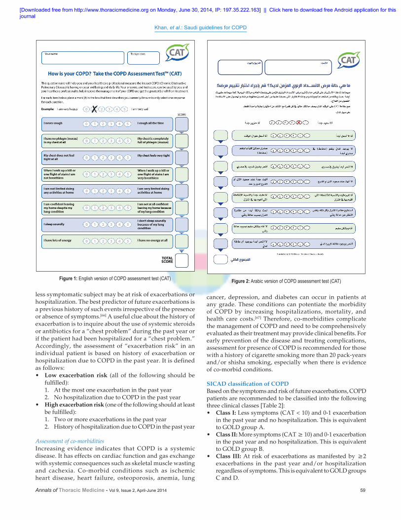
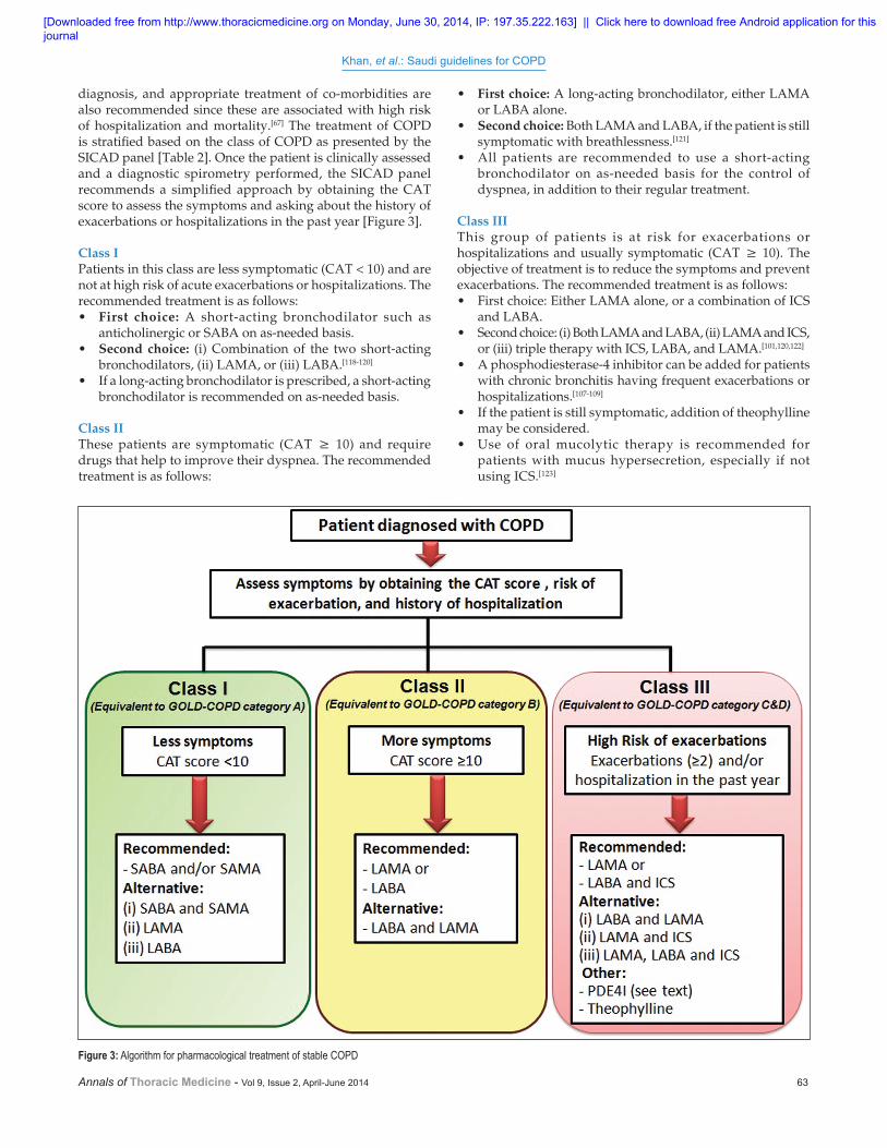
Assessment of symptomsThe cardinal symptoms of COPD, such as cough, sputum production, and dyspnea on exertion, should be assessed in all patients. An objective assessment of symptoms by a validated tool is recommended, such as the COPD Assessment Test (CAT) [Figure 1].[58] The CAT provides a comprehensive assessment of the impact of dyspnea and health status impairment in COPD. The CAT has been translated and validated in Arabic language [Figure 2].[59] It is an eight-item questionnaire with a six-item Likert scale ranging from 0 to 5. It correlates very well with the health status questionnaire known as the St George Respiratory Questionnaire.[60] The score ranges from zero (completely asymptomatic) to 40 (extremely symptomatic). A CAT score ≥10 is associated with a significantly impaired health status [Table 1].[61]
Recommendation• CAT score is recommended as a tool for the comprehensive
assessment of all patients with COPD. (Evidence C)
Assessment of exacerbation riskIt is important to identify patients who are at risk of exacerbations, as it has been recently recognized that exacerbations can be prevented with treatment. [62] COPD exacerbation is simply defined as worsening of patient’s usual symptoms, which may require a change in usual medications.[63] COPD exacerbations cause a significant decline in health status, accelerate the decline in lung function, and increase the risk of mortality.[64,65] A symptomatic patient may have no exacerbations, whereas a
Table 1: Interpretation of the COPD Assessment Test CAT score Interpretation >30 Very high>20 High10-20 Medium <10 Low
[Downloaded free from http://www.thoracicmedicine.org on Monday, June 30, 2014, IP: 197.35.222.163] || Click here to download free Android application for thisjournal
Khan, et al.: Saudi guidelines for COPD
Annals of Thoracic Medicine - Vol 9, Issue 2, April-June 2014 59
less symptomatic subject may be at risk of exacerbations or hospitalization. The best predictor of future exacerbations is a previous history of such events irrespective of the presence or absence of symptoms.[66] A useful clue about the history of exacerbation is to inquire about the use of systemic steroids or antibiotics for a “chest problem” during the past year or if the patient had been hospitalized for a “chest problem.” Accordingly, the assessment of “exacerbation risk” in an individual patient is based on history of exacerbation or hospitalization due to COPD in the past year. It is defined as follows:• Low exacerbation risk (all of the following should be
fulfilled):1. At the most one exacerbation in the past year2. No hospitalization due to COPD in the past year
• High exacerbation risk (one of the following should at least be fulfilled):1. Two or more exacerbations in the past year2. History of hospitalization due to COPD in the past year
Assessment of co-morbiditiesIncreasing evidence indicates that COPD is a systemic disease. It has effects on cardiac function and gas exchange with systemic consequences such as skeletal muscle wasting and cachexia. Co-morbid conditions such as ischemic heart disease, heart failure, osteoporosis, anemia, lung
cancer, depression, and diabetes can occur in patients at any grade. These conditions can potentiate the morbidity of COPD by increasing hospitalizations, mortality, and health care costs.[67] Therefore, co-morbidities complicate the management of COPD and need to be comprehensively evaluated as their treatment may provide clinical benefits. For early prevention of the disease and treating complications, assessment for presence of COPD is recommended for those with a history of cigarette smoking more than 20 pack-years and/or shisha smoking, especially when there is evidence of co-morbid conditions.
SICAD classification of COPDBased on the symptoms and risk of future exacerbations, COPD patients are recommended to be classified into the following three clinical classes [Table 2]: • Class I: Less symptoms (CAT < 10) and 0-1 exacerbation
in the past year and no hospitalization. This is equivalent to GOLD group A.
• Class II: More symptoms (CAT ≥ 10) and 0-1 exacerbation in the past year and no hospitalization. This is equivalent to GOLD group B.
• Class III: At risk of exacerbations as manifested by ≥2 exacerbations in the past year and/or hospitalization regardless of symptoms. This is equivalent to GOLD groups C and D.
Figure 1: English version of COPD assessment test (CAT) Figure 2: Arabic version of COPD assessment test (CAT)
[Downloaded free from http://www.thoracicmedicine.org on Monday, June 30, 2014, IP: 197.35.222.163] || Click here to download free Android application for thisjournal

Khan, et al.: Saudi guidelines for COPD
60 Annals of Thoracic Medicine - Vol 9, Issue 2, April-June 2014
Pharmacological Treatment
Pharmacological treatment for COPD is primarily directed toward symptom control and prevention of acute exacerbations, thereby slowing the progression of disease and mortality. Table 3 shows the classes of medications used in treating COPD and available in the Saudi market.
BronchodilatorsBronchodilators are the mainstay of treatment for symptomatic COPD patients. They improve the expiratory flow by
altering bronchial smooth muscle tone. This effect improves emptying of the lungs and reduces hyperinflation at rest and during exercise.[68] Despite symptomatic improvement, bronchodilators do not modify the decline in FEV1 in COPD patients.[69] Bronchodilators are given either on as-needed basis for acute symptom relief or on a regular basis to prevent or reduce symptoms. The inhalation route is preferred for administration. The choice of inhaler device depends on availability, cost, and the patient’s ability to use the device. In Saudi Arabia, inhalers are available in the form of pressurized meter-dose inhalers (MDI), evohalers, breath-actuated (Easyhaler) and dry-powder inhalers (DPIs), such as turbuhaler, discus, aerohalers, and handihalers. Patients with poor inspiratory force may obtain some symptomatic benefit with drugs administered through nebulization; however, drug nebulization is generally not recommended for regular treatment because of cost and practicality issues.[70] Bronchodilators are generally safe. The side effects are usually mild, dose dependent, and tend to resolve after treatment withdrawal. However, as COPD patients are usually elderly and more likely to have co-morbidities, the risk of developing side effects is considered to be greater than in asthmatic patients.
Table 2: SICAD classification of COPD Class Characteristics Exacerbation in
the past yearCAT
scoreGOLD
equivalent Class I Less symptoms 0-1 ≤10 Group A
At low risk of exacerbation
Class II More symptoms 0-1 ≥10 Group BAt low risk of exacerbation
Class III At high risk of exacerbation
≥2 Any score Group C and D
Table 3: The classes of medications used in treating COPD and available in the Saudi marketDrug Dose Route of administration Duration of action (h)b2-agonists
Short acting
Salbutamol100, 200 μg MDI 4-6
5 mg/ml Nebulizer solution 4-6Long acting
Formoterol 4.5-12 μg DPI 12+
Salmeterol 25-50 μg MDI, DPI 12+
Indacaterol 150-300 μg DPI 24AnticholinergicsShort acting
Ipratropium bromide 20, 40 μg MDI 6-80.25-0.5 μg Nebulizer solution 6-8
Long actingTiotropium 18 μg DPI 24+
Inhaled steroidsBeclomethasone 50-400 μg MDI, DPI 6-12
Budesonide100, 200 μg DPI 12
0.25, 0.5 mg/ml Nebulizer solution 12Fluticasone propionate 50-500 μg MDI, DPI 12Ciclesonide 80-320 μg MDI 24
Combination of long-acting b2-agonist and inhaled steroid
Formoterol/budesonide 4.5/160 DPI 12Salmeterol/fluticasone 25/50, 25/125, 25/250
50/100, 50/250, 50/500MDIDPI
12
MethylxanthinesAminophylline 200-600 mg OralTheophylline (SR) 100-600 mg Oral 24
Phosphodiesterase-4 inhibitorsRoflumilast 500 mcg Oral 24
Systemic steroidsPrednisolone/prednisone 5-60 mg Oral 24
MDI = Meter-dose inhaler, DPI = Dry-powder inhaler
[Downloaded free from http://www.thoracicmedicine.org on Monday, June 30, 2014, IP: 197.35.222.163] || Click here to download free Android application for thisjournal

Khan, et al.: Saudi guidelines for COPD
Annals of Thoracic Medicine - Vol 9, Issue 2, April-June 2014 61
b2-agonistsThe main action of b2-agonists is to induce airway smooth muscle relaxation by stimulating b2-adrenergic receptors and increase the production of cyclic AMP (cAMP). Inhaled b2-agonists have a rapid onset of action. This effect is probably slower in COPD than in asthma. The bronchodilator effects of short-acting b2-agonists (SABA) usually last for 4-6 h.[71] SABA such as salbutamol is recommended as rescue medication to relieve the acute symptoms of bronchospasm. On the other hand, long-acting inhaled b2-agonists (LABA), such as salmeterol and formoterol, have an effect that last for 12 h or more with no evidence of tolerance or tachyphylaxis with regular use in COPD patients.[72,73] Indacaterol is a new medication characterized by its 24-h action, and thus can be used as a single daily dose.[74] LABA are recommended as maintenance therapy, and are more effective and convenient than treatment with SABA medication and should always be considered in patients requiring chronic therapy.[75] Treatment with LABA has been shown to improve health status, quality of life, exercise tolerance, and exacerbation rate in COPD patients.[72-74] Contrary to patients with asthma, COPD patients can be treated with LABA as monotherapy.
The side effects are related to the stimulation of b2-adrenergic receptors that may cause sinus tachycardia and rarely other cardiac rhythm abnormalities in susceptible patients. Symptomatic tremor may be troublesome in some older patients treated with high doses of b2-agonists. Hypokalemia can occur, especially when treatment is combined with diuretics, and should be monitored in susceptible patients.
Recommendations• The central role of bronchodilators in COPD patients
is directed toward symptom relief and not toward improvement in FEV1. (Evidence A)
• SABA, such as salbutamol, are recommended to relieve acute symptoms related to bronchospasm. (Evidence A)
• Regular treatment with LABA is more effective and convenient than treatment with SABA, and is recommended for symptomatic patients (Class II and III). (Evidence A)
• The regular use of LABA bronchodilators has shown to improve health status, quality of life, and exercise tolerance in COPD patients. (Evidence A)
AnticholinergicsAnticholinergic medications, such as ipratropium bromide and tiotropium, inhibit the effect of acetylcholine on M3 receptors. The bronchodilatation effect of short-acting inhaled ipratropium may last for up to 8 h. Tiotropium has duration of action of more than 24 h and is used once daily.[71,76] Treatment with a long-acting muscarinic antagonist (LAMA) drug, tiotropium, is effective in reducing the symptoms, improving the quality of life, and decreasing the exacerbations and hospitalizations at any stage in COPD patients.[77-81] It improves the effectiveness of pulmonary rehabilitation (PR) and may also show reduction in the rate of decline in lung function compared with placebo, in patients who are not on other maintenance drugs.[82,83] Anticholinergic drugs are considered to be very safe. The main side effect is dryness of mouth. Although mild prostatic symptoms have been reported, there is no evidence of a direct causal relationship. The use of nebulized ipratropium with a facemask has been reported to precipitate acute glaucoma.[77]
Recommendations • Ipratropium bromide is recommended as the rescue
medication to relieve the acute symptoms of bronchospasm. (Evidence A)
• Treatment with LAMA such as tiotropium is effective in reducing the exacerbation rate in COPD patients and improves the effectiveness of PR. (Evidence A)
• Tiotropium may reduce the rate of decline in lung function compared with placebo in patients who are not on other maintenance drugs. (Evidence B)
Corticosteroids
The effects of oral and inhaled corticosteroids in COPD are much less defined than in asthma. Therefore, every effort should be made to differentiate COPD from asthma.
Inhaled corticosteroidsThe role of inhaled corticosteroids (ICS) in stable COPD as a maintenance treatment is still controversial. Although regular treatment with ICS does not modify the long-term decline of FEV1 in patients with COPD, substantial evidence suggests that regular treatment with ICS might be beneficial in symptomatic COPD patients with an FEV1 less than 60% of the predicted value and with a history of frequent exacerbations.[84-86] This treatment has been shown to reduce the frequency of exacerbations, and thus improve the health status.[87] Furthermore, withdrawal from ICS may lead to exacerbations in certain COPD patients.[88,89] A combination of ICS and LABA is more effective than any one alone.[90,91]
ICS are generally safe and well tolerated by COPD patients. The majority of the patients may not have any side effects even with long-term use. Minor side effects, such as hoarseness of the voice and higher incidence of skin bruising that increases with age, were reported in COPD patients. Treatment with ICS for very long periods may be associated with a reduction in bone density, slightly increased risk of fractures and pneumonia, and adrenal insufficiency.[92-96]
Oral corticosteroidsOral corticosteroids are recommended to be used in acute COPD exacerbation. However, long-term treatment with systemic corticosteroids has many side effects, including steroid myopathy and glucose intolerance. These side effects might have a negative impact on COPD co-morbidities. Therefore, based on the large body of evidence on side effects, and because of a lack of evidence of benefit, long-term treatment with oral corticosteroids is not recommended for COPD.[93]
Recommendations• ICS are recommended in Class III (equivalent to GOLD
groups C and D). (Evidence C)• A therapeutic trial with oral corticosteroids in stable COPD
patients is not recommended. (Evidence B)• Long-term treatment with oral corticosteroids is not
recommended for stable COPD. (Evidence A)
Combination therapyWhen comparing combination of ICS with LABA to either of them alone in a single device, the combination is more effective in improving the lung function and quality of life and reducing
[Downloaded free from http://www.thoracicmedicine.org on Monday, June 30, 2014, IP: 197.35.222.163] || Click here to download free Android application for thisjournal

Khan, et al.: Saudi guidelines for COPD
62 Annals of Thoracic Medicine - Vol 9, Issue 2, April-June 2014
the exacerbations in moderate and severe disease, and it may also reduce mortality.[90,97,98] The addition of tiotropium to a combination of ICS and LABA further reduces the exacerbation rate and improves the lung functions and quality of life.[99-101]
Recommendations• Patients with COPD should not be treated with ICS alone,
and patients with asthma should not receive LABA alone. (Evidence A)
• Addition of regular treatment with ICS to bronchodilator treatment might be beneficial in symptomatic COPD patients with a history of frequent exacerbations. (Evidence A)
• A combination of an ICS and LABA is more effective than the individual components in all parameters of COPD management. (Evidence A)
• The addition of tiotropium to a combination of ICS plus LABA further reduces the exacerbation rate and improves the lung functions and quality of life. (Evidence B)
Methylxanthines The role of methylxanthines in COPD is controversial and the exact mechanism of action in COPD patients is yet to be determined. Although methylxanthines are considered to be bronchodilators, a range of non-bronchodilator actions has been reported. An anti-inflammatory effect through the non-selective inhibition of phosphodiesterase enzymes has been suggested.[102] Theophylline has been shown to improve steroid resistance through up-regulation of histone deacetylase pathway.[103] In addition, changes in inspiratory muscle function have also been reported in COPD patients treated with theophylline; however, whether this is a primary effect on the muscle or just reflecting changes in dynamic lung volumes is not yet clear.[104] Low-dose theophylline appears to reduce exacerbations without improvement in bronchodilatation.[105]
Dose-related toxicity is the main concern for using this class of medications. Unfortunately, most of the benefits reported are only observed when near-toxic doses are used. Serious side effects include the development of atrial and ventricular arrhythmias (which can prove fatal) and seizures (which can occur without prior history). More common, but less significant side effects include headaches, insomnia, and gastrointestinal disturbances such as nausea and heartburn. As the drugs are metabolized by cytochrome P450, drug–drug interactions can occur.
Recommendations• Low-dose theophylline appears to reduce exacerbations.
(Evidence C)
Phosphodiesterase-4 inhibitorsRoflumilast is an anti-inflammatory drug and a specific phosphodiesterase-4 enzyme inhibitor, which is responsible for the breakdown of intracellular cAMP. The drug is taken orally and has been approved for COPD patients with chronic bronchitis symptoms and suffering from frequent exacerbations. It has been shown to reduce moderate to severe exacerbations in moderate to severe COPD treated with corticosteriods.[106] The drug’s beneficial effects on exacerbation and lung functions are also seen when it is added to LABA, LAMA, or ICS.[107-109] The most common side effects are nausea, abdominal pain, diarrhea, reduced appetite, headache, and
sleep disturbance. Most of these adverse effects improve over time. Mild weight reduction was also reported.
Recommendations• Roflumilast is indicated in Class III COPD patients with
chronic bronchitis phenotype. (Evidence A)• The drug has beneficial effects in reducing exacerbations
when added to LAMA, LABA, or ICS. (Evidence B)
Mucolytic and antioxidant agentsThe regular use of mucolytics in COPD is controversial. Mucolytic agents may have some beneficial effects on exacerbation rates and health-related quality of life in patients not treated with standard COPD therapy.[110,111] Mucolytics are often considered as an “add-on” therapy in patients with severe COPD who have recurrent exacerbations and remain symptomatic despite maximum standard therapy. They are generally safe and well tolerated. In a recent randomized, double-blind, placebo-controlled study, 1 year treatment with high-dose N-acetylcysteine (1200 mg/day) resulted in significant improvement of small airways function and decreased frequency of exacerbations in stable COPD patients.[112]
Recommendations• Mucolytics may be considered as an “add on” therapy in
COPD. (Evidence C)• Long-term treatment with high-dose N-acetylcysteine (1200
mg/day) may result in a significant improvement of small airways function and decreased frequency of exacerbations in stable COPD patients. (Evidence C)
AntibioticsAntibiotics should be used to treat infectious exacerbations of COPD. The prophylactic use of antibiotics has been shown to be promising; however, additional studies are required before the regular use of these agents can be recommended.[113-115]
Recommendations• Antibiotics should be used to treat infectious exacerbations
of COPD. (Evidence A)
AntitussivesThe regular use of antitussives is not recommended for stable COPD patients.
Recommendations• The regular use of antitussives is not recommended for
stable COPD patients. (Evidence D)
a-1 antitrypsina-1 antitrypsin augmentation therapy can be considered in young patients with severe hereditary a-1 antitrypsin deficiency and established emphysema. However, this therapy is very expensive and only available in very specialized tertiary care hospitals.[116,117]
Treatment for Stable COPD Based on SICAD Classification
The current management strategy for COPD primarily focuses on control of symptoms, prevention of exacerbations, and improving the quality of life. Furthermore, prevention,
[Downloaded free from http://www.thoracicmedicine.org on Monday, June 30, 2014, IP: 197.35.222.163] || Click here to download free Android application for thisjournal
Khan, et al.: Saudi guidelines for COPD
Annals of Thoracic Medicine - Vol 9, Issue 2, April-June 2014 63
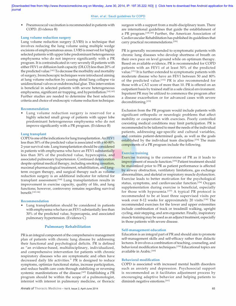
diagnosis, and appropriate treatment of co-morbidities are also recommended since these are associated with high risk of hospitalization and mortality.[67] The treatment of COPD is stratified based on the class of COPD as presented by the SICAD panel [Table 2]. Once the patient is clinically assessed and a diagnostic spirometry performed, the SICAD panel recommends a simplified approach by obtaining the CAT score to assess the symptoms and asking about the history of exacerbations or hospitalizations in the past year [Figure 3].
Class I Patients in this class are less symptomatic (CAT < 10) and are not at high risk of acute exacerbations or hospitalizations. The recommended treatment is as follows:• First choice: A short-acting bronchodilator such as
anticholinergic or SABA on as-needed basis.• Second choice: (i) Combination of the two short-acting
bronchodilators, (ii) LAMA, or (iii) LABA.[118-120]
• If a long-acting bronchodilator is prescribed, a short-acting bronchodilator is recommended on as-needed basis.
Class II These patients are symptomatic (CAT ≥ 10) and require drugs that help to improve their dyspnea. The recommended treatment is as follows:
• First choice: A long-acting bronchodilator, either LAMA or LABA alone.
• Second choice: Both LAMA and LABA, if the patient is still symptomatic with breathlessness.[121]
• All patients are recommended to use a short-acting bronchodilator on as-needed basis for the control of dyspnea, in addition to their regular treatment.
Class III This group of patients is at risk for exacerbations or hospitalizations and usually symptomatic (CAT ≥ 10). The objective of treatment is to reduce the symptoms and prevent exacerbations. The recommended treatment is as follows:• First choice: Either LAMA alone, or a combination of ICS
and LABA.• Second choice: (i) Both LAMA and LABA, (ii) LAMA and ICS,
or (iii) triple therapy with ICS, LABA, and LAMA.[101,120,122]
• A phosphodiesterase-4 inhibitor can be added for patients with chronic bronchitis having frequent exacerbations or hospitalizations.[107-109]
• If the patient is still symptomatic, addition of theophylline may be considered.
• Use of oral mucolytic therapy is recommended for patients with mucus hypersecretion, especially if not using ICS.[123]
Figure 3: Algorithm for pharmacological treatment of stable COPD
[Downloaded free from http://www.thoracicmedicine.org on Monday, June 30, 2014, IP: 197.35.222.163] || Click here to download free Android application for thisjournal

Khan, et al.: Saudi guidelines for COPD
64 Annals of Thoracic Medicine - Vol 9, Issue 2, April-June 2014
• All patients can use short-acting bronchodilators as needed for control of dyspnea, in addition to their regular treatment.
Non-pharmacological Therapies
Smoking cessationSmoking cessation has the greatest proven impact on the natural history of COPD by slowing the accelerated decline of lung function.[124] The younger the age at which a smoker quits, the more likely the rate of decline will parallel that of non-smokers.[125] Smoking cessation is recommended at any age, since it can decrease the COPD symptoms and the number of exacerbations and improve the health status and exercise tolerance. It is one of the few interventions with proven benefit on decreasing mortality in COPD due to its effect on lung cancer, cardiovascular disease, and other co-morbid conditions.[126] The following strategy may help patients who are willing to quit smoking to achieve this goal:
CounselingObtaining the patient’s smoking history and offering a brief conseling to the patient to quit smoking by a health care professional can result in quit rates of 5-10%. Tobacco dependence is a chronic disease and quitting may require multiple attempts. Long-term quit rates of 25% are achievable with more intensive interventions.[127]
MedicationsCombining professional counseling, group support with pharmacotherapy significantly increases the success rate of smoking cessation.[128] Nicotine replacement therapy increases the long-term quit rate and is significantly more effective than placebo. This therapy is contraindicated in those with recent myocardial infarction, unstable angina, stroke, or untreated peptic ulcer disease. Oral medications, such as varenicline, bupropion, and nortryptiline, have increased long-term quit rates and are more effective when combined with counseling. Bupropion can be combined with nicotine replacement therapy for better results.[129,130] Health care professionals should familiarize themselves with the contraindications and possible side effects of these medications, such as possible increased risk of suicide with varenicline and seizures with bupropion.
Recommendation• Smoking cessation is recommended at any age; it improves
the health status and exercise tolerance and decreases COPD symptoms, the number of exacerbations, and mortality. (Evidence A)
Long-term oxygen therapyThere is unequivocal evidence that long-term oxygen therapy (LTOT) improves survival and quality of life in hypoxemic patients with COPD. LTOT is recommended when the PaO2 is less than 55 mmHg (less than 7.3 kPa or arterial saturation less than 88%), while the patient is breathing room air and is free of acute exacerbation for at least 2 months while receiving maximum therapy. LTOT is also recommended if the PaO2 is between 55 and 60 mmHg (7.4-8 kPa) in a patient with COPD associated with cor-pulmonale, peripheral edema, or hematocrit ≥55%. Despite the lack of solid evidence, the LTOT recommendation may be extended to COPD patients
with PaO2 more than 60 mmHg (more than 8 kPa or arterial saturation more than 88%) and having nocturnal hypoxemia or hypoxemia during exercise.
Once LTOT is commenced, it is recommended to re-evaluate the patients at 3 months and 1 year intervals to optimize oxygen prescription.[131,132] The oxygen should be titrated to achieve a resting PaO2 of 60-65 mmHg or arterial saturation between 88% and 94%. The usual dose is 1-2.5 L/min, typically administered by nasal cannula, for a minimum of 18 h/day to derive benefit in terms of survival. A practical way of supplying oxygen at home is the use of an oxygen concentrator. Long tubes may be needed to ensure continuous use of oxygen during normal activity and exercise. Lightweight portable delivery systems (e.g. cylinders) are now available for outdoor use.
Recommendation• LTOT is recommended when the PaO2 is less than 55 mmHg
(less than 7.3 kPa or arterial saturation less than 88%), or PaO2 is between 55 and 60 mmHg (7.4-8 kPa) with cor-pulmonale, peripheral edema, or hematocrit ≥55%. (Evidence A)
Non-invasive ventilationNon-invasive ventilation (NIV) is a method of providing ventilatory support using a nasal or full-face mask without the placement of an endotracheal tube. Benefits of NIV in the treatment of acute respiratory failure in COPD have been well documented.[133] However, the benefit of NIV for stable COPD patients with chronic respiratory failure is less clear. Recent studies revealed that it may improve survival and dyspnea in hypercapnic COPD patients, particularly in conjunction with PR, but the results about the effect on quality of life are conflicting.[134,135] A Cochran review and meta-analysis did not confirm any benefit from long-term use of non-invasive positive pressure ventilation (NIPPV) in stable COPD.[136,137] Systematic review revealed a benefit of NIPPV in a subset of selected patients with severe COPD on maximal therapy.[138]
Recommendation• Benefits of NIV in the treatment of acute respiratory failure
in COPD have been well documented. (Evidence B)
VaccinationAn acute exacerbation of COPD is a major cause of morbidity and mortality worldwide. Most acute exacerbations are triggered by community-acquired respiratory infections. Influenza and pneumococcus are the major causes of morbidity and mortality in people with COPD. Influenza vaccination clearly reduces acute exacerbations of COPD and may reduce hospitalizations and mortality. Patients with COPD should receive influenza vaccination annually.[139]
Pneumococcal vaccination reduces invasive pneumococcal disease, especially in smokers. The US Centers for Disease Control and Prevention (CDC) recommend the administration of pneumococcal vaccination to all patients at least 65 years of age, as well as to younger patients with chronic medical illnesses, including COPD.[140]
Recommendations• Patients with COPD are recommended to receive influenza
vaccination annually. (Evidence A)
[Downloaded free from http://www.thoracicmedicine.org on Monday, June 30, 2014, IP: 197.35.222.163] || Click here to download free Android application for thisjournal
Khan, et al.: Saudi guidelines for COPD
Annals of Thoracic Medicine - Vol 9, Issue 2, April-June 2014 65
• Pneumococcal vaccination is recommended in patients with COPD. (Evidence B)
Lung volume reduction surgeryLung volume reduction surgery (LVRS) is a technique that involves reducing the lung volume using multiple wedge excisions of emphysematous areas. LVRS is reserved for highly selected patients with upper lobe predominant heterogeneous emphysema who do not improve significantly with a PR program. It is contraindicated in very severely ill patients with either FEV1 or diffusion lunh capacity (DLCO) less than 20% of the predicted value.[141] To decrease the morbidity and mortality of surgery, bronchoscopic techniques were introduced aiming at lung volume reduction by causing distal lung collapse via unidirectional valves or endobronchial glue. This intervention is beneficial in selected patients with severe heterogeneous emphysema, significant air trapping, and hyperinflation.[142,143] Further studies are needed to determine the best selection criteria and choice of endoscopic volume reduction technique.
Recommendation• Lung volume reduction surgery is reserved for a
highly selected small group of patients with upper lobe predominant heterogeneous emphysema who do not improve significantly with a PR program. (Evidence B)
Lung transplantCOPD is one of the indications for lung transplantation. An FEV1 less than 30% of the predicted value is associated with a 60-80% 2-year survival rate. Lung transplantation should be considered in patients with emphysema who have an FEV1 substantially less than 30% of the predicted value, hypercapnia, and associated pulmonary hypertension. Continued deterioration despite optimal medical therapy, including smoking cessation, maximal pharmacological treatment, rehabilitation, and long-term oxygen therapy, and surgical therapy such as volume reduction surgery is an additional indicator for referral for transplant assessment.[144] Lung transplantation results in improvement in exercise capacity, quality of life, and lung functions; however, controversy remains regarding survival benefit.[145,146]
Recommendation• Lung transplantation should be considered in patients
with emphysema who have an FEV1 substantially less than 30% of the predicted value, hypercapnia, and associated pulmonary hypertension. (Evidence C)
Pulmonary Rehabilitation
PR is an integral component of the comprehensive management plan of patients with chronic lung disease by addressing their functional and psychological deficits. PR is defined as “an evidence-based, multidisciplinary, individualized, and comprehensive intervention for patients with chronic respiratory diseases who are symptomatic and often have decreased daily life activities.” PR is designed to reduce symptoms, optimize functional status, increase participation, and reduce health care costs through stabilizing or reversing systemic manifestations of the disease.[147] Establishing a PR program should be within the scope of a pulmonologist, internist with interest in pulmonary medicine, or thoracic
surgeon with a support from a multi-disciplinary team. There are international guidelines that guide the establishment of a PR program.[148,149] Further, the American Association of Cardiovascular Rehabilitation has published its guidelines that carry practical recommendations.[150]
PR is generally recommended to symptomatic patients with chronic lung diseases who develop shortness of breath on their own pace on level ground while on optimum therapy. Based on available evidence, PR is recommended for COPD patients with an FEV1 of at least 50% of the predicted value. [151] It is further extended to symptomatic patients with moderate disease who have an FEV1 between 50 and 80% of the predicted value.[152] PR is also recommended for those with a CAT score of more than 10. PR is offered on an outpatient basis by trained staff in a safe clinical environment. Inpatient PR may be utilized to commence the program after a disease exacerbation or for advanced cases with severe deconditioning.[153]
Exclusion from the PR program would include patients with significant orthopedic or neurologic problems that affect mobility or cooperation with exercises. Poorly controlled coexisting medical conditions may limit participation. PR is recommended to be tailored to meet the needs of the individual patients, addressing age-specific and cultural variables, and contains patient-determined goals, as well as the goals established by the individual team discipline.[154] The main components of a PR program include the following.
Exercise trainingExercise training is the cornerstone of PR as it leads to improvement of muscle function.[155] Patient treatment should be maximized prior to PR as patient performance is affected by airway obstruction, ventilatory limitations, gas exchange abnormalities, and skeletal or respiratory muscle dysfunction. Exercise leads to better motivation for the psychological status, symptoms, and cardiovascular function.[156] Oxygen supplementation during exercise is beneficial, especially for those with hypoxemia.[157] A typical PR protocol is recommended to be at least three supervised visits per week over 8-12 weeks for approximately 20 visits.[158] The recommended exercises for the lower and upper extremities include combination of track or treadmill walking, upright cycling, stair stepping, and arm ergometer. Finally, inspiratory muscle training may be used as an adjunct treatment, especially in those patients with severe disease.
Self-management educationEducation is an integral part of PR and should aim to promote self-management skills and self-efficacy rather than didactic lectures. It involves a combination of teaching, counseling, and behavioral modification techniques.[159] Educational topics are available in Arabic.[160]
Behavioral modificationCOPD is associated with increased mental health disorders such as anxiety and depression. Psychosocial support is recommended as it facilitates adjustment process by encouraging adaptive behavior and helping patients to diminish negative emotions.[161]
[Downloaded free from http://www.thoracicmedicine.org on Monday, June 30, 2014, IP: 197.35.222.163] || Click here to download free Android application for thisjournal

Khan, et al.: Saudi guidelines for COPD
66 Annals of Thoracic Medicine - Vol 9, Issue 2, April-June 2014
Outcome assessmentAssessment in COPD should be patient-centered, and range from unstructured clinical assessment to validated tests and instruments such as six minutes walk distance (6MWD) or health-related quality of life instruments.[162] The Chronic Respiratory Disease Questionnaire (CRQ) is recommended as it is reliable, valid, and available in Arabic.[163,164] The Arabic version of CRQ is based on the CRQ with standardized dyspnea domain and is self-administered. It is a 20-item questionnaire with the four domains of the CRQ calculated separately, which are dyspnea, fatigue, emotion, and mastery. Higher numbers indicate better quality of life. For patients with COPD, the CAT was recently introduced to measure the impact of the disease [Figure 1]. The CAT is an 8-item test where higher numbers reflect a higher impact of the disease [Table 1]. The CAT is also available in Arabic [Figure 2].[59]
Benefits from PR program may continue up to 18 months.[165] Health-related quality of life is maintained for a longer period compared to exercise.[166] Strategies to maintain the benefits of PR include continuing rehabilitation, maintenance program, and recall program.[167,168]
Recommendations• PR is recommended for COPD patients with an FEV1 of at
least 50% of the predicted value and those with moderate disease who have an FEV1 between 50 and 80% of the predicted value. (Evidence B)
• A typical PR protocol is recommended to be at least three supervised visits per week over 8-12 weeks for approximately 20 visits. (Evidence B)
• Benefits from PR program may continue up to 18 months. (Evidence B)
• Health-related quality of life is maintained for a longer period compared to exercise. (Evidence A)
Systemic Effects of COPD and Co-morbidities
COPD is associated with low-grade systemic inflammation with increased levels of systemic inflammatory markers such as leukocytosis, fibrinogen, C-reactive protein (CRP), cytokines [interleukin (IL)-6], and tumor necrosis factor alpha (TNF-α). Its intensity increases during COPD exacerbations.[169,170] It has also been implicated in the pathogenesis of weight loss, skeletal muscle dysfunction, wasting of muscle mass, cardiovascular disease, and depression.[171-174]
Nutrition and Body Mass Index
Malnutrition is a significant problem in COPD patients. The estimated prevalence of malnutrition is 10-15% in patients with mild to moderate COPD and 50% in patients with advanced stage of the disease. This percentage increases to approximately 50-70% in hospitalized patients with acute exacerbation.[175-177]
Malnutrition is defined as weight less than 90% of the predicted value or body mass index (BMI) less than 18.4 kg/m2. A recent weight loss of more than 10% over 6 months carries a negative prognostic effect as well. Malnutrition is frequently associated with anemia, increased susceptibility to infection and muscle wasting that aggravates dyspnea, and limitation
in exercise capacity.[146] BMI is an independent prognostic factor in patients with COPD. A decreased BMI is associated with increased mortality and increased risk of developing COPD.[178-182] An increased BMI may even improve lung function in patients with COPD.[148,149] COPD patients who are underweight or who experience loss of weight over the course of follow-up should be given nutritional supplements. [150] The long-term benefits of nutritional supplementation are yet to be determined.[151]
Recommendation• COPD patients who are underweight or with a decrease in
weight over the course of follow-up are recommended to receive nutritional supplements. (Evidence C)
Vitamin D and COPD
Vitamin D insufficiency seems to be a common problem that affects the general population worldwide, including the sunny countries such as Saudi Arabia.[183,184] Vitamin D deficiency has been associated with decline in lung function, increased inflammation, and reduced immunity.[185] The prevalence of Vitamin D deficiency is particularly high in COPD patients and increases with the severity of COPD from 60% to 77% in patient’s advanced disease.[154] It is closely associated with prevalence of osteoporosis, which is particularly higher in older patients and in those on steroids.[155]
Several factors may be attributed to Vitamin D deficiency in COPD patients, including poor diet, reduced outdoor activities and exposure to sun, impaired activation because of renal dysfunction, and a lower storage capacity in muscles or fat due to wasting.[186] Given the high prevalence of osteoporosis in the Saudi population compared to western countries and the increased risk of osteopenia and osteoporosis in COPD, measurement of 25-hydroxyvitamin D (25-OHD) levels and dual-energy X-ray absorptiometry (DXA) are recommended for all patients with obvious risk of osteoporosis.[187,188] There is no adequate evidence for routine supplementation with vitamin D in patients with COPD. However, vitamin D and calcium supplementation should be considered in all patients at risk, including those receiving corticosteroid regardless of the steroid dose or duration.[189,190]
Recommendation• COPD patients at risk for osteoporosis, especially the
patients on glucocorticoids at any dose with an anticipated duration more than 3 months, are recommended to be screened for osteoporosis. (Evidence D)
• Routine supplementation of vitamin D in patients with COPD is not recommended. (Evidence D)
Sleep and COPD
Worsening of hypoxemia during sleep in COPD patients is well known. The degree of hypoxemia during sleep depends on the severity of COPD and the baseline PaO2 level. Hypoxemia in COPD patients during sleep is attributed to a drop in ventilation due to decrease in the ventilatory drive, decreased chemosensitivity, decreased basal metabolic rate, and increased upper airway resistance. Although these changes occur in normal individuals, the alterations are much more profound
[Downloaded free from http://www.thoracicmedicine.org on Monday, June 30, 2014, IP: 197.35.222.163] || Click here to download free Android application for thisjournal

Khan, et al.: Saudi guidelines for COPD
Annals of Thoracic Medicine - Vol 9, Issue 2, April-June 2014 67
in COPD patients and are particularly worse during REM sleep. Moreover, during sleep, there is decreased mucociliary clearance, a decrease in the functional residual capacity, and closing of small airway; these changes consequently increase ventilation-perfusion (V/Q) mismatching and worsening of hypoxemia.[191-193]
Sleep disorders in COPDSleep disorders are common among patients with COPD and may have a major impact on the patients’ quality of life and health outcomes. Sleep disturbances may occur due to the effects of breathing abnormalities on sleep and sleep disruption. However, other etiologies may include the medications used to treat COPD, concomitant anxiety and depression, or the presence of co-morbid sleep disorders such as obstructive sleep apnea or obesity hypoventilation syndrome. COPD per se in the absence of sleep apnea or hypopnea does not affect the quality of sleep.[194] Insomnia is the most common sleep disorder reported in COPD.[191,195] Other common sleep disorders are obstructive sleep apnea and obesity hypoventilation syndrome.
Obstructive sleep apnea and overlap syndromeObstructive sleep apnea syndrome (OSAS) and COPD are two distinct diseases. Overlap syndrome is the co-existence of COPD and OSAS within an individual. The estimated prevalence is approximately 1% in adult males.[195,196] A patient with COPD, who also has OSAS has more significant oxygen desaturation and is more prone to hypercapnea and pulmonary hypertension than a patient with the same degree of obstruction but without coexisting OSAS.[197] Furthermore, overlap syndrome is associated with an increased risk of death and hospitalization because of COPD exacerbation. The treatment with continuous positive airway pressure (CPAP) was associated with higher survival and decreased hospitalizations in patients with overlap syndrome and COPD.[198,199]
Recommendations• Long-term oxygen therapy is not indicated in patients with
oxygen desaturation only during sleep. (Evidence B)• The co-existence of obstructive sleep apnea (overlap
syndrome) or obesity hypoventilation syndrome should be suspected in COPD patients with significant hypoxemia or hypercapnea relative to mild airflow limitation, pulmonary hypertension, or daytime sleepiness, or in patients with daytime sleepiness, snoring, and high BMI. Referral for sleep study to rule out OSAS is recommended. (Evidence C)
Heart Failure and COPD
Heart failure and COPD are common. The clinical symptoms and signs of both diseases overlap, and a combination of both diseases in a particular patient may present a diagnostic challenge. B-type natriuretic peptide (BNP) may be helpful in differentiating COPD exacerbation from heart failure.[200] Clinicians are encouraged to use a combination of clinical presentation, spirometry, and echocardiography findings to differentiate both conditions. The use of b-blockers for treatment of heart failure in patients with COPD appears to be safe and had been shown to decrease exacerbations, hospitalizations, and all-cause mortality.[201-203]
Recommendation• The use of b-blockers for treatment of heart failure in
patients with COPD appears to be safe and had been shown to decrease exacerbations, hospitalizations, and all-cause mortality. (Evidence C)
Pulmonary Hypertension and COPD
Pulmonary hypertension is a common co-morbidity associated with increased mortality in COPD patients. The incidence of pulmonary hypertension in COPD patients depends on the severity of airway obstruction and varies from 6 to 60%. It is often mild to moderate, while severe pulmonary hypertension is estimated to be present in 1-3.7% of patients with severe COPD.[204,205] Echocardiography is adequate for initial screening, but is not recommended as routine screening except for patients with hypoxia or dyspnea that is out of proportion with the severity of airway obstruction. Standard maximum COPD therapy along with oxygen supplementation in hypoxic patients is recommended as a treatment for pulmonary hypertension as well. Any COPD patient with severe pulmonary hypertension should be evaluated for other causes of pulmonary hypertension. Few studies had shown limited improvement of pulmonary hypertension in COPD with specific pulmonary vasodilators.[206] In a recent Aspire registry report, compassionate treatment with targeted vasodilator therapy in 43 out of 59 patients with COPD and severe pulmonary hypertension did not show any survival benefit, though in a subset of patients, improvement in functional class was observed. However, currently, there is no clear benefit and it is not recommended.[207,208]
Recommendation• Screening for pulmonary hypertens ion us ing
echocardiogram is recommended for patients with shortness of breath or hypoxia that is out of proportion with the degree of airway obstruction. (Evidence D)
• There is no clearly proven benefit of treating pulmonary hypertension in COPD with pulmonary vasodilators. (Evidence C)
Surgery in COPD
Surgery and general anesthesia are generally not contraindicated in patients with COPD. However, postoperative complications are more frequent in COPD depending on the site of surgery. Upper abdominal and thoracic surgeries have more postoperative complications compared to lower abdominal surgeries. Other risk factors for postoperative complications include smoking, presence of bronchospasm, and secretions.[209] It is very important that the patient with COPD undergoing surgery be carefully assessed for symptoms of ongoing infection, bronchospasm, smoking, and site of surgery. Comorbidity risks that need to be assessed include: hypoxia, hypercapnea, risk for thromboembolism, pulmonary hypertension, and associated cardiac co-morbidities such as coronary artery disease and heart failure. Before undergoing any elective surgery, patients should stop smoking for at least 6-8 weeks and should receive maximal therapy for COPD and PR. Early mobilization, chest physiotherapy, and incentive spirometry may reduce postoperative complications.[147,210]
[Downloaded free from http://www.thoracicmedicine.org on Monday, June 30, 2014, IP: 197.35.222.163] || Click here to download free Android application for thisjournal
Khan, et al.: Saudi guidelines for COPD
68 Annals of Thoracic Medicine - Vol 9, Issue 2, April-June 2014
Recommendation• Before undergoing any elective surgery, COPD patients
should stop smoking for at least 6-8 weeks and should receive maximal therapy for COPD and PR. Early mobilization, chest physiotherapy, and incentive spirometry may reduce postoperative complications. (Evidence C)
Air Travel
COPD patients are liable to develop potentially serious oxygen desaturation during commercial flights. Commercial airplane cabin is pressurized to a level of 6000-8000 m that is equivalent to inspired oxygen of 15% at sea level.[211] It is recommended to keep PaO2 above 50 mmHg (6.7 kPa) in patients with COPD during flight. Supplemental oxygen by nasal cannula usually compensates for the hypoxemia of air travel.[212] However, during a flight, the oxygen pressure should be maintained at the same level at which the patient is clinically stable at sea level. Patients with COPD are recommended to ask their doctors to fill the oxygen supplement request form provided by airlines. Attention also should be paid to co-existing conditions that could preclude air travel like unstable angina, severe anemia, uncontrolled heart failure, large emphysematous bullae, or pneumothorax.
Acute COPD Exacerbation
Exacerbations are frequent events in the natural history of COPD and can adversely affect health-related quality of life, cause deterioration in lung functions, frequent hospitalization, and may even increase mortality.[64] Exacerbations are often under-diagnosed and unreported.[213] It is important to recognize these events, treat effectively, and adopt measures for their prevention. An exacerbation may be defined as an acute sustained deterioration of respiratory symptoms in a COPD patient beyond day-to-day variability which could not be explained by any other cause, and this event may or may not lead to change in therapy.[214] These events may resolve spontaneously or with treatment over a period of time.
Clinical presentationAcute exacerbations present as acute change in symptoms, including coughing, change in the quantity and quality of sputum, and increase in baseline dyspnea. Signs of severe or life-threatening exacerbation include deterioration in the level of consciousness, marked respiratory distress, use of accessory muscles, PaO2 less than 50 mmHg or pH less than 7.3, or a rapid increase in PaCO2 to above 70 mmHg despite the use of oxygen and bronchodilators.
Precipitating factorsIt is estimated that 70-80% of COPD exacerbations are due to respiratory infections. Viral and bacterial infections cause most exacerbations, whereas atypical bacteria are a relatively uncommon cause.[215] The remaining 20-30% is due to environmental pollution or has an unknown etiology.[216] Less likely causes of exacerbation are heart failure, pneumonia, pulmonary embolism, pneumothorax, improper oxygen management, inappropriate drugs such as sedatives, and electrolyte imbalance.
Risk factorsAccording to observational studies, the risk of developing an exacerbation of COPD correlates with advanced age, duration
of COPD, history of antibiotic therapy, COPD-related hospitalization within the previous year, chronic mucus hypersecretion, and having one or more co-morbidities (e.g. ischemic heart disease, chronic heart failure, or diabetes mellitus).[217] A high FEV1 is associated with a lower risk of COPD exacerbation. In the ECLIPSE study, 2138 patients with moderate to severe COPD (GOLD stages II, III, or IV) were followed for 3 years. The single best predictor of exacerbations was a history of exacerbations, regardless of COPD severity.[66,218]
Differential diagnosisPatients with COPD who present to the health care facility with acute worsening of dyspnea should be evaluated for potential alternative diagnoses, such as heart failure, pulmonary thromboembolism, and pneumonia. This was illustrated in an autopsy study of 43 patients with COPD who died within 24 h of admission for a COPD exacerbation.[219] The primary causes of death were heart failure (37%), pneumonia (28%), pulmonary thromboembolism (21%), and COPD (14%).
Goals of treatmentThe goals of treatment of acute exacerbation of COPD include the following:• Identify and ameliorate the cause of the acute exacerbation,
if possible.• Optimize lung function by administering bronchodilators
and other pharmacological agents.• Ensure adequate oxygenation and secretion clearance.• Avoid the need for intubation, if possible.• Prevent the complications of immobility, such as
thromboembolism and deconditioning.• Address nutritional needs.
ManagementIn general, mild exacerbations may be treated with inhaled bronchodilators only, whereas moderate and severe exacerbations require treatment with steroids and/or antibiotics.
AntibioticsThe use of antibiotics appears to improve clinical success rate, lung functions, and time to next exacerbation; however, some patients may improve without the use of antibiotics. A practical approach is to use antibiotics in patients who have two of three important symptoms: increasing breathlessness, increasing sputum volume, and/or increasing sputum purulence.[220] However, patients with severe or life-threatening exacerbations or requiring admission to the hospital should receive antibiotics regardless of the above factors. Around one-third of the exacerbations are associated with viral infection (with rhinovirus and influenza virus predominating). Bacterial colonization is reported in 20-30% of cases during remissions and increases to 30-50% during exacerbations. The usual organisms are Haemophilus influenzae, Streptococcus pneumoniae, and Branhamella catarrhalis. Pseudomonas aeruginosa is occasionally present in severe or long-standing COPD. The following antibiotics are useful in COPD exacerbation: Second-generation cephalosporins, amoxicillin-clavulanate, macrolide, quinolones such as moxifloxacin or levofloxacin, and doxycycline.
[Downloaded free from http://www.thoracicmedicine.org on Monday, June 30, 2014, IP: 197.35.222.163] || Click here to download free Android application for thisjournal
Khan, et al.: Saudi guidelines for COPD
Annals of Thoracic Medicine - Vol 9, Issue 2, April-June 2014 69
Recommendations• Antibiotics should be used to treat exacerbations in
ambulatory patients with at least two symptoms: increasing breathlessness, increasing sputum volume, and/or increasing sputum purulence or presence of purulent sputum alone. (Evidence C)
• Hospitalized patients due to severe exacerbation of COPD should receive antibiotics. (Evidence A)
Bronchodilator therapyBronchodilator therapy should be maximized at home, using a combination of ipratropium and a b2-agonist by inhaler (with or without a spacer) or air-driven nebulization at more frequent intervals (2-4 per hour). The recommended doses for hospitalized patients are: ipratropium 250-500 mcg plus salbutamol 2.5-5 mg in 3 ml normal saline nebulized with 7-8 L of air per minute to avoid CO2 narcosis.
CorticosteroidsIntravenous methylprednisolone or oral prednisolone should be routinely used for life-threatening exacerbations or moderately severe episodes which have failed to respond to the above measures. Studies have confirmed their efficacy in acute exacerbations, with a more rapid improvement of FEV1 and shorter hospital stay.[221,222] Oral corticosteroids are equally efficacious as intravenous corticosteroids for treatment of most exacerbations of COPD.[223] The dose of systemic corticosteroids for treating a COPD exacerbation ranges from prednisone 30-60 mg once daily to methylprednisolone 60-125 mg 2-4 times daily. A growing body of evidence favors using a moderate rather than high dose of corticosteroids for the majority of patients with an exacerbation of COPD. A comparative analysis of corticosteroid dosing examined the outcomes of 79,985 patients admitted to a hospital with an exacerbation of COPD, excluding those individuals requiring intensive care. The median corticosteroid dose administered in the first 2 days was 60 mg for those individuals on oral therapy and 556 mg for those on intravenous therapy. The risk of treatment failure was no greater with the lower dose.[224] As this was an observational study and did not include objective measures of airflow limitation, it is possible that less ill patients were more likely to receive oral treatment. Recently, a multicenter randomized clinical trial using shorter duration of prednisone in the management of acute exacerbations had shown that prednisone 40 mg daily for 5 days was non-inferior to conventional prednisone 40 mg daily for 14 days in reducing exacerbations during 6 months study period. Moreover, the time to the first exacerbation, need for mechanical ventilation, improvement in lung function, duration of hospitalization, and mortality were almost the same in the two treatment groups.[225]
Recommendations• All hospitalized patients with acute exacerbation of COPD
should, in addition to other therapies, receive oral steroids or equivalent intravenous steroids if unable to take oral therapy. (Evidence A)
• The use of short course of oral steroids for a period of 5 days for moderate exacerbation is effective in reducing dyspnea, risk of treatment failure, and risk of hospitalization. (Evidence A)
TheophyllinePublished evidence and recent recommendations do not support the use of theophylline in the treatment of COPD exacerbations.[226,227]
OxygenOxygen should be administered by venturi mask for accuracy, starting with low concentration of 24% or 28% to avoid inducing CO2 narcosis. The aim is to maintain oxygen saturation greater than 90%. Following a repeat of arterial blood gases, oxygen saturation should be cautiously adjusted to keep PaO2 around 60 mmHg, while ensuring that the pH does not drop below 7.30.
Non-invasive ventilationNIV by face or nasal mask is rapidly establishing itself as the method of choice for ventilatory support in patients with COPD exacerbation, as it is effective with reported success rate of 80-85% and avoids many of the problems of invasive ventilation and intubation.[228,229] NIV is well documented to significantly decrease both mortality and the need for intubation.[230,231] Contraindications to NIV include: hypotension, arrhythmias, drowsiness, un-cooperative patients, and increased risk of aspiration like vomiting or copious secretions.
Mechanical ventilationIntubation and mechanical ventilation are required if there is severe acidosis (pH < 7.25), marked hypercapnea (PaCO2 > 60 mmHg), life-threatening hypoxemia (PaO2/FIO2 < 200), or if NIV failed to improve arterial blood gases. The selection of patients for invasive ventilation is of utmost importance.[232,233] Consider avoiding mechanical ventilation in patients with much advanced disease and without reversible factors and who may prove highly difficult to extubate. Even after extubation, such patients have poor survival rates.[234]
Other treatmentCautious oral or intravenous hydration should be considered in the presence of signs of hypovolemia. Prophylaxis against pulmonary embolism and deep venous thrombosis must be administered in immobilized patients.
PrognosisIt is estimated that 14% of patients admitted for an exacerbation of COPD will die within 3 months of admission.[235] Even if the acute exacerbation resolves, many patients never return to their baseline level of health.[236] Among patients with an acute exacerbation and a PaCO2 of 50 mmHg or more, the 6 and 12 months mortality rates are approximately 33% and 43%, respectively.[237]
References
1. Lopez AD, Shibuya K, Rao C, Mathers CD, Hansell AL, Held LS, et al. Chronic obstructive pulmonary disease: Current burden and future projections. Eur Respir J 2006;27:397-412.
2. Menezes AM, Perez-Padilla R, Jardim JR, Muino A, Lopez MV, Valdivia G, et al. Chronic obstructive pulmonary disease in five Latin American cities (the PLATINO study): A prevalence study. Lancet 2005;366:1875-81.
3. Pena VS, Miravitlles M, Gabriel R, Jimenez-Ruiz CA, Villasante C, Masa JF, et al. Geographic variations in prevalence and underdiagnosis of COPD: Results of the IBERPOC multicentre epidemiological study. Chest 2000;118:981-9.
[Downloaded free from http://www.thoracicmedicine.org on Monday, June 30, 2014, IP: 197.35.222.163] || Click here to download free Android application for thisjournal

Khan, et al.: Saudi guidelines for COPD
70 Annals of Thoracic Medicine - Vol 9, Issue 2, April-June 2014
4. Pride NB, Soriano JB. Chronic obstructive pulmonary disease in the United Kingdom: Trends in mortality, morbidity, and smoking. Current opinion in pulmonary medicine. 2002;8: 95-101.
5. Lozano R, Naghavi M, Foreman K, Lim S, Shibuya K, Aboyans V, et al. Global and regional mortality from 235 causes of death for 20 age groups in 1990 and 2010: A systematic analysis for the Global Burden of Disease Study 2010. Lancet 2012;380:2095-128.
6. Eisner MD, Anthonisen N, Coultas D, Kuenzli N, Perez-Padilla R, Postma D, et al. An official American Thoracic Society public policy statement: Novel risk factors and the global burden of chronic obstructive pulmonary disease. Am J Respir Crit Care Med 2010;182:693-718.
7. Halpin DM, Miravitlles M. Chronic obstructive pulmonary disease: The disease and its burden to society. Proc Am Thorac Soc 2006;3:619-23.
8. Idrees M, Koniski ML, Taright S, Shahrour N, Polatli M, Ben Kheder A, et al. Management of chronic obstructive pulmonary disease in the Middle East and North Africa: Results of the BREATHE study. Respir Med 2012;(106 Suppl) 2:S33-44.
9. Al Moamary MS. Health care utilization among chronic obstructive pulmonary disease patients and the effect of pulmonary rehabilitation. Med Princ Pract 2010;19:373-8.
10. Al-Ghimlas F. The philosophy of evidence-based clinical practice: Is evidence enough? Ann Thorac Med 2013;8:131-2.
11. Al-Moamary MS, Alhaider SA, Al-Hajjaj MS, Al-Ghobain MO, Idrees MM, Zeitouni MO, et al. The Saudi initiative for asthma — 2012 update: Guidelines for the diagnosis and management of asthma in adults and children. Ann Thorac Med 2012;7:175-204.
12. Soriano JB, Lamprecht B. Chronic obstructive pulmonary disease: A worldwide problem. Med Clin North Am 2012;96:671-80.
13. Peto R, Speizer FE, Cochrane AL, Moore F, Fletcher CM, Tinker CM, et al. The relevance in adults of air-flow obstruction, but not of mucus hypersecretion, to mortality from chronic lung disease. Results from 20 years of prospective observation. Am Rev Respir Dis 1983;128:491-500.
14. The definition of emphysema. Report of a National Heart, Lung, and Blood Institute, Division of Lung Diseases workshop. The American review of respiratory disease. 1985;132:182-5.
15. Miravitlles M, Calle M, Soler-Cataluna JJ. Clinical phenotypes of COPD: Identification, definition and implications for guidelines. Arch Bronconeumol 2012;48:86-98.
16. Stoller JK, Aboussouan LS. Alpha1-antitrypsin deficiency. Lancet 2005;365:2225-36.
17. Eisner MD, Balmes J, Katz PP, Trupin L, Yelin EH, Blanc PD. Lifetime environmental tobacco smoke exposure and the risk of chronic obstructive pulmonary disease. Environ Health 2005;4:7.
18. Raad D, Gaddam S, Schunemann HJ, Irani J, Abou Jaoude P, Honeine R, et al. Effects of water-pipe smoking on lung function: A systematic review and meta-analysis. Chest 2011;139:764-74.
19. Blanc PD, Iribarren C, Trupin L, Earnest G, Katz PP, Balmes J, et al. Occupational exposures and the risk of COPD: Dusty trades revisited. Thorax 2009;64:6-12.
20. Orozco-Levi M, Garcia-Aymerich J, Villar J, Ramirez-Sarmiento A, Anto JM, Gea J. Wood smoke exposure and risk of chronic obstructive pulmonary disease. Eur Respir J 2006;27:542-6.
21. Dossing M, Khan J, al-Rabiah F. Risk factors for chronic obstructive lung disease in Saudi Arabia. Respir Med 1994;88:519-22.
22. Prescott E, Lange P, Vestbo J. Socioeconomic status, lung function and admission to hospital for COPD: Results from the Copenhagen City Heart Study. Eur Respir J 1999;13:1109-14.
23. McCloskey SC, Patel BD, Hinchliffe SJ, Reid ED, Wareham NJ, Lomas DA. Siblings of patients with severe chronic obstructive pulmonary disease have a significant risk of airflow obstruction. Am J Respir Crit Care Med 2001;164:1419-24.
24. Lin K, Watkins B, Johnson T, Rodriguez JA, Barton MB ; U.S. Preventive Services Task Force. Screening for chronic obstructive pulmonary disease using spirometry: Summary of the evidence for the U.S. Preventive Services Task Force. Ann Intern Med 2008;148:535-43.
25. Lundback B, Lindberg A, Lindstrom M, Ronmark E, Jonsson AC, Jonsson E, et al. Not 15 but 50% of smokers develop COPD? — Report from the Obstructive Lung Disease in Northern Sweden Studies. Respir Med 2003;97:115-22.
26. Lokke A, Lange P, Scharling H, Fabricius P, Vestbo J. Developing COPD: A 25 year follow up study of the general population. Thorax 2006;61:935-9.
27. Miller MR, Jordan RE, Adab P. Gender differences in COPD: Are women more susceptible to smoking effects than men? Thorax 2011;66:921-2.
28. Sorheim IC, Johannessen A, Gulsvik A, Bakke PS, Silverman EK, DeMeo DL. Gender differences in COPD: Are women more susceptible to smoking effects than men? Thorax 2010;65:480-5.
29. Ben Abdallah FC, Taktak S, Chtourou A, Mahouachi R, Kheder AB. Burden of Chronic Respiratory Diseases (CRD) in Middle East and North Africa (MENA). World Allergy Organ J 2011;4(1 Suppl):S6-8.
30. Chan-Yeung M, Ait-Khaled N, White N, Ip MS, Tan WC. The burden and impact of COPD in Asia and Africa. Int J Tuberc Lung Dis 2004;8:2-14.
31. WHO. WHO REPORT on the global TOBACCO epidemic, 2011: Warning about the dangers of tobacco. Switzerland 2011.
32. Khattab A, Javaid A, Iraqi G, Alzaabi A, Ben Kheder A, Koniski ML, et al. Smoking habits in the Middle East and North Africa: Results of the BREATHE study. Respir Med 2012;(106 Suppl) 2:S16-24.
33. Daldoul H, Denguezli M, Jithoo A, Gnatiuc L, Buist S, Burney P, et al. Prevalence of COPD and Tobacco Smoking in Tunisia — Results from the BOLD Study. Int J Environ Res Public Health 2013;10:7257-71.
34. Fakhfakh R, Hsairi M, Maalej M, Achour N, Nacef T. Tobacco use in Tunisia: Behaviour and awareness. Bull World Health Organ 2002;80:350-6.
35. Gunen H, Hacievliyagil SS, Yetkin O, Gulbas G, Mutlu LC, Pehlivan E. Prevalence of COPD: First epidemiological study of a large region in Turkey. Eur J Intern Med 2008;19:499-504.
36. Al Moamary MS, Al Ghobain MO, Al Shehri SN, Gasmelseed AY, Al-Hajjaj MS. Predicting tobacco use among high school students by using the global youth tobacco survey in Riyadh, Saudi Arabia. Ann Thorac Med 2012;7:122-9.
37. Al Moamary MS, Al Ghobain MA, Al Shehri SN, Alfayez AI, Gasmelseed AY, Al-Hajjaj MS. The prevalence and characteristics of water-pipe smoking among high school students in Saudi Arabia. J Infect Public Health 2012;5:159-68.
38. Al Moamary MS. Tobacco consummation: Is it still a dilemma? Ann Thorac Med 2010;5:193-4.
39. Alzeidan RA, Mandil AA, Fayed AA, Wahabi HA. The effectiveness of breath carbon monoxide analyzer in screening for environmental tobacco smoke exposure in Saudi pregnant women. Ann Thorac Med 2013;8:214-7.
40. Jarallah JS, al-Rubeaan KA, al-Nuaim AR, al-Ruhaily AA, Kalantan KA. Prevalence and determinants of smoking in three regions of Saudi Arabia. Tob Control 1999;8:53-6.
41. Tageldin MA, Nafti S, Khan JA, Nejjari C, Beji M, Mahboub B, et al. Distribution of COPD-related symptoms in the Middle East and North Africa: Results of the BREATHE study. Respir Med 2012;(106 Suppl) 2:S25-32.
42. Al-Haqwi AI, Tamim H, Asery A. Knowledge, attitude and practice of tobacco smoking by medical students in Riyadh, Saudi Arabia. Ann Thorac Med 2010;5:145-8.
43. Al Ghobain M, Al-Hajjaj MS, Wali SO. Prevalence of chronic obstructive pulmonary disease among smokers attending primary healthcare clinics in Saudi Arabia. Ann Saudi Med 2011;31:129-33.
[Downloaded free from http://www.thoracicmedicine.org on Monday, June 30, 2014, IP: 197.35.222.163] || Click here to download free Android application for thisjournal

Khan, et al.: Saudi guidelines for COPD
Annals of Thoracic Medicine - Vol 9, Issue 2, April-June 2014 71
44. Nakahama C, Watanabe M, Kawanishi M, Niki Y, Kato O, Kawane H, et al. [Prevalence of antibodies to Legionella pneumophila in healthy adults and hospitalized patients with respiratory diseases in Okayama Prefecture]. Kansenshogaku Zasshi 1985;59:373-80.
45. Al Moamary MS, Tamim HM, Al-Mutairi SS, Al-Khouzaie TH, Mahboub BH, Al-Jawder SE, et al. Quality of life of patients with chronic obstructive pulmonary disease in the Gulf Cooperation Council countries. Saudi Med J 2012;33:1111-7.
46. Qaseem A, Wilt TJ, Weinberger SE, Hanania NA, Criner G, van der Molen T, et al. Diagnosis and management of stable chronic obstructive pulmonary disease: A clinical practice guideline update from the American College of Physicians, American College of Chest Physicians, American Thoracic Society, and European Respiratory Society. Ann Intern Med 2011;155:179-91.
47. Qaseem A, Snow V, Shekelle P, Sherif K, Wilt TJ, Weinberger S, et al. Diagnosis and management of stable chronic obstructive pulmonary disease: A clinical practice guideline from the American College of Physicians. Ann Intern Med 2007;147:633-8.
48. Schols AM, Slangen J, Volovics L, Wouters EF. Weight loss is a reversible factor in the prognosis of chronic obstructive pulmonary disease. Am J Respir Crit Care Med 1998;157:1791-7.
49. Standardization of Spirometry, 1994 Update. American Thoracic Society. Am J Respir Crit Care Med 1995;152:1107-36.
50. Miller MR, Hankinson J, Brusasco V, Burgos F, Casaburi R, Coates A, et al. Standardisation of spirometry. Eur Respir J 2005;26:319-38.
51. Brusasco V, Crapo R, Viegi G, American Thoracic S, European Respiratory S. Coming together: The ATS/ERS consensus on clinical pulmonary function testing. Eur Respir J 2005;26:1-2.
52. Hanania NA, Celli BR, Donohue JF, Martin UJ. Bronchodilator reversibility in COPD. Chest 2011;140:1055-63.
53. Cotes JE. Bronchodilator reversibility testing in COPD. Thorax 2004;59:178-9.
54. Alhamad EH. Interstitial lung diseases in Saudi Arabia: A single-center study. Ann Thorac Med 2013;8:33-7.
55. Vestbo J, Hurd SS, Rodriguez-Roisin R. The 2011 revision of the global strategy for the diagnosis, management and prevention of COPD (GOLD)--why and what? Clin Respir J 2012;6:208-14.
56. Fabbri LM, Hurd SS, Committee GS. Global Strategy for the Diagnosis, Management and Prevention of COPD: 2003 update. Eur Respir J 2003;22:1-2.
57. Tsiligianni I, Kocks J, Tzanakis N, Siafakas N, van der Molen T. Factors that influence disease-specific quality of life or health status in patients with COPD: A review and meta-analysis of Pearson correlations. Prim Care Respir J 2011;20:257-68.
58. Jones PW, Harding G, Berry P, Wiklund I, Chen WH, Kline Leidy N. Development and first validation of the COPD Assessment Test. Eur Respir J 2009;34:648-54.
59. Al-Moamary MS, Al-Hajjaj MS, Tamim HM, Al-Ghobain MO, Al-Qahtani HA, et al. The reliability of an Arabic translation of the chronic obstructive pulmonary disease assessment test. Saudi Med J 2011;32:1028-33.
60. Dodd JW, Hogg L, Nolan J, Jefford H, Grant A, Lord VM, et al. The COPD assessment test (CAT): Response to pulmonary rehabilitation. A multicentre, prospective study. Thorax 2011;66:425-9.
61. Jones PW, Tabberer M, Chen WH. Creating scenarios of the impact of COPD and their relationship to COPD Assessment Test (CAT) scores. BMC Pulm Med 2011;11:42.
62. Barnes N, Calverley PM, Kaplan A, Rabe KF. Chronic obstructive pulmonary disease and exacerbations: Clinician insights from the global Hidden Depths of COPD survey. Curr Med Res Opin 2013 Dec 12. [Epub ahead of print].
63. Rodriguez-Roisin R. Toward a consensus definition for COPD exacerbations. Chest 2000;117(5 Suppl 2):398S-401S.
64. Miravitlles M, Ferrer M, Pont A, Zalacain R, Alvarez-Sala JL, Masa F, et al. Effect of exacerbations on quality of life in patients
with chronic obstructive pulmonary disease: A 2 year follow up study. Thorax 2004;59:387-95.
65. Agusti A, Calverley PM, Celli B, Coxson HO, Edwards LD, Lomas DA, et al. Characterisation of COPD heterogeneity in the ECLIPSE cohort. Respir Res 2010;11:122.
66. Hurst JR, Vestbo J, Anzueto A, Locantore N, Mullerova H, Tal-Singer R, et al. Susceptibility to exacerbation in chronic obstructive pulmonary disease. N Engl J Med 2010;363:1128-38.
67. Mannino DM, Thorn D, Swensen A, Holguin F. Prevalence and outcomes of diabetes, hypertension and cardiovascular disease in COPD. Eur Respir J 2008;32:962-9.
68. O’Donnell DE, Fluge T, Gerken F, Hamilton A, Webb K, Aguilaniu B, et al. Effects of tiotropium on lung hyperinflation, dyspnoea and exercise tolerance in COPD. Eur Respir J 2004;23:832-40.
69. Guyatt GH, Townsend M, Pugsley SO, Keller JL, Short HD, Taylor DW, et al. Bronchodilators in chronic air-flow limitation. Effects on airway function, exercise capacity, and quality of life. Am Rev Respir Dis 1987;135:1069-74.
70. Boe J, Dennis JH, O’Driscoll BR, Bauer TT, Carone M, Dautzenberg B, et al. European Respiratory Society Guidelines on the use of nebulizers. Eur Respir J 2001;18:228-42.
71. In chronic obstructive pulmonary disease, a combination of ipratropium and albuterol is more effective than either agent alone. An 85-day multicenter trial. COMBIVENT Inhalation Aerosol Study Group. Chest 1994;105:1411-9.
72. Boyd G, Morice AH, Pounsford JC, Siebert M, Peslis N, Crawford C. An evaluation of salmeterol in the treatment of chronic obstructive pulmonary disease (COPD). Eur Respir J 1997;10:815-21.
73. Tashkin DP, Fabbri LM. Long-acting beta-agonists in the management of chronic obstructive pulmonary disease: Current and future agents. Respir Res 2010;11:149.
74. Donohue JF, Fogarty C, Lotvall J, Mahler DA, Worth H, Yorgancioglu A, et al. Once-daily bronchodilators for chronic obstructive pulmonary disease: Indacaterol versus tiotropium. Am J Respir Crit Care Med 2010;182:155-62.
75. Baker WL, Baker EL, Coleman CI. Pharmacologic treatments for chronic obstructive pulmonary disease: A mixed-treatment comparison meta-analysis. Pharmacotherapy 2009;29:891-905.
76. van Noord JA, Bantje TA, Eland ME, Korducki L, Cornelissen PJ. A randomised controlled comparison of tiotropium nd ipratropium in the treatment of chronic obstructive pulmonary disease. The Dutch Tiotropium Study Group. Thorax 2000;55:289-94.
77. Casaburi R, Mahler DA, Jones PW, Wanner A, San PG, ZuWallack RL, et al. A long-term evaluation of once-daily inhaled tiotropium in chronic obstructive pulmonary disease. Eur Respir J 2002;19:217-24.
78. Vincken W, van Noord JA, Greefhorst AP, Bantje TA, Kesten S, Korducki L, et al. Improved health outcomes in patients with COPD during 1 yr’s treatment with tiotropium. Eur Respir J 2002;19:209-16.
79. Sampablo I, Carone M, Antoniu SA. Tiotropium therapy and mortality risk in COPD patients: The most severe, the most protected? Evaluation of Celli B, Decramer M, Kesten S, et al. Mortality in the 4-year trial of tiotropium (UPLIFT) in patients with chronic obstructive pulmonary disease. Am J Respir Crit Care Med 2009;180:948-55. Expert Opin Pharmacother 2010;11:1439-41.
80. Antoniu SA. UPLIFT Study: The effects of long-term therapy with inhaled tiotropium in chronic obstructive pulmonary disease. Evaluation of: Tashkin DP, Celli B, Senn S et al. A 4-year trial of tiotropium in chronic obstructive pulmonary disease. N Engl J Med 2008:359:1543-1554. Expert Opin Pharmacother 2009;10:719-22.
81. Tashkin DP, Celli B, Senn S, Burkhart D, Kesten S, Menjoge S, et al. A 4-year trial of tiotropium in chronic obstructive pulmonary disease. New Engl J Med 2008;359:1543-54.
[Downloaded free from http://www.thoracicmedicine.org on Monday, June 30, 2014, IP: 197.35.222.163] || Click here to download free Android application for thisjournal

Khan, et al.: Saudi guidelines for COPD
72 Annals of Thoracic Medicine - Vol 9, Issue 2, April-June 2014
82. Casaburi R, Kukafka D, Cooper CB, Witek TJ Jr., Kesten S. Improvement in exercise tolerance with the combination of tiotropium and pulmonary rehabilitation in patients with COPD. Chest 2005;127:809-17.
83. Troosters T, Celli B, Lystig T, Kesten S, Mehra S, Tashkin DP, et al. Tiotropium as a first maintenance drug in COPD: Secondary analysis of the UPLIFT trial. Eur Respir J 2010;36:65-73.
84. Vestbo J, Sorensen T, Lange P, Brix A, Torre P, Viskum K. Long-term effect of inhaled budesonide in mild and moderate chronic obstructive pulmonary disease: A randomised controlled trial. Lancet 1999;353:1819-23.
85. Burge PS, Calverley PM, Jones PW, Spencer S, Anderson JA, Maslen TK. Randomised, double blind, placebo controlled study of fluticasone propionate in patients with moderate to severe chronic obstructive pulmonary disease: The ISOLDE trial. BMJ 2000;320:1297-303.
86. Jones PW, Willits LR, Burge PS, Calverley PM. Inhaled Steroids in Obstructive Lung Disease in Europe study i. Disease severity and the effect of fluticasone propionate on chronic obstructive pulmonary disease exacerbations. Eur Respir J 2003;21:68-73.
87. Spencer S, Calverley PM, Burge PS, Jones PW. Impact of preventing exacerbations on deterioration of health status in COPD. Eur Respir J 2004;23:698-702.
88. van der Valk P, Monninkhof E, van der Palen J, Zielhuis G, van Herwaarden C. Effect of discontinuation of inhaled corticosteroids in patients with chronic obstructive pulmonary disease: The COPE study. Am J Respir Crit Care Med 2002;166:1358-63.
89. Al-Kassimi FA, Alhamad EH, Al-Hajjaj MS, Abba AA, Raddaoui E, Shaikh SA. Abrupt withdrawal of inhaled corticosteroids does not result in spirometric deterioration in chronic obstructive pulmonary disease: Effect of phenotyping? Ann Thorac Med 2012;7:238-42.
90. Calverley P, Pauwels R, Vestbo J, Jones P, Pride N, Gulsvik A, et al. Combined salmeterol and fluticasone in the treatment of chronic obstructive pulmonary disease: A randomised controlled trial. Lancet 2003;361:449-56.
91. Szafranski W, Cukier A, Ramirez A, Menga G, Sansores R, Nahabedian S, et al. Efficacy and safety of budesonide/formoterol in the management of chronic obstructive pulmonary disease. Eur Respir J 2003;21:74-81.
92. Singh S, Amin AV, Loke YK. Long-term use of inhaled corticosteroids and the risk of pneumonia in chronic obstructive pulmonary disease: A meta-analysis. Arch Intern Med 2009;169:219-29.
93. Burge PS, Calverley PM, Jones PW, Spencer S, Anderson JA. Prednisolone response in patients with chronic obstructive pulmonary disease: Results from the ISOLDE study. Thorax 2003;58:654-8.
94. Ai-Kassimi FA, Alhamad EH. Chronic obstructive pulmonary disease lost in translation: Why are the inhaled corticosteroids skeptics refusing to go? Ann Thorac Med 2013;8:8-13.
95. Kuan YC, How SH, Azian AA, Liam CK, Ng TH, Fauzi AR. Bone mineral density in asthmatic patients on inhaled corticosteroids in a developing country. Ann Thorac Med 2012;7:69-73.
96. Fahim A, Faruqi S, Wright CE, Kastelik JA, Morice AH. Comparison of the effect of high-dose inhaled budesonide and fluticasone on adrenal function in patients with severe chronic obstructive pulmonary disease. Ann Thorac Med 2012;7:140-4.
97. Mahler DA, Wire P, Horstman D, Chang CN, Yates J, Fischer T, et al. Effectiveness of fluticasone propionate and salmeterol combination delivered via the Diskus device in the treatment of chronic obstructive pulmonary disease. Am J Respir Crit Care Med 2002;166:1084-91.
98. Hanania NA, Darken P, Horstman D, Reisner C, Lee B, Davis S, et al. The efficacy and safety of fluticasone propionate (250 microg)/salmeterol (50 microg) combined in the Diskus inhaler for the treatment of COPD. Chest 2003;124:834-43.
99. Antoniu SA, Carone M, Sampablo I. Triple inhaled therapy in stable chronic obstructive pulmonary disease: The earlier, the better? Evaluation of Welte T, Miravitlles M, Hernandez P, et al. Efficacy and tolerability of budesonide/formoterol added to tiotropium in patients with chronic obstructive pulmonary disease. Am J Respir Crit Care Med 2009;180:741-50. Expert Opin Pharmacother 2010;11:1039-42.
100. Welte T, Miravitlles M, Hernandez P, Eriksson G, Peterson S, Polanowski T, et al. Efficacy and tolerability of budesonide/formoterol added to tiotropium in patients with chronic obstructive pulmonary disease. Am J Respir Crit Care Med 2009;180:741-50.
101. Aaron SD, Vandemheen KL, Fergusson D, Maltais F, Bourbeau J, Goldstein R, et al. Tiotropium in combination with placebo, salmeterol, or fluticasone-salmeterol for treatment of chronic obstructive pulmonary disease: A randomized trial. Ann Intern Med 2007;146:545-55.
102. Fredholm BB. On the mechanism of action of theophylline and caffeine. Acta Med Scand 1985;217:149-53.
103. Barnes PJ. Reduced histone deacetylase in COPD: Clinical implications. Chest 2006;129:151-5.
104. van der Hei jden HF, Dekhui jzen PN, Folgering H, van Herwaarden CL. Pharmacotherapy of respiratory muscles in chronic obstructive pulmonary disease. Respir Med 1996;90:513-22.
105. Zhou Y, Wang X, Zeng X, Qiu R, Xie J, Liu S, et al. Positive benefits of theophylline in a randomized, double-blind, parallel-group, placebo-controlled study of low-dose, slow-release theophylline in the treatment of COPD for 1 year. Respirology 2006;11:603-10.
106. Wedzicha JA, Rabe KF, Martinez FJ, Bredenbroker D, Brose M, Goehring UM, et al. Efficacy of roflumilast in the COPD frequent exacerbator phenotype. Chest 2013;143:1302-11.
107. Fabbri LM, Calverley PM, Izquierdo-Alonso JL, Bundschuh DS, Brose M, Martinez FJ, et al. Roflumilast in moderate-to-severe chronic obstructive pulmonary disease treated with longacting bronchodilators: Two randomised clinical trials. Lancet 2009;374:695-703.
108. Beghe B, Rabe KF, Fabbri LM. Phosphodiesterase-4 inhibitor therapy for lung diseases. Am J Respir Crit Care Med 2013;188:271-8.
109. Bateman ED, Rabe KF, Calverley PM, Goehring UM, Brose M, Bredenbroker D, et al. Roflumilast with long-acting beta2-agonists for COPD: Influence of exacerbation history. Eur Respir J 2011;38:553-60.
110. Poole P, Black PN, Cates CJ. Mucolytic agents for chronic bronchitis or chronic obstructive pulmonary disease. Cochrane Database Syst Rev 2012;8:CD001287.
111. Decramer M, Rutten-van Molken M, Dekhuijzen PN, Troosters T, van Herwaarden C, Pellegrino R, et al. Effects of N-acetylcysteine on outcomes in chronic obstructive pulmonary disease (Bronchitis Randomized on NAC Cost-Utility Study, BRONCUS): A randomised placebo-controlled trial. Lancet 2005;365:1552-60.
112. Tse HN, Raiteri L, Wong KY, Yee KS, Ng LY, Wai KY, et al. High-dose N-acetylcysteine in stable COPD: The 1-year, double-blind, randomized, placebo-controlled HIACE study. Chest 2013;144:106-18.
113. Albert RK, Connett J, Bailey WC, Casaburi R, Cooper JA Jr., Criner GJ, et al. Azithromycin for prevention of exacerbations of COPD. N Engl J Med 2011;365:689-98.
114. Seemungal TA, Wilkinson TM, Hurst JR, Perera WR, Sapsford RJ, Wedzicha JA. Long-term erythromycin therapy is associated with decreased chronic obstructive pulmonary disease exacerbations. Am J Respir Crit Care Med 2008;178:1139-47.
115. Sethi S, Jones PW, Theron MS, Miravitlles M, Rubinstein E, Wedzicha JA, et al. Pulsed moxifloxacin for the prevention of exacerbations of chronic obstructive pulmonary disease: A randomized controlled trial. Respir Res 2010;11:10.
[Downloaded free from http://www.thoracicmedicine.org on Monday, June 30, 2014, IP: 197.35.222.163] || Click here to download free Android application for thisjournal

Khan, et al.: Saudi guidelines for COPD
Annals of Thoracic Medicine - Vol 9, Issue 2, April-June 2014 73
116. Mohanka M, Khemasuwan D, Stoller JK. A review of augmentation therapy for alpha-1 antitrypsin deficiency. Expert Opin Biol Ther 2012;12:685-700.
117. Gupta R, Sridhara S, Wood J. A rare case of alpha 1-antitrypsin deficiency associated with hypogammaglobulinemia and recurrent pulmonary thrombosis. Ann Thorac Med 2014;9:39-41.
118. Takayama T, Arakawa I, Kakihara H, Tachibana K, Ozono S. [Pharmacoeconomic evaluation of combination therapy with dutasteride and alpha1 blocker for treatment of benign prostatic hyperplasia in Japan]. Hinyokika kiyo 2012;58:61-9.
119. Appleton S, Poole P, Smith B, Veale A, Lasserson TJ, Chan MM. Long-acting beta2-agonists for poorly reversible chronic obstructive pulmonary disease. Cochrane Database Syst Rev 2006:CD001104.
120. van Noord JA, Aumann JL, Janssens E, Smeets JJ, Verhaert J, Disse B, et al. Comparison of tiotropium once daily, formoterol twice daily and both combined once daily in patients with COPD. Eur Respir J 2005;26:214-22.
121. Tashkin DP, Pearle J, Iezzoni D, Varghese ST. Formoterol and tiotropium compared with tiotropium alone for treatment of COPD. COPD 2009;6:17-25.
122. Short PM, Williamson PA, Elder DH, Lipworth SI, Schembri S, Lipworth BJ. The impact of tiotropium on mortality and exacerbations when added to inhaled corticosteroids and long-acting beta-agonist therapy in COPD. Chest 2012;141:81-6.
123. Zheng JP, Kang J, Huang SG, Chen P, Yao WZ, Yang L, et al. Effect of carbocisteine on acute exacerbation of chronic obstructive pulmonary disease (PEACE Study): A randomised placebo-controlled study. Lancet 2008;371:2013-8.
124. Anthonisen NR, Connett JE, Murray RP. Smoking and lung function of Lung Health Study participants after 11 years. Am J Respir Crit Care Med 2002;166:675-9.
125. Kohansal R, Martinez-Camblor P, Agusti A, Buist AS, Mannino DM, Soriano JB. The natural history of chronic airflow obstruction revisited: An analysis of the Framingham offspring cohort. Am J Respir Crit Care Med 2009;180:3-10.
126. Anthonisen NR, Skeans MA, Wise RA, Manfreda J, Kanner RE, Connett JE, et al. The effects of a smoking cessation intervention on 14.5-year mortality: A randomized clinical trial. Ann Intern Med 2005;142:233-9.
127. Williams C. Dissemination of the agency for health care policy and research guideline. Tob Control 1998;(7 Suppl):S17-8; discussion S24-5.
128. Anthonisen NR, Connett JE, Kiley JP, Altose MD, Bailey WC, Buist AS, et al. Effects of smoking intervention and the use of an inhaled anticholinergic bronchodilator on the rate of decline of FEV1. The Lung Health Study. JAMA 1994;272:1497-505.
129. Stapleton J, West R, Hajek P, Wheeler J, Vangeli E, Abdi Z, et al. Randomized trial of nicotine replacement therapy (NRT), bupropion and NRT plus bupropion for smoking cessation: Effectiveness in clinical practice. Addiction 2013;108:2193-201.
130. Cahill K, Stead LF, Lancaster T. Nicotine receptor partial agonists for smoking cessation. Cochrane Database Syst Rev 2011;(2):CD006103.
131. Guyatt GH, Nonoyama M, Lacchetti C, Goeree R, McKim D, Heels-Ansdell D, et al. A randomized trial of strategies for assessing eligibility for long-term domiciliary oxygen therapy. Am J Respir Crit Care Med 2005;172:573-80.
132. Corrado A, Renda T, Bertini S. Long-term oxygen therapy in COPD: Evidences and open questions of current indications. Monaldi Arch Chest Dis 2010;73:34-43.
133. Clini E, Sturani C, Rossi A, Viaggi S, Corrado A, Donner CF, et al. The Italian multicentre study on noninvasive ventilation in chronic obstructive pulmonary disease patients. Eur Respir J 2002;20:529-38.
134. McEvoy RD, Pierce RJ, Hillman D, Esterman A, Ellis EE, Catcheside PG, et al. Nocturnal non-invasive nasal ventilation in
stable hypercapnic COPD: A randomised controlled trial. Thorax 2009;64:561-6.
135. Tsolaki V, Pastaka C, Karetsi E, Zygoulis P, Koutsokera A, Gourgoulianis KI, et al. One-year non-invasive ventilation in chronic hypercapnic COPD: Effect on quality of life. Respir Med 2008;102:904-11.
136. Wijkstra PJ, Lacasse Y, Guyatt GH, Goldstein RS. Nocturnal non-invasive positive pressure ventilation for stable chronic obstructive pulmonary disease. Cochrane Database Syst Rev 2002(3):CD002878.
137. Wijkstra PJ, Lacasse Y, Guyatt GH, Casanova C, Gay PC, Meecham Jones J, et al. A meta-analysis of nocturnal noninvasive positive pressure ventilation in patients with stable COPD. Chest 2003;124:337-43.
138. Kolodziej MA, Jensen L, Rowe B, Sin D. Systematic review of noninvasive positive pressure ventilation in severe stable COPD. Eur Respir J 2007;30:293-306.
139. Furumoto A, Ohkusa Y, Chen M, Kawakami K, Masaki H, Sueyasu Y, et al. Additive effect of pneumococcal vaccine and influenza vaccine on acute exacerbation in patients with chronic lung disease. Vaccine 2008;26:4284-9.
140. Nuorti JP, Whitney CG. Centers for Disease C, Prevention. Prevention of pneumococcal disease among infants and children — use of 13-valent pneumococcal conjugate vaccine and 23-valent pneumococcal polysaccharide vaccine — recommendations of the Advisory Committee on Immunization Practices (ACIP). MMWR Recomm Rep 2010;59:1-18.
141. Fishman A, Martinez F, Naunheim K, Piantadosi S, Wise R, Ries A, et al. A randomized trial comparing lung-volume-reduction surgery with medical therapy for severe emphysema. N Engl J Med 2003;348:2059-73.
142. Song L, Zhao F, Ti X, Chen W, Wang G, Wu C, et al. Bronchoscopic lung volume reduction for pulmonary emphysema: Preliminary experience with endobronchial occluder. Respir Care 2013;58:1351-9.
143. Galluccio G, Lucantoni G. Bronchoscopic lung volume reduction for pulmonary emphysema: Preliminary experience with a new NOVATECH endobronchial silicone one-way valve. Interact Cardiovasc Thorac Surg 2010;11:213-5.
144. Orens JB, Estenne M, Arcasoy S, Conte JV, Corris P, Egan JJ, et al. International guidelines for the selection of lung transplant candidates: 2006 update--a consensus report from the Pulmonary Scientific Council of the International Society for Heart and Lung Transplantation. J Heart Lung Transplant 2006;25:745-55.
145. Cano JR, Algar FJ, Cerezo F, Moreno P, Espinosa D, Alvarez A, et al. Results of lung transplantation in patients with chronic obstructive pulmonary disease. Transplant Proc 2008;40:3073-5.
146. Stavem K, Bjortuft O, Borgan O, Geiran O, Boe J. Lung transplantation in patients with chronic obstructive pulmonary disease in a national cohort is without obvious survival benefit. J Heart Lung Transplant 2006;25:75-84.
147. Spruit MA, Singh SJ, Garvey C, ZuWallack R, Nici L, Rochester C, et al. An official American Thoracic Society/European Respiratory Society statement: Key concepts and advances in pulmonary rehabilitation. Am J Respir Crit Care Med 2013;188:e13-64.
148. Grone O, Garcia-Barbero M. Services WHOEOfIHC. Integrated care: A position paper of the WHO European Office for Integrated Health Care Services. Int J Integr Care 2001;1:e21.
149. Troosters T, Casaburi R, Gosselink R, Decramer M. Pulmonary rehabilitation in chronic obstructive pulmonary disease. Am J Respir Crit Care Med 2005;172:19-38.
150. Ries AL, Bauldoff GS, Carlin BW, Casaburi R, Emery CF, Mahler DA, et al. Pulmonary Rehabilitation: Joint ACCP/AACVPR Evidence-Based Clinical Practice Guidelines. Chest 2007;131(5 Suppl):4S-42S.
151. van Wetering CR, Hoogendoorn M, Mol SJ, Rutten-van Molken MP, Schols AM. Short- and long-term efficacy of a community-based
[Downloaded free from http://www.thoracicmedicine.org on Monday, June 30, 2014, IP: 197.35.222.163] || Click here to download free Android application for thisjournal
Khan, et al.: Saudi guidelines for COPD
74 Annals of Thoracic Medicine - Vol 9, Issue 2, April-June 2014
COPD management programme in less advanced COPD: A randomised controlled trial. Thorax 2010;65:7-13.
152. Maltais F, Bourbeau J, Shapiro S, Lacasse Y, Perrault H, Baltzan M, et al. Effects of home-based pulmonary rehabilitation in patients with chronic obstructive pulmonary disease: A randomized trial. Ann Intern Med 2008;149:869-78.
153. Puhan MA, Gimeno-Santos E, Scharplatz M, Troosters T, Walters EH, Steurer J. Pulmonary rehabilitation following exacerbations of chronic obstructive pulmonary disease. Cochrane Database Syst Rev 2011;(10):CD005305.
154. Al-Moamary MS, Alorainy HS, Al-Hajjaj MS. Pulmonary rehabilitation: A regional perspective evidenced-based review. Ann Thor Med 2014;9:3-7.
155. Sala E, Roca J, Marrades RM, Alonso J, Gonzalez De Suso JM, Moreno A, et al. Effects of endurance training on skeletal muscle bioenergetics in chronic obstructive pulmonary disease. Am J Respir Crit Care Med 1999;159:1726-34.
156. Emery CF, Schein RL, Hauck ER, MacIntyre NR. Psychological and cognitive outcomes of a randomized trial of exercise among patients with chronic obstructive pulmonary disease. Health Psychol 1998;17:232-40.
157. O’Donnell DE, D’Arsigny C, Webb KA. Effects of hyperoxia on ventilatory limitation during exercise in advanced chronic obstructive pulmonary disease. Am J Respir Crit Care Med 2001;163:892-8.
158. Rossi G, Florini F, Romagnoli M, Bellantone T, Lucic S, Lugli D, et al. Length and clinical effectiveness of pulmonary rehabilitation in outpatients with chronic airway obstruction. Chest 2005;127:105-9.
159. Bourbeau J, Nault D, Dang-Tan T. Self-management and behaviour modification in COPD. Patient Educ Couns 2004;52:271-7.
160. Al-Moamary M. Pulmonary Rehabilitation: An illustrated text book in Arabic. Riyadh, Saudi Arabia: Al-Jeraisy for distribution; 2011.
161. Singer HK, Ruchinskas RA, Riley KC, Broshek DK, Barth JT. The psychological impact of end-stage lung disease. Chest 2001;120:1246-52.
162. Laboratories ATSCoPSfCPF. ATS statement: Guidelines for the six-minute walk test. Am J Respir Crit Care Med 2002;166:111-7.
163. Jones PW, Quirk FH, Baveystock CM, Littlejohns P. A self-complete measure of health status for chronic airflow limitation. The St. George’s Respiratory Questionnaire. Am Rev Respir Dis 1992;145:1321-7.
164. Al Moamary MS, Tamim HM. The reliability of an Arabic version of the self-administered standardized chronic respiratory disease questionnaire (CRQ-SAS). BMC Pulm Med 2011;11:21.
165. California Pulmonary Rehabilitation Collaborative G. Effects of pulmonary rehabilitation on dyspnea, quality of life, and healthcare costs in California. J Cardiopulm Rehabil 2004;24:52-62.
166. Bestall JC, Paul EA, Garrod R, Garnham R, Jones RW, Wedzicha AJ. Longitudinal trends in exercise capacity and health status after pulmonary rehabilitation in patients with COPD. Respir Med 2003;97:173-80.
167. Ries AL, Kaplan RM, Myers R, Prewitt LM. Maintenance after pulmonary rehabilitation in chronic lung disease: A randomized trial. Am J Respir Crit Care Med 2003;167:880-8.
168. Grosbois JM, Lamblin C, Lemaire B, Chekroud H, Dernis JM, Douay B, et al. Long-term benefits of exercise maintenance after outpatient rehabilitation program in patients with chronic obstructive pulmonary disease. J Cardiopulm Rehabil 1999;19:216-25.
169. Gan WQ, Man SF, Senthilselvan A, Sin DD. Association between chronic obstructive pulmonary disease and systemic inflammation: A systematic review and a meta-analysis. Thorax 2004;59:574-80.
170. Hurst JR, Perera WR, Wilkinson TM, Donaldson GC, Wedzicha JA. Systemic and upper and lower airway inflammation at
exacerbation of chronic obstructive pulmonary disease. Am J Respir Crit Care Med 2006;173:71-8.
171. Agusti A. Thomas a. Neff lecture. Chronic obstructive pulmonary disease: A systemic disease. Proc Am Thorac Soc 2006;3:478-81.
172. Agusti AG, Noguera A, Sauleda J, Sala E, Pons J, Busquets X. Systemic effects of chronic obstructive pulmonary disease. Eur Respir J 2003;21:347-60.
173. Sin DD, Man SF. Why are patients with chronic obstructive pulmonary disease at increased risk of cardiovascular diseases? The potential role of systemic inflammation in chronic obstructive pulmonary disease. Circulation 2003;107:1514-9.
174. Wouters EF. Chronic obstructive pulmonary disease. 5: Systemic effects of COPD. Thorax 2002;57:1067-70.
175. King DA, Cordova F, Scharf SM. Nutritional aspects of chronic obstructive pulmonary disease. Proc Am Thorac Soc 2008;5:519-23.
176. Giron R, Matesanz C, Garcia-Rio F, de Santiago E, Mancha A, Rodriguez-Salvanes F, et al. Nutritional state during COPD exacerbation: Clinical and prognostic implications. Ann Nutr Metab 2009;54:52-8.
177. Laaban JP, Kouchakji B, Dore MF, Orvoen-Frija E, David P, Rochemaure J. Nutritional status of patients with chronic obstructive pulmonary disease and acute respiratory failure. Chest 1993;103:1362-8.
178. Landbo C, Prescott E, Lange P, Vestbo J, Almdal TP. Prognostic value of nutritional status in chronic obstructive pulmonary disease. Am J Respir Crit Care Med 1999;160:1856-61.
179. Harik-Khan RI, Fleg JL, Wise RA. Body mass index and the risk of COPD. Chest 2002;121:370-6.
180. Vestbo J, Prescott E, Almdal T, Dahl M, Nordestgaard BG, Andersen T, et al. Body mass, fat-free body mass, and prognosis in patients with chronic obstructive pulmonary disease from a random population sample: Findings from the Copenhagen City Heart Study. Am J Respir Crit Care Med 2006;173:79-83.
181. Prescott E, Almdal T, Mikkelsen KL, Tofteng CL, Vestbo J, Lange P. Prognostic value of weight change in chronic obstructive pulmonary disease: Results from the Copenhagen City Heart Study. Eur Respir J 2002;20:539-44.
182. Marti S, Munoz X, Rios J, Morell F, Ferrer J. Body weight and comorbidity predict mortality in COPD patients treated with oxygen therapy. Eur Respir J 2006;27:689-96.
183. Ginde AA, Mansbach JM, Camargo CA Jr. Vitamin D, respiratory infections, and asthma. Curr Allergy Asthma Rep 2009;9:81-7.
184. Ardawi MS, Sibiany AM, Bakhsh TM, Qari MH, Maimani AA. High prevalence of vitamin D deficiency among healthy Saudi Arabian men: Relationship to bone mineral density, parathyroid hormone, bone turnover markers, and lifestyle factors. Osteoporos Int 2012;23:675-86.
185. Sundar IK, Rahman I. Vitamin d and susceptibility of chronic lung diseases: Role of epigenetics. Front Pharmacol 2011;2:50.
186. Herr C, Greulich T, Koczulla RA, Meyer S, Zakharkina T, Branscheidt M, et al. The role of vitamin D in pulmonary disease: COPD, asthma, infection, and cancer. Respir Res 2011;12:31.
187. Ardawi MS, Maimany AA, Bahksh TM, Nasrat HA, Milaat WA, Al-Raddadi RM. Bone mineral density of the spine and femur in healthy Saudis. Osteoporos Int 2005;16:43-55.
188. Raef H, Al-Bugami M, Balharith S, Moawad M, El-Shaker M, Hassan A, et al. Updated recommendations for the diagnosis and management of osteoporosis: A local perspective. Ann Saudi Med 2011;31:111-28.
189. Lehouck A, Boonen S, Decramer M, Janssens W. COPD, bone metabolism, and osteoporosis. Chest 2011;139:648-57.
190. Grossman JM, Gordon R, Ranganath VK, Deal C, Caplan L, Chen W, et al. American College of Rheumatology 2010 recommendations for the prevention and treatment of glucocorticoid-induced osteoporosis. Arthritis Care Res (Hoboken) 2010;62:1515-26.
191. Weitzenblum E, Chaouat A. Sleep and chronic obstructive pulmonary disease. Sleep Med Rev 2004;8:281-94.
[Downloaded free from http://www.thoracicmedicine.org on Monday, June 30, 2014, IP: 197.35.222.163] || Click here to download free Android application for thisjournal

Khan, et al.: Saudi guidelines for COPD
Annals of Thoracic Medicine - Vol 9, Issue 2, April-June 2014 75
192. Bahammam AM, Alsaeed M, AlAhmari M, AlBalawi I, Sharif M. Sleep medicine services in Saudi Arabia: The 2013 national survey. Ann Thorac Med 2014;9:45-7.
193. Garg R, Singh A, Prasad R, Saheer S, Jabeed P, Verma R. A comparative study on the clinical and polysomnographic pattern of obstructive sleep apnea among obese and non-obese subjects. Ann Thorac Med 2012;7:26-30.
194. Sanders MH, Newman AB, Haggerty CL, Redline S, Lebowitz M, Samet J, et al. Sleep and sleep-disordered breathing in adults with predominantly mild obstructive airway disease. Am J Respir Crit Care Med 2003;167:7-14.
195. Collop N. Sleep and sleep disorders in chronic obstructive pulmonary disease. Respiration 2010;80:78-86.
196. McNicholas WT. Chronic obstructive pulmonary disease and obstructive sleep apnea: Overlaps in pathophysiology, systemic inflammation, and cardiovascular disease. Am j Respire Crit Care Med 2009;180:692-700.
197. Jones PW, Brusselle G, Dal Negro RW, Ferrer M, Kardos P, Levy ML, et al. Properties of the COPD assessment test in a cross-sectional European study. Eur Respir J 2011;38:29-35.
198. Machado MC, Vollmer WM, Togeiro SM, Bilderback AL, Oliveira MV, Leitao FS, et al. CPAP and survival in moderate-to-severe obstructive sleep apnoea syndrome and hypoxaemic COPD. Eur Respir J 2010;35:132-7.
199. Bahammam AS. Sleep medicine: Present and future. Ann Thorac Med 2012;7:113-4.
200. Hawkins NM, Petrie MC, Jhund PS, Chalmers GW, Dunn FG, McMurray JJ. Heart failure and chronic obstructive pulmonary disease: Diagnostic pitfalls and epidemiology. Eur J Heart Fail 2009;11:130-9.
201. Rutten FH, Groenwold RH, Sachs AP, Grobbee DE, Hoes AW. Beta-Blockers and All-Cause Mortality in Adults with Episodes of Acute Bronchitis: An Observational Study. PloS One 2013;8:e67122.
202. Etminan M, Jafari S, Carleton B, FitzGerald JM. Beta-blocker use and COPD mortality: A systematic review and meta-analysis. BMC Pulm Med 2012;12:48.
203. Farland MZ, Peters CJ, Williams JD, Bielak KM, Heidel RE, Ray SM. Beta-Blocker use and incidence of chronic obstructive pulmonary disease exacerbations. Ann Pharmacother 2013;47:651-6.
204. Chaouat A, Bugnet AS, Kadaoui N, Schott R, Enache I, Ducolone A, et al. Severe pulmonary hypertension and chronic obstructive pulmonary disease. Am J Respirat Crit Care Med 2005;172:189-94.
205. Wright JL, Levy RD, Churg A. Pulmonary hypertension in chronic obstructive pulmonary disease: Current theories of pathogenesis and their implications for treatment. Thorax 2005;60:605-9.
206. Kessler R, Chaouat A, Schinkewitch P, Faller M, Casel S, Krieger J, et al. The obesity-hypoventilation syndrome revisited: A prospective study of 34 consecutive cases. Chest 2001;120:369-76.
207. Hurdman J, Condliffe R, Elliot CA, Swift A, Rajaram S, Davies C, et al. Pulmonary hypertension in COPD: Results from the ASPIRE registry. Eur Respire J 2013;41:1292-301.
208. Madden BP, Allenby M, Loke TK, Sheth A. A potential role for sildenafil in the management of pulmonary hypertension in patients with parenchymal lung disease. Vascul Pharmacol 2006;44:372-6.
209. Smetana GW, Lawrence VA, Cornell JE, American College of P. Preoperative pulmonary risk stratification for noncardiothoracic surgery: Systematic review for the American College of Physicians. Ann Intern Med 2006;144:581-95.
210. Nici L, Donner C, Wouters E, Zuwallack R, Ambrosino N, Bourbeau J, et al. American Thoracic Society/European Respiratory Society statement on pulmonary rehabilitation. Am j Respire Crit Care Med 2006;173:1390-413.
211. Dillard TA, Berg BW, Rajagopal KR, Dooley JW, Mehm WJ. Hypoxemia during air travel in patients with chronic obstructive pulmonary disease. Ann Intern Med 1989;111:362-7.
212. Seccombe LM, Peters MJ. Oxygen supplementation for chronic obstructive pulmonary disease patients during air travel. Curr Opin Pulm Med 2006;12:140-4.
213. Langsetmo L, Platt RW, Ernst P, Bourbeau J. Underreporting exacerbation of chronic obstructive pulmonary disease in a longitudinal cohort. Am J Respire Crit Care Med 2008;177:396-401.
214. Burge S, Wedzicha JA. COPD exacerbations: Definitions and classifications. Eur Respire J Suppl 2003;41:46s-53s.
215. Sethi S, Murphy TF. Infection in the pathogenesis and course of chronic obstructive pulmonary disease. N Engl J Med 2008;359:2355-65.
216. Sapey E, Stockley RA. COPD exacerbations. 2: Aetiology. Thorax 2006;61:250-8.
217. Miravitlles M, Guerrero T, Mayordomo C, Sanchez-Agudo L, Nicolau F, Segu JL. Factors associated with increased risk of exacerbation and hospital admission in a cohort of ambulatory COPD patients: A multiple logistic regression analysis. The EOLO Study Group. Respiration 2000;67:495-501.
218. Magda S. Susceptibility to exacerbation in chronic obstructive pulmonary disease - Data from the ECLIPSE study. Maedica (Buchar) 2010;5:223-4.
219. Zvezdin B, Milutinov S, Kojicic M, Hadnadjev M, Hromis S, Markovic M, et al. A postmortem analysis of major causes of early death in patients hospitalized with COPD exacerbation. Chest 2009;136:376-80.
220. Miravitlles M, Anzueto A. Antibiotics for acute and chronic respiratory infection in patients with chronic obstructive pulmonary disease. Am J Respire Crit Care Med 2013;188:1052-7.
221. Adelman A. Effect of systemic glucocorticoids on exacerbations of chronic obstructive pulmonary disease. J Fam Prac 1999;48:750-1.
222. Niewoehner DE, Erbland ML, Deupree RH, Collins D, Gross NJ, Light RW, et al. Effect of systemic glucocorticoids on exacerbations of chronic obstructive pulmonary disease. Department of Veterans Affairs Cooperative Study Group. N Engl J Med 1999;340:1941-7.
223. de Jong YP, Uil SM, Grotjohan HP, Postma DS, Kerstjens HA, van den Berg JW. Oral or IV prednisolone in the treatment of COPD exacerbations: A randomized, controlled, double-blind study. Chest 2007;132:1741-7.
224. Lindenauer PK, Pekow PS, Lahti MC, Lee Y, Benjamin EM, Rothberg MB. Association of corticosteroid dose and route of administration with risk of treatment failure in acute exacerbation of chronic obstructive pulmonary disease. JAMA 2010;303:2359-67.
225. Leuppi JD, Schuetz P, Bingisser R, Bodmer M, Briel M, Drescher T, et al. Short-term vs conventional glucocorticoid therapy in acute exacerbations of chronic obstructive pulmonary disease: The REDUCE randomized clinical trial. JAMA 2013;309:2223-31.
226. Niewoehner DE. Review: Methylxanthines are not effective for acute exacerbations of chronic obstructive pulmonary disease. ACP J Club 2004;140:60.
227. Barr RG, Rowe BH, Camargo CA Jr. Methylxanthines for exacerbations of chronic obstructive pulmonary disease: Meta-analysis of randomised trials. BMJ 2003;327:643.
228. Conti G, Antonelli M, Navalesi P, Rocco M, Bufi M, Spadetta G, et al. Noninvasive vs. conventional mechanical ventilation in patients with chronic obstructive pulmonary disease after failure of medical treatment in the ward: A randomized trial. Intensive Care Med 2002;28:1701-7.
229. Esquinas AM, Koksal G. Failure of non-invasive mechanical ventilation in acute hypercapnic respiratory failure: Still, there are more things to learn. Ann Thorac Med 2013;8:66.
230. Hess DR. The evidence for noninvasive positive-pressure ventilation in the care of patients in acute respiratory failure: A systematic review of the literature. Respir Care 2004;49:810-29.
231. Gursel G, Aydogdu M, Tasyurek S, Gulbas G, Ozkaya S, Nazik S, et al. Factors associated with noninvasive ventilation response in
[Downloaded free from http://www.thoracicmedicine.org on Monday, June 30, 2014, IP: 197.35.222.163] || Click here to download free Android application for thisjournal

Khan, et al.: Saudi guidelines for COPD
76 Annals of Thoracic Medicine - Vol 9, Issue 2, April-June 2014
the first day of therapy in patients with hypercapnic respiratory failure. Ann Thorac Med 2012;7:92-7.
232. Berkius J, Engerstrom L, Orwelius L, Nordlund P, Sjoberg F, Fredrikson M, et al. A prospective longitudinal multicentre study of health related quality of life in ICU survivors with COPD. Crit Care 2013;17:R211.
233. Arabi YM, Taher S, Berenholtz SM, Alamry A, Hijazi R, Alatassi A, et al. Building capacity for quality and safety in critical care: A roundtable discussion from the second international patient safety conference in April 9-11, 2013, Riyadh, Saudi Arabia. Ann Thorac Med 2013;8:183-5.
234. Alaithan AM, Memon JI, Rehmani RS, Qureshi AA, Salam A. Chronic obstructive pulmonary disease: Hospital and intensive care unit outcomes in the Kingdom of Saudi Arabia. Int J Chron Obstruct Pulmon Dis 2012;7:819-23.
235. Roberts CM, Lowe D, Bucknall CE, Ryland I, Kelly Y, Pearson MG. Clinical audit indicators of outcome following admission to hospital with acute exacerbation of chronic obstructive pulmonary disease. Thorax 2002;57:137-41.
236. Cote CG, Dordelly LJ, Celli BR. Impact of COPD exacerbations
on patient-centered outcomes. Chest 2007;131:696-704.237. Connors AF Jr, Dawson NV, Thomas C, Harrell FE Jr, Desbiens N,
Fulkerson WJ, et al. Outcomes following acute exacerbation of severe chronic obstructive lung disease. The SUPPORT investigators (Study to Understand Prognoses and Preferences for Outcomes and Risks of Treatments). Am J Respir Crit Care Med 1996;154:959-67.
How to c i te th is a r t i c le : Khan JH , Labab id i HM, Al-Moamary MS, Zeitouni MO, AL-Jahdali HH, Al-Amoudi OS, et al. The Saudi guidelines for the diagnosis and management of copd. Ann Thorac Med 2014;9:55-76.Source of Support: SICAD received full and unlimited support from the Saudi Thoracic Society. No direct or indirect support was received from pharmaceutical or medical equipment industry during the process of the development of this guideline, Conflict of Interest: This guideline was solely sponsored by the Saudi Thoracic Society. The authors did not have shared interest with pharmaceutical or medical equipments.
Author Help: Online submission of the manuscripts
Articles can be submitted online from http://www.journalonweb.com. For online submission, the articles should be prepared in two files (first page file and article file). Images should be submitted separately.1) First Page File: Prepare the title page, covering letter, acknowledgement etc. using a word processor program. All information related to your identity
should be included here. Use text/rtf/doc/pdf files. Do not zip the files.2) Article File: The main text of the article, beginning with the Abstract to References (including tables) should be in this file. Do not include any information
(such as acknowledgement, your names in page headers etc.) in this file. Use text/rtf/doc/pdf files. Do not zip the files. Limit the file size to 1 MB. Do not incorporate images in the file. If file size is large, graphs can be submitted separately as images, without their being incorporated in the article file. This will reduce the size of the file.
3) Images: Submit good quality color images. Each image should be less than 4 MB in size. The size of the image can be reduced by decreasing the
actual height and width of the images (keep up to about 6 inches and up to about 1800 x 1200 pixels). JPEG is the most suitable file format. The image quality should be good enough to judge the scientific value of the image. For the purpose of printing, always retain a good quality, high resolution image. This high resolution image should be sent to the editorial office at the time of sending a revised article.
4) Legends: Legends for the figures/images should be included at the end of the article file.
[Downloaded free from http://www.thoracicmedicine.org on Monday, June 30, 2014, IP: 197.35.222.163] || Click here to download free Android application for thisjournal