the single-macro domain protein lrp16 is an essential...
TRANSCRIPT

Endocrine-Related Cancer (2009) 16 139–153
The single-macro domain protein LRP16 isan essential cofactor of androgen receptor
J Yang1*, Y-L Zhao1*, Z-Q Wu1*, Y-L Si1, Y-G Meng1, X-B Fu1,Y-M Mu2 and W-D Han1
1Department of Molecular Biology, Institute of Basic Medicine and 2Department of Endocrinology, Chinese PLA General Hospital,
Beijing 100853, P. R. China
(Correspondence should be addressed to W-D Han; Email: [email protected])
*J Yang, Y-L Zhao and Z-Q Wu contributed equally to this work
Abstract
LRP16 is a special member of the macro domain superfamily, containing only a stand-alonemacro domain functional module. Previous study demonstrated that the estrogenically regulatedLRP16 cooperates with the estrogen receptor a and enhances the receptor’s transcriptionalactivity in an estrogen-dependent manner. Here, we discovered that LRP16 binds to androgenreceptor (AR) via its macro domain and amplifies the transactivation function of AR in response toandrogen. Similarly, we also discovered that LRP16 acts as a potential coactivator to amplify thetransactivation of at least other four nuclear receptors (NRs). Importantly, we show that the singlemacro domain in LRP16 can serve as the AR coactivator. RNA interference knockdown of LRP16leads to impaired AR function and greatly attenuates the coactivation of AR by other ARcoactivators such as ART-27 and steroid receptor coactivator-1. This interference also markedlyinhibits the androgen-stimulated proliferation of androgen-sensitive LNCaP prostate cancer cells.However, LRP16 knockdown did not significantly affect the growth rate of AR-negative PC-3prostate cancer cells. Furthermore, we observed the induction effect of LRP16 expression byandrogen and established a feedforward mechanism that activated AR transactivation. Ourresults suggest that the macro domain protein LRP16 represents a novel type of cofactor of NR.They also indicate that LRP16 plays an essential role in AR transactivation.
Endocrine-Related Cancer (2009) 16 139–153
Introduction
The androgen receptor (AR) is a ligand-dependent
transcription factor of the nuclear receptor (NR)
superfamily. Like other NRs, AR is composed of a
N-terminal transactivation domain (NTD) harboring
one or more hormone-independent activation function
collectively named AF1, a highly conserved central
DNA binding domain (DBD), and a C-terminal domain
(CTD) that contains both the AR ligand-binding
domain (LBD) and harbors a ligand-dependent
activation function (AF2; Brinkmann et al. 1989). In
the absence of androgen, AR is inactive and unable to
influence the transcription rate of its target gene
promoters. Androgen-bound AR engages with andro-
gen response elements in the promoter and enhancer
regions of target genes and regulates transcription
(White & Parker 1998).
Endocrine-Related Cancer (2009) 16 139–153
1351–0088/09/016–139 q 2009 Society for Endocrinology Printed in Great
AR-mediated transcription is a highly complex
process involving multiple of coregulatory factors
(McKenna et al. 1999, McKenna & O’Malley 2002,
Rosenfeld et al. 2006). The AR LBD is the major
protein–protein interaction surface used to recruit
coactivators (Chang & McDonnell 2005). The last
decade has seen the identification of an overwhelming
number of AR coregulators, including ART-27 (Markus
et al. 2002, Heemers & Tindall 2007). Most coactivator
proteins contain different activation domains or enzyme
activity modules such as classical histone acetylase
(HAT), bromo, chromo, SET (Su(var) 3–9, enhancer-
of-zeste, Trithorax) and ATPase domains, by which
coactivators facilitate AR transactivation (Heemers &
Tindall 2007). The AR activity is not only critical for
male sex determination but also plays an essential role
for normal prostate development and malignant growth
(Lee & Chang 2003, Dehm & Tindall 2006, 2007).
Britain
DOI: 10.1677/ERC-08-0150
Online version via http://www.endocrinology-journals.org

J Yang, Y-L Zhao, Z-Q Wu et al.: Functional regulation of AR by LRP16
The macro domain is an evolutionary conserved
sequence (130–190 amino acids) that can be found in
all organisms (Karras et al. 2005). This domain was
initially described as the non-histone region of the
variant histone macroH2A (Pehrson & Fried 1992). In
vertebrates, macro domains are sometimes found
physically associated with proteins involved in ADP-
ribosylation or poly(ADP-ribose) polymerization, as
well as ATP-dependent chromatin remodeling (Aguiar
et al. 2005, Goenka & Boothby 2006). Recent
biochemical and structural analyses have revealed
that macro domains are robust ADP-ribose and
poly(ADP-ribose) binding modules (Karras et al.
2005, Egloff et al. 2006). However, the macro domain
of histone macroH2A does not bind poly (ADP-
ribose), but does bind the monomeric SirT1 metabolite
O-acetyl-ADP-ribose with high affinity through its
macro domain (Kustatscher et al. 2005). Emerging
evidence indicates that macro domains modulate
transcription by two models that are mutually
exclusive. First, macro domains repress transcription
when the domains are positioned to a promoter
(Ladurner 2003); for instance, the macro domain of
histone macroH2A has been implicated in the direct
silencing of transcription by interfering with NF-kB
binding to its cognate sequence (Angelov et al. 2003)
and that of the BAL family proteins represses
transcription when tethered to a promoter (Aguiar
et al. 2005). Second, macro domains activate tran-
scription by functioning as a coactivator of some
specific transcription factors. For example, a triplicated
macro domain embedded within CoaSt6 protein can
serve as a cofactor significantly enhancing IL4-induced
Stat6 transactivation (Goenka & Boothby 2006).
LRP16 is a special member of macro domain
superfamily, the structure of which is simple in
contrast to other macro domain protein members,
composed of only a stand-alone macro module at its
C-terminal region (Han et al. 2002, Aguiar et al. 2005).
Given the unicity of its structural module, LRP16
should become an ideal model gene for clarifying and
extending the biological function of macro domains. In
a previous report, we demonstrated the potential of the
macro domain acting as a coactivator by providing
evidence that LRP16 cooperates with estrogen receptor
a (ERa) and amplifies the receptor’s transactivation
(Han et al. 2007).
The LRP16 gene was originally isolated from human
lymphocyte cells and predominantly localizes in the
nucleus (Han et al. 2001, 2002). Although the
expression pattern of LRP16 is ubiquitous in multiple
human tissues and tumor cells (Han et al. 2001,
Imagama et al. 2007), the expression of LRP16 strongly
140
depends on the estrogen actions in ERa-positive breast
cancer cell lines (Zhao et al. 2005, Han et al. 2007,
2008). Testosterone can also activate the responsive-
ness of the LRP16 gene promoter via AR mediation in
COS-7 cells (Han et al. 2003), which suggests that
LRP16 is an AR response target in AR-positive prostate
cancer cells. The expression level of LRP16 was
positively linked to the proliferation and invasion of
the ERa-positive breast cancer and endometrial cancer
cell lines and to the progression of primary breast
cancers (Han et al. 2003, 2007, Liao et al. 2006, Meng
et al. 2007). Furthermore, we have demonstrated that
the estrogenically regulated LRP16 can interact with
ERa and enhance the receptor’s transcriptional activity
in a ligand-dependent manner, thus establishing a
positive feedback regulatory loop between LRP16 and
ERa signal transduction (Han et al. 2007). Recently, a
RUNX1-LRP16 fusion has been identified in a patient
with acute monocytic leukemia (Imagama et al. 2007),
which also suggests the transcriptional regulatory role
of LRP16 when it is engaged with a transcription factor.
To further identify the transcription factors that
potentially interact with LRP16, we herein performed a
yeast two-hybrid screen with the full-length human
LRP16 bait and a MCF-7 cDNA library. The well-
known AR coactivator ART-27 was identified to
physically interact with LRP16. We further provided
evidence that LRP16 is able to interact with AR
independently of ART-27 mediation. The molecular
determinants of LRP16-AR interaction and the
functional significance were further characterized.
We first uncovered a direct link between the
coregulation of AR and the single macro domain of
LRP16. Finally, we investigated the induction effect of
LRP16 expression by androgen and established the
existence of a feedforward mechanism that activated
AR transactivation.
Materials and methods
Chemicals, cell lines and siRNA
Testosterone (T), 17b-estradiol (E2), dexamethasone,
and fenofibrate were purchased from Sigma. Troglita-
zone was obtained from Sankyo Pharmaceuticals
(Tokyo, Japan). Steroid-deprived serum was prepared
as described previously (Han et al. 2008). MCF-7,
COS-7, LNCaP, PC-3, DU145, HeLa, and 293T cells
were originally obtained from the American Type
Culture Collection (ATCC, Rockvile, MD, USA)
and cultured according to the instructions. The
duplexes of LRP16 specific siRNA 374, 668, and the
www.endocrinology-journals.org

Endocrine-Related Cancer (2009) 16 139–153
control-siRNA were synthesized as in a previous report
(Han et al. 2007).
Plasmids
The pcDNA3.1-LRP16 was described previously (Han
et al. 2003). The luciferase reporter pGL3-S0 and
pGL3-S2, driven by an LRP16 promoter region, were
described previously (Zhao et al. 2005). The reporter
3!ERE-TATA-Luc was provided by Prof. Donald P
McDonnell (Duke University Medical Center,
Durham, NC, USA). The reporters MMTV-Luc and
PPRE-TK-Luc were provided by Dr Masatoshi
Nomura (Kyushu University, Fukuoka, Japan) and Dr
Jianzhong Zhang (Peiking Medical University, Beij-
ing, China) respectively. The plasmids Gal4-Luc and
Gal4-VP16 were obtained from Dr Guangchun Chen
(Kyushu University). The steroid receptor coactivator
1 (SRC-1) expression vector pSG5-SRC-1 was
described previously (Chauchereau et al. 2000). The
pcDNA3-Flag plasmid was provided by Jie Liu
(Beijing Institute of Biotechnology, Beijing, China).
The full-length cDNA of AR, estrogen receptor b(ERb), glucocorticoid receptor (GR), and peroxisome
proliferator-activated receptor a (PPARa) and g(PPARg) were PCR-amplified from their expression
plasmids as previously described (Han et al. 2003),
then cloned into pcDNA3.1 by using appropriate sites.
The GAL4-VP16 was PCR-amplified from pACT
plasmid and inserted into pcDNA3.1 at BamH I and
Kpn I sites.
The yeast expression plasmid pGBKT7-LRP16
(Gal4 BD:bait gene fusion) was generated by inserting
the full-length LRP16 cDNA in-frame at EcoR I sites of
pGBKT7. For generating the glutathione S-transferase
(GST)-LRP16 fusion plasmid and its mutants GST-
LRP16-N (1–160) and GST-LRP16-C (161–324), the
corresponding fragments were PCR-amplified and
inserted at the EcoR I/Hind III sites of pGEX-6p-1
(Amersham Biosciences). For generating AR deletion
mutants Flag-AR-N1 (1–266), AR-N2 (1–557), AR-N3
(1–621), and AR-C (622–920), the corresponding
fragments were PCR-amplified and inserted at the
Xho I/Xba I sites of pcDNA3-Flag. The full-length
coding region of human ART-27 was amplified from
GAL4 AD:ART-27 (pGADT7-ART-27) and then
cloned into pcDNA3.1 at BamH I/EcoR I sites.
Generation of cDNA library and yeast two-hybrid
screening
Total RNA of MCF-7 cells was extracted by use of
TRIzol reagent (Invitrogen). The MCF-7 cDNA library
was generated by using the BD SMART kit from
www.endocrinology-journals.org
Clontech according to the manufacturer’s instructions.
Yeast two-hybrid screening used to identify LRP16
interacting proteins involved the Matchmaker two-
hybrid system 3 kit from Clontech according to the
manufacturer’s instructions.
Cell transfection and cell proliferation assays
pcDNA3.1-LRP16 or empty vector was stably or
transiently transfected into LNCaP cells by use of
Superfect reagent (Qiagen) according to the manufac-
turer’s instructions. For stable transfection, cells were
selected with 0.8 mg/ml G418 (Invitrogen) for 2 weeks
after 48-h transfection. All of the LNCaP parental cells
were killed by G418 within this period. The selected
cells were maintained as stable LRP16-expressing cells
in media supplemented with 0.4 mg/ml G418.
For cell proliferation assay, cells were plated onto
24-well plates at 1!104 cells/well in culture medium
supplemented with 1 or 5% fetal bovine serum
(Hyclone, Logon, UT, USA) and the medium was
changed every 2 days. Cell proliferation rate was
quantified by the use of CellTiter 96 Aqueous One
Solution Cell Proliferation Assay (Promega). Each
experiment was performed in triplicate and repeated on
three occasions.
GST pull-down assay
GST and GST fusion proteins were prepared as
described previously (Han et al. 2007). [35S]methion-
ine-labeled proteins were produced by the use of
Promega’s TNT coupled in vitro transcription and
translation (IVT) system, with the expression vectors
for ERb, GR, PPARa, PPARg, ART-27, VP16, AR, and
its derivatives in pcDNA3.1 or pcDNA3-Flag. An
amount of 20 ml of the IVT reactions was incubated with
GST or GST-fusion proteins bound to glutathione-
sepharose beads. The binding reaction was performed
for 1 h at room temperature, and the beads were washed
four times and resuspended in 20 ml of 2!SDS-PAGE
loading buffer, boiled for 5 min, and resolved on a SDS-
12% PAGE gel followed by radiophotography.
Immunoprecipitation (Co-IP) and immunoblotting
Cells were cultured in 10-cm dishes and transfected
with expression vectors. Forty-eight hours after
transfection, cells were harvested and lysed in 500 ml
lysis buffer (20 mM Tris (pH 7.4), 50 mM NaCl, 1 mM
EDTA, 0.5% NP-40, 0.5% SDS, 0.5% deoxycholate,
and protease inhibitors). An amount of 500 mg of lysate
(1 mg/ml) was precleared with 50 ml protein
A-Sepharose beads (Upstate Biotechnology, Lake
141

J Yang, Y-L Zhao, Z-Q Wu et al.: Functional regulation of AR by LRP16
Placid, NY, USA) for 2 h at 4 8C. An appropriate
amount of rabbit anti-AR antibody (Abcam,
Cambridge, UK), rabbit anti-LRP16 antibody, rabbit
anti-Flag antibody (Sigma) or rabbit non-specific
immunoglobulin G (IgG) (Clontech) was then added
and incubated overnight at 4 8C. An amount of 100 ml
of preblocked agarose beads was added to the
antibody/lysate mixture for another 2 h at 4 8C, and
the beads were pelleted and washed thrice with lysis
buffer. Bound proteins were eluted in SDS sample
buffer, subjected to SDS-PAGE and analyzed by
immunoblotting. The rabbit anti-LRP16 antibody was
described previously (Han et al. 2007). Rabbit anti-
ART-27 (Aviva Systems Biology, Beijing, China),
mouse monoclonal anti-Flag (Sigma), mouse mono-
clonal anti-PSA, mouse monoclonal anti-Gal4, rabbit
anti-ERb, rabbit anti-GR, rabbit anti-b-actin (Santa
Cruz Biotechnology, Santa Cruz, CA, USA), rabbit
anti-PPARa and anti-RRARg (Aviva Systems
Biology) were used for immunoblotting analyses.
Luciferase assays
Cells that had reached a 50% confluency rate in 35 mm
dishes were cotransfected using Superfect (Qiagen).
An amount of 0.25 mg reporter, 0.25 mg AR or other
NR expression vectors or GAL4-VP16, and 0.5 mg
ART-27 and/or LRP16 expression vector were used to
cotransfect cells. pRL-SV40 was cotransfected
(1 ng/per well) with reporters to normalize the
transfection efficiency. The total DNA was adjusted
to 2 mg per well with pcDNA3.1 empty vector. Cell
extracts were prepared 42 h after transfection, and the
relative luciferase activities were measured as
described previously (Han et al. 2007).
An amount of 1 mg of siRNA duplexes, 0.5 mg
MMTV-Luc, 0.25 mg AR, 0.5 mg ART-27 or SRC-1
expression vector, and 1 ng pRL-SV40 were cotrans-
fected using Lipofectamine 2000 according to the
manufacturer’s recommendations (Invitrogen). The
total amount of nucleotides was adjusted to 4 mg per
well with pcDNA3.1 empty vector. Then, 48 h later,
cells were harvested and the relative luciferase
activities were measured as described previously
(Han et al. 2007).
Northern blot and quantitative real-time PCR
Total RNA was extracted by TRIzol reagent (Invi-
trogen) and underwent northern blot analysis as
previously described (Han et al. 2007). The mem-
branes were probed with a 550 bp fragment of LRP16,
a 621 bp fragment of AR, 243 bp of PSA and a 515 bp
of b-actin fragment labeled with [a-32P]dCTP by
142
random priming. For quantitative real-time PCR
(qPCR) experiment, cDNA was prepared using Super-
script II RNase HK reverse transcriptase (Invitrogen)
and 1 to 2 mg total RNA. cDNA was subjected to qPCR
using the SYBR Green PCR Reagents kit (Bio-Rad).
PCR primers were as follows: PSA sense, 5 0-
ACCCTCAGAAGGTGACCAAGT-3 0; PSA antisense,
5 0-TGAAGCACACCATTACAGACAA-3 0. HPRT
sense, 5 0-TTGCTCGAGATGTGATGAAAGGA-3 0;
HPRT antisense, 5 0-TTCCAGTTAAAGTTGAGA-
GATCA-3 0. Reactions were run on a LightCycler
(Roche). Experiments were performed in triplicate for
each data point.
Statistical analysis
Experimental results were expressed as the meanGS.E.M. Statistical analysis was performed using Chess
software. All data were evaluated for paired variables
to compare two groups. P!0.05 was considered to be
statistically significant.
Results
LRP16 interacts with AR coactivator
ART-27 and AR
To identify new interactors of LRP16, we performed
yeast two-hybrid screening from a human breast
cancer MCF-7 cDNA library using LRP16 as bait.
This screening yielded 9 validated cDNA clones
corresponding to 12 different LRP16-associated
proteins. Among them, two clones code for the
a-class prefoldin family protein ART-27. Sequence
analysis of the two ART-27 clones revealed that
both clones retained a full-length coding fragment of
ART-27 (amino acids 1–157).
ART-27 was previously identified to directly
interact with AR and act as its coactivator (Markus
et al. 2002). In addition, ART-27 is involved in other
steroid and thyroid hormone receptor-mediated tran-
scriptional activation (Markus et al. 2002). Therefore,
we assessed the interaction among LRP16, ART-27,
and AR in pull-down assays with the in vitro-
translated, [35S]methionine-labeled full-length ART-
27 alone, AR alone or their combination incubated
with GST-LRP16 or GST alone. ART-27 and AR were
pulled down by GST-LRP16 but not by GST (Fig. 1A).
Both ART-27 and AR were simultaneously pulled
down by GST-LRP16 (Fig. 1A). These results
suggested the reciprocal interaction among LRP16,
ART-27, and AR proteins in a cell-free system.
To identify which domain in AR is necessary
for AR-LRP16 association, we used a series of
www.endocrinology-journals.org
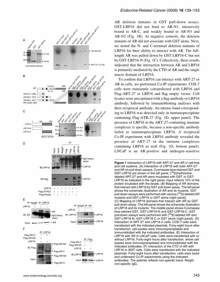
Endocrine-Related Cancer (2009) 16 139–153
www.endocrinology-journals.org
AR deletion mutants in GST pull-down assays.
GST-LRP16 did not bind to AR-N1, intensively
bound to AR-C, and weakly bound to AR-N3 and
AR-N2 (Fig. 1B). As negative controls, the deletion
mutants of AR did not associate with GST alone. Next,
we tested the N- and C-terminal deletion mutants of
LRP16 for their ability to interact with AR. The full-
length AR was pulled down by GST-LRP16-C but not
by GST-LRP16-N (Fig. 1C). Collectively, these results
indicated that the interaction between AR and LRP16
is primarily mediated by the CTD of AR and the single
macro domain of LRP16.
To confirm that LRP16 can interact with ART-27 or
AR in cells, we performed Co-IP experiments. COS-7
cells were transiently cotransfected with LRP16 and
Flag-ART-27 or LRP16 and flag empty vector. Cell
lysates were precipitated with a flag antibody or LRP16
antibody, followed by immunoblotting analyses with
their reciprocal antibody. An intense band correspond-
ing to LRP16 was detected only in immunoprecipitate
containing Flag-ATR-27 (Fig. 1D, upper panel). The
presence of LRP16 in the ART-27-containing immune
complexes is specific, because a non-specific antibody
failed to immunoprecipitate LRP16. A reciprocal
Co-IP experiment with LRP16 antibody revealed the
presence of ART-27 in the immune complexes
containing LRP16 as well (Fig. 1D, bottom panel).
LNCaP is an AR-positive and androgen-sensitive
Figure 1 Interaction of LRP16 with ART-27 and AR in cell-freeand cell systems. (A) Interaction of LRP16 with both ART-27and AR on pull down assays. Coomassie blue-stained GST andGST-LRP16 are shown in the left panel. [35S]methionine-labeled ART-27 and AR were incubated with GST or GST-LRP16 as indicated in the right panel. Input reflects 10% of theprotein incubated with the beads. (B) Mapping of AR domainsthat interact with LRP16 by GST pull-down assay. The left panelshows the schematic illustration of AR and its mutants. GSTpull-down assays were performed with various [35S]-labeled ARmutants and GST-LRP16 or GST alone (right panel).(C) Mapping of LRP16 domains that interact with AR by GSTpull-down assay. The left panel shows the schematic illustrationof LRP16 and its mutants. The middle panel shows Coomassieblue-stained GST, GST-LRP16-N and GST-LRP16-C. GSTpull-down assays were performed with [35S]-labeled AR andGST-LRP16-N, GST-LRP16-C or GST alone (right panel). (D)Interaction of ART-27 and LRP16 in cells. COS-7 cells weretransfected with the indicated plasmids. Forty-eight hours aftertransfection, cell lysates were immunoprecipitated andimmunoblotted with the indicated antibodies. (E) Interaction ofLRP16 with AR in LNCaP cells. Cells were transfected with orwithout LRP16. Forty-eight hours after transfection, whole celllysates were immunoprecipitated and immunoblotted with theindicated antibodies. (F) Interaction of the CTD of AR withLRP16 in 393T cells. Cells were transfected with the indicatedplasmids. Forty-eight hours after transfection, cells were lysedand underwent Co-IP experiments using the indicatedantibodies. The asterisk reflects non-specific band. NsIgG:non-specific IgG.
143

J Yang, Y-L Zhao, Z-Q Wu et al.: Functional regulation of AR by LRP16
prostate cancer cell line. The interaction between
LRP16 and AR was determined by Co-IP experiments
in LNCaP cells with or without LRP16 transfection in
the absence of androgen treatment. Cell lysates were
immunoprecipitated with a LRP16 antibody, and then
underwent immunoblotting analysis with an anti-AR
antibody. The endogenous AR was revealed in the
immune complexes containing overexpressed or only
endogenous LRP16 (Fig. 1E, left panel). Next, the
same experiments performed in the reciprocal manner
revealed both the exogenous and endogenous LRP16
detected in the AR antibody immunoprecipitated
complex (Fig. 1E, right panel). To confirm whether
the C-terminal region of AR is the primary region for
AR-LRP16 association, we performed Co-IP assays
using LRP16 and Flag-AR-N3 or LRP16 and Flag-
AR-C cotransfected 293T cells. Immunoblotting
results showed that the AR-C and LRP16 were
intensively and reciprocally coprecipitated (Fig. 1F).
Although AR-N3 appeared to be coprecipitated in
LRP16 antibody immunoprecipitated complex, its
expression was very weak when compared with that
of AR-C. Collectively, these results confirmed the
interaction of LRP16 with ART-27 and AR in cells in
an androgen-independent manner.
Analysis of LRP16 interaction with other NR
Given the high homology among NR family members,
we presumed that LRP16 might be able to interact with
other members of NR family proteins. To test this
presumption, we performed GST pull-down assays
using a panel of in vitro-translated, [35S]methionine-
labeled transcription factors and GST-fused LRP16
protein. The full-length ERb, GR, PPARa, and PPARgbut not VP16 could be pulled down by LRP16 (Fig. 2).
Thus, these preliminary results suggested that LRP16
can interact not only with AR and ERa but also with at
least four other NRs.
Figure 2 Interaction of LRP16 with other nuclear receptors inGST pull-down assays. GST pull-down assays were carried outwith [35S]methionine-labeled ERb, GR, PPARa, PPARg, orVP16 in the presence of GST-LRP16. GST protein was used asa control. Input reflects 10% of the protein incubated with thebeads.
144
Overexpression of LRP16 enhances the
transcriptional activation of its interacting NR
To explore the possible functional significance of the
interaction of LRP16 with ART-27 and AR, we
examined the effect of LRP16 on AR-mediated
transcription activity. AR-deficient HeLa cells were
cotransfected with AR, AR-responsive reporter con-
struct MMTV-Luc, LRP16, and/or ART-27. Cotrans-
fection with LRP16 or ART-27 did not affect the
AR-mediated transcription in the absence of testo-
sterone stimulation (Fig. 3A). In the presence of
testosterone (10K7 M/l), AR up-regulated the reporter
gene activity, which was further increased about
threefold by either ART-27 or LRP16 cotransfection.
Although ART-27 and LRP16 showed an enhanced
effect on AR-mediated transcription, no additive or
synergetic effect was observed on cotransfection of
ART-27 and LRP16. Similar results were observed in
AR-positive LNCaP human prostate cancer cells
(Fig. 3B). These results indicate that LRP16 enhances
AR-mediated transcriptional activation in a ligand-
dependent manner.
The ability of LRP16 to affect transactivation by
other NRs was also tested using transient transfection
assays. Similar to the effect of LRP16 on AR
transcriptional activation, LRP16 significantly
enhanced reporter gene activities mediated by ERb,
GR, PPARa, and PPARg on stimulation with their
corresponding ligands (Fig. 3C–F). Recall that VP16
did not interact with LRP16 in a cell-free system
(Fig. 2A) and in mammalian two-hybrid hybridization
(Han et al. 2007). We next tested the effect of LRP16
on VP16-dependent transcription. Consistent with this
lack of interaction, LRP16 expression had no effect on
GAL4-VP16 activity from a reporter plasmid
containing five GAL4-binding sites upstream of the
TK promoter (Fig. 3G). These results suggest that
LRP16 might enhance the functional activity of its
interacting NRs in a ligand-dependent manner.
The macro domain of LRP16 enhances
ligand-induced AR transactivation
Two of the most salient features of the LRP16 open
reading frame (ORF) are its lack of modules
represented in conventional transcriptional coregula-
tors (e.g.,HAT, bromo, chromo, SET, or ATPase
domains) and its membership in a small family of
proteins containing macro domains. As a special
member of the macro domain protein family, LRP16
exclusively contains a stand-alone macro domain,
which mediates its interaction with AR as illustrated
in Fig. 1C. A mutant of LRP16 consisting exclusively
www.endocrinology-journals.org
Endocrine-Related Cancer (2009) 16 139–153
of a C-terminal region was compared with the full-
length LRP16. The macro domain of LRP16 increased
AR-mediated reporter gene activity nearly as high as
did the full-length LRP16 in transfection assays;
however, the N-terminal region of LRP16 failed to
enhance reporter gene activity relative to the back-
ground (Fig. 4A). To test whether the single macro
domain of LRP16 can induce AR-mediated gene
expression, we performed qPCR to measure the
mRNA level of the well-known AR target gene PSA.
Transduction experiments using endogenous AR-posi-
tive LNCaP cells confirmed that both the LRP16-C and
the full-length LRP16 significantly amplified AR-me-
diated induction of PSA mRNA by testosterone
(10K8 M/l), when compared with the empty vector
and the LRP16-N (Fig. 4B). As negative controls,
neither the LRP16-C nor the full-length LRP16
changed the mRNA level of HPRT gene (Fig. 4B).
Collectively, these findings demonstrate that the
potentiation of ligand-induced AR transactivation by
LRP16 relies on the presence of the macro domain.
Knockdown of LRP16 sharply diminishes
ligand-induced AR transactivation and hampers
coactivation of the AR by ART-27 and SRC-1
To further test whether LRP16 is required for AR
transactivation, we used the MMTV-Luc reporter gene
to evaluate the effect of LRP16 knockdown on AR
activation status. Compared with the control siRNA,
both LRP16-siRNA374 and LRP16-siRNA668 caused
a specific reduction of LRP16 expression at both
mRNA and protein levels in LNCaP cells but no
change in expression of AR and b-actin genes
(Fig. 5A). Consistent with our previous report (Han
et al. 2007), LRP16-siRNA374 was reproducibly better
than LRP16-siRNA668 and therefore was used more
frequently in later experiments. Luciferase assays
revealed that the decrease of endogenous LRP16
markedly inhibited testosterone-induced AR transacti-
vation (Fig. 5B). To determine whether knockdown of
LRP16 affected the AR transactivation enhanced by
ART-27 and other coactivators, we also cotransfected
AR, MMTV-Luc, ART-27, or SRC-1 in LRP16-
inhibited LNCaP cells. The AR transcriptional
response to ART-27 or SRC-1 was markedly
attenuated by LRP16-siRNA374 relative to the
control-siRNA and LRP16-siRNA668 (Fig. 5B).
These results indicate that LRP16 is essential for
ligand-induced AR transactivation and suggested that
LRP16 is required for the potentiation of AR
transactivation by some other coactivators.
www.endocrinology-journals.org
To address the necessity for LRP16 in regulation of
AR target genes, we used PSA to detect its expression
in response to LRP16 knockdown. PSA expression at
both mRNA and protein levels was significantly
decreased by LRP16-siRNA374 and was partially
decreased by LRP16-siRNA668 in LNCaP cells
(Fig. 5A).
Knockdown of LRP16 markedly inhibits the
growth of androgen-sensitive LNCaP prostate
cancer cells
To determine the effect of the LRP16-AR association
on the growth of androgen-dependent prostate cancer
cells, we stably transfected LRP16 or empty vector into
LNCaP cells for cell proliferation assays. However,
ectopic LRP16 expression did not markedly promote
cell growth when cells were cultured in normal or
testosterone (10K9 M/l)-supplemented medium (data
not shown). Next, we transiently transfected LRP16-
siRNA374 or control-siRNA into LNCaP cells for cell
proliferation assays. Cells carrying LRP16-siRNA374
or control-siRNA showed no significant growth
difference on culture in steroid-deprived medium
(Fig. 6A). Testosterone (10K12 M/l and 10K9 M/l)
stimulated proliferation of LNCaP cells carrying either
control siRNA or LRP16-siRNA 374, but the degree of
testosterone-stimulated proliferation of LRP16-
siRNA374 expressing cells was much less than that
of control-siRNA expressing cells (Fig. 6B and C). To
determine whether LRP16 is required for cell growth
independent of its effect on AR activity, we transiently
transfected LRP16-siRNA374 or control-siRNA into
AR-deficient PC-3 prostate cancer cells and performed
cell proliferation assays. As shown in Fig. 6D, knock-
down of the endogenous LRP16 did not significantly
inhibited growth of PC-3 cells. Immunoblotting
analysis revealed that the endogenous LRP16 in
LNCaP and PC-3 cells was still partially inhibited by
LRP16-siRNA374 at days 9 and 6 respectively after
transfection (Fig. 6E). These findings suggest that
inhibition of LRP16 expression blocks the proliferation
of androgen-dependent prostate cancer cells through
suppressing AR functional activity.
Androgen stimulates LRP16 promoter response
and upregulates LRP16 expression in
AR-positive prostate cancer cells
Previously, we demonstrated that the synthesis of
LRP16 depends on estrogen activities in breast cancer
cells, and in turn, LRP16 enhances ERa functional
activity by cooperating with ERa (Han et al. 2007). We
also previously observed that androgen can upregulate
145
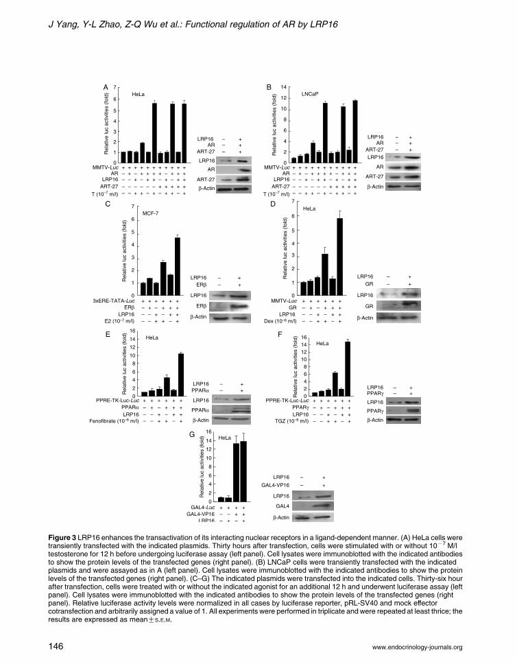
Figure 3 LRP16 enhances the transactivation of its interacting nuclear receptors in a ligand-dependent manner. (A) HeLa cells weretransiently transfected with the indicated plasmids. Thirty hours after transfection, cells were stimulated with or without 10K7 M/ltestosterone for 12 h before undergoing luciferase assay (left panel). Cell lysates were immunoblotted with the indicated antibodiesto show the protein levels of the transfected genes (right panel). (B) LNCaP cells were transiently transfected with the indicatedplasmids and were assayed as in A (left panel). Cell lysates were immunoblotted with the indicated antibodies to show the proteinlevels of the transfected genes (right panel). (C–G) The indicated plasmids were transfected into the indicated cells. Thirty-six hourafter transfection, cells were treated with or without the indicated agonist for an additional 12 h and underwent luciferase assay (leftpanel). Cell lysates were immunoblotted with the indicated antibodies to show the protein levels of the transfected genes (rightpanel). Relative luciferase activity levels were normalized in all cases by luciferase reporter, pRL-SV40 and mock effectorcotransfection and arbitrarily assigned a value of 1. All experiments were performed in triplicate and were repeated at least thrice; theresults are expressed as meanGS.E.M.
J Yang, Y-L Zhao, Z-Q Wu et al.: Functional regulation of AR by LRP16
www.endocrinology-journals.org146
Endocrine-Related Cancer (2009) 16 139–153
LRP16 promoter activity via AR mediation in COS-7
cells (Han et al. 2003). On the basis of these findings
and the fact that LRP16 serves as a coactivator of AR,
we hypothesized the existence of a feedback loop
between AR function and LRP16 expression. To
evaluate this hypothesis, we first examined the
androgen responsiveness of LRP16 promoter-driven
reporter genes pGL-3S0 and pGL3-S2 in LNCaP cells.
Treatment with 10K10 M/l testosterone resulted in a
1.2- to 1.3-fold higher pGL3-S0 and pGL3-S2 reporter
gene activities than without testosterone stimulation
(Fig. 7A). The testosterone induction of the reporter
activity of pGL3-S0 and pGL3-S2 was further
enhanced (five- to sixfold) by their cotransfection
with AR. These findings indicated that LRP16 is an
androgen-responsive target gene in AR-positive pros-
tate cancer cells. Next, we examined the induction
effect of androgen on LRP16 expression at the
protein level in LNCaP cells. LNCaP cells were
treated with various concentrations of testosterone
(10K12–10K6 M/l) for 24 h, and then underwent
western blot analyses. Testosterone, from 10K10 to
10K6 M/l, significantly increased LRP16 expression,
with the low concentration (10K10 M/l) producing
maximal induction (Fig. 7B). We further investigated
the effect of 10K10 M/l testosterone on the expression
level of LRP16 over 48 h (Fig. 7B). A significant
increase of LRP16 expression was observed as early as
3 h after the addition of testosterone, which indicates
that LRP16 is a testosterone-induced early response
gene. The testosterone induction of LRP16 expression
reached to the peak at 6 h and remained for at least
48 h. To further confirm the regulatory effect of AR on
LRP16 expression, we measured the expression of
LRP16 protein in AR-positive and AR-negative human
prostate cancer cell lines. LRP16 protein level was
much lower in AR-negative PC-3 and DU145 cells
than in AR-positive LNCaP cells (Fig. 7C). Finally, we
overexpressed AR in PC-3 cells and observed a marked
increase of LRP16 protein level (Fig. 7D). The
induction of LRP16 in AR-overexpressed PC-3 cells
indicated that the culture medium containing non-
stripped serum provided the source of ligand for AR.
Altogether, these data confirmed the induction effect of
LRP16 expression by androgen via AR mediation and
established a feedforward mechanism that activated the
AR in human prostate cancer cells.
Discussion
The transactivation potential of an NR depends on the
cofactors it recruits, a key mechanism by which NRs
regulate transcriptional programs (McKenna et al.
www.endocrinology-journals.org
1999, McKenna & O’Malley 2002, Heemers & Tindall
2007). A central finding of the present study is that the
protein LRP16 serves as a coactivator that amplifies the
transactivation function of AR in response to androgen.
Similarly, LRP16 was suggested to amplify the
transactivation of other NRs, including ERb, GR,
PPARa, and PPARg, in the presence of their
corresponding agonists, by functioning as a potential
coactivator. Importantly, we have uncovered a direct
link between the coregulation of AR and the macro
domain of LRP16. Knockdown of the endogenous
LRP16 in cells significantly inhibited androgen-
induced AR transactivation and diminished the coacti-
vation of AR by other AR coactivators such as ART-27
and SRC-1. These findings suggest that the single macro
domain protein LRP16 represents a novel type of
cofactor of NRs and underscore a critical role of the
macro domain in transcription modulation of multiple
NRs.
Protein ADP-ribosylation modification is emerging
as a regulatory process of protein function in
prokaryotes and eukaryotes and may act on the level
of signaling transduction, modulation of chromatin
structure and epigenetic histone code (Hassa et al.
2006). The increased negative charge of ADP-
ribosylated proteins may alter their spatial confor-
mation and biological properties (Corda & Girolamo
2003, Hassa et al. 2006). Previously identified as
displaying an Appr-1 00-p (ADP-ribose-1 00-mono-
phosphate) processing activity and robust ADP-ribose
binding activity (Culver et al. 1994, Karras et al. 2005,
Kumaran et al. 2005, Egloff et al. 2006), the macro
domain may play roles in distinct ADP-ribose
pathways, such as the ADP-ribosylation of proteins,
an important post-translational modification (Kraus &
Lis 2003, Ame et al. 2004). In this study, we validated
that a single macro domain within LRP16 is able to act
as a coactivator of AR. Considering the biochemical
characterization of macro domains binding to ADP-
ribose, we proposed that LRP16 may act as a donor
protein of ADP-ribose for ADP-ribosylation modifi-
cation of NRs. Although the capacity of macro
domains to transfer poly(ADP-ribose) to its partners
is not yet authenticated, this hypothesis appears to be
reasonable in that the poly(ADP-ribose) was indeed
found to be transferred from its binding protein to
another partner protein. For example, tankyrase, a
member of PARP superfamily proteins, was found to
confer poly(ADP-ribose) to its interacting protein
TRF1, then ADP-ribosylation of TRF1 diminished its
ability to bind to telomeric DNA (Smith et al. 1998). In
general, because of the well-known function of
the macro domain and the dependence of AR
147

Figure 5 Knockdown of LRP16 sharply diminishes AR-mediatedtranscription. (A) LNCaP cells were transfected with the indicatedsiRNA. Forty-eight hour after transfection, the endogenousexpression of LRP16, AR, PSA, and b-actin was monitored bynorthern blot and western blot analysis. (B) The indicated siRNAoligonucleotides were cotransfected with MMTV-Luc and theindicated plasmids into LNCaP cells. Thirty-six hour aftertransfection, cells were treated with 10K10 M/l testosterone ordimethyl sulfoxide (DMSO) for 12 h before luciferase assay. Therelative luciferase activity levels were normalized by the use ofmock effector transfection and arbitrarily assigned a value of 1. Allexperiments were performed in triplicate and were repeated atleast thrice; the results are expressed asmeanGS.E.M. (#P!0.01;* P!0.05; N PO0.05).
Figure 4 The macro domain of LRP16 enhances AR-mediatedtranscription. (A) Segments of LRP16 were transfected intoLNCaP cells along with an AR-dependent MMTV-Luc reporterand AR. Thirty hour after transfection, cells were stimulated with10K10 M/l testosterone for 12 h before luciferase assay. Therelative normalized luciferase activity level for empty vectortransfection was arbitrarily assigned a value of 1. Shown aremeanGS.E.M. from at least three independent experiments.(B) LNCaP cells were transfected with the indicated segmentsof LRP16 or empty vector. Twelve or 32 h after transfection,cells were stimulated with 10K10 M/l testosterone for anadditional 12 h. Endogenous mRNA expression of PSA andHPRT were measured by qPCR. Data represent meanGS.E.M.from independent three experiments.
J Yang, Y-L Zhao, Z-Q Wu et al.: Functional regulation of AR by LRP16
www.endocrinology-journals.org148

Endocrine-Related Cancer (2009) 16 139–153
transactivation on LRP16 as we show, we propose that
the ADP-ribose binding/processing activity may be
required for NR activation and might depend on
interaction with LRP16.
Several areas of high sequence conservation are
observed in macro domains. Structure analysis also
revealed a conserved ADP-ribose binding pocket in
www.endocrinology-journals.org
different macro domains (Karras et al. 2005, Egloff
et al. 2006). However, only about 30–40% sequence
homology is shared between any two macro domains
(Aguiar et al. 2005, Egloff et al. 2006). A significant
degree of sequence variation exists among different
macro domains. These variations should be the
determinants of predisposing the selectivity of macro
domain binding partners. Through binding to different
transcription factors, different macro domains are
disposed to affect different cellular signaling transduc-
tion and transcription regulation. CoaSt6 was pre-
viously identified to cooperate with Stat6 but not Stat1
and amplify IL4-induced transactivation (Goenka &
Boothby 2006). In this study, several NRs were
affected by LRP16. Further identification of LRP16-
interacting transcription factors will help clarify the
partner selectivity of a single macro domain in LRP16
and the importance of the macro domain in transcrip-
tion regulation.
GST pull-down and CoIP assay has shown to
primarily interact with the AR CTD and probably the
DBD and AF1b regions. By contrast, LRP16 was
previously shown to primarily interact with the ERa N
terminus by the two mammalian two-hybrid assays
(Han et al. 2007). On the basis of the homologous
sequence derived from the AR C terminus and ERa N
terminus, we analyzed homology of NR along with the
other receptors affected by LRP16; however, this
search failed to reveal a definite LRP16 interaction
motif. One plausible explanation is that LRP16 may
associate with NRs through multiple interactions with
different affinity. In addition, the AR differs from other
Figure 6 Knockdown of LRP16 blunts the androgen-responsiveproliferation ability of LNCaP cells but does not inhibitproliferation of PC-3 cells. (A–C) LNCaP cells were cultured inmedium supplemented with steroid-stripped FBS (5%) for3 days and then transiently transfected with LRP16-siRNA374or control-siRNA. Forty-eight hour after transfection, cells weretreated with testosterone at different concentrations for theindicated times. Cell proliferation rate was quantified byCellTiter 96 AQueous assay. Each data point represents themeanGS.E.M. of at least three independent experiments.(*P!0.05, #P!0.01). (D) PC-3 cells were transfected witheither LRP16-siRNA374 or control-siRNA. Forty-eight hourafter transfection, cells were plated onto 24-well plates at1!104 cells/well in the culture medium supplemented with 5%FBS. Cell proliferation rate was quantified by CellTiter 96AQueous assay. Each data point represents the meanGS.E.M.of at least three independent experiments. (E) LNCaP cellstransfected with the indicated siRNAs were treated as in C. 9 dafter siRNA transfection, cells were cultured as in C and theendogenous expression of LRP16 and b-actin was monitoredby immunoblotting (left panel). PC-3 cells transfected with theindicated siRNAs were treated as in D. 2 d and 6 d aftertransfection, the endogenous expression of LRP16 and b-actinwas monitored by immunoblotting (right panel).
149

Figure 7 Androgen stimulates LRP16 promoter activity via ARand up-regulates LRP16 expression. (A) LNCaP cells werecotransfected with the indicated plasmids. Thirty-six hour aftertransfection, cells were treated with or without 10K10 M/ltestosterone for an additional 12 h and underwent luciferaseassay. The relative luciferase activity levels were normalized inall cases by mock effector transfection and arbitrarily assigned avalue of 1. All experiments were performed in triplicate andwere repeated at least three times; results are expressed asmeanGS.E.M. (B) LNCaP cells were cultured in mediumsupplemented with steroid-stripped FBS (5%) for 3 days, thentreated with testosterone at the indicated concentration for theindicated times. Immunoblots were probed for LRP16 andb-actin. (C) Total protein from the indicated cell lines wasextracted and used for monitoring the expression of AR, LRP16,and b-actin by immunoblotting assays. (D) PC-3 cells weretransfected with AR or the empty vector and cultured in amedium containing non-stripped serum. Immunoblots wereprobed for AR, LRP16, and b-actin.
J Yang, Y-L Zhao, Z-Q Wu et al.: Functional regulation of AR by LRP16
NRs in that AR-mediated transcription requires the
interaction of the AR NTD with the AR LBD in a
hormone-dependent manner when the AR is not bound
to DNA (He et al. 2000, van Royen et al. 2007), thus
possibly generating a new conformation that is
150
important for recruitment of at least some of AR
coactivators (Toumazou et al. 2008). So, another
explanation for differential LRP16-binding motifs
between AR and ERa is that the interaction of
LRP16 with the AR C terminus may be more suitable
for the special AR configuration rather than ERa.
ART-27 is a well-known AR coactivator that interacts
with the AR N terminus and activates AR activity in a
ligand-dependent manner (Markus et al. 2002, Taneja
et al. 2004). LRP16 also interacts with ART-27, which
indicates that LRP16 communicates with at least one
other transcriptional regulatory cofactor of AR. SRC-1
interacts directly and ligand dependently with the AR to
enhance AR-mediated transcription (Powell et al. 2004).
Although SRC-1 has been shown to interact with AR AF-
2, it interacts primarily with the AR N-terminus and
possibly the DBD (Bevan et al. 1999, Powell et al. 2004,
Heemers & Tindall 2007). Moreover, SRC-1 functions as
a scaffold protein that attracts additional coactivator
proteins such as p300, the p300 homolog CREB-binding
protein (CBP), and p300/CBP-associated factor (P/CAF;
Chen et al. 1997, Spencer et al. 1997, Heemers & Tindall
2007). Despite the lack of binding competition for the AR
motif between LRP16 and ART-17 or SRC-1, we found
the AR-induced transcriptional activation facilitated by
both ART-27 and SRC-1 markedly diminished in
LRP16-inhibited LNCaP cells (Fig. 5B), which suggests
that LRP16 may play an essential role in AR transcription
complex assembly after AR is exposed to ligand.
Overexpression of LRP16 cannot amplify the coactiva-
tion of AR by ART-27 (Fig. 3A and B), which suggests
that the endogenous LRP16 in cells is enough to support
the excessive activities of other AR coactivators.
By promoter analyses and expression measurement
of the LRP16 gene in androgen-sensitive LNCaP
prostate cancer cells (Fig. 7), we confirmed the
induction effect of LRP16 expression by androgen
via AR mediation. In a 2.6-kb region of LRP16
upstream regulatory sequence (pGL3-S0), which
confers the androgen responsiveness as illustrated in
Fig. 7A, we failed to find the canonical androgen
response element (ARE) (imperfect palindrome
GGTACAnnnTGTTCT) or either of the perfect half-
sites. However, three sites containing imperfect
AR-binding sequence (nGTACnnnnnGnnCn) were
found across a 1.6-kb region of LRP16 upstream
regulatory sequence. In addition, four imperfect half-
sites across a 2.0-kb region of LRP16 upstream
regulatory sequence were also observed. We arbitrarily
proposed that one of the imperfect AREs or ARE half-
sites may mainly contribute to the androgen respon-
siveness through AR binding to it, because it has been
reported that there is a wide range of sequences that AR
www.endocrinology-journals.org

Endocrine-Related Cancer (2009) 16 139–153
can bind, typically containing the core requirement of
three out of four guanines contacts at GGTA-
CAnnnTGTTCT (Nelson et al. 1999). Certainly, we
cannot exclude the possibility that LRP16 is a
secondary response gene of androgen. The molecular
mechanism by which androgen upregulates LRP16
expression is being investigated in our laboratory.
Considering the essential roles of the AR activity in
normal and cancerous prostate tissues, a precise
understanding of the mechanisms of AR regulation is
of utmost importance. The expression of most AR
coactivators is out of control by AR activity. Yet,
emerging evidence has demonstrated that some of the
AR coactivators such as SRG3 and FHL2 are
AR-regulated targets (Hong et al. 2005, Heemers
et al. 2007). This positive regulatory mechanism of AR
transactivation by its response targets may be
important for the rapid proliferation of cells during
prostate development and may be essential for AR
activation during prostate cancer progression (Hong
et al. 2005, Heemers et al. 2007). In this study, we
established the existence of a feedforward mechanism
whereby the androgen-responsive protein LRP16
activates the AR in human prostate cancer cells,
which is similar to the LRP16-ERa feedforward
regulation in estrogen-dependent breast cancer cells
(Han et al. 2007). Furthermore, we also demonstrated
that the inhibition of the endogenous LRP16 in
androgen-sensitive prostate cancer cells markedly
diminished androgen-stimulated cell growth (Fig. 6),
although its overexpression did not significantly
promote cell proliferation. These results indicated
that the AR-LRP16 feedback pathway may play an
essential role in maintaining androgen-induced AR
activation. Appropriate expression of LRP16 in
androgen-sensitive prostate cancer cells is critical for
cell proliferation. The breakdown of this feedforward
signaling might be beneficial in AR-targeting therapy
of androgen-dependent prostate cancer.
Declaration of interest
The authors declare that there is no conflict of interest that
could be perceived as prejudicing the impartiality of the
research reported.
Funding
This study was supported by the National Natural Science
Foundation of China (grants 30670809 and 30870968) and
partially supported by a grant from the Ministry of Science
and Technology of China (2005CB522603).
www.endocrinology-journals.org
Acknowledgements
We thank Prof. Donald P McDonnell from Duke University
Medical Center for providing 3!ERE-TATA-Luc plasmid
and Dr Masatoshi Nomura from Kyushu University for
providing MMTV-Luc and other plasmids.
References
Aguiar RCT, Takeyama K, He C, Kreinbrink K & Shipp MA
2005 B-aggressive lymphoma family protein have unique
domains that modulate transcription and exhibit poly
(ADP-ribose) polymerase activity. Journal of Biological
Chemistry 280 33756–33765.
Ame JC, Spenlehauer C & de Murcia G 2004 The PARP
superfamily. BioEssays 26 882–893.
Angelov D, Molla A, Perche PY, Hans F, Cote J, Khochbin S,
Bouvet P & Dimitrov S 2003 The histone variant
macroH2A interferes with transcription factor binding
and SWI/SNF nucleosome remodeling. Molecular Cell 11
1033–1041.
Bevan CL, Hoare S, Claessens F, Heery DM & Parker MG
1999 The AF1 and AF2 domains of the androgen receptor
interact with distinct regions of SRC-1. Molecular and
Cellular Biology 19 8383–8392.
Brinkmann AO, Faber PW, van Rooij HCJ, Kuiper GGJM,
Ris C, Klaassen P, van der Korput JAHM, Voorhorst MM,
van Laar JH, Mulder E et al. 1989 The human androgen
receptor: domain structure, genomic organization and
regulation of expression. Journal of Steroid Biochemistry
34 307–310.
Chang CY & McDonnell DP 2005 Androgen receptor–
cofactor interactions as targets for new drug discovery.
Trends in Pharmacological Sciences 26 225–228.
Chauchereau A, Georgiakaki M, Perrin-Wolff M, Milgrom E
& Loosfelt H 2000 JAB1 interacts with both the
progesterone receptor and SRC-1. Journal of Biological
Chemistry 275 8540–8548.
Chen H, Lin RJ, Schiltz RL, Chakravarti D, Nash A, Nagy L,
Privalsky ML, Nakatani Y & Evans RM 1997 Nuclear
receptor coactivator ACTR is a novel histone acetyl-
transferase and forms a multimeric activation complex
with P/CAF and CBP/p300. Cell 90 569–580.
Corda D & Girolamo MD 2003 Functional aspects of protein
mono-ADP-ribosylation. EMBO Journal 22 1953–1958.
Culver GM, Consaul SA, Tycowski KT, Filipowicz W &
Phizicky EM 1994 tRNA splicing in yeast and wheat
germ. A cyclic phosphodiesterase implicated in the
metabolism of ADP-ribose 1 00, 2 00-cyclic phosphate.
Journal of Biological Chemistry 269 24928–24934.
Dehm SM & Tindall DJ 2006 Molecular regulation of
androgen action in prostate cancer. Journal of Cellular
Biochemistry 99 333–344.
Dehm SM & Tindall DJ 2007 Androgen receptor structural
and functional elements: role and regulation in prostate
cancer. Molecular Endocrinology 21 2855–2863.
Egloff MP, Malet H, Putics A, Heinonen M, Dutartre H,
Frangeul A, Gruez A, Campanacci V, Cambillau C,
151

J Yang, Y-L Zhao, Z-Q Wu et al.: Functional regulation of AR by LRP16
Ziebuhr J et al. 2006 Structural and functional basis for
ADP-ribose and poly(ADP-ribose) binding by viral macro
domains. Journal of Virology 80 8493–8502.
Goenka S & Boothby M 2006 Selective potentiation of Stat-
dependent gene expression by collaborator of Stat6
(CoaSt6), a transcriptional cofactor. PNAS 103 4210–4215.
Han WD, Yu L, Lou FD, Wang QS, Zhao Y, Shi ZJ, Jiao HY
& Zhou JJ 2001 Cloning and expression characteristics of
the full length cDNA for a novel leukemia-associated
gene LRP16. Academic Journal of PLA Postgraduate
Medical School 17 209–214.
Han WD, Lou FD, Yu L, Han XP, Wang QS, Li N & Zhou
CX 2002 Bioinformatic analysis and subcellular distri-
bution of LRP16 protein. Academic Journal of PLA
Postgraduate Medical School 23 277–279.
Han WD, Mu YM, Lu XC, Xu ZM, Li XJ, Yu L, Song HJ, Li M,
Lu JM, Zhao YL et al.2003 Up-regulation of LRP16 mRNA
by 17b-estradiol through activation of estrogen receptor a
ERa, but not estrogen receptor b ERb and promotes human
breast cancer MCF-7 cell proliferation: a preliminary report.
Endocrine-Related Cancer 10 217–224.
Han WD, Zhao YL, Meng YG, Zang L, Wu ZQ, Li Q, Si YL,
Huang K, Ba JM, Morinaga H et al. 2007 Estrogenically
regulated ERa target gene LRP16 interacts with ERa and
enhances the receptor’s transcriptional activity. Endo-
crine-Related Cancer 14 741–753.
Han WD, Si YL, Zhao YL, Li Q, Wu ZQ, Hao HJ & Song HJ
2008 GC-rich promoter elements maximally confer
estrogen-induced transactivation of LRP16 gene through
ERa/Sp1 interaction in MCF-7 cells. Journal of Steroid
Biochemistry and Molecular Biology 109 47–56.
Hassa PO, Haenni SS, Elser M & Hottiger MO 2006 Nuclear
ADP-ribosylation reactions in mammalian cells: where
are we today and where are we going? Microbiology and
Molecular Biology Reviews 70 789–829.
He B, Kemppainen JA & Wilson EM 2000 FXXLF and
WXXLF sequences mediate the NH2-terminal interaction
with the ligand binding domain of the androgen receptor.
Journal of Biological Chemistry 275 22986–22994.
Heemers HV & Tindall DJ 2007 Androgen receptor
coregulators: a diversity of functions converging on and
regulating the AR transcriptional complex. Endocrine
Reviews 28 778–808.
Heemers HV, Regan KM, Dehm SM & Tindall DJ 2007
Androgen induction of the androgen coactivator four and
a half LIM domain protein-2: evidence for a role for
serum response factor in prostate cancer. Cancer
Research 67 10592–10599.
Hong CY, Suh JH, Kim K, Gong EY, Jeon SH, Ko M, Seong
RH, Kwon HB & Lee K 2005 Modulation of androgen
receptor transactivation by the SWI3-related gene product
(SRG3) in multiple ways. Molecular and Cellular Biology
25 4841–4852.
Imagama S, Abe A, Suzuki M, Hayakawa F, Katsumi A, Emi
N, Kiyoi H & Naoe T 2007 LRP16 is fused to RUNX1 in
monocytic leukemia cell line with t(11;21)(q13;q22).
European Journal of Haematology 79 25–31.
152
Karras GI, Kustatscher G, Buhecha HR, Allen MD, Pugieux
C, Sait F, Bycroft M & Ladurner AG 2005 The macro
domain is an ADP-ribose module. EMBO Journal 24
1911–1920.
Kraus WL & Lis JT 2003 PARP goes transcription. Cell 113
677–683.
Kumaran D, Eswaramoorthy S, Studier FW & Swaminathan
S 2005 Structure and mechanism of ADP-ribose-1 00-
monophosphatase (Appr-1 00-pase), a ubiquitous cellular
processing enzyme. Protein Science 14 719–726.
Kustatscher G, Hothorn M, Pugieux C, Scheffzek K &
Ladurner AG 2005 Splicing regulation NAD metabolite
binding to histone macroH2A. Nature Structural and
Molecular Biology 12 624–625.
Ladurner AG 2003 Inactivating chromosomes: a macro domain
that minimizes transcription. Molecular Cell 12 1–4.
Lee HJ & Chang C 2003 Recent advances in androgen
receptor action. Cellular and Molecular Life Sciences 60
1613–1622.
Liao DX, Han WD, Zhao YL, Pu YD, Mu YM, Luo CH & Li
XH 2006 The expression and clinical significance of
LRP16 gene in human breast cancer. Ai Zheng 25
866–870.
Markus SM, Taneja SS, Logan SK, Li WH, Ha S, Hittelman
AB, Rogatsky I & Garabedian MJ 2002 Identification and
characterization of ART-27, a novel coactivator for the
androgen receptor N terminus. Molecular and Cellular
Biology 13 670–682.
McKenna NJ & O’Malley BW 2002 Combinatorial control of
gene expression by nuclear receptors and coregulators.
Cell 108 465–474.
McKenna NJ, Lanz RB & O’Malley BW 1999 Nuclear
receptor coregulators: cellular and molecular biology.
Endocrine Reviews 20 321–344.
Meng YG, Han WD, Zhao YL, Huang K, Si YL, Wu ZQ &
Mu YM 2007 Induction of LRP16 gene by estrogen
promotes the invasive growth of Ishikawa human
endometrial cancer cells through down-regulation of
E-cadherin. Cell Research 17 869–880.
Nelson CC, Hendy SC, Shukin RJ, Cheng H, Bruchovsky N,
Koop BF & Rennie PS 1999 Determinants of DNA
sequence specificity of the androgen, progesterone, and
glucocorticoid receptors: evidence for differential steroid
receptor response elements. Molecular Endocrinology 13
2090–2107.
Pehrson JR & Fried VA 1992 MacroH2A, a core histone
containing a large nonhistone region. Science 257 1398–1400.
Powell SM, Christiaens V, Voulgaraki D, Waxman J,
Claessens F & Bevan CL 2004 Mechanisms of androgen
receptor signaling via steroid receptor coactivator-1 in
prostate. Endocrine-Related Cancer 11 117–130.
Rosenfeld MG, Lunyak VV & Glass CK 2006 Sensors
and signals: a coactivator/corepressor/epigenetic code
for integrating signal-dependent programs of
transcriptional response. Genes and Development 20
1405–1428.
www.endocrinology-journals.org

Endocrine-Related Cancer (2009) 16 139–153
van Royen ME, Cunha SM, Brink MC, Mattern KA, Nigg AL,
Dubbink HJ, Verschure PJ, Trapman J & Houtsmuller AB
2007 Compartmentalization of androgen receptor protein–
protein interactions in living cells. Journal of Cell Biology
177 63–72.
Smith S, Giriat I, Schmitt A & de Lange T 1998 Tankyrase, a
poly(ADP-ribose) polymerase at human telomeres.
Science 282 1484–1487.
Spencer TE, Jenster G, Burcin MM, Allis CD, Zhou J,
Mizzen CA, McKenna NJ, Onate SA, Tsai SY, Tsai MJ
et al. 1997 Steroid receptor coactivator-1 is a histone
acetyltransferase. Nature 389 194–198.
Taneja SS, Ha S, Swenson NK, Torra IP, Rome S, Walden
PD, Huang HY, Shapiro E, Garabedian MJ & Logan SK
www.endocrinology-journals.org
2004 ART-27, an androgen receptor coactivator regulated
in prostate development and cancer. Journal of Biological
Chemistry 279 13944–13952.
Toumazou C, Li J & Wong J 2008 Cofactor restriction by
androgen receptor N-terminal and C-terminal interaction
Molecular Endocrinology [in press].
White R & Parker MG 1998 Molecular mechanisms of
steroid hormone action. Endocrine-Related Cancer 5
1–14.
Zhao YL, Han WD, Li Q, Mu YM, Lu XC, Yu L, Song HJ,
Lu JM & Pan CY 2005 Mechanism of transcriptional
regulation of LRP16 gene expression by 17-b estradiol in
MCF-7 human breast cancer cells. Journal of Molecular
Endocrinology 34 77–89.
153