the skeletal system - complete
DESCRIPTION
The Skeletal System - Complete, With Elaborate PicturesTRANSCRIPT

THE S
KELETA
L SYS
TEM
VINNCI A
NGELICA G
. DELA
CRUZ

THE SKELETAL SYSTEM
COMPOSED OF 206 BONES
NEW BORN: 350 BONESINFANCY: 275 BONESADULT: 206 BONES

FUNCTIONS:
1.SUPPORT
2.PROTECTION
3.MOVEMENT
4.STORAGE > STOREHOUSE FOR MINERALS
5.BLOOD CELL FORMATION (HEMATOPOIESIS)

CLASSIFICATION OF BONES:
1.COMPACT BONES > ARE PACKED TIGHTLY TOGETHER TO FORM WHAT APPEARS TO BE SOLID MASS.

CLASSIFICATION OF BONES:
2. SPONGY (CANCELLOUS) BONES > COMPOSED OF SMALL NEEDLELIKE PIECES OF BONE & LOTS OF OPEN SPACES.

CLASSIFICATION OF BONES:

4 SHAPES OF BONES:
1. LONG BONE>FOUND IN LIMBS

LONG BONE

4 SHAPES OF BONES:
2. SHORT BONE> EX: KNEE-CAP

SHORT BONE: PATELLA (KNEECAP)

4 SHAPES OF BONES:
3. FLAT BONE> EX: SKULL, RIBS, STERNUM

FLAT BONE: SCAPULA (SHOULDER BLADE)

4 SHAPES OF BONES:
4. IRREGULAR BONE> EX:VERTEBRAE

IRREGULAR BONE: VERTEBRAE

STRUCTURE OF A LONG BONE:
1.DIAPHYSIS/ SHAFTCOMPOSED OF COMPACT BONE.
PERIOSTEUM > A FIBROUS CONNECTIVE TISSUE THAT COVER AND PROTECT THE DIAPHYSIS.
PERFORATING/SHARPEY’S FIBERS
> CONNECTIVE TISSUE FIBERS THAT SECURE THE PERIOSTEUM TO THE UNDERLYING TISSUE.

STRUCTURE OF A LONG BONE:
2. EPIPHYSES (PROXIMAL AND DISTAL) END PART OF A LONG BONE.
ARTICULAR CARTILAGE > COVERS THE EPIPHYSES AND PROVIDE A SMOOTH, SLIPPERY SURFACE THAT DECREASES FRICTION AT JOINT SURFACES.

STRUCTURE OF A LONG BONE

SKELETON IS DIVIDED INTO 2 PARTS:1.AXIAL SKELETON FORMS THE LONGITUDINAL AXIS OF
THE BODY. (SKULL, VERTEBRAL COLUMN, AND RIB CAGE)
2. APPENDICULAR SKELETONBONES OF THE ARMS AND LEGS
AND THE SHOULDER AND PELVIC GIRDLES.

THE AXIAL AND APPENDICULAR SKELETON

THE AXIAL AND APPENDICULAR SKELETON

AXIAL SKELETON
SKULLIS FORMED BY 2 SETS OF
BONES: CRANIUM AND FACIAL
BONES.

SKULL

CRANIUM
1.FRONTAL BONE2.PARIETAL BONES > THEY MEET IN THE MIDLINE OF
THE SKULL AT THE SAGITTAL SUTURE AND FORM THE CORONAL SUTURE WHERE THEY MEET WITH THE FRONTAL BONE.

FRONTAL BONE

PARIETAL BONES

SAGITTAL AND CORONAL SUTURES

CRANIUM
3. TEMPORAL BONES
> LIES INFERIOR TO THE PARIETAL BONE, THEY JOIN THEM AT THE SQUAMOUS SUTURES.

TEMPORAL BONE AND SQUAMOUS SUTURE

CRANIUMIMPORTANT BONE MARKINGS:A.EXTERNAL ACOUSTIC
(AUDITORY) MEATUS> A CANAL THAT LEADS TO THE
EARDRUM AND MIDDLE EAR.

EXTERNAL AUDITORY MEATUS

CRANIUMIMPORTANT BONE MARKINGS:B. STYLOID PROCESS>ATTACHMENT POINT FOR
SOME NECK MUSCLES.

CRANIUMIMPORTANT BONE MARKINGS:C. MASTOID PROCESS>ATTACHMENT SITE FOR SOME
MUSCLES OF THE NECK

STYLOID PROCESS AND MASTOID PROCESS

CRANIUMIMPORTANT BONE MARKINGS:D. JUGULAR FORAMEN>ALLOWS PASSAGE OF THE
JUGULAR VEIN

JUGULAR FORAMEN

CRANIUMIMPORTANT BONE MARKINGS:E. INTERNAL ACOUSTIC
MEATUS >TRANSMITS CRANIAL NERVE VII AND VIII (FACIAL AND VESTIBULOCOCHLEAR NERVES)

INTERNAL ACOUSTIC MEATUS

CRANIUMIMPORTANT BONE MARKINGS:F. CAROTID CANAL > IT IS WHERE THE INTERNAL
CAROTID ARTERY RUNS, SUPPLYING BLOOD TO MOST OF THE BRAIN.

CAROTID CANAL AND CAROTID ARTERY

CRANIUM4. OCCIPITAL BONETHE MOST POSTERIOR BONE
OF THE CRANIUM. IT FORMS THE FLOOR AND BACK WALL OF THE SKULL.
*lambdoid suture > joins the parietal bones anteriorly
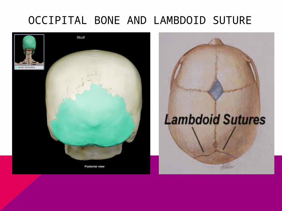
OCCIPITAL BONE AND LAMBDOID SUTURE

CRANIUMOCCIPITAL BONE
A.FORAMEN MAGNUM
> A LARGE OPENING THAT ALLOWS THE SPINAL CORD TO CONNECT TO THE BRAIN.

FORAMEN MAGNUM

CRANIUMOCCIPITAL BONE
B. OCCIPITAL CONDYLES
> LIES ON THE FIRST VERTEBRA OF THE SPINAL COLUMN.

OCCIPITAL CONDYLE

CRANIUM5. SPHENOID BONE
> FORMS THE PART OF THE FLOOR OF THE CRANIAL CAVITY.

SPHENOID BONE

CRANIUMSPHENOID BONEA.SELLA TURCICA/ TURK’S
SADDLE> HOLDS THE PITUITARY
GLAND IN PLACE.

SELLA TURCICA

CRANIUMSPHENOID BONEB. FORAMEN OVALE> A LARGE OVAL OPENING
THAT ALLOWS FIBERS OF CRANIAL NERVE V (TRIGEMINAL NERVE) TO PASS TO THE CHEWING MUSCLES OF THE LOWER JAW (MANDIBLE).

FORAMEN OVALE

CRANIUMSPHENOID BONEC. OPTIC CANAL> ALLOWS OPTIC NERVE TO
PASS TO THE EYE.

OPTIC CANAL

CRANIUMSPHENOID BONED. SUPERIOR ORBITAL FISSURE> ALLOWS CRANIAL NERVES III,
IV, VI RESPONSIBLE FOR CONTROLLING EYE MOVEMENTS.

SUPERIOR ORBITAL FISSURE

CRANIUMSPHENOID BONEE. SPHENOID SINUSES>CAVITIES OF THE SPHENOID
BONE

SPHENOID SINUSES

CRANIUM
6. ETHMOID BONE
> FORMS THE ROOF OF THE NASAL CAVITY

ETHMOID BONE

CRANIUM
ETHMOID BONEA. CRISTA GALLI> THE OUTERMOST
COVERING OF THE BRAIN ATTACHES TO THIS PROJECTION.

CRANIUM
ETHMOID BONEB. CRIBIFORM PLATE>HOLEY AREAS THAT ALLOW
NERVE FIBERS CARRYING IMPULSES FROM THE OLFACTORY (SMELL) RECEPTORS OF THE NOSE TO REACH THE BRAIN.

CRANIUMETHMOID BONEC. SUPERIOR AND MIDDLE
NASAL CONCHAE> FORM PART OF THE
LATERAL WALLS OF THE NASAL CAVITY AND INCREASE THE TURBULENCE OF AIR FLOWING THROUGH THE NASAL PASSAGES.

ETHMOID BONE

FACIAL BONES1. MAXILLAE/ MAXILLARY BONE MAIN OR KEYSTONE BONES
OF THE FACE. CARRY THE UPPER TEETH
WITH THE ALVEOLAR MARGIN

MAXILLA

FACIAL BONES1. MAXILLARY BONEA.PALATINE PROCESSHARD PALATE OF THE MOUTHB. PARANASAL SINUSESLIGHTEN THE SKULL BONES AND ACT TO AMPLIFY THE SOUNDS WE MAKE AS WE SPEAK.

FACIAL BONES2. PALATINE BONES> CLEFT PALATE RESULTS IF UNFUSED MEDIALLY.

PALATINE PROCESS AND PALATINE BONES

FACIAL BONES3. ZYGOMATIC BONES> CHEEKBONES
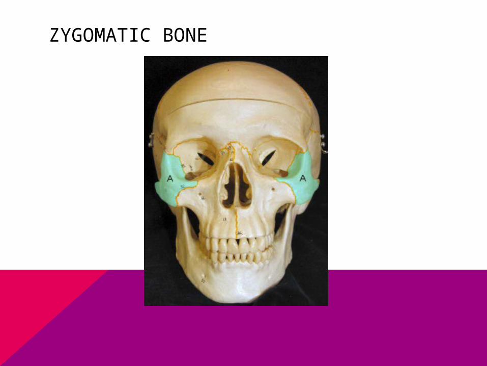
ZYGOMATIC BONE

FACIAL BONES4. LACRIMAL BONES>PASSAGEWAY OF TEARS

LACRIMAL BONES

FACIAL BONES5. NASAL BONES> BRIDGE OF THE NOSE

NASAL BONE

FACIAL BONES6. VOMER BONE>SINGLE BONE IN THE MEDIAN LINE OF THE NASAL CAVITY.

VOMER BONE

FACIAL BONES7. INFERIOR NASAL CONCHAE

FACIAL BONES8. MANDIBLE (LOWER JAW) > LARGEST AND STRONGEST BONE OF THE FACE

FACIAL BONES9. HYOID BONE>SERVES AS A MOVABLE BASE FOR THE TONGUE AND AN ATTACHMENT POINT FOR NECK MUSCLES THAT RAISE AND LOWER THE LARYNX WHEN WE SWALLOW AND SPEAK.

HYOID BONE

THE SKELETAL SYSTEM
B. VERTEBRAL COLUMN (spine)> EXTENDS FROM THE SKULL, AND IT SUPPORTS, TO THE PELVIS, WHERE IT TRANSMITS THE WEIGHT OF THE BODY TO THE LOWER LIMBS.

THE SKELETAL SYSTEM
VERTEBRAL COLUMN (spine)1.CERVICAL VERTEBRAE (7)ARE THOSE WITHIN THE NECK.ATLAS: FIRST VERTEBRAEAXIS: SECOND VERTEBRAE

VERTEBRAE

THE SKELETAL SYSTEM
VERTEBRAL COLUMN (spine)2. THORACIC VERTEBRAE> FORMS JOINTS AND THE RIBS ON THE POSTERIOR SIDE OF THE TRUNK.

THE SKELETAL SYSTEM
VERTEBRAL COLUMN (spine)3. LUMBAR VERTEBRAE>LARGEST AND STRONGEST BONES OF THE SPINE

THE SKELETAL SYSTEM
VERTEBRAL COLUMN (spine)4. SACRUM> PERMITS ARTICULATION OF THE 2 HIP BONES (SACROILIAC JOINT)

THE SKELETAL SYSTEM
VERTEBRAL COLUMN (spine)5. COCCYX>REMNANT OF TAIL VERTEBRAE AND SOME MUSCLES OF THE PERINEUM (PELVIC FLOOR) ARE ANCHORED TO IT.

THE SKELETAL SYSTEM
VERTEBRAL COLUMN (spine)*vertebral canal > a continuous tunnel within the brain that contains the spinal cord and protects it from mechanical injury.

THE SKELETAL SYSTEM
VERTEBRAL COLUMN (spine)*vertebral canal > a continuous tunnel within the brain that contains the spinal cord and protects it from mechanical injury.

THE SKELETAL SYSTEMVERTEBRAL COLUMN (spine)*intervertebral discs > are fibrous cartilage which separate the bodies of adjacent vertebrae.> cushion and absorb shock and permits some movement bet. vertebrae (symphysis joints)

INTERVERTEBRAL DISCS

THE SKELETAL SYSTEMABNORMALITIES OF THE CURVE OF THE
SPINE
1.Scoliosis
> abnormal lateral curvature which may be congenital, the result of having 1 leg longer than the other, or the result of chronic poor posture during childhood while the vertebrae are still growing. Usually, the thoracic vertebrae are affected which displaces the rib cage to one side.

SCOLIOSIS

THE SKELETAL SYSTEMABNORMALITIES OF THE CURVE OF THE
SPINE
2. Kyphosis
> Exaggerated thoracic curve; referred to as hunchback

KYPHOSIS

THE SKELETAL SYSTEMABNORMALITIES OF THE CURVE OF THE
SPINE
3. LordosisExaggerated lumbar curve; referred to as swaybackThe pride of pregnancy curve

LORDOSIS

THE SKELETAL SYSTEM
C. RIB CAGE>consists of 10 pairs of ribs, and the sternum or breastbone.

THE SKELETAL SYSTEM
*True ribs > first 7 pair of ribs*False ribs > next 3 pairs; their cartilages join the 7th rib cartilage*Floating ribs > 2 pairs (last) because they do not articulate with the sternum at all.

RIB CAGE

THE SKELETAL SYSTEM
3 PARTS OF THE STERNUM
1.Manubrium2.Body3.Xiphoid process

APPENDICULAR SKELETON
A. Shoulder and Arm1. Scapula > is a large, flat
bone withe several projection (coracoid process) that anchor some of the muscles that move upper arm and the forearm (shoulder blade)

SCAPULA

APPENDICULAR SKELETON
A. Shoulder and Arm*Glenoid fossa > a shallow
depression that forms a ball-and-socket joint with the humerus.

APPENDICULAR SKELETON
A. Shoulder and Arm2. Clavicle > act as braces
for the scapulae and prevent the shoulders from coming too far forward.

APPENDICULAR SKELETON
A. Shoulder and Arm3. Humerus > long bone of the
upper arm.Proximal end> is the ball-and-
socket jointDistal end> hinge joint that
permits the elbow to move in 1 plane ; restricted lateral mov’t.
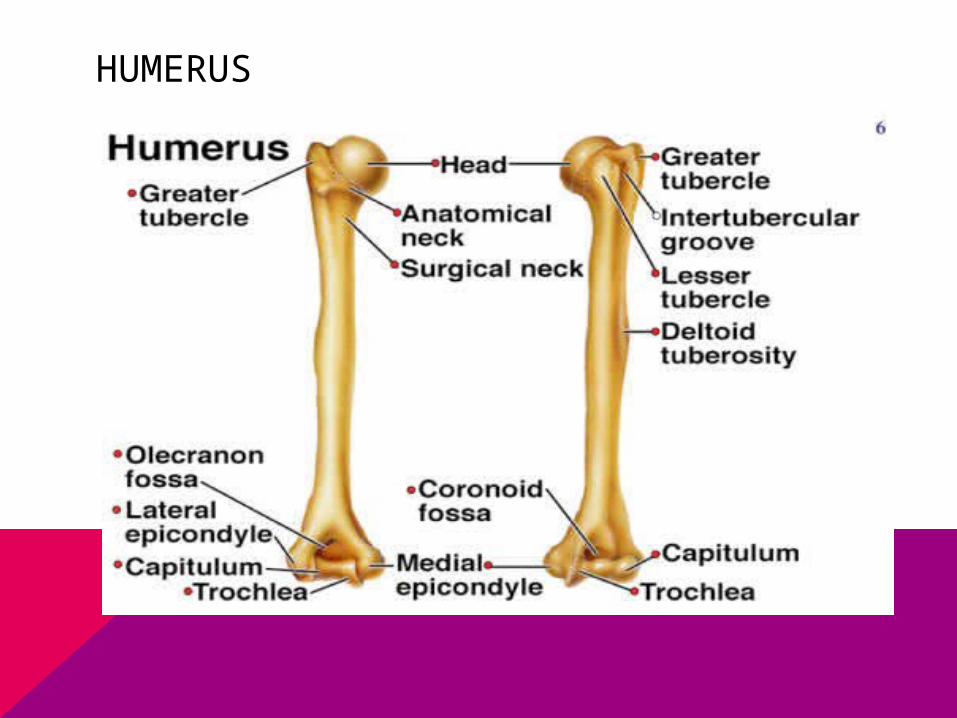
HUMERUS

APPENDICULAR SKELETON
A. Shoulder and Arm4. Ulna > on the side of the
little finger

APPENDICULAR SKELETON
A. Shoulder and Arm5. Radius >thumbsidePivot joint> permit turning
the hand palm up and down.

APPENDICULAR SKELETON
A. Shoulder and Arm6. Carpals > are 8 small
bones in the wrist.Sliding joints> permit a
sliding mov’t.

APPENDICULAR SKELETON
A. Shoulder and Arm7. Metacarpals > 5 bones of
the palm of the hand.Saddle joints> enables the
thumb to cross over the palm which permits gripping.

APPENDICULAR SKELETON
A. Shoulder and Arm8. Phalanges > bones of the
fingers.

APPENDICULAR SKELETON
B. Hip and Leg (Pelvic Girdle/ Pelvic Bone)1. Hip Bones > ilium, ischium (part that we sit on), and pubis.

APPENDICULAR SKELETON
B. Hip and Leg (Pelvic Girdle/ Pelvic Bone)2. Acetabulum>is the socket in the hip bone that forms a ball-and-socket with the femur, has much deeper socket.

APPENDICULAR SKELETON
B. Hip and Leg (Pelvic Girdle/ Pelvic Bone)3. Femur > long bone of the thigh.

APPENDICULAR SKELETON
B. Hip and Leg (Pelvic Girdle/ Pelvic Bone)4. Patella or kneecap>anterior to the kneejoint.

APPENDICULAR SKELETON
B. Hip and Leg (Pelvic Girdle/ Pelvic Bone)5. Tibia > weight-bearing bone of the lower leg.Inner malleolus> inner ankle boneLateral malleolus> outer ankle bone

APPENDICULAR SKELETON
B. Hip and Leg (Pelvic Girdle/ Pelvic Bone)6. Fibula > does not bear much weight. Help stabilize the tibia.

APPENDICULAR SKELETON
B. Hip and Leg (Pelvic Girdle/ Pelvic Bone)7. Tarsals >composed of 7 bones in the ankle Calcaneus (heelbone) > largest tarsalTalus > transmit wt. bet. the calcaneus and the tibia.

APPENDICULAR SKELETON
B. Hip and Leg (Pelvic Girdle/ Pelvic Bone)8. Metatarsals> 5 long bones of each foot.

APPENDICULAR SKELETON
B. Hip and Leg (Pelvic Girdle/ Pelvic Bone)9. Phalanges>bones of the toes.

TARSALS, METATARSALS, PHALANGES, CALCANEUS AND TALUS

THE SKELETAL SYSTEM
JOINTSIS WHERE 2 BONES MEET OR ARTICULATE.

THE SKELETAL SYSTEM
CLASSIFICATION OF JOINTS1. Synarthrosis > is an immovable joint, such as a suture between 2 cranial bones.

THE SKELETAL SYSTEM
CLASSIFICATION OF JOINTS2. Amphiarthrosis > is a slightly movable joint, such as the symphysis joint between adjacent vertebrae.

SYNARTHROSIS AND AMPHIARTHROSIS

THE SKELETAL SYSTEM
CLASSIFICATION OF JOINTS3. Diarthrosis > is a freely movable joint which includes ball-and-socket joint, the pivot, hinge, and others. > is under synovial joints.

THE SKELETAL SYSTEM
*articular cartilage > provides a smooth surface*joint capsule > made of fibrous connective tissue which encloses the joint in a strong sheath like a sleeve.

THE SKELETAL SYSTEM
*synovial membrane > lines the joint capsule which secretes synovial fluid into the joint cavity *synovial fluid > prevents friction as the bones move.
THANK YOU!!!