the skull of tapejara wellnhoferi (reptilia, pterosauria ... · in the state of ceará, chapada do...
TRANSCRIPT

© Biodiversity Heritage Library, http://www.biodiversitylibrary.org/; www.zobodat.at
I Mitt. Bayer. Staatsslg. Paliiont. hist. Gcol. 31 89—106 München, 31. 12. 1991L - ____________ :_______________ 0 ___________________________ _l _________ _L_ __L _________
The Skull of Tapejara w e l ln ho fe r i K ellner (Reptilia, Pterosauria) from the Lower Cretaceous Santana Formation
of the Araripe Basin, Northeastern Brazil
By P eter W fllnhofer and A lexander W . A . K ellner-"')
With 7 text figures and 1 plate
Abstract
An almost complete skull and mandible and an anterior portion of a skull of the remarkable pterodactyloid pterosaur, Tapejara wellnhoferi K ellner, are figured and described. For the first time the material provides the possibility of a detailed restoration of the skull of this toothless pterosaur from the Lower Cretaceous Santana formation of the Araripe Basin in Brazil. Tapejara wellnhoferi has a relatively short skull, a high premaxillary crest anteriorly, and a short fronto-parietal crest posteriorly. The toothless beak is slender and pointed, and seems best adapted for plucking or picking. Therefore a frugivorous feeding habit is suggested. Pterosaurs like Tapejara mav have been important seed dispersers for early angiosperms in the Lower Cretaceous.
Kurzfassung
Ein nahezu vollständiger Schädel mit Unterkiefer sowie eine vordere Schädelpartie des bemerkenswerten pterodactyloiden Pterosauriers Tapejara wellnhoferi K ellner werden beschrieben und abgebildet. Zum ersten Mal erlaubt dieses Material eine detaillierte Rekonstruktion des Schädels dieses zahnlosen Flugsauriers aus der Santana-Formation (untere Kreide) des Araripe-Beckens von Brasilien. Tapejara wellnhoferi hat einen relativ kurzen Schädel, vorne einen hohen Knochenkamm und hinten eine kurze Fronto-Parietal-Crista. Der zahnlose Schnabel ist schlank und spitz und scheint am besten zum Pflücken oder Picken geeignet gewesen zu sein. Es wird deshalb eine frugivore Ernährungsweise von Tapejara wellnhoferi vermutet. Möglicherweise spielten in der Unterkreide derartige Flugsaurier eine bedeutende Rolle als Samenverbreiter früher Angiospermen.
P. W fllnhofer, Bayerische Staatssammlung für Paläontologie und historische Geologie, Richard- Wagner-Siraße 10, 8000 München 2, F. R. G.. A. W. A. Kellner, presently at Department of Vertebrate Paleontology, American Museum of Natural History, Central Park West at 79th Sir., New Ü ork, N. Y. 10024-5192, U.S.A.
89

© Biodiversity Heritage Library, http://www.biodiversitylibrary.org/; www.zobodat.at
Resumo
Um cränio e mandíbula, praticamente completos, e a parte anterior de um segundo cränio do pterodactiloide Tapejara wellnhoferi Kellner, säo descritos e figurados. Com base nestes exemplares é possível, pela primeira vez, apresentar a reconstrugáo detalhada do cränio desta interessante espécie de pterossauro desprovida de dentes, que é procedente da formado Santana, Cretáceo Inferior da bacia do Araripe, nordeste do Brasil.
Tapejara wellnhoferi possui um cränio relativamente curto, com urna crista premaxilar muito desenvolvida e urna crista fronto-parietal baixa, esta última se estendendo para a regiáo posterior do cränio. O bico é delgado e pontudo, indicando ser possivelmente melhor adaptado para „bicar“ ou „arrancar“. Desta maneira, um hábito alimentar frugívoro é sugerido para esta espécie. Com base nesta hipótese é aventada a possibilidade de que pterossauros como Tapejara wellnhoferi tenham sido importantes na dispersäo de sementes das primitivas angiospermas do Cretáceo Inferior.
Introduction
During the last twenty years the Lower Cretaceous Santana formation of the Araripe Basin in northeastern Brazil, widely known for its fish-bearing concretions, has increasingly become one of the most productive fossillagerstatten for pterosaurs. So far, fourteen different species of pterodactyloid pterosaurs have been recognized belonging to nine genera and at least five distinct families ( W ellnhofer 1991 a). Until recently, mostly relatively large and strongly toothed forms were known. But for the first time K ellner and C ampos (1988) described cranial and postcranial remains of a new toothless pterodactyloid as Tupuxuara longicristatus, and K ellner (1989) recognized a second toothless pterosaur on the basis of an incomplete skull which he assigned to a new genus and species as Tapejara wellnhoferi. In the same publication he established for both genera, Tupuxuara and Tapejara, a new family, Tapejaridae.
In 1989 Dr. John Maisey, New York, offered to the senior author (P. W.) a pterosaur skull for study. It is preserved in a calcareous concretion typical for the Santana formation, from the Dr. Herbert Axelrod Collection donated to the American Museum of Natural History New York (AMNH). Subsequently, after further preparation, it turned out that the skull belongs to Tapejara wellnhoferi, and Alexander Kellner was invited to coauthor the study of this almost complete skull and mandible which can add much more to our knowledge of this genus and species than it was possible on the basis of the previously known single specimen, the holotype.
During this study an additional specimen of the same species has been brought to our attention by Urs Oberli, St. Gallen, Switzerland, consisting of the front end of the skull, premaxillary crest, the tip of the beak and part of the palate. It supplements the New York skull, AMNH 24440, as well as the holotype very nicely and allows a highly reliable restoration of the skull of Tapejara wellnhoferi.
For stratigraphic and paleoenvironmental discussions about the Santana formation we refer to K ellner (1984), Silva (1986), M aisey (1990, 1991) and W ellnhofer (1991 a, b), and the literature cited therein. The origin of the concretion containing these specimens is the Romualdo member, the upper stratigraphic unit of the Santana formation (Beurlen 1971), which has been dated as Aptian by previous authors as Braun (1966), M abesonf. and T inooo (1973), or Late Ap- tian/Albian by S ilva (1986) and others. A definitive age determination for the fossil-bearing concretions of the Lower Cretaceous Araripe Basin seems not yet to be established, however. For detailed discussions see M aisey (1991).
90

© Biodiversity Heritage Library, http://www.biodiversitylibrary.org/; www.zobodat.at
Acknowledgements
We wish to thank several individuals who have contributed to this study in many ways: Dr. John Maisey, Curator at the American Museum of Natural History, New York, provided the specimen of Tapejara wellnboferi (AMNH 24440) for study. From his own collection Urs Oberli, St. Gallen, Switzerland, made an additional specimen of Tapejara from the Santana formation available. Encouraging comments on the possible frugivory of Tapejara were given by Dr. Theodore Fleming, Professor of Biology at the University of Miami, USA. The senior author (P. Wellnhofer) benefitted greatly from discussions with Dr. M. Kirchner and E. Rieber, Munich, who also contributed valuable informations on paleobotanical data concerning Lower Cretaceous plants. Udo Kandler, Augsburg, provided informations about the fossil macroflora of the Santana formation. Ernst Schmieja carried out the preparation of specimen AMNH 24440 in a very skillfull way and has made the casts, and Franz Hock took the photographs. The junior author (A. W. A. Kellner) wishes to thank the Columbia University, Department of Geological Sciences, for its financial support.
Systematic Description
Pterodactyloidea Plifninger 1901 Tapejaridae K ellner 1989
Tapejara K ellner 1989
Tapejara wellnboferi K ellner 1989
1989 Tapejara wellnboferi Kellnf.r, p. 439, figs. 1 - 9 ; holotype.1991 Tapejara wellnboferi Kellner, p. 3711991 Tapejara t sp., KELLNER, p. 371; AMNH 24440.
M a te r ia l: 1. Specimen AMNH 24440: skull, lower jaw and anterior cervical vertebrae encased in a calcareous concretion. American Museum of Natural History, New York (Dr. Herbert Axelrod Collection). 2. Specimen UOSG 12891: front end of skull with premaxillary crest surrounding the anterior half of the nasopreorbital fenestra. Urs Oberli Collection, St. Gallen, Switzerland.
H o rizon and lo c a li ty : Lower Cretaceous, Late Aptian/Albian, Santana formation, Romualdo member. The exact locality is not known, but is probably Jardim or Santana do Cariri in the state of Ceará, Chapada do Araripe, northeastern Brazil.
Specim en AM N H 24440 (Figs 1—6; plate 1, fig. 1)
P reserv a tio n : Skull and lower jaw remained in almost natural articulation with the beak open. A certain degree of compression occurred in the postorbital region of the skull and in the posterior section of the lower jaw, so that the left mandibular ramus was broken behind the symphysis and displaced medially. Dislocation took also place in the lower and anterior surroundings of the orbit, in certain elements of the occiput and the ceratobranchialia of the hyoid apparatus. The upper and anterior extremity of the previously high sagittal crest is broken away, and so are the posterior extremity of the parietal crest and the anterior and ventral extensions of the mandibular crest.
The skull was prepared mechanically and exposed from its right side as well as the anterior portion of the palate, the upper part of the occiput and the dorsal surface of the braincase and
91

© Biodiversity Heritage Library, http://www.biodiversitylibrary.org/; www.zobodat.at
parietal crest. Fragments of a cervical vertebra and the left squamosal have been separated completely, whereas the anterior cervicals remained in the matrix.
Sku ll (Fig. 1):As already stated by Kfi lner (1989) the high sagittal crest in front of and above the nasopre-
orbital fenestra is the most outstanding feature of this species. In addition to the parts of this crest already known from the holotype (CD-R-080), the new specimen reveals also a posterior extension of the crest which separates from the skull above the middle of the nasopreorbital fenestra extending parallel to the midline of the frontals and tapering towards the rear of the skull. Posteriorly, a short sagittal crest formed by the frontals and parietals was developed.
In comparison with other pterodactyloids the skull is relatively short and narrow with the nasopreorbital fenestra occupying more than one third of the total (restored) skull length. The orbit, still with the sclerotic ring in place, is relatively small and situated well below the level of upper margin of nasopreorbital fenestra.
Fig. 1. Tapejara ivellnboferi Kellner, AMNH 24440, skull in right lateral aspect as preserved. Hatched areas indicate broken edges, dashed lines indicate restored outlines. Abbreviations: a angular, ana neurapo- physis of atlas, cv cervical vertebra, d dentary, ec ectoptervgoidl f frontal, j (d) right jugal, j (s) left jugal, 1 (d) right lachrymal, 1 (s) left lachrymal, m maxillary, n (d) right nasal, ?n (s) left (?) nasal, op opisthotic, pa parietal, pm premaxillary, pmcr premaxillary crest, po postorbital, pra proatlas, pt pterygoid, q (d) right quadrate, q (s) left quadrate, scl slcaral ring, sq squamosal, sp splenial.
92

© Biodiversity Heritage Library, http://www.biodiversitylibrary.org/; www.zobodat.at
P rem a x illa ry/ m a x illa ry : There is no visible suture separating the premaxillary and the maxillary. The front end forming the tip of the upper jaw is missing. The margins of the upper jaw are toothless. Anteriorly, beginning at the level of the anterior margin of the nasopreorbital fenestra, the palate is excavated so that it forms a shallow, concave depression with sharp lateral margins. This section is also inclined downwards at an angle of about 26° relative to the posterior margin of the upper jaw. Approximately three centimeters of the anterior tip of the premaxillary are missing.
The premaxillary sagittal crest rises high above the anterior part of the skull. At the broken edges it becomes obvious that the crest was formed by only a very thin outer layer of bone and a trabecular internal structure. On the basis of X-ray images K ellner (1989) suggested that the sagittal crest in Tapejara contains channels indicating a high vascularisation of the crest for possibly thermoregulatory function.
The premaxillary forms the antero-dorsal margin of the nasopreorbital fenestra, at the middle of which the premaxillary crest separates from the skull roof formed by the frontals, and extends backwards closely following the midline of the fused frontoparietals with only a few millimetres distance. The upper edge of this posterior extension of the premaxillary crest reveals its slender shape, sharp upper egde and trigonal cross section. The posterior extremity is missing. It must have tapered into a pointed tip overlying the upper margin of the parietal crest.
The posterior lateral margins of the upper jaw are formed by the maxillaries. A distinction is possible against the palatines, however. Posteriorly the lateral margins of the maxillaries are rounded turning gradually sharper anteriorly, especially at the region of the premaxillaries. The maxillary is in close contact to the anterior process of the jugal. Both elements together form the lower margin of the nasopreorbital fenestra.
N asal: The right nasal is preserved in its natural position as a triangular curved element situated in the upper posterior corner of the nasopreorbital fenestra. It is separated from the frontal by a smooth suture and is ventrally overlapping the lachrymal.
Whether the thin elongated bone in front of the right nasal is the left nasal seems uncertain. It is displaced from its natural position to the front and has a long tapering anterior extremity approaching the upper margin of the nasopreorbital fenestra. Ventrally it has three pointed extensions. The element is part of the upper margin of the nasopreorbital fenestra but is set off rather medially. It could possibly be interpreted as the left nasal revealing its interior concave surface.
F ro n ta l/ p ar ie ta l: The skull roof is essentially built up by the fused frontals and parietals. In the dorsal midline they meet at an angle turning above the temporal region gradually into a narrow parietal crest to which also the supraoccipital contributes ventrally. The frontals have their maximum width above the orbits. The postorbital process of the frontal is broken away on the left side but partly preserved on the right, joining the postorbital and forming the posterior border of the orbit, which is rather deeply excavated providing room for a globular eye ball of about 2 cm in diameter.
If additional small circumorbital elements were preserved, such as postfrontal, supraorbital or prefrontal, they are broken off.
Ju g a l: The jugal appears to be a tetraradiate element with a slender ascending process which was in contact with the lachrymal. Due to slight dislocation of both elements this connection has been lost. Also exposed is the ascending process of the left jugal which was shifted to the front showing its medial aspect. The anterior process of the jugal is long and slender, now lying displaced in the nasopreorbital fenestra. Originally, it must have overlapped the lateral surface of the maxillary forming the lower margin of the nasopreorbital opening. Posteriorly, the jugal has two pronged processes which frame the anterior part of the lower temporal fenestra. The
93

© Biodiversity Heritage Library, http://www.biodiversitylibrary.org/; www.zobodat.at
upper and stronger of the two prongs borders the orbit postero-ventrally and has an overlap contact with the postorbital. The lower and much smaller prong was in close contact with the quadrate and probably also with the squamosal, which are both slightly dislocated.
L ach rym al: Both lachrymals are exposed and show their external surfaces, which means that the left lachrymal has been rotated by 180°. Also the right lachrymal has slipped from its contact with the nasal and jugal. The element is roughly triangular in outline and is highly fenestrated. The anterior margin is rounded forming the posterior border of the nasopreorbital fenestra. Posteriorly, there is a bony tuberosity which might have entered the orbit.
P o sto rb ita l: The postorbital is preserved on the right side and is still in contact with the jugal, squamosal and frontal, although is has been slightly displaced. It is a typically triangular bone which forms the posterior margin of the orbit, the upper margin of the lower temporal fenestra and the lower margin of the upper temporal opening.
Squam osal: The right squamosal remained in almost natural position. The left one was incomplete and dislocated towards the occiput. It could be isolated from the nodule. The squamosal is a relatively thick bony plate bridgeing the parietal and opisthotic posteriorly and the postorbital anteriorly. It is extended ventrally into two pronged slender processes which are closely connected with the quadrate and the jugal respectively. There was probably also a postero-dorsal ascending process for the contact with the parietal.
Fig. 2. Tapcjara wellnboferi Kellner, AMNH 24440, restoration of upper jaw in ventral view (a), and lower jaw in dorsal (b) and right lateral (c) views. Abbreviations: ar articular, ch choanae, d dentary, m maxillary, pi palatine, pm premaxillary, pplf postpalatinal fenestra, q quadrate articulation, v vomer.
94

© Biodiversity Heritage Library, http://www.biodiversitylibrary.org/; www.zobodat.at
Q u ad ra to ju g a l: This element can not be identified with any certainty. On the right side a slender bone ventral and parallel to the quadrate could be interpreted as the right quadratojugal with the anterior portion missing. There must have been a contact to the lateral extremity of the articular end of the quadrate which shows a broken surface at this point.
Fig. 3. Tapejara wellnhoferi KELLNER, AMNH 24440, posterior part of skull in left lateral view (a), and occiput in posterior view (b), as preserved, Abbreviations: bs/ps basisphenoid/parasphenoid, cv cervical vertebra, f frontal, feov foramen for external occipital veine, fm foramen magnum, Is laterosphenoid, op opisthotic, per parietal crest, pr prootic, so supraoccipital.
P ala te (Fig. 2 a): Behind the depressed surface of the anterior part of the upper jaw the palate is slightly constricted in ventral view, as shown by K ellner ( 1989, fig. 8) in the holotype. The surface is smooth and bears no medial ridge. Sutures between premaxillary, maxillary and palatine can not be observed. Posterior to the shallow depression of the premaxillary a slight medial bump is developed, and behind it a medial foramen entering the maxillo-palatines.
Underneath the middle of the nasopreorbital fenestra the anterior margins of the choanae divided by very thin vomers could be exposed .
Not quite to the same anterior position extend on both sides the postpalatinal fenestrae, long and narrow openings in the palate. Crossing the right choana a displaced pterygoid fragment is preserved with two slender processes forming the posterior margin of the choana. Medially attached to it are small fragmentary elements which could be parts of the ectopterygoid. It is difficult, however, to identify these palatal elements in this damaged area.
Q u adrate : The right quadrate is partly exposed underneath the squamosal and jugal which has been rotated antero-dorsally post mortem. Its distal end is still in articulation whith the lower jaw. The lateral extremity of the distal articular condyle is broken off, probably where the quadratojugal was attached. The proximal head of the quadrate, originally in articulation
95

© Biodiversity Heritage Library, http://www.biodiversitylibrary.org/; www.zobodat.at
with the squamosal, is hidden under this element. A similarly robust bone showing up near the lower margin of the orbit could be the proximal part of the left quadrate. It shows a well developed articular head.
O cciput (Fig. 3): The occiput could be exposed in its dorsal section, but ventrally it is covered by matrix containing cervical vertebrae and the hyoids. They could not have been removed without the danger of destroying them. However, some elements of the occiput, such as the opisthotic, parasphenoid/basisphenoid complex and the (?) prootic show up at least partially.
In relation to the ventral margin of the maxillary as datum line the occipital plane slopes downward antero-ventrally at about an angle of 40°. The su p rao cc ip ita l is a relatively flat, triangular bone with the median suture obscured. Dorsally along the midline a shallow groove is developed. The dorsal extremity is broken off (or weathered away). Obviously, it was extended into a long tapering base for the parietal sagittal crest to which is was firmly fused. At a distance of 35 mm from the preserved dorsal end of the supraoccipital the upper margin of the foramen magnum can be observed . Laterally, two large oval grooves terminating into foramina enter the braincase. They have the same position as described in „Aranpesaurns“ santanae by W ellnhofer (1985: 155), now Anhanguera santanae ( W ellnhoeew 1991a: 46). They are interpreted as the foramina for the external occipital veine.
The right o p is th o tic is preserved in lateral view and shows up between the squamosal and the second cervical vertebra. It has lost its natural contact with the supraoccipital. The left opisthotic is dislocated and has been rotated by about 90° from its supraoccipital contact. Therefore its internal surface is exposed and shows several foramina and protrusions.
Also exposed on the left side of the skull is a long element oriented parallel to the quadrates. This is interpreted as the p arasp h en o id / b asisp h en o id complex. Dorsally it is an expanded, somewhat concave bony plate which continues ventrally into a long, flattened and slightly bent bone. Its distal ends which must have met the quadrates via the basipterygoid processes are not exposed.
A bone which shows up underneath the left opisthotic being still in contact with the parasphenoid/basisphenoid is probably the p ro o tic . However, very little details can be observed of its shape and size.
S c lera l ring : In the right orbit the scleral ring, a typically pterosaurian osseous strengthening structure in the eyeball, remained almost intact in natural position. It is composed of probably 13 thin, flattened plates which overlap each other. As usual, there is dorsally a triangular element which is overlapped by both neighbour plates. Whether there is a “negative” or “positive” plate on the opposite side of the ring can not be determined, because the ring arrangement is in disorder ventrally. The outer diameter of the ring is about 18 mm, the inner opening, the “pupil”, is about 9 mm in diameter. However, the ring appears to be slightly oval in outline. As in most pterosaurs the inner margin of the scleral ring is bent upward.
H yo id ap p ara tu s : Both first ceratobranchials of the hyoid apparatus are preserved, lying behind the lower jaw. These are slightly curved, slender, rodlike bones as known in other pterosaurs, too. They are about 7 cm long and have a diameter throughout their entire length of less than 2 mm. The hyoid apparatus was originally lying in the floor of the mouth and pharynx and is mainly associated with the support and movement of the tongue. Except for the first ceratobranchials the hyoid apparatus usually remains cartilaginous. As in all pterosaurs, the ceratobranchials correspond to the length of the lower jaw and of the tongue.
Lower jaw (Fig. 2b, c):The lower jaw is toothless. It has a ventral symphyseal bony crest corresponding to the pre
maxillary crest of the upper jaw. Although anterior and ventral parts are missing the shape and size of this crest can be tentatively restored. The right mandibular ramus is complete and still
96

© Biodiversity Heritage Library, http://www.biodiversitylibrary.org/; www.zobodat.at
Fig. 4. Tapcjara u ’ellnhoferi K ellner, restoration of skull and lower jaw, based on specimens CD-R-080, AMNH 24440 and UOSG 12891, showing dorsal aspect (a), right lateral aspect (b) and cross section through anterior premaxillary crest and lower jaw along A - B (c).
in articular connection with the quadrate. For this reason the articular surface is obscured. The left mandibular ramus is broken off the symphysis and displaced medially. Its articular region is missing.
Dorsally the symphysis is excavated in order to form a shallow depression with sharp lateral edges, in the same way as on the opposite side, in the upper jaw. This front end of the lower jaw is also inclined downward parallel to the front end of the upper jaw. Provided that the jaws were covered by horny bills, they would have functioned as a perfect picking or plucking tool. Posterior to the shallow depression the upper surface of the symphysis is convex and blunt. Almost no sutures can be observed. The upper surface of the retroarticular process slopes downward at an angle of 45°. The mandibular rami are high, blade-like thin bones.
97

© Biodiversity Heritage Library, http://www.biodiversitylibrary.org/; www.zobodat.at
A xial ske le ton (Figs 1, 3, 5, 6):C erv ica l verteb rae : The proximal four vertebrae of the cervical series are preserved, but
rather disarticulated from their natural positions. However, they could not be separated from the matrix except for an anterior fragment of the third cervical. The vertebrae are uncrushed and three-dimensionally preserved. They are procoelous as usual in pterosaurs.
A tlas/ax is (Fig. 1): Dorsal to the right opisthotic and close to the anterior end of the axis there are three small bones which are interpreted as the median proatlas and the left and right atlantal neurapophyses. The proatlas is a triangular element with a medial pointed tip and two ventral prong-like extensions. The neurapophyses of the atlas are curved elements with extended bases, forming an arch over the neural canal. These small elements are similar in shape as in the specimen of Anhanguera santanae (AMNH 22555) described by W ellnhoffr (1991 a: 50, fig. 4). The atlas intercentrum is not exposed and covered by matrix. The axis is visible in dorsal aspect. It lies behind the right opisthotic and is partly hidden by this bone.
The neural spine of the axis is broken away, but must have been situated more posteriorly. The postzygapophyses are widely spaced giving the vertebra a triangular appearance in dorsal view. Again, this is similar to the axis of Anhanguera. In Tapejara, however, the axis is much smaller and lacks lateral pneumatic foramina at the transition from the centrum to the neural arch which are present in Anhanguera santanae ( W ellnhoffr 1990 a: 50, fig. 4). The centrum of the axis seems to be lower than in Anhanguera.
T hird cerv ica l (Fig. 5): This vertebra is incompletely preserved and is present in two parts. The left anterior portion of the centrum could be removed from the matrix. In anterior view it shows the left half of the saddle-shaped concave anterior cotylus, above it in the middle the neu-
Fig. 5. Tapejara wellnbofert Kellner, AMNH 24440, third cervical vertebra, partly restored, in anterior (a) and ventral (b) view. Abbreviations: can anterior cotylus, fpn foramen pneumaticum, nc neural canal, prz prezygapophysis.
98
© Biodiversity Heritage Library, http://www.biodiversitylibrary.org/; www.zobodat.at
ral canal and lateral to it a large pneumatic foramen on each side and of almost the same diameter. This condition is different from Anhanguera where there are no foramina lateral to the neural canal but rather only one foramen above it.
The left prezygapophysis is partly preserved reaching far beyond the centrum to the front and revealing an articular facet which is dorsomedially oriented. In the midline a ventral hypa- pophysis is developed.
The posterior part of the third cervical is still partly covered by matrix and can be seen from the left side of the skull, right in front of the fourth cervical. It is exposed from its ventral surface revealing the short postexapophyses and part of the posterior condyle. Laterally, the posterior and ventral border of a large pneumatic foramen can be recognized.
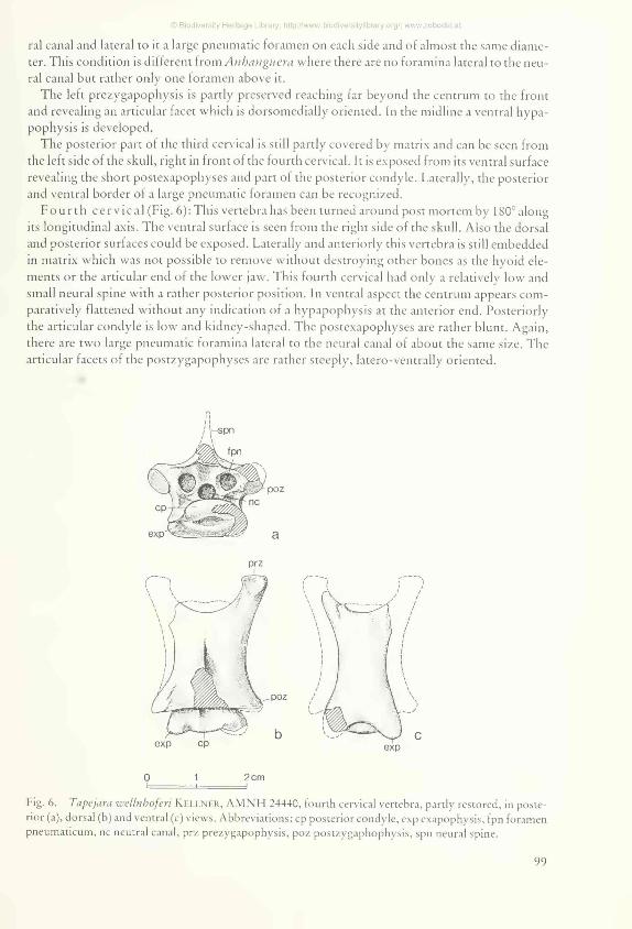
Fourth cerv ica l (Fig. 6): This vertebra has been turned around post mortem by 180° along its longitudinal axis. The ventral surface is seen from the right side of the skull. Also the dorsal and posterior surfaces could be exposed. Laterally and anteriorly this vertebra is still embedded in matrix which was not possible to remove without destroying other bones as the hyoid elements or the articular end of the lower jaw. This fourth cervical had only a relatively low and small neural spine with a rather posterior position. In ventral aspect the centrum appears comparatively flattened without any indication of a hypapophysis at the anterior end. Posteriorly the articular condyle is low and kidney-shaped. The postexapophyses are rather blunt. Again, there are two large pneumatic foramina lateral to the neural canal of about the same size. The articular facets of the postzygapophyses are rather steeply, latero-ventrally oriented.
0 1 2cmi- i — i
Fig. 6. Tapejara wellnbofen K ellner, AMNH 24440, fourth cervical vertebra, partly restored, in posterior (a), dorsal (b) and ventral (c) views. Abbreviations: cp posterior condyle, exp exapophysis, fpn foramen pneumaticum, nc neutral canal, prz prezygapophysis, poz postzygaphophysis, spn neural spine.
99
© Biodiversity Heritage Library, http://www.biodiversitylibrary.org/; www.zobodat.at
M easurem ents (in mm):Length of skull as preserved 185Length of skull between squamosal and anterior
margin of nasopreorbital fenestra 132Length of skull beween squamosal and premaxillary (restored) ca. 183Total length of skull (restored) ca. 220Height of skull above quadrate 67Height of premaxillary crest (restored) ca. 120Length of nasopreorbital fenestra (restored) 80Diameter of orbit through scleral ring 25Length of lower jaw as preserved 120Length of lower jaw (restored) ca. 150Length of axis 17,5Width of axis 20Length of third cervical vertebra (restored) 30Width of third cervical vertebra (restored) 24Length of fourth cervical vertebra 32Width of fourth cervical vertebra 24
Specim en UOSG 12891 (Plate 1, figs 2 a, b)
P reserv atio n : A calcareous concretion typical for the Romualdd member of the Santana formation contains the anterior portion of a skull including the very tip of the premaxillary and the premaxillary crest. The concretion had been split, one side exhibiting mainly the exterior bony layer of the right side in internal view, the other containing the main portion of the bones in right lateral aspect, three-dintensionally preserved. The specimen was prepared by Urs Oberli in order to clear the nasopreorbital fenestra and to expose the palate. Dorsally the premaxillary crest is broken and does not reveal its natural margin. Here the right bony wall of the crest is broken away and shows a trabecular internal structure.
P rem ax illa ry : The front end of the upper jaw is completely preserved and therefore complements the holotype and specimen AMNH 24440. The beak appears to be very much pointed. Its length from the anterior margin of the nasopreorbital fenestra is about 60 mm. The pointed tip is inclined downwards by an angle of about 35° relative to the palate. At the front the premaxillary crest has a sharp edge, its margin rises in a smooth concave line to a vertical orientation and curves back high above the beak. The maximum height of the crest as preserved is about 107 mm above the lower margin of the jaw. However, it must have been even higher. The crest ist very thin at the broken edges. Its surface is smooth and only slightly sculptured by the internal trabecular system. Towards the top the surface becomes more coarse.
P alate : The ventral surface of the beak is concave as in specimen AMNH 24440. Behind this a convex bump is delveloped followed by a medial foramen entering the maxillo-palatines. Posteriorly the choanae are exposed, just before the nodule is broken. Except for a medial suture separating the palatines no clear sutures are visible. The palate has no medial ridge but is slightly convex with rounded lateral margins.
R em arks: The specimen of the collection Urs Oberli comes from an individual about the same size as specimen AMNH 24440. In its typical characters it is exactly the same and can be assigned to Tapcjara ivellnhofcri.
100
© Biodiversity Heritage Library, http://www.biodiversitylibrary.org/; www.zobodat.at
Comparison and relationships
The new genus and species, Tapejara wellnhoferi,was founded by K ellner (1989) on an incomplete skull (Romualdo member, Santana formation, Araripe Basin, North-east Brazil) consisting of the anterior part with the premaxillary crest and elements ventrally surrounding the nasopreorbital fenestra, orbit, and infratemporal fenestra, i. e. maxillary, jugal and lachrymal. In shape and size these bones in the holotype (Desiree-Collection of R. A. v. Blittersdorff, Rio de Janeiro, CD-R-080) are identical with specimen AMNH 24440 which, thanks to its completeness, provides many more details of skull, lower jaw and cervical vertebrae and allows, in addition to specimen UOSG 12891, a reliable reconstruction of the skull of this remarkable pterosaur.
Based on this new evidence an emended diagnosis of the genus can be given:
Genus Tapejara K ellner 1989Type species T. wellnboferi K f.llner 1989
D iagn o sis : “Toothless pterodactyloids with short skull, high premaxillary sagittal crest on anterior part of skull tapering to a low posterior extension closely following the midline of the skull roof. Short fronto-parietal crest extending posteriorly present. Very large nasopreorbital fenestra, relatively small orbit situated below level of upper margin of nasopreorbital fenestra. Rostrum inclined downward with concave depression in palatal view and terminating in a pointed tip anteriorly. Palate lacks mesial ridge. Lower jaw ventrally with sagittal crest on the symphysis. Upper margin of symphysis inclined downward with concave depression dorsally. Cervical vertebrae short with low neural spines and two large pneumatic foramina lateral to the neural canal. The axis is lacking lateral pneumatic foramina.”
Originally, K ellner (1989) included in his family Tapejaridae also the toothless genus Tupu- xuara K ellner & C ampos (1988). The only species, Tupuxuara longicristatus, was based on an incomplete skull and a few wing bones. In 1990, K illner and C ampos described another incomplete skull from the Santana formation with a high sagittal crest, apparently toothless, and assigned it tentatively to the Tapejaridae. The premaxillary sagittal crest of Tupuxuara is much lower, the palate has a mesial ridge, and there is no downward inclination of the rostrum, in contrast to Tapejara. In addition, nothing is known about the post-nasopreorbital section of the skull in Tupuxuara, unless the tapejarid skull mentioned by K ellner and C ampos (1990) could clearly be assigned to the genus Tupuxuara.
More, yet undescribed material from the Santana formation indicates new toothless pterosaurs suggesting that already in Late Aptian times the earliest toothless pterosaurs in the fossil record represent different forms, at least at the generic level. At present, the available material is still too incomplete as to evaluate the relationships of Tapejara to other pterosaurs of the Santana formation documented by postcranial material only, as Araripesaurus Price 1971 andAra- ripedactylus W f.llnhoffr 1977, or to the toothed genera, Santanadactylus B uisonje 1980, Brasi- leodactylus K ellner 1984, Anhanguera C ampos & K ellner 1985, Cearadactylus L eonardi & B orgomanero 1985, and Tropeognathus W ellnhofer 1987. Tapejara appears to be so different from other Cretaceous toothless pterodactyloids, as the Pteranodontidae, Nyctosauridae or Azhdarchidae, that any closer relationships can be excluded for now, at least at the family level.
101

© Biodiversity Heritage Library, http://www.biodiversitylibrary.org/; www.zobodat.at
Paleobiological Significance and Life Style of Tapejara
On the basis of the neck vertebrae comparisons with more complete Santana pterosaur skeletons suggest a wing span of Tapejara wellnhoferi between 1.35 m and 1.5 m. This relatively small size, the short head with a high anterior premaxillary crest and the toothless jaws are in sharp contrast to other Cretaceous, toothed and toothless, pterosaurs which are supposed to have been piscivorous. Adaptations for piscivory include long pointed jaws, as in Pteranodon, and/or an effective dentition with long, pointed, intermeshing teeth as a useful gripping device, as for example in the Anhangueridae.
Also an insectivorous life style seems unlikely for Tapejara, since its head is very narrow and teeth are lacking. Pterosaurs supposed to represent insectivores have a broad mouth and few peg-like teeth, as documented in Armrognathus and Batracbognatbus from the Upper Jurassic (W ellnhofer 1991 c).
It is possible that Tapejara was a carrion feeder using its short pointed, hooked beak, covered with horny sheaths and sharp cutting edges in order to cut pieces of meat out of a dinosaur carcass. However, the high crest at the front seemed to be rather an obstacle, and a long flexible neck would be necessary. The neck vertebrae preserved suggest an estimated length of the neck of only 16 cm, considerably less than the skull length.
A more likely feeding habit of Tapejara was frugivory. Recently, Fleming and L ips (1991) hypothesized that some Cretaceous pterosaurs could have been the ecological analogues of extant fruit-eating birds and bats which are the most important consumers and dispersers of modern gymnosperm and angiosperm seeds, today. They considered even a co-evolutive relationship between pterosaurs and early angiosperms, having been involved in the early evolutionary radiation of angiosperms, in the Cretaceous, when neither frugivorous birds nor bats extisted.
Fig. 7. Life restoration of Tapejara Hkellnbofcn based on restoration of skull in figure 4. Upper and lower jaws, premaxillary and fronto-parietal crests are considered to have been covered by horny sheaths.
102

© Biodiversity Heritage Library, http://www.biodiversitylibrary.org/; www.zobodat.at
Tapejara wellnhoferi meets several requirements for a frugivorous life style. It is a relatively small pterosaur with a maximum wing span of 1.5 m. (Modern fruit bats like the Pteropodidae reach a maximum wing span of 1.7 m.). According to the general bauplan of pterodactyloids it must have been able to scramble around on shrubs or to climb in trees quadrupedally using its strong finger claws. Its head was short and narrow with the front end of the jaws bent downward. Only the anterior six centimetres of the beak are functional with concave, shallow excavations in the upper and lower jaws with sharp lateral edges (Fig. 2). When the jaws were closed only the pointed tips were in contact leaving an oval gap or diastema behind. This would have made a perfect tool for picking or plucking fleshy fruits, either from non-angiosperms as cy- cads, Gnetales and different gymnosperms, or from angiosperms which, in Late Aptian/Albian time, had undergone their first major evolutionary radiation (Friis, C haloner & C ranf. 1987).
In the upper and lower jaws there are konvex bumps posterior to the shallow depressions which could have functioned as a cracking device for hard objects, such as seeds or fruit capsules.
The high anterior bony crests of the premaxillary and the dentary were supposedly covered by horny sheaths. Internal bone structures have been interpreted as a highly vascularized tissue with a body temperature control function (K fllnfr 1989). Besides being simply display structures, these crests could also have been used for separating the foliage and tangle of tendrils in order to get easier access to fruits on shrubs and trees. As T heodore H. F leming states (personal correspondence, August 15, 1991): “The frontal crest is most reminiscent to me of the casque on the skull of modern hornbills (Bucerotidae), many of which are, of course, consumers of canopy fruits.”
Of course, there arises the question whether there is any evidence of fleshy-fruited plants in the fossil record of the Santana formation. First of all, is should be remembered that the fossilisation potenial for fleshy fruits is low in general, so that we hardly can expect to find them in the calcareous concretions of the Romualdo member from which the remains of Tapejara originated. The macroflora record is rather poorly known. D uarte (1985) described two gymnosperms, Brachyphyllurn and Podozamites, and one angiosperm, Nymphyaeites. According to C rane & M aisey (1991) conifers are one of the most conspicuous elements in the palynofloras from the Santana formation, probably representing Cheirolepidiaceae and Araucariaceae, also present as macrofossils. Characteristic pollen grains point also to producers similar to extant Gnetales. Spores recovered from the Santana formation were probably produced by different ferns. A variety of angiosperm pollen suggest that nonmagnoliid dicotyledons of five different subclasses were present, as well as several types of monocotyledons or dicotyledons of the ma- gnoliid grade. However, C rane & M aisey (1991: 416) state that “the macrofossil flora from the Santana formation concretions so far includes no unequivocal angiosperm fossils”.
On the other hand, a great quantity of macroflora fossils have been collected from the Santana formation and await detailed study and research. Prof. M. R. D e L ima, Sito Paulo, estimates that there are about 12 different families of angiosperms as well as many gymnosperms represented in the Santana formation, both of which could have produced fruits as possible diet of Tapejara and other fruit eating pterosaurs (personal communication by U. Kandler).
Early Cretaceous pterosaurs, such as Tapejara, could therefore well have played an important role in the dispersion of angiosperm seeds. We agree with Fleming & L ips (1991) that their metabolism, fast food passage rate (and therefore gentle treatment of seeds in the gut), plus the ability to fly long distances and possibly migrate seasonally, would have made pterosaurs excellent long-distance seed dispersers, superior to herbivorous dinosaurs (W eishampel 1984). The rapid early radiation and dispersion of angiosperms beginning in the Lower Cretaceous, the little evidence of birds and the lack of bats as possible seed dispersers make it probable that
103

© Biodiversity Heritage Library, http://www.biodiversitylibrary.org/; www.zobodat.at
frugivorous pterosaurs were involved. At the Araripe Basin there is until now just one fossil record of birds, an undeterminated feather from the Crato member of the Santana formation underlying the Romualdo member from where most pterosaurs have been discovered (K ellner et al. 1991). Based on these assumptions Tapejeara w d ln h o f e r i could be regarded as the first candidate for frugivorous feeding habits in pterosaurs. Possibly it belongs to a group of rather diverse highly spezialized frugivorous pterosaurs.
Literature Cited
Beurlfn, K. (1971: As condicóes ecológicas e faciológicas da formacào Santana na Chapada do Araripe (Nordeste do Brasil). — An. Acad, brasil. Cieñe., 43: 4 1 1—415 (suplemento); Rio de Janeiro.
Braun, O. P. G. (1966): Estratigrafía dos sedimentos da parte interior da regiáo Nordeste do Brasil (Badas de Tucano-Jatoba, Mirandiba e Araripe). - D . N. P. M./Div. Geol. Miner., Bol., No. 236: 1 — 15; Rio de Janeiro.
Buisonjé, P. H. de (1980): Santanadactylus brasilensis nov. gen., nov. sp., a long-necked, large pterosauricr from the Aptian of Brasil. — Proc. Koninkl. Nederl. Akad. Wetensch., (B), 83, 2: 145—172; Amsterdam.
CAMPOS, D. A. & K ellner, A. W. A. (1985): Panorama of the Flying Reptiles Study in Brazil and South America. — An. Acad. Brasil, Cienc., (1985), 57 (4): 453 — 466; Rio de Janeiro.
C rane, P. R. Sc Maisey, J. G. (1991): Fossil Plants. - in: Santana Fossils (J. G. Maisey ed.): 4 14 -42 9 ; T. F. H. Publications Inc., Neptune City, N. J.
Duarte, F. (1985): Vegetáis Fosseis da Chapada do Araripe, Br. — Coletanea deTrabalhos Paleontológicos, Série Geología, no. 27, Seçâo Paleontología e Estratigrafía, no. 2: 557—563; Brasilia.
Fleming, T. H. & FlPS, K. R. (1991): Angiosperm Endozoochory: Were Pterosaurs Cretaceous Seed Dispersers? — Amer. Naturalist, 138 (4): in press; Chicago.
Friis, F. M., C halonlr, W. G. A C rane, P. R. (editors) (1987): The origins of angiosperms and their biological consequences. — 358 p; Cambridge University Press, Cambridge.
Kellner, A. W. A. (1984): Ocorrencia de urna mandibula de Ptersosauria (Brasileodactyltts anmpensis, nov. gen., nov. sp.) na Formaçào Santana, Cretáceo da Chapada do Araripe, Cearà, Brasil. — An. XXXIII Congr. Brasil. Geol. Rio de Janeiro, 1984: 578-590.
---- (1989): A New Edentate Pterosaur of the Fower Cretaceous from the Araripe Basin, Northeast Brazil.- An. Acad, brasil. Ci., 1989, 61 (4): 439 -446 ; Rio de Janeiro (published in 1990).
---- (1991): Supplementary Notes and Comments to The Santana Pterosaurs. — in: Santana Fossils(J. G. Maisey ed.): 370 —371 ; T. F. FI. Publications Inc., Neptune City, N. J.
K ellner, A. W. A. & C ampos, D. A. (1988): Sobre um Novo Pterossauro com Crista Sagital de Bada do Araripe, Cretáceo Inferior do Nordeste do Brasil. — An. Acad, brasil. Ci., (1988), 60 (4): 459 —469; Rio de Janeiro (published in 1989).
---- (1990): Preliminary description of an unusual pterosaur skull of the Fower Cretaceous from the Araripe Basin. - Atas 1. Simposio sobre a Bada do Araripe e Bacías Interiores do Nordeste: 40 1-40 5 ; Crato.
Kellner, A. W. A., Martins Neto, R. G. Sc Maisey, J. G. (1991): Underminated feather. - in: Santana Fossils (J. G. Maisey ed.): 376 -377 ; T. F. H. Publications. Inc., Neptune City, N. J.
Ffonardi, G. & Borgomanfro, G. (1985): Cearadactylus atrox nov. gen., nov. sp.: Novo Pterosauria (Ptcrodactyloidea) da Chapada do Araripe, Ceará, Brasil. - Coletanea de Trabalhos Paleontológicos, Sér. Geol., 27, Seçâo de Paleontología e Estratigrafía, no. 2: 75 — 80; Brasilia.
Mabesone, J. M. & T inoco, 1. M. (1973): Paleoecology of the Aptian Santana Formation (Northeastern Brazil). — Paleogeogr., Paleoclimat., Paleoecol., 14 (2): 97—118; Amsterdam.
Maisey, J. G. (1990): Stratigraphy and depositional environment of the Crato Member (Santana Formation, Fower Cretaceous of N. F. Brazil). — in: Insects from the Santana Formation, Fower Cretaceous of Brazil (D. A. G rimaldi, ed.). - Bull. Amer. Mus. Nat. Hist., 195: 15 -19 ; New York.
---- (ed.) (1991): Santana Fossils. An Illustrated Atlas. — 459p.;T. F. H. Publications, Inc., NeptuneCitv,N.J.
104

© Biodiversity Heritage Library, http://www.biodiversitylibrary.org/; www.zobodat.at
Price, L. 1. (1971): A Presenta de Pterosauria no Cretáceo Inferior da Chapada do Araripe, Brasil. — An. Acad, brasil. Ci., 43 (Suppl.): 452—461; Rio de Janeiro.
Su va, M. A. M. da (1986): Lower Cretaceous sedimentary sequences in the Basin, northeastern Brazil: a revision. — Rev. Brasil. Geoci., 16 (3): 3 1 1—319; Sao Paulo.
W eishampel, D. B. (1984): Interactions between Mesozoic plants and vertebrates: fructifications and seed predation. — N. Jb. Geol. Paläont. Abh., 167 (2): 224 — 250; Stuttgart.
W ellnhofer, P. (1977): Araripedactylus dehrni nov. gen., nov. sp., ein neuer Flugsaurier aus der Unterkreide von Brasilien. — Mitt. Bayer. Staatsslg. Paläont. hist. Geol., 17: 157— 167; München.
---- (1985): Neue Pterosaurier aus der Santana-Formation (Apt) der Chapada do Araripe, Brasilien. — Pa-laeontographica, A. 187: 105—182; Stuttgart.
---- (1987): New Crested Pterosaurs from the Lower Cretaceous of Brazil. — Mitt. Bayer. Staatsslg. Paläont. hist. Geol., 27: 175— 186; München.
---- (1991a): Weitere Pterosaurierfunde aus der Santana-Formation (Apt) der Chapada do Araripe, Brasilien. — Paleontographica A, 215: 43—101; Stuttgart.
---- (1991 b): Santana Formation Pterosaurs. — in: Santana Fossils (J. G Maisey ed.): 351—370; T. F. H.Publications, Inc., Neptune City, N. J.
---- (1991c): The Illustrated Encyclopedia of Pterosaurs. — 192p ; Salamander Books Ltd., London.
Plate 1
Tapejara wellnhoferi K ellner, Santana formation (Late Aptian/Albian), Araripe Basin, Ceara, Brazil.
Fig. 1: Skull and mandible, cervical vertebrae 1—4, specimen AMNPI 24440, New York.
Fig. 2: Front end of skull with premaxillary crest, from qne calcareous concretion showing the right side (2a) and the counterpart (2b), specimen UOSG 12891, Collection Urs Oberli, St. Gallen, Switzerland.
105

M 'tt Bt St t 1 h itage( Libro 'y ’ 31'/wYW b id ive rs itylibrary .° rg/; www.zobodat.at
W i-llnhoffr, P. & K l-.llnf.r, A. W. A.: T apeja ra Plate 1